bioorganic & medicinal chemistry letters · 2017-01-03 · bcl-2/bid interaction, resulting in...
TRANSCRIPT

Bioorganic & Medicinal Chemistry Letters 24 (2014) 717–724
Contents lists available at ScienceDirect
Bioorganic & Medicinal Chemistry Letters
journal homepage: www.elsevier .com/ locate/bmcl
BMCL Digest
a-Helix mimetics: Outwards and upwards
(b) (c)(a)
Figure 1. Distribution of residues on a canonical a-helix; (a) side-view wii + 7, i + 11 residues displayed on the same face (orange); (b) top-down viehelical wheel.
0960-894X � 2013 The Authors. Published by Elsevier Ltd.http://dx.doi.org/10.1016/j.bmcl.2013.12.003
⇑ Corresponding authors.E-mail addresses: [email protected] (S. Thompson), andrew.hamil-
[email protected] (A.D. Hamilton).
Open access under CC BY-NC-ND license.
Madura K. P. Jayatunga, Sam Thompson ⇑, Andrew D. Hamilton ⇑Department of Chemistry, Chemistry Research Laboratory, University of Oxford, Oxford OX1 3TA, UK
a r t i c l e i n f o
Article history:Received 12 August 2013Revised 23 November 2013Accepted 1 December 2013Available online 8 December 2013
Keywords:Protein–protein interactionRational designPeptidomimeticProteomimetic
a b s t r a c t
a-Helices are common secondary structural elements forming key parts of the large, generally featurelessinterfacial regions of many therapeutically-relevant protein–protein interactions (PPIs). The rationaldesign of helix mimetics is an appealing small-molecule strategy for the mediation of aberrant PPIs, how-ever the first generation of scaffolds presented a relatively small number of residues on a single recogni-tion surface. Increasingly, helices involved in PPIs are found to have more complex binding modes,utilizing two or three recognition surfaces, or binding with extended points of contact. To address theseunmet needs the design and synthesis of new generations of multi-sided, extended, and supersecondarystructures are underway.
� 2013 The Authors. Published by Elsevier Ltd. Open access under CC BY-NC-ND license.
Proteomics has emerged as an important tool in unraveling theintricate, dynamic nature of many cellular processes.1 The preva-lence of protein–protein interactions (PPIs) in signal transduction,transcription, and apoptosis, make them attractive therapeutictargets for numerous diseases (e.g., HIV, cancer, and neurodegener-ative disorders).2–8 The development of chemical probes is impor-tant to gain further insights into these critical biological systemsbut despite their potential clinical importance, few small moleculeinhibitors of PPIs have been developed. Many established enzy-matic targets have small, well-defined catalytic domains whichgovern the majority of the enthalpic contribution to substratebinding.9 Conversely, PPIs are mediated by the cumulative bindingenergy of many amino acid residues, over an extended surface (aslarge as 4500 Å2).10–12 Much of this surface is solvent exposedwhen unbound, with binding domains arranged in a noncontigu-ous manner, often necessitating the development of large molecu-lar weight inhibitors.13 Strategies to disrupt PPIs frequently focuson key ‘hot spot’ residues which contribute heavily to binding.14
An a-helix is the most common protein secondary structuralelement, defined by a tight helical turn (3.4 residues per turn),thereby creating three distinct binding surfaces, with the i, i + 4and i + 7, residues aligned on the same face (Fig. 1). Of the PPIsfound in the Protein Data Bank (PDB), 62% have an a-helix at theinterface, illustrating the importance of this structural element in
protein-protein recognition.15,16 They are also found at more com-plex recognition domains such as DNA binding motifs and mem-brane-bound proteins.17,18
The increase in structural data over the last 20 years (Fig. 2) hasfacilitated research into the mediation of therapeutically relevantPPIs, with many groups targeting key helical elements. Whilstthe ratio of two- and three-sided helix-mediated PPIs relative tosingle-sided analogues has not changed, there has been a growingawareness of their presence at important interfaces.
Many groups have developed conformationally restrictedpeptides employing covalent tethers including lactams,19,20 disul-fide bridges,21 triazoles,22 and hydrocarbon linkers.23–26 Grubbsshowed that olefin metathesis can be used to promote helicity,27
with Verdine later showing that stapled peptides inhibit the
th i, i + 4,w, (c) a-

0
50
100
150
200
250
1990-1995 1996-2000 2001-2005 2006-2011
1-sided helix PPI 2-sided helix PPI 3-sided helix PPI
Figure 2. PDB structures with helices at the PPI interface over time. Subcategoriesillustrate which proportion contained helices with one, two or three key bindingdomains.15,16
718 M. K. P. Jayatunga et al. / Bioorg. Med. Chem. Lett. 24 (2014) 717–724
Bcl-2/Bid interaction, resulting in decreased growth of human leu-kemia xenografts in vivo.28 Strategies developed by Cabezas andArora involve replacement of the internal a-helix hydrogen bond-ing network with a covalent linkage.29–32 A number of differentapproaches have since been developed to introduce constraintsin an effort to promote helicity (Fig. 3).33,34 Arora et al. demon-strated inhibition of the HIF-1a/p300 PPI with an olefin-stapledhydrogen bond surrogate (HBS) where key residues are found atthe i, i + 2, i + 5, i + 6 and i + 10 positions.35 This strategy has thebenefit of retaining the i, i + 4 and i + 7 residues and avoiding theuse of synthetically challenging a,a-di-substituted amino acids.In another strategy, tethers have been used to staple long chainsand stabilize extended helical structures. An impressive exampleis a 36 amino acid anti-viral sequence stabilized by two hydrocar-bon bridges formed through ring-closing metathesis (RCM),increasing potency of a peptide inhibitor, enfuvirtide, four-fold to2.1 nM and at the same time improving its pharmacokinetics.36
Short peptide truncates are frequently unstructured, promptingresearchers to explore the helical propensities of more metaboli-cally stable b-peptides.37,38 Variations of this strategy have beenemployed to promote additional helicity, with many groupsexploiting conformationally constrained variants,39–42 and mixedsequences of a/b-amino acids.43–46 Seebach and Gellman
(a) (b)
(c) (d)
Figure 3. a-Helix tethering strategies; (a) canonical a-helix with 13-membered H-bonding, (b) stapled a-helix through residues on the i and i + 4 positions (22-ringmacrocycle), (c) Cabezas’ hydrazone HBS (13-ring macrocycle), (d) Arora’s olefinHBS (13-ring macrocycle). HBS = hydrogen bond surrogate.
pioneered the use of b-peptides to mimic a-helical secondarystructure,47–49 with Schepartz later demonstrating inhibition ofthe p53/MDM2 PPI.50 An example by Gellman showed an extendedhelix which accurately mimicked the native sequence, and success-fully inhibited the Bcl-2/Bim PPI.51
A fundamentally different strategy to mediate PPIs is to mimicelements of protein secondary structure with entirely non-peptidicconstructs. The rational design of helix mimics aims to projectfunctional groups from a scaffold in order to reproduce the orien-tation of the native side-chains, whilst avoiding the expression,purification or chemical synthesis of peptidic elements. Such pep-tidomimetics present an opportunity to design compounds whichhave the same binding mode as the native protein, yet are meta-bolically stable, can permeate membranes and are readilyabsorbed.11 The structural diversity provided by chemical synthe-sis allows a much greater array of groups, with varied steric andelectronic properties, to be projected than those provided by theside chains of proteogenic amino acids. Majmudar et al. identifiedphenol carboxylic acid natural products, sekikaic- and lobaric acid,that inhibit the PPI between p300 and both MLL (Mixed-LineageLeukemia) and pKID (phosphorylated kinase-inducible domain ofCREB). These molecules bind at the same site as the helical regionof peptides, hinting that Nature has evolved its own helixmimics.52
Early work focused on the mimicry of hydrophobic residues ona single helical face, yet important targets often have amphiphilichelices with hot-spot regions projected from multiple faces. Impor-tant helical sequences with hot-spot residues in the i, i + 4 and i + 7positions were frequently targeted since they are found on a com-mon face (Fig. 1).14 Several classes of helix mimetic have beendeveloped, many inspired by the terphenyl scaffold introducedby Orner et al. in 2001.53 The p53/MDM2 and Bcl-xl/Bak interac-tions are two well-studied targets, with a-helices projectingcrucial residues on a single face.54,55 Both interactions inhibit cel-lular apoptosis: MDM2 binds and inactivates p53, the principalarchitect of cell-death, thereby arresting apoptosis.56 Similarly,Bak is a pro-apoptotic protein whose function is disrupted on bind-ing of Bcl-xl (Fig. 4a).57 Both interactions are thought to be prom-ising cancer targets since neoplastic cells often disable intracellularapoptotic mechanisms. Yin et al. designed terphenyl helix mimet-ics that inhibited the p53/HDM2 interaction in vitro with a Ki of182 nM where a native peptide truncate had a Ki of 3.51 lM.54
Arora has highlighted the importance of binding to multiplesurfaces of a single helix in many interactions.15 The currentcollection of single-sided helix mimetics are ill-equipped to simul-taneously mimic hot-spot residues displayed on more than oneface, presenting a need for the development of more complex scaf-folds (Fig. 4).58 Moreover, the restricted length of mimics of twoa-helix turns is often insufficient to inhibit interactions of ex-tended helices. The Bcl-2/Bid interaction is an example with keyresidues orientated on two faces of the a-helix.59 Key hydrophobicresidues Ile82, Ile86 and Leu90 lie on the same face, with Asp95projected on a different face to form important hydrogen bondsto Bcl-2 arginine and asparagine residues (Fig. 4b). Enzymes E1and E2, involved in the replication of papillomavirus, are anotherimportant cancer target displaying a two-faced a-helix bindingmode.60
Examples with hot-spot residues found on all three faces of ana-helix include the interaction of calmodulin with CaM kinase I.61
Peptide truncates show that a particularly dense array of residuesbound in the N-terminal domain of calmodulin is crucial, with the i(Val), i + 1 (Arg), i + 2 (His), i + 3 (Met) and i + 4 (Arg) positions ofthe peptide contributing strongly to binding (Fig. 4c).
Burgess has reasoned that since there is homology in the waymany secondary structural elements display residues, it is possibleto design ‘universal peptidomimetics’, able to reproduce the
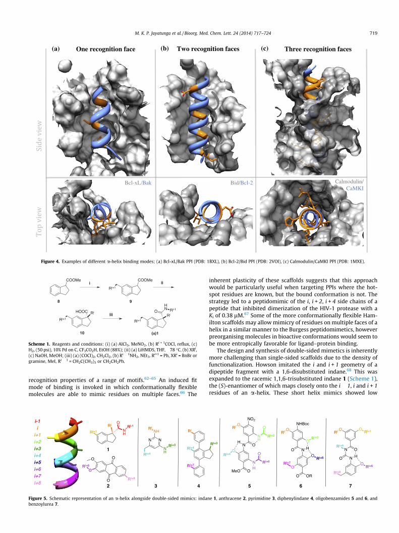
(a) (b) (c)One recognition face Two recognition faces Three recognition faces
Bcl-xL/Bak Bid/Bcl-2 Calmodulin/CaMKI
Figure 4. Examples of different a-helix binding modes; (a) Bcl-xL/Bak PPI (PDB: 1BXL), (b) Bcl-2/Bid PPI (PDB: 2VOI), (c) Calmodulin/CaMKI PPI (PDB: 1MXE).
Scheme 1. Reagents and conditions: (i) (a) AlCl3, MeNO2, (b) Ri + 1COCl, reflux, (c)H2, (50 psi), 10% Pd on C, CF3CO2H, EtOH (88%); (ii) (a) LiHMDS, THF, �78 �C, (b) XRi,(c) NaOH, MeOH; (iii) (a) (COCI)2, CH2Cl2, (b) Ri � 1NH2, NEt3. Ri+1 = Ph, XRi = BnBr orgramine, MeI, Ri � 1 = CH2C(CH3)3 or CH2CH2Ph.
M. K. P. Jayatunga et al. / Bioorg. Med. Chem. Lett. 24 (2014) 717–724 719
recognition properties of a range of motifs.62–65 An induced fitmode of binding is invoked in which conformationally flexiblemolecules are able to mimic residues on multiple faces.66 The
Figure 5. Schematic representation of an a-helix alongside double-sided mimics: indanbenzoylurea 7.
inherent plasticity of these scaffolds suggests that this approachwould be particularly useful when targeting PPIs where the hot-spot residues are known, but the bound conformation is not. Thestrategy led to a peptidomimic of the i, i + 2, i + 4 side chains of apeptide that inhibited dimerization of the HIV-1 protease with aKi of 0.38 lM.67 Some of the more conformationally flexible Ham-ilton scaffolds may allow mimicry of residues on multiple faces of ahelix in a similar manner to the Burgess peptidomimetics, howeverpreorganising molecules in bioactive conformations would seem tobe more entropically favorable for ligand–protein binding.
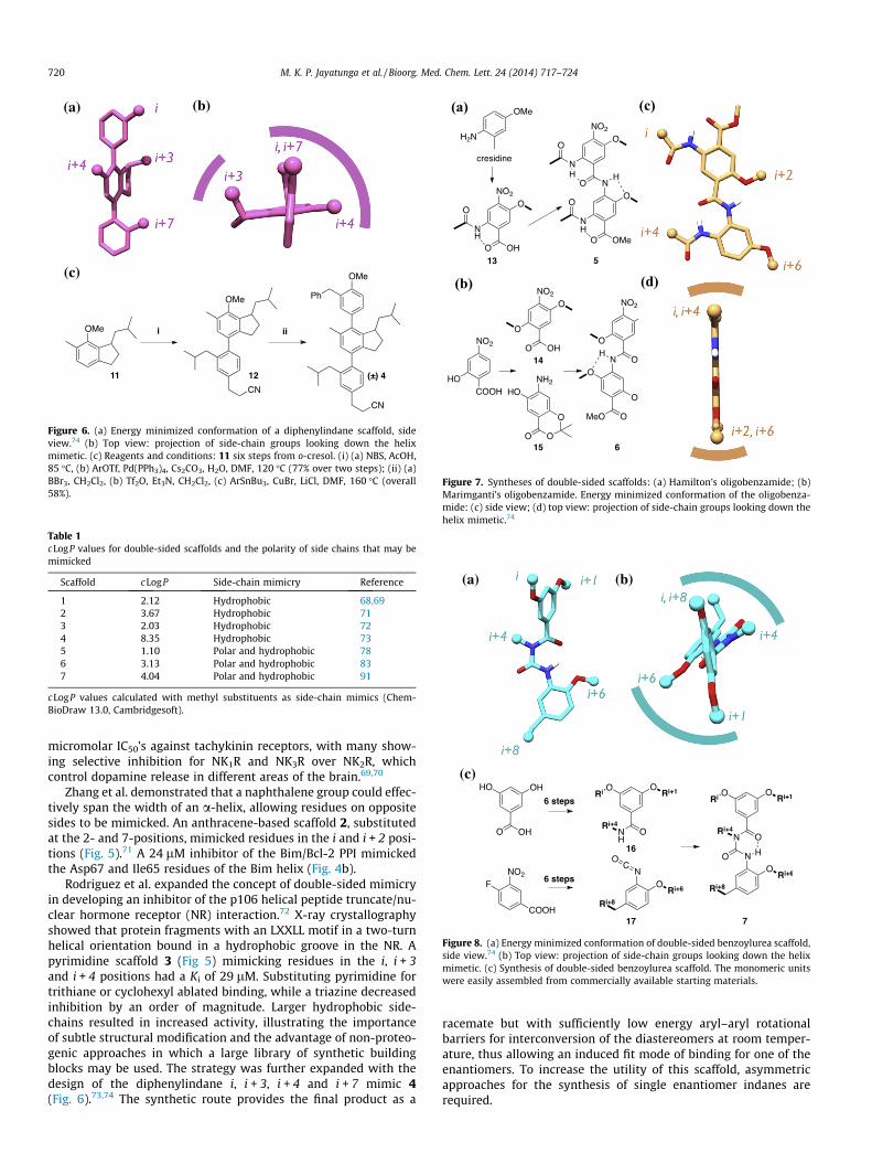
The design and synthesis of double-sided mimetics is inherentlymore challenging than single-sided scaffolds due to the density offunctionalization. Howson imitated the i and i + 1 geometry of adipeptide fragment with a 1,6-disubstituted indane.68 This wasexpanded to the racemic 1,1,6-trisubstituted indane 1 (Scheme 1),the (S)-enantiomer of which maps closely onto the i � 1, i and i + 1residues of an a-helix. These short helix mimics showed low
e 1, anthracene 2, pyrimidine 3, diphenylindane 4, oligobenzamides 5 and 6, and
(a) (b)
(c)
Figure 6. (a) Energy minimized conformation of a diphenylindane scaffold, sideview.74 (b) Top view: projection of side-chain groups looking down the helixmimetic. (c) Reagents and conditions: 11 six steps from o-cresol. (i) (a) NBS, AcOH,85 �C, (b) ArOTf, Pd(PPh3)4, Cs2CO3, H2O, DMF, 120 �C (77% over two steps); (ii) (a)BBr3, CH2Cl2, (b) Tf2O, Et3N, CH2Cl2, (c) ArSnBu3, CuBr, LiCl, DMF, 160 �C (overall58%).
Table 1cLogP values for double-sided scaffolds and the polarity of side chains that may bemimicked
Scaffold cLogP Side-chain mimicry Reference
1 2.12 Hydrophobic 68,692 3.67 Hydrophobic 713 2.03 Hydrophobic 724 8.35 Hydrophobic 735 1.10 Polar and hydrophobic 786 3.13 Polar and hydrophobic 837 4.04 Polar and hydrophobic 91
cLogP values calculated with methyl substituents as side-chain mimics (Chem-BioDraw 13.0, Cambridgesoft).
(a)
(b)
(c)
(d)
Figure 7. Syntheses of double-sided scaffolds: (a) Hamilton’s oligobenzamide; (b)Marimganti’s oligobenzamide. Energy minimized conformation of the oligobenza-mide: (c) side view; (d) top view: projection of side-chain groups looking down thehelix mimetic.74
(a) (b)
(c)
Figure 8. (a) Energy minimized conformation of double-sided benzoylurea scaffold,side view.74 (b) Top view: projection of side-chain groups looking down the helixmimetic. (c) Synthesis of double-sided benzoylurea scaffold. The monomeric unitswere easily assembled from commercially available starting materials.
720 M. K. P. Jayatunga et al. / Bioorg. Med. Chem. Lett. 24 (2014) 717–724
micromolar IC50’s against tachykinin receptors, with many show-ing selective inhibition for NK1R and NK3R over NK2R, whichcontrol dopamine release in different areas of the brain.69,70
Zhang et al. demonstrated that a naphthalene group could effec-tively span the width of an a-helix, allowing residues on oppositesides to be mimicked. An anthracene-based scaffold 2, substitutedat the 2- and 7-positions, mimicked residues in the i and i + 2 posi-tions (Fig. 5).71 A 24 lM inhibitor of the Bim/Bcl-2 PPI mimickedthe Asp67 and Ile65 residues of the Bim helix (Fig. 4b).
Rodriguez et al. expanded the concept of double-sided mimicryin developing an inhibitor of the p106 helical peptide truncate/nu-clear hormone receptor (NR) interaction.72 X-ray crystallographyshowed that protein fragments with an LXXLL motif in a two-turnhelical orientation bound in a hydrophobic groove in the NR. Apyrimidine scaffold 3 (Fig 5) mimicking residues in the i, i + 3and i + 4 positions had a Ki of 29 lM. Substituting pyrimidine fortrithiane or cyclohexyl ablated binding, while a triazine decreasedinhibition by an order of magnitude. Larger hydrophobic side-chains resulted in increased activity, illustrating the importanceof subtle structural modification and the advantage of non-proteo-genic approaches in which a large library of synthetic buildingblocks may be used. The strategy was further expanded with thedesign of the diphenylindane i, i + 3, i + 4 and i + 7 mimic 4(Fig. 6).73,74 The synthetic route provides the final product as a
racemate but with sufficiently low energy aryl–aryl rotationalbarriers for interconversion of the diastereomers at room temper-ature, thus allowing an induced fit mode of binding for one of theenantiomers. To increase the utility of this scaffold, asymmetricapproaches for the synthesis of single enantiomer indanes arerequired.
(a) (b)
Figure 9. Superimposition of lowest energy conformations of diphenylindane(pink), benzamide (brown) and benzoylurea (cyan) scaffolds showing differing sidechain projections: (a) side view; (b) top view.74
M. K. P. Jayatunga et al. / Bioorg. Med. Chem. Lett. 24 (2014) 717–724 721
Many of the early mimics mediate binding predominantlythrough hydrophobic interactions and the exclusion of water—alsoa major thermodynamic driving force in the association of pro-teins.75,76 Interactions of this type are not necessarily non-specific:for example a 1-naphthyl-substituted terphenyl displayed selec-tive inhibition of the HDM2/p53 PPI over the Bcl-2/Bak PPI, whilea 2-naphthyl analogue reversed the selectivity.54 The hydrophobic-ity of these molecules is an important factor in the good cellpermeability of this series,77 but limits aqueous solubility,58
providing an impetus for the development of new scaffolds. Forexample scaffolds 1–3 have moderate c logP values (Table 1), how-ever only hydrophobic groups can be appended, further increasingtheir hydrophobicity. Later scaffolds, for example, 5–7 incorporateheteroatoms that act to increase aqueous solubility and provideadditional synthetic versatility for the incorporation of a greaterrange of side-chains.
(a) (b) (c
Figure 10. (a) Extended benzoylurea 18 and enaminone 19 scaffolds, (b) nine turn a-helreceptor.
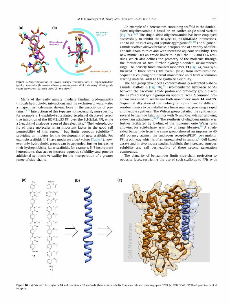
An example of a heteroatom-containing scaffold is the double-sided oligobenzamide 5 based on an earlier single-sided variant(Fig. 7a).78,79 The single-sided oligobenzamide has been employedsuccessfully to inhibit the Bak/Bcl-xL, p53/hMDM2 interactions,and to inhibit islet amyloid peptide aggregation.80–82 The oligoben-zamide scaffold allows for facile incorporation of a variety of differ-ent side-chain mimics and with increased aqueous solubility. Thisnew mimic uses an amide linker to install the i + 2 and i + 6 resi-dues, which also defines the geometry of the molecule throughthe formation of two further hydrogen-bonded six-memberedrings. The densely functionalized monomer 13 (Fig. 7a) was syn-thesized in three steps (50% overall yield) from meta-cresidine.Sequential coupling of different monomeric units from a commonstarting material adds to the synthetic flexibility.
The Ahn group developed a conformationally restricted bisben-zamide scaffold 6 (Fig. 7b).83 Five-membered hydrogen bondsbetween the backbone amide proton and ortho-oxy group placesthe i + 2/i + 5 and i/i + 7 groups on opposite faces. A common pre-cursor was used to synthesize both monomeric units 14 and 15.Sequential alkylation of the hydroxyl groups allows for differentresidue mimics to be installed in a linear manner, providing a rapidand flexible synthesis. The Wilson group detailed the synthesis ofseveral benzamide helix mimics with N- and O-alkylation allowingside-chain attachment.84,85 The synthesis of oligobenzamides wasfurther facilitated by loading of the monomer onto Wang resinallowing for solid-phase assembly of large libraries.86 A singlesided benzamide from the same group showed an impressive 40nM potency against the androgen receptor/PELP1 co-regulatorPPI, a pathway which is often upregulated in tumors.87 Cell-basedassays and in vivo mouse studies highlight the increased aqueoussolubility and cell permeability of these second generationcompounds.
The planarity of benzamides limits side-chain projection toopposite faces, restricting the use of such scaffolds to PPIs with
)
Cell
Mem
bra
ne
ix from a membrane spanning opsin GPCR, (c) PDB: 3CAP. GPCR = G protein-coupled

Figure 11. Common super secondary structural motifs containing a-helices(cylinders), b-strands (arrows) and loop sequences (red).
722 M. K. P. Jayatunga et al. / Bioorg. Med. Chem. Lett. 24 (2014) 717–724
hot-spot residues at 180� to one another (Fig. 7c and d). The Spiveygroup overcame this limitation by designing a scaffold with anazabicyclo[2.2.2]octane moiety coupled to an aryl fragment,mimicking the i, i + 1, i + 2, i + 4 and i + 5 residues.88
Figure 12. (a) HTH (red) of Pax class homeodomain dimer bound to DNA (green, PDB: 1mimic 20.105
The benzoylurea scaffold can similarly mimic a more complexpresentation of residues, with a convergent synthesis allowingincorporation of hydrophobic and hydrophilic groups.89,90 TheHamilton group synthesized double-sided mimic 7 from a benzam-ide 16 and an isocyanate 17 (Fig. 8c).91 The helical backbone isreminiscent of earlier benzamide helices created by the group,82,91
but allows for a more comprehensive distribution of side chains(Fig. 8b). Existing scaffolds reproduce groups on a number of differ-ent faces of the helix (Fig. 9), suggesting that while a three-sidedhelical peptidomimetic remains elusive, several frameworks existfor targeting PPIs with complex binding modes.
Extended helices are found in important therapeutic targets;for example transmembrane GPCR (G protein-coupled receptor)helices which span cell membranes and assemble due to bothhydrophobic and hydrophilic side-chain interactions.92 Mem-brane-spanning amphiphilic a-helices form artificial ion-chan-nels, mimicking the acetylcholine receptor.93 Synthetic variantsmay be able to replicate this behavior or inhibit endogenous
FJL). (b) Structure and energy minimized conformation of bis-pentabenzamide HTH

M. K. P. Jayatunga et al. / Bioorg. Med. Chem. Lett. 24 (2014) 717–724 723
ion channels. The Hamilton group elongated the single-sidedbenzoylurea scaffold to an oligomeric species mimicking a 37 Åa-helix 18 (Fig. 10). The easily assembled benzoylurea ring surro-gate enabled facile extension of the scaffold resulting in a mimicthat exhibited a preference for the extended conformation.89 Asimilar result was attained by the same group with an enaminonescaffold 19 which was extended to a ten turn a-helix mimic.94 Itwas also possible to incorporate polar side-chains, thus broaden-ing the potential for therapeutic applications. Other extendedscaffolds have been designed by the Rebek, Ahn and Wilsongroups, with the latter exploiting solid phase synthesis to facili-tate oligomerisation.95–98
Peptidomimetic design principles may be harnessed to repli-cate elements of supersecondary structure.17 Commonly occur-ring combinations of secondary structural elements constitutea-hairpins, a-corners and b-a-b motifs (Fig. 11). Helical hairpinsare important structures aiding protein insertion and transportacross cell membranes.99 Given the majority of folded protein isburied in the hydrophobic core, loop sequences are found atprotein surfaces. To replicate these more complex structures,non-peptidic loop or hairpin sequences have been designed totemplate the display of a-helical mimics.100–104 A series of linkedoligobenzamide and oligopyridylamide mimetics have the poten-tial to function in a similar way.105 Flexible syntheses with a vari-ety of linking groups give versatile helix-turn-helix (HTH) mimicsthat can be tuned for inter-helix angle and length (Fig. 12b).Linked helix mimetics may find a role in inhibiting important pro-tein–DNA interactions, such as the helix-turn-helix (Fig. 12a) orbasic-helix-loop-helix, which regulate gene transcription. TheDNA binding domain of the proto-oncogene c-Myc is an exampleof a HTH tri-helical protein.106 The helical turn forms a hydropho-bic cavity, while simultaneously projecting polar residues to-wards the negatively charged phosphate DNA backbone. Theseclasses of HTH mimic allow controlled projection of side chainsin three-dimensional space and thus the reproduction of rela-tively large protein surfaces. Surface mimicry has been success-fully attempted with both cyclic peptides and backbone graftingonto protein scaffolds.107,108 Synthetic scaffolds have also beenemployed by Ghosh and Hamilton in immobilizing peptide loopson G-quadruplexes and thus imitating multi-loop protein sufac-es.109 The same group previously designed a large protein surfacemimic by coupling cyclic peptides on to a calixarene scaffold,which showed strong binding to cytochrome c.110 Surface recog-nition has been demonstrated with porphyrin species,111 copper-and ruthenium-complexes,112,113 illustrating the diversity ofscaffolds available.
If the lessons learned from the development of early peptidom-imetics are extended to produce synthetic antibodies, these non-peptidic constructs may allow several problems with the use ofbiologics in vivo to be circumvented. Despite the growing numberof this type of therapeutic, prohibitive cost, poor oral bioavailabil-ity and the difficulties associated with maintaining homogeneitycurrently present major obstacles.
The last two decades have seen extensive efforts in the designand synthesis of a-helix mimetics as synthetic agents for themediation of PPIs. Many of these constructs have been thoroughlycharacterized and shown to provide good structural mimicry, withsome demonstrating functional mimicry in vitro. Researchers havetargeted a relatively small number of interfaces, which havefrequently relied on single face binding mediated by hydrophobicgroups. Given the myriad of therapeutically relevant PPIs thechallenge for researchers is to extend the scope of these mimeticsto provide a diverse collection allowing intervention at a broaderrange of interfaces, and with improved pharmacokineticproperties.
Acknowledgments
We thank Cancer Research UK (M.K.P.J.) and the University ofOxford (S.T.) for funding, and Drs. Hayden Peacock and HannahLingard (Oxford) for helpful discussions.
References and notes
1. Patterson, S. D.; Aebersold, R. H. Nat. Genet. 2003, 33, 311.2. Saksela, K.; Cheng, G.; Baltimore, D. EMBO J. 1995, 14, 484.3. Shangary, S.; Wang, S. In Annual Review of Pharmacology and Toxicology;
Annual Reviews: Palo Alto, 2009; Vol. 49, p 223.4. Chene, P. Nat. Rev. Cancer 2003, 3, 102.5. Lee, J. W.; Bae, S. H.; Jeong, J. W.; Kim, S. H.; Kim, K. W. Exp. Mol. Med. 2004, 36,
1.6. Grelle, G.; Kostka, S.; Otto, A.; Kersten, B.; Genser, K. F.; Muller, E. C.; Walter,
S.; Boddrich, A.; Stelzl, U.; Hanig, C.; Volkmer-Engert, R.; Landgraf, C.; Alberti,S.; Hofeld, J.; Strodicke, M.; Wanker, E. E. Mol. Cell. Proteomics 2006, 5, 234.
7. Lipinski, M. M.; Zheng, B.; Lu, T.; Yan, Z.; Py, B. F.; Ng, A.; Xavier, R. J.; Li, C.;Yankner, B. A.; Scherzer, C. R.; Yuan, J. Proc. Natl. Acad. Sci. U.S.A. 2010, 107,14164.
8. Kilby, J. M.; Hopkins, S.; Venetta, T. M.; DiMassimo, B.; Cloud, G. A.; Lee, J. Y.;Alldredge, L.; Hunter, E.; Lambert, D.; Bolognesi, D.; Mathews, T.; Johnson, M.R.; Nowak, M. A.; Shaw, G. M.; Saag, M. S. Nat. Med. 1998, 4, 1302.
9. Babine, R. E.; Bender, S. L. Chem. Rev. 1997, 97, 1359.10. Stites, W. E. Chem. Rev. 1997, 97, 1233.11. Fletcher, S.; Hamilton, A. D. Curr. Opin. Chem. Biol. 2005, 9, 632.12. Jones, S.; Thornton, J. M. Proc. Natl. Acad. Sci. U.S.A. 1996, 93, 13.13. Morelli, X.; Bourgeas, R.; Roche, P. Curr. Opin. Chem. Biol. 2011, 15, 475.14. Moreira, I. S.; Fernandes, P. A.; Ramos, M. J. Proteins 2007, 68, 803.15. Bullock, B. N.; Jochim, A. L.; Arora, P. S. J. Am. Chem. Soc. 2011, 133, 14220.16. The data presented in Figure 2 is taken from Ref. 15 and was compiled by
analysis of backbone dihedral angles using a modified version of the Rosettaprogram. We thank a reviewer for pointing out that the parameters used maylead to the inclusion of a small number of 310 and p helices.
17. Brändén, C.-I.; Tooze, J. Introduction to Protein Structure; Introduction to ProteinStructure Series; Garland Publishing, 1999.
18. Doherty, A. J.; Serpell, L. C.; Ponting, C. P. Nucleic Acids Res. 1996, 24, 2488.19. Osapay, G.; Taylor, J. W. J. Am. Chem. Soc. 1990, 112, 6046.20. Shepherd, N. E.; Hoang, H. N.; Desai, V. S.; Letouze, E.; Young, P. R.; Fairlie, D. P.
J. Am. Chem. Soc. 2006, 128, 13284.21. Jackson, D. Y.; King, D. S.; Chmielewski, J.; Singh, S.; Schultz, P. G. J. Am. Chem.
Soc. 1991, 113, 9391.22. Kawamoto, S. A.; Coleska, A.; Ran, X.; Yi, H.; Yang, C.-Y.; Wang, S. J. Med. Chem.
2012, 55, 1137.23. Schafmeister, C. E.; Po, J.; Verdine, G. L. J. Am. Chem. Soc. 2000, 122, 5891.24. Bernal, F.; Tyler, A. F.; Korsmeyer, S. J.; Walensky, L. D.; Verdine, G. L. J. Am.
Chem. Soc. 2007, 129, 2456.25. Kim, Y.-W.; Grossmann, T. N.; Verdine, G. L. Nat. Protoc. 2011, 6, 761.26. Blackwell, H. E.; Sadowsky, J. D.; Howard, R. J.; Sampson, J. N.; Chao, J. A.;
Steinmetz, W. E.; O’Leary, D. J.; Grubbs, R. H. J. Org. Chem. 2001, 66, 5291.27. Blackwell, H. E.; Grubbs, R. H. Angew. Chem., Int. Ed. 1998, 37, 3281.28. Walensky, L. D.; Kung, A. L.; Escher, I.; Malia, T. J.; Barbuto, S.; Wright, R. D.;
Wagner, G.; Verdine, G. L.; Korsmeyer, S. J. Science 2004, 305, 1466.29. Cabezas, E.; Satterthwait, A. C. J. Am. Chem. Soc. 1999, 121, 3862.30. Liu, J.; Wang, D.; Zheng, Q.; Lu, M.; Arora, P. S. J. Am. Chem. Soc. 2008, 130,
4334.31. Bao, J.; Dong, X. Y.; Zhang, J. Z. H.; Arora, P. S. J. Phys. Chem. B 2009, 113, 3565.32. Henchey, L. K.; Porter, J. R.; Ghosh, I.; Arora, P. S. ChemBioChem 2010, 11, 2104.33. Patgiri, A.; Jochim, A. L.; Arora, P. S. Acc. Chem. Res. 2008, 41, 1289.34. Vernall, A. J.; Cassidy, P.; Alewood, P. F. Angew. Chem., Int. Ed. 2009, 48, 5675.35. Henchey, L. K.; Kushal, S.; Dubey, R.; Chapman, R. N.; Olenyuk, B. Z.; Arora, P.
S. J. Am. Chem. Soc. 2010, 132, 941.36. Bird, G. H.; Madani, N.; Perry, A. F.; Princiotto, A. M.; Supko, J. G.; He, X.;
Gavathiotis, E.; Sodroski, J. G.; Walensky, L. D. Proc. Natl. Acad. Sci. U.S.A. 2010,107, 14093.
37. Seebach, D.; Matthews, J. L. Chem. Commun. 1997, 2015.38. Frackenpohl, J.; Arvidsson, P. I.; Schreiber, J. V.; Seebach, D. ChemBioChem
2001, 2, 445.39. Appella, D. H.; Christianson, L. A.; Klein, D. A.; Powell, D. R.; Huang, X.; Barchi,
J. J.; Gellman, S. H. Nature 1997, 387, 381.40. Appella, D. H.; Christianson, L. A.; Klein, D. A.; Richards, M. R.; Powell, D. R.;
Gellman, S. H. J. Am. Chem. Soc. 1999, 121, 7574.41. Appella, D. H.; Barchi, Joseph J.; Durell, S. R.; Gellman, S. H. J. Am. Chem. Soc.
1999, 121, 2309.42. Hamuro, Y.; Schneider, J. P.; DeGrado, W. F. J. Am. Chem. Soc. 1999, 121, 12200.43. Krauthäuser, S.; Christianson, L. A.; Powell, D. R.; Gellman, S. H. J. Am. Chem.
Soc. 1997, 119, 11719.44. Seebach, D.; Jaun, B.; Sebesta, R.; Mathad, R. I.; Flögel, O.; Limbach, M.; Sellner,
H.; Cottens, S. Helv. Chim. Acta 2006, 89, 1801.45. Horne, W. S.; Johnson, L. M.; Ketas, T. J.; Klasse, P. J.; Lu, M.; Moore, J. P.;
Gellman, S. H. Proc. Natl. Acad. Sci. U.S.A. 2009, 106, 14751.

724 M. K. P. Jayatunga et al. / Bioorg. Med. Chem. Lett. 24 (2014) 717–724
46. Van der Knaap, M.; Otero, J. M.; Llamas-Saiz, A.; van Raaij, M. J.; Lageveen, L. I.;Busscher, H. J.; Grotenbreg, G. M.; van der Marel, G. A.; Overkleeft, H. S.;Overhand, M. Tetrahedron 2012, 68, 2391.
47. Seebach, D.; Overhand, M.; Kühnle, F. N. M.; Martinoni, B.; Oberer, L.;Hommel, U.; Widmer, H. Helv. Chim. Acta 1996, 79, 913.
48. Appella, D. H.; Christianson, L. A.; Karle, I. L.; Powell, D. R.; Gellman, S. H. J. Am.Chem. Soc. 1996, 118, 13071.
49. Cheng, R. P.; Gellman, S. H.; DeGrado, W. F. Chem. Rev. 2001, 101, 3219.50. Kritzer, J. A.; Lear, J. D.; Hodsdon, M. E.; Schepartz, A. J. Am. Chem. Soc. 2004,
126, 9468.51. Boersma, M. D.; Haase, H. S.; Peterson-Kaufman, K. J.; Lee, E. F.; Clarke, O. B.;
Colman, P. M.; Smith, B. J.; Horne, W. S.; Fairlie, W. D.; Gellman, S. H. J. Am.Chem. Soc. 2012, 134, 315.
52. Majmudar, C. Y.; Højfeldt, J. W.; Arevang, C. J.; Pomerantz, W. C.; Gagnon, J. K.;Schultz, P. J.; Cesa, L. C.; Doss, C. H.; Rowe, S. P.; Vásquez, V.; Tamayo-Castillo,G.; Cierpicki, T.; Brooks, C. L.; Sherman, D. H.; Mapp, A. K. Angew. Chem., Int. Ed.2012, 51, 11258.
53. Orner, B. P.; Ernst, J. T.; Hamilton, A. D. J. Am. Chem. Soc. 2001, 123, 5382.54. Yin, H.; Lee, G.; Park, H. S.; Payne, G. A.; Rodriguez, J. M.; Sebti, S. M.;
Hamilton, A. D. Angew. Chem., Int. Ed. 2005, 44, 2704.55. Yin, H.; Lee, G.; Sedey, K. A.; Kutzki, O.; Park, H. S.; Orner, B. P.; Ernst, J. T.;
Wang, H.-G.; Sebti, S. M.; Hamilton, A. D. J. Am. Chem. Soc. 2005, 127, 10191.56. Lane, D. P. Nature 1992, 358, 15.57. Chao, D. T.; Korsmeyer, S. J. Annu. Rev. Immunol. 1998, 16, 395.58. Cummings, C. G.; Hamilton, A. D. Curr. Opin. Chem. Biol. 2010, 341.59. Smits, C.; Czabotar, P. E.; Hinds, M. G.; Day, C. L. Structure 2008, 16, 818.60. Abbate, E. A.; Berger, J. M.; Botchan, M. R. Genes Dev. 1981, 2004, 18.61. Clapperton, J. A.; Martin, S. R.; Smerdon, S. J.; Gamblin, S. J.; Bayley, P. M.
Biochemistry 2002, 41, 14669.62. Ko, E.; Liu, J.; Perez, L. M.; Lu, G.; Schaefer, A.; Burgess, K. J. Am. Chem. Soc.
2011, 133, 462.63. Chen, D.; Brahimi, F.; Angell, Y.; Li, Y.-C.; Moscowicz, J.; Saragovi, H. U.;
Burgess, K. ACS Chem. Biol. 2009, 4, 769.64. Ko, E.; Burgess, K. Org. Lett. 2011, 13, 980.65. Raghuraman, A.; Ko, E.; Perez, L. M.; Ioerger, T. R.; Burgess, K. J. Am. Chem. Soc.
2011, 133, 12350.66. Ko, E.; Liu, J.; Burgess, K. Chem. Soc. Rev. 2011, 40, 4411.67. Ko, E.; Raghuraman, A.; Perez, L. M.; Ioerger, T. R.; Burgess, K. J. Am. Chem. Soc.
2013, 135, 167.68. Horwell, D. C.; Howson, W.; Nolan, W. P.; Ratcliffe, G. S.; Rees, D. C.; Willems,
H. M. G. Tetrahedron 1995, 51, 203.69. Horwell, D. C.; Howson, W.; Ratcliffe, G. S.; Willems, H. M. G. Bioorg. Med.
Chem. 1996, 4, 33.70. Tremblay, L.; Kemel, M.; Desban, M.; Gauchy, C.; Glowinski, J. Proc. Natl. Acad.
Sci. U.S.A. 1992, 89, 11214.71. Zhang, Z.; Li, X.; Song, T.; Zhao, Y.; Feng, Y. J. Med. Chem. 2012, 55, 10735.72. Rodriguez, A. L.; Tamrazi, A.; Collins, M. L.; Katzenellenbogen, J. A. J. Med.
Chem. 2004, 47, 600.73. Kim, I. C.; Hamilton, A. D. Org. Lett. 2006, 8, 1751.74. Minimizations were performed using the program Molecular Operating
Environment, MMFF94(s); Chemical Computing Group Inc.75. Ross, P. D.; Subramanian, S. Biochemistry 1981, 20, 3096.76. Nicholls, A.; Sharp, K. A.; Honig, B. Proteins 1991, 11, 281.77. Chen, L.; Yin, H.; Farooqi, B.; Sebti, S.; Hamilton, A. D.; Chen, J. Mol. Cancer Ther.
2005, 4, 1019.
78. Thompson, S.; Vallinayagam, R.; Adler, M. J.; Scott, R. T. W.; Hamilton, A. D.Tetrahedron 2012, 68, 4501.
79. Ernst, J. T.; Becerril, J.; Park, H. S.; Yin, H.; Hamilton, A. D. Angew. Chem., Int. Ed.2003, 42, 535.
80. Shaginian, A.; Whitby, L. R.; Hong, S.; Hwang, I.; Farooqi, B.; Searcey, M.; Chen,J.; Vogt, P. K.; Boger, D. L. J. Am. Chem. Soc. 2009, 131, 5564.
81. Plante, J. P.; Burnley, T.; Malkova, B.; Webb, M. E.; Warriner, S. L.; Edwards, T.A.; Wilson, A. J. Chem. Commun. 2009, 5091.
82. Saraogi, I.; Hebda, J. A.; Becerril, J.; Estroff, L. A.; Miranker, A. D.; Hamilton, A.D. Angew. Chem., Int. Ed. 2010, 49, 736.
83. Marimganti, S.; Cheemala, M. N.; Ahn, J.-M. Org. Lett. 2009, 11, 4418.84. Campbell, F.; Plante, J. P.; Edwards, T. A.; Warriner, S. L.; Wilson, A. J. Org.
Biomol. Chem. 2010, 8, 2344.85. Plante, J.; Campbell, F.; Malkova, B.; Kilner, C.; Warriner, S. L.; Wilson, A. J. Org.
Biomol. Chem. 2007, 6, 138.86. Long, K.; Edwards, T. A.; Wilson, A. J. Bioorg. Med. Chem. 2013, 21, 4034.87. Ravindranathan, P.; Lee, T.-K.; Yang, L.; Centenera, M. M.; Butler, L.; Tilley, W.
D.; Hsieh, J.-T.; Ahn, J.-M.; Raj, G. V. Nat. Commun. 2013, 4, 1923.88. Bayly, A. R.; White, A. J. P.; Spivey, A. C. Eur. J. Org. Chem. 2013, 2013, 5566.89. Rodriguez, J. M.; Hamilton, A. D. Angew. Chem., Int. Ed. 2007, 46, 8614.90. Lessene, G.; Smith, B. J.; Gable, R. W.; Baell, J. B. J. Org. Chem. 2009, 74, 6511.91. Thompson, S.; Hamilton, A. D. Org. Biomol. Chem. 2012, 10, 5780.92. Gibson, N. J.; Cassim, J. Y. Biophys. J. 1989, 56, 769.93. Lear, J.; Wasserman, Z.; DeGrado, W. Science 1988, 240, 1177.94. Adler, M. J.; Hamilton, A. D. J. Org. Chem. 2011, 76, 7040.95. Restorp, P.; Rebek, J., Jr. Bioorg. Med. Chem. Lett. 2008, 18, 5909.96. Murphy, N. S.; Prabhakaran, P.; Azzarito, V.; Plante, J. P.; Hardie, M. J.; Kilner,
C. A.; Warriner, S. L.; Wilson, A. J. Chem. Eur. J. 2013, 19, 5546.97. Ahn, J. M.; Beinborn, M. U.S. Patent 056,920, 2008.98. Ahn, J. M.; Ganesh, R. U.S. Patent 066,212, 2013.99. Engelman, D. M.; Steitz, T. A. Cell 1981, 23, 411.
100. Nair, C. M.; Vijayan, M.; Venkatachalapathi, Y. V.; Balaram, P. J. Chem. Soc.,Chem. Commun. 1979, 1183.
101. Feigel, M. J. Am. Chem. Soc. 1986, 108, 181.102. Gramberg, D.; Weber, C.; Beeli, R.; Inglis, J.; Bruns, C.; Robinson, J. A. Helv.
Chim. Acta 1995, 78, 1588.103. Pfeifer, M. E.; Linden, A.; Robinson, J. A. Helv. Chim. Acta 1997, 80, 1513.104. Nevola, L.; Rodrigeuz, J. M.; Thompson, S.; Hamilton, A. D. Supramol. Chem.
2013, 25, 586.105. Kulikov, O. V.; Thompson, S.; Xu, H.; Incarvito, C. D.; Scott, R. T. W.; Saraogi, I.;
Nevola, L.; Hamilton, A. D. Eur. J. Org. Chem. 2013, 3433.106. Ogata, K.; Hojo, H.; Aimoto, S.; Nakai, T.; Nakamura, H.; Sarai, A.; Ishii, S.;
Nishimura, Y. Proc. Natl. Acad. Sci. U.S.A. 1992, 89, 6428.107. Villén, J.; Rodríguez-Mias, R. A.; Núñez, J. I.; Giralt, E.; Sobrino, F.; Andreu, D.
Chem. Biol. 2006, 13, 815.108. Azoitei, M. L.; Correia, B. E.; Ban, Y.-E. A.; Carrico, C.; Kalyuzhniy, O.; Chen, L.;
Schroeter, A.; Huang, P.-S.; McLellan, J. S.; Kwong, P. D.; Baker, D.; Strong, R.K.; Schief, W. R. Science 2011, 334, 373.
109. Ghosh, P. S.; Hamilton, A. D. J. Am. Chem. Soc. 2012, 134, 13208.110. Hamuro, Y.; Calama, M. C.; Park, H. S.; Hamilton, A. D. Angew. Chem., Int. Ed.
1997, 36, 2680.111. Jain, R. K.; Hamilton, A. D. Org. Lett. 2000, 2, 1721.112. Fazal, M. A.; Roy, B. C.; Sun, S.; Mallik, S.; Rodgers, K. R. J. Am. Chem. Soc. 2001,
123, 6283.113. Muldoon, J.; Ashcroft, A. E.; Wilson, A. J. Chem. Eur. J. 2010, 16, 100.