bioreactors and tissue engineering
TRANSCRIPT
Bioreactors for Tissue EngineeringBioreactors for Tissue Engineering
Chris CannizzaroChris CannizzaroGordana VunjakGordana Vunjak--NovakovicNovakovic
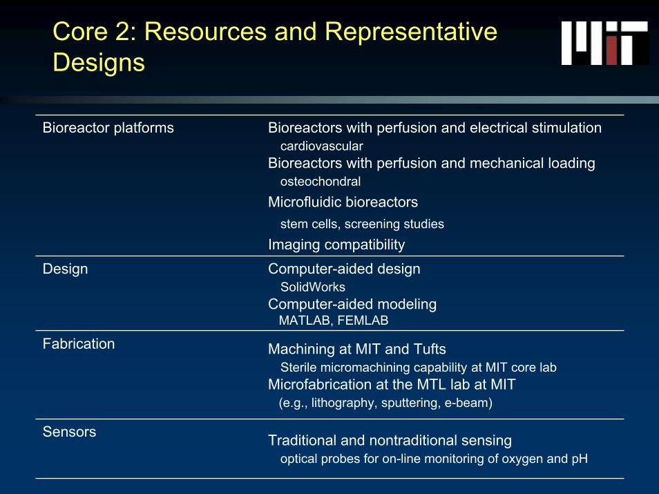
Core 2: Resources and RepresentativeDesigns
Bioreactor platforms Bioreactors with perfusion and electrical stimulationcardiovascular
Bioreactors with perfusion and mechanical loadingosteochondral
Microfluidic bioreactorsstem cells, screening studies
Imaging compatibilityDesign Computer-aided design
SolidWorksComputer-aided modeling
MATLAB, FEMLAB
Fabrication Machining at MIT and TuftsSterile micromachining capability at MIT core lab
Microfabrication at the MTL lab at MIT (e.g., lithography, sputtering, e-beam)
Sensors Traditional and nontraditional sensing optical probes for on-line monitoring of oxygen and pH
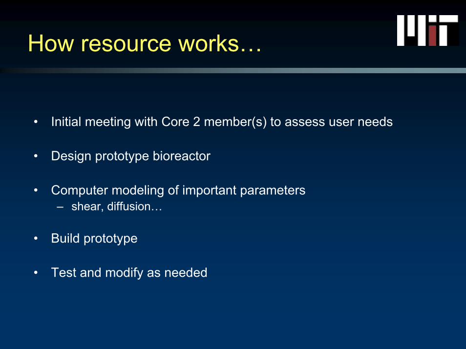
How resource works…
• Initial meeting with Core 2 member(s) to assess user needs
• Design prototype bioreactor
• Computer modeling of important parameters– shear, diffusion…
• Build prototype
• Test and modify as needed
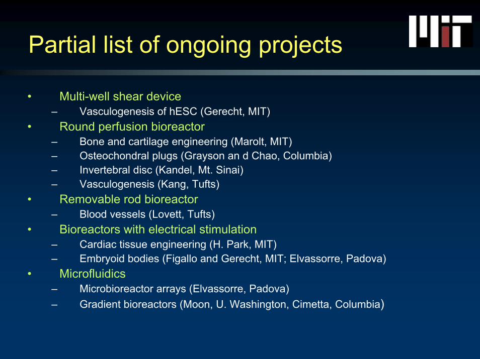
Partial list of ongoing projects
• Multi-well shear device– Vasculogenesis of hESC (Gerecht, MIT)
• Round perfusion bioreactor– Bone and cartilage engineering (Marolt, MIT)– Osteochondral plugs (Grayson an d Chao, Columbia)– Invertebral disc (Kandel, Mt. Sinai)– Vasculogenesis (Kang, Tufts)
• Removable rod bioreactor– Blood vessels (Lovett, Tufts)
• Bioreactors with electrical stimulation– Cardiac tissue engineering (H. Park, MIT)– Embryoid bodies (Figallo and Gerecht, MIT; Elvassorre, Padova)
• Microfluidics– Microbioreactor arrays (Elvassorre, Padova)– Gradient bioreactors (Moon, U. Washington, Cimetta, Columbia)
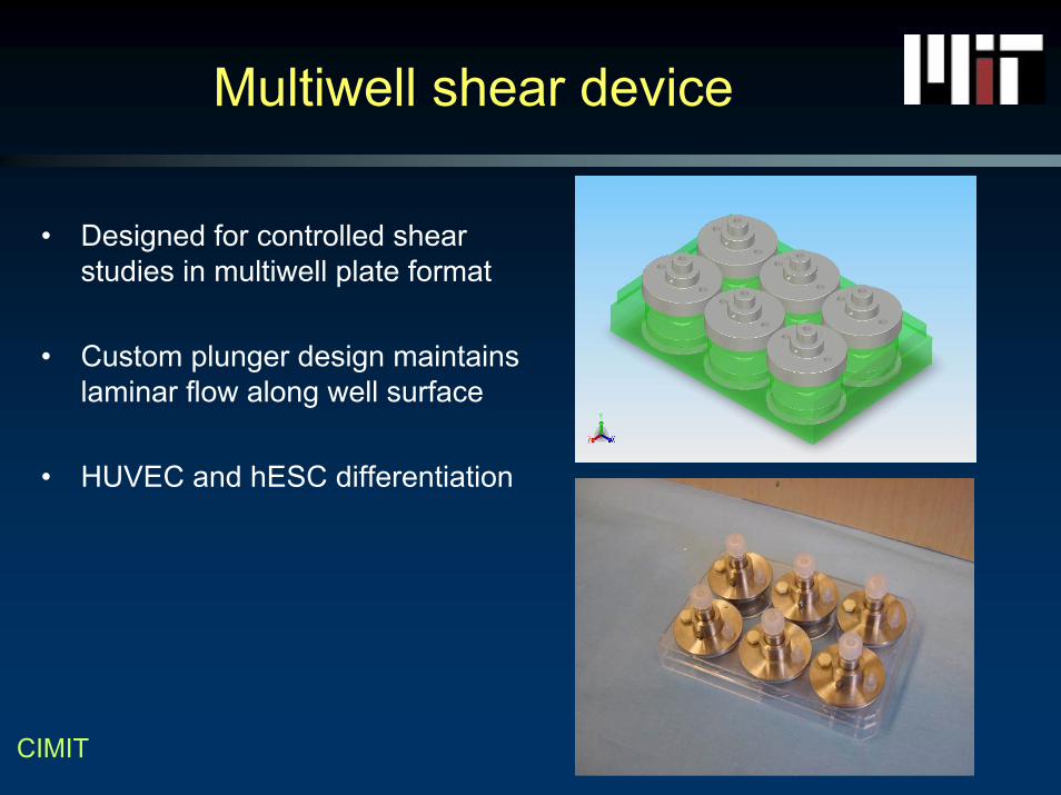
Multiwell shear device
• Designed for controlled shear studies in multiwell plate format
• Custom plunger design maintains laminar flow along well surface
• HUVEC and hESC differentiation
CIMIT
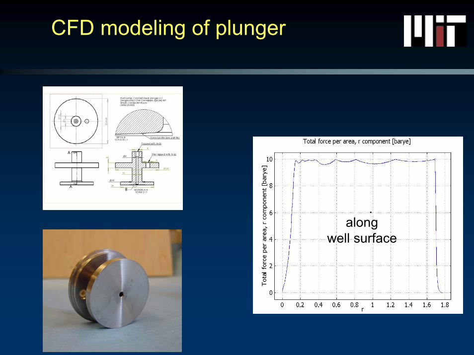
CFD modeling of plunger
�������� �����along
well surface
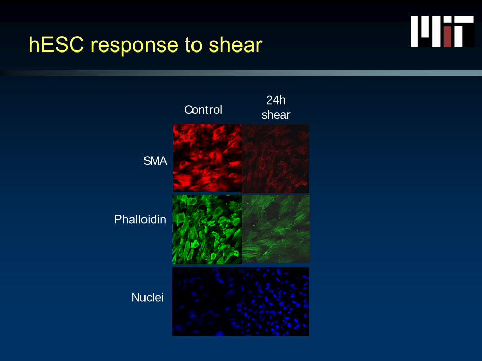
hESC response to shear
SMA
Phalloidin
Control24h
shear
Nuclei
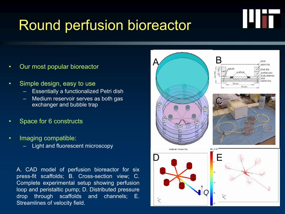
Round perfusion bioreactor
• Our most popular bioreactor
• Simple design, easy to use– Essentially a functionalized Petri dish– Medium reservoir serves as both gas
exchanger and bubble trap
• Space for 6 constructs
• Imaging compatible:– Light and fluorescent microscopy
A. CAD model of perfusion bioreactor for six press-fit scaffolds; B. Cross-section view; C. Complete experimental setup showing perfusion loop and peristaltic pump; D. Distributed pressure drop through scaffolds and channels; E. Streamlines of velocity field.
BBAA
CC
Q
DD EE
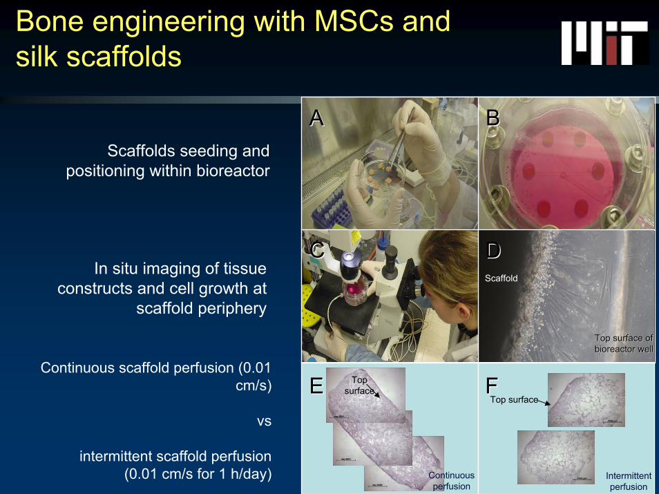
Bone engineering with MSCs and silk scaffolds
Scaffolds seeding and positioning within bioreactor
In situ imaging of tissue constructs and cell growth at
scaffold periphery
Continuous scaffold perfusion (0.01 cm/s)
vs
intermittent scaffold perfusion (0.01 cm/s for 1 h/day)
Scaffold
Top surface ofTop surface ofbioreactor wellbioreactor well
Continuousperfusion
Top surface
Intermittentperfusion
Top surface
AA BB
CC DD
EE FF
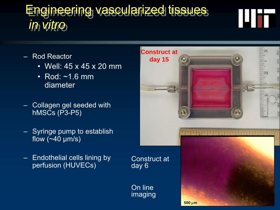
– Rod Reactor• Well: 45 x 45 x 20 mm• Rod: ~1.6 mm
diameter
– Collagen gel seeded with hMSCs (P3-P5)
– Syringe pump to establish flow (~40 µm/s)
– Endothelial cells lining by perfusion (HUVECs)
Engineering vascularized tissuesin vitro
Engineering vascularized tissuesin vitro
Construct at day 15
500 μm
Construct at day 6
On line imaging
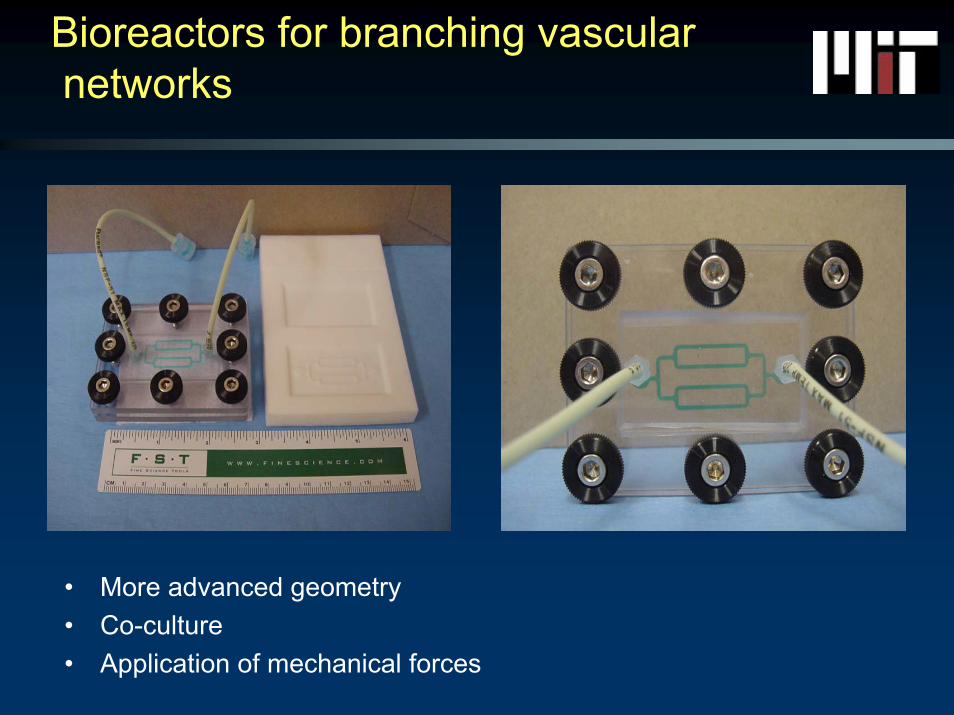
Bioreactors for branching vascularnetworks
• More advanced geometry• Co-culture• Application of mechanical forces
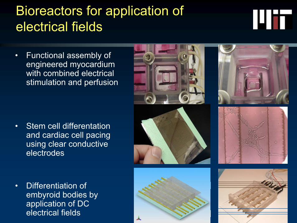
Bioreactors for application of electrical fields
• Functional assembly of engineered myocardium with combined electrical stimulation and perfusion
• Stem cell differentationand cardiac cell pacing using clear conductive electrodes
• Differentiation of embyroid bodies by application of DC electrical fields
Advanced culture systems for Advanced culture systems for controlled growth and differentiation of controlled growth and differentiation of human embryonic stem cellshuman embryonic stem cells
Sharon Gerecht & myself HST, MITGordana Vunjak-Novakovic Biomed Eng, Columbia UniversityElisa Figallo & Nicola Elvassore Chem Eng, University of PadovaJason Burdick Bioengineering, University of Pennsylvania
Chris Cannizzaro
Outline
• Introduction• 2D monolayer bioreactor system
– Bioreactor design– Image analysis– Differentiation of hES’s
• 3D hydrogel bioreactor system– Bioreactor design– Hyaluronic acid– Proliferation and differentiation of hES’s
• Conclusions
Advanced culture systems for controlled growth and differentiation of human embryonic stem cellshuman embryonic stem cells
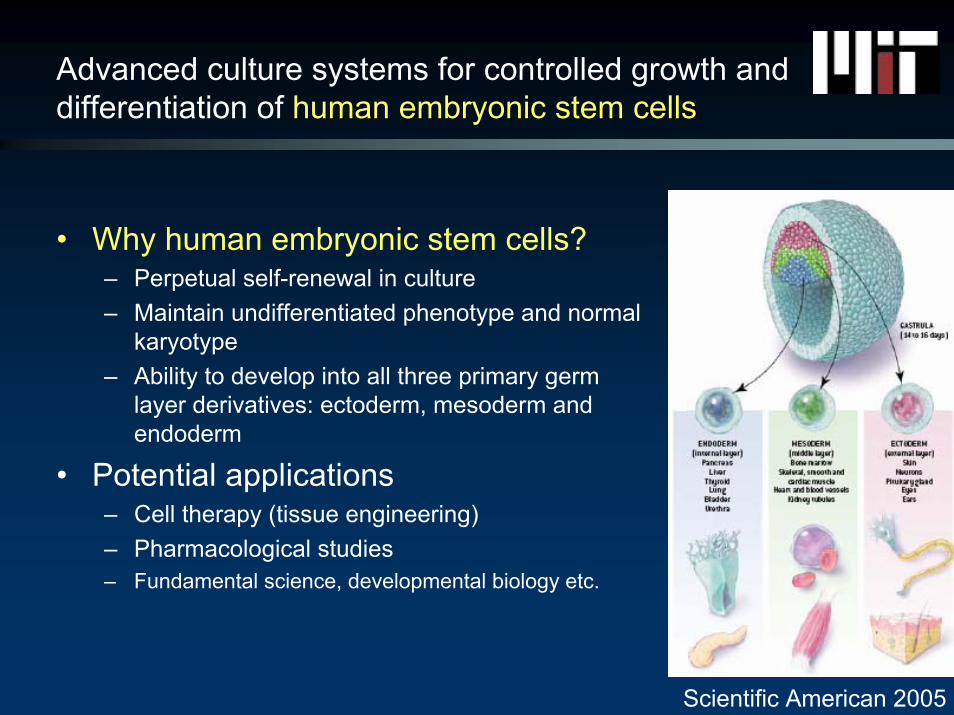
• Why human embryonic stem cells?– Perpetual self-renewal in culture– Maintain undifferentiated phenotype and normal
karyotype– Ability to develop into all three primary germ
layer derivatives: ectoderm, mesoderm and endoderm
• Potential applications– Cell therapy (tissue engineering)– Pharmacological studies– Fundamental science, developmental biology etc.
Scientific American 2005
Advanced culture systems for controlled growth and controlled growth and differentiationdifferentiation of human embryonic stem cells
• Why controlled growth and differentiation?– Stem cells must be differentiated in vitro for
therapeutic applications– Growth must be well controlled to expand cells to
req’d large quantities– Would like to recapitulate stem cell “niche”
• micro-environment surrounding the stem cells
Advanced culture systemsAdvanced culture systems for controlled growth and differentiation of human embryonic stem cells
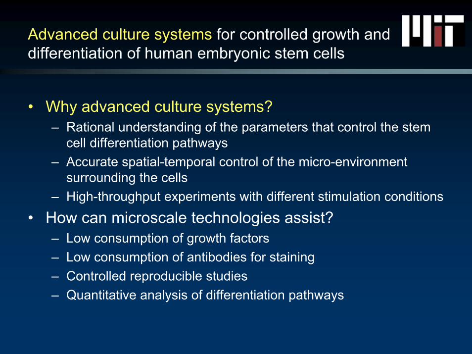
• Why advanced culture systems?– Rational understanding of the parameters that control the stem
cell differentiation pathways– Accurate spatial-temporal control of the micro-environment
surrounding the cells– High-throughput experiments with different stimulation conditions
• How can microscale technologies assist?– Low consumption of growth factors– Low consumption of antibodies for staining– Controlled reproducible studies– Quantitative analysis of differentiation pathways
2D monolayer bioreactor system
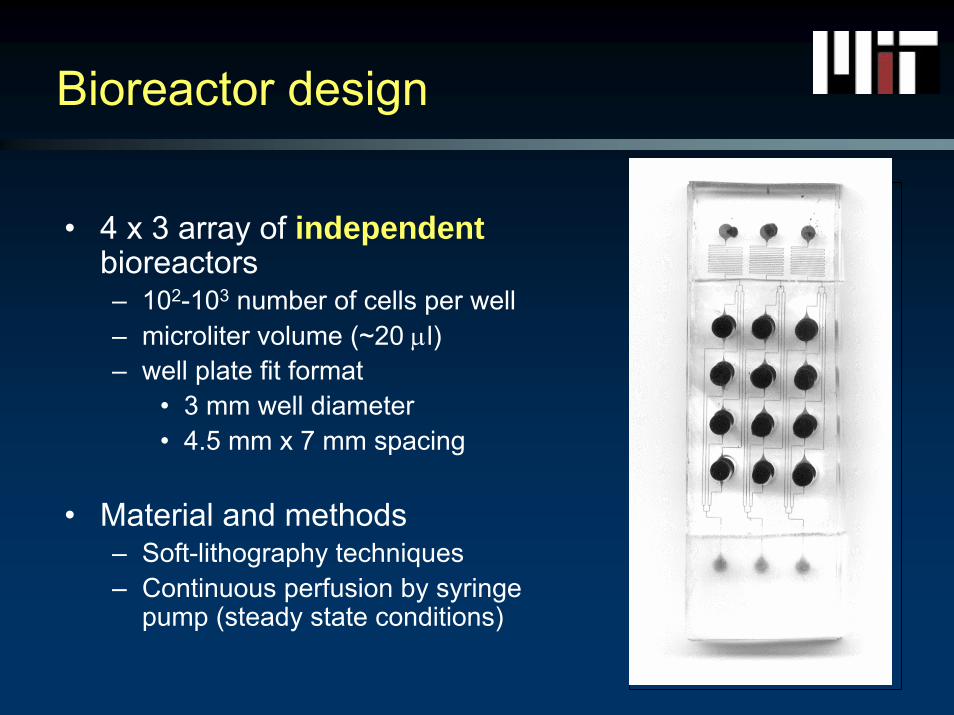
Bioreactor design
• 4 x 3 array of independentbioreactors– 102-103 number of cells per well– microliter volume (~20 μl)– well plate fit format
• 3 mm well diameter• 4.5 mm x 7 mm spacing
• Material and methods– Soft-lithography techniques – Continuous perfusion by syringe
pump (steady state conditions)
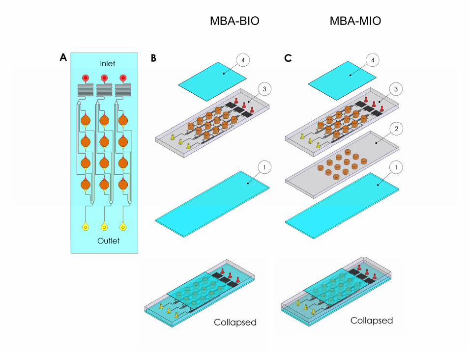
Bioreactor SchematicMBA-MIOMBA-BIO
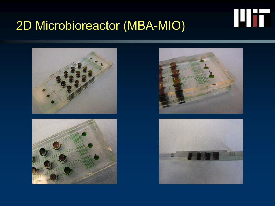
2D Microbioreactor (MBA-MIO)
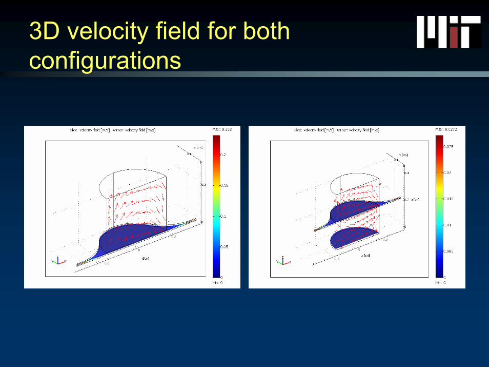
3D velocity field for bothconfigurations
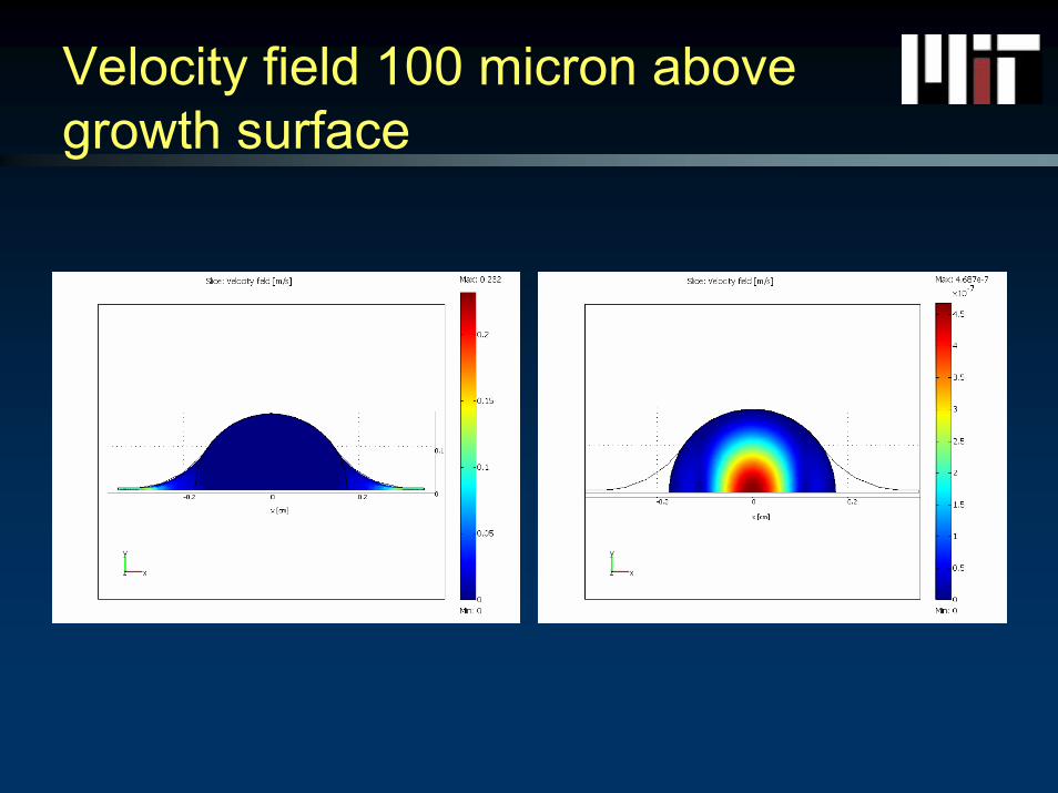
Velocity field 100 micron above growth surface
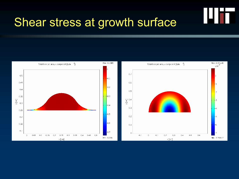
Shear stress at growth surface
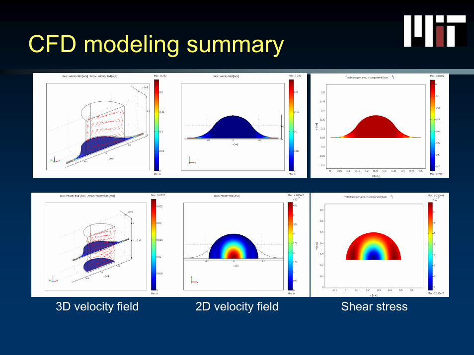
CFD modeling summary
3D velocity field 2D velocity field Shear stress
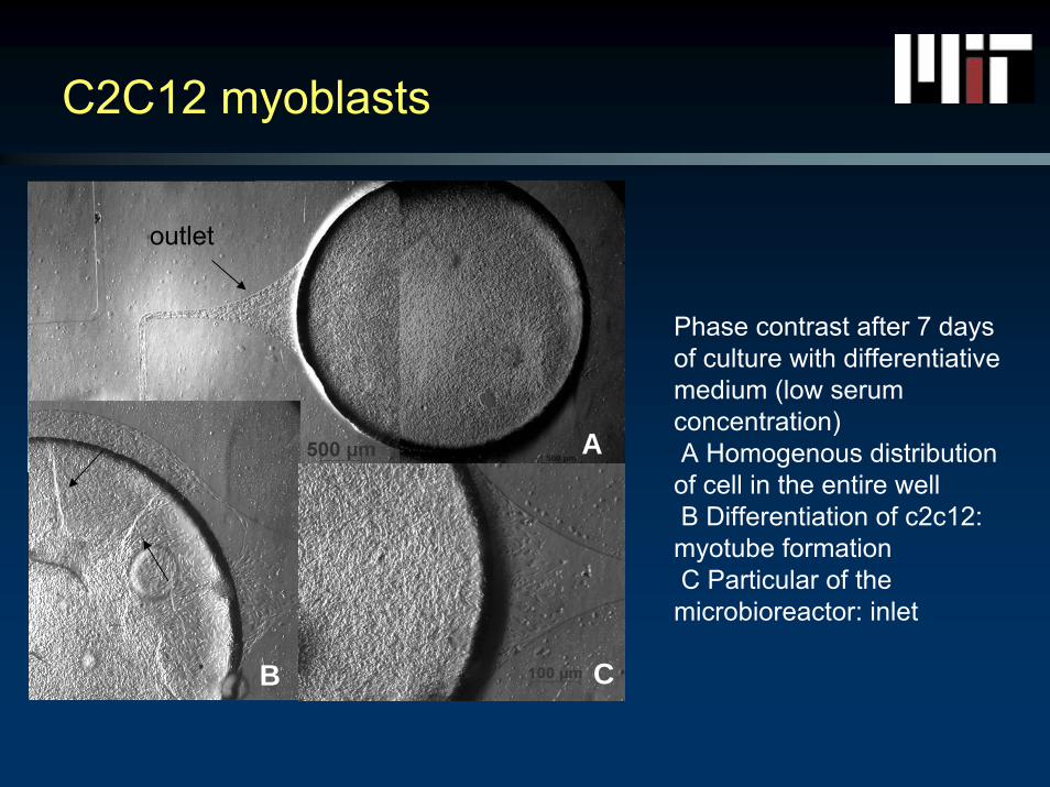
C2C12 myoblasts
Phase contrast after 7 days of culture with differentiativemedium (low serum concentration)A Homogenous distribution of cell in the entire wellB Differentiation of c2c12: myotube formationC Particular of the microbioreactor: inlet
A
B C
outlet
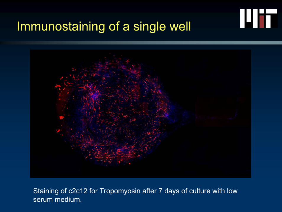
Immunostaining of a single well
Staining of c2c12 for Tropomyosin after 7 days of culture with low serum medium.
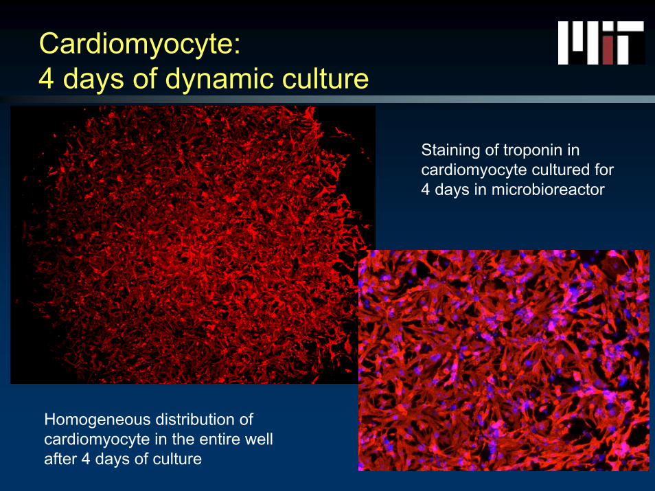
Cardiomyocyte: 4 days of dynamic culture
Homogeneous distribution of cardiomyocyte in the entire well after 4 days of culture
Staining of troponin in cardiomyocyte cultured for 4 days in microbioreactor
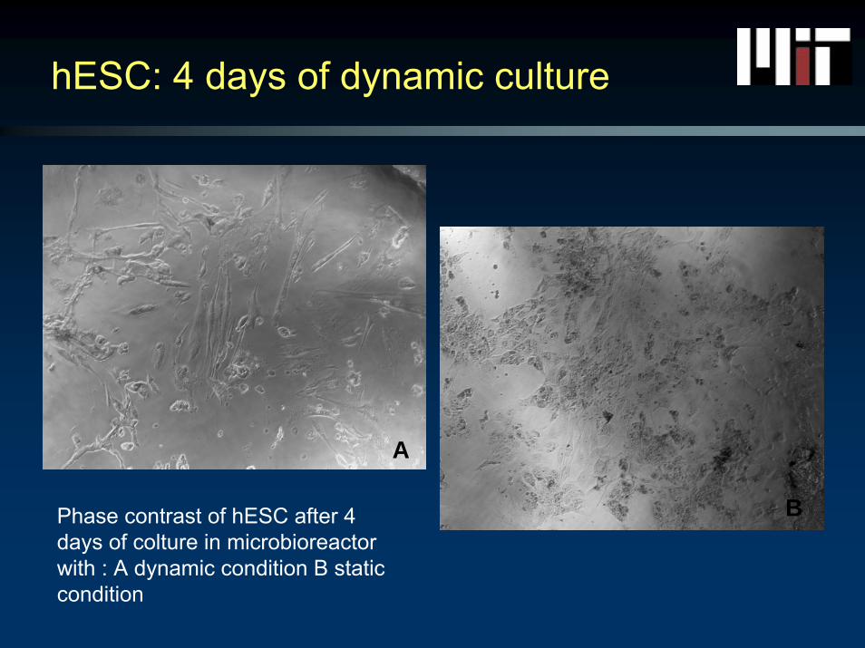
hESC: 4 days of dynamic culture
A
BPhase contrast of hESC after 4 days of colture in microbioreactor with : A dynamic condition B static condition

Immunostaining of hES’s after 4 days
SMAD
DAPI
high seeding density
low seeding density
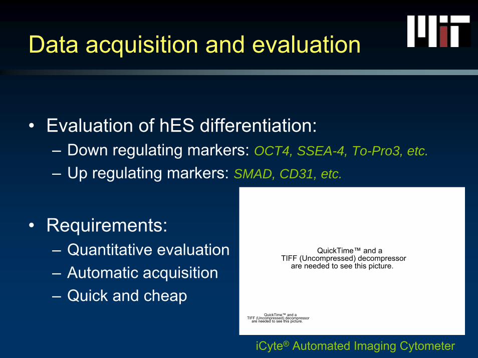
Data acquisition and evaluation
• Evaluation of hES differentiation:– Down regulating markers: OCT4, SSEA-4, To-Pro3, etc.
– Up regulating markers: SMAD, CD31, etc.
• Requirements:– Quantitative evaluation– Automatic acquisition– Quick and cheap
QuickTime™ and aTIFF (Uncompressed) decompressor
are needed to see this picture.
QuickTime™ and aTIFF (Uncompressed) decompressor
are needed to see this picture.
iCyte® Automated Imaging Cytometer
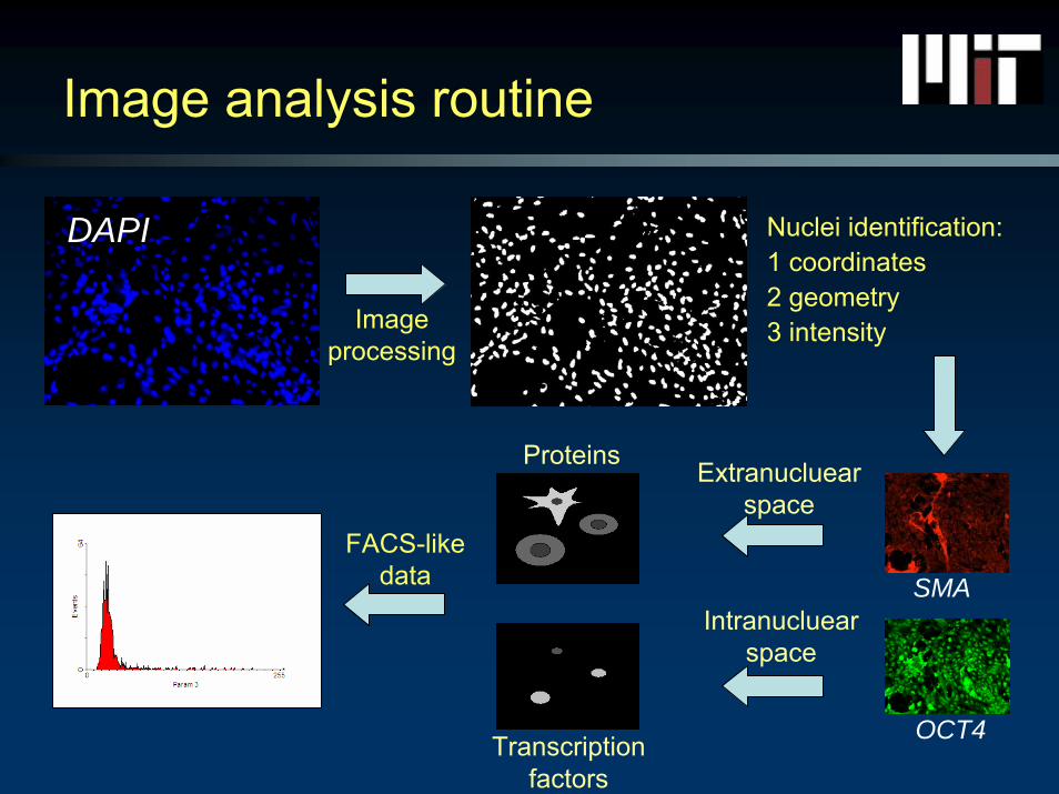
Image analysis routine
DAPI Nuclei identification:1 coordinates2 geometry3 intensityImage
processing
FACS-like data
Extranucluearspace
Proteins
SMA
Transcriptionfactors
Intranucluearspace
OCT4
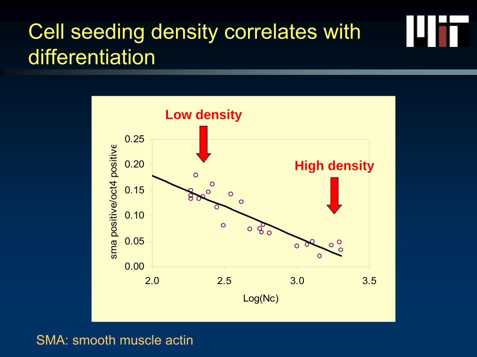
Cell seeding density correlates with differentiation
0.00
0.05
0.10
0.15
0.20
0.25
2.0 2.5 3.0 3.5
Log(Nc)
sma
posi
tive/
oct4
pos
itive
Low density
High density
SMA: smooth muscle actin
How do perfusion and growth factors influence differentiation?
• Experimental parameters:– Perfusion + and - with thermostated syringe pump:
• Flow rate of 0.3 μL/min– VEGF + and - medium– Slide precoated with collagen (IV)– 4 replicates for each condition– 4 day culture
• Immunofluorescence analysis:– Down regulating transcription factor: OCT4– Up regulating transcription factor: SMAD
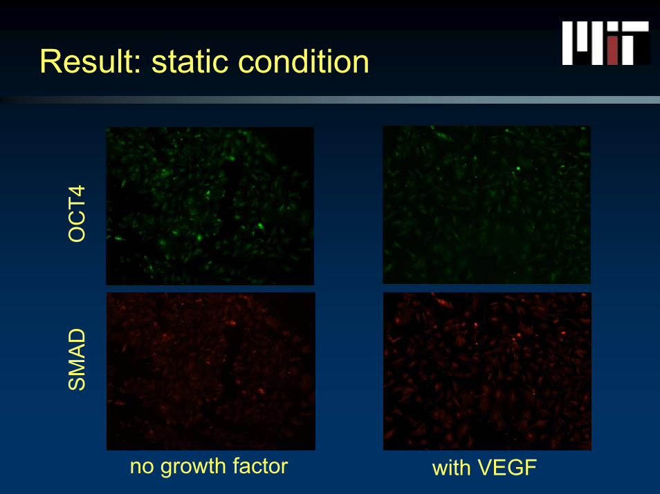
Result: static condition
no growth factor with VEGF
OC
T4S
MA
D
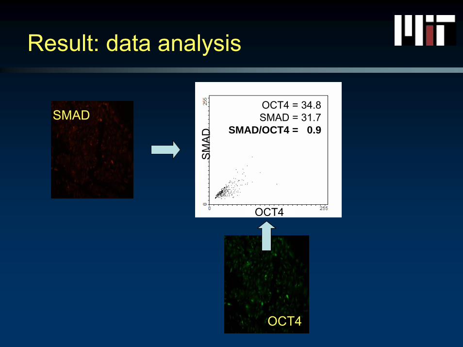
Result: data analysis
OCT4
SMADOCT4 = 34.8SMAD = 31.7
SMAD/OCT4 = 0.9
OCT4S
MA
D
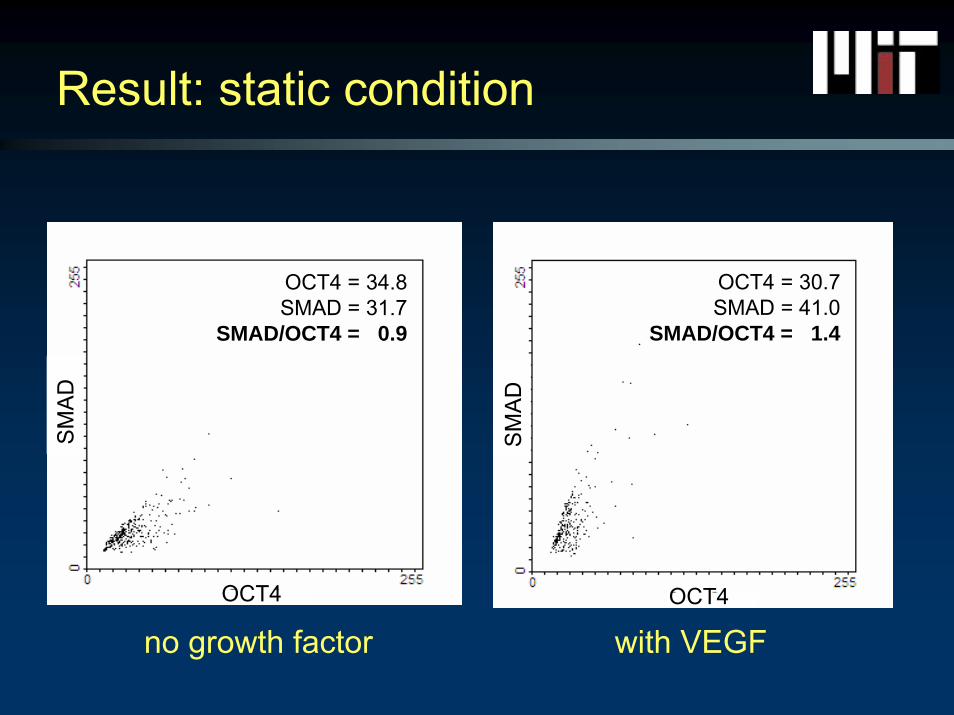
Result: static condition
no growth factor with VEGF
SM
AD
OCT4
SM
AD
OCT4
OCT4 = 30.7SMAD = 41.0
SMAD/OCT4 = 1.4
OCT4 = 34.8SMAD = 31.7
SMAD/OCT4 = 0.9
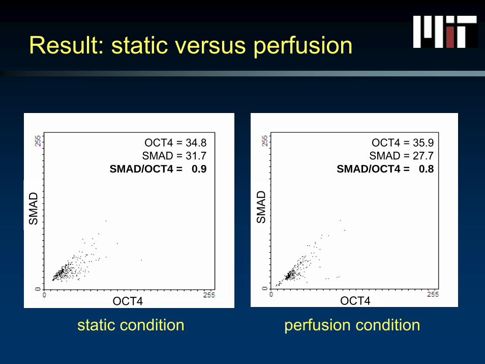
Result: static versus perfusionS
MA
D
OCT4
static condition perfusion condition
SM
AD
OCT4
OCT4 = 35.9SMAD = 27.7
SMAD/OCT4 = 0.8
OCT4 = 34.8SMAD = 31.7
SMAD/OCT4 = 0.9
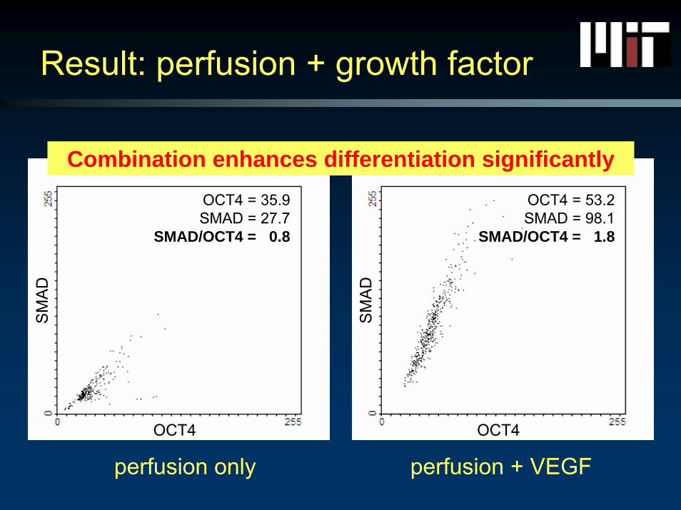
Result: perfusion + growth factorS
MA
D
OCT4
SM
AD
OCT4
perfusion only perfusion + VEGF
Combination enhances differentiation significantlyOCT4 = 35.9SMAD = 27.7
SMAD/OCT4 = 0.8
OCT4 = 53.2SMAD = 98.1
SMAD/OCT4 = 1.8
3D hydrogel bioreactor system
Bioreactor design
Control and regulation of differentiation & Control and regulation of differentiation &
proliferation/selfproliferation/self--renewal processesrenewal processes
⇓⇓
Why culture cells in a hydrogel?
• Use chemically defined surroundings for human ESC cultures
• 3D cultures affect cellular response of mature cells• Mimic 3D setting of a developing blastocyst• Future tissue engineering applications
Why hyaluronic acid (HA) hydrogel?
• HA is involved in the growth of undifferentiated human ESCs and is therefore an excellent substrate for their propagation
• HA hydrogel maintains hESCs in their undifferentiated state in contrast to other 3D systems
• HA hydrogel is a chemically defined and controllable environment in contrast to Matrigel and MEF

Levels decrease at the onset of
differentiationToole BP. Semin Cell Dev Biol. 2001
Improves IVF embryo survival and
developmentFurnus et al., Theriogenology 1998 Kim et al., Theriogenology 2005
HA receptor expressions during early embryo
development Campbell et al., Hum Reprod. 1995 Furnus et al.., Theriogenology. 2003
High expressionlevels during
embryogenesis Toole BP. Nat Rev Cancer . 2004
HA
Role of HA in early development
++
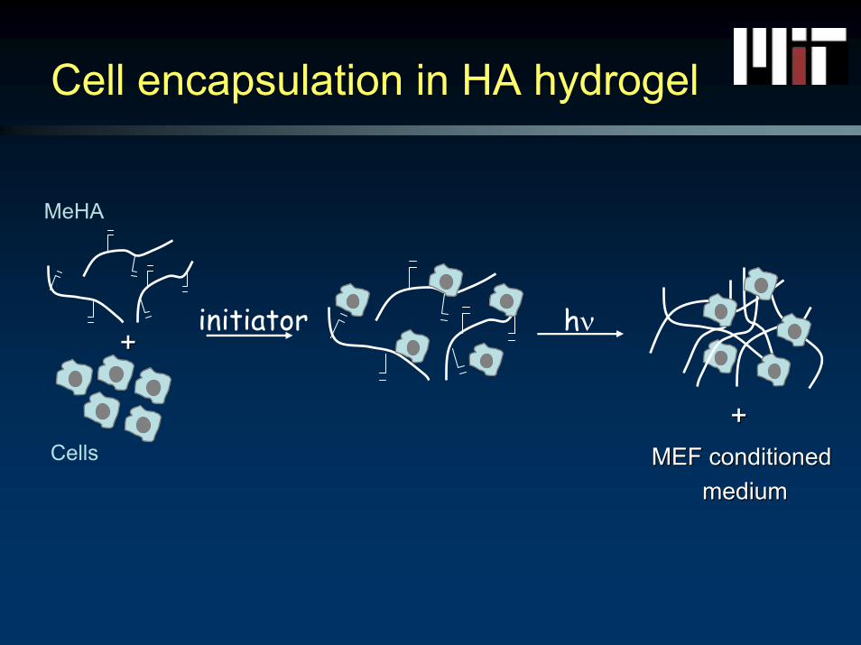
MeHA
Cells
hν
MEF conditionedMEF conditionedmedium medium
++
initiator
Cell encapsulation in HA hydrogel
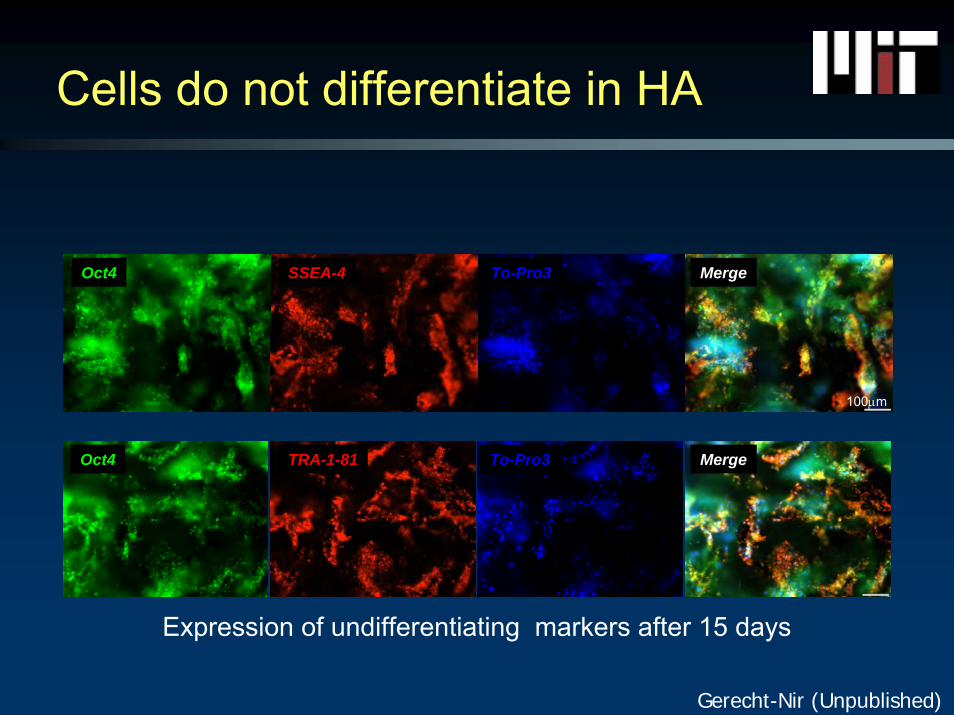
Oct4 SSEA-4 To-Pro3 Merge
Oct4 TRA-1-81 To-Pro3 Merge
100μm
Expression of undifferentiating markers after 15 days
Gerecht-Nir (Unpublished)
Cells do not differentiate in HA
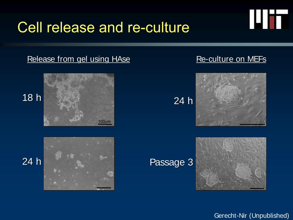
18 h18 h
24 h24 h
Release from gel using Release from gel using HAseHAse
100μm
24 h24 h
Passage 3Passage 3
ReRe--culture on culture on MEFsMEFs
Gerecht-Nir (Unpublished)
Cell release and re-culture

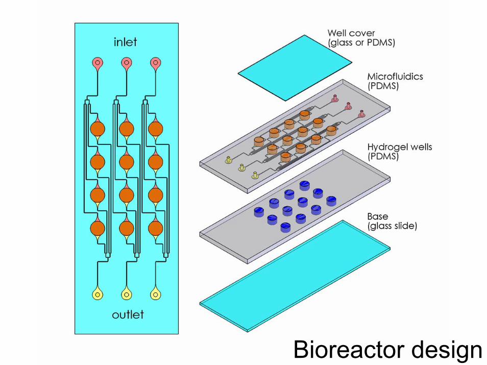
Bioreactor design
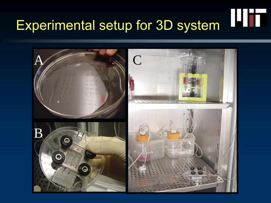
Experimental setup for 3D system
A
B
C
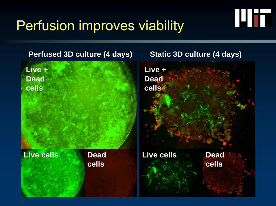
Perfusion improves viability
Live + Dead cells
Live cells Dead cells
Live + Dead cells
Live cells Dead cells
Perfused Perfused 3D culture (4 3D culture (4 daysdays)) Static Static 3D culture (4 3D culture (4 daysdays))

Differentiation can be studied by adding growth factors to medium
DAPI + CD31
CD31 DAPI
Cellular morphology Cellular morphology &&sproutingsprouting
7 days, VEGF+ medium
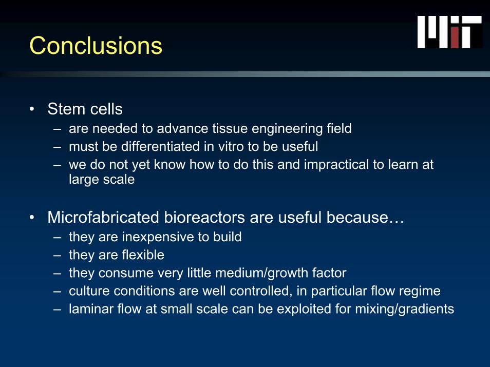
Conclusions
• Stem cells– are needed to advance tissue engineering field– must be differentiated in vitro to be useful– we do not yet know how to do this and impractical to learn at
large scale
• Microfabricated bioreactors are useful because…– they are inexpensive to build– they are flexible– they consume very little medium/growth factor– culture conditions are well controlled, in particular flow regime– laminar flow at small scale can be exploited for mixing/gradients
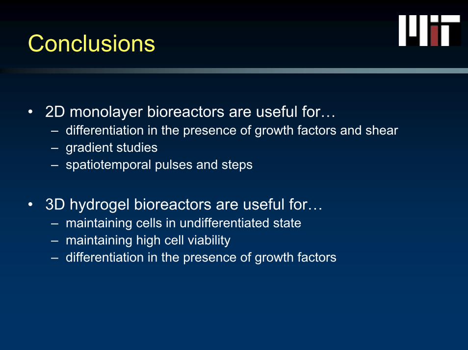
Conclusions
• 2D monolayer bioreactors are useful for…– differentiation in the presence of growth factors and shear– gradient studies– spatiotemporal pulses and steps
• 3D hydrogel bioreactors are useful for…– maintaining cells in undifferentiated state– maintaining high cell viability– differentiation in the presence of growth factors
Acknowledgments
University of Padova
Fulbright Scholar Program
Juvenile Diabetes Research FoundationQuickTime™ and aTIFF (Uncompressed) decompressor
are needed to see this picture.

Original Abstract
• The in situ environment of an embryonic cell in a developing blastocyte has a three-dimensional (3D) architecture, in contrast to the commonly used monolayer cultures. In vivo, cells are surrounded by other cells and a complex network of extracellular matrix (ECM) fibers, and subjected to cascades of regulatory signals (molecular and physical) which interact according to specific spatial and temporal patterns. Together, the factors associated with this environment regulate the self renewal and differentiation of human embryonic stem cells (hESCs). So far, ECM-cell interactions have not been extensively studied in hESCs. Furthermore, most advanced existing bioreactors can provide either local control of oxygen and pH or biophysical stimuli, but not the two sets of factors concurrently. Our goal was to develop culture systems that have the necessary cues and signals to provide microenvironmental control and biophysical regulation of cultured hESCs. First, a bioactive hydrogel based culture system was designed to provide a defined 3D environment for propagation of hESCs. This was followed by the development of a bioreactor for microenvironmental control and biophysical regulation. Both 2D and 3D settings were developed to study hESC growth and differentiation.
• The ECM polysaccharide hyaluronic acid (HA) was found to be a developmentally relevant material for the growth of hESCs. hESCs encapsulated in HA hydrogels propagated as undifferentiating cells and could be released and re-cultured, illustrating the importance of both the HA hydrogel structure and chemical specificity. HA internalization and gel remodeling by hESCs validated the specific bioactivity of the HA hydrogel culture system. This was followed by the development of microfluidic systems for cell culture in 2D and 3D settings, with perfusion and environmental control, consisting of an array of independent micro-bioreactors fabricated using soft lithography. This optically transparent microfluidic device integrates multi-parametric changes in culture conditions with non-destructive monitoring of events in individual living hESCs. First, the steady-state and reproducible culture conditions including mass transport (i.e. gas, metabolite, and soluble factor concentration) and flow rate (i.e. hydrodynamics) within the 2D microbioreactor were confirmed, and regulated Smad 2/3, Oct4 and actin during hESC mesodermal differentiation. Cell density, soluble factors and substrate components were all found to affect hESC shape and differentiation. Further steps were taken to study hESCs in a 3D setting, utilizing this highly defined and controllable culture system. Continuous perfusion of 3D scaffolding was found to improve hESCviability and survival within HA hydrogels. Experiments focusing on understanding developmental cues of 3D differentiating hESCs are ongoing. Thus far, the utilization of bio- materials and bioreactors facilitated control of cell behavior and can help improve our understanding of the signaling pathways involved in hESC growth and differentiation.