bioreactors for liver tissue engineering - oulun … · catapano and gerlach bioreactors for liver...
TRANSCRIPT

G. Catapano* and J. C. Gerlach
Summary
A cute liver failure (ALF) patients have a high risk of mortality. The elective treatment for ALF still is orthotopic liver transplantation. Donor organ scarcity and the associated high costs make transplantation possible for only about one third of the patients on the transplantation waiting list. Engineering a biological liver substitute is an interesting alternative to the traditional ALF therapy. Cells seeded in three-dimensional scaffolds made of bioresorbable biomaterials might be implanted in vivo to replace part of, or the whole, liver and integrate with time. Two- or three-dimensional non-implantable constructs may be used ex vivo for the EC support of ALF patients until a tissue compatible organ is available or the patient’s own liver heals. Non-implantable three-dimensional constructs may be also useful to investigate liver cell metabolism or as an alternative to animal tests for drug screening or toxicity assessment in vitro. Over the years, research has mainly focused on the development of new culture techniques or new immortalized hepatic cell lines. A major obstacle to the generation of functional substitutes is the limited understanding of the role of specific physical-chemical parameters on tissue development. Bioreactors provide controlled environmental conditions to improve the quality of tissue and to study liver cell metabolism. This review focuses on the bioreactors that have been proposed to develop enhanced biological liver substitutes or study liver cell metabolism. Keywords: Bioreactors; Cell; Liver; Metabolism; Tissue engineering
Bioreactors for Liver Tissue Engineering
Topics in Tissue Engineering, Vol. 3, 2007. Eds. N Ashammakhi, R Reis & E Chiellini © 2007.
*Correspondence to: Prof. Gerardo Catapano, Department of Chemical Engineering and Materials, University of Calabria, Via P. Bucci, I-87030 Rende (CS), Italy. Phone: +39 0984 496706. Fax: +39 0984 496655. E-mail: [email protected]
V B
IOR
EA
CT
OR
S
C H A P T E R 8

Catapano and Gerlach Bioreactors for Liver Tissue Engineering
2Topics in Tissue Engineering, Vol. 3, 2007. Eds. N Ashammakhi, R Reis & E Chiellini © 2007
Introduction Acute liver failure (ALF) caused by drug poisoning, surgical complications, viral infections or
decompensated chronic liver disorders has a high risk of mortality. ALF may progress within a
few weeks from the first symptoms (e.g., increased ammonia levels or the onset of jaundice)
onto life threatening complications (e.g., generalized oedema, bleeding and hepatic
encephalopathy) that culminate in multiorgan failure and death [1]. ALF has a poor prognosis
under conservative management, with survival rates ranging from 79% (for amanita
intoxication) to 10% (cryptogenic genesis) depending on its etiology [2]. The elective treatment
for ALF is orthotopic liver transplantation (OLTx) with survival rates up to 60-80% [3]. Until
2002, about one in three patients died while on the transplant waiting list and the number of
donor organs keeps decreasing since [4]. Donor organ shortage and the high social costs of
transplantation and the associated year-long immunosuppressive therapy limit the number of
liver transplants. However, liver tissue has the potential to regenerate and heal [5].
ALF is generally associated with the accumulation of plasmatic toxins (such as ammonia,
mercaptans, free phenols, bile acids, benzodiazepines, etc.) [6]. Thus, over the years, many
extracorporeal (EC) artificial liver support devices have been proposed for the treatment of
ALF, or to bridge a patient to OLTx in good mental conditions, that have not brought about
significant improvements over conventional patient management. In these devices, perm-
selective membranes or adsorption cartridges are used to simulate the liver detoxification
functions and remove from the blood some of the putative toxins damaging the liver. More
advanced systems have been reported to remove also protein-bound toxins by transferring them
to an albumin-rich solution across porous polysulphone membranes [7-9]. In one of them,
albumin is stably adsorbed in the membrane pores [7]. In these devices, the stripping albumin
solution is continuously regenerated over an adsorption column or goes to waste. Interesting
results have been reported in the treatment of acute-on-chronic (AoC) hepatic patients,
although the efficacy of these devices has not yet been proven in blind trials.
The need of liver-specific metabolic products to relieve some of the symptoms (e.g., cerebral
oedema and bleeding) and promote liver tissue regeneration started the quest for alternative
cell-based therapies aimed at providing ALF patients also with the liver biosynthetic and
biotransformation functions.
Tissue engineering (TE) is a multidisciplinary science aimed at developing “biological
substitutes to restore, maintain or augment tissue function” [10], and holds promise for the

Catapano and Gerlach Bioreactors for Liver Tissue Engineering
3Topics in Tissue Engineering, Vol. 3, 2007. Eds. N Ashammakhi, R Reis & E Chiellini © 2007
development of innovative alternatives to ALF treatment. The ideal biological liver substitute
should perform most or all of the liver-specific detoxification, synthetic and biotransformation
functions. As most of these functions are still unknown, mature liver cells (e.g., primary or
immortalized) or cells that may differentiate into hepatocytes (e.g., stem or progenitor oval
cells) have been used in these substitutes to perform liver-specific functions. Typically, liver
constructs are engineered in vitro by culturing liver (or liver-like) cells in/on synthetic scaffolds
which provide the template for cell adhesion, re-arrangement, proliferation and development.
Three-dimensional (3D) implantable constructs are made of porous biomaterials that degrade
and resorb at controlled rates to permit their replacement with the extracellular matrix (ECM)
produced by the cells, and cell colonization of the construct. Once implanted in vivo, the graft
has to fully integrate into the body of the host. Two-dimensional (2D) or 3D non-implantable
constructs may be used ex vivo for the EC support of ALF patients till a tissue compatible
organ is available or the patient’s own liver heals. These constructs are expected to provide the
patients only with the relevant liver functions that stop progression of the damages and promote
liver regeneration, and to function for a limited time from a few weeks to a few months. Non-
implantable constructs may be also useful to investigate liver cell metabolism or as an
alternative to animal tests for the in vitro drug screening or toxicity assessment. In this case, the
cells and the device in which these constructs are used, as a whole, should express enzymatic
activities and yield metabolite profiles similar to those in the natural liver for at least a few
months to represent a feasible and convenient alternative to animal tests.
The development of constructs with metabolic functions equivalent to those of the liver poses
technical challenges well beyond that of developing new culture techniques (e.g., 2D
monolayer vs. 3D culture) for the complexity of liver cell physical-chemical requirements and
the scale of the constructs. In fact, the liver is a highly structured organ with many distinct cell
sub-populations. In the liver parenchyma, the hepatocytes perform most of the liver-specific
metabolic functions. Hepatocytes are arranged in plates in repeating units termed “lobules” in
which they spread outward from a central vein. At the lobule vertices, a bile duct and a branch
of the hepatic artery and portal vein are located close to one another in an arrangement often
termed “the portal triads”. Blood flows from the two vessel branches towards the central vein
through small vascular channels, termed “sinusoids”, lined with a fenestrated layer of
endothelial cells. Plasma filters through the endotelium into the space that separates it from the
hepatocytes (i.e., the space of Disse), and exchanges nutrients and metabolites with the
hepatocytes through their apical surface. Bile is secreted into the canaliculi formed between the

Catapano and Gerlach Bioreactors for Liver Tissue Engineering
4Topics in Tissue Engineering, Vol. 3, 2007. Eds. N Ashammakhi, R Reis & E Chiellini © 2007
basal surfaces of adjacent hepatocytes, and flows through the bile ducts into the common bile
duct that dumps it into the duodenum. Kuppfer cells, ECM-producing stellate cells, biliary
epithelial cells, hepatocyte precursor cells and fibroblasts are also present and perform
important metabolic functions [11]. Liver cells are spatially organized to optimize
communication and transport. Cells communicate directly through cellular and gap junctions,
and via chemical signals dissolved and blood-borne or present in the macromolecules forming
the ECM that surrounds them. The signals that cells exchange promote differentiation,
proliferation and functions [12-14]. Furthermore, metabolic (e.g., carbohydrate metabolism)
and detoxification (e.g., CYP450 enzymes) activities of the hepatocytes change spatially along
the length of the sinusoid, apparently regulated by gradients of oxygen, hormones and ECM
composition, a phenomenon termed “liver zonation” [15,16]. Information on the structure-
function relationship for normal and pathological liver tissue is still lacking. Fostering the same
cellular relationships existing in the normal liver also in the TE liver construct is considered
fundamental for cells to function as in the natural liver. This has suggested to pattern the
biological substitutes after the liver micro-architecture, although reproducing the whole liver
architecture in constructs for EC liver support might not be necessary for cells to perform only
the subset of hepatic functions relevant to stop progression of ALF. Hepatectomy studies
suggest that TE liver constructs should perform metabolic functions quantitatively equivalent
to at least 30% of the natural liver mass to be effective in the treatment of ALF [5], and set the
scale of the construct at one hosting an average of at least 500 g cells. An additional
complication is that the engineering of liver tissue in vitro occurs through processes whose
length and time scale spans over orders of magnitude. In fact, cells adhere on the biomaterial
surface by recognizing morphological and biochemical patterns in the nanometer range but
have to organize in hierarchical structures a few centimeters large. Proteins from serum adsorb
on, and mediate cell interactions with, the biomaterial surface within milliseconds but it may
take weeks before liver cells differentiate and re-arrange in functional liver-like cellular
structures [7]. That said above clearly outlines the formidable technical challenges posed by
liver tissue engineering. Over the years, research has mainly focused on the procurement of
large amounts of suitable liver cells and the development of in vitro culture techniques [17-19].
Increasing research effort is being devoted to the development of biomaterials for liver cell
scaffolding at whose surface topological, morphological and biochemical signals are present to
attract cells and make them adhere and rearrange their cytoskeleton, and to the characterization
of the dissolved biochemical and physical signals that control cell differentiation, proliferation
and apoptosis [20].

Catapano and Gerlach Bioreactors for Liver Tissue Engineering
5Topics in Tissue Engineering, Vol. 3, 2007. Eds. N Ashammakhi, R Reis & E Chiellini © 2007
This chapter focuses on the bioreactors proposed for liver tissue engineering. A bioreactor may
possibly be defined as a volume (or vessel) in which one or more biochemical or biological
processes take place. Indeed, bioreactors are extensively used at any step of the assembly of a
TE liver construct. In fact, primary cells enzymatically isolated from autogenous or allogeneic
liver tissue (or the whole organ) are often cultured in Petri dishes or T-flasks prior to their
seeding in the scaffold to let them recover from the isolation and purification stress.
Immortalized and primary progenitor or stem liver cells are generally expanded in Petri dishes
or T-flasks, often under conditions facilitating differentiation to mature hepatocytes of the first
two cell types, or inhibiting differentiation to maximize proliferation of the third cell type.
Liver cells (often of different types) are harvested and seeded in/on synthetic scaffolds in
bioreactors under conditions that should facilitate cell attachment to the scaffold surface and
penetration into its pores. The cell-seeded scaffolds are then cultured in bioreactors under
tightly controlled and closely monitored environmental conditions to provide cells with
biochemical and physical cues that should promote cell reorganization into liver-like
aggregates and differentiation to make the construct functionally equivalent to liver tissue.
Non-implantable constructs used for EC liver support are eventually cultured in bioreactors
under conditions that maximize their therapeutic efficacy, prevent their rejection (if allogeneic
cells are used) and intoxication caused by the ALF plasma, and preserve the liver cell
phenotype for the treatment time. Non-implantable constructs used for metabolic investigation
are eventually used in bioreactors under controlled, measurable and reproducible metabolite or
drug concentrations to study liver cell metabolic behavior and differentiation. Below, the
problems in bioreactor design for liver tissue engineering are briefly discussed and the
proposed bioreactors presented.
Bioreactor design issues The first step in the development of constructs for liver replacement is to seed a large mass of
liver cells uniformly on, or throughout, a scaffold. Then, the adherent cells have to be provided
with adequate amounts of oxygen and nutrients to survive and proliferate, and adequate
biochemical/physical signals to re-organize and differentiate to yield cellular structures and
metabolic zonation resembling that of the liver. Attaining adequate cell seeding, nutrient and
oxygen supply to cells, and the control of biochemical signals gradients and concentrations in
large scaffolds is not easy and depends on the bioreactor configuration and operation, on how

Catapano and Gerlach Bioreactors for Liver Tissue Engineering
6Topics in Tissue Engineering, Vol. 3, 2007. Eds. N Ashammakhi, R Reis & E Chiellini © 2007
mass is transported outside and inside the scaffold, and on cell metabolism. Bioreactors used
for extracorporeal liver support have also to be connected to the patient so as to guarantee the
unhindered transport of soluble species from the patient’s circulation to the cells in the
bioreactor and vice versa. Below, this last issue shall only be briefly discussed.
Cell seeding
Cell seeding is thought to play a critical role in the development of in vitro engineered tissue.
Seeding cells at high initial density may favor tissue formation [21]. High seeding efficiency
would also limit the amount of organ tissue from which primary cells are isolated and cell
expansion. Uniform initial cell distribution on 2D or throughout 3D scaffolds has been related
to the uniformity of engineered cartilage and bone tissue [22-24]. Uneven cell distribution in
the scaffold might lead to spatial variations in nutrients, oxygen and metabolite concentrations
that would condition the survival and metabolism of cells at different positions in the scaffold.
Seeding cells efficiently and uniformly on/in a scaffold is challenging, in particular throughout
3D porous scaffolds. In fact, closed or tight pores inside the scaffold may limit cell access and
the seeding efficiency and distribution. Cell distribution in accessible pores depends on the
balance between the rate at which cells are physically transported from the medium bulk to the
outer scaffold surface (i.e., external transport) and from there towards its innermost pores
(internal transport), and the rate at which cells bind to the biomaterial pore surface or to other
cells in the feed suspension and form clusters. Resistance to external transport and cell
uniformity in the feed suspension may be varied by changing the mixing intensity in the
bioreactor where seeding takes place. Resistance to internal transport depends on the cell-to-
pore size ratio but also on the transport mechanism within the scaffold. In fact, drag associated
with medium convection through the pores enhances cell transport as compared to gravity or
capillary forces only, although to an extent that depends on the actual pore size distribution and
tortuosity of the scaffold. Cell association in clusters increases the size of the transported
aggregate and limits, or hinders, their mobility within 3D scaffolds. Fast cell adhesion to the
scaffold surface closer to its external surface may reduce the effective pore size and hinder
farther cell penetration into 3D porous scaffolds.
Nutrient and metabolite transport
Growing liver tissue in vitro for liver replacement or support is more difficult than other
tissues. In fact, cells have to be cultured at the high density typical of the natural liver, much

Catapano and Gerlach Bioreactors for Liver Tissue Engineering
7Topics in Tissue Engineering, Vol. 3, 2007. Eds. N Ashammakhi, R Reis & E Chiellini © 2007
higher than in many other tissues. Liver cells have also important nutrient requirements and are
sensitive to waste metabolites [25-29]. In vivo, the liver is efficiently provided with soluble
nutrients by a high blood flow that reaches the innermost cells in the organ by means of a fine
network of capillaries (i.e., the sinusoids). This keeps the diffusion distance between cells and
the blood small (i.e., within a few hundreds micron). Providing an analogous system to supply
basic substrates (e.g., oxygen, glucose and amino acids) to or clear waste metabolites (e.g.,
CO2, ammonia, urea, lactate) from liver cells in large 3D constructs is a formidable challenge,
and a pre-requisite to promote cell growth, differentiation and long-term survival [30-33]. In
fact, nutrients are continuously consumed (and products formed) by the cells while they are
transported from the source into the cell mass (or from the cells to the sink). This causes the
concentration of soluble nutrients in the cell mass to be generally less than that near their
source (for neo-synthesized species it would be higher than that near the sink), and the
formation of concentration gradients across the cell construct. These gradients may induce
different cell behaviors at different spatial positions in the construct, and even induce
chemotaxis if the gradients are steep enough to be sensed by the cells [34]. Although not basic
for cell survival, transport of large proteins may also have important effects on cell behavior
and the therapeutic efficacy of constructs used in bioartificial livers (BALs) for EC liver
support. In fact, their mobility is significantly lower than that of small nutrients. This causes a
mass transport resistance that may substantially reduce the concentration of proteic effectors
(e.g., EGF) with respect to their value in the feed medium and may hinder transport of neo-
synthesized liver-specific proteins (e.g., clotting proteins and growth factors) back into the
patient’s blood. Three compartments may be identified in the bioreactors proposed for the in
vitro culture of liver cell-seeded constructs: the medium compartment, where synthetic culture
medium, plasma or blood from the patient’s circulation flows; the membrane compartment,
when membranes are used as immuno-selective barriers to protect the cells from rejection or to
scaffold the cells; the cell compartment, where cells are seeded and cultured attached to a
biocompatible scaffold or microcarrier, or in suspension. To reach the cells in the construct,
soluble nutrients have to be transported across each compartment, in particular: external to the
construct, from the medium bulk to the membrane (or the construct) outer surface; across the
membrane wall; inside the construct, from the membrane inner (or the construct outer) surface
into the cell construct across the cell mass. The spatial profile of soluble nutrients and wastes in
the construct, and the rate at which they are transferred from one compartment to the other,
depends on the mass transport resistance of each compartment. In the following, the factors
affecting the resistance in each compartment are shortly presented and discussed as a means to

Catapano and Gerlach Bioreactors for Liver Tissue Engineering
8Topics in Tissue Engineering, Vol. 3, 2007. Eds. N Ashammakhi, R Reis & E Chiellini © 2007
understanding the rationale of the different bioreactors proposed to optimize the in vitro culture
of liver cells in large constructs. For the sake of the example, reference is made to the transport
of oxygen, an important nutrient playing a key role in all aerobic metabolic processes, often
limiting cell functions and tissue growth in vitro. For additional information, the Reader is
referred to other available reviews on mass transport in bioreactors for tissue culture [30-34].
External transport – In conventional batch bioreactors (e.g., Petri dishes or T-flasks) for 2D
cell or tissue culture, soluble species are transported by diffusion from the medium bulk to the
cell surface across a stagnant layer of medium. When oxygen is supplied from the gas above
the medium (as is often the case), the dissolved oxygen concentration at the gas-medium
interface Ceq is limited by the scarce oxygen solubility in aqueous media. In fact, medium
equilibrated with air would contain only ca. 0.2 mmoloxygen/L. The actual dissolved oxygen
concentration at the cell surface Csurface is generally lower than Ceq. In particular, Csurface
increases with increasing Ceq (hence the gaseous O2 partial pressure) and oxygen diffusivity in
the medium D (ca. 2 x 10⁻⁵ cm2/s), and decreases with increasing medium height above the
cells h and cell oxygen consumption rate OCR. The balance at the liver cell surface of the
oxygen diffusive flux (i.e., D (Ceq – Csurface)/h) and the cellular rate of oxygen consumption
yields oxygen concentrations at the cell surface decreasing from ca. 50 to 3% of the
concentration at the gas-medium interface as the medium height above the cells increases from
0.5 to 2 mm (i.e., for OCR=Vmax Csurface/(Km + Csurface) and Vmax=koxy Ccell with koxy=0.38
nmol/106 cells/s, Km=6.6 nmol/mL [28], for a confluent cell monolayer with ca. Ccell=105
cells/cm2).
Culturing large cell-seeded 3D scaffolds in stagnant medium (e.g., in Falcon tubes) may result
in even steeper spatial oxygen concentration gradients along the construct length. This causes
the dissolved oxygen concentration to decrease significantly at zones of the outer construct
surface farther away from the gaseous oxygen source.
External mass transport resistance can be reduced by superimposing convective transport to
pure diffusion to increase the mixing. Under these conditions, the rate of solute transport
to/from the outer membrane or construct surface may be expressed proportional to the
dissolved oxygen concentration difference between the medium bulk and the outer construct
surface through the solute transport coefficient kc (i.e., the reciprocal external resistance to
solute transport). The external mass transport resistance is often lumped in a thin stagnant
liquid film adherent to the outer surface of the membrane/construct. Its actual value is generally

Catapano and Gerlach Bioreactors for Liver Tissue Engineering
9Topics in Tissue Engineering, Vol. 3, 2007. Eds. N Ashammakhi, R Reis & E Chiellini © 2007
provided in non-dimensional semi-empirical equations correlating the Sherwood number Sh=kc
d/D with powers of non-dimensional groups accounting for the geometry of the flow channel
and the liquid velocity field (e.g., the Reynolds number Re=ρ v d/μ), and the liquid and solute
transport and physical properties (e.g., the Schmidt number Sc=μ/ρ D , where μ is solvent
viscosity, D solute diffusivity in the solvent, ρ solvent density), such as: Sh ∝ Reβ Scγ [35].
These correlations suggest that kc is proportional to the β-th power of the liquid velocity
relative to the construct v, and inversely proportional to the (1-β)-th power of the characteristic
length of the flow channel or construct d, as follows: kc ∝ vβ / d (1-β) . Hence, solute transport
could be enhanced by increasing v and by decreasing d to an extent that depends on the value
of β. The occurrence of turbulence and secondary flows promoted by mechanical stirring or
tortuous flow around the membrane/construct generally yields higher β values and a stronger
dependence of kc on the liquid velocity than for liquid flowing in a tube under laminar
conditions [36, 37].
In the analysis of external mass transport, it is often assumed that matter is ideally distributed
outside the membrane/construct. The irregular and time-varying geometry of the construct, the
geometry of the bioreactor inlet and outlet ports, improper construct fitting into the bioreactor
and the actual operating conditions may all cause a non-ideal distribution of matter in the
bioreactor which exposes the external surface of the construct to solute concentrations spatially
varying in unpredictable fashion. As very nicely pointed out in [33], the occurrence of
important back-mixing (e.g., caused by velocity gradients in planes orthogonal to the bioreactor
or construct axis) may even off-set some of the advantages of external convective transport.
The intensity of these non-ideal behaviors is often expressed in terms of a dispersion
coefficient, or similar model parameters, accounting also for diffusion [38]. Although
information is available in literature to estimate the dispersion coefficient for some industrial
chemical reactors, its value and the occurrence of flow maldistribution in bioreactors for liver
tissue engineering is more effectively estimated in the course of tracing experiments [39].
In a few bioreactors proposed for EC liver support, the patient’s blood flows in direct contact
with the membranes protecting the liver cell-seeded constructs [40, 41]. The use of whole
blood significantly enhances oxygen external transport to the liver cell construct. In fact, the
arterial blood of the average healthy individual may carry ca. 8.6 mmoloxygen/Lblood for the
oxygen binding capacity of hemoglobin in the red blood cells, an amount far higher than that of
aqueous medium [42]. However, in these bioreactors the enhancement of transport by

Catapano and Gerlach Bioreactors for Liver Tissue Engineering
10Topics in Tissue Engineering, Vol. 3, 2007. Eds. N Ashammakhi, R Reis & E Chiellini © 2007
convection is limited by the possible occurrence of hemolysis or clotting caused by steep
velocity gradients or blood stagnation in the bioreactor.
Membrane transport – Semi-permeable membranes are effectively used as immune barriers to
protect xeno- or allogeneic liver cells in a construct implanted in vivo or used for EC liver
support from immuno-competent proteins and cells [43]. Their presence causes an additional
resistance to mass transport. Depending on their charge, size and physical-chemical properties
(e.g., hydrophobicity) relative to that of the membranes, soluble metabolites may partition
between the medium/blood and the membrane ending up with concentrations in the membrane
at times significantly different than those in the medium. Solutes may be transported across the
membrane by both diffusion and convection in response to concentration or pressure gradients
across the membrane wall. The rate at which they are transported is effectively expressed as a
function of the solute concentrations in the medium near the membrane face CB and in the cell
compartment near the membrane face CC according to the irreversible thermodynamics as
follows [44]:
1. ( ) ( ) CJ1CCPJ vCBMS σ−+−=
with ( )CB
CB
CCLnCCC/
−= .
A flat membrane resistance to solute diffusive transport RM may be expressed in terms of the
reciprocal membrane diffusive permeability towards the given solute PM, with:
2. MM
M DKP1R δ
==
where δ is the membrane wall thickness, DM the effective solute diffusivity in the membrane,
and K = CBM / CB = CMC /CC the solute partition coefficient in the case that the solute partitions
equally between the medium and the membrane, where its concentration is CBM, and the cells
and the membrane, where its concentration is CMC. The radial change of membrane surface area
is also generally accounted for in RM in the case of thick cylindrical membranes. DM was shown
to decrease with increasing solute-to-pore size ratios and with the solute molecular weight
more rapidly than that predicted by the Einstein-Stokes equation [45]. The solute ability to be
freely dragged across the membrane is expressed in terms of σ, the Stavermann coefficient,
which is 0 for fully permeable and 1 for fully rejected solute. The medium flow through the
membrane Jv = Lp ΔP is related to the membrane hydraulic permeability Lp and the pressure
difference that develops, or is applied, across the membrane ΔP. The accumulation and
Catapano and Gerlach Bioreactors for Liver Tissue Engineering
11Topics in Tissue Engineering, Vol. 3, 2007. Eds. N Ashammakhi, R Reis & E Chiellini © 2007
adsorption of partially or fully rejected solutes at the membrane surface may significantly
change the membrane transport properties and decrease its permeability to both aqueous
medium and soluble metabolites, in particular those of medium to high molecular weight [46].
Internal transport – Transport inside 3D cell constructs of soluble metabolites that are
consumed or produced by the cells occurs simultaneously to their metabolic consumption or
production. Under steady conditions, the metabolite concentration profiles that establish across
the construct balance out the rate of physical transport into the construct with the rate of its
consumption or production. In densely packed cell constructs, and in many bioreactors
proposed in BALs for EC liver assist, soluble species are mainly transported by diffusion into
the construct. This is particularly true for small solutes. Both the increasingly high cell density
as cells grow and the amount of synthetic or newly formed ECM decrease solute mobility in
the construct Deff ≈ D ε/τ as compared to that in medium D by decreasing its void fraction ε and
by increasing its tortuosity τ. For purely diffusive transport, reduced solute diffusivity, high cell
packing density and metabolic activity are all expected to contribute to a sharp decrease of
soluble nutrient concentration into the construct away from the nutrient source. A more
quantitative description of phenomena occurring in the construct may be provided according to
a pseudo-homogeneous approach to predict the solute concentration profile across liver cell
constructs formed inside or outside hollow fiber membranes [47-49]. As an example, let us
consider the simple case of steady oxygen transport across a cell-seeded construct shaped as a
slab and zero-th order oxygen consumption kinetics. The one-dimensional mass balance
equation for oxygen may be written in terms of its non-dimensional concentration CO2/CO2,C
and the spatial coordinate x/L as follows:
3. 22
C2O2Oeff
celloxy2
C2O2O2
LCD
CkLxdCCd
φ==,,
,
)/()/(
Equation 3 suggests that steep non-dimensional oxygen concentration profiles establish across
the slab at high cell packing density Ccell, oxygen consumption rate on a per cell basis koxy, and
slab half thickness L, and at low oxygen effective diffusivity in the construct Deff,O2 and
dissolved oxygen concentration at the outer slab surface CO2,C (equal to that in the medium bulk
CO2,b for negligible external and membrane mass transport resistance). The actual oxygen
concentration profile is determined by these variables grouped into the non-dimensional Thiele
modulus φ rather than independently, values of φ2>1 causing a steep oxygen concentration
decay into the construct. For Ccell=108 cell/mL, diffusivity in the cell mass Deff,O2=0.6 DO2, koxy,

Catapano and Gerlach Bioreactors for Liver Tissue Engineering
12Topics in Tissue Engineering, Vol. 3, 2007. Eds. N Ashammakhi, R Reis & E Chiellini © 2007
Km and CO2,C=Ceq as reported above, integration of Equation 3 yields at the center of a slab
200μm thick a dissolved oxygen concentration only 16% of that at the external face, and that
anoxic conditions establish already at ca. 70μm from the external face of a slab 240μm thick.
In the presence of external resistance to mass transport, the concentration of metabolic nutrients
at the external slab face is lower than that in the medium bulk (it is higher for metabolic
products) further reducing its average value within the construct. Under these conditions, the
dimensional analysis of Equation 3 and its new boundary conditions suggests that the actual
solute concentration profile across the construct depends also on the Biot number Bi, where
Bi=kc L/Deff, and kc is the solute transport coefficient that accounts for the serial transport
external to the construct and across the membrane. The balance of external transport and
metabolic consumption at the external slab surface suggests that the actual nutrient
concentration at the external face is better determined by the Thiele-to-Biot number ratio φ2/Bi
[38]. The above analysis evidences that soluble nutrients transported only by diffusion may be
supplied to the cells located in the center of only relatively small constructs or cell clumps and
prevent their necrosis. Similar considerations apply also to proteic effectors (e.g., EGF) or
toxins externally supplied to the cells in the construct. Steep effector concentration profiles
across the construct might cause heterogeneous responses of cells at varying positions in the
construct during new tissue formation. Steep toxin concentration profiles might cause the
inefficient use of the cell detoxification activity, because only cells in the outer construct layers
would contribute to the elimination of blood/plasma-borne toxins in EC liver support.
Pressure-driven perfusion of the construct adds some convection and enhances internal
transport particularly of large, slowly diffusing molecules. Medium or plasma convective
transport through liver cell clusters cultured around and in between hollow fiber membranes
has been promoted by Starling flows [40,50] or by directly applying a pressure difference
[51,52]. Convection-enhanced oxygen and nutrient transport was reported to maintain primary
porcine and human cells viable and functional in a differentiated state for up to a few months.
However, its effect on cell re-organization and new liver tissue formation depends on the
balance of nutrients and waste metabolite transport to and away from the cells, respectively,
and the retention of newly synthesized ECM components as liver-like tissue forms and its
hydraulic resistance increases [31,53,54]. Pressure-driven medium or plasma perfusion of
highly packed aggregates of different liver cell types co-cultured outside a 3D membrane
network was even reported to promote the formation of sinusoid-like canaliculi among

Catapano and Gerlach Bioreactors for Liver Tissue Engineering
13Topics in Tissue Engineering, Vol. 3, 2007. Eds. N Ashammakhi, R Reis & E Chiellini © 2007
aggregates of parenchymal cells [55,56]. When medium flows in the voids (also termed pores)
around and among cell clumps, so dense that soluble species mainly diffuse to/from the cells
into the clump, internal mass transport lends itself to a similar analysis as that described at the
beginning of this paragraph. In each pore into the construct, the relative importance of
convective and diffusive transport along the pore axis may be estimated in terms of the non-
dimensional Peclet number Pe=v d/Deff, where d and v are the pore size and liquid velocity into
that pore respectively.
Cell metabolism – Cell sourcing for liver TE has been the subject of controversial discussions
and remains a topic of major importance. The metabolic functions that a TE liver construct may
have to provide are often limited to those typical of the hepatocytes, but might encompass all
those performed by the liver as a whole. Primary liver cells may be isolated from mammalian
tissue (as adult or stem cells) or may be genetically engineered (to proliferate quickly and be
available off-the-shelf in large amounts). Primary cells may derive from the patient (i.e.,
autologous cells, including adult stem cells), from human donor tissue (i.e., homologous or
allogeneic cells, including embryonic stem cells), or from an animal source (i.e., xenogeneic
cells). The ideal source should provide cells functionally equivalent to human liver cells, with
unlimited (but controllable) expansion capability, and should expose the patient to minimal
risks of either an immunogenic response or transfer of infections. Cells should withstand the
toxicity of the blood of ALF patients and should not lead to the liver construct premature
failure. An important safety issue with important consequences on the TE construct function is
the immunogenic response elicited by the cells. In traditional organ transplantation practice,
immunogenic responses are categorized as either graft-vs.-host or host-vs.-graft. Depending on
how the construct contacts the tissues and fluids of the host, these responses may be primarily
mediated either by cells or by humoral factors. The characteristics of the primary mediator
shall dictate the barrier properties of the membranes that may be used to prevent the construct
rejection but may also affect the transport of other species, in particular large proteins [43].
The level of cell differentiation affects the liver-specific metabolic functions expressed and the
cell proliferation capability. Along the differentiation spectrum, early embryonic cells are the
prototypical undifferentiated cells that may become any cell type in the body (totipotency) and
possess remarkable proliferative potential. As differentiation progresses, cells lose these two
characteristics till they become terminally differentiated and express the whole repertoire of
liver-specific functions but with little, if any, proliferative potential.

Catapano and Gerlach Bioreactors for Liver Tissue Engineering
14Topics in Tissue Engineering, Vol. 3, 2007. Eds. N Ashammakhi, R Reis & E Chiellini © 2007
Adult primary hepatocytes are terminally differentiated and may express the full range of
hepatocyte-specific functions in the proper in vitro culture environment. In fact, hepatocytes
rapidly lose the liver-specific gene expression and become phenotypically unstable following
isolation [57]. Initial clinical studies employed primary adult porcine liver cells for
extracorporeal liver support [50,57]. Their use is controversial for various reasons, including
the possible transfer of porcine endogenous retroviruses and possible immunologic reactions
elicited by newly synthesized xenogeneic proteins [59]. Moreover, although the liver functions
of different mammals are remarkably similar, differences do exist. A multivariate analysis of
variance of patients treated with extracorporeal liver perfusion from 1964 to 2001 showed that
only baboon and human livers provide an independent positive prognostic marker for improved
survival [60]. This analysis suggests that xenogeneic hepatocytes are not the ideal substitute for
the complex metabolic tasks of a human liver. Primary human liver cells isolated from
explanted organs unsuitable for OLTx are an interesting cell source as they are ethically
acceptable and capable of performing the human liver metabolic and regulatory tasks.
According to the data of Eurotransplant and the European and American organ procurement
organizations, approximately 20-25% of all explanted livers are unsuitable for transplantation
and are discarded. Their number corresponds to that of the ALF patients that should be bridged
to OLTx [61]. However, the viability of cells from these organs immediately after isolation
may be impaired by suboptimal organ preservation and cell isolation procedures. It is
worthwhile recalling that the survival, growth and metabolic functions of adult primary liver
cells cultured in vitro in a scaffold are also affected by the culture technique, the medium
composition, the dissolved oxygen tension, and the scaffold geometry and surface properties.
Various immortalized cell lines have been developed that express liver-specific functions
[17,18]. They are available off-the-shelf, have less stringent metabolic requirements than adult
liver cells, and proliferate fast. However, they perform only some of the hepatocyte metabolic
functions [62]. Concerns also exist for the oncogenic potential of cell lines derived from
tumour cells.
Above it was discussed that the rate at which cellular metabolic reactions occur anywhere in
the construct has important effects on the concentration profiles and gradients of soluble
nutrients, metabolic wastes, toxins and proteic effectors across liver cell-seeded constructs.
Reliable quantitative predictions of the relevant metabolic reaction rates for cells cultured
according to different techniques, under different metabolite concentrations (i.e., their kinetics
or rate equation) and at varying stages of liver tissue formation and maturation is essential to

Catapano and Gerlach Bioreactors for Liver Tissue Engineering
15Topics in Tissue Engineering, Vol. 3, 2007. Eds. N Ashammakhi, R Reis & E Chiellini © 2007
make liver cells in the construct survive, migrate, differentiate, function and re-arrange in liver-
like hierarchical structures [13,34,63]. Nonetheless, only a few rate equations are currently
available for liver cell metabolic reactions. This is partly due to the fact that, currently, there is
no clear definition of the functions that need to be replaced in EC liver support, nor of the
minimal metabolic requirements [64]. Recent research has focused mainly on the quantitative
characterization of the cell oxygen consumption kinetics. In fact, the dissolved oxygen
concentration at the cell surface was shown to limit liver cell survival and function [26,28,29]
and to control the enzymatic activities expressed by the liver cells [65]. The kinetics of cell
metabolic reactions was mostly expressed independent of metabolites concentration (i.e., as
zero-th order kinetics) and often estimated under largely uncontrolled environmental
conditions. A few reliable equations correlate the cell oxygen consumption rate (OCR) to the
dissolved oxygen concentration at the surface of primary porcine or rat hepatocytes
immediately, or shortly, after seeding on planar scaffolds [28,29]. Some rate equations are
available that correlate the ammonia elimination rate, urea synthesis rate and oxygen
consumption rate of primary rat liver cells with the ammonia concentration at the cell surface
in the post-attachment phase [66]. However, liver cells do not always respire at basal rates. In
fact, challenging rat liver cells with increasing ammonia concentrations was reported to
increase OCR [67]. Several factors were qualitatively shown to affect liver cell metabolism.
The dissolved oxygen concentration and medium composition were reported to affect the
enzymatic activities expressed by the liver cells [26,68]. Liver cells cultured on scaffolds
differing for geometry (i.e., 2D vs. 3D), chemical composition and morphology were reported
to synthesize proteins and to eliminate ammonia to a different extent [69-71]. High lactate and
ammonia concentrations were reported to be toxic for liver cells to an extent depending on the
exposure time [27]. In the co-culture of different liver cell types, cells with different oxygen
metabolic requirements might also compete for the available dissolved oxygen. In spite of its
importance, quantitative information on these effects is still lacking and complicates the
optimization of bioreactor design and operation for the culture of liver cell constructs.
Bioreactors proposed for liver tissue engineering Bioreactors for cell seeding
Cells are generally seeded on/in scaffolds in batch bioreactors that are sometimes also used for
cell culture and for promoting tissue formation. They may be classified as static or dynamic,

Catapano and Gerlach Bioreactors for Liver Tissue Engineering
16Topics in Tissue Engineering, Vol. 3, 2007. Eds. N Ashammakhi, R Reis & E Chiellini © 2007
depending on whether cell transport towards and into the scaffolds occurs by gravity or is
promoted by a net mass flow across the scaffold, respectively.
Static bioreactors – Liver cells are still mostly manually spread on 2D or 3D scaffolds and let
settle in stagnant medium (i.e., static seeding), with an efficacy strongly dependent on the
operator’s skills. Gravity or capillary forces promote cell transport from the suspension bulk to
the outer scaffold surface and into its pores. It is well known that the initial cell density on 2D
scaffolds has to exceed a critical value for cells to form monolayers. Below such critical value,
liver cells adhere as singlets or doublets on biomaterials presenting adhesion peptide
sequences. On very hydrophobic biomaterials, cells preferentially interact with one another and
re-organize in clusters [72,73]. For its simplicity, static seeding has been and still is used for
distributing liver and other cells (as such or in clusters) on/into different types of small and
large 3D porous scaffolds [40,50,74,75]. However, static operation was reported to result in
low seeding efficiency and cell density rapidly decreasing from the outer to the innermost
zones of the scaffold [76,77].
Dynamic bioreactors – A few batch bioreactors have been proposed for the dynamic seeding of
cells in scaffolds a few mm’s thick and large. Slightly higher seeding efficiency and uniformity
than in static bioreactors were reported by seeding chondrocytes and fibroblasts into porous
scaffolds in stirred bioreactors (i.e., spinner flasks) [78,79]. In spinner flasks, the scaffolds are
held in stationary positions in the medium and cells are dragged towards the scaffolds by the
relative motion of the cell suspension promoted by mechanically or magnetically driven
stirrers. With liver and other cell types and scaffolds, the cell seeding efficiency was not always
reported to be higher than in static bioreactors [76,77]. In fact, the unidirectional medium flow
causes cells to adhere more densely along the leading edge of the scaffold directly exposed to
the suspension flow than in its trailing part. Other bioreactors exploit convection promoted by
direct perfusion of the liver cell suspension through the scaffold driven by pressure gradients
applied across the scaffold [52] or by centrifugal forces [21]. Independent of the cell type,
direct perfusion of the scaffold pores enables the seeding of concentrated cell suspensions, and
yields higher seeding efficiency and more uniform cell distribution throughout the scaffold as
compared to seeding under static conditions or in spinner flasks [77,80]. However, long-term
scaffold colonization requires high initial cell densities [21].

Catapano and Gerlach Bioreactors for Liver Tissue Engineering
17Topics in Tissue Engineering, Vol. 3, 2007. Eds. N Ashammakhi, R Reis & E Chiellini © 2007
The size and structure of clinical-scale TE liver constructs used in BALs limits the use of
dynamic seeding techniques. However, some degree of mixing is often generated after
statically seeding the cells to improve the uniformity of the distribution and cell penetration in
3D porous scaffolds. Good liver cell penetration and distribution was reported in a 3D network
of different semi-permeable hollow fiber membranes by static seeding a concentrated cell
suspension (i.e., ca. 6x10¹⁰ cells/ml) followed by slow rotation of the bioreactor for the next 8
hours in the absence of direct perfusion [55,81].
A rather controlled sparse distribution of hepatocytes was obtained inside semipermeable
hollow fiber membranes by injecting in the membrane lumen a viscous cell suspension in
collagen and by letting it gel at 37°C. As the collagen gel forms, it blocks the cells in spatially
ordered positions [82].
Bioreactors for tissue culture
Immediately after isolation or thawing, primary or immortalized liver cells are often seeded and
cultured directly in the bioreactors where tissue should form. Only when used for investigating
their metabolism, primary liver cells are shortly cultured under static conditions to make them
recover from the isolation and purification stress. Most bioreactors proposed for liver tissue
culture have been designed and operated so as to optimize the transport of nutrients to and
waste metabolites away from cells densely packed into the construct, to enhance cell-cell
contact and communication and the formation of neo-sinusoids to foster cell zonation, to avoid
cell necrosis at the center of the construct, and to promote the expression of similar enzymatic
activities as in the natural liver. Most bioreactors are dynamic because pressure-driven
convective transport is promoted to minimize the mass transport resistance external and
internal to the construct, and across the membranes when they are used. The requirements that
the bioreactors should fulfil change with the application and have led to a great variety of
bioreactors differing for design, operation mode, cell culture technique and scaffolding [47,83].
In the following, the bioreactors proposed for the BALs used for the EC liver support of ALF
patients, for growing implantable TE liver constructs, and for investigating liver cell
metabolism or for toxicological drug screening are presented.
Bioreactors for BALs
EC liver support aims at maintaining ALF patients in a stable neurological status till a tissue-
compatible liver is available for OLTx or until the own liver heals. In BALs, liver cell
Catapano and Gerlach Bioreactors for Liver Tissue Engineering
18Topics in Tissue Engineering, Vol. 3, 2007. Eds. N Ashammakhi, R Reis & E Chiellini © 2007
constructs are cultured in bioreactors to provide ALF patients with the biosynthetic, regulatory
and detoxification capacity lacking to their diseased liver and necessary for stopping
progression of the damages of liver failure or promoting healing of the liver parenchyma. At
least in principle, the bioreactor is required to perform only a subset of all the functions of the
natural liver, to an extent equivalent to roughly one third of the natural liver cell mass,
consistently for a time ranging from a few days to a few weeks. Moreover, the bioreactor
should feature a low priming volume, be ready and easy to use in the clinical setting, and
should not be hazardous to the patient or too expensive. In some BALs, passive membrane-
based or adsorptive blood/plasma detoxification devices provide for the needed detoxification
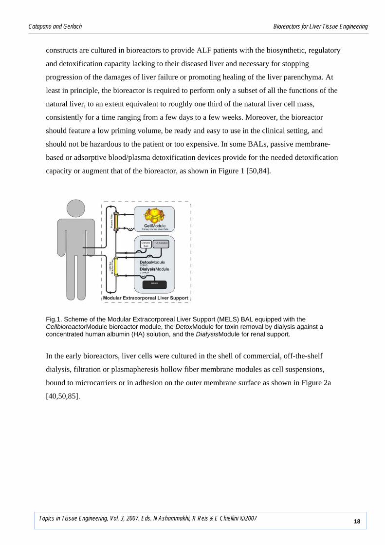
capacity or augment that of the bioreactor, as shown in Figure 1 [50,84].
Fig.1. Scheme of the Modular Extracorporeal Liver Support (MELS) BAL equipped with the CellbioreactorModule bioreactor module, the DetoxModule for toxin removal by dialysis against a concentrated human albumin (HA) solution, and the DialysisModule for renal support.
In the early bioreactors, liver cells were cultured in the shell of commercial, off-the-shelf
dialysis, filtration or plasmapheresis hollow fiber membrane modules as cell suspensions,
bound to microcarriers or in adhesion on the outer membrane surface as shown in Figure 2a
[40,50,85].

Catapano and Gerlach Bioreactors for Liver Tissue Engineering
19Topics in Tissue Engineering, Vol. 3, 2007. Eds. N Ashammakhi, R Reis & E Chiellini © 2007
Fig. 2A. Scheme of the bioreactors proposed for BALs used for extracorporeal liver assist and tested in the clinics: a) liver cells cultured outside hollow fiber membranes in shell-and-tube configuration as a suspension of cell clumps, bound to microcarriers, or in adhesion on membrane surface, with blood, plasma or medium flowing in the membrane lumina.
Starting in the mid 90’s, a new generation of bioreactors became available in which liver cells
were cultured in 3D scaffolds (e.g., in a gel bed, non-woven fabrics, foams, or a membrane
network) that were to replace the natural ECM (Table 1), and oxygen supply to the cells was
enhanced by inclusion in the construct of distributed oxygen sources, as schematically shown
in Figures 2 c-e [51,75]. Direct construct perfusion with medium, plasma or whole blood was
also exploited to minimize the external or internal mass transport resistance, or both (Figures 2
b-e). Most of the proposed bioreactors are operated in recycle mode to minimize transport
resistance and maximize the yield of liver cell reactions. Some of the proposed laboratory-
scale continuous-flow bioreactors were tested in vitro (Table 2), and were rapidly scaled-up to
treat small-to-large animal models of ALF (Table 3). Only a few were scaled-up to treat ALF
patients and are under clinical assessment (Table 4 and Figure 2).
Blood
Membrane
Blood orPlasma
Cells adherent on membranes Cell clump suspension Cells bound to microcarriers
Blood
Membrane
Blood orPlasma
Cells adherent on membranes Cell clump suspension Cells bound to microcarriers

Catapano and Gerlach Bioreactors for Liver Tissue Engineering
20Topics in Tissue Engineering, Vol. 3, 2007. Eds. N Ashammakhi, R Reis & E Chiellini © 2007
Table 1. In vitro liver cell culture techniques
Bioreactors where liver cells are cultured outside hollow fiber membranes arranged in a shell-
and-tube configuration in suspension, as cell clumps, or bound to microcarriers have been the
first ones proposed for BALs (Figure 2a). They have a number of interesting features: cell
seeding is easy, quick and can be done on demand by using cryopreserved tissue [50]; the size
of cell aggregates is not limited by the membrane diameter; cell grafts need not to be
vascularized; membranes protect the allografts against rejection; commercial membrane
modules may be used. However, in bioreactors equipped with hemodialysis or low flux
hemofiltration membranes the liquid in the cell compartment is generally stagnant and causes a
large resistance to metabolite transport to/from the cells possibly leading to cell starvation and
the accumulation of waste metabolites. In recent commercial versions of these bioreactors
mixing in the shell-side is increased by changing their aspect ratio and by using more
permeable membranes so as to promote the occurrence of Starling flows. Concern exists for
cell sedimentation in use, and for cell viability and capacity to express liver-specific metabolic
functions in the long term in particular when cell suspensions are used. Bioreactors of this type,
seeded with cryopreserved porcine liver cells attached to dextran microbeads, have been used
Culture technique Cell Type References
Suspension of single or aggregated cells Rat hepatocytes 86
Cell entrapment in microcapsules Rat hepatocytes or Chang liver
cell line 87,88,89
Cell aggregates adherent on beads Porcine hepatocytes 50
Cells adherent on the external surface of parallel
hollow fiber membranes
Human hepatoblastoma C3A
cell line 40
Cells embedded in a collagen gel Rat or human hepatocytes 2, 90
Cells outside and in between hollow fiber
membranes orderly organized in a 3D network Porcine or human liver cells 51,91
Cells in 3D foams Porcine hepatocytes 92,93
Cells in 3D non-woven fabrics Porcine hepatocytes 75,94
Cells in porous biodegradable polymeric scaffolds
prepared by 3D microprinting Rat hepatocytes 95
Cells adherent on borosilicate wafers with
micropatterned biochemical cues
Rat hepatocytes and 3T3
fibroblasts 96

Catapano and Gerlach Bioreactors for Liver Tissue Engineering
21Topics in Tissue Engineering, Vol. 3, 2007. Eds. N Ashammakhi, R Reis & E Chiellini © 2007
in a BAL shortly after cell seeding to treat the plasma of ALF patients for up to 4-6 h at a time
[113].
In another early bioreactor concept for BALs, cells of the immortalized C3A line are cultured
in adhesion on the external surface of hollow fiber (HF) membranes arranged in an acrylic
housing in shell-and-tube configuration while blood or plasma flows in the membrane lumina.
Sussmann et al. reported that bioreactors of this type containing ca. 200g of cells in the shell
reversed ALF in a dog model [40], permitted ALF patients to regain consciousness and
promoted liver regeneration [112]. Later on, Millis et al. reported similar results treating the
plasma of ALF patients in EC circulation with 4 of these bioreactors in parallel [119].
Culturing liver cells outside and among HF membranes permits cell 3D organization and the
culture of a large cell mass. The metabolic requirements of immortalized cells derived from
tumour cells are also less important than those of primary cells. However, the dense cell
packing in the shell and the low hydraulic permeability of the hemodialysis membranes used
permit transport of soluble metabolites to/from the cells only by diffusion. This may cause
steep concentration gradients in the cell mass and may lead to depletion of essential nutrients
and cell necrosis in the regions farther away from the membrane surface, as has been reported.
Since tumour-derived liver cells are used, a concern is the potential release of soluble
carcinogenic species or the leak of cells into the patient’s circulation. For this reason, Millis et
al. filtered the treated plasma across microfiltration membranes prior to returning it to the
patient.
Liver cell inclusion in a homogeneous collagen or alginate gel bed provides a large surface area
for 3D cell scaffolding and was reported to promote cell polarization, differentiation and cell-
cell contacts, and to yield viability and differentiated functions higher than monolayer culture
and stable for weeks. It may also permit a good control over cell distribution across the bed
depth and along its length. Nyberg et al. [82] used primary liver cells embedded in a collagen
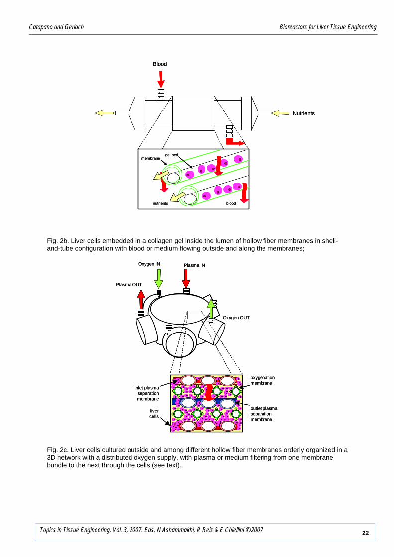
gel inside the bore of hollow fiber semi-permeable membranes assembled in a shell-and-tube
configuration for the EC treatment of the blood of ALF patients flowing outside the membranes
(Figure 2b). Blood flow around the membranes decreases mass transport resistance external to
the membranes that protect the allograft against rejection. However, suboptimal design of the
blood flow paths may cause blood clotting. Cell necrosis at the innermost regions of the bed
has also been reported possibly caused by the steep nutrient and oxygen concentration
gradients that develop across the bed depth.
Catapano and Gerlach Bioreactors for Liver Tissue Engineering
22Topics in Tissue Engineering, Vol. 3, 2007. Eds. N Ashammakhi, R Reis & E Chiellini © 2007
oxygenationmembrane
inlet plasmaseparationmembrane
outlet plasma separationmembrane
Plasma IN
Plasma OUT
Oxygen IN
Oxygen OUT
livercells
oxygenationmembrane
inlet plasmaseparationmembrane
outlet plasma separationmembrane
Plasma IN
Plasma OUT
Oxygen IN
Oxygen OUT
livercells
Fig. 2c. Liver cells cultured outside and among different hollow fiber membranes orderly organized in a 3D network with a distributed oxygen supply, with plasma or medium filtering from one membrane bundle to the next through the cells (see text).
Fig. 2b. Liver cells embedded in a collagen gel inside the lumen of hollow fiber membranes in shell-and-tube configuration with blood or medium flowing outside and along the membranes;
Nutrients
Blood
nutrients blood
gel bedmembrane
Nutrients
Blood
nutrients blood
gel bedmembrane

Catapano and Gerlach Bioreactors for Liver Tissue Engineering
23Topics in Tissue Engineering, Vol. 3, 2007. Eds. N Ashammakhi, R Reis & E Chiellini © 2007
Fig. 2d. Liver cells attached to a 3D fabric with a distributed oxygen supply, with plasma or medium axially flowing along the fabric.
Fig. 2e. Liver cells attached to a 3D fabric with a distributed oxygen supply, with plasma or medium radially flowing across the fabric.
Oxygen
Plasma
Plasma
Oxygen
Oxygenationmembrane
Livercells
Non-wovenfabrics
Plasma
Plasma Oxygen
Oxygen
Oxygenationmembrane
Livercells
Non-wovenfabrics
Perforated tube
Plasma
Plasma Oxygen
Oxygen
Oxygenationmembrane
Livercells
Non-wovenfabrics
Perforated tube

Catapano and Gerlach Bioreactors for Liver Tissue Engineering
24Topics in Tissue Engineering, Vol. 3, 2007. Eds. N Ashammakhi, R Reis & E Chiellini © 2007
A four compartment bioreactor based on a network of interwoven hollow fiber membranes was
proposed for liver cell culture by Gerlach et al. [51,91] (Figure 2c). Aiming at reproducing the
liver vascular network, the bioreactor consists of a 3D network of HF membranes with
different separation and transport properties orderly woven in planar mats enclosed in a
polyurethane housing. The repeating basic network unit consists of two mats of hydrophilic
microporous HF membranes, with the membranes in a mat angled at 90 degrees with respect to
those in the underlying mat, in between which a mat of hydrophobic microporous HF
membranes is interposed for oxygen supply to and CO2 removal from the cells. The
membranes in mats with the same properties and orientation are bundled up together and fitted
with headers. Porcine or human primary parenchymal and non-parenchymal liver cells are co-
cultured outside and among the membranes in the network. Medium or plasma is fed to the
bore of one bundle of hydrophilic membranes, a fraction (i.e., cross-flow operation) or the
whole stream (dead-end operation) filters across the membranes, bathes the cells, contacts the
oxygenation membranes and is replenished with oxygen, is re-absorbed in the bore of the
hydrophilic membranes in the other bundle, and is returned to the patient’s circulation or the
accumulation vessel. Up to two more bundles of different membrane mats may be added to the
network to introduce additional functions (e.g., to augment gas exchange, dialyze out small
solutes, feed nutrients etc.). In this bioreactor, oxygen is supplied to the cells with the medium
but is also locally supplied through the hydrophobic microporous membranes causing more
physiological oxygen gradients to establish across the cell mass. Pressure-driven direct cell
perfusion enhances transport of large solutes and species rapidly consumed/produced to/from
the cells and should lead to the prompt return of large liver-specific factors to the plasma,
reduced accumulation of waste metabolites near the cells, enhanced cell survival and functions,
and the efficient use of the available cellular activity. Indeed, liver cells cultured in the 3D
membrane network were shown to spontaneously re-organize in liver-like aggregates forming
sinusoid-like microchannels with a neo-space of Disse. Cells produced biomatrix and expressed
liver-specific functions consistently for several weeks. BALs using this bioreactor for the EC
treatment of pig models of ALF gave encouraging results [107,108]. Bioreactors seeded with
porcine liver cells were used in BALs as a bridge to OLTx to treat ALF patients (Figure 3),
coma stage III-IV, leading to a 100% survival rate after 3 years from treatment and OLTx
[116]. A pilot study aimed at using the same bioreactor in the treatment of ALF patients but
seeded with human liver cells harvested from donor organs discarded for steatosis, cirrhosis or
mechanical injury is currently underway and is giving promising results [117].

Catapano and Gerlach Bioreactors for Liver Tissue Engineering
25Topics in Tissue Engineering, Vol. 3, 2007. Eds. N Ashammakhi, R Reis & E Chiellini © 2007
Fig. 3. Clinical treatment of an ALF patient with the Modular Extracorporeal Liver Support (MELS) BAL utilizing the bioreactor developed by Gerlach et al. [51] loaded with 600 g of primary porcine cells.
In 1997, Flendrig et al. proposed another packed-bed bioreactor with decentralized oxygen
supply permitting direct perfusion of high density liver cells with low nutrients concentration
gradients [75]. Primary porcine liver cells were cultured attached to the fibers of a spiral wound
3D polyester non-woven fabric packed in a cylindrical acrylic enclosure, and were directly
perfused with medium or plasma flowing along the bioreactor length (Figure 2d). Microporous
membranes interposed in between adjacent fabric layers provided for a distributed oxygen
supply and CO2 removal. Hepatocytes were reported to arrange in the fabric in in vivo-like
aggregates, to synthesize urea and proteins, and to transform lidocaine into MEGX and xilidine
for up to 2 weeks. Use of the bioreactor for the EC treatment of animal models of ALF caused
a significant enhancement of the survival rate of small and large laboratory animals [109] and
was proven safe in the treatment of ALF patients [118]. Recently, Ambrosino et al. proposed to
couple the polyester fabric with a porcine autologous biomatrix to enhance cell attachment to
the 3D scaffold [99]. In 1996, Naruse et al. [94] and later on Morsiani et al. [103] modified this
concept by arranging the fabric in an annular packed-bed bioreactor and by flowing medium or
plasma radially across the fabric to enhance oxygen transport to the cells and reduce the
bioreactor inlet/outlet pressure drop (Figure 2e).
Up to ca. 230 g primary hepatocytes could be cultured in such bioreactor in a high
metabolically active state [120]. BALs based on this bioreactor are under clinical testing.
Catapano and Gerlach Bioreactors for Liver Tissue Engineering
26Topics in Tissue Engineering, Vol. 3, 2007. Eds. N Ashammakhi, R Reis & E Chiellini © 2007
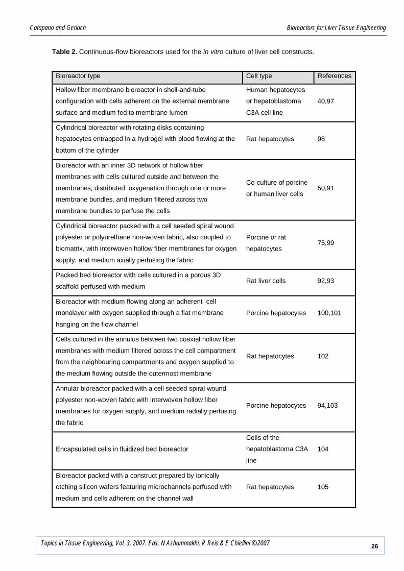
Table 2. Continuous-flow bioreactors used for the in vitro culture of liver cell constructs.
Bioreactor type Cell type References
Hollow fiber membrane bioreactor in shell-and-tube
configuration with cells adherent on the external membrane
surface and medium fed to membrane lumen
Human hepatocytes
or hepatoblastoma
C3A cell line
40,97
Cylindrical bioreactor with rotating disks containing
hepatocytes entrapped in a hydrogel with blood flowing at the
bottom of the cylinder
Rat hepatocytes 98
Bioreactor with an inner 3D network of hollow fiber
membranes with cells cultured outside and between the
membranes, distributed oxygenation through one or more
membrane bundles, and medium filtered across two
membrane bundles to perfuse the cells
Co-culture of porcine
or human liver cells 50,91
Cylindrical bioreactor packed with a cell seeded spiral wound
polyester or polyurethane non-woven fabric, also coupled to
biomatrix, with interwoven hollow fiber membranes for oxygen
supply, and medium axially perfusing the fabric
Porcine or rat
hepatocytes 75,99
Packed bed bioreactor with cells cultured in a porous 3D
scaffold perfused with medium Rat liver cells 92,93
Bioreactor with medium flowing along an adherent cell
monolayer with oxygen supplied through a flat membrane
hanging on the flow channel
Porcine hepatocytes 100,101
Cells cultured in the annulus between two coaxial hollow fiber
membranes with medium filtered across the cell compartment
from the neighbouring compartments and oxygen supplied to
the medium flowing outside the outermost membrane
Rat hepatocytes 102
Annular bioreactor packed with a cell seeded spiral wound
polyester non-woven fabric with interwoven hollow fiber
membranes for oxygen supply, and medium radially perfusing
the fabric
Porcine hepatocytes 94,103
Encapsulated cells in fluidized bed bioreactor
Cells of the
hepatoblastoma C3A
line
104
Bioreactor packed with a construct prepared by ionically
etching silicon wafers featuring microchannels perfused with
medium and cells adherent on the channel wall
Rat hepatocytes 105
Catapano and Gerlach Bioreactors for Liver Tissue Engineering
27Topics in Tissue Engineering, Vol. 3, 2007. Eds. N Ashammakhi, R Reis & E Chiellini © 2007
Bioreactor type Cell type References
Hollow fiber membrane bioreactor in shell-and-tube
configuration with cells cultured in the shell as clumps
adherent on microbeads and medium fed to membrane lumen
Rat or porcine
hepatocytes 106
Hollow fiber membrane bioreactor in shell-and-tube
configuration with cells adherent on the external membrane
surface and medium fed to membrane lumen
Human
hepatoblastoma C3A
cell line
40
Bioreactor with an inner 3D network whose repeating module
consists of two overlaid hydrophilic microporous hollow fiber
membrane mats and a mat of microporous hydrophobic
membranes interposed among the other mats for oxygen
supply. Cells cultured outside and among the membranes are
perfused by the medium filtered from the first to the second
hydrophilic membrane mat.
Porcine liver cells 107,108
Cylindrical bioreactor packed with a cell seeded spiral wound
polyester non-woven fabric with interwoven hollow fiber
membranes for oxygen supply, and medium axially perfusing
the fabric
Porcine hepatocytes 109
Encapsulated cells in fixed bed bioreactor Porcine hepatocytes 110
Cylindrical bioreactor packed with cell seeded polyurethane
foam Porcine hepatocytes 92,93
Bioreactor with plasma flowing along an adherent cell
monolayer with oxygen supplied through a flat membrane Porcine hepatocytes 101
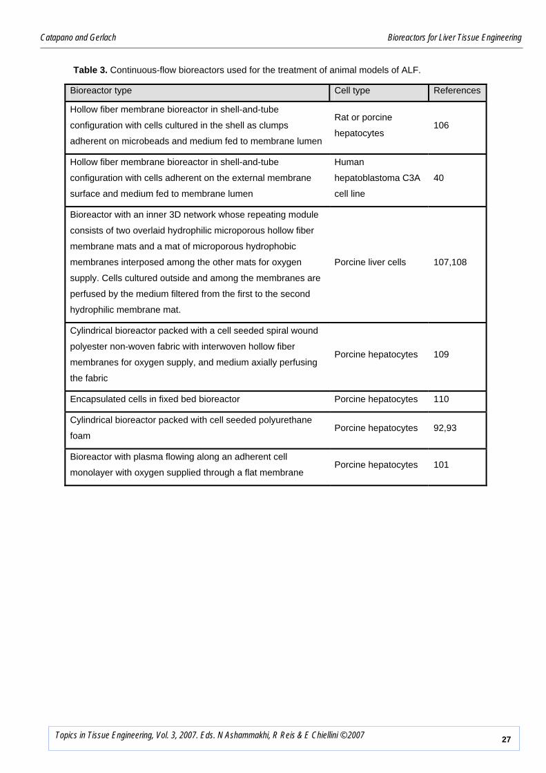
Table 3. Continuous-flow bioreactors used for the treatment of animal models of ALF.

Catapano and Gerlach Bioreactors for Liver Tissue Engineering
28Topics in Tissue Engineering, Vol. 3, 2007. Eds. N Ashammakhi, R Reis & E Chiellini © 2007
Table 4. Continuous-flow bioreactors used for the clinical treatment of ALF patients
Bioreactor brand name & type
Additional
detoxification
treatment
Perfusate Cell number
& type References
Cell and charcoal suspension in a housing
perfused with blood
Activated
charcoal
suspension
Blood
40x106
porcine
hepatocytes
111
Vital Therapies ELAD®
Shell-and-tube HF membrane bioreactor
with cells adherent on the external
membrane surface and blood fed to
membrane lumen
Blood
2x10¹⁰
(ca. 200 g)
cells of
human
hepatoblasto-
ma C3A line
112
Fig. 2a
Arbios Systems HepatAssist®
Shell-and-tube HF membrane bioreactor
with cryopreserved cell clumps adherent on
dextran microbeads in the shell and plasma
fed to membrane lumen
Activated
charcoal
adsorbent
cartridge
Plasma
4-6x10⁹
(ca. 50 g)
cryopreser-
ved porcine
hepatocytes
113,114
Fig. 2a
Excorp Medical BLSS®
Shell-and-tube HF membrane bioreactor
with cells in the membrane lumen
embedded in collagen and blood flowing
around the membranes in the shell
Blood
8x10⁹
(ca. 100 g)
porcine
hepatocytes
115
Fig. 2b
MELS CellModule
3D membrane network bioreactor with
repeating units consisting of two overlaid
hydrophilic microporous HF membrane
mats and a mat of microporous hydrophobic
membranes interposed among them for
oxygen supply. Cells cultured outside and
among the membranes are perfused by the
plasma filtered from the first to the second
hydrophilic membrane mat
Dialysis and
stripping of
hydrophobic
toxins with a
concentrated
albumin
solution
possible
Plasma
2.2x10¹⁰
(ca. 500 g)
porcine or
human liver
cells
116,117
Fig. 2c

Catapano and Gerlach Bioreactors for Liver Tissue Engineering
29Topics in Tissue Engineering, Vol. 3, 2007. Eds. N Ashammakhi, R Reis & E Chiellini © 2007
AMC BAL
Cylindrical packed bioreactor with cell-
seeded spiral wound polyester non-woven
fabric with HF membranes for oxygen
supply, and plasma axially perfusing the
fabric
Plasma 2x10⁹
porcine cells
118
Fig. 2d
Vitagen ELAD®
Shell-and-tube HF membrane bioreactor
with cells adherent on the external
membrane surface and plasma fed to
membrane lumen
Plasma
ca. 4 x 200 g
cells of
human
hepatoblasto-
ma C3A line
119
Fig. 2a
RAnD BAL
Annular packed bioreactor with cell seeded
spiral wound polyester non-woven fabric
with HF membranes for oxygen supply, and
plasma radially perfusing the fabric
Bilirubin
adsorption
cartridge
Plasma
2-2.3x10¹⁰
porcine
hepatocytes
120
Fig. 2e
Bioreactors for implantable TE liver constructs
The therapeutic success of split liver transplantation suggests a possible role for implantable
engineered liver constructs in the treatment of acute, chronic or AoC liver failure. To make its
implantation and subsequent integration possible, the TE liver construct should exhibit as
similar an architecture as possible to that of the natural liver and should be easily connected to
the vascular network of the liver. Bioreactors may be used to provide liver cell types with the
biochemical and mechanical cues that promote the organization of different liver cell types in
the construct. Only a few bioreactors have been proposed thus far to this purpose that permit
the culture of only rather small liver cell-seeded constructs.
In the rotating wall vessel (RWV) bioreactor, liver cells may be cultured in small 3D
biodegradable scaffolds suspended in medium in the space between two concentric cylinders.
The continuous rotation of the medium was proven to reduce the effect of gravity causing cells
to distribute uniformly throughout biodegradable scaffold, establish tight cellular junctions,
form bile duct-like structures and synthesize proteins for up to 60 days [121]. The laminar flow
of the medium with respect to the construct, and the continuous oxygen supply through
membranes placed either on a cylinder or a flat wall, was reported to effectively reduce the
resistance to essential nutrients and waste transport external to the construct without subjecting

Catapano and Gerlach Bioreactors for Liver Tissue Engineering
30Topics in Tissue Engineering, Vol. 3, 2007. Eds. N Ashammakhi, R Reis & E Chiellini © 2007
the cells to high shear. However, only TE constructs of limited size may be produced with the
RWV bioreactor.
Starting from the good results reported with other cell types, Kim et al. proposed to culture in a
cylindrical continuous-flow bioreactor rat hepatocytes attached to tubes a few millimeters thick
and long prepared from non-woven sheets of a biodegradable polyglycolic acid [95]. Dynamic
cell seeding permitted cells to distribute uniformly throughout the scaffold (see above). The
good 3D scaffold accessibility, made possible by 250 μm large inner pores and ca. 96%
porosity, and the optimization of the oxygenated feed flow rate were reported to enhance
significantly the transport to/from the cells and to yield a stable albumin production for about a
week of culture.
Bioreactors for metabolic studies
Knowledge of the rate at which metabolites or drugs are produced or consumed by the liver
cells in a construct under given operating conditions (e.g., metabolite and proteic effector
concentrations, medium composition, pH, etc.) is essential for preventing necrosis at the core
of the construct and for designing bioreactors for therapeutically effective BALs, and is
generally expressed in terms of the rate equations (i.e., the kinetics) of the metabolic reactions
in which a given metabolite or drug participates [38]. Kinetic characterization of the liver cell
metabolic reactions in the presence of known amounts of drugs or toxins may also provide
information on drug clearance and cytotoxicity that are important for the development of new
drugs. The ideal bioreactor for liver cell kinetic characterization should permit cell culture
according to the given technique, should feature a fluid dynamics that enable a controlled
distribution of metabolite concentrations at the cell surface and their accurate measurement or
estimation. Its design and operation should minimize the resistance to metabolite transport to
the cells and should easily permit to extract the reaction rate from the overall bioreactor
performance, by separating kinetics from physical transport. Such bioreactors do not need to
host large amounts of cells, which makes the resistance to soluble metabolite transport internal
to the construct less critical than in large TE liver constructs. However, similarly to those
considered thus far, these bioreactors should promote liver cell re-organization in liver-like
structures and the expression of the same enzymatic activities as in the natural liver. In
consideration of the multiple reaction steps leading to the elimination of a drug, the bioreactors
for drug screening as a whole should also yield metabolite concentration profiles as close as
possible to those in vivo.

Catapano and Gerlach Bioreactors for Liver Tissue Engineering
31Topics in Tissue Engineering, Vol. 3, 2007. Eds. N Ashammakhi, R Reis & E Chiellini © 2007
Static bioreactors - The kinetics of liver cell metabolic reactions is traditionally characterized
in static bioreactors, such as the Petri dishes. They are well established, inexpensive and easy to
use. The rate of the investigated metabolic reaction is generally estimated as the ratio of the
bulk metabolite concentration difference to the finite time elapsed after adding the metabolite
to the medium, and is related to the initial bulk metabolite concentration. However, such an
estimate is the compound effect of reaction events taking place at varying rates as the
metabolites (or drugs) are consumed or produced and should not be related to the initial
metabolite concentration [63]. Time varying cell number or metabolic activity, and the
existence of spatial metabolite concentration gradients caused by poor mixing or resistance to
metabolite transport may make these estimates even less reliable. The effect of mass transport
resistance is particularly significant when cells are cultured in 3D scaffolds or in adhesion at
high surface density under a thick layer of medium [47].
Dynamic bioreactors – The intrinsic kinetics (i.e., unaffected by mass transport) of liver cell
metabolic reactions is conveniently characterized in continuous-flow bioreactors operated
under time-invariant conditions. In these bioreactors, fresh medium containing nutrients and
metabolites continuously enters the bioreactor, flows in contact with the cells and leaves the
bioreactor carrying away spent medium and waste metabolites, similarly to that occurring in
the natural liver. Many dynamic bioreactors have been proposed for the kinetic characterization
of liver cells cultured on/in 2D/3D scaffolds. In most cases the distribution of matter (i.e., the
actual metabolite or drug spatial concentration profiles) in the bioreactor is unknown, nor is it
clear the extent to which the reaction rate estimated as the overall bioreactor performance is
affected by the physical transport of metabolite or drug to/from the cells. When the distribution
of matter in the bioreactor and the transport resistance to/from the cells is characterized,
quantitative information on the rate of the investigated metabolic reactions can be gathered
from the steady state mass balance about the bioreactor for a given metabolite or drug. Rotem
et al. [28] and later Smith et al. [122] characterized the oxygen consumption rate of liver cells
embedded in a flat collagen sandwich or in adhesion on collagen, respectively, at the bottom of
a modified T-flask operated at steady state under a constant medium height. The kinetics of
oxygen consumption was characterized under the assumption that medium flows as a plug
above the cells and that oxygen in the gaseous phase above the cells is in equilibrium with the
dissolved oxygen in the medium anywhere in the bioreactor. For non-gaseous metabolites, a
macroscopic mass balance about the bioreactor for the metabolite does not provide a direct
estimate of the rate at which a metabolite is consumed or produced for their concentration

Catapano and Gerlach Bioreactors for Liver Tissue Engineering
32Topics in Tissue Engineering, Vol. 3, 2007. Eds. N Ashammakhi, R Reis & E Chiellini © 2007
continuously varies along the bioreactor length, as the reaction rate does [38]. This complicates
the characterization of metabolic reactions with complex, non-linear kinetics. Kinetic studies
can be conveniently performed with continuous-flow well mixed bioreactors where metabolites
are uniformly distributed. In fact, everywhere in the bioreactor cells are exposed to the same
metabolite, effector and drug concentration, and metabolic reactions occur everywhere at the
same rate. Concentrations in the bioreactor equal those in the stream leaving the bioreactor, that
are easily and reliably measured. Moreover, at steady state, the metabolite concentration
difference in the streams entering and leaving the bioreactor provides a direct estimate of the
investigated metabolic reaction rate. Gebhardt et al. [123] and Smith et al. [122] proposed a
one-compartment and a two-compartment continuous-flow bioreactor, respectively, that should
have prevented spatial metabolite concentration gradients. However, no evidence was provided
that soluble metabolites indeed distribute uniformly in the bioreactor, nor that the effect of
mass transport resistance to/from the cells is indeed negligible (i.e., that cells were operated
under kinetic control). Catapano and De Bartolo proposed a well mixed continuous-flow,
recycle bioreactor for the characterization of the metabolic reactions of primary rat liver cells in
adhesion on collagen [66]. The bioreactor was designed to host planar supports that can be
inserted in standard Petri dishes to let liver cells recover from the isolation stress prior to the
kinetic characterization. A fraction of the stream leaving the bioreactor is continuously
recycled back to the bioreactor inlet where it mixes with fresh medium. The recycle makes it
possible to separate control of the mixing in the bioreactor from the hold-up time (i.e., the
bioreactor volume-to-feed flow rate ratio) to minimize the shear cells are subject to and to
maximize the metabolite concentration difference about the bioreactor. The bioreactor design
and operation (i.e., bioreactor volume, design of the inlet and outlet ports, feed flow rate,
recycle ratio, etc.) was optimized to culture liver cells at uniform metabolite concentrations and
under negligible mass transport resistance. This bioreactor was successfully used to
characterize the dependence of the rate of oxygen consumption, ammonia and
phenolsulphonphtalein elimination on ammonia concentration in the medium. Dynamic
bioreactors have also been proposed as an alternative to traditional methods in drug screening
and cytotoxicity assessment. Recently, a flat plate continuous-flow bioreactor similar to that of
Rotem et al. was proposed by Allen & Bathia [124] to impose steady physiological gradients of
dissolved oxygen and nutrient concentrations over hepatocytes in adhesion on collagen, as an
in vitro model of liver zonation. In this bioreactor, the high metabolic consumption rates cause
metabolite or dissolved oxygen concentration gradients to form along the bioreactor similarly
to that occurring internal to 3D liver constructs. The distribution of matter and the dissolved

Catapano and Gerlach Bioreactors for Liver Tissue Engineering
33Topics in Tissue Engineering, Vol. 3, 2007. Eds. N Ashammakhi, R Reis & E Chiellini © 2007
oxygen gradients in the bioreactor were validated by comparing experimental dissolved oxygen
concentrations in the stream leaving the bioreactor with predictions from a plug flow transport
model. The Authors reported that the oxygen gradients contribute to an in vivo-like
heterogeneous induction of enzymatic activities (e.g., CYP2B and CYP3A), modulate the
enzymatic activities induced with drugs, and reproduce well in vitro the zonal toxicity of
acetaminophen reported in vivo [125]. A scaled-down version of the dynamic 3D membrane
network bioreactor developed by Gerlach et al. [51] for a BAL, seeded with porcine liver cells
and batch-wise operated by filtering the medium across the cell compartment in dead-end
mode, was also shown to promote cell re-arrangement in liver-like structures and yield MEGX
concentration profiles similar to those found in vivo when challenged with physiological
lidocaine concentrations [126]. The kinetic analysis yielded lidocaine clearance comparable to
those reported in literature and evidenced the importance of accounting for the adsorption of
hydrophobic drugs to materials when assessing the drug clearance of liver cell constructs in
bioreactors. A micro-bioreactor fostering the formation of 3D liver tissue has been recently
proposed for the assessment of drug safety and efficacy by Powers et al. [127]. The bioreactor
core is a 3D scaffold produced by deep reactive ion etching of silicon wafers featuring through
microchannels approx 300μm x 300μm wide with collagen-coated cell adhesive walls. Rat
hepatocytes were reported to adhere to the channel walls and re-arrange into liver-like
structures when the scaffold pores were perfused at flow rates at which the oxygen transport
rate to the cells matches their estimated OCR. However, cells are still exposed to physiological
shear stresses. Seeding cell spheroids rather than single cells was reported to maintain tissue-
like architecture and viability for long times. The Authors report that in this micro-bioreactor
the hepatocytes expressed at near in vivo levels many drug metabolizing enzymatic activities
that are rapidly lost in vitro in many other bioreactor systems [128].
Concluding remarks The large number of bioreactors proposed for liver tissue engineering testifies to the ingenuity
of researchers operating in the field. However, cell response to soluble and immobilized
biochemical cues is only now beginning to be unraveled. Quantitative information on liver cell
metabolic reactions at each stage of cell maturation and the type and amount of biochemical
stimuli needed to promote liver-like tissue formation in vitro still needs to be gathered. Much
work is still to be done towards the understanding and the modeling of transport phenomena in
the proposed bioreactors. Within this framework, continuous-flow bioreactors purposely

Catapano and Gerlach Bioreactors for Liver Tissue Engineering
34Topics in Tissue Engineering, Vol. 3, 2007. Eds. N Ashammakhi, R Reis & E Chiellini © 2007
designed, seeded and operated shall contribute to establishing in vitro the proper environment
to promote cell re-organization in 3D scaffolds in liver-like hierarchical structures and cell
differentiation to a stable adult phenotype. This way, EC treatments based on these bioreactors
shall be able to provide for the needed functions to support ALF patients until OLTx or the
own liver tissue regenerates. New bioreactors shall also constitute the core of effective in vitro
model systems to investigate liver cell metabolism and replace costly, at times also unreliable,
animal tests in the assessment of drug clearance and toxicity.

Catapano and Gerlach Bioreactors for Liver Tissue Engineering
35Topics in Tissue Engineering, Vol. 3, 2007. Eds. N Ashammakhi, R Reis & E Chiellini © 2007
References 1 Stockmann HB, JN IJ. Prospects for the temporary treatment of acute liver failure. Eur J
Gastroenterol Hepatol 2002; 14:195-203. 2 Dunn JC, Yarmush ML, Koebe HG, Tompkins RG. Hepatocyte function and extracellular
matrix geometry: Long-term culture in a sandwich configuration. FASEB J 1989; 3: 174-177.
3 Miura Y, Akimoto T, Yoshikawa N, Yagi K. Characterization of immobilized hepatocytes as liver support. Biomater Artif Cells Artif Organs 1990; 18: 549-554.
4 OPTN/SRTR data. 1 August 2002. www.optn.org . 5 Lin TY, Lee CS, Chen CC, Lian KY, Lin WSJ. Regeneration of human liver after hepatic
lobectomy studies by repeated liver scanning and repeated needle biopsy. Ann Surg 1979; 190:48-53.
6 Matsushita M, Nosé Y. Artificial liver. Artif organs 1986; 10(5):378-384. 7 Mitzner SR, Stange J, Klammt S, et al. Extracorporeal detoxification using the Molecular
Adsorbent Recirculating System for critically ill patients with liver failure. J Am Soc Nephrol 2001; 12(suppl 17):S75-S82.
8 Sauer IM, Zeilinger K, Pless G, Kardassis D, Theruvath T, Pascher A, Mueller AR, Steinmueller T, Neuhaus P, Gerlach JC: Extracorporeal Liver Support based on Human Liver Cells and Albumin Dialysis – Treatment of a Patient with Primary Graft Non-Function. J Hepatol 2003; 39 (4):649-653.
9 Willinger M, Schima H, Schmidt C, Huber L, Vogt G, Falkenhagen D, Losert U. Microspheres based detoxification system: in vitro study and mathematical estimation of filter performance. Int J Artif Organs 1999; 22: 573-582.
10 Langer R, Vacanti JP. Tissue engineering. Science 1993; 260:920-926. 11 Kulig KM, Vacanti JP. Hepatic tissue engineering. Transplant Immunology 2004;
12:303-310. 12 Mooney D, Hansen L, Vacanti J, Langer R, Farmer S, Ingber D. Switching from
differentiation to growth in hepatocytes: control by extracellular matrix. J Cell Physiol 1992; 151(3):497-505.
13 Goulet F, Normad C, Morin O. Cellular interactions promote tissue-specific function, biomatrix deposition and junctional communication of primary cultured hepatocytes. Hepatology 1988; 8(5):1010-1018.
14 Crossin KL. Cell adhesion molecules activate signaling networks that influence proliferation, gene expression, and differentiation. Ann NY Acad Sci 2003; 961(1):159.
15 Jungermann K, Thurmann RG. Hepatocyte heterogeneity in the metabolism of carbohydrates. Enzyme 1992; 46:33-58.
16 Gebhardt R. Metabolic zonation of the liver: regulation and implications for liver function. Pharmacol Ther 1992; 53:275-354.
17 Jauregui HO. Hepatocyte cell line as the biological component of liver support. Artif Organs 2001, 25(7): 509-512.
18 Hoekstra R, Chamuleau RAFM. Recent developments on human cell lines for the bioartificial liver. Int J Artif Organs 2002, 25(3): 182-191.
19 Bader A, Knop E, Fruehauf N, Crome O, Boeker K, Christians U, Oldhafer K, Ringe B, Pichlmayr R, Sewing K-F. Reconstruction of liver tissue in vitro: geometry of charactersitic flat bed, hollow fiber and spouted bed bioreactors with reference to the in vivo liver. Artif Organs 1995; 19(9):941-950.
20 Hutmacher DW, Teoh SH, Zein I, Ranawake M, Lau S. Tissue engineering: the engineer’s role. Medical Device Technol 2000; january/february: 33-36.

Catapano and Gerlach Bioreactors for Liver Tissue Engineering
36Topics in Tissue Engineering, Vol. 3, 2007. Eds. N Ashammakhi, R Reis & E Chiellini © 2007
21 Dvir-Ginzberg M, Gamlieli-Bonshtein I, Agbaria R, Cohen S. Liver tissue engineering with alginate scaffolds: effects of cell-seeding density on hepatocyte viability, morphology, and function. Tissue Eng 2003; 9(4):757-766.
22 Freed LE, Marquis JC, Vunjak-Novakovic G, Emmanual J, Langer R. Composition of cell-polymer cartilage implants. Biotechnol Bioeng 1994; 43:605-614.
23 Holy CE, Shoichet MS, Davies JE. Engineering three-dimensional bone tissue in vitro using biodegradable scaffolds: investigating initial cell-seeding density and culture period. J Biomater Mater Res 2000; 51:376-382.
24 Ishaug-Riley SL, Crane-Kuger GM, Yaszemski MJ, Mikos AG. Three-dimensional culture of rat calvarial osteoblasts in porous biodegradable polymers. Biomaterials 1998; 19:1405-1412.
25 Freshney R. Culture of animal cells – a manual of basic techniques, 4th ed. New York: Wiley Liss; 2000.
26 Catapano G, De Bartolo L, Lombardi CP, Drioli E. The effect of oxygen transport resistances on the viability and functions of isolated rat hepatocytes. Int. J. of Artif. Organs 1996; 19(1):31-41.
27 Catapano G, De Bartolo L, Lombardi CP, Drioli E. The effect of catabolite concentration on the viability and functions of isolated rat hepatocytes. Int J of Artif Organs 1996; 19(4):245-250.
28 Rotem A, Toner M, Tompkins RG, Yarmush ML. Oxygen uptake rates in cultured rat hepatocytes. Biotechnol Bioeng 1992, 40:1286-1291.
29 Balis UJ, Behnia K, Dwarakanath B, Bathia SN, Sullivan SJ, Yarmush ML, Toner M. Oxygen consumption characteristics of porcine hepatocytes. Metab Eng 1999, 1:49.
30 Catapano G. Mass transfer limitations to the performance of membrane bioartificial liver support devices. Int J Artif Organs 1996; 19(1):18-35.
31 Martin I, Wendt D, Heberer M. The role of bioreactors in tissue engineering. Trends in Biotechnol 2004; 22(2):80-86.
32 Muschler GF, Nakamoto C, Griffith LG. Engineering principles of clinical cell-based tissue engineering. J Bone Joint Surg 2004; 86(7):1541-1558.
33 Martin Y, Vermette P. Bioreactors for tissue mass culture: Design, characterization and recent advances. Biomaterials 2005; 26:7481-7503.
34 Griffith LG, Swartz MA. Capturing complex 3D tissue physiology in vitro. Nature Rev 2006; 7:211-224.
35 Bird RB, Stewart WE, Lightfoot EN. Transport phenomena. Wiley, New York, 1960. 36 Ozisik NM, Heat Transfer, McGraw-Hill, New York, 1985. 37 Catapano G, Bergins M, Wodetzki A, Papenfuss HD, Rintelen T, Baurmeister U. Mass
and momentum transport in extra-luminal flow (ELF) membane devices for blood oxygenation. J Membrane Sci 2001; 184:123.
38 Fogler HS. Elements of chemical reaction engineering. Prentice Hall Intl 1992 Englewood Cliffs, New Jersey USA.
39 Catapano G, Euler M, Gaylor JDS, Gerlach JC. Characterization of the distribution of matter in hybrid liver support devices where cells are cultured in a 3-D membrane network or on flat substrata. Int J of Artif Organs 2001; 24(2):102-109.
40 Sussman NL, Chong MG, Koussayer T, He DE, Shang TA, Whisennand HH, Kelly JH. Reversal of fulminant hepatic failure using an extracorporeal liver assist device. Hepatology 1992; 16: 60-65.
41 Mazariegos GV, Patzer JF, 2nd, Lopez RC, Giraldo M, Devera ME, Grogan TA, Zhu Y, Fulmer ML, Amiot BP, Kramer DJ. First clinical use of a novel bioartificial liver support system (BLSS). Am J Transplant 2002; 2: 260-266.

Catapano and Gerlach Bioreactors for Liver Tissue Engineering
37Topics in Tissue Engineering, Vol. 3, 2007. Eds. N Ashammakhi, R Reis & E Chiellini © 2007
42 Fournier RL. Basic transport phenomena in biomedical engineering. 1999 Taylor & Francis, Philadelphia PA (USA).
43 Nyberg SL, Platt JL, Shirabe K, Payne WD, Hu W-S, Cerra FB. Immunoprotection of xenocytes in a hollow fiber bioartificial liver. Hepatology 1992; 16:60-65.
44 Kedem O, Katschalsky A. Thermodynamic analysis of the permeability of biological membranes to non-electrolytes. Biochem Biophys Acta 1958; 27:229.
45 Klein E, Holland FF, Donnaud A, Leboeuf A, Eberle K. Diffusive and hydraulic permeabilities of commercially available cellulosic hemodialysis films and hollow fibers. J Membrane Sci 1977; 2:349.
46 Feldhoff P, Turnham T, Klein E. Effect of plasma proteins on the sieving spectra of hemofilters. Artif Organs 1984; 8(2):186.
47 Catapano G. Mass transfer limitations to the performance of membrane bioartificial liver support devices. Int J Artif Organs 1996; 19(1):18-35.
48 Hay PD, Veitch AR, Smith MD, Cousins RB, Gaylor JDS. Oxygen transfer in a diffusion limited hollow fiber bioartificial liver. Artif Organs 2000; 24:278-288.
49 McClelland RE, MacDonald JM, Coger RN. Modeling O2 transport within engineered hepatic devices. Biotechnol Bioeng 2003; 82(1):12-27.
50 Demetriou AA, Whiting JF, Levenson SM, Chowdury NR, Schechner R, Michalski S, Feldman D, Chowdury JR. New method of hepatocyte transplantation and extracorporeal liver support. Ann Surg 1986; 204(3):259-270.
51 Gerlach JC, Encke J, Hole O, Muller C, Courtney JM, Neuhaus P. Hepatocyte culture between three dimensionally arranged biomatrix-coated independent artificial capillary systems and sinusoidal endothelial cell co-culture compartments. Int J Artif Organs 1994; 17: 301-306.
52 Kim S, Sundback C, Kaihara S, Benvenuto M, Kim B-S, Mooney et al. Dynamic seeding and in vitro culture of hepatocytes in a flow perfusion system. Tissue Eng 2000; 6(1):39-44.
53 Catapano G, Iorio G, Drioli E, et al. Theoretical and experimental analysis of a hybrid bioartificial membrane pancreas: a distributed parameter model taking into account Starling fluxes. J Membrane Sci 1990; 52:351-378.
54 Todd DG, Moscioni AD, Rozga J, Demetriou AA. Mass transfer in a hollow fiber device used as a bioartificial liver. ASAIO J 1993, 886-892.
55 Gerlach JC, Schnoy N, Encke J, Smith MD, Muller C, Neuhaus P. Improved hepatocyte in vitro maintenance in a culture model with woven multicompartment capillary systems: Electron microscopy studies. Hepatology 1995; 22: 546-552.
56 Gerlach JC, Botsch M, Kardassis D, Lemmens P, Schon M, Janke J, Puhl G, Unger J, Kraemer M, Busse B, Bohmer C, Belal R, Ingenlath M, Kosan M, Kosan B, Sultmann J, Patzold A, Tietze S, Rossaint R, Muller C, Monch E, Sauer IM, Neuhaus P. Experimental evaluation of a cell module for hybrid liver support. Int J Artif Organs 2001; 24: 793-798.
57 Skett P. Problems in using isolated and cultured hepatocytes for xenobiotic metabolism/metabolism-based toxicity testing-solutions? Toxicol In Vitro 1994; 8:491-504.
58 Gerlach J, Brombacher J, Smith M, Neuhaus P. High yield hepatocyte isolation from pig livers for investigation of hybrid liver support systems: Influence of collagenase concentration and body weight. J Surg Res 1996; 62: 85-89.
59 Baquerizo A, Mhoyan A, Kearns-Jonker M, Arnaout WS, Shackleton C, Busuttil RW, Demetriou AA, Cramer DV. Characterization of human xenoreactive antibodies in liver failure patients exposed to pig hepatocytes after bioartificial liver treatment: an ex vivo model of pig to human xenotransplantation. Transplantation 1999 Jan 15; 67(1):5-18

Catapano and Gerlach Bioreactors for Liver Tissue Engineering
38Topics in Tissue Engineering, Vol. 3, 2007. Eds. N Ashammakhi, R Reis & E Chiellini © 2007
60 Pascher A, Sauer IM, Hammer C, Gerlach JC, Neuhaus P. Extracorporeal liver perfusion as hepatic assist in acute liver failure - a review of world experience. Xenotransplantation 2002; 9:309-324.
61 Annual Report/Eurotransplant International Foundation– Leiden: Annual report 2000 /Guido G. Persijn and Bernard Cohen. CIP-Gegevens Koninklijke Bibliotheek, Den Haag ISBN 90-71658-19-8.
62 Nyberg SL, Remmel RP, Mann HJ, Peshwa MV, Hu WS, Cerra FB. Primary hepatocytes outperform Hep G2 cells as the source of biotransformation functions in a bioartificial liver. Ann Surg 1994; 220:59–67.
63 Catapano G, De Bartolo L. The importance of the kinetic characterization of liver cell metabolic reactions to the design of hybrid liver support devices. Int J of Artif Organs 1996; 19(11):670-676.
64 Selden C, Hodgson H. Cellular therapies for liver replacement. Transplant Immunol 2004; 12: 273-288.
65 Kietzmann T, Jungermann K. Modulation by oxygen of zonal gene expression in liver studied in primary rat hepatocyte cultures. Cell Biol Toxicol 1997; 13:243-255.
66 Catapano G, De Bartolo L. Technique for the kinetic characterization of the metabolic reactions of hepatocytes in adhesion culture. Biotechnol Progress 1998; 14(3)-500-507.
67 Catapano G, De Bartolo L. Combined effect of oxygen and ammonia on the kinetics of ammonia elimination and oxygen consumption of adherent rat liver cells. Int. J. of Artificial Organs 2002; 25(2):151-157.
68 Unger JK, Catapano G, Horn NA, Schroers A, Gerlach JC, Rossaint R. Comparative analysis of metabolism of medium- and plasma-perfused primary pig hepatocytes cultured around a 3-D membrane network. Int. J. of Artif Organs 2000; 23(2):104-110.
69 Gerlach J, Stoll P, Schnoy N, Neuhaus P. Comparison of hollow fibre membranes for hepatocyte immobilisation in bioreactors. Int J Art Org 1996;19:610-616
70 Bhatia SN, Yarmush ML, Toner M. Controlling cell interactions by micropatterning in co-cultures: Hepatocytes and 3T3 fibroblasts. J Biomed Mater Res 1997; 34: 189-199.
71 Catapano G, De Bartolo L, Vico V, Ambrosio L. Morphology and metabolism of hepatocytes cultured in Petri dishes on films and in non-woven fabrics of hyaluronic acid esters. Biomaterials 2001; 22:659-665.
72 Powers MJ, Griffith LG. Adhesion-guided in vitro morphogenesis in pure and mixed cell cultures. Microsc Res Tech 1998; 43:379-384.
73 Catapano G, Di Lorenzo MC, Della Volpe C, De Bartolo L, Migliaresi C. Polymeric membranes for hybrid liver support devices: the effect of membrane surface wettability on hepatocyte viability and functions. J Biomater Sci - Polymer Edn 1996; 7(11):1017-1027.
74 Naruse K, Sakai Y, Nagashima JGX, Suzuki M, Muto T. Development of a new bioartificial liver module filled with porcine hepatocytes immobilized on non-woven fabric. Int J Artif Organs 1996; 6:347-352.
75 Flendrig LM, la Soe JW, Joerning GGA, Steenbeck A, Karlsen OT, Bovèe WMMJ, Ladiges NCJJ, te Velde AA, Chamuleau AFM. In vitro evaluation of a novel bioreactor based on an integral oxygenator ands a spirally wound nonwoven polyester matrix for hepatocyte culture as small aggregates. J Hepatol 1997; 26:1379-1392.
76 Glicklis R, Shapiro L, Agbaria R, Merchuk JC, Cohen S. Hepatocyte behavior within three-dimensional porous alginate scaffolds. Biotechnol Bioeng 2000; 67(3): 344-353.
77 Wendt D, Marsano A, Jakob M, Heberer M, Martin I. Oscillating perfusion of cell suspensions through three-dimensional scaffolds enhances cell seeding efficiency and uniformity. Biotechnol Bioeng 2003; 84(2): 205-214.

Catapano and Gerlach Bioreactors for Liver Tissue Engineering
39Topics in Tissue Engineering, Vol. 3, 2007. Eds. N Ashammakhi, R Reis & E Chiellini © 2007
78 Vunjak-Novakovic G, Freed LE, Biron LJ, Langer R. Effects of mixing on the composition and morphology of tissue engineered cartilage. AIChE J 1996; 42:850-860.
79 Xiao Y-L, Riesle J, Van Blitterswijk CA. Static and dynamic fibroblast seeding and cultivation in porous PEO/PBT scaffolds. J Mater Sci Mater in Medicine 1999; 10:773-777.
80 Li Y, Ma T, Kniss D, Lasky L, Yang S-T. Effects of filtration seeding on cell density, spatial distribution, and proliferation in nonwoven fibrous matrices. Biotechnol Prog 2001; 17:935-944.
81 Gerlach JC, Botsch M, Kardassis D, Lemmens P, Schon M, Janke J, Puhl G, Unger J, Kraemer M, Busse B, Bohmer C, Belal R, Ingenlath M, Kosan M, Kosan B, Sultmann J, Patzold A, Tietze S, Rossaint R, Muller C, Monch E, Sauer IM, Neuhaus P. Experimental evaluation of a cell module for hybrid liver support. Int J Artif Organs 2001; 24: 793-798.
82 Nyberg SL, Shatford RA, Peshwa MV, White JG, Cerra FB, Hu WS. Evaluation of a hepatocyte-entrapment hollow fiber bioreactor: a potential bioartificial liver. Biotechnol Bioeng 1993; 41:194-203.
83 Allen JW, Bathia SN. Engineering live therapies for the future. Tissue Eng 2002; 8(5):725-737.
84 Jalan R, Sen S, Williams R. Prospects for extracorporeal liver support. Gut 2004; 53:890-898.
85 Wolf CFW, Minick CR, McCoy CH. Morphologic examination of a prototype liver assist device composed of cultured cells and artificial capillaries. Int J rtif Organs 1978; 1(1):45-51.
86 Sakai Y, Naruse K, Nagashima I, Muto T, Suzuki M. A new bioartificial liver using porcine hepatocyte spheroids in high-cell density suspension perfusion culture: in vitro performance in synthesized medium culture and in 100% human plasma. Cell Transplant 1999; 8:531-541.
87 Miura Y, Akimoto T, Kanazawa H, Yagi K. Synthesis and secretion of protein by hepatocytes entrapped within calcium alginate. Artif Organs 1986; 10: 460-465.
88 Wong H, Chang TMS. Bioartificial liver: implanted artificial cells microencapsulated living hepatocytes increases survival of liver failure rats. Int J Artif Organs 1986; 9(5):335-336.
89 Cai Z, Shi Z, O’Shea GM, Sun AM. Microencapsulated hepatocytes for bioartificial liver support. Artif Organs 1988; 12(5):388-393.
90 Koebe HG, Wick M, Cramer U, Lange V, Schildberg FW. Collagen gel immobilisation provides a suitable cell matrix for long term human hepatocyte cultures in hybrid reactors. Int J Artif Organs 1994; 17: 95-106.
91 Sauer IM, Zeilinger K, Obermayer N, Pless G, Grunwald A, Pascher A, Mieder T, Roth S, Goetz M, Kardassis D, Mas A, Neuhaus P, Gerlach JC. Primary human liver cells as source for modular extracorporeal liver support--a preliminary report. Int J Artif Organs 25: 1001-1005, 2002.
92 Gion T, Shimada M, Shirabe K, Nakazawa K, Ijima H, Matsushita T, Funatsu K, Sugimachi K. Evaluation of a hybrid artificial liver using a polyurethane foam packed-bed culture system in dogs. J Surg Res 1999, 82:131.
93 Nakazawa K, Ijima H, Fukuda J, Sakiyama R, Yamashita Y, Shimada M, Shirabe K, Tsujita E, Sugimachi K, Funatsu K. Development of a hybrid artificial liver using polyurethane foam/ hepatocyte spheroid culture in a pre-clinical pig experiment. Int J Artif Organs 2002; 25(1):51-60.
94 Naruse K, Sakai Y, Nagashima I, Jiang GX, Suzuki M, Muto T. Development of a new bioartificial liver module filled with porcine hepatocytes immobilized on non-woven fabric. Int J Artif Organs 1996, 19(6):347-352.

Catapano and Gerlach Bioreactors for Liver Tissue Engineering
40Topics in Tissue Engineering, Vol. 3, 2007. Eds. N Ashammakhi, R Reis & E Chiellini © 2007
95 Kim SS, Utsunomiya H, Koski JA, Wu BM, Cima MJ, Sohn J, Mukai K, Griffith LG, Vacanti JP. Survival and function of hepatocytes on a novel three-dimensional synthetic biodegradable polymer scaffold with an intrinsic network of channels. Ann Surg 228: 8-13, 1998.
96 Bhatia SN, Yarmush ML, Toner M. Controlling cell interactions by micropatterning in co-cultures: hepatocytes and 3T3 fibroblasts. J Biomed Mater Res 1997; 34: 189-199.
97 Hager JC, Carman R, Porter LE, et al. Neonatal hepatocyte culture on artificial capillaries: a model for drug metabolism and the artificial liver. ASAIO J 6: 26-35, 1983.
98 Yanagi K, ookawa K, Mizuno S, Ohshima N. Performance of a new hybrid artificial liver support system using hepatocytes entrapped within a hydrogel. Trans ASAIO 1989; XXXV:570-572.
99 Ambrosino G, Varotto S, Basso S, Galavotti D, Cecchetto A, Carraro P, Naso A, De Silvestro G, Plebani M, Giron G, Abatangelo G, Donato D, Braga GP, Cestrone A, Marrelli L, Trombetta M, Lorenzelli V, Picardi A, Valente ML, Palu G, Colantoni A, Van Thiel D, Ricordi C, D'Amico DF. Alex (artificial liver for extracorporeal xenoassistance): A new bioreactor containing a porcine autologous biomatrix as hepatocyte support. Preliminary results in an ex vivo experimental model. Int J Artif Organs 25: 960-965, 2002.
100 De Bartolo L, Jarosch-Von Schweder G, Haverich A, Bader A. A novel full-scale flat membrane bioreactor utilizing porcine hepatocytes: cell viability and tissue-specific functions. Biotechnol Prog 2000; 16:102-108.
101 Shito M, Kim NH, Baskaran H, Tilles AW, Tompkins RG, Yarmush ML, Toner M. In vitro and in vivo evaluation of albumin synthesis rate of porcine hepatocytes in a flat-plate bioreactor. Artif Organs 25: 571-578, 2001.
102 MacDonald JM, Wolfe SP, Roy-Chowdhury I, Kubota H, Reid LM. Effect of flow configuration and membrane characteristics on membrane fouling in a novel multicoaxial hollow-fiber bioartificial liver. Ann N Y Acad Sci 944: 334-343, 2001.
103 Morsiani E, Brogli M, Gavalotti D, et al.. Long-term expression of highly differentiated functions by isolated porcine hepatocytes perfused in a radial-flow bioreactor. Artif Organs, 2001, 25:329-337.
104 David B, Dufresne M, Nagel MD, Legallais C. In vitro assessment of encapsulated C3A hepatocytes functions in a fluidized bed bioreactor. Biotechnol Prog. 2004; 20(4):1204-1212.
105 Powers MJ, Janigian DM, Wack KE, Baker CS, Beer Stolz D, Griffith LG. Functional behavior of primary rat liver cells in a three-dimensional perfused microarray bioreactor. Tissue Eng 8: 499-513, 2002.
106 Arnaout WS, Moscioni AD, Barbour RL, Demetriou AA. Development of bioartificial liver: bilirubin conjugation in Gunn rats. J Surg Res 1990; 48:379-382.
107 Gerlach J, Trost T, Ryan CJ, Meissler M, Hole O, Muller C, Neuhaus P. Hybrid liver support system in a short term application on hepatectomized pigs. Int J Artif Organs 1994; 17: 549-553.
108 Janke J, Gerlach J, Kardassis D, Bohmer C, Rossaint R. Effect of a hybrid liver support system on cardiopulmonary function in healthy pigs. Int J Artif Organs 1997; 20: 570-575.
109 Flendrig LM, Calise F, Di Florio E, Mancini A, Ceriello A, Santaniello W, Mezza E, Sicoli F, Belleza G, Bracco A, Cozzolino S, Scala D, Mazzone M, Fattore M, Gonzales E, Chamuleau RA. Significantly improved survival time in pigs with complete liver ischemia treated with a novel bioartificial liver. Int J Artif Organs 1999; 22: 701-709.
110 Dixit V, Gitnik G. The bioartificial liver: state-of-the-art. Eur J Surg Suppl 1998; 36:71-76.

Catapano and Gerlach Bioreactors for Liver Tissue Engineering
41Topics in Tissue Engineering, Vol. 3, 2007. Eds. N Ashammakhi, R Reis & E Chiellini © 2007
111 Margulis MS, Erukhimov EA, Andreiman LA, Viksna LM. Temporary organ substitution
by hemoperfusion through suspension of active donor hepatocytes in a total complex of intensive therapy in patients with acute hepatic insufficiency. Resuscitation 1989; 18:85-94.
112 Sussman NL, Kelly JH. Improved liver function following treatment with an extracorporeal liver assist device. Artif Organs 1993; 17(1): 27-30.
113 Watanabe FD, Mullon CJ, Hewitt WR, Arkadopoulos N, Kahaku E, Eguchi S, Khalili T, Arnaout W, Shackleton CR, Rozga J, Solomon B, Demetriou AA. Clinical experience with a bioartificial liver in the treatment of severe liver failure. A phase I clinical trial. Ann Surg 1997; 225: 484-491.
114 Stevens AC, Busuttil R, Han S, et al. An interim analysis of a phase II/III prospective randomized, multicenter, controlled trial of the HepatAssist® bioartificial liver support system for the treatment of fulminant hepatic failure. Hepatology 2001; 34(Suppl S, Part 2):509.
115 Mazariegos GV, Patzer JF, 2nd, Lopez RC, Giraldo M, Devera ME, Grogan TA, Zhu Y, Fulmer ML, Amiot BP, Kramer DJ. First clinical use of a novel bioartificial liver support system (BLSS). Am J Transplant 2002; 2: 260-266.
116 Sauer IM, Kardassis D, Zeillinger K, Pascher A, Gruenwald A, Pless G, Irgang M, Kraemer M, Puhl G, Frank J, Muller AR, Steinmuller T, Denner J, Neuhaus P, Gerlach JC. Clinical extracorporeal hybrid liver support - phase I study with primary porcine liver cells. Xenotransplantation 2003; 10: 460-469.
117 Sauer IM, Zeilinger K, Pless G, Kardassis D, Theruvath T, Pascher A, Goetz M, Neuhaus P, Gerlach JC. Extracorporeal liver support based on primary human liver cells and albumin dialysis - treatment of a patient with primary graft non-function. J Hepatol 2003; 39: 649-653.
118 Van de Kerkhove MP, Di Florio E Scuderi V, Mancini A, Belli A, Bracco A, Scala S, Zeuli L, Di Nicuolo G, Amoroso P, Calise F, Chamuleau RA. Bridiging a patient with acute liver failure to liver transplantion by the AMC-bioartifical liver. Cell Transplant 2003; 12(6): 563-568
119 Millis JM, Maguire PJ, Cronin HC, Johnson R, Conlin CA, Brotherton J, O’Lauglhlin R, Triglia D. Continuous human liver support as a bridge to transplantation. Hepatology 30, 168A, 1999.
120 Morsiani E, Pazzi P, Puviani AC, Brogli M, Valieri L, Gorini P, Scoletta P, Marangoni E, Ragazzi R, Azzena A, Frazzoli E, Di Luca D, Cassai E, Lombardi G, Cavallari A, Faenza S, Pasetto A, Girardis M, Jovine E, Pinna AD. Early experiences with a porcine hepatocyte-based bioartificial liver in acute hepatic failure patients. Int J Artif Organs, 2002, 25(3): 192-121 Khaoustov VI, Darlington GJ, Soriano HE, Krishnan B, Risen D, Pellis NR, Yoffe B. Induction of Three-Dimensional Assembly of Human Liver Cells by Simulated Microgravity. In Vitro Cell Dev Biol Animal 1999; 35:501-9.
121 Smith MD, Smirthwaite AD, Cairns DE, Cousins RB, Gaylor JD. Techniques for the measurement of oxygen consumption rates of hepatocytes during attachment and post-attachment. Int J Artif Organs 1996; 19(1):36-44.
122 Gebhardt R, Mecke D. Perifused monolayer cultures of rat hepatocytes as an improved in vitro system for studies on ureogenesis. Exp Cell Res 1979; 124: 349-359.
123 Allen JW, Bhatia SN. Formation of steady-state oxygen gradients in vitro. Biotechnol Bioeng 2003; 253-262.
124 Allen JW, Khetani SR, Bhatia SN. In vitro zonation and toxicity in a hepatocyte bioreactor. Toxicol Sci 2005; 84:110-119.

Catapano and Gerlach Bioreactors for Liver Tissue Engineering
42Topics in Tissue Engineering, Vol. 3, 2007. Eds. N Ashammakhi, R Reis & E Chiellini © 2007
125 Catapano G, Unger JK, Gerlach JC. Pig liver cells cultured around a 3-D membrane network form MEGX in more physiological pattern than in monolayer culture. Congress EUROMEMBRANE 2004, Hamburg (Germany), September 28-October 1 2004.
126 Powers MJ, Domandsky K, Kaazempur-Mofrad MR, Kalezi A, Capitano A, Upadhyaya A, Kurzawski P, Wack KE, Stolz DB, Kamm R, Griffith LG. A microfabricated array bioreactor for perfused 3D liver culture. Biotechnol Bioeng 2002; 78(3);257-269.
127 Powers MJ, Janigian D, Baker C, Wack K, Stolz DB, Griffith LG. Functional behavior of primary rat liver cells in a 3D perfused microarray bioreactor. Tissue Eng 2002; 8:499-513.