biosensors and bioelectronics - sogang situ... · md.k. hossain et al. / biosensors and...
TRANSCRIPT

Biosensors and Bioelectronics 71 (2015) 300–305
Contents lists available at ScienceDirect
Biosensors and Bioelectronics
http://d0956-56
n Corrogy, Sog
E-m1 Eq
journal homepage: www.elsevier.com/locate/bios
In situ monitoring of doxorubicin release from biohybrid nanoparticlesmodified with antibody and cell-penetrating peptides in breast cancercells using surface-enhanced Raman spectroscopy
Md. Khaled Hossain a,1, Hyeon-Yeol Cho b,1, Kyeong-Jun Kimb, Jeong-Woo Choi a,b,n
a Interdisciplinary Program of Integrated Biotechnology, Sogang University, 121-742 Seoul, Republic of Koreab Department of Chemical and Biomolecular Engineering, Sogang University, 121-742 Seoul, Republic of Korea
a r t i c l e i n f o
Article history:Received 4 February 2015Received in revised form9 April 2015Accepted 17 April 2015Available online 20 April 2015
Keywords:In situ monitoringIntracellular releaseCell-penetrating peptideSurface-enhanced Raman spectroscopy
x.doi.org/10.1016/j.bios.2015.04.05363/& 2015 Elsevier B.V. All rights reserved.
esponding author at: Interdisciplinary Prograng University, 121-742 Seoul, Republic of Koail address: [email protected] (J.-W. Choi).ual contribution.
a b s t r a c t
In situ monitoring of drug release in cancer cells is very important for real-time assessment of drugrelease dynamics in chemotherapy. In this study, we report label-free in situ monitoring and control ofintracellular anti-cancer drug delivery process using biohybrid nanoparticles based on surface-enhancedRaman spectroscopy (SERS) for the first time. Each biohybrid nanoparticle consisted of gold nanoparticle,cell-penetrating peptide (Tat peptide), and cancer-targeting antibody to increase the efficacy of the anti-cancer drug delivery with specific targeting and increased uptake rate. The doxorubicin (Dox)-loadedbiohybrid nanoparticles were showed specific SERS spectra of Dox, specifically immobilized on the targetcell membrane and quickly penetrated into the cells when treated on the mixed cell culture condition.The intracellular release of Dox from the biohybrid nanoparticle was continuously monitored with time-dependent change of intracellular SERS signals of Dox. The releasing rate of Dox was successfully con-trolled with the addition of glutathione on the cells. The anti-cancer effect of intracellular released Doxwas confirmed with cell viability assay. With the proposed monitoring system, specific cancer cell tar-geting and improved uptake of the anti-cancer drug were detected and time-dependent intracellularrelease of the anti-cancer drug was monitored successfully. The proposed novel in situmonitoring systemcan be used as a spectroscopic analysis tool for label-free monitoring of the time-dependent release ofvarious kinds of anti-cancer drugs inside cells.
& 2015 Elsevier B.V. All rights reserved.
1. Introduction
In situ monitoring of drug release is very important for real-time assessment of drug release kinetics (Hu et al., 2009; Wanget al., 2014) in cancer therapy. Several analytical techniques havebeen employed to study drug release from therapeutic nano-particles, including high-performance liquid chromatography(HPLC) (Zagotto et al., 2001) and fluorescence microscopy (Isbenet al., 2013; Nakamura et al., 2015). HPLC is the most popularmethod for quantifying released drugs by reading the character-istic UV absorbance upon elution from a proper HPLC column, butthis technique lacks real-time monitoring capability and suffersfrom excessive down-time and a lack of a sensitive, universal de-tector (Lurie et al., 1984). In some cases, to measure drug releasekinetics, dialysis devices are used to collect the released drugs(Zhang et al., 2008; Chan et al., 2009). While these techniques are
am of Integrated Biotechnol-rea. Fax: þ82 2 3273 0331.
capable of quantifying the drug release profile, they usually in-volve complex procedures and labor-intensive sample preparation.In the case of fluorescence microscopy, additional dye is needed.Therefore, there is still a need for an easy and suitable techniquefor the effective monitoring of drug release.
Since its development, surface-enhanced Raman spectroscopy(SERS) has been widely used for biological sensing and molecularimaging as an ultrasensitive spectroscopic tool (Pallaoro et al.,2010). The SERS technique has shown promise in overcoming theproblem of low sensitivity inherent in conventional Raman spec-troscopy (Doering et al., 2007). In addition, Raman microscopy hasmade unique contributions to intracellular activity monitoring(Pully et al., 2010; Boyd et al., 2011; Zong et al., 2011; Zachariaet al., 2010). Considering the advantages above, we used SERS inthis study for label-free in situ monitoring of time-dependent anti-cancer drug release at the single-cell level.
SERS has recently been used for label-free in situ monitoring ofanti-cancer drug release by Kang et al. (2013) and Oak et al. (2012).In their studies, nanomaterials were used to monitor the in-tracellular drug release and these particles were loaded with anti-cancer drugs. Furthermore, light exposure (Kang et al., 2013) and

Fig. 1. Schematic diagram for (a) conjugation of the biohybrid nanoparticle, (b) time-dependent monitoring of the nanoparticle's specific targeting, cellular uptake, and drugrelease, and (c) uptake of the Dox-loaded biohybrid nanoparticles by the cells and the intracellular release of Dox by GSH.
Md.K. Hossain et al. / Biosensors and Bioelectronics 71 (2015) 300–305 301
addition of glutathione (GSH) (Oak et al., 2012) were tested tocontrol intracellular drug release. However, techniques for tar-geting of specific cells and fast uptake of nanoparticles have notbeen reported in the above studies. Non-targeted therapeutic na-noparticles not taken up can cause harmful effects to healthy cells.
In this study, an in situ label-free intracellular drug releasemonitoring system based on biohybrid nanoparticle was proposedfor the first time. The biohybrid nanoparticle, consist of a goldnanoparticle (AuNP), a cell-penetrating peptide (CPP), and a breastcancer-targeting antibody, was newly conjugated for facilitatedspecific cell targeting, increased uptake, and time-dependent in-tracellular anti-cancer drug release using SERS (Fig. 1a). The cy-steine-modified Tat peptide (Tat-C) was used as a CPP in the bio-hybrid nanoparticles for increased uptake by the cancer cells. Inaddition, the anti-HER2 antibody was used to target the breastcancer cells (SK-BR-3). We monitored the GSH-dependent in-tracellular release rate of doxorubicin (Dox) as an anti-cancer drugand conducted a cell cytotoxicity assay to observe the effects ofintracellular anti-cancer drug release in the target cells (Fig. 1band c) (Fig. 1).
2. Materials and methods
2.1. Materials
Unless otherwise stated, all chemicals were obtained fromSigma-Aldrich (St. Louis, MO, USA), and were of the highest purityavailable.
2.2. Formation of biohybrid nanoparticles
The Dox loaded biohybrid nanoparticles were prepared inconjugation with AuNP, Dox, Tat-C, polyethylene glycol (PEG) andanti-HER2 antibody (described in Supplementary material).
2.3. Cancer cell culture
The human breast cancer cell line (SK-BR-3) and neuroblastomacell line (SH-SY5Y) were obtained from ATCC (Manassas, VA, USA).The cells were cultured at 37 °C in an RPMI-1640 medium sup-plemented with 10% heat-inactivated fetal bovine serum and 1%antibiotics (streptomycin and penicillin) in a humidified atmo-sphere of 95% air with 5% CO2. The cells were grown in TC-gradepetri dishes. After every 48 h of incubation, the medium was re-placed with a fresh medium.
2.4. Treatment of mixed cultured cells with biohybrid nanoparticles
At first, SK-BR-3 and SH-SY5Y cells were seeded on a glasssubstrate attached with chamber at a concentration of2�104 cells/ml media and incubated at 37 °C in a cell culture in-cubator. After a 48 h incubation period, the cells were treated withthe biohybrid nanoparticles and incubated at 37 °C for 2 h forspecific targeting of, and uptake by, the SK-BR-3 cells. After in-cubation, the biohybrid nanoparticle-containing media was re-moved. Cells were rinsed five times with PBS (10 mM, pH 7.4),fresh media was added, and then the cells were incubated at 37 °Cfor next 22 h for Dox-release measurements.
2.5. Measurement of SERS on biohybrid nanoparticle-treated cells
The SERS signal was measured on the biohybrid nanoparticle-treated cells through confocal Raman spectroscopy (NTEGRASpectra, NT-MDT) (An et al., 2014; Chae et al., 2013; Kim et al.,2013). The distribution of biohybrid nanoparticles inside the cellswas detected with SERS map imaging. The specific Raman band ofDox was selected to create an SERS map. Then, the intensity of theSERS spectra was measured at ten different spots on three in-dividual cells and the spectra were averaged to create every singlecurve (El-Said et al., 2011a,b; An et al., 2011a, b; El-Said et al.,2010). The release of Dox from the biohybrid nanoparticle surface
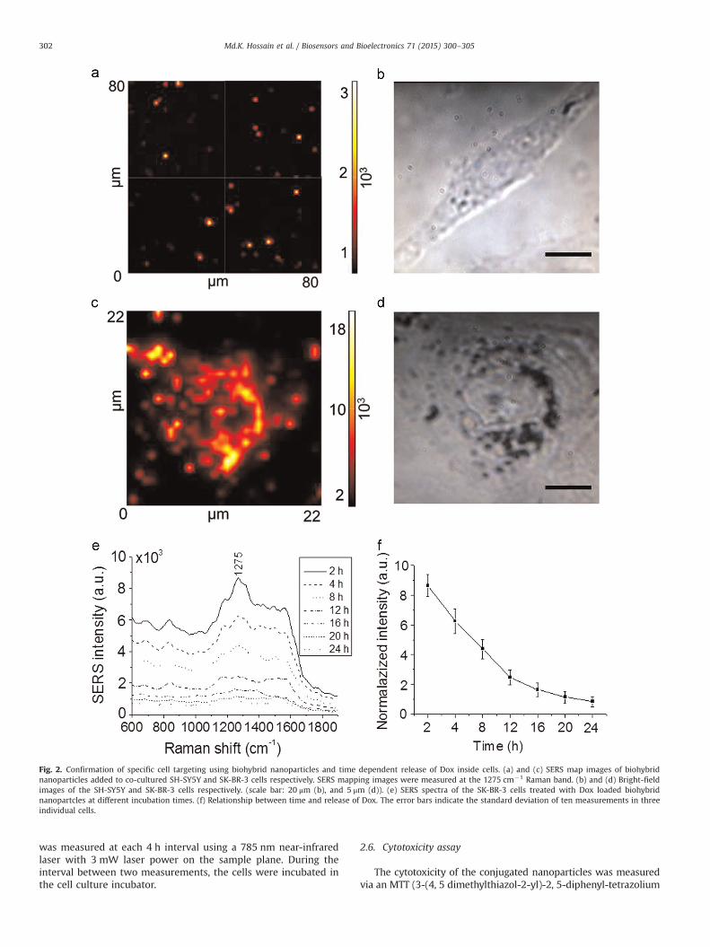
Fig. 2. Confirmation of specific cell targeting using biohybrid nanoparticles and time dependent release of Dox inside cells. (a) and (c) SERS map images of biohybridnanoparticles added to co-cultured SH-SY5Y and SK-BR-3 cells respectively. SERS mapping images were measured at the 1275 cm�1 Raman band. (b) and (d) Bright-fieldimages of the SH-SY5Y and SK-BR-3 cells respectively. (scale bar: 20 μm (b), and 5 μm (d)). (e) SERS spectra of the SK-BR-3 cells treated with Dox loaded biohybridnanopartcles at different incubation times. (f) Relationship between time and release of Dox. The error bars indicate the standard deviation of ten measurements in threeindividual cells.
Md.K. Hossain et al. / Biosensors and Bioelectronics 71 (2015) 300–305302
was measured at each 4 h interval using a 785 nm near-infraredlaser with 3 mW laser power on the sample plane. During theinterval between two measurements, the cells were incubated inthe cell culture incubator.
2.6. Cytotoxicity assay
The cytotoxicity of the conjugated nanoparticles was measuredvia an MTT (3-(4, 5 dimethylthiazol-2-yl)-2, 5-diphenyl-tetrazolium

Md.K. Hossain et al. / Biosensors and Bioelectronics 71 (2015) 300–305 303
bromide) assay following the method described previously (Arouiet al., 2009). After 48 h of seeding, the cells (5000 cells per well of a96-well plate) were treated with the biohybrid nanoparticles andincubated at 37 °C for 2 h for the specific targeting of, and uptakeby, the SK-BR-3 cells. After incubation, the biohybrid nanoparticle-containing media was removed, the cells were rinsed five timeswith PBS, and fresh media was added to the cells. MTT solution wasadded to each well at a final concentration of 1.2 mM and incubatedat 37 °C for 3 h. After incubation, the MTT solution-containingmedia was removed and 200 μl of dimethyl sulfoxide (DMSO) wasadded to each well. The optical density was measured with a uni-versal microplate reader (EL-800, BioTek Instrument Inc.) at a wa-velength of 540 nm.
3. Results and discussion
3.1. Particle conjugation and characterization
Biohybrid nanoparticles were prepared by the conjugation ofAuNPs, Tat-C, PEG, and the anti-HER2 antibody. To increase thepayload of the anti-cancer drug and to prevent the degradation ofbiological elements during conjugation, Dox was immobilized tothe AuNPs first, followed by Tat-C, PEG, and the anti-HER2 anti-body. The size of the conjugated nanoparticle was characterized byTEM imaging and DLS. The TEM image shows three 30 nm con-jugated AuNP (Fig. S1a). According to the DLS results, the averagesize of the bare AuNPs was 31.2 nm in diameter. After every step ofthe conjugations of Dos, Tat-C, PEG and antibody the diameter ofthe conjugated nanoparticles increased (Fig. S1b, S2) and, at theend of the conjugation, the average size of the nanoparticles was50 nm, but after GSH treatment the average size is decreased to44 nm (Fig. S1b). The decrease in size was due to release of dox-orubicin from the nanoparticle's surface after GSH treatment.
The zeta potential of the conjugated nanoparticles was mea-sured for confirmation of component conjugation at each step. TheAuNPs, HS-PEG-COOH, and anti-HER2 antibody were negativelycharged (Ock et al., 2012; Chen et al., 2013; Koopaei et al., 2011),and Dox, Tat-C and GSH were positively charged (Yousefpour et al.,2011; Kaplan et al., 2005; Ock et al., 2012). According to Fig. S1c,the zeta potential of the bare AuNPs was �68.64 mV. Duringconjugation of Dox, Tat-C, PEG, and the antibody to the AuNPs, thezeta potential of the conjugated nanoparticles became either po-sitive or negative, depending upon the components conjugated. Atthe end of the conjugation, the zeta potential of the Dox-loadedbiohybrid nanoparticle was �11.22 mV, but after GSH treatmentthe zeta potential was decreased to �47.75. The decrease in zetapotential was due to release of Dox from the surfaces of AuNPsafter GSH treatment. Figs. S1d and S3 show the intensity of theSERS spectra of different concentration of Dox loaded on the bio-hybrid nanoparticles. The SERS intensity of Dox increased alongwith the increase in loading concentration.
The SERS spectra were measured from the conjugated nano-particles (Fig. S4). The Dox exhibited a strong Raman band at1275 cm�1, which was due to the (νC-O) vibrational mode of ringA of the Dox molecule (Lee et al., 2004). After the addition of Tat-C,PEG, and the antibody, the SERS intensity of Dox reduced slightly.This occurs due to the scattering shielding effect caused by theincreasing thickness of the coating layer (Park et al., 2009). TheDox-loaded biohybrid nanoparticles were stable for up to 28 days(Fig. S5).
3.2. Specific targeting of SK-BR-3 cells
To study specific targeting, HER2-expressing cells (SK-BR-3)(Lee et al., 2013a, b) and HER2-negative cells (SH-SY5Y) were co-
cultured and treated with Dox-loaded biohybrid nanoparticles. Todetect the SERS signal from the treated cells, SERS mapping imageswere measured at the 1275 cm�1 Raman band. Fig. 2a and c showsthe SERS map images of the biohybrid nanoparticle–treated, co-cultured SH-SY5Y and SK-BR-3 cells respectively, and Fig. 2b and dshows the bright-field images of both cell types. The mappingimages demonstrate that the SERS signals of the biohybrid nano-particles were detected from SK-BR-3 cells in the immobilizedarea, but only non-specific and low SERS signals in the SH-SY5Ycell area. These results indicate that the biohybrid nanoparticlescan specifically target cells that express HER2 on the membrane.
To confirm the SERS results for specific targeting of the breastcancer cells, a fluorescence microscopy experiment was also con-ducted. Fig. S6a shows the SERS map image and fluorescencemicroscopy image, while Fig. S6b shows the bright-field images ofco-cultured cells containing SK-BR-3 and SH-SY5Y (arrow). In thecase of the SERS experiment, Dox-loaded biohybrid nanoparticleswere added to the cells, while for fluorescence microscopy FITC-labeled biohybrid nanoparticles were added. The mapping imagedemonstrates that the SERS signals of the biohybrid nanoparticleswere detected from the SK-BR-3 cell-immobilized area only andthat the areas with the SH-SY5Y cells did not exhibit any SERSsignal. The fluorescence microscopy image shows that the FITC-labeled (green) conjugated nanoparticles were immobilized on theround SK-BR-3 cells only, while the SH-SY5Y cells did not have anyconjugated nanoparticles. Hoechst 33342 dye was used to locatethe nuclei of the cells (blue). Therefore, the fluorescently labeledbiohybrid nanoparticle-treatment experiment demonstrated thatthe biohybrid nanoparticles are capable of specific targeting at thebulk cell level as well.
3.3. Detecting the effect of Tat-C on nanoparticle uptake by the SK-BR-3 cells
Before selecting Tat-C as a CPP, the cell penetration efficacy offour kinds of CPPs, Tat-C, Penetratin-C, pVEC-C, and Pep-1-C (TableS1), was studied through fluorescence microscopy. Among them,Tat-C exhibited the highest cell penetration efficacy (Fig. S7) and,hence, Tat-C was selected for further experiments in this study. Oftwo groups of SK-BR-3 cells, one group was treated with biohybridnanoparticles containing Tat-C and other group was treated withbiohybrid nanoparticles without Tat-C. After 2 h of particle treat-ment, the cells were washed and their gold content measuredusing inductively coupled plasma atomic emission spectroscopy(ICP-AES). According to the ICP-AES results, the uptake of Tat-C-modified biohybrid nanoparticles was approximately 5,670 percell, while the uptake of the biohybrid nanoparticles not con-taining Tat-C was too low to detect (detection limit was 0.05 ppm)(Table S2) (Fig. 2).
3.4. Monitoring intracellular Dox release in SK-BR-3 cells
Since the chemical structure of Dox contains an aromatic ring,it produces enhanced Raman signals when immobilized on AuNPsurfaces. Dox can be released from biohybrid nanoparticle surfacesby intracellular GSH. GSH is the most abundant thiol species in thecell cytoplasm, with a concentration range of 1–10 mM, and hasbeen used as an in situ releasing reagent in living cells, owing to itsbiochemical reducing capability (Oak et al., 2012). The thiol groupin the GSH has strong affinity to AuNPs and can bind with theAuNPs through covalent coupling. For this reason, amine con-taining Dox can easily be replaced from AuNP surfaces. Beforestudying its effects on cells, we studied the release characteristicsof Dox from biohybrid nanoparticle surfaces in the absence of cells,but in the presence of different concentrations of GSH using SERS.Fig. S8 shows the GSH concentration-dependent Dox release from

Fig. 3. Results of a cytotoxicity assay with the SK-BR-3 cell line. (a) Level of Dox-dependent cytotoxicity under different treatment conditions. (b) Cytotoxicity oftime-dependent intracellular Dox release from the biohybrid nanoparticle surfaces.The error bars indicate the standard deviation of five independent measurements.
Md.K. Hossain et al. / Biosensors and Bioelectronics 71 (2015) 300–305304
the biohybrid nanoparticle surfaces in the absence of cells. Thedecrease in intensity at the 1275 cm�1 Raman band of Dox in-dicated the release of Dox from the surface of the biohybrid na-noparticles. Ten spectra were measured and averaged to plot eachcurve. The results show that the Dox release rate was high in thepresence of 5–12 mM GSH. Time-dependent Dox release from thebiohybrid nanoparticle surface in the absence of cells but in thepresence 10 mM GSH was also studied using SERS (Fig. S9). Ac-cording to the SERS results, the Dox release rate was high within1 h of GSH treatment.
Time-dependent intracellular Dox release in live SK-BR-3 cellswas studied using SERS. Fig. 2d shows a bright-field image of anSK-BR-3 cell that was treated with Dox-loaded biohybrid nano-particles. The SERS map image of the cells (Fig. 2c) shows the SERSsignal of nanoparticles taken up by the cell. The SERS mappingimage was measured at the 1275 cm�1 Raman band. The SERSspectra were measured from ten different spots on three in-dividual cells at 4 h intervals after 2 h of nanoparticle treatmentand the spectra were averaged to make a single curve. The SERSresults (Fig. 2e and f) show that the intracellular Dox release ratefrom the biohybrid nanoparticles was high up to 12 h after na-noparticle treatment, followed by a decrease in concentration andrelease rate. The intracellular Dox release rate was comparativelyslower than the Dox release rate in the absence of cells but in thepresence of 10 mM GSH (Fig. S9). To observe the effect of addi-tional GSH on intracellular Dox release, another experiment wasconducted. GSH-OEt (5 mM) was added to the cell medium after2 h of nanoparticle treatment and SERS spectra were measuredevery 15 minutes a few minutes after GSH-OEt treatment. Theresults show that after adding 5 mM of additional GSH-OEt to thecells, the Dox release rate increased and most of the Dox was re-leased within 1 h of GSH-OEt treatment (Fig. S10).
3.5. Cytotoxicity assay
To study the cytotoxicity of the biohybrid nanoparticles, anMTT assay was conducted with different time condition. Fig. 3ashows that the cytotoxic effect depends on the concentration ofDox under different treatment conditions over the 24 h incubationperiod and, in contrast with the Dox-containing treatment con-dition, bare AuNPs (0 μM Dox) did not cause any cytotoxicity. TheDox-conjugated AuNP treatment caused higher cytotoxic effectsunder every tested condition and had a role as a carrier of Dox intocells. Furthermore, in AuNP-based Dox delivery, Tat-C-modifiedAuNPs exhibited higher cytotoxic effects than non-modifiedAuNPs, based on their increased uptake. Fig. 3b shows the time-dependent cytotoxicity of Dox-loaded biohybrid nanoparticles.After 2 h of treatment with biohybrid nanoparticles, cell viabilitieswere measured up to 24 h every 4 h. The results show that Doxwas released continuously under intracellular conditions, asshown in Fig. 2e, and induced an anti-cancer effect in the cells.Eight hours after the particle treatment, 9.53% of cells died and cellviability decreased continuously until 24 h (39.48%). The Doxworked as a DNA intercalator when it was released from thebiohybrid nanoparticles and that is the reason the Dox-mediatedcytotoxicity was not obtained directly. However, the biohybridnanoparticles did not cause any significant cell mortality. There-fore, the biohybrid nanoparticles can be used as carrier for anti-cancer drugs and as a monitoring probe for drug release (Fig. 3).
4. Conclusion
In this study, we proposed label-free in situ monitoring andcontrol of intracellular anti-cancer drug delivery process usingbiohybrid nanoparticles based on surface-enhanced Raman
spectroscopy (SERS) for the first time. The biohybrid nanoparticleswere successfully conjugated with AuNP, Tat-C, PEG and anti-HER2antibody and enhanced Raman signal of Dox when Dox was loa-ded. The HER2-positive cancer cell (SK-BR-3) was specifically tar-geted with biohybrid nanoparticles and showed the SERS signal ofDox from entire cell. Due to the addition of Tat-C to the biohybridnanoparticles, an average of 5670 nanoparticles were taken up byeach cell within 2 h of treatment, but uptake was very low to inthe case of nanoparticles not containing Tat-C. Time-dependentintracellular Dox release from biohybrid nanoparticles was mon-itored using SERS. According to the SERS results, 90.23% of the Doxwas released by the action of intracellular GSH with 24 h of par-ticle treatment and releasing time of Dox can be reduced until 2 hwith addition of GSH. Cell mortality was linearly increasing until60.86% at 24 h with Dox-loaded biohybrid nanoparticle-treatedcells, but there was no anti-cancer effect in unloaded biohybridnanoparticle-treated cells. Thus, by using the biohybrid nano-particles, we successfully monitored in situ Dox release inside SK-BR-3 cells. The proposed biohybrid nanoparticles suitable for ap-plication of aromatic anti-cancer drugs but SERS signal of otherkinds of drugs may not be enough to distinguish from other sig-nals. However, our newly proposed system can be applied as a

Md.K. Hossain et al. / Biosensors and Bioelectronics 71 (2015) 300–305 305
spectroscopic biosensor for label-free, in situ monitoring of thetime-dependent release of other anti-cancer drugs in cells.
Acknowledgements
This research was supported by the National Research Foun-dation of Korea (NRF) grant funded by the Korea government(MSIP) (No. 2014R1A2A1A10051725).
Appendix A. Supplementary material
Supplementary data associated with this article can be found inthe online version at http://dx.doi.org/10.1016/j.bios.2015.04.053.
References
An, J.H., et al., 2014. Biosens. Bioelectron. 10. 1016/j.bios.2014.049An, J.H., et al., 2011a. J. Nanosci. Nanotechnol. 11, 1585–1588.An, J.H., et al., 2011b. J. Nanosci. Nanotechnol. 11, 4424–4429.Aroui, S., et al., 2009. Cancer Lett. 285, 28–38.Boyd, A.R., et al., 2011. J. Mater. Sci.: Mater. Med. 22, 1923–1930.
Chan, J.M., et al., 2009. Biomaterials 30, 1627–1634.Chae, E.J., et al., 2013. J. Biomed. Nanotechnol. 9, 659–663.Chen, H., et al., 2013. Nanotechnology 24, 355101.Doering, W.E., et al., 2007. Adv. Mater. 19, 3100–3108.El-Said, W.A., et al., 2011a. PLoS One 6, e15836.El-Said, W.A., et al., 2011b. J. Nanosci. Nanotechnol. 11, 768–772.El-Said, W.A., et al., 2010. Biosens. Bioelectron. 26, 1486–1492.Hu, S.H., et al., 2009. Adv. Funct. Mater. 19, 3396–3403.Isben, S., et al., 2013. Ultrasonics 53, 178–184.Kang, B., et al., 2013. ACS Nano 7, 7420–7427.Kaplan, I.M., et al., 2005. J. Control. Release 102, 247–253.Kim, T.H., et al., 2013. Biomaterials 34, 8660–8670.Koopaei, M.N., et al., 2011. Int. J. Nanomed. 6, 1904–1912.Lurie, L.S., et al., 1984. J. Forensic Sci. 29, 4.Lee, C.J., et al., 2004. Bull. Korean Chem. Soc. 25, 1211–1216.Lee, H.J., et al., 2013a. Angew. Chem. 52, 8337–8340.Lee, H.J., et al., 2013b. Biosens. Bioelectron. 47, 508–514.Nakamura, T., et al., 2015. Chem. Sci. 10:1039/c4sc03549fOck, K., et al., 2012. Anal. Chem. 84, 2172–2178.Pallaoro, A., et al., 2010. Small 6, 618–622.Park, H., et al., 2009. Phys. Chem. Chem. Phys. 11, 7444–7449.Pully, V.V., et al., 2010. Anal. Chem. 82, 1844–1850.Wang, J., et al., 2014. J. Mater. Chem. B. 2, 4379–4386.Yousefpour, P., et al., 2011. Int. J. Nanomedicine 6, 1487–1496.Zhang, L.F., et al., 2008. ACS Nano 2, 1696–1702.Zacharia, E., et al., 2010. J. Photochem. Photobiol. B 100, 113–116.Zagotto, G., et al., 2001. J. Chromatogr. B 764 161-148.Zong, S., et al., 2011. Anal. Chem. 83, 4178–4183.