blockade of stimulus convergence in amygdala neurons disrupts taste associative learning
TRANSCRIPT
Brief Communications
Blockade of Stimulus Convergence in Amygdala NeuronsDisrupts Taste Associative Learning
Koichi Hashikawa,1 Masamitsu Naka,1 Daisuke Nakayama,1 Nobuyoshi Matsumoto,1 Rachael Neve,2
and Norio Matsuki1
1Laboratory of Chemical Pharmacology Graduate School of Pharmaceutical Sciences, The University of Tokyo, Tokyo 113-0033, Japan, and 2PicowerInstitute, Massachusetts Institute of Technology, Cambridge, Massachusetts 02139
Humans and non-human animals learn associations of temporally contingent stimuli to better cope with the changing environment. Inanimal models of classical conditioning, a neutral conditioned stimulus (CS) predicts an aversive unconditioned stimulus (US). Severallines of indirect evidence indicate that this learning may rely on stimulus convergence in a subset of neurons, but this hypothesis has notbeen directly tested. In the current study, we tested this hypothesis using a pharmacogenetic approach, the cAMP response element-binding protein (CREB)/Allatostatin Receptor system, to target a subset of amygdala neurons receiving convergent stimuli in mice duringconditioned taste aversion. Virally infected basolateral amygdala neurons with higher CREB levels were predominantly active during CSpresentation. Blocking stimulus convergence in infected neurons by silencing them during US disrupted taste associative memory.Moreover, silencing infected neurons only during CS also disrupted associative memory formation. These results provide support for thenotion that convergent inputs of CS and US in a subpopulation of neurons are critical for associative memory formation.
IntroductionThrough associative learning, humans and non-human animalslearn that a sensory stimulus predicts the occurrence of an event.It has been proposed that associative memory formation criticallyrelies on convergent inputs of the conditioned stimulus (CS) andthe unconditioned stimulus (US) to a subpopulation of neurons(Blair et al., 2001; Johansen et al., 2010). Despite substantial prog-ress in identifying brain regions involved in several associativelearning tasks (Rogan et al., 1997; Yasoshima et al., 2000) andvisualizing subpopulations of neurons responding to convergentstimuli (Barot et al., 2008; Chung et al., 2011), the requirement ofstimulus convergence for associative learning has not been di-rectly examined, because of the lack of a method for selectivelytargeting stimulus convergence. Selectively blocking stimulusconvergence requires the selection of a subpopulation of neuronsthat are activated by the CS, and silencing them during US expo-sure. Since neurons active during CS are going to be active (stim-ulus convergence) or inactive during US, by silencing them
during US, stimulus convergence is blocked in the former neu-rons while the latter neurons keep inactivated during US.
We used a pharmacogenetic approach to achieve cell-specificmanipulation of CREB (cAMP response element-binding pro-tein) levels, and reversible inactivation of targeted cells to selec-tively block stimulus convergence (Zhou et al., 2009). Recentstudies have suggested that amygdala neurons with relatively highCREB levels are preferentially recruited in associative memorytraces, such as fear and taste aversive memories (Han et al., 2007,2009; Zhou et al., 2009). Neurons with higher CREB levels arepredominantly active during fear conditioning and fear memoryretrieval (Han et al., 2007). Manipulation of CREB levels in asubset of neurons may also allow investigators to specifically tar-get the neuronal population that is activated by CS.
Conditioned taste aversion (CTA) is a simple form of associa-tive learning during which a novel taste (CS) is paired with vis-ceral malaise (US) (Bermudez-Rattoni, 2004). In associativelearning tasks such as fear conditioning, the CS and US are typi-cally separated for no more than several seconds, or at most acouple of minutes. In contrast, CTA can be acquired with delaysof several minutes or even hours between CS and US presenta-tion. This longer interval allows for the silencing of targeted neu-rons during CS or US using a pharmacogenetic approach. Wefocused on the basolateral nucleus of the amygdala (BLA), whichis critically involved in CTA acquisition (Yasoshima et al., 2000;Josselyn et al., 2004), and where a subpopulation of neuronsreceiving convergent inputs of CS and US has previously beenvisualized (Barot et al., 2008).
Materials and MethodsSubjects. Adult male C57BL/6J mice were group-housed on a 12 h light/dark cycle. Food and water were available ad libitum before the behav-
Received Nov. 26, 2012; revised Jan. 30, 2013; accepted Feb. 5, 2013.Author contributions: K.H., D.N., N. Matsumoto, and N. Matsuki designed research; K.H., M.N., D.N., N. Matsu-
moto, and R.L.N. performed research; K.H., M.N., D.N., and N. Matsumoto analyzed data; K.H. wrote the paper.This work was supported by grants from the Japan Society for the Promotion of Science (70126168) to N. Matsuki.
We thank Dr. Alcino Silva for amplicon vectors of HSV-CREB and HSV-LacZ, Dr. Paul Worley for Arc cDNA, and Dr.Sheena Josselyn for GFP cDNA. We also thank Dr. Yu Zhou, Dr. Masanori Sakaguchi, Dr. Yasunori Hayashi, AakaneSano, Dr. Takuya Takahashi for technical advice, Dr. Dayu Lin, and Yoshiko Yamasaki for helpful comments on themanuscript.
The authors declare no competing financial interests.Correspondence should be addressed to Koichi Hashikawa, Laboratory of Chemical Pharmacology, Graduate
School of Pharmaceutical Sciences, The University of Tokyo, 7-3-1 Hongo, Bunkyo-ku, Tokyo 113-0033, Japan.E-mail: [email protected].
DOI:10.1523/JNEUROSCI.5462-12.2013Copyright © 2013 the authors 0270-6474/13/334958-06$15.00/0
4958 • The Journal of Neuroscience, March 13, 2013 • 33(11):4958 – 4963

ioral experiments. Mice were treated in accordance with guidelinesapproved by The University of Tokyo.
HSV vectors. HSV-CREB-AlstR (HSV-CREB) and HSV-LacZ-AlstR(HSV-LacZ) were used (Zhou et al., 2009). The HSV amplicons were kindlyprovided by Dr. Alcino Silva (The University of California, Los Angeles). Forvisualization of transgene expression, eGFP was fused to the end of the CREBand LacZ cDNA. Creb 1 and LacZ were expressed using an IE4/5 promoter,and AlstR was expressed with a CMV promoter. The amplicons were pack-aged according to a published method (Lim and Neve, 2001).
Surgery and virus infusion. Anesthesia was induced with Nembutal (5mg/kg body weight, injected intraperitoneally). The scalp was opened,and holes were drilled in the skull bilaterally above the BLA (AP, �1.4;LM, �3.5, DV, �4.9 mm from bregma) according to a mouse brain atlas(Paxinos and Franklin, 2001). For behavioral experiments, guide cannu-lae (Plastics One, 26 gauge) were implanted just dorsal to the BLA andfixed on the skull with dental cement. Virus infusion was delayed until10 d after surgery to ensure physical recovery. A virus solution (1.5 �l,bilateral) was delivered to the BLA at a flow rate of 0.075 �l min �1
through inner injection cannulae (Plastics One, 33 gauge) using an infu-sion pump (Muromachi). The injection cannula was left in place for anadditional 10 min to ensure diffusion of the virus. For electrophysiolog-ical or fluorescence in situ hybridization (FISH) studies, virus solution(1.5 �l, bilateral) was delivered to the BLA at a flow rate of 0.075 �lmin �1 through glass micropipettes (World Precision Instruments) im-mediately after the holes were drilled. The micropipette was left in placefor an additional 10 min. Electrophysiological, FISH, and behavioralexperiments were performed 3 d following virus infusion.
Allatostatin administration. The peptide allatostatin (Phoenix Phar-maceuticals) was dissolved in double-distilled water to make a 2.5 mM
stock solution. Saline was added to dilute it to the final concentrationfor each experiment. For behavioral studies, allatostatin solution (100�M, 0.5 �l) or the vehicle saline (0.5 �l) was infused bilaterally to theBLA of freely moving mice at a flow rate of 0.25 �l min �1 throughinner injection cannulae. The cannulae were left in place for an addi-tional 2 min.
Arc and GFP double FISH. Five minutes afterthe onset of CS or US exposure, brains were ex-tracted, frozen, and stored at �80°C. Brains werecut into 20 �m coronal slices using a cryostat andmounted onto slides. Digoxigenin-labeled Arc ri-boprobes and fluorescein-labeled GFP ribo-probes were generated from modified cDNAplasmids. FISH for Arc and GFP mRNA followedprevious reports (Han et al., 2007; Hashikawa etal., 2011). Arc and GFP signals were visualizedusing the Cyanine 3 and Cyanine 5 TSA system(PerkinElmer). Nuclei were counterstained withHoechst (Invitrogen). Three to four sections in-cluding the BLA were analyzed per mouse. Im-ages were acquired using a Leica SP5 confocalmicroscope with a 40� objective lens. Z-seriesstacks (1-�m-thick optical sections) were con-structed and analyzed using the MetaMorph 6.0program. Analysis was performed by three exper-imenters blind to group membership. Only neu-rons with fully intact nuclei were scored. Forneurons to be considered Arc positive for nuclearstaining, one or two robust foci within their nu-clei were required. Perinuclear signals were re-quired for neurons to be considered cytoplasmicArc positive.
CTA. Mice were deprived of water for 24 h,then adapted to a water-restriction schedulefor 4 d with two daily drinking sessions at 9:00A.M. (60 min initially, gradually reduced to 5min) and at 5:00 P.M. (for 60 min) from twotubes filled with water. HSV was bilaterally in-fused into the BLA on the second day of thewater restriction. Conditioning took place 3 dafter the viral injection.
On the conditioning day, a tube filled with 0.2% saccharin sodium salt(w/v, CS) was presented for 5 min (short-interval conditioning) or 30min (long-interval conditioning). Mice were treated with an intraperito-neal injection of 0.06 M LiCl (2% of body weight) 30 min (short interval)or 2.5 h (long interval) after the CS.
Testing for aversion to saccharin took place 24 h after the condition-ing. Two tubes (one filled with water and the other filled with saccharin)were presented for 10 min. The intake of each fluid was measured. Theaversion index was defined as (milliliters of water consumption)/(milli-liters of water consumption � milliliters of saccharin consumption) andused as an index for learned aversion for saccharin.
After the behavioral experiments, mice were killed by transcardialperfusion with PBS, followed by 4% paraformaldehyde (w/v). Follow-ing perfusion, brains were left in 4% paraformaldehyde for 1 d andthen moved to a 30% sucrose solution (w/v) in PBS for 2–3 d. Brainswere frozen and sliced coronally (40 �m) with a cryostat. To identifycannula tip locations, relevant sections were mounted on slides andstained with crystal violet. Only those mice with bilateral placementsin the BLA were included in the analysis.
Electrophysiology. Acute brain slices were prepared from BLA neurons ofadult mice 3 d after viral infusion as previously described (Miura et al., 2012).Slices were perfused with artificial CSF containing the following (in mM): 127NaCl, 1.6 KCl, 1.24 KH2PO4, 1.3 mM MgSO4, 2.4 CaCl2, 26 NaHCO3, and 10glucose at 31°C. Cells were visualized using infrared or epifluorescent illumi-nation, and whole-cell current-clamp recordings were made from neuronsin BLA. Patch electrodes (3–6 M�) contained (in mM): 120 K-gluconate, 5KCl, 10 HEPES, 1 MgCl2, 10 phosphocreatine-Na2, 2 MgATP, 0.2 Na2GTP,0.2 EGTA, and 0.04 Alexa Fluor 568, pH 7.2–7.3, 280–295 mOsm. Accessresistance was monitored throughout the experiments. Only putative pyra-midal neurons in BLA defined as healthy (resting membrane potential morenegative than �55 mV) were included in the analysis. Allatostatin (20 nM)was applied by adding it to the superfusate. Physiological measures before,after 5 min of perfusion with allatostatin, and 15–20 min after washout werecompared.
Figure 1. Selective viral expression in the BLA with HSV-CREB or HSV-LacZ. A, Left, Outline of the BLA. Right, A representativeimage of localized GFP (green) expression in the BLA 3 d following viral injection. Blue, Nuclei. Scale bar, 80 �m. B, Approximately20% of BLA neurons expressed GFP in the infected region following HSV-CREB or HSV-LacZ infusion (n � 4 mice/group). C,Representative traces showing that allatostatin selectively and quickly (�5 min) inactivated an infected pyramidal neuron in theBLA (top). Error bars represent SEM.
Hashikawa et al. • Blocking Convergence Disrupts Associative Learning J. Neurosci., March 13, 2013 • 33(11):4958 – 4963 • 4959

Data analysis. All values were reported asmeans � SEM. ANOVA was used for data anal-ysis with post hoc comparisons performed us-ing Student’s t test to determine thesignificance of differences between two groups.Comparisons within an individual mouse wereperformed using paired t test.
The probability of Arc � neurons in GFP �
or GFP � cells was calculated as follows: Arc(�) in GFP (�) (%) � (the number of cellswith Arc and GFP signals/total number of GFP(�) cells) � 100. Arc (�) in GFP (�) (%) �(the number of cells with Arc and no GFP sig-nals/total number of GFP (�) cells) � 100.
ResultsTo selectively target and silence cells thatare active during CS presentation, theBLA was injected with CREB-AlstR HSV(HSV-CREB), which coexpresses CREBtagged with green fluorescent protein(GFP) and the Drosophila allatostatinG-protein-coupled receptor (AlstR). Forthe control virus (HSV-LacZ), CREB wasreplaced with �-galactosidase (LacZ)(Zhou et al., 2009). Allatostatin adminis-tration selectively and reversibly silencesneurons expressing AlstR through theactivation of endogenous mammalianG-protein-coupled inwardly rectifyingK� channels (Tan et al., 2006). Indeed, weconfirmed that administration of alla-tostatin selectively silenced BLA neuronsinfected with the viruses by performingwhole-cell patch-clamp recording (Fig.1C). Detection of GFP mRNA by FISHrevealed that �20% of neurons in the in-jected region in the BLA were GFP� withboth HSVs, and these cells were preferen-tially localized in the BLA. No significantdifference was found between the infec-tion rates for the HSV-CREB and HSV-LacZ viruses (19 � 2% versus 17 � 1%,n � 4 mice per group, Student’s t test, t(6)
� 0.915, p � 0.395; Fig. 1A,B).We investigated whether virally in-
fected cells overexpressing CREB are pref-erentially active during CS. To visualizecellular activation, we used the activity-dependent gene Arc (activity-regulatedcytoskeleton-associated protein; alsotermed Arg3.1) (Guzowski et al., 1999; Barot et al., 2008). ArcmRNA signal first presents in the nucleus (5–10 min) and thentranslocates to the cytoplasm over time (�30 min). We injectedHSV-CREB or HSV-LacZ into the BLA 3 d before saccharin presenta-tion. Five minutes after saccharin presentation, brains were harvestedfor double FISH to detect intranuclear Arc and GFP mRNA in the BLA.Overall, the percentage of Arc� cells was higher following saccharinpresentation compared with home cage controls in mice injected withHSV-CREB (Student’s t test, t(6) �5.533, p�0.0015; Fig. 2B). Further-more, the probability of detecting Arc� nuclei in the saccharin groupwas higher in neurons with HSV-CREB than in their noninfectedneighbors (paired t test, t(3) � 16.684, p � 0.00047; Fig. 2C). Incontrast, neurons with HSV-LacZ were no more likely to be Arc�
than were their noninfected neighbors (paired t test, t(3) � �0.158,p � 0.88; Fig. 2C). These results indicate that amygdala neurons withincreased CREB function were preferentially activated by saccharinpresentation.
In addition, infected neurons with HSV-CREB were pref-erentially activated by convergent stimuli (Fig. 2D–F ). HSV-CREB was injected in the BLA 3 d before CS and USpresentation. Double FISH for detecting Arc and GFP mRNAin the BLA revealed that the percentage of double-labeled Arc-positive neurons with intranuclear and cytoplasmic signalswas significantly higher in GFP (�) neurons than in GFP (�)neurons after CS and US presentation (paired t test, t(2) �5.336, p � 0.033; Fig. 2F ).
Figure 2. BLA neurons with high CREB levels are preferentially active during CS presentation. A, Representative confocal imagesof Arc (red) and GFP (green) expression in the BLA neurons. Mice were injected with HSV-CREB (top) or HSV-LacZ (bottom) 3 dbefore CS presentation. They were killed 5 min after the onset of CS presentation. Blue, Nuclei; yellow arrows, colocalized neurons(GFP � and intranuclear Arc �). Scale bar, 20 �m. B, Percentage of Arc � neurons following CS or in the home cage in mice injectedwith HSV-CREB (n � 4 mice/group). **p � 0.01. C, The proportion of Arc � neurons in GFP � or GFP � neurons following CS inHSV-CREB group (left) and HSV-LacZ group (right). **p � 0.01. D, Mice were infused with HSV-CREB 3 d before training (n � 3mice). They were presented with CS for 5 min (30 min before they were killed) and US (5 min before they were killed). E,Representative images of GFP � (green) neurons with intranuclear (left), cytoplasmic (middle), or double-labeled (right) Arc signal(red). Blue, Nuclei. Scale bar, 10 �m. F, The proportion of double-labeled Arc � neurons in GFP � or GFP � neurons. *p � 0.05.Error bars represent SEM.
4960 • J. Neurosci., March 13, 2013 • 33(11):4958 – 4963 Hashikawa et al. • Blocking Convergence Disrupts Associative Learning
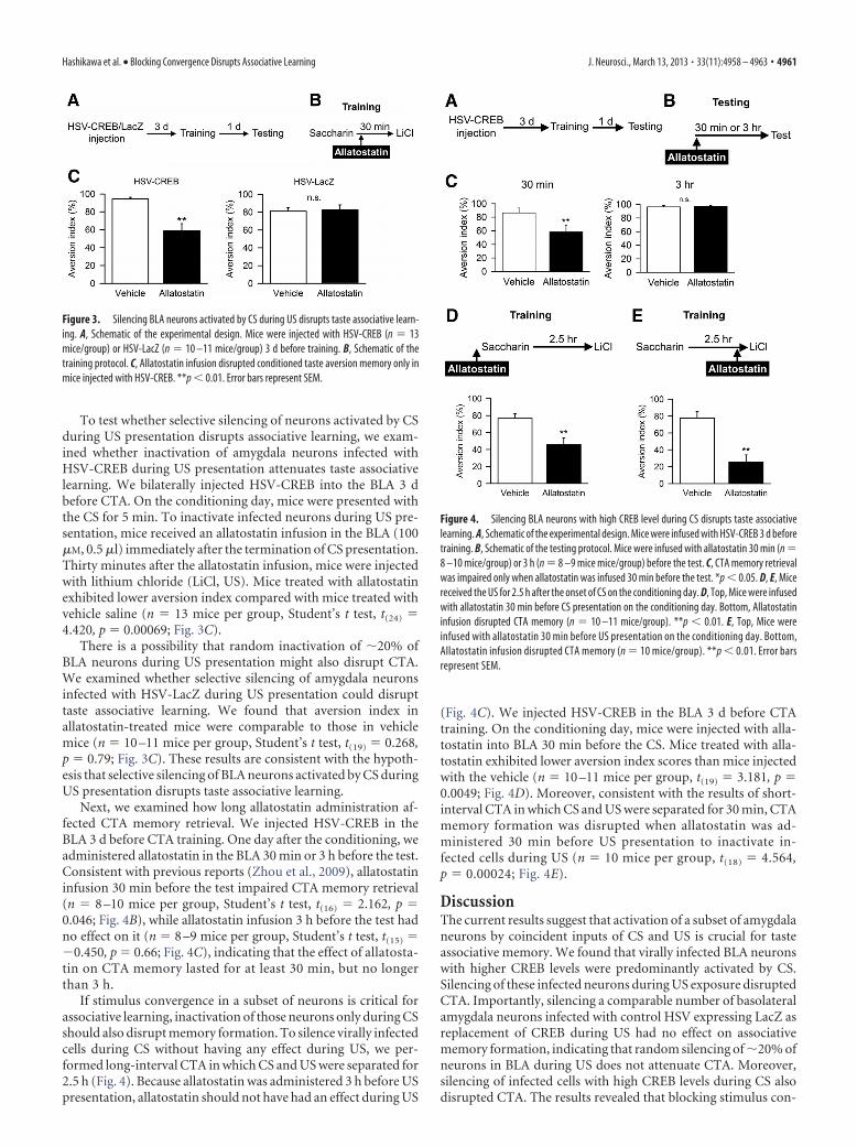
To test whether selective silencing of neurons activated by CSduring US presentation disrupts associative learning, we exam-ined whether inactivation of amygdala neurons infected withHSV-CREB during US presentation attenuates taste associativelearning. We bilaterally injected HSV-CREB into the BLA 3 dbefore CTA. On the conditioning day, mice were presented withthe CS for 5 min. To inactivate infected neurons during US pre-sentation, mice received an allatostatin infusion in the BLA (100�M, 0.5 �l) immediately after the termination of CS presentation.Thirty minutes after the allatostatin infusion, mice were injectedwith lithium chloride (LiCl, US). Mice treated with allatostatinexhibited lower aversion index compared with mice treated withvehicle saline (n � 13 mice per group, Student’s t test, t(24) �4.420, p � 0.00069; Fig. 3C).
There is a possibility that random inactivation of �20% ofBLA neurons during US presentation might also disrupt CTA.We examined whether selective silencing of amygdala neuronsinfected with HSV-LacZ during US presentation could disrupttaste associative learning. We found that aversion index inallatostatin-treated mice were comparable to those in vehiclemice (n � 10 –11 mice per group, Student’s t test, t(19) � 0.268,p � 0.79; Fig. 3C). These results are consistent with the hypoth-esis that selective silencing of BLA neurons activated by CS duringUS presentation disrupts taste associative learning.
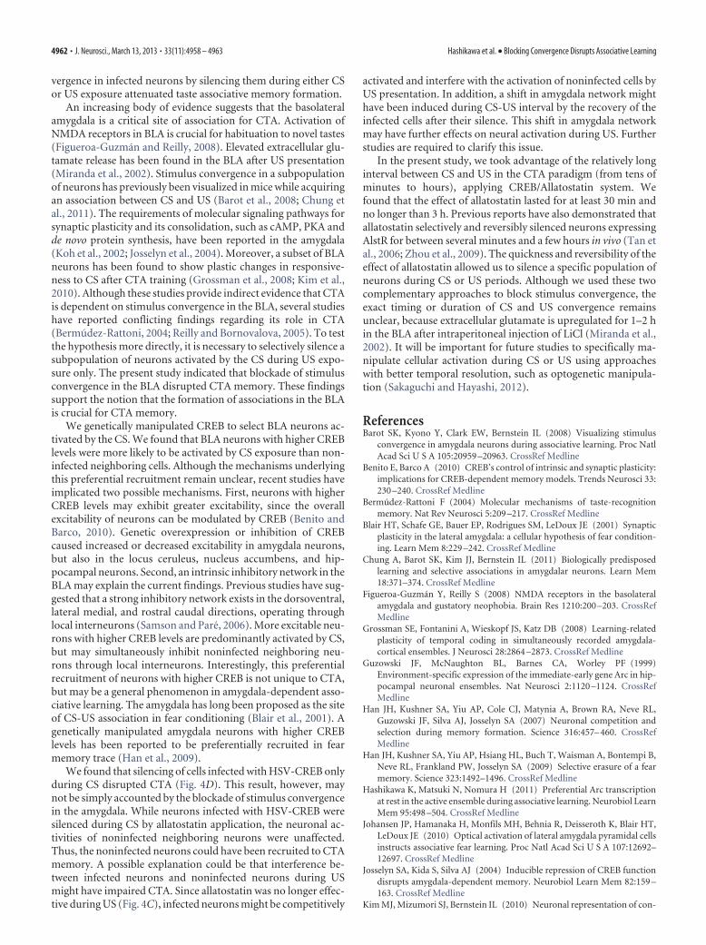
Next, we examined how long allatostatin administration af-fected CTA memory retrieval. We injected HSV-CREB in theBLA 3 d before CTA training. One day after the conditioning, weadministered allatostatin in the BLA 30 min or 3 h before the test.Consistent with previous reports (Zhou et al., 2009), allatostatininfusion 30 min before the test impaired CTA memory retrieval(n � 8 –10 mice per group, Student’s t test, t(16) � 2.162, p �0.046; Fig. 4B), while allatostatin infusion 3 h before the test hadno effect on it (n � 8 –9 mice per group, Student’s t test, t(15) ��0.450, p � 0.66; Fig. 4C), indicating that the effect of allatosta-tin on CTA memory lasted for at least 30 min, but no longerthan 3 h.
If stimulus convergence in a subset of neurons is critical forassociative learning, inactivation of those neurons only during CSshould also disrupt memory formation. To silence virally infectedcells during CS without having any effect during US, we per-formed long-interval CTA in which CS and US were separated for2.5 h (Fig. 4). Because allatostatin was administered 3 h before USpresentation, allatostatin should not have had an effect during US
(Fig. 4C). We injected HSV-CREB in the BLA 3 d before CTAtraining. On the conditioning day, mice were injected with alla-tostatin into BLA 30 min before the CS. Mice treated with alla-tostatin exhibited lower aversion index scores than mice injectedwith the vehicle (n � 10 –11 mice per group, t(19) � 3.181, p �0.0049; Fig. 4D). Moreover, consistent with the results of short-interval CTA in which CS and US were separated for 30 min, CTAmemory formation was disrupted when allatostatin was ad-ministered 30 min before US presentation to inactivate in-fected cells during US (n � 10 mice per group, t(18) � 4.564,p � 0.00024; Fig. 4E).
DiscussionThe current results suggest that activation of a subset of amygdalaneurons by coincident inputs of CS and US is crucial for tasteassociative memory. We found that virally infected BLA neuronswith higher CREB levels were predominantly activated by CS.Silencing of these infected neurons during US exposure disruptedCTA. Importantly, silencing a comparable number of basolateralamygdala neurons infected with control HSV expressing LacZ asreplacement of CREB during US had no effect on associativememory formation, indicating that random silencing of �20% ofneurons in BLA during US does not attenuate CTA. Moreover,silencing of infected cells with high CREB levels during CS alsodisrupted CTA. The results revealed that blocking stimulus con-
Figure 3. Silencing BLA neurons activated by CS during US disrupts taste associative learn-ing. A, Schematic of the experimental design. Mice were injected with HSV-CREB (n � 13mice/group) or HSV-LacZ (n � 10 –11 mice/group) 3 d before training. B, Schematic of thetraining protocol. C, Allatostatin infusion disrupted conditioned taste aversion memory only inmice injected with HSV-CREB. **p � 0.01. Error bars represent SEM.
Figure 4. Silencing BLA neurons with high CREB level during CS disrupts taste associativelearning. A, Schematic of the experimental design. Mice were infused with HSV-CREB 3 d beforetraining. B, Schematic of the testing protocol. Mice were infused with allatostatin 30 min (n �8 –10 mice/group) or 3 h (n � 8 –9 mice mice/group) before the test. C, CTA memory retrievalwas impaired only when allatostatin was infused 30 min before the test. *p � 0.05. D, E, Micereceived the US for 2.5 h after the onset of CS on the conditioning day. D, Top, Mice were infusedwith allatostatin 30 min before CS presentation on the conditioning day. Bottom, Allatostatininfusion disrupted CTA memory (n � 10 –11 mice/group). **p � 0.01. E, Top, Mice wereinfused with allatostatin 30 min before US presentation on the conditioning day. Bottom,Allatostatin infusion disrupted CTA memory (n � 10 mice/group). **p � 0.01. Error barsrepresent SEM.
Hashikawa et al. • Blocking Convergence Disrupts Associative Learning J. Neurosci., March 13, 2013 • 33(11):4958 – 4963 • 4961
vergence in infected neurons by silencing them during either CSor US exposure attenuated taste associative memory formation.
An increasing body of evidence suggests that the basolateralamygdala is a critical site of association for CTA. Activation ofNMDA receptors in BLA is crucial for habituation to novel tastes(Figueroa-Guzman and Reilly, 2008). Elevated extracellular glu-tamate release has been found in the BLA after US presentation(Miranda et al., 2002). Stimulus convergence in a subpopulationof neurons has previously been visualized in mice while acquiringan association between CS and US (Barot et al., 2008; Chung etal., 2011). The requirements of molecular signaling pathways forsynaptic plasticity and its consolidation, such as cAMP, PKA andde novo protein synthesis, have been reported in the amygdala(Koh et al., 2002; Josselyn et al., 2004). Moreover, a subset of BLAneurons has been found to show plastic changes in responsive-ness to CS after CTA training (Grossman et al., 2008; Kim et al.,2010). Although these studies provide indirect evidence that CTAis dependent on stimulus convergence in the BLA, several studieshave reported conflicting findings regarding its role in CTA(Bermudez-Rattoni, 2004; Reilly and Bornovalova, 2005). To testthe hypothesis more directly, it is necessary to selectively silence asubpopulation of neurons activated by the CS during US expo-sure only. The present study indicated that blockade of stimulusconvergence in the BLA disrupted CTA memory. These findingssupport the notion that the formation of associations in the BLAis crucial for CTA memory.
We genetically manipulated CREB to select BLA neurons ac-tivated by the CS. We found that BLA neurons with higher CREBlevels were more likely to be activated by CS exposure than non-infected neighboring cells. Although the mechanisms underlyingthis preferential recruitment remain unclear, recent studies haveimplicated two possible mechanisms. First, neurons with higherCREB levels may exhibit greater excitability, since the overallexcitability of neurons can be modulated by CREB (Benito andBarco, 2010). Genetic overexpression or inhibition of CREBcaused increased or decreased excitability in amygdala neurons,but also in the locus ceruleus, nucleus accumbens, and hip-pocampal neurons. Second, an intrinsic inhibitory network in theBLA may explain the current findings. Previous studies have sug-gested that a strong inhibitory network exists in the dorsoventral,lateral medial, and rostral caudal directions, operating throughlocal interneurons (Samson and Pare, 2006). More excitable neu-rons with higher CREB levels are predominantly activated by CS,but may simultaneously inhibit noninfected neighboring neu-rons through local interneurons. Interestingly, this preferentialrecruitment of neurons with higher CREB is not unique to CTA,but may be a general phenomenon in amygdala-dependent asso-ciative learning. The amygdala has long been proposed as the siteof CS-US association in fear conditioning (Blair et al., 2001). Agenetically manipulated amygdala neurons with higher CREBlevels has been reported to be preferentially recruited in fearmemory trace (Han et al., 2009).
We found that silencing of cells infected with HSV-CREB onlyduring CS disrupted CTA (Fig. 4D). This result, however, maynot be simply accounted by the blockade of stimulus convergencein the amygdala. While neurons infected with HSV-CREB weresilenced during CS by allatostatin application, the neuronal ac-tivities of noninfected neighboring neurons were unaffected.Thus, the noninfected neurons could have been recruited to CTAmemory. A possible explanation could be that interference be-tween infected neurons and noninfected neurons during USmight have impaired CTA. Since allatostatin was no longer effec-tive during US (Fig. 4C), infected neurons might be competitively
activated and interfere with the activation of noninfected cells byUS presentation. In addition, a shift in amygdala network mighthave been induced during CS-US interval by the recovery of theinfected cells after their silence. This shift in amygdala networkmay have further effects on neural activation during US. Furtherstudies are required to clarify this issue.
In the present study, we took advantage of the relatively longinterval between CS and US in the CTA paradigm (from tens ofminutes to hours), applying CREB/Allatostatin system. Wefound that the effect of allatostatin lasted for at least 30 min andno longer than 3 h. Previous reports have also demonstrated thatallatostatin selectively and reversibly silenced neurons expressingAlstR for between several minutes and a few hours in vivo (Tan etal., 2006; Zhou et al., 2009). The quickness and reversibility of theeffect of allatostatin allowed us to silence a specific population ofneurons during CS or US periods. Although we used these twocomplementary approaches to block stimulus convergence, theexact timing or duration of CS and US convergence remainsunclear, because extracellular glutamate is upregulated for 1–2 hin the BLA after intraperitoneal injection of LiCl (Miranda et al.,2002). It will be important for future studies to specifically ma-nipulate cellular activation during CS or US using approacheswith better temporal resolution, such as optogenetic manipula-tion (Sakaguchi and Hayashi, 2012).
ReferencesBarot SK, Kyono Y, Clark EW, Bernstein IL (2008) Visualizing stimulus
convergence in amygdala neurons during associative learning. Proc NatlAcad Sci U S A 105:20959 –20963. CrossRef Medline
Benito E, Barco A (2010) CREB’s control of intrinsic and synaptic plasticity:implications for CREB-dependent memory models. Trends Neurosci 33:230 –240. CrossRef Medline
Bermudez-Rattoni F (2004) Molecular mechanisms of taste-recognitionmemory. Nat Rev Neurosci 5:209 –217. CrossRef Medline
Blair HT, Schafe GE, Bauer EP, Rodrigues SM, LeDoux JE (2001) Synapticplasticity in the lateral amygdala: a cellular hypothesis of fear condition-ing. Learn Mem 8:229 –242. CrossRef Medline
Chung A, Barot SK, Kim JJ, Bernstein IL (2011) Biologically predisposedlearning and selective associations in amygdalar neurons. Learn Mem18:371–374. CrossRef Medline
Figueroa-Guzman Y, Reilly S (2008) NMDA receptors in the basolateralamygdala and gustatory neophobia. Brain Res 1210:200 –203. CrossRefMedline
Grossman SE, Fontanini A, Wieskopf JS, Katz DB (2008) Learning-relatedplasticity of temporal coding in simultaneously recorded amygdala-cortical ensembles. J Neurosci 28:2864 –2873. CrossRef Medline
Guzowski JF, McNaughton BL, Barnes CA, Worley PF (1999)Environment-specific expression of the immediate-early gene Arc in hip-pocampal neuronal ensembles. Nat Neurosci 2:1120 –1124. CrossRefMedline
Han JH, Kushner SA, Yiu AP, Cole CJ, Matynia A, Brown RA, Neve RL,Guzowski JF, Silva AJ, Josselyn SA (2007) Neuronal competition andselection during memory formation. Science 316:457– 460. CrossRefMedline
Han JH, Kushner SA, Yiu AP, Hsiang HL, Buch T, Waisman A, Bontempi B,Neve RL, Frankland PW, Josselyn SA (2009) Selective erasure of a fearmemory. Science 323:1492–1496. CrossRef Medline
Hashikawa K, Matsuki N, Nomura H (2011) Preferential Arc transcriptionat rest in the active ensemble during associative learning. Neurobiol LearnMem 95:498 –504. CrossRef Medline
Johansen JP, Hamanaka H, Monfils MH, Behnia R, Deisseroth K, Blair HT,LeDoux JE (2010) Optical activation of lateral amygdala pyramidal cellsinstructs associative fear learning. Proc Natl Acad Sci U S A 107:12692–12697. CrossRef Medline
Josselyn SA, Kida S, Silva AJ (2004) Inducible repression of CREB functiondisrupts amygdala-dependent memory. Neurobiol Learn Mem 82:159 –163. CrossRef Medline
Kim MJ, Mizumori SJ, Bernstein IL (2010) Neuronal representation of con-
4962 • J. Neurosci., March 13, 2013 • 33(11):4958 – 4963 Hashikawa et al. • Blocking Convergence Disrupts Associative Learning

ditioned taste in the basolateral amygdala of rats. Neurobiol Learn Mem93:406 – 414. CrossRef Medline
Koh MT, Thiele TE, Bernstein IL (2002) Inhibition of protein kinase A ac-tivity interferes with long-term, but not short-term, memory of condi-tioned taste aversions. Behav Neurosci 116:1070 –1074. CrossRef Medline
Lim F, Neve RL (2001) Generation of high-titer defective HSV-1 vectors.Curr Protoc Neurosci Chapter 4:Unit4.13.
Miranda MI, Ferreira G, Ramirez-Lugo L, Bermudez-Rattoni F (2002) Glu-tamatergic activity in the amygdala signals visceral input during tastememory formation. Proc Natl Acad Sci U S A 99:11417–11422. CrossRefMedline
Miura Y, Naka M, Matsuki N, Nomura H (2012) Differential calcium de-pendence in basal and forskolin-potentiated spontaneous transmitter re-lease in basolateral amygdala neurons. Neurosci Lett 529:1– 6.
Paxinos G, Franklin KBJ (2001) The mouse brain in stereotaxic coordinates,Ed 2. San Francisco: Academic.
Reilly S, Bornovalova MA (2005) Conditioned taste aversion and amygdalalesions in the rat: a critical review. Neurosci Biobehav Rev 29:1067–1088.CrossRef Medline
Rogan MT, Staubli UV, LeDoux JE (1997) Fear conditioning induces asso-
ciative long-term potentiation in the amygdala. Nature 390:604 – 607.CrossRef Medline
Sakaguchi M, Hayashi Y (2012) Catching the engram: strategies to examinethe memory trace. Mol Brain 5:32. CrossRef Medline
Samson RD, Pare D (2006) A spatially structured network of inhibitory andexcitatory connections directs impulse traffic within the lateral amygdala.Neuroscience 141:1599 –1609. CrossRef Medline
Tan EM, Yamaguchi Y, Horwitz GD, Gosgnach S, Lein ES, Goulding M,Albright TD, Callaway EM (2006) Selective and quickly reversible inac-tivation of mammalian neurons in vivo using the Drosophila allatostatinreceptor. Neuron 51:157–170. CrossRef Medline
Yasoshima Y, Morimoto T, Yamamoto T (2000) Different disruptive effectson the acquisition and expression of conditioned taste aversion by block-ades of amygdalar ionotropic and metabotropic glutamatergic receptorsubtypes in rats. Brain Res 869:15–24. CrossRef Medline
Zhou Y, Won J, Karlsson MG, Zhou M, Rogerson T, Balaji J, Neve R, PoiraziP, Silva AJ (2009) CREB regulates excitability and the allocation of memoryto subsets of neurons in the amygdala. Nat Neurosci 12:1438–1443. CrossRefMedline
Hashikawa et al. • Blocking Convergence Disrupts Associative Learning J. Neurosci., March 13, 2013 • 33(11):4958 – 4963 • 4963