body temperature measurements for metabolic phenotyping in ... · body temperature data is of...
TRANSCRIPT

METHODSpublished: 31 July 2017
doi: 10.3389/fphys.2017.00520
Frontiers in Physiology | www.frontiersin.org 1 July 2017 | Volume 8 | Article 520
Edited by:
Martin Jastroch,
Helmholtz Zentrum München,
Germany
Reviewed by:
Jens Mittag,
University of Lübeck, Germany
Dmitry Zaretsky,
Intarcia Therapeutics, Inc.,
United States
Miklós Székely,
Medical School, University of Pécs,
Hungary
*Correspondence:
Carola W. Meyer
Specialty section:
This article was submitted to
Integrative Physiology,
a section of the journal
Frontiers in Physiology
Received: 20 May 2017
Accepted: 06 July 2017
Published: 31 July 2017
Citation:
Meyer CW, Ootsuka Y and
Romanovsky AA (2017) Body
Temperature Measurements
for Metabolic Phenotyping in Mice.
Front. Physiol. 8:520.
doi: 10.3389/fphys.2017.00520
Body Temperature Measurementsfor Metabolic Phenotyping in Mice
Carola W. Meyer 1*, Youichirou Ootsuka 2 and Andrej A. Romanovsky 3
1Department of Pharmacology, Max-Planck Institute for Heart and Lung Research, Bad Nauheim, Germany, 2Centre for
Neuroscience, School of Medicine, Flinders University of South Australia, Adelaide, SA, Australia, 3 FeverLab, St. Joseph’s
Hospital and Medical Center, Phoenix, AZ, United States
Endothermic organisms rely on tightly balanced energy budgets to maintain a regulated
body temperature and body mass. Metabolic phenotyping of mice, therefore, often
includes the recording of body temperature. Thermometry in mice is conducted at
various sites, using various devices andmeasurement practices, ranging from single-time
probing to continuous temperature imaging. Whilst there is broad agreement that
body temperature data is of value, procedural considerations of body temperature
measurements in the context of metabolic phenotyping are missing. Here, we provide an
overview of the various methods currently available for gathering body temperature data
from mice. We explore the scope and limitations of thermometry in mice, with the hope
of assisting researchers in the selection of appropriate approaches, and conditions, for
comprehensive mouse phenotypic analyses.
Keywords: mouse, phenotyping, body temperature, thermography, metabolism, telemetric recordings, mouse
models
KEY POINTS
• Rectal probing is subject to procedural bias. This method is suitable for first-line phenotyping,provided probe depth and measurement duration are standardized. It is also useful for detectingindividuals with out-of-range body temperatures (during hypothermia, torpor).
• The colonic temperature attained by inserting the probe >2 cm deep is a measure of deep (core)body temperature.
• IR imaging of the skin is useful for detecting heat leaks and autonomous thermoregulatoryalterations, but it does not measure body temperature.
• Temperature of the hairy or shaved skin covering the inter-scapular brown adipose tissue can beused as a measure of BAT thermogenesis. However, obtaining such measurements of sufficientquality is very difficult, and interpreting them can be tricky. Temperature differences betweenthe inter-scapular and lumbar areas can be a better measure of the thermogenic activity ofinter-scapular brown adipose tissue.
• Implanted probes for precise determination of BAT temperature (changes) should be fixed closeto the Sulzer’s vein. For measurement of BAT thermogenesis, core body temperature and BATtemperature should be recorded simultaneously.
• Tail temperature is suitable to compare the presence or absence of vasoconstriction orvasodilation.
• Continuous, longitudinal monitoring of core body temperature is preferred over single probing,as the readings are taken in a non-invasive, physiological context.
• Combining core body temperature measurements with metabolic rate measurements yieldsinsights into the interplay between heat production and heat loss (thermal conductance),potentially revealing novel thermoregulatory phenotypes.
Meyer et al. Body Temperature in Mouse Phenotyping
INTRODUCTION
Precise phenotyping of mice strains and genetically modifiedmice has become increasingly important for revealingcorrelations and inferring causality amongst specificphysiological pathways. Important targets in mouse phenotypinginclude changes in energy balance, which can result in alteredbody composition (Tschöp et al., 2012; Rozman et al., 2014).The majority of studies targeting energy balance involvemeasurements of body mass, fat mass, and food (energy) intake(Moir et al., 2016). Many authors also report energy expenditureby using indirect calorimetry (Speakman, 2013; Meyer et al.,2015). In addition, body temperature is often measured.
At present, mouse body temperature data for metabolicphenotyping is mainly obtained by one of three methods: (1)inserting a probe into the rectum (or, less typically, into thesigmoid colon); (2) measuring temperature in the abdominalcavity or in the subcutaneous compartment with a pre-implantedprobe; or (3) measuring surface temperature with infrared(IR) thermography (e.g., from the tail, trunk areas, externalauditory meatus, or eyes). In many cases, thermometry dataobtained by any of these methods is presented as a single (oftenthe mean) value labeled “body temperature.” Unfortunately,this simplistic approach ignores the fact that the body of amouse is thermally heterogeneous - and that all sites producedifferent output values. For example, IR thermography of amouse tail provides information about the vasomotor tone ofthe tail vasculature, whereas colonic thermometry produces avalue of deep body temperature. This creates a need for theappropriate labeling of temperature data. Condensing complexdata into a single mean value also dismisses the pronouncedcircadian rhythms of body temperature in mice and, in general,ignores the fact that body temperature is constantly affectedby changes in both the external and internal environments(Gordon, 1993).
We think that the method of thermometry (i.e., whereand how body temperature is measured) and the experimentalconditions (e.g., ambient temperature, whether or not theanimals are restrained, and whether or not they are acclimatedto the experimental setups and procedures) are of enormousimportance for the interpretation of any body temperaturedata obtained during metabolic phenotyping of genetically
BOX 1 | Thermocouples, thermistors, and RTDs.
A thermocouple is formed by two dissimilar metals. The voltage produced by such a junction is temperature-dependent. Different types of thermocouples use different
metal combinations and, therefore, have different characteristics. By convention, the color of the thermocouple connector identifies the type. For most physiological
purposes, including rectal thermometry, copper-constantan (T-type) thermocouples are used; their plugs are usually made of blue plastic. The response time of these
temperature probes is often around 0.5 s, but it can vary widely based on the probe size and insulating materials used. The accuracy of thermocouple probes can
be rather high (<0.1◦C), but in most commercially available devices for rectal thermometry, it is somewhere between 0.1 and 0.5◦C.
Thermistors and the so-called RTDs (resistance temperature detectors) are used to measure temperature because their electrical resistance depends on temperature.
Thermistors are generally made from certain metal oxides, often incased in ceramic, and their resistance decreases with increasing temperatures. RTDs are made
of metals, such as platinum and nickel, and their resistance increases with temperature. In general, RTDs have technical characteristics that are superior to those of
thermistors and thermocouples, but these minor technical differences are of little relevance to the typical tasks of rectal thermometry in mice. RTDs are also more
expensive.
The material of the probe shaft is relevant when environmental parameters are significantly deviating from physiological, i.e., during cold exposure. If the metal holder
of the probe is short, it is able to transfer energy to the sensor, hence pushing the readings toward cooler values in cold. In this case, non-metal shafts are preferred
in order to reduce bias.
modified mice. Below, we overview the main methods used forthermometry at different sites of the body in the mouse (withoccasional reference to the rat) and address the utility of thesemethods in the context of mouse metabolic phenotyping.
Rectal (or Colonic) ThermometryRectal thermometry is a common method of measuring bodytemperature in rodents. It involves inserting a small-diametertemperature probe through the anus. The temperature-sensitiveelement of the probe is either a thermistor, a resistancetemperature detector (RTD), or the “hot” (active) junction of athermocouple (Box 1). As the core of the body is warmer thanthe shell, the body temperature value obtained with a rectalprobe critically depends on the insertion depth. In adult mice,an insertion depth of >2 cm will yield colonic temperatures. Infact, the temperature measured by this method is one of highesttemperatures in the body of endotherms, corresponding to deep(core) body temperature (Donhoffer, 1980).
Less-deep insertion of the probe results in somewhatlower and more variable readings, corresponding to rectaltemperatures. Despite the fact that rectal temperature issomewhat inferior to colonic temperature; it is still a validmeasure. Rectal thermometry is the simplest, and sometimeslowest-cost, method for obtaining body temperature data inconscious mice. It is also the lowest-impact method, as theprocedure is fast and painless for mice, and no surgery isrequired. To obtain a rectal temperature, the mouse is usuallyhand-restrained and placed on a horizontal surface, e.g., a cagelid. The tail is then lifted, and a probe (covered with Vaseline)is gently inserted into the rectum to a fixed depth (typically,up to 2 cm). Although different laboratories and manufacturersrecommend different insertion depths, it is critical that the depthbe exactly the same for each measurement to reduce within-group variability. For comparative purposes, insertion depthshould be routinely reported in publications involving rectalthermometry.
The time of rectal readings depends on the time constantof the probe and is often specified by the manufacturer. If thereading is taken too fast, the value obtained underestimates thereal temperature. However, waiting too long may also createa problem as body temperature increases rapidly (i.e., withinseconds) as part of the stress response of the mouse to being
Frontiers in Physiology | www.frontiersin.org 2 July 2017 | Volume 8 | Article 520
Meyer et al. Body Temperature in Mouse Phenotyping
handled and fixated (Clement et al., 1989). Hence, rectal bodytemperature readings obtained at longer time periods are morelikely to be “contaminated” by stress hyperthermia. Acute stress-induced increases in body temperature can be greatly alleviatedif the mice are trained to the measurement procedure and torestraint (Garami et al., 2011). Rodents are readily adaptable toconfinement, and-when habituated-incur neither a stress fever(Romanovsky et al., 1998) nor show any other signs of stress(Hashimoto et al., 1988; Melia et al., 1994; Stamp and Herbert,1999).
Rectal probing is particularly useful in diagnosing bodytemperatures outside the normal range, e.g., during conditionsof torpor and hypothermia (Haemmerle et al., 2006). Providedthat variations in probe depth, mouse age and sex, as well asother factors impacting body temperature are controlled for,rectal thermometry can also be used for first-line phenotyping ofcohorts of genetically modified mice (Willershäuser et al., 2012).Because the method is prone to environmental and proceduralvariations (Zethof et al., 1994), it is more useful for screeningfor large effects on body temperature rather than for studyingmechanisms of effects on body temperature - especially if theeffects are moderate or small. Assuming an insertion depth of2 cm and a standard deviation of 0.4◦C for inbred C57BL/6Jmice (Willershäuser et al., 2012), detecting a moderate differenceof 0.5◦C in rectal temperature at p < 0.05 and a power of 80%(two-sided t-test) would require the use of at least 12 animals pergroup.
Wireless Measurements of BodyTemperature Using Implanted ProbesWireless monitoring of temperature can be achieved usingprobes that are firmly anchored to the inside of the body toobtain temperature data from freely-moving, conscious animals.Table 1 gives an overview of the current products availablefor measuring internal temperatures in unrestrained mice and
other small mammals. Some of the probes not only providetemperature readings, but also enable the measurement of grossmotor activity. The devices listed are transmitters, transpondersand data loggers (Boxes 2–4). In addition, novel implantabletransponder-logger hybrids have been made available to enablethe identification and monitoring of multiple animals in onecage (see Table 1). This relieves the constraint to house animalsindividually for measuring body temperature, thus allowing forno-contact thermometry in a social context.
Owing to their size, miniature transponders, e.g., IPTT-300, are typically implanted subcutaneously. They representadvanced versions of animal identification chips and, as such,provide subcutaneous temperature measurements as an “addedbonus.” At most locations (e.g., on the back of the animal), thesubcutaneous temperature determined by a transponder will beclose to the temperatures of the adjacent hairy (non-glabrous)skin (Romanovsky, 2014). Subcutaneous temperatures can varywidely and are strongly affected by the ambient temperature.They cannot serve as a measure of deep body temperature.
In some mouse studies, miniature transponders are implantedin proximity to the inter-scapular brown adipose tissue (BAT)depots (e.g., Bal et al., 2012; Gerhart-Hines et al., 2013; Mulleret al., 2013; Lateef et al., 2014). Although some of these studiesclaim successful use of such implants for assessing the (changein) temperature of BAT (as a measure of thermogenic activity),we are skeptical about such applications and do not recommendthem - considering the rather large size of the transponderin relation to the BAT tissue. There are also some technicalconcerns: when thermogenesis in BAT is activated, the generatedheat is collected throughout the brown fat pads by venous bloodand leaves the tissue through Sulzer’s vein (Smith and Horwitz,1969). The amount of heat generated is so high that it is sufficientto rapidly warm the entire body of an animal by a few degreesCelsius. Hence, in active BAT, a proper measurement of brownfat temperature is substantially (sometimes by more than 1◦C)
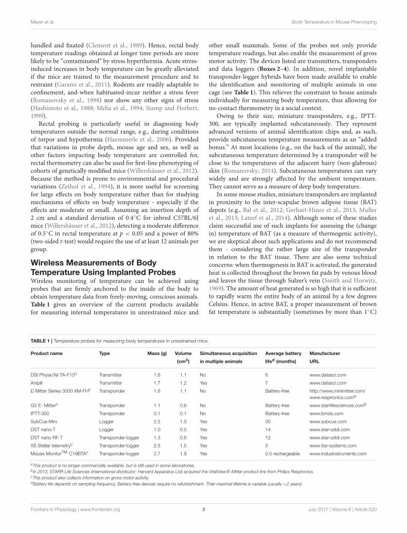
TABLE 1 | Temperature probes for measuring body temperatures in unrestrained mice.
Product name Type Mass (g) Volume Simultaneous acquisition Average battery Manufacturer
(cm3) in multiple animals lifed (months) URL
DSI PhysioTel TA-F10c Transmitter 1.6 1.1 No 6 www.datasci.com
Anipill Transmitter 1.7 1.2 Yes 7 www.datasci.com
E-Mitter Series 3000 XM-FHc Transponder 1.6 1.1 No Battery-free http://www.minimitter.com/
www.respironics.coma
G2 E- Mitterc Transponder 1.1 0.6 No Battery-free www.starrlifesciences.comb
IPTT-300 Transponder 0.1 0.1 No Battery-free www.bmds.com
SubCue Mini Logger 2.5 1.5 Yes 30 www.subcue.com
DST nano-T Logger 1.0 0.5 Yes 14 www.star-oddi.com
DST nano RF-T Transponder-logger 1.3 0.6 Yes 12 www.star-oddi.com
XS Stellar telemetryc Transponder-logger 2.5 1.5 Yes 5 www.tse-systems.com
Mouse MonitorTM C19BTAc Transponder-logger 2.7 1.9 Yes 0.5 rechargeable www.indusinstruments.com
aThis product is no longer commercially available, but is still used in some laboratories.b In 2013, STARR Life Sciences (international distributor: Harvard Apparatus Ltd) acquired the VitalView/E-Mitter product line from Philips Respironics.cThis product also collects information on gross motor activity.dBattery life depends on sampling frequency. Battery-free devices require no refurbishment. Their maximal lifetime is variable (usually >2 years).
Frontiers in Physiology | www.frontiersin.org 3 July 2017 | Volume 8 | Article 520

Meyer et al. Body Temperature in Mouse Phenotyping
BOX 2 | Transponders.
A transponder is an electronic device that can receive radiofrequency signals in response to predetermined signals. Hence, it can function as both a transmitter and
a responder. If used for animal identification purpose, its function is to send out an identifier signal when an outside signal requests identification. A transponder
consists of a coil antenna coupled to an integrated circuit chip, both covered by biocompatible glass or plastic. For temperature-measuring transponders, the chip
includes a thermistor to measure temperature and a memory unit to store temperature data. An example of a transponder, IPTT-300, is listed in Table 1. This is a
low-accuracy (0.5◦C) device used mostly in large-scale animal husbandry operations, where some rough estimate of body temperature is obtained “for free” along
with the animal’s identifier. A main advantage of transponders is the ease of their implantation (using a syringe-like injector) and their battery-free operation. Once
implanted, a transponder can provide temperature data for the entire life of the animal.
BOX 3 | Telemetry transmitters.
A transmitter is an electronic device that generates and amplifies a carrier wave, modulates it according to its temperature, and broadcasts the resulting signal from
an antenna. Signal modulation by temperature is typically achieved through changes in either the frequency (frequency modulated, FM) or the amplitude (amplitude
modulated, AM). For implantable transmitters used to measure body temperature, all the electronics involved in sensing temperature, modulating and transmitting
the signal are hidden inside a miniature biocompatible capsule. Several examples of transmitters are listed in Table 1. Their accuracy is generally greater than 0.2◦C.
For those experimental conditions where a radio signal from a transmitter implanted in an animal can be easily detected by a receiver, transmitter-based telemetry is
often a method of choice for real-time thermometry.
BOX 4 | Data loggers.
A temperature data logger is an electronic device that stores temperature signals in digital form on silicon or wax-coated memory chips integrated with a temperature
probe. The device is programmed and implanted into an animal. The temperature information can be read only after the logger is removed from the animal, which
usually happens at the end of the experiment when the animal is euthanized. The sampling frequency is set during programming, and the amount of data that can be
stored and retrieved depends on the memory size. For example, if the memory size is 2,000 data points, the logger can store the data for ∼1.4 days of recordings
at 1-min sampling, or for about 85 days of recordings at 1-h sampling. The major advantage of data loggers is that they can be used under those conditions that
do not permit communication between a transmitter and receiver, e.g., in a field experiment, or when an animal is placed in an environment impermeable to radio
waves, such as in some designs of a thermogradient apparatus (Almeida et al., 2006; Garami et al., 2011).
higher than core body temperature, as was shown in rats withthermocouples acutely implanted in the inter-scapular depotsof brown fat immediately before an experiment (Szekely et al.,1973; Szekely and Szelenyi, 1979), and in some experimentsusing chronic implantation techniques (Romanovsky et al.,1997; Ootsuka et al., 2007; Almeida et al., 2012). In chronicexperiments, it is difficult to achieve the quality of measurementsthat would allow one to accurately quantify this phenomenon. Toachieve a high-quality BAT temperature measurement, a probemust be fixed in the immediate proximity of Sulzer’s vein withspecial techniques (e.g., Almeida et al., 2012; and Figure 1).
Even when a probe is properly positioned near Sulzer’s vein,BAT temperature can be influenced by temperature changes inthe muscles underneath the BAT depots, and in the skin above.To control for these influences, at least partially, it is important tomeasure core body temperature at the same time. The differencebetween BAT and core body temperature, sometimes referredto as “BAT themogenic index,” can be used to assess BAT heatproduction (Almeida et al., 2012). Comparing the slope of anincrease in BAT temperature during the initial phase of BATthermogenesis with the slope of core body temperature is away to assess the validity of BAT temperature measurements(Mohammed et al., 2014).
Most often, the probes listed in Table 1 are implanted intothe peritoneal cavity to measure what is commonly referred toas “abdominal temperature.” Abdominal temperature is a validmeasure of deep (core) body temperature. The associated surgicalintervention involves a small midline incision through the skin,followed by an opening of the peritoneal cavity through thelinea alba. In order to prevent probe migration in the peritoneal
FIGURE 1 | Twenty four-hour brown adipose tissue (BAT) thermometry and
abdominal body temperature readings in a freely-moving C57BL/6 mouse
maintained at 26◦C. Abdominal temperature was measured using an
implanted telemetry transmitter (DSI, ETA-F10). BAT temperature was
measured using a thermistor (NTH5G10P, muRata, Kyoto, Japan) implanted
between the BAT and the underlying muscle layer in the inter-scapular region
near Sulzer’s vein, and was connected to a swivel. The resolution of readings
was set to 1 Hz. Y. Ootsuka, unpublished data.
cavity, some devices have small loops for suture-anchoring tothe abdominal wall. Alternatively, the probe is sometimes leftfree-floating in the peritoneal cavity, which increases the intra-and inter-animal variation in recorded temperature, and is notrecommended.
Un-tethered, continuous monitoring of body temperatureis more informative compared to single-point probing, e.g.,by rectal thermometry, as periodic temperature changes are
Frontiers in Physiology | www.frontiersin.org 4 July 2017 | Volume 8 | Article 520
Meyer et al. Body Temperature in Mouse Phenotyping
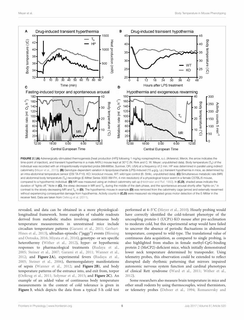
FIGURE 2 | (A) Adrenergically-stimulated thermogenesis [heat production (HP)] following 1 mg/kg norepinephrine, s.c. (Arterenol, Merck; the arrow indicates the
time-point of injection), and transient hyperthermia in a male AKR/J mouse kept at 30◦C (N. Rink and C. W. Meyer; unpublished data). Body temperature (Tb) of the
individual was recorded with an intraperitoneally-implanted probe (MiniMitter, Sunriver, OR, USA) at a frequency of 2 min. HP was determined in parallel using indirect
calorimetry (Meyer et al., 2015). (B) Genotype-dependent variation in lipopolysaccharide (LPS)-induced (15 µg/g, i.p.) transient hypothermia in mice, as determined by
an intra-abdominal temperature sensor (DSI TA-F10). KO: knockout mouse, WT: wild-type control (B. Strilic, unpublished data). (C) Simultaneous metabolic rate (MR)
and abdominal body temperature (Tb) recordings (E-Mitter Series 3000 XM-FH, 4-min resolution) of a physiological torpor event in a female C57BL/6 mouse,
compared to a hypothermic individual. (D) MR was measured using an indirect calorimetry set-up (Heldmaier and Ruf, 1992). In (C,D), shaded areas indicate the
duration of “lights off.” Note in (C), the steep decrease in MR and Tb during the middle of the dark phase, and the spontaneous arousal shortly after “lights-on,” in
contrast to the slowly-decreasing MR and Tb in (D). The hypothermic mouse in example (D) was removed from the calorimetry cage (arrow) and externally rewarmed
without experiencing consequential damage from hypothermia. Activity counts in (C,D) were measured via integrated gross motor detection of the E-Mitter in the
receiver field. Data are taken from Oelkrug et al. (2011).
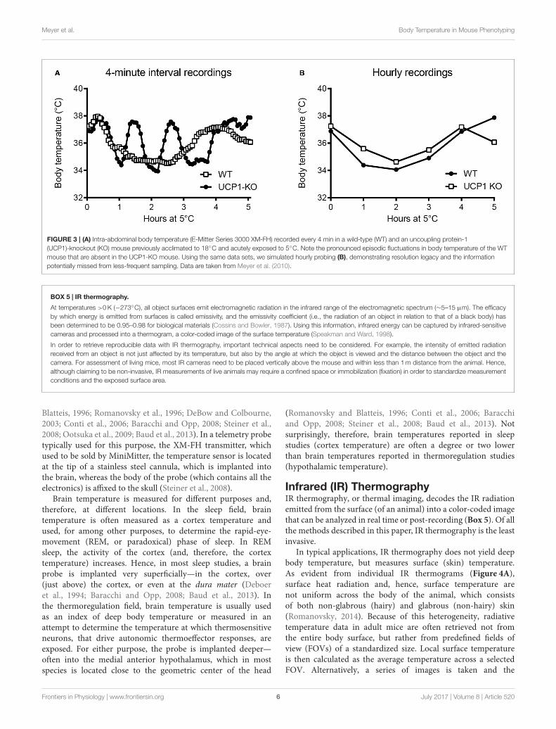
revealed, and data can be obtained in a more physiological-longitudinal framework. Some examples of valuable readoutsderived from metabolic studies involving continuous bodytemperature measurements in unrestrained mice includecircadian temperature patterns (Garami et al., 2011; Gerhart-Hines et al., 2013), ultradian-episodic (“jaggy”) events (Blessingand Ootsuka, 2016; Miyata et al., 2016), genotype- or sex-specificheterothermy (Wither et al., 2012), hyper- or hypothermicresponses to pharmacological treatments (Rudaya et al.,2005; Steiner et al., 2007; Garami et al., 2011; Wanner et al.,2012; and Figure 2A), experimental fevers (Rudaya et al.,2005; Steiner et al., 2006), thermoregulatory manifestationsof sepsis (Wanner et al., 2012; and Figure 2B), and bodytemperature patterns of the entrance into, and exit from, torpor(Oelkrug et al., 2011; Solymar et al., 2015; and Figure 2C). Anexample of an added value of continuous body temperaturemeasurements in the context of cold tolerance is given inFigure 3, which depicts the data from a typical 5-h cold test
performed at 4–5◦C (Meyer et al., 2010). Hourly probing wouldhave correctly identified the cold-tolerant phenotype of theuncoupling protein-1 (UCP1)-KO mouse after pre-acclimationto moderate cold, but this experimental setup would have failedto uncover the absence of periodic fluctuations in abdominaltemperature, compared to wild-type. The translational value ofcontinuous data acquisition, as compared to single probing, isalso highlighted from studies in female methyl-CpG-bindingprotein 2 (MeCP2)-deficient mice, which initially demonstratedlower neck temperature determined by transponder. Usingtelemetry probes, this observation could be extended to reflectdisrupted daily rhythmic patterning that mirrors impairedautonomic nervous system function and cardinal phenotypesof clinical Rett syndrome (Ward et al., 2011; Wither et al.,2012).
Some researchers also measure brain temperature in mice andother small rodents by using thermocouples, wired thermistors,or telemetry probes (Deboer et al., 1994; Romanovsky and
Frontiers in Physiology | www.frontiersin.org 5 July 2017 | Volume 8 | Article 520
Meyer et al. Body Temperature in Mouse Phenotyping
FIGURE 3 | (A) Intra-abdominal body temperature (E-Mitter Series 3000 XM-FH) recorded every 4 min in a wild-type (WT) and an uncoupling protein-1
(UCP1)-knockout (KO) mouse previously acclimated to 18◦C and acutely exposed to 5◦C. Note the pronounced episodic fluctuations in body temperature of the WT
mouse that are absent in the UCP1-KO mouse. Using the same data sets, we simulated hourly probing (B), demonstrating resolution legacy and the information
potentially missed from less-frequent sampling. Data are taken from Meyer et al. (2010).
BOX 5 | IR thermography.
At temperatures >0K (−273◦C), all object surfaces emit electromagnetic radiation in the infrared range of the electromagnetic spectrum (∼5–15 µm). The efficacy
by which energy is emitted from surfaces is called emissivity, and the emissivity coefficient (i.e., the radiation of an object in relation to that of a black body) has
been determined to be 0.95–0.98 for biological materials (Cossins and Bowler, 1987). Using this information, infrared energy can be captured by infrared-sensitive
cameras and processed into a thermogram, a color-coded image of the surface temperature (Speakman and Ward, 1998).
In order to retrieve reproducible data with IR thermography, important technical aspects need to be considered. For example, the intensity of emitted radiation
received from an object is not just affected by its temperature, but also by the angle at which the object is viewed and the distance between the object and the
camera. For assessment of living mice, most IR cameras need to be placed vertically above the mouse and within less than 1m distance from the animal. Hence,
although claiming to be non-invasive, IR measurements of live animals may require a confined space or immobilization (fixation) in order to standardize measurement
conditions and the exposed surface area.
Blatteis, 1996; Romanovsky et al., 1996; DeBow and Colbourne,2003; Conti et al., 2006; Baracchi and Opp, 2008; Steiner et al.,2008; Ootsuka et al., 2009; Baud et al., 2013). In a telemetry probetypically used for this purpose, the XM-FH transmitter, whichused to be sold by MiniMitter, the temperature sensor is locatedat the tip of a stainless steel cannula, which is implanted intothe brain, whereas the body of the probe (which contains all theelectronics) is affixed to the skull (Steiner et al., 2008).
Brain temperature is measured for different purposes and,therefore, at different locations. In the sleep field, braintemperature is often measured as a cortex temperature andused, for among other purposes, to determine the rapid-eye-movement (REM, or paradoxical) phase of sleep. In REMsleep, the activity of the cortex (and, therefore, the cortextemperature) increases. Hence, in most sleep studies, a brainprobe is implanted very superficially—in the cortex, over(just above) the cortex, or even at the dura mater (Deboeret al., 1994; Baracchi and Opp, 2008; Baud et al., 2013). Inthe thermoregulation field, brain temperature is usually usedas an index of deep body temperature or measured in anattempt to determine the temperature at which thermosensitiveneurons, that drive autonomic thermoeffector responses, areexposed. For either purpose, the probe is implanted deeper—often into the medial anterior hypothalamus, which in mostspecies is located close to the geometric center of the head
(Romanovsky and Blatteis, 1996; Conti et al., 2006; Baracchiand Opp, 2008; Steiner et al., 2008; Baud et al., 2013). Notsurprisingly, therefore, brain temperatures reported in sleepstudies (cortex temperature) are often a degree or two lowerthan brain temperatures reported in thermoregulation studies(hypothalamic temperature).
Infrared (IR) ThermographyIR thermography, or thermal imaging, decodes the IR radiationemitted from the surface (of an animal) into a color-coded imagethat can be analyzed in real time or post-recording (Box 5). Of allthe methods described in this paper, IR thermography is the leastinvasive.
In typical applications, IR thermography does not yield deepbody temperature, but measures surface (skin) temperature.As evident from individual IR thermograms (Figure 4A),surface heat radiation and, hence, surface temperature arenot uniform across the body of the animal, which consistsof both non-glabrous (hairy) and glabrous (non-hairy) skin(Romanovsky, 2014). Because of this heterogeneity, radiativetemperature data in adult mice are often retrieved not fromthe entire body surface, but rather from predefined fields ofview (FOVs) of a standardized size. Local surface temperatureis then calculated as the average temperature across a selectedFOV. Alternatively, a series of images is taken and the
Frontiers in Physiology | www.frontiersin.org 6 July 2017 | Volume 8 | Article 520
Meyer et al. Body Temperature in Mouse Phenotyping
Positions 1, 3, and 5: Trpv1-/-
Positions 2, 4, and 6: Trpv1+/+
34.0
32.0
38.0
36.0
A B
C D
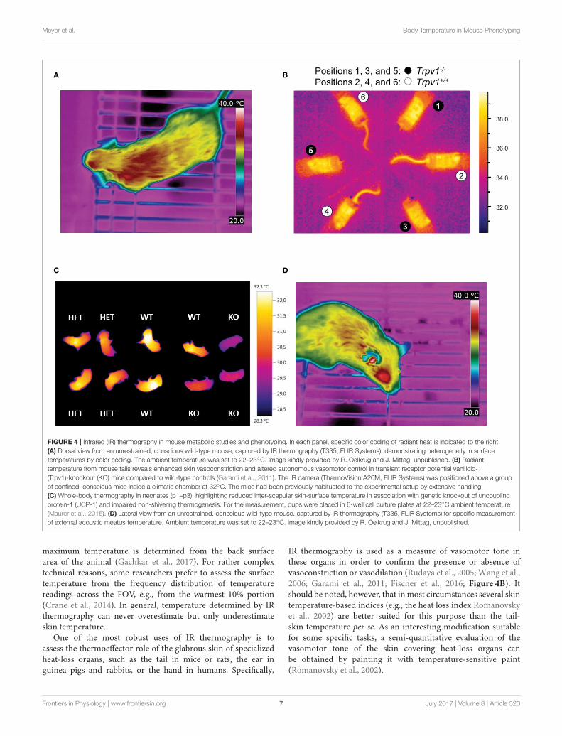
FIGURE 4 | Infrared (IR) thermography in mouse metabolic studies and phenotyping. In each panel, specific color coding of radiant heat is indicated to the right.
(A) Dorsal view from an unrestrained, conscious wild-type mouse, captured by IR thermography (T335, FLIR Systems), demonstrating heterogeneity in surface
temperatures by color coding. The ambient temperature was set to 22–23◦C. Image kindly provided by R. Oelkrug and J. Mittag, unpublished. (B) Radiant
temperature from mouse tails reveals enhanced skin vasoconstriction and altered autonomous vasomotor control in transient receptor potential vanilloid-1
(Trpv1)-knockout (KO) mice compared to wild-type controls (Garami et al., 2011). The IR camera (ThermoVision A20M, FLIR Systems) was positioned above a group
of confined, conscious mice inside a climatic chamber at 32◦C. The mice had been previously habituated to the experimental setup by extensive handling.
(C) Whole-body thermography in neonates (p1–p3), highlighting reduced inter-scapular skin-surface temperature in association with genetic knockout of uncoupling
protein-1 (UCP-1) and impaired non-shivering thermogenesis. For the measurement, pups were placed in 6-well cell culture plates at 22–23◦C ambient temperature
(Maurer et al., 2015). (D) Lateral view from an unrestrained, conscious wild-type mouse, captured by IR thermography (T335, FLIR Systems) for specific measurement
of external acoustic meatus temperature. Ambient temperature was set to 22–23◦C. Image kindly provided by R. Oelkrug and J. Mittag, unpublished.
maximum temperature is determined from the back surfacearea of the animal (Gachkar et al., 2017). For rather complextechnical reasons, some researchers prefer to assess the surfacetemperature from the frequency distribution of temperaturereadings across the FOV, e.g., from the warmest 10% portion(Crane et al., 2014). In general, temperature determined by IRthermography can never overestimate but only underestimateskin temperature.
One of the most robust uses of IR thermography is toassess the thermoeffector role of the glabrous skin of specializedheat-loss organs, such as the tail in mice or rats, the ear inguinea pigs and rabbits, or the hand in humans. Specifically,
IR thermography is used as a measure of vasomotor tone inthese organs in order to confirm the presence or absence ofvasoconstriction or vasodilation (Rudaya et al., 2005; Wang et al.,2006; Garami et al., 2011; Fischer et al., 2016; Figure 4B). Itshould be noted, however, that inmost circumstances several skintemperature-based indices (e.g., the heat loss index Romanovskyet al., 2002) are better suited for this purpose than the tail-skin temperature per se. As an interesting modification suitablefor some specific tasks, a semi-quantitative evaluation of thevasomotor tone of the skin covering heat-loss organs canbe obtained by painting it with temperature-sensitive paint(Romanovsky et al., 2002).
Frontiers in Physiology | www.frontiersin.org 7 July 2017 | Volume 8 | Article 520

Meyer et al. Body Temperature in Mouse Phenotyping
Another application of IR thermometry is to use theinformation from vasomotor tone of heat-loss organs todetermine the thermoneutral zone (TNZ; see Romanovsky et al.,2002). The authoritative sources (IUPS Thermal Commission,see Bligh and Johnson, 1973; Mercer and Werner, 2001) definethe TNZ as the range of ambient temperatures at which bodytemperature regulation is achieved only by control of sensibleheat loss, i.e., without changes in metabolic heat production orevaporative heat loss. “Sensible,” or “Newtonian,” heat loss is thetotal heat loss due to all heat exchange mechanisms, except forevaporation. In practice, the major physiological mechanism ofsensible heat loss is cutaneous vasodilation, especially in bodyparts that serve as heat exchangers with the environment, suchas the tail of a mouse. Hence, in a subneutral (cold) environment,the tails of mice exhibit constant maximal vasoconstriction (andare difficult to see in an IR thermogram as they have nearly thesame temperature as the environment). In a supraneutral (hot)environment, the tails exhibit constant maximal vasodilation(and may or may not be well-seen on thermograms, dependingon the ambient temperature). In a neutral environment (i.e.,within the TNZ), the tails constantly change their vasomotortone from mild vasoconstriction to mild vasodilation, thusbecome intermittently from almost invisible to highly visible(see Romanovsky et al., 2002, for more detailed information;see Garami et al., 2011, and Figure 4B for examples in mice).For studying thermogenic responses by IR thermography, tightcontrol of the ambient temperature is essential. In such studies,mice should be conscious, as most anesthetics decrease thethreshold body temperature for activation of cold defenses - thus,effectively inhibiting thermogenesis (Garami et al., in press).
The utility of IR thermography in assessing the vasomotorcontrol in mice tails is exemplified by the metabolic phenotypeof mice expressing a mutant thyroid hormone receptor alpha 1(TRa1+m). In these mice, impaired vasoconstriction of the tailarteries leads to increased heat loss in cold environments andpromotes hypothermia, despite elevated brown fat activity andenergy expenditure. At first sight, the hypermetabolic phenotypewas “paradoxical” (Warner andMittag, 2014), since the mutationwas expected to reduce the affinity of TRa1 to thyroid hormonesand, hence, was predicted to lower thermogenesis. Subsequentstudies of the thermoregulatory effector organs revealed thatTRa1+m mice had a greater need for adaptive thermogenesis,as their vasomotor responses were ineffective at maintainingeuthermia. These studies revealed an unexpected role of thyroidhormones in thermoregulation (Sjogren et al., 2007; Warneret al., 2013). Furthermore, these findings demonstrate thathypermetabolism is not necessarily associated with high bodytemperature.
IR thermography is also used to assess BAT thermogenesis inrodents. For this purpose, most researchers use the differencebetween radiative temperatures of the inter-scapular back-skin (covers the inter-scapular BAT depots) and the lumbarback-skin (reference point) as an index of thermogenesis in theinter-scapular BAT (e.g., Marks et al., 2009; Pazos et al., 2015).We find this approach to be well-grounded. Other researcherscapture the absolute values of, or the changes (from basal value)in, the inter-scapular skin temperature for the same purpose
(Gerhart-Hines et al., 2013; Crane et al., 2015). This latterapproach is more prone to error, but still seems to work insome cases. For example, the increase in inter-scapular skintemperature observed in wild-type mice (1.7◦C) in response tothe selective beta-3 adrenergic agonist CL-316,243 was absentin UCP1-KO mice, corroborating compromised BAT-function(Crane et al., 2014). In contrast, this approach did not allowfor the successful quantification of BAT thermogenic capacityfollowing beta-adrenergic stimulation or cold acclimation in astudy in voles (Microtus agrestis; Jackson et al., 2001).
Some researchers prefer shaving the skin in trunk FOVs,whilst others do not. Under most conditions, the shaved patchesgive higher surface temperature readings, as any insulative effectof the pelage is eliminated. In addition, shaving lessens theconfounding diffusion and reflection effects of the pelage. Ofcourse, this is not an issue in experiments with nude pups(Figure 4C) or genetically hairless animals (Chen et al., 2013;Schulz et al., 2013; Romanovsky, 2014; Maurer et al., 2015).Whether or not shaving is required depends on the IR signalintensity and the specific goal of measurements. However, wefeel that shaving should be avoided whenever possible, as itcreates “thermal windows,” whichmay substantially increase heatloss, and thus, potentially change deep body temperature andaffects the recruitment of thermoeffectors. Removing the hairalso irritates the skin and can lead to inflammation, which canaffect local temperatures. If repeatedmeasurements, over a longerperiod of time, are required - shaving needs to be repeatedfrequently, as the trunk hair re-grows relatively fast (∼3 weeksin adult mice Muller-Rover et al., 2001). This alters conductivityand may subsequently change the cold-stress responsiveness.
IR thermometry also has the potential of assessing deep bodytemperature, but not by measuring trunk-skin temperature. Therecent study by Vogel et al. (2016) proposes measuring radiativetemperature of the eyes in mice, as an index of their braintemperature. The external acoustic meatus is another “windowto the brain” that can possibly be used for assessing braintemperature of rodents by IR thermography or thermometry(Romanovsky, 2014; Hoefig et al., 2015, 2016, and Figure 4D).
CONCLUDING REMARKS
Metabolic phenotypes of genetically modified mice can helpto establish causality between a specific gene or pathway andenergy metabolism in vivo. In this context, animal temperatureis often measured to uncover thermoregulatory alterations. Here,we have summarized the main thermometry methods usedfor the purposes of metabolic phenotyping in mice, and haveinvestigated their utility. It is not only the type of sensor, butalso the experimental conditions, probe location and samplingfrequency, that determine the biological value of the results,driving successful phenotyping.
For comprehensive phenotyping involving thermometry, thethermal environment of the animals measured must be tightlycontrolled. Mice are small-sized endotherms with relativelyhigh basal metabolic costs, owing to their unfavorable surfacearea to volume ratio (Kleiber, 1961). Mouse pelage hasrelatively poor insulative capacities, making this species sensitive
Frontiers in Physiology | www.frontiersin.org 8 July 2017 | Volume 8 | Article 520
Meyer et al. Body Temperature in Mouse Phenotyping
to cold and specifically reliant on creating and exploitingthermally advantageous microenvironments for cost-efficientbody temperature regulation (Hart, 1971). Any metabolicstudy in mice, therefore, not only requires considerations ofroom temperature (gradients and variation) in the animalfacility/experimental location, but also of the “operative ambienttemperature” created by the animals inside the cage (Gordonet al., 1998). Factors known to affect individual metaboliccosts, at otherwise constant room air temperature, are thetype and amount of bedding material, nest material, and theopportunity to huddle with cagemates (Himms-Hagen andVillemure, 1992; Gordon et al., 1998; Gordon, 2004; Davidet al., 2013; Maher et al., 2015). Hence, control of any ofthese parameters potentially reduces variance in output values.Following the same reasoning, researchers should also keep inmind that any intervention or incident that disrupts the integrityof the skin/fur (e.g., shaving, surgery, alopecia) may cause specificthermoregulatory adjustments potentially contributing to extravariation in metabolic costs.
In order to provide a more complete thermoregulatoryportrait of a certain genotype, it is essential that thermometrynot be limited to measurements of body temperature understandard housing conditions. Rather, temperature readingsshould be obtained and compared at thermoneutral vs. sub-thermoneutral (cold) and supra-neutral (warm) conditions- aiming to reveal specific deficiencies in cold and heatdefenses. For the same purpose, we advocate combiningmeasurements of core body temperature with tail thermographyand back thermography (Gachkar et al., 2017). In order toreveal thermoregulatory phenotypes associated with behavioralthermoregulation, temperature readings could be combinedwith, for example, food restriction challenges (Haemmerle et al.,2006; Meyer et al., 2010), behavioral tasks, e.g., temperaturepreference chambers, thermal gradients (Gordon et al., 1998;
Bautista et al., 2007), or operant conditioning for thermal reward(Baldwin, 1968; Carlisle and Dubuc, 1982).
It is advantageous to conduct high-resolution core bodytemperature measurements in conjunction with measurementsof metabolic rate, for the assessment for heat production ratesat tightly controlled ambient temperatures. This experimentalscheme not only determines the energy expenditure andassociated body temperature variations, but also allows for theassessment of thermal conductance of the animals (Box 6)-aquantitative measure of the rate of heat exchange between theanimal’s body and the environment (McNab, 1980). In theexample shown in Figure 5, thermal conductance is comparedin the context of cold tolerance in UCP1-KO mice. Conductanceis consistently lower in cold-acclimated UCP1-KO mice, whichsupports the conclusion that cold acclimation in the absence offunctional BAT involves specific heat-conserving mechanisms,including improved tail vasoconstriction (Wang et al., 2006).
In terms of animal welfare, hyper- or hypothermic thresholdsare to be established and applied carefully, as periodic deviationsfrom “normal” temperature ranges are context-, time- andstrain-dependent, and may not unequivocally indicate anacute life-threatening condition. For example, during torpor, aphysiological state of metabolic depression (Jastroch et al., 2016),the abdominal body temperature of a mouse may transientlyreach values that are only half a degree higher than the ambienttemperature. Murine torpor commonly occurs during the earlymorning hours and can, in principle, be diagnosed by rectalprobing. Following handling, acoustic, haptic or mechanicalstimulation, a healthy mouse “alarm-arouses” and regains what isoften referred to as normal metabolism and normothermy within1–2 h, depending on the degree of metabolic depression and theambient temperature. Of note, the classification of normothermyvs. torpor is often based on metrics of heterothermy, e.g., theamplitude of daily body temperature fluctuations (see Levesque
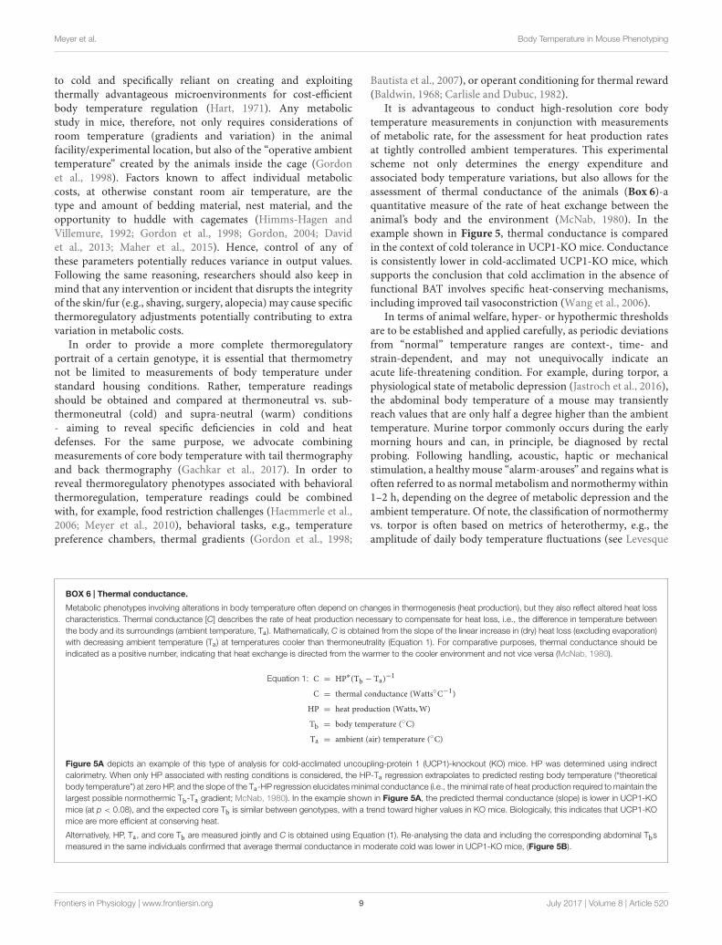
BOX 6 | Thermal conductance.
Metabolic phenotypes involving alterations in body temperature often depend on changes in thermogenesis (heat production), but they also reflect altered heat loss
characteristics. Thermal conductance [C] describes the rate of heat production necessary to compensate for heat loss, i.e., the difference in temperature between
the body and its surroundings (ambient temperature, Ta). Mathematically, C is obtained from the slope of the linear increase in (dry) heat loss (excluding evaporation)
with decreasing ambient temperature (Ta) at temperatures cooler than thermoneutrality (Equation 1). For comparative purposes, thermal conductance should be
indicated as a positive number, indicating that heat exchange is directed from the warmer to the cooler environment and not vice versa (McNab, 1980).
Equation 1: C = HP∗(Tb − Ta)−1
C = thermal conductance (Watts◦C−1)
HP = heat production (Watts,W)
Tb = body temperature (◦C)
Ta = ambient (air) temperature (◦C)
Figure 5A depicts an example of this type of analysis for cold-acclimated uncoupling-protein 1 (UCP1)-knockout (KO) mice. HP was determined using indirect
calorimetry. When only HP associated with resting conditions is considered, the HP-Ta regression extrapolates to predicted resting body temperature (“theoretical
body temperature”) at zero HP, and the slope of the Ta-HP regression elucidates minimal conductance (i.e., the minimal rate of heat production required to maintain the
largest possible normothermic Tb-Ta gradient; McNab, 1980). In the example shown in Figure 5A, the predicted thermal conductance (slope) is lower in UCP1-KO
mice (at p < 0.08), and the expected core Tb is similar between genotypes, with a trend toward higher values in KO mice. Biologically, this indicates that UCP1-KO
mice are more efficient at conserving heat.
Alternatively, HP, Ta, and core Tb are measured jointly and C is obtained using Equation (1). Re-analysing the data and including the corresponding abdominal Tbs
measured in the same individuals confirmed that average thermal conductance in moderate cold was lower in UCP1-KO mice, (Figure 5B).
Frontiers in Physiology | www.frontiersin.org 9 July 2017 | Volume 8 | Article 520
Meyer et al. Body Temperature in Mouse Phenotyping
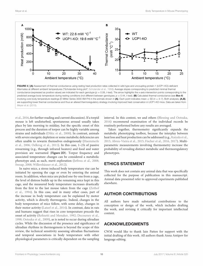
FIGURE 5 | (A) Assessment of thermal conductance using resting heat production rates collected in wild-type and uncoupling-protein-1 (UCP1-KO)-deficient
littermates at different ambient temperatures (“Scholander-Irving plot”; Scholander et al., 1950). Average slopes corresponding to predicted minimal thermal
conductance (expressed as positive values) are indicated for each genotype (p = 0.08, t-test). The arrow highlights the x-axis intersection points corresponding to the
predicted average body temperature during resting conditions (not different between genotypes; p = 0.44, t-test). (B) Calculated thermal conductance (see Box 6)
involving core body temperature readings (E-Mitter Series 3000 XM-FH) in the animals shown in (A). Each point indicates mean ± SD (n = 4–7). Both analyses, (A,B),
are supporting lower thermal conductance and thus an altered thermoregulatory strategy involving improved heat conservation in UCP1-KO mice. Data are taken from
Meyer et al. (2010).
et al., 2016, for further reading and current discussion). If a torpidmouse is left undisturbed, spontaneous arousal usually takesplace by late morning to midday, but the specific onset of thisprocess and the duration of torpor can be highly variable amongstrains and individuals (Dikic et al., 2008). In contrast, animalswith severe energetic depletion or somemetabolic deficiencies areoften unable to rewarm themselves endogenously (Haemmerleet al., 2006; Oelkrug et al., 2011). In this case, 1–2 h of passiverewarming (e.g., through infrared heaters) and food and waterprovision are warranted (Figure 2D). Torpor frequency andassociated temperature changes can be considered a metabolicphenotype and, as such, merit exploration (Jethwa et al., 2008;Swoap, 2008; Willershäuser et al., 2012).
In naive mice, a stress-induced body temperature rise can beinitiated by opening the cage or even by entering the animalroom. In addition, whenmice are picked one-by-one from a cage,the level of distress builds up in the remaining mice kept in thiscage, and the measured body temperature increases drasticallyfrom the first to the last mouse taken from the cage (Zethofet al., 1994). In this case, and in many other cases, part ofthe increase in body temperature can be explained by motoractivity, which is directly thermogenic. Indeed, changes in thebody temperature of mice follow, with some delay, changes intheir motor activity (Lateef et al., 2014). In contrast, data in ratsand humans suggest that rises in core temperature precede theonset of activity (Refinetti and Menaker, 1992; Decoursey et al.,1998; Ootsuka et al., 2009), as is noted to occur during ultradiancycles. While the discussion of the presence and significance ofultradian rhythms in thermogenesis is beyond the scope of thisreview, the technical sensitivity assessing ultradian fluctuationsand temporal associations in body temperature with otherphysiological parameters is critically dependent on the sampling
interval. In this context, we and others (Blessing and Ootsuka,2016) recommend examination of the individual records beroutinely performed before any results are averaged.
Taken together, thermometry significantly expands themetabolic phenotyping toolbox, because the interplay betweenheat loss and heat production can be addressed (e.g., Kaiyala et al.,2015; Abreu-Vieira et al., 2015; Fischer et al., 2016, 2017). Multi-parametric measurements involving thermometry increase theprobability of revealing distinct metabolic and thermoregulatoryphenotypes.
ETHICS STATEMENT
This work does not contain any animal data that was specificallycollected for the purpose of publication in this manuscript.Animal data presented refer to approved experiments publishedelsewhere.
AUTHOR CONTRIBUTIONS
All authors have made substantial contributions to theconception or design of the work, which includes draftingthe work, and revising it critically for important intellectualcontent.
ACKNOWLEDGMENTS
CWM would like to thank Iain Patten for support with theinitial drafting of this work. All authors thank Anna Antipov forlanguage editing.
Frontiers in Physiology | www.frontiersin.org 10 July 2017 | Volume 8 | Article 520
Meyer et al. Body Temperature in Mouse Phenotyping
REFERENCES
Abreu-Vieira, G., Xiao, C., Gavrilova, O., and Reitman, M. L. (2015). Integration
of body temperature into the analysis of energy expenditure in the mouse.Mol.
Metab. 4, 461–470. doi: 10.1016/j.molmet.2015.03.001
Almeida, M. C., Hew-Butler, T., Soriano, R. N., Rao, S., Wang, W., Wang, J.,
et al. (2012). Pharmacological blockade of the cold receptor TRPM8 attenuates
autonomic and behavioral cold defenses and decreases deep body temperature.
J. Neurosci. 32, 2086–2099. doi: 10.1523/JNEUROSCI.5606-11.2012
Almeida, M. C., Steiner, A. A., Branco, L. G., and Romanovsky, A. A. (2006). Cold-
seeking behavior as a thermoregulatory strategy in systemic inflammation. Eur.
J. Neurosci. 23, 3359–3367. doi: 10.1111/j.1460-9568.2006.04854.x
Bal, N. C., Maurya, S. K., Sopariwala, D. H., Sahoo, S. K., Gupta, S. C., Shaikh,
S. A., et al. (2012). Sarcolipin is a newly identified regulator of muscle-
based thermogenesis in mammals. Nat. Med. 18, 1575–1579. doi: 10.1038/
nm.2897
Baldwin, B. A. (1968). Behavioural thermoregulation in mice. Physiol. Behav. 3,
401–407. doi: 10.1016/0031-9384(68)90069-3
Baracchi, F., and Opp, M. R. (2008). Sleep-wake behavior and responses to
sleep deprivation of mice lacking both interleukin-1 beta receptor 1 and
tumor necrosis factor-alpha receptor 1. Brain Behav. Immun. 22, 982–993.
doi: 10.1016/j.bbi.2008.02.001
Baud,M.O.,Magistretti, P. J., and Petit, J.M. (2013). Sustained sleep fragmentation
affects brain temperature, food intake and glucose tolerance in mice. J. Sleep
Res. 22, 3–12. doi: 10.1111/j.1365-2869.2012.01029.x
Bautista, D. M., Siemens, J., Glazer, J. M., Tsuruda, P. R., Basbaum, A. I., Stucky,
C. L., et al. (2007). The menthol receptor TRPM8 is the principal detector of
environmental cold. Nature 448, 204–208. doi: 10.1038/nature05910
Blessing, W., and Ootsuka, Y. (2016). Timing of activities of daily life is
jaggy: how episodic ultradian changes in body and brain temperature
are integrated into this process. Temperature (Austin) 3, 371–383.
doi: 10.1080/23328940.2016.1177159
Bligh, J., and Johnson, K. G. (1973). Glossary of terms for thermal physiology. J.
Appl. Physiol. 36, 941–961.
Carlisle, H. J., and Dubuc, P. U. (1982). Unchanged thermoregulatory set-point in
the obese mouse. Nature 297, 678–679. doi: 10.1038/297678a0
Chen, Y., Siegel, F., Kipschull, S., Haas, B., Frohlich, H., Meister, G., et al. (2013).
miR-155 regulates differentiation of brown and beige adipocytes via a bistable
circuit. Nat. Commun. 4:1769. doi: 10.1038/ncomms2742
Clement, J. G., Mills, P., and Brockway, B. (1989). “Use of telemetry to record
body temperature and activity in mice.” J. Pharmacol. Methods 21, 129–140.
doi: 10.1016/0160-5402(89)90031-4
Conti, B., Sanchez-Alavez, M., Winsky-Sommerer, R., Morale, M. C., Lucero,
J., Brownell, S., et al. (2006). Transgenic mice with a reduced core
body temperature have an increased life span. Science 314, 825–828.
doi: 10.1126/science.1132191
Cossins, A. R., and Bowler, K. (1987). Temperature Biology of Animals. London;
New York, NY: Chapman and Hall.
Crane, J. D., Mottillo, E. P., Farncombe, T. H., Morrison, K. M., and Steinberg,
G. R. (2014). A standardized infrared imaging technique that specifically
detects UCP1-mediated thermogenesis in vivo. Mol. Metab. 3, 490–494.
doi: 10.1016/j.molmet.2014.04.007
Crane, J. D., Palanivel, R., Mottillo, E. P., Bujak, A. L., Wang, H., Ford, R. J., et al.
(2015). Inhibiting peripheral serotonin synthesis reduces obesity and metabolic
dysfunction by promoting brown adipose tissue thermogenesis. Nat. Med. 21,
166–172. doi: 10.1038/nm.3766
David, J. M., Knowles, S., Lamkin, D. M., and Stout, D. B. (2013). Individually
ventilated cages impose cold stress on laboratory mice: a source of systemic
experimental variability. J. Am. Assoc. Lab. Anim. Sci. 52, 738–744.
Deboer, T., Franken, P., and Tobler, I. (1994). Sleep and cortical temperature in
the Djungarian hamster under baseline conditions and after sleep deprivation.
J. Com. Physiol. A 174, 145–155. doi: 10.1007/bf00193782
DeBow, S., and Colbourne, F. (2003). Brain temperature measurement and
regulation in awake and freely moving rodents. Methods 30, 167–171.
doi: 10.1016/s1046-2023(03)00080-x
Decoursey, P. J., Pius, S., Sandlin, C.,Wethey, D., and Schull, J. (1998). Relationship
of circadian temperature and activity rhythms in two rodent species. Physiol.
Behav. 65, 457–463. doi: 10.1016/S0031-9384(98)00187-5
Dikic, D., Heldmaier, G., and Meyer, C. W. (2008). “Induced torpor in different
strains of laboratory mice,” inHypometabolism in Animals: Torpor, Hibernation
and Cryobiology, eds B. G. Lovegrove and A. E. McKechnie (Pietermaritzburg:
University of KwaZulu-Natal), 223–230.
Donhoffer, S. (1980). Homeothermia of the Brain: Cerebral Blood Flow, Metabolic
Rate, and Brain Temperature in the Cold: the Possible Role of Neuroglia.
Budapest, Akadémiai Kiadó.
Fischer, A. W., Hoefig, C. S., Abreu-Vieira, G., de Jong, J. M., Petrovic, N., Mittag,
J., et al. (2016). Leptin raises defended body temperature without activating
thermogenesis. Cell Rep. 14, 621–1631. doi: 10.1016/j.celrep.2016.01.041
Fischer, K., Ruiz, H. H., Jhun, K., Finan, B., Oberlin, D. J., van der Heide, V., et al.
(2017). Alternatively activated macrophages do not synthesize catecholamines
or contribute to adipose tissue adaptive thermogenesis. Nat. Med. 23, 623–630.
doi: 10.1038/nm.4316
Gachkar, S., Oelkrug, R., Martinez-Sanchez, N., Rial-Pensado, E., Warner, A.,
Hoefig, C. S., et al. (2017). 3-Iodothyronamine induces tail vasodilation
through central action in male mice. Endocrinology 158, 1977–1984.
doi: 10.1210/en.2016-1951
Garami, A., Ibrahim, M., Gilbrait, K., Khanna, R., Pakai, E., Miko, A., et al.
(in press). TRPV1 antagonists prevent anesthesia-induced hypothermia and
decrease post-incisional opioid dose requirements in rodents. Anesthesiology.
Garami, A., Pakai, E., Oliveira, D. L., Steiner, A. A., Wanner, S. P., Almeida, M.
C., et al. (2011). Thermoregulatory phenotype of the Trpv1 knockout mouse:
thermoeffector dysbalance with hyperkinesis. J. Neurosci. 31, 1721–1733.
doi: 10.1523/JNEUROSCI.4671-10.2011
Gerhart-Hines, Z., Feng, D., Emmett, M. J., Everett, L. J., Loro, E., Briggs, E. R.,
et al. (2013). The nuclear receptor Rev-erbalpha controls circadian thermogenic
plasticity. Nature 503, 410–413. doi: 10.1038/nature12642
Gordon, C. J. (1993). Temperature Regulation in Laboratory Rodents. Cambridge:
Cambridge University Press.
Gordon, C. J. (2004). Effect of cage bedding on temperature regulation and
metabolism of group-housed female mice. Comp. Med. 54, 63–68.
Gordon, C. J., Becker, P., and Ali, J. S. (1998). Behavioral thermoregulatory
responses of single- and group-housed mice. Physiol. Behav. 65, 255–262.
doi: 10.1016/S0031-9384(98)00148-6
Haemmerle, G., Lass, A., Zimmermann, R., Gorkiewicz, G., Meyer, C., Rozman, J.,
et al. (2006). Defective lipolysis and altered energy metabolism in mice lacking
adipose triglyceride lipase. Science 312, 734–737. doi: 10.1126/science.1123965
Hart, J. S. (1971). “Rodents,” in Comparative Physiology of Thermoregulation, Vol.
2, eds G. C. Whittow (New York, NY; London: Academic Press), 1–149.
Hashimoto, K., Suemaru, S., Takao, T., Sugawara, M., Makino, S.,
and Ota, Z. (1988). Corticotropin-releasing hormone and pituitary-
adrenocortical responses in chronically stressed rats. Regul. Pept. 23, 17–126.
doi: 10.1016/0167-0115(88)90019-5
Heldmaier, G., and Ruf, T. (1992). Body temperature and metabolic rate during
natural hypothermia in endotherms. J. Comp. Physiol. B. Biochem. Syst.
Environ. Physiol. 162, 696–706. doi: 10.1007/BF00301619
Himms-Hagen, J., and Villemure, C. (1992). Number of mice per cage influences
uncoupling protein content of brown adipose tissue. Proc. Soc. Exp. Biol. Med.
200, 502–506. doi: 10.3181/00379727-200-43461
Hoefig, C. S., Harder, L., Oelkrug, R., Meusel, M., Vennstrom, B., Brabant, G.,
et al. (2016). Thermoregulatory and cardiovascular consequences of a transient
Thyrotoxicosis and recovery in male mice. Endocrinology 157, 2957–2967.
doi: 10.1210/en.2016-1095
Hoefig, C. S., Jacobi, S. F., Warner, A., Harder, L., Schanze, N., Vennstrom, B.,
et al. (2015). 3-Iodothyroacetic acid lacks thermoregulatory and cardiovascular
effects in vivo. Br. J. Pharmacol. 172, 3426–3433. doi: 10.1111/bph.13131
Jackson, D. M., Hambly, C., Trayhurn, P., and Speakman, J. R. (2001).
Can non-shivering thermogenesis in brown adipose tissue following NA
injection be quantified by changes in overlying surface temperatures using
infrared thermography? J. Therm. Biol. 26, 85–93. doi: 10.1016/S0306-4565(00)
00023-1
Jastroch, M., Giroud, S., Barrett, P., Geiser, F., Heldmaier, G., and Herwig, A.
(2016). Seasonal control of mammalian energy balance: recent advances in
the understanding of daily torpor and hibernation. J. Neuroendocrinol. 28.
doi: 10.1111/jne.12437
Jethwa, P. H., I’Anson, H., Warner, A., Prosser, H. M., Hastings, M. H., Maywood,
E. S., et al. (2008). Loss of prokineticin receptor 2 signaling predisposes mice
Frontiers in Physiology | www.frontiersin.org 11 July 2017 | Volume 8 | Article 520

Meyer et al. Body Temperature in Mouse Phenotyping
to torpor. Am. J. Physiol. Regul. Integr. Comp. Physiol. 294, R1968–R1979.
doi: 10.1152/ajpregu.00778.2007
Kaiyala, K. J., Ogimoto, K., Nelson, J. T., Schwartz, M. W., and Morton, G. J.
(2015). Leptin signaling is required for adaptive changes in food intake, but
not energy expenditure, in response to different thermal conditions. PLoS ONE
10:e0119391. doi: 10.1371/journal.pone.0119391
Kleiber, M. (1961). The Fire of Life. New York, NY; London: John Wiley and Sons
Inc.
Lateef, D. M., Abreu-Vieira, G., Xiao, C., and Reitman, M. L. (2014). Regulation
of body temperature and brown adipose tissue thermogenesis by bombesin
receptor subtype-3. Am. J. Physiol. Endocrinol. Metab. 306, E681–E687.
doi: 10.1152/ajpendo.00615.2013
Levesque, D., Nowack, J., and Stawski, C. (2016). Modelling mammalian
energetics: the heterothermy problem. Clim. Change Respon. 3:7.
doi: 10.1186/s40665-016-0022-3
Maher, R. L., Barbash, S. M., Lynch, D. V., and Swoap, S. J. (2015). Group housing
and nest building only slightly ameliorate the cold stress of typical housing
in in female C57BL/6J mice. Am. J. Physiol. Regul. Integr. Comp. Physiol. 308,
R1070–R1079. doi: 10.1152/ajpregu.00407.2014
Marks, A., Vianna, D. M., and Carrive, P. (2009). Nonshivering thermogenesis
without interscapular brown adipose tissue involvement during conditioned
fear in the rat. Am. J. Physiol. Regul. Integr. Comp. Physiol. 296, R1239–R1247.
doi: 10.1152/ajpregu.90723.2008
Maurer, S. F., Fromme, T., Grossman, L. I., Huttemann, M., and Klingenspor,
M. (2015). The brown and brite adipocyte marker Cox7a1 is not required for
non-shivering thermogenesis in mice. Sci. Rep. 5:17704. doi: 10.1038/srep17704
McNab, B. K. (1980). On estimating thermal conductance in endotherms. Physiol.
Zool. 53, 145–156. doi: 10.1086/physzool.53.2.30152577
Melia, K. R., Ryabinin, A. E., Schroeder, R., Bloom, F. E., andWilson, M. C. (1994).
Induction and habituation of immediate early gene expression in rat brain by
acute and repeated restraint stress. J. Neurosci. 14, 5929–5938.
Mercer, J. G., andWerner, J. (2001). Glossary of terms for thermal physiology. Jpn.
J. Physiol. 51, 245–280.
Meyer, C. W., Reitmeir, P., and Tschop, M. H. (2015). Exploration of Energy
Metabolism in the Mouse Using Indirect Calorimetry: measurement
of daily energy expenditure (DEE) and basal metabolic rate (BMR).
Curr. Protoc. Mouse Biol. 5, 205–222. doi: 10.1002/9780470942390.
mo140216
Meyer, C. W., Willershauser, M., Jastroch, M., Rourke, B. C., Fromme, T., Oelkrug,
R., et al. (2010). Adaptive thermogenesis and thermal conductance in wild-
type and UCP1-KO mice. Am. J. Physiol. Regul. Integr. Comp. Physiol. 299,
R1396–R1406. doi: 10.1152/ajpregu.00021.2009
Miyata, K., Kuwaki, T., and Ootsuka, Y. (2016). The integrated ultradian
organization of behavior and physiology in mice and the contribution
of orexin to the ultradian patterning. Neuroscience 334, 119–133.
doi: 10.1016/j.neuroscience.2016.07.041
Mohammed, M., Ootsuka, Y., Yanagisawa, M., and Blessing, W. (2014). Reduced
brown adipose tissue thermogenesis during environmental interactions in
transgenic rats with ataxin-3-mediated ablation of hypothalamic orexin
neurons. Am. J. Physiol. Regul. Integr. Comp. Physiol. 307, R978–R989.
doi: 10.1152/ajpregu.00260.2014
Moir, L., Bentley, L., and Cox, R. D. (2016). Comprehensive energy
balance measurements in mice. Curr. Protoc. Mouse Biol. 6, 211–222.
doi: 10.1002/cpmo.13
Muller, T. D., Lee, S. J., Jastroch, M., Kabra, D., Stemmer, K., Aichler, M.,
et al. (2013). p62 links beta-adrenergic input to mitochondrial function and
thermogenesis. J. Clin. Invest. 123, 469–478. doi: 10.1172/JCI64209
Muller-Rover, S., Handjiski, B., van der Veen, C., Eichmuller, S., Foitzik, K., McKay,
I. A., et al. (2001). A comprehensive guide for the accurate classification of
murine hair follicles in distinct hair cycle stages. J. Invest. Dermatol. 117, 3–15.
doi: 10.1046/j.0022-202x.2001.01377.x
Oelkrug, R., Heldmaier, G., and Meyer, C. W. (2011). Torpor patterns, arousal
rates, and temporal organization of torpor entry in wildtype and UCP1-
ablated mice. J. Comp. Physiol. B. Biochem. Syst. Environ. Physiol. 181, 137–145.
doi: 10.1007/s00360-010-0503-9
Ootsuka, Y., de Menezes, R. C., Zaretsky, D. V., Alimoradian, A., Hunt,
J., Stefanidis, A., et al. (2009). Brown adipose tissue thermogenesis heats
brain and body as part of the brain-coordinated ultradian basic rest-
activity cycle. Neuroscience 164, 849–861. doi: 10.1016/j.neuroscience.2009.
08.013
Ootsuka, Y., Heidbreder, C. A., Hagan, J. J., and Blessing, W. W. (2007).
Dopamine D2 receptor stimulation inhibits cold-initiated thermogenesis
in brown adipose tissue in conscious rats. Neuroscience 147, 127–135.
doi: 10.1016/j.neuroscience.2007.04.015
Pazos, P., Lima, L., Tovar, S., Gonzalez-Touceda, D., Dieguez, C., and Garcia, M. C.
(2015). Divergent responses to thermogenic stimuli in BAT and subcutaneous
adipose tissue from interleukin 18 and interleukin 18 receptor 1-deficient mice.
Sci. Rep. 5:17977. doi: 10.1038/srep17977
Refinetti, R., and Menaker, M. (1992). The circadian rhythm of body temperature.
Physiol. Behav. 51, 613–637. doi: 10.1016/0031-9384(92)90188-8
Romanovsky, A. (2014). Skin temperature: its role in thermoregulation. Acta
Physiol. 210, 498–507. doi: 10.1111/apha.12231
Romanovsky, A. A., and Blatteis, C. M. (1996). Heat stroke: opioid-mediated
mechanisms. J. Appl. Physiol. 81, 2565–2570.
Romanovsky, A. A., Ivanov, A. I., and Shimansky, Y. P. (2002). Selected
contribution: ambient temperature for experiments in rats: a new method for
determining the zone of thermal neutrality. J. Appl. Physiol. 92, 2667–2679.
doi: 10.1152/japplphysiol.01173.2001
Romanovsky, A. A., Kulchitsky, V. A., Akulich, N. V., Koulchitsky, S. V., Simons,
C. T., Sessler, D. I., et al. (1996). First and second phases of biphasic fever:
two sequential stages of the sickness syndrome? Am. J. Physiol. 271(1 Pt 2),
R244–R253.
Romanovsky, A. A., Simons, C. T., and Kulchitsky, V. A. (1998). Biphasic fevers
often consist of more than two phases. Am. J. Physiol. 275(1 Pt 2), R323–R331.
Romanovsky, A. A., Simons, C. T., Szekely, M., and Kulchitsky, V. A. (1997). The
vagus nerve in the thermoregulatory response to systemic inflammation. Am. J.
Physiol. 273(1 Pt 2), R407–R413.
Rozman, J., Klingenspor, M., and Hrabe de Angelis, M. (2014). A review of
standardized metabolic phenotyping of animal models. Mamm. Genome 25,
497–507. doi: 10.1007/s00335-014-9532-0
Rudaya, A. Y., Steiner, A. A., Robbins, J. R., Dragic, A. S., and Romanovsky, A.
A. (2005). Thermoregulatory responses to lipopolysaccharide in the mouse:
dependence on the dose and ambient temperature. Am. J. Physiol. Regul. Integr.
Comp. Physiol. 289, R1244–R1252. doi: 10.1152/ajpregu.00370.2005
Scholander, P. F., Hock, R., Walters, V., and Irving, L. (1950). Adaptation
to cold in arctic and tropical mammals and birds in relation to body
temperature insulation, and basal metabolic rate. Biol. Bull. 99, 259–271.
doi: 10.2307/1538742
Schulz, T. J., Huang, P., Huang, T. L., Xue, R., McDougall, L. E., Townsend,
K. L., et al. (2013). Brown-fat paucity due to impaired BMP signalling
induces compensatory browning of white fat. Nature 495, 379–383.
doi: 10.1038/nature11943
Sjogren, M., Alkemade, A., Mittag, J., Nordstrom, K., Katz, A., Rozell, B.,
et al. (2007). Hypermetabolism in mice caused by the central action of
an unliganded thyroid hormone receptor alpha1. EMBO J. 26, 4535–4545.
doi: 10.1038/sj.emboj.7601882
Smith, R. E., and Horwitz, B. A. (1969). Brown fat and thermogenesis. Physiol. Rev.
49, 330–425.
Solymar, M., Petervari, E., Balasko, M., and Szelenyi, Z. (2015). The onset
of daily torpor is regulated by the same low body mass in lean mice
and in mice with diet-induced obesity. Temperature (Austin) 2, 129–134.
doi: 10.1080/23328940.2015.1014250
Speakman, J. R. (2013). Measuring energy metabolism in the mouse -
theoretical, practical, and analytical considerations. Front. Physiol. 4:34.
doi: 10.3389/fphys.2013.00034
Speakman, J. R., and Ward, S. (1998). Infrared thermography: principles and
applications. Zool. Anal. Complex Systems 101, 224–232.
Stamp, J. A., and Herbert, J. (1999). Multiple immediate-early gene expression
during physiological and endocrine adaptation to repeated stress. Neuroscience
94, 1313–1322. doi: 10.1016/S0306-4522(99)00368-1
Steiner, A. A., Chakravarty, S., Rudaya, A. Y., Herkenham, M., and
Romanovsky, A. A. (2006). Bacterial lipopolysaccharide fever is initiated
via Toll-like receptor 4 on hematopoietic cells. Blood 107, 4000–4002.
doi: 10.1182/blood-2005-11-4743
Frontiers in Physiology | www.frontiersin.org 12 July 2017 | Volume 8 | Article 520
Meyer et al. Body Temperature in Mouse Phenotyping
Steiner, A. A., Oliveira, D. L., Roberts, J. L., Petersen, S. R., and Romanovsky,
A. A. (2008). Nicotine administration and withdrawal affect survival
in systemic inflammation models. J. Appl. Physiol. 105, 1028–1034.
doi: 10.1152/japplphysiol.90619.2008
Steiner, A. A., Turek, V. F., Almeida, M. C., Burmeister, J. J., Oliveira,
D. L., Roberts, J. L., et al. (2007). Nonthermal activation of transient
receptor potential vanilloid-1 channels in abdominal viscera tonically
inhibits autonomic cold-defense effectors. J. Neurosci. 27, 7459–7468.
doi: 10.1523/JNEUROSCI.1483-07.2007
Swoap, S. J. (2008). The pharmacology and molecular mechanisms underlying
temperature regulation and torpor. Biochem. Pharmacol. 76, 817–824.
doi: 10.1016/j.bcp.2008.06.017
Szekely, M., and Szelenyi, Z. (1979). Endotoxin fever in the rat. Acta Physiol. Acad.
Sci. Hung. 53, 265–277.
Szekely, M., Szelenyi, Z., and Sumegi, I. (1973). Brown adipose tissue as a source
of heat during pyrogen-induced fever. Acta Physiol. Acad. Sci. Hung. 43,
85–88.
Tschöp, M. H., Speakman, J. R., Arch, J. R., Auwerx, J., Bruning, J. C., Chan, L.,
et al. (2012). A guide to analysis of mouse energy metabolism. Nat. Methods 9,
57–63. doi: 10.1038/nmeth.1806
Vogel, B., Wagner, H., Gmoser, J., Worner, A., Loschberger, A., Peters,
L., et al. (2016). Touch-free measurement of body temperature using
close-up thermography of the ocular surface. MethodsX 3, 407–416.
doi: 10.1016/j.mex.2016.05.002
Wang, Y., Kimura, K., Inokuma, K., Saito, M., Kontani, Y., Kobayashi, Y., et al.
(2006). Potential contribution of vasoconstriction to suppression of heat loss
and homeothermic regulation in UCP1-deficient mice. Pflugers Arch. 452,
363–369. doi: 10.1007/s00424-005-0036-3
Wanner, S. P., Garami, A., Pakai, E., Oliveira, D. L., Gavva, N. R., Coimbra, C.
C., et al. (2012). Aging reverses the role of the transient receptor potential
vanilloid-1 channel in systemic inflammation from anti-inflammatory to
proinflammatory. Cell Cycle 11, 343–349. doi: 10.4161/cc.11.2.18772
Ward, C. S., Arvide, E. M., Huang, T. W., Yoo, J., Noebels, J. L., and Neul, J.
L. (2011). MeCP2 is critical within HoxB1-derived tissues of mice for normal
lifespan. J. Neurosci. 31, 10359–10370. doi: 10.1523/JNEUROSCI.0057-11.2011
Warner, A., and Mittag, J. (2014). Brown fat and vascular heat dissipation: the new
cautionary tail. Adipocyte 3, 221–223. doi: 10.4161/adip.28815
Warner, A., Rahman, A., Solsjo, P., Gottschling, K., Davis, B., Vennstrom, B., et al.
(2013). Inappropriate heat dissipation ignites brown fat thermogenesis in mice
with a mutant thyroid hormone receptor alpha1. Proc. Natl. Acad. Sci. U.S.A.
110, 16241–16246. doi: 10.1073/pnas.1310300110
Willershäuser, M., Ehrhardt, N., Elvert, R., Wirth, E. K., Schweizer, U., Gailus-
Durner, V., et al. (2012). “Systematic screening for mutant mouse lines
with defects in body temperature regulation,” in Living in a Seasonal World:
Thermoregulatory and Metabolic Adaptations, eds T. Ruf, C. Bieber, W. Arnold,
and E. Millesi (Berlin, Heidelberg: Springer-Verlag), 459–469.
Wither, R. G., Colic, S., Wu, C., Bardakjian, B. L., Zhang, L., and Eubanks,
J. H. (2012). Daily rhythmic behaviors and thermoregulatory patterns are
disrupted in adult female MeCP2-deficient mice. PLoS ONE 7:e35396.
doi: 10.1371/journal.pone.0035396
Zethof, T. J., Van der Heyden, J. A., Tolboom, J. T., and Olivier, B. (1994). Stress-
induced hyperthermia in mice: a methodological study. Physiol. Behav. 55,
109–115. doi: 10.1016/0031-9384(94)90017-5
Conflict of Interest Statement: The authors declare that the research was
conducted in the absence of any commercial or financial relationships that could
be construed as a potential conflict of interest.
Copyright © 2017 Meyer, Ootsuka and Romanovsky. This is an open-access article
distributed under the terms of the Creative Commons Attribution License (CC BY).
The use, distribution or reproduction in other forums is permitted, provided the
original author(s) or licensor are credited and that the original publication in this
journal is cited, in accordance with accepted academic practice. No use, distribution
or reproduction is permitted which does not comply with these terms.
Frontiers in Physiology | www.frontiersin.org 13 July 2017 | Volume 8 | Article 520