brain, 141: 688-697 hot-spot kif5a mutations cause
TRANSCRIPT
http://www.diva-portal.org
This is the published version of a paper published in Brain.
Citation for the original published paper (version of record):
Brenner, D., Yilmaz, R., Müller, K., Grehl, T., Petri, S. et al. (2018)Hot-spot KIF5A mutations cause familial ALSBrain, 141: 688-697https://doi.org/10.1093/brain/awx370
Access to the published version may require subscription.
N.B. When citing this work, cite the original published paper.
Permanent link to this version:http://urn.kb.se/resolve?urn=urn:nbn:se:umu:diva-146237
Hot-spot KIF5A mutations cause familial ALS
David Brenner,1 Rustem Yilmaz,1 Kathrin Muller,1 Torsten Grehl,2 Susanne Petri,3
Thomas Meyer,4 Julian Grosskreutz,5 Patrick Weydt,1,6 Wolfgang Ruf,1
Christoph Neuwirth,7 Markus Weber,7 Susana Pinto,8,9 Kristl G. Claeys,10,11,12,13
Berthold Schrank,14 Berit Jordan,15 Antje Knehr,1 Kornelia Gunther,1 Annemarie Hubers,1
Daniel Zeller,16 The German ALS network MND-NET,* Christian Kubisch,17,18
Sibylle Jablonka,19 Michael Sendtner,19 Thomas Klopstock,20,21,22 Mamede de Carvalho,8,23
Anne Sperfeld,15 Guntram Borck,17 Alexander E. Volk,17,18 Johannes Dorst,1
Joachim Weis,10 Markus Otto,1 Joachim Schuster,1 Kelly Del Tredici,1 Heiko Braak,1
Karin M. Danzer,1 Axel Freischmidt,1 Thomas Meitinger,24,25 Tim M. Strom,24,25
Albert C. Ludolph,1 Peter M. Andersen1,9 and Jochen H. Weishaupt1
*Appendix 1.
Heterozygous missense mutations in the N-terminal motor or coiled-coil domains of the kinesin family member 5A (KIF5A) gene
cause monogenic spastic paraplegia (HSP10) and Charcot-Marie-Tooth disease type 2 (CMT2). Moreover, heterozygous de novo
frame-shift mutations in the C-terminal domain of KIF5A are associated with neonatal intractable myoclonus, a neurodevelop-
mental syndrome. These findings, together with the observation that many of the disease genes associated with amyotrophic lateral
sclerosis disrupt cytoskeletal function and intracellular transport, led us to hypothesize that mutations in KIF5A are also a cause of
amyotrophic lateral sclerosis. Using whole exome sequencing followed by rare variant analysis of 426 patients with familial
amyotrophic lateral sclerosis and 6137 control subjects, we detected an enrichment of KIF5A splice-site mutations in amyotrophic
lateral sclerosis (2/426 compared to 0/6137 in controls; P = 4.2 � 10�3), both located in a hot-spot in the C-terminus of the
protein and predicted to affect splicing exon 27. We additionally show co-segregation with amyotrophic lateral sclerosis of two
canonical splice-site mutations in two families. Investigation of lymphoblast cell lines from patients with KIF5A splice-site muta-
tions revealed the loss of mutant RNA expression and suggested haploinsufficiency as the most probable underlying molecular
mechanism. Furthermore, mRNA sequencing of a rare non-synonymous missense mutation (predicting p.Arg1007Gly) located in
the C-terminus of the protein shortly upstream of the splice donor of exon 27 revealed defective KIF5A pre-mRNA splicing in
respective patient-derived cell lines owing to abrogation of the donor site. Finally, the non-synonymous single nucleotide variant
rs113247976 (minor allele frequency = 1.00% in controls, n = 6137), also located in the C-terminal region [p.(Pro986Leu) in exon
26], was significantly enriched in familial amyotrophic lateral sclerosis patients (minor allele frequency = 3.40%; P = 1.28 � 10�7).
Our study demonstrates that mutations located specifically in a C-terminal hotspot of KIF5A can cause a classical amyotrophic
lateral sclerosis phenotype, and underline the involvement of intracellular transport processes in amyotrophic lateral sclerosis
pathogenesis.
1 Neurology Department, Ulm University, Ulm, Germany2 Department of Neurology, Alfried Krupp Hospital, Essen, Germany3 Department of Neurology, Hannover Medical School, Hannover, Germany4 Charite University Hospital, Humboldt-University, Berlin, Germany5 Department of Neurology, Jena University Hospital, Jena, Germany6 Department for Neurodegenerative Disorders and Gerontopsychiatry, Bonn University, Bonn, Germany7 Kantonsspital St. Gallen, ALS Outpatient Clinic, St. Gallen, Switzerland
doi:10.1093/brain/awx370 BRAIN 2018: 141; 688–697 | 688
Received November 24, 2017. Revised December 19, 2017. Accepted December 20, 2017. Advance Access publication January 12, 2018
� The Author(s) (2018). Published by Oxford University Press on behalf of the Guarantors of Brain.
This is an Open Access article distributed under the terms of the Creative Commons Attribution Non-Commercial License (http://creativecommons.org/licenses/by-nc/4.0/), which permits
non-commercial re-use, distribution, and reproduction in any medium, provided the original work is properly cited. For commercial re-use, please contact [email protected]
Downloaded from https://academic.oup.com/brain/article-abstract/141/3/688/4797069by Umea University Library useron 09 April 2018

8 Department of Neurosciences and Mental Health, Hospital de Santa Maria-CHLN, Lisbon, Portugal9 Department of Pharmacology and Clinical Neuroscience, Umea University, Umea, Sweden
10 Institute of Neuropathology, RWTH Aachen University Hospital, Aachen, Germany11 Department of Neurology, RWTH Aachen University Hospital, Aachen, Germany12 Department of Neurology, University Hospitals Leuven, Leuven, Belgium13 Laboratory for Muscle Diseases and Neuropathies, Department of Neurosciences, Experimental Neurology, KU Leuven -
University of Leuven, Leuven, Belgium14 Department of Neurology, DKD HELIOS Klinik Wiesbaden, Wiesbaden, Germany15 Department of Neurology Martin-Luther-University Halle-Wittenberg, Halle/Saale, Germany16 Department of Neurology, University of Wurzburg, Wurzburg, Germany17 Institute of Human Genetics, Ulm University, Ulm, Germany18 Institute of Human Genetics, University Medical Center Hamburg-Eppendorf, Hamburg, Germany19 Institute of Clinical Neurobiology, University Hospital of Wurzburg, Wurzburg, Germany20 Department of Neurology with Friedrich-Baur-Institute, University of Munich, Munich, Germany21 German Center for Neurodegenerative Diseases (DZNE), Munich, Germany22 Munich Cluster for Systems Neurology (SyNergy), Munich, Germany23 Instituto de Medicina Molecular and Institute of Physiology, Faculty of Medicine, University of Lisbon, Portugal24 SyNergy, Munich Cluster for Systems Neurology, Ludwig Maximilians Universitat Munchen, Germany25 Institute of Human Genetics, Technische Universitat Munchen, Munchen, Germany
Correspondence to: Jochen H. Weishaupt, MD
Neurology Department
Ulm University
Albert-Einstein-Allee 11, 89081 Ulm
Germany
E-mail: [email protected]
Keywords: ALS; KIF5A mutations; axonal transport; whole exome sequencing
Abbreviations: ALS = amyotrophic lateral sclerosis; CMT = Charcot-Marie-Tooth disease; HSP = hereditary spastic paraplegia;MAF = minor allele frequency; SNV = single nucleotide variant
IntroductionKIF5A is a member of the kinesin family of proteins that is
mainly expressed in neurons (Niclas et al., 1994; Fagerberg
et al., 2014). As part of a multi-subunit complex, it acts as
a microtubule motor in intracellular protein and organelle
transport, including mitochondria (Hirokawa et al. 2009).
Missense mutations in the kinesin family member 5A
(KIF5A) gene at 12q13.3 are the third most frequent
cause of autosomal dominant hereditary spastic paraplegia
(HSP10, SPG10, OMIM#604187) in European popula-
tions, affecting primarily the long pyramidal tracts and
sometimes also the peripheral nervous system (Reid et al.,
2002; Goizet et al., 2009; Morais et al., 2017).
Additionally, the known phenotypic spectrum of KIF5A
mutations comprises also an autosomal dominant axonal
sensorimotor peripheral neuropathy (Charcot-Marie-Tooth
disease type 2; CMT2) (Liu et al., 2014) and a complex
infantile neurological syndrome with leukencephalopathy,
myoclonus, hypotonia, optic nerve abnormalities, dyspha-
gia, apnoea, hearing loss, and early developmental arrest
[neonatal intractable myoclonus (NEIMY; OMIM#
617235); Duis et al., 2016; Rydzanicz et al., 2017].
Some 5% of patients with the motor neuron disease
amyotrophic lateral sclerosis (ALS) self-report a positive
family history (familial ALS), most frequently as a
Mendelian autosomal dominant trait. Since 1993, muta-
tions in over 36 genes have been associated with ALS
pathogenesis, and mutations in several of these have been
predicted to disrupt cytoskeletal function and intracellular
transport (PFN1, NEFH, PRPH, ALSIN, DCTN1;
TUBA4A Figlewicz et al., 1994; Yang et al., 2001; Puls
et al., 2003; Gros-Louis et al., 2004; Wu et al., 2012;
Smith et al., 2014). Datasets of two large studies based
on whole exome sequencing or genome-wide association
testing suggested also an association between variants in
KIF5A and ALS (Kenna et al., 2016; McLaughlin et al.,
2017). In both studies, the association did not achieve
genome-wide statistical significance and the studies also
lacked data on a possible co-segregation of KIF5A variants
with ALS. Furthermore, detailed clinical information
beyond the phenotype ‘ALS’ was not available with
regard to the possibly KIF5A-linked patients. Altogether,
we hypothesized that mutations affecting KIF5A can also
be a cause of ALS. Consequently, we here assessed a pos-
sible association between KIF5A and ALS by first compar-
ing the mutation burden of KIF5A in a cohort of 426
familial ALS patients with 144 769 control individuals (com-
prising 6137 in-house controls and the gnomAD dataset)
followed by co-segregation analysis, detailed clinical descrip-
tion of the patients, as well as RNA expression and splicing
analysis of KIF5A mutations in patient-derived cell lines.
KIF5A mutations in familial ALS BRAIN 2018: 141; 688–697 | 689
Downloaded from https://academic.oup.com/brain/article-abstract/141/3/688/4797069by Umea University Library useron 09 April 2018

Material and methods
Patients and ethics statement
All ALS patients were diagnosed according to the EFNS
Consensus criteria (Andersen et al., 2005, 2012). With in-
formed written consent and approval by the national med-
ical ethical review boards in accordance with the
Declaration of Helsinki, EDTA blood samples were
drawn from controls, ALS patients, and their unaffected
relatives. DNA was extracted from EDTA blood samples
according to standard procedures.
Genotyping for SOD1 and C9orf72mutations
Mutations in SOD1 and C9orf72 were excluded prior to
exome sequencing of familial ALS cases as described before
(Freischmidt et al., 2015).
Whole-exome sequencing
We sequenced exomes of 426 European familial ALS index
patients and 6137 control subjects. Controls comprised
healthy parents of children with various diseases, healthy
tissues of individuals with tumour diseases, and 200 indi-
viduals from the KORA studies (Kooperative
Gesundheitsforschung in der Region Augsburg) (Herder
et al., 2013). Sequencing, read mapping and variant calling
was performed on HiSeq2000/2500 systems (Illumina) as
described previously (Freischmidt et al., 2015).
RNA expression and splicing analysis
RNA was isolated from the immortalized lymphoblast cell
lines derived from the mutation carriers and their unaffected
mutation-negative relatives. To test the effect of the variants
on mRNA level, fragments were amplified using the cDNA
template with primers binding to exon 25 and 28/29. Primer
sequences are available on request. PCR products were
sequenced on ABI 3130xl Genetic Analyzer using BigDye
v3.1 cycle sequencing kit (Life Technologies), according to
the manufacturer’s protocol. Mutation nomenclature is ac-
cording to the transcript NM_004984.
To calculate the relative expression of KIF5A mRNA in
the subjects, quantitative real-time PCR was performed
with SYBR� Green chemistry. Primers spanning exons
2/3 and exon 3/4 were used. TBP (TATA-binding protein)
was used as an internal control for normalization. The
��ct method was used for quantification (Livak and
Schmittgen, 2001).
Statistics
Fisher’s exact test was used to compare sequence variant fre-
quencies between ALS and control groups. A significance level
a5 0.05 was applied in all tests statistical tests (two-tailed).
For linkage analysis of Families A–C, we assumed an
autosomal dominant model. Penetrance was set at 0.8.
The frequency of the deleterious allele was set at 0.0001,
the phenocopy rate at 0.003, and the marker allele fre-
quency to 0.0001. Linkage analysis was performed using
Merlin software (version 1.1.2). We set the phenotype to
unknown if an unaffected individual was either younger
than 60 years or had died before the age of 60 years.
Results
Exome sequencing and associationanalysis
To assess a possible association between KIF5A variants and
familial ALS we analysed whole exome sequence data of 426
familial ALS index patients. The frequency of KIF5A variants
was compared to 6137 in-house whole exome datasets from
control individuals with non-neurological diseases and the
138 632 exomes and genomes of the gnomAD dataset
(http://gnomad.broadinstitute.org/, Lek et al., 2016). The fa-
milial ALS index patients were selected for whole exome
sequencing from families with at least two individuals af-
fected by ALS or frontotemporal dementia from the six
European countries: Germany, Denmark, Finland, Sweden,
Switzerland, and Portugal, subsequent to a negative screen
for pathogenic mutations in SOD1 and C9orf72.
We analysed missense variants of KIF5A at a minor allele
frequency (MAF) below the thresholds of 1% and 0.1%,
respectively. We did not observe a significant overall en-
richment of KIF5A rare missense variants in the familial
ALS group when compared to our in-house control group
(n = 6137) or the gnomAD dataset (n = 138 632; Tables 1
and 2).
Although we did not detect a significant enrichment of
rare missense variants in the patient group, the non-syn-
onymous single nucleotide variant (SNV) rs113247976
showed a trend towards enrichment in ALS patients in a
previous GWAS (McLaughlin et al., 2017). We found this
SNV highly enriched in our familial ALS cohort [29/426
patients; allele frequency (AF) = 3.4%] compared to in-
house controls (123/6137 individuals; AF = 1.00%;
P = 1.28 � 10�7) or the gnomAD database (3132/138 632
individuals; AF = 1.13%; P = 3.11 � 10�7). This SNV
predicts an amino acid exchange in KIF5A [hg19:
g.57,975,700C4T; c.2957C4T; p.(Pro986Leu)] (Table 2).
Rs113247976 represents the only non-synonymous variant
with a frequency 40.1% in the normal population (gnomAD
dataset and in-house controls); thus the rest of KIF5A displays
high evolutionary conservation. Remarkably, 11 of 29 pa-
tients carrying rs113247976 also had a heterozygous genetic
variant in one of the following ALS genes (Supplementary
Table 2): HNRNPA1 (p.M137V), VCP (p.R95C), ERBB4
(p.T271I), TARDBP (p.A315T and p.N352S each in one
patient), FUS (p.G405R and p.R514T each in one case),
690 | BRAIN 2018: 141; 688–697 D. Brenner et al.
Downloaded from https://academic.oup.com/brain/article-abstract/141/3/688/4797069by Umea University Library useron 09 April 2018

FIG4 (p.R699H), SPG11 [c.7152-1G4C (acceptor splice
site) and p.V2426M, each in one case; only bi-allelic muta-
tions regarded to be pathogenic], as well as NEK1
(p.Ser1036Ter) and UBQLN2 (p.P509S) in the same
individual.
Furthermore, we separately analysed loss-of-function vari-
ants, defined as nonsense, canonical splice sites (within two
nucleotides of exon boundary), read-through, and frameshift
variants. We identified a significant enrichment of KIF5A loss-
of-function variants in the patient group (2/426 patients;
AF = 0.23%) compared to the in-house control group (0/
6137 individuals; AF = 0%; P = 4.2 � 10�3) or the gnomAD
dataset (13/138 632 individuals; AF = 4.7 � 10�3; P =
9.6 � 10�4) (Tables 1 and 2). Consistent with the low
abundance of loss-of-function variants in control datasets,
the pLI score of KIF5A is 1 (http://exac.broadinstitute.org/
gene/ENSG00000155980), which indicates a high probability
of KIF5A loss-of-function mutation intolerance. Mutations in
established ALS disease genes were not detected in the patients
with KIF5A loss-of-function mutation.
Segregation analysis
Next, we aimed to further corroborate the observed asso-
ciation of deleterious mutations with ALS by segregation
analysis. We extended the two families with loss-of-func-
tion mutations and reanalysed them using Sanger sequen-
cing. As shown in Fig. 1A and B, both splice site variants
Table 1 KIF5A splice site and rare missense variants (MAF 51%) found in this study (426 index patients) and basic
clinical characteristics of index patients
Varianta Predicted
consequence at
protein level
MAF, % Onset Age at
onset,
Disease
duration,
Phenotype
c: NM_004984.2: Allele count years months
g: NC_000012.11 (gnomAD)
Missense variants
c.1238A4G p.Glu413Gly 4.062 � 10�6 Spinal, right UL both MN 35 28 Classical ALS
g.57968879A4G (1/246 210) LMN4UMN
c.1422A4T p.Gln474His 4,065 � 10�6 Spinal, left LL both MN 68 41 Classical ALS
plus FTDg.57965903A4T (1/246 010)
c.1729A4G p.Ser577Gly 4.062 � 10�6 Bulbar 35 60 ALS
g.57968879A4G (1/246 210)
c.3019A4G p.(Arg1007Gly)b 0 Spinal, LMN right UL 53 45 Classical ALS
g.57976411A4G LMN4UMN
Splice site
c.2993-1G4Ag.57976384G4A
c 4.061 � 10�6 Spinal, UMN left LL 56 436 (alive) Classical ALS
(1/246 246) UMN = LMN
c.3020+2T4C p.(Asn999Valfs*39) 0 Spinal, LMN left UL 29 34 Classical ALS
g.57976414T4C UMN4 LMN
c.3020+1G4Ad p.(Asn999Valfs*39) 0 n.a. n.a. n.a. n.a.
g.57976413G4A
aGenomic positions according to the GRCh37/hg19.bPredicted missense mutation, experimentally shown to abrogate function of splice donor site in intron 27 resulting in the predicted change p.Asn999Valfs*39 (see ‘Results’ section).cSplice acceptor site predicted to be abrogated, resulting protein change unpredictable.dSplice site variant found in the familial ALS exome data of the ALS Variant Server (AVS) (http://als.umassmed.edu/), no clinical information available.
LL = lower limb; LMN = lower motor neuron; n.a. = not available; UMN = upper motor neuron; UL = upper limb.
Table 2 KIF5A variants with the respective allele frequencies in case and control whole exome datasets
Familial
ALS
In-house
exomes (AF)
P-value (Fisher’s
exact test)
gnomAD
dataset (AF)
P-value (Fisher’s
exact test)
n 426 6137 - 138 632 -
Loss-of-function 2 0 4.2 � 10�3 13 9.6 � 10�4
(0.23%) (0%) (4.7 � 10�3%)
Missense 5 88 n.s. 1596 n.s.
(MAF4 1%) (0.59%) (0.72%) (0.58%)
Missense 4 n.a n.a. 714 n.s.
(MAF4 0.01%) (0.47%) (0.26%)
SNV rs113247976 29 123 1.28 � 10�7 3132 3.11 � 10�7
[p.(Pro986Leu)] (3.40%) (1.00%) (1.13%)
n.a. = not applicable; n.s. = not significant.
KIF5A mutations in familial ALS BRAIN 2018: 141; 688–697 | 691
Downloaded from https://academic.oup.com/brain/article-abstract/141/3/688/4797069by Umea University Library useron 09 April 2018
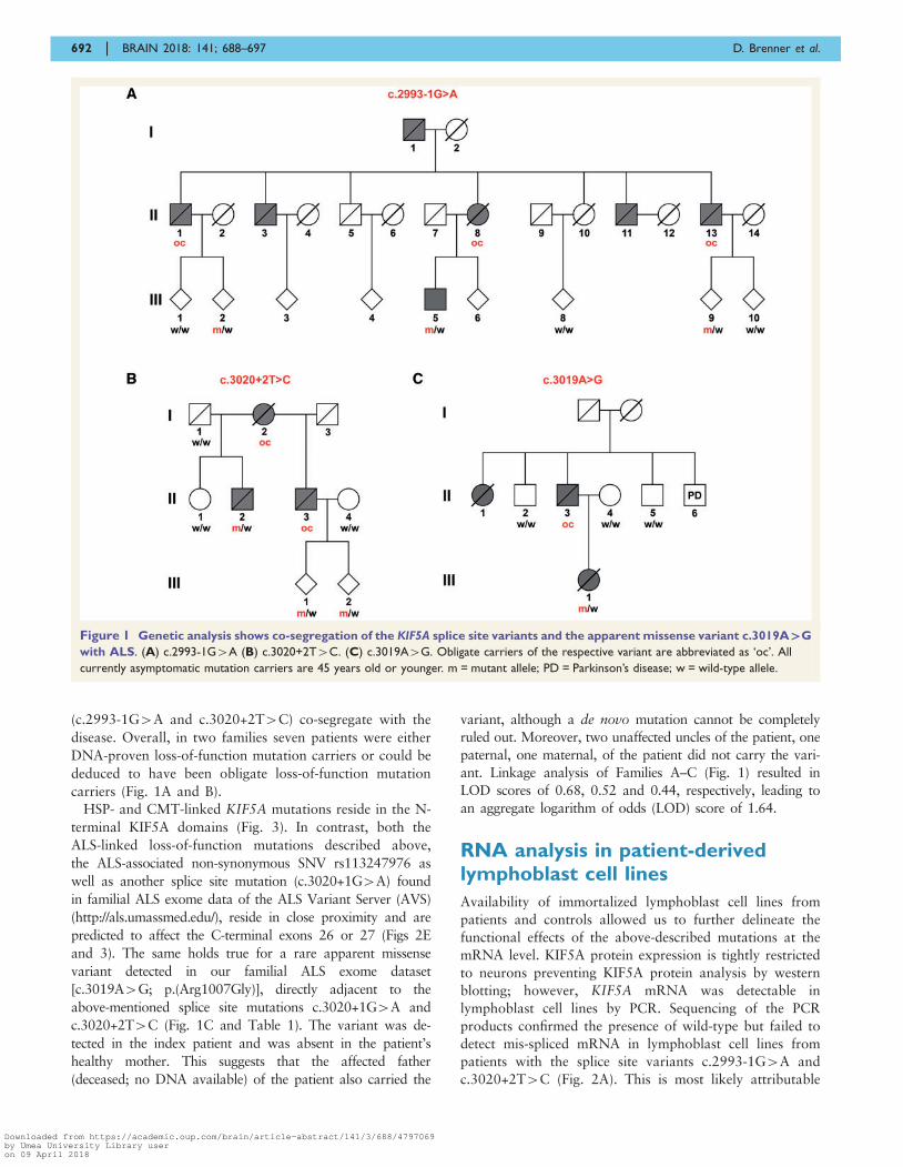
(c.2993-1G4A and c.3020+2T4C) co-segregate with the
disease. Overall, in two families seven patients were either
DNA-proven loss-of-function mutation carriers or could be
deduced to have been obligate loss-of-function mutation
carriers (Fig. 1A and B).
HSP- and CMT-linked KIF5A mutations reside in the N-
terminal KIF5A domains (Fig. 3). In contrast, both the
ALS-linked loss-of-function mutations described above,
the ALS-associated non-synonymous SNV rs113247976 as
well as another splice site mutation (c.3020+1G4A) found
in familial ALS exome data of the ALS Variant Server (AVS)
(http://als.umassmed.edu/), reside in close proximity and are
predicted to affect the C-terminal exons 26 or 27 (Figs 2E
and 3). The same holds true for a rare apparent missense
variant detected in our familial ALS exome dataset
[c.3019A4G; p.(Arg1007Gly)], directly adjacent to the
above-mentioned splice site mutations c.3020+1G4A and
c.3020+2T4C (Fig. 1C and Table 1). The variant was de-
tected in the index patient and was absent in the patient’s
healthy mother. This suggests that the affected father
(deceased; no DNA available) of the patient also carried the
variant, although a de novo mutation cannot be completely
ruled out. Moreover, two unaffected uncles of the patient, one
paternal, one maternal, of the patient did not carry the vari-
ant. Linkage analysis of Families A–C (Fig. 1) resulted in
LOD scores of 0.68, 0.52 and 0.44, respectively, leading to
an aggregate logarithm of odds (LOD) score of 1.64.
RNA analysis in patient-derivedlymphoblast cell lines
Availability of immortalized lymphoblast cell lines from
patients and controls allowed us to further delineate the
functional effects of the above-described mutations at the
mRNA level. KIF5A protein expression is tightly restricted
to neurons preventing KIF5A protein analysis by western
blotting; however, KIF5A mRNA was detectable in
lymphoblast cell lines by PCR. Sequencing of the PCR
products confirmed the presence of wild-type but failed to
detect mis-spliced mRNA in lymphoblast cell lines from
patients with the splice site variants c.2993-1G4A and
c.3020+2T4C (Fig. 2A). This is most likely attributable
Figure 1 Genetic analysis shows co-segregation of the KIF5A splice site variants and the apparent missense variant c.3019A`G
with ALS. (A) c.2993-1G4A (B) c.3020+2T4C. (C) c.3019A4G. Obligate carriers of the respective variant are abbreviated as ‘oc’. All
currently asymptomatic mutation carriers are 45 years old or younger. m = mutant allele; PD = Parkinson’s disease; w = wild-type allele.
692 | BRAIN 2018: 141; 688–697 D. Brenner et al.
Downloaded from https://academic.oup.com/brain/article-abstract/141/3/688/4797069by Umea University Library useron 09 April 2018
to nonsense-mediated decay (NMD) of the RNA tran-
scribed from the mutant alleles. To confirm a loss of ex-
pression of the splice site mutant alleles, we tried to
quantify the total KIF5A mRNA levels in c.2993-1G4A
and c.3020+2T4C splice site mutant and wild-type
lymphoblast cell lines. However, possibly as a result of
generally low KIF5A expression in non-neuronal cells, the
results were too variable for quantification. Interestingly,
the above-mentioned C-terminal rare variant, which theor-
etically leads to a single amino acid exchange in exon 27
[c.3019A4G; p.(Arg1007Gly)], deviates from the expected
effect at the mRNA level: Bioinformatic splice site analysis
(Berkeley Drosophila Genome Project; Celniker et al.,
2002; http://www.fruitfly.org/seq_tools/splice.html) already
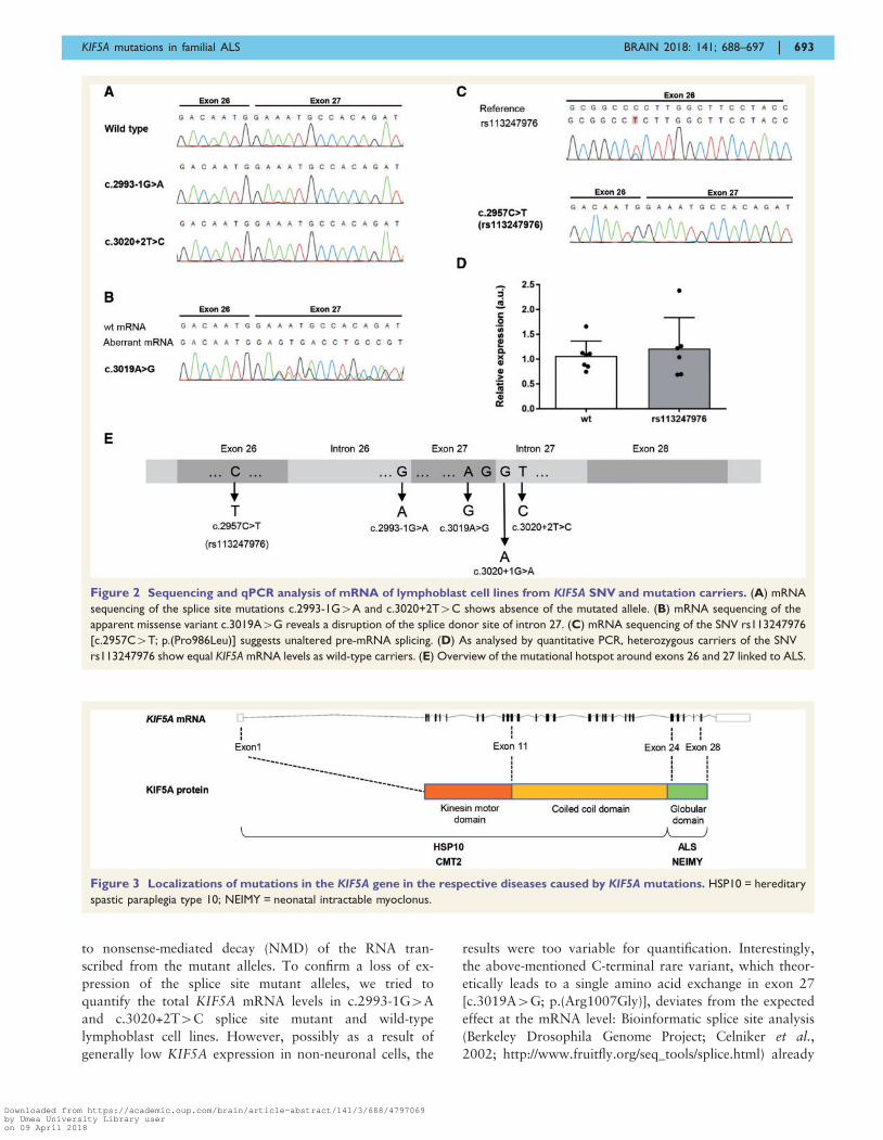
Figure 2 Sequencing and qPCR analysis of mRNA of lymphoblast cell lines from KIF5A SNV and mutation carriers. (A) mRNA
sequencing of the splice site mutations c.2993-1G4A and c.3020+2T4C shows absence of the mutated allele. (B) mRNA sequencing of the
apparent missense variant c.3019A4G reveals a disruption of the splice donor site of intron 27. (C) mRNA sequencing of the SNV rs113247976
[c.2957C4T; p.(Pro986Leu)] suggests unaltered pre-mRNA splicing. (D) As analysed by quantitative PCR, heterozygous carriers of the SNV
rs113247976 show equal KIF5A mRNA levels as wild-type carriers. (E) Overview of the mutational hotspot around exons 26 and 27 linked to ALS.
Figure 3 Localizations of mutations in the KIF5A gene in the respective diseases caused by KIF5A mutations. HSP10 = hereditary
spastic paraplegia type 10; NEIMY = neonatal intractable myoclonus.
KIF5A mutations in familial ALS BRAIN 2018: 141; 688–697 | 693
Downloaded from https://academic.oup.com/brain/article-abstract/141/3/688/4797069by Umea University Library useron 09 April 2018

predicts a decrease of the splice site score from 0.79 for the
wild-type splice donor site in intron 27 to 0.16 upon the
c.3019A4G change. In agreement with this prediction,
sequencing of the PCR product from the respective lympho-
blast cell line shows that this variant, separated only by one
nucleotide from the 50 end of intron 27, disrupts the splice
donor site of intron 27 (Fig. 2B). This results in a skipping
of exon 27 and the predicted truncating change
p.Asn999Valfs*39 at the protein level. Thus, this variant,
one of the ALS-linked canonical splice site mutations from
this study (c.3020+2T4C) and the splice site mutation
(c.3020+1G4A) found in familial ALS exome data of the
AVS, are all expected or experimentally proven to disrupt
the splice donor site of intron 27. However, contrary to the
canonical splice site mutations, mis-spliced mRNA resulting
from the c.3019A4G [p.(Arg1007Gly)] missense mutation
is detectable in the lymphoblast cell lines from the respect-
ive patient (data not shown). We speculate that this result
may be explained by a differential stability of the respective
pre-mRNAs before or during the splicing event.
In our analysis of lymphoblast cell line RNA, the above-
described SNV rs113247976 [p.(Pro986Leu)] did not have
an impact on splicing of introns 26 or 27, and the respect-
ive mRNA level was unaltered compared to wild-type
lymphoblast cell lines (Fig. 2C and D). Together with our
data and the splice site mutation derived from the AVS, we
identified four different mutations predicted or experimen-
tally shown to affect splicing of introns 26 or 27 of KIF5A
in familial ALS (Fig. 2E).
Clinical phenotypes of KIF5A muta-tion carriers
The phenotypes of the patients with a canonical KIF5A
splice site mutation or with the c.3019A4G predicted mis-
sense variant that led to abrogation of the intron 27 splice
donor site were compatible with a classical ALS syndrome:
Specifically, adult-onset of initial focal asymmetric onset of
affection of both the upper and lower motor neuron sys-
tems with later generalization, bulbar motor involvement,
emotional lability, absence of vegetative symptoms and sen-
sory symptoms in most patients, as well as rapid disease
progression and early death—all of which were fully com-
patible with a typical ALS phenotype rather than HSP or
CMT (Table 1 and Supplementary Table 1)
Patients carrying the SNV rs113247976 [c.2957C4T;
p.(Pro986Leu)] found to be enriched in familial ALS
tended to have a (non-significantly) shorter disease duration
and a significantly decreased proportion of FTD co-morbid-
ity compared to the background cohort of German patients
with familial ALS (Supplementary Table 3). However, the
number of and clinical information on SNV carriers
rs113247976 needs to be extended before it can be defin-
itely ascertained whether rs113247976 is modifying the dis-
ease phenotype.
Finally, we screened nine patients with sporadic ALS, from
whom post-mortem brain tissue was available, for the SNV
rs113247976. One of nine patients carried this SNV.
Neuropathological examination revealed typical stage 2 TDP-
43 neuropathology (Braak et al., 2013, 2017; Brettschneider
et al., 2014; Tan et al., 2013) (Supplementary Fig. 1).
DiscussionThe present data, supported by previous suggestive findings
(Kenna et al., 2016; McLaughlin et al., 2017), indicate that
KIF5A is a novel ALS gene. In our study, the association of
rare KIF5A variants with ALS was predominantly carried
by loss-of-function mutations, in particular the two splice
site mutations found among the studied 426 ALS families.
Notably, in the familial ALS group of the AVS, another
KIF5A splice variant is reported in a familial ALS patient
(c.3020+1G4A), affecting the same splice donor site as the
c.3020+2T4C mutation found in our cohort.
Corroborating the link to ALS, both of the splice site mu-
tations (c.2993-1G4A and c.3020+2T4C) we identified co-
segregated with the disease. The splice site mutations were
present in all seven patients from whom DNA or indirect
information about their mutation status (obligate carriers)
was available. Furthermore, no healthy individual older
than the expected age of disease onset and carrying the mu-
tation was identified in the two families studied. This infor-
mation, although in its present form limited, is suggestive of a
high penetrance of KIF5A loss-of-function mutations.
In contrast to loss-of-function mutations, we found no
significant association between familial ALS and rare mis-
sense variants (MAF51%). Nevertheless, it is well pos-
sible that at least a subset of missense variants is
pathogenic. KIF5A has a z-score of 4.38 (http://exac.broad-
institute.org/gene/ENSG00000155980), i.e. fewer missense
variants than expected, which indicates a general intoler-
ance towards variation in this gene. Remarkably, RNA
analysis of patient-derived cell lines revealed that one of
the rare (predicted) missense variants (c.3019A4G; pre-
dicting p.Arg1007Gly) turned out to abrogate the splice
donor site in intron 27, the same donor site that is affected
by two of the above mentioned canonical splice site muta-
tions (c.3020+1G4A and c.3020+2T4C).
The KIF5A mutations previously described in HSP or
CMT2 patients are restricted to missense mutations in the
kinesin motor domain (amino acid positions 9–327) and in
the alpha-helical coiled-coil domain (amino acid positions
331–906) (summarized in Kaji et al., 2016; Guinto et al.,
2017). By contrast, the ALS-associated mutations described
here (Fig. 2E) are predicted to affect the C-terminal part of
KIF5A or to represent loss-of-function mutations, as experi-
mentally shown for two of them (overview in Fig. 3).
Moreover, deletion mutations in the same C-terminal
domain that are predicted to cause a frame-shift with
stop loss and an elongated protein (c.2854delC,
c.2921delC, c.2934delC), have been associated with the
694 | BRAIN 2018: 141; 688–697 D. Brenner et al.
Downloaded from https://academic.oup.com/brain/article-abstract/141/3/688/4797069by Umea University Library useron 09 April 2018

severe developmental syndrome neonatal intractable myo-
clonus (NEIMY) (Duis et al., 2016; Rydzanicz et al., 2017).
Given our and previously reported data, it is thus possible
to link the N-terminal and the C-terminal mutational hot-
spots in KIF5A to HSP and to CMT or to ALS and
NEIMY (Fig. 3). The C-terminal globular tail has been
demonstrated to be necessary for the binding of cargo-
adaptor proteins (e.g. TRAK1/2, GABARAP) (Nakajima
et al., 2012; Randall et al., 2013). Mutations predicted to
affect the C-terminal tail, if translated into protein, might
therefore lead to altered binding of cargo to KIF5A, al-
though this awaits experimental confirmation. By contrast,
mutations in the N-terminal kinesin motor domain linked
to HSP and CMT decrease the velocity and flux of cargo
transport (Wang et al., 2010). This could explain why mis-
sense mutations in the N-terminal kinesin motor or the
central coiled-coil domain cause the milder HSP/CMT2
phenotype, whereas mutations in the C-terminal small globular
domain induce the more severe ALS and NEIMY syndromes,
possibly owing to altered cargo binding, haploinsufficiency or a
dominant-negative effect of truncated KIF5A.
In this study, we only included ALS patients who had
been diagnosed by experienced ALS clinical specialists
adhering to stringent ALS criteria (Andersen et al., 2005,
2012). This is important, not the least because two previ-
ous reports suggested a possible link between ALS and
KIF5A, but lacked precise clinical information (Kenna
et al., 2016; McLaughlin et al., 2017). Considering the
relative phenotypic similarities between ALS, HSP, and
CMT, it cannot be excluded that part of the association
signal in the previous large studies arose from HSP or
CMT patients misdiagnosed as (slowly progressive) ALS.
The patients in our study showed a classical ALS pheno-
type. Nevertheless, overlapping syndromes may exist and
could complicate the distinction between ALS, CMT and
HSP in some instances. Along this line, it remained unclear
if the pronounced sensory symptoms of Patient A/II.13
were the consequence of a paraneoplastic syndrome or indi-
cated CMT2 co-morbidity.
Interestingly, we observe a co-occurrence of rs113247976
with rare genetic missense variants in other known ALS
genes in 11 of 29 patients carrying this particular SNV.
Half of these variants have been reported to be pathogenic
for ALS in earlier studies (Gitcho et al., 2008; Kimonis
et al., 2008; Kuhnlein et al., 2008; Chio et al., 2009;
Orlen et al., 2009; Deng et al., 2011; Brenner et al.,
2016; Nguyen et al., 2018), while the rest are of uncertain
significance (Supplementary Table 2). Supporting our ob-
servation of a bigenic effect for ALS causality, one of the
patients who carried the rs113247976 also had a TARDBP
p.N352S mutation, in agreement with van Blitterswijk et al.
who described that 50% of patients with familial ALS car-
rying the TARDBP p.N352S mutation also have a muta-
tion in another ALS gene (Van Blitterswijk et al., 2012).
Moreover, only homozygous or compound heterozygous
SPG11 mutations have been shown to cause juvenile
ALS, HSP or CMT2. Nevertheless, considering that both
SPG11 and KIF5A are involved in axonal transport and
cause the same phenotypic spectrum when mutated, one is
inclined to speculate about an additive effect of the
observed co-occurrence of heterozygous SPG11 mutations
and the SNV rs113247976 in KIF5A. Similarly, the NEK1
variant (p.Ser1036Ter) observed in combination with
rs113247976 and the UBQLN2 mutation p.P509S—previ-
ously described by Deng et al. (2011)—shows incomplete
penetrance (Brenner et al., 2016; Nguyen et al., 2018).
Thus, penetrance of NEK1 mutations may require one or
more additional genetic variants in the same patient. Taken
together, we therefore hypothesize that the SNV rs113247976
lowers the threshold for phenoconversion in carriers of add-
itional ALS gene mutations. This would be compatible with
an oligogenic mode of inheritance and could possibly also
explain a substantial proportion of sporadic ALS cases.
A patient with the rare KIF5A missense variant
(c.1422A4T; p.Gln474His, Table 1) suffered from ALS
and FTD. Co-segregation analysis of this variant was unfor-
tunately not possible. Preliminary results suggested a reduced
frequency of FTD co-morbidity among ALS patients carry-
ing the SNV rs113247976 (Supplementary Table 3); how-
ever, this result requires replication in larger cohorts.
In conclusion, we demonstrate here that highly penetrant
C-terminal KIF5A splice site mutations can cause ALS and
we present detailed clinical information on the KIF5A-
linked ALS phenotype. The type of mutations together
with RNA analysis in patient-derived cell lines indicates
that haploinsufficiency is the most likely molecular genetic
mechanism for highly penetrant KIF5A mutations. In add-
ition, we report that the SNV rs113247976 is associated
with familial ALS and possibly involved in digenic/poly-
genic inheritance of the disease, representing thus far, to
our knowledge, the most frequent genetic factor contribut-
ing to ALS pathogenesis. Our findings underline the im-
portance of intracelluar transport molecules for ALS
pathogenesis. Finally, we outline a hypothesis on how the
type and location of KIF5A variants determine the mani-
festation of four different neurological syndromes.
Web resourcesALS Variant Server, Worcester, MA (http://als.umassmed.
edu/).
AcknowledgementsWe are indebted to the patients and their families for their
participation in this project. We are grateful to Eva
Jonsson, Ann-Charloth Nilsson, and Helena Alstermark
for skilful technical assistance. The authors would like to
thank the ALS Variant Server (als.umassmed.edu), which is
supported by funds from NIH/NINDS (1R01NS065847),
AriSLA (EXOMEFALS, NOVALS), the ALS Association,
and the Motor Neuron Disease Association.
KIF5A mutations in familial ALS BRAIN 2018: 141; 688–697 | 695
Downloaded from https://academic.oup.com/brain/article-abstract/141/3/688/4797069by Umea University Library useron 09 April 2018

FundingThis work was supported in whole or in part by grants from
the German society for patients with muscular diseases
(DGM), the German Federal Ministry of Education and
Research [JPND ‘STRENGTH’ consortium (01ED1408);
JPND ‘PreFrontAls’ (01ED1512), German Network for ALS
Research MND-NET (01GM1103A), German FTLDc net-
work (O1GI1007A)], the DFG-funded Swabian ALS
Registry, SFB1279, the ALS association, EU: FAIR-PARK II
633190, the foundation of the state Baden-Wurttemberg
(D.3830), Boehringer Ingelheim Ulm University BioCenter
(D.5009), Thierry Latran Foundation, the Swedish Brain
Foundation, the Swedish Science Council, the Knut and
Alice Wallenberg Foundation, the Bertil Hallsten Foundation,
the Ulla-Carin Lindquist Foundation, the Neuroforbundet
Association, the Torsten and Ragnar Soderberg Foundation,
the Stratneuro Initiative, and the Vasterbotten County
Council. The work of A.E.V was funded by the Deutsche
Forschungsgemeinschaft (DFG, VO 2028/1-1).
Supplementary materialSupplementary material is available at Brain online.
Appendix 1List of participants of The German ALS network MND-
NET (see also Supplementary material): Ute Weyen,
Andreas Hermann, Tim Hagenacker, Jan Christoph Koch,
Paul Lingor, Bettina Goricke, Stephan Zierz, Petra Baum,
Joachim Wolf, Andrea Winkler, Peter Young,
Ulrich Bogdahn, Johannes Prudlo, and Jan Kassubek.
ReferencesAndersen PM, Borasio GD, Dengler R, Hardiman O, Kollewe K, Leigh
PN, et al. EFNS task force on management of amyotrophic lateral
sclerosis: guidelines for diagnosing and clinical care of patients and
relatives. An evidence-based review with good practice points. Eur J
Neurol 2005; 12: 921–38.Andersen PM, Abrahams S, Borasio GD, de Carvalho M, Chio A, Van
Damme P, et al. EFNS guidelines on the Clinical Management of
Amyotrophic Lateral Sclerosis (MALS)—revised report of an EFNS
task force. Eur J Neurol 2012; 19: 360–75.
Braak H, Brettschneider J, Ludolph AC, Lee VM, Trojanowski JQ, Del
Tredici K. Amyotrophic lateral sclerosis–a model of corticofugal
axonal spread. Nat Rev Neurol 2013; 9: 708–14.Braak H, Ludolph AC, Neumann M, Ravits J, Del Tredici K.
Pathological TDP-43 changes in Betz cells differ from those in
bulbar and spinal a-motoneurons in sporadic amyotrophic lateral
sclerosis. Acta Neuropathol 2017; 133: 79–90.
Brenner D, Muller K, Wieland T, Weydt P, Bohm S, Lule D, et al.
NEK1 mutations in familial amyotrophic lateral sclerosis. Brain
2016; 139: e28.
Brettschneider J, Arai K, Del Tredici K, Toledo JB, Robinson JL, Lee
EB, et al. TDP-43 pathology and neuronal loss in amyotrophic lat-
eral sclerosis spinal cord. Acta Neuropathol 2014; 128: 423–37.
Celniker SE, Wheeler DA, Kronmiller B, Carlson JW, Halpern A, Patel
S, et al. Finishing a whole-genome shotgun: release 3 of the
Drosophila melanogaster euchromatic genome sequence. Genome
Biol 2002; 3: RESEARCH0079.
Chio A, Restagno G, Brunetti M, Ossola I, Calvo A, Mora G, et al.
Two Italian kindreds with familial amyotrophic lateral sclerosis due
to FUS mutation. Neurobiol Aging 2009; 30: 1272–5.Deng H-X, Chen W, Hong S-T, Boycott KM, Gorrie GH, Siddique N,
et al. Mutations in UBQLN2 cause dominant X-linked juvenile and
adult-onset ALS and ALS/dementia. Nature 2011; 477: 211–5.Duis J, Dean S, Applegate C, Harper A, Xiao R, He W, et al. KIF5A
mutations cause an infantile onset phenotype including severe myo-
clonus with evidence of mitochondrial dysfunction. Ann Neurol
2016; 80: 633–7.
Fagerberg L, Hallstrom BM, Oksvold P, Kampf C, Djureinovic D,
Odeberg J, et al. Analysis of the human tissue-specific expression
by genome-wide integration of transcriptomics and antibody-based
proteomics. Mol Cell Proteomics 2014; 13: 397–406.
Figlewicz DA, Krizus A, Martinoli MG, Meininger V, Dib M, Rouleau
GA, et al. Variants of the heavy neurofilament subunit are asso-
ciated with the development of amyotrophic lateral sclerosis. Hum
Mol Genet 1994; 3: 1757–61.Freischmidt A, Wieland T, Richter B, Ruf W, Schaeffer V, Muller K,
et al. Haploinsufficiency of TBK1 causes familial ALS and fronto-
temporal dementia. Nat Neurosci 2015; 18: 631–6.Gitcho MA, Baloh RH, Chakraverty S, Mayo K, Norton JB, Levitch
D, et al. TDP-43 A315T mutation in familial motor neuron disease.
Ann Neurol 2008; 63: 535–8.Goizet C, Boukhris A, Mundwiller E, Tallaksen C, Forlani S, Toutain A,
et al. Complicated forms of autosomal dominant hereditary spastic
paraplegia are frequent in SPG10. Hum Mutat 2009; 30: E376–85.
Gros-Louis F, Lariviere R, Gowing G, Laurent S, Camu W, Bouchard
J-P, et al. A frameshift deletion in peripherin gene associated with
amyotrophic lateral sclerosis. J Biol Chem 2004; 279: 45951–6.
Guinto CO, Diarra S, Diallo S, Cisse L, Coulibaly T, Diallo SH, et al.
A novel mutation in KIF5A in a Malian family with spastic para-
plegia and sensory loss. Ann Clin Transl Neurol 2017; 4: 272–5.
Herder C, Bongaerts BWC, Rathmann W, Heier M, Kowall B, Koenig
W, et al. Association of subclinical inflammation with polyneurop-
athy in the older population: KORA F4 study. Diabetes Care 2013;
36: 3663–70.
Hirokawa N, Noda Y, Tanaka Y, Niwa S. Kinesin superfamily motor
proteins and intracellular transport. Nat Rev Mol Cell Biol 2009;
10: 682–96.
Kaji S, Kawarai T, Miyamoto R, Nodera H, Pedace L, Orlacchio A,
et al. Late-onset spastic paraplegia type 10 (SPG10) family present-
ing with bulbar symptoms and fasciculations mimicking amyo-
trophic lateral sclerosis. J Neurol Sci 2016; 364: 45–9.
Kenna KP, van Doormaal PTC, Dekker AM, Ticozzi N, Kenna BJ,
Diekstra FP, et al. NEK1 variants confer susceptibility to amyo-
trophic lateral sclerosis. Nat Genet 2016; 48: 1037–42.
Kimonis VE, Fulchiero E, Vesa J, Watts G. VCP disease associated
with myopathy, Paget disease of bone and frontotemporal dementia:
review of a unique disorder. Biochim Biophys Acta Mol Basis Dis
2008; 1782: 744–8.
Kuhnlein P, Sperfeld A-D, Vanmassenhove B, Van Deerlin V, Lee VM-
Y, Trojanowski JQ, et al. Two German kindreds with familial
amyotrophic lateral sclerosis due to TARDBP mutations. Arch
Neurol 2008; 65: 1185–9.
Lek M, Karczewski KJ, Minikel EV, Samocha KE, Banks E, Fennell T,
et al. Analysis of protein-coding genetic variation in 60,706 humans.
Nature 2016; 536: 285–91.Livak KJ, Schmittgen TD. Analysis of relative gene expression data
using real-time quantitative PCR and the 2���CT method.
Methods 2001; 25: 402–8.
696 | BRAIN 2018: 141; 688–697 D. Brenner et al.
Downloaded from https://academic.oup.com/brain/article-abstract/141/3/688/4797069by Umea University Library useron 09 April 2018

Liu Y-T, Laura M, Hersheson J, Horga A, Jaunmuktane Z, Brandner S,et al. Extended phenotypic spectrum of KIF5A mutations: from spastic
paraplegia to axonal neuropathy. Neurology 2014; 83: 612–19.
McLaughlin RL, Schijven D, van Rheenen W, van Eijk KR, O’Brien
M, Kahn RS, et al. Genetic correlation between amyotrophic lateralsclerosis and schizophrenia. Nat Commun 2017; 8: 14774.
Morais S, Raymond L, Mairey M, Coutinho P, Brandao E, Ribeiro P,
et al. Massive sequencing of 70 genes reveals a myriad of missing
genes or mechanisms to be uncovered in hereditary spastic paraple-gias. Eur J Hum Genet 2017; 25: 1217–28.
Nakajima K, Yin X, Takei Y, Seog D-H, Homma N, Hirokawa N.
Molecular motor KIF5A is essential for GABAA receptor transport,and KIF5A deletion causes epilepsy. Neuron 2012; 76: 945–61.
Nguyen HP, Van Mossevelde S, Dillen L, De Bleecker JL, Moisse M,
Van Damme P, et al. NEK1 genetic variability in a Belgian cohort of
ALS and ALS-FTD patients. Neurobiol Aging 2018; 61: 255.e1–e7.Niclas J, Navone F, Hom-Booher N, Vale RD. Cloning and localiza-
tion of a conventional kinesin motor expressed exclusively in neu-
rons. Neuron 1994; 12: 1059–72.
Orlen H, Melberg A, Raininko R, Kumlien E, Entesarian M, SoderbergP, et al. SPG11 mutations cause Kjellin syndrome, a hereditary spastic
paraplegia with thin corpus callosum and central retinal degeneration.
Am J Med Genet B Neuropsychiatr Genet 2009; 150B: 984–92.
Puls I, Jonnakuty C, LaMonte BH, Holzbaur ELF, Tokito M, Mann E,et al. Mutantdynactin in motorneuron disease. NatGenet2003; 33: 455–6.
Randall TS, Moores C, Stephenson FA. Delineation of the TRAK
binding regions of the kinesin-1 motor proteins. FEBS Lett 2013;587: 3763–9.
Reid E, Kloos M, Ashley-Koch A, Hughes L, Bevan S, Svenson IK,et al. A kinesin heavy chain (KIF5A) mutation in hereditary spastic
paraplegia (SPG10). Am J Hum Genet 2002; 71: 1189–94.
Rydzanicz M, Jagla M, Kosinska J, Tomasik T, Sobczak A, Pollak A,
et al. KIF5A de novo mutation associated with myoclonic seizuresand neonatal onset progressive leukoencephalopathy. Clin Genet
2017; 91: 769–73.
Smith BN, Ticozzi N, Fallini C, Gkazi AS, Topp S, Kenna KP, et al.
Exome-wide rare variant analysis identifies TUBA4A mutationsassociated with familial ALS. Neuron 2014; 84: 324–31.
Tan RH, Shepherd CE, Kril JJ, McCann H, McGeachie A, McGinley
C, et al. Classification of FTLD-TDP cases into pathological sub-types using antibodies against phosphorylated and non-phosphory-
lated TDP43. Acta Neuropathol Commun 2013; 1: 33.
Van Blitterswijk M, van Es MA, Hennekam EAM, Dooijes D, van
Rheenen W, Medic J, et al. Evidence for an oligogenic basisof amyotrophic lateral sclerosis. Hum Mol Genet 2012; 21: 3776–84.
Wang L, Brown A. A hereditary spastic paraplegia mutation in kine-
sin-1A/KIF5A disrupts neurofilament transport. Mol Neurodegener
2010; 5: 52.Wu C-H, Fallini C, Ticozzi N, Keagle PJ, Sapp PC, Piotrowska K,
et al. Mutations in the profilin 1 gene cause familial amyotrophic
lateral sclerosis. Nature 2012; 488: 499–503.
Yang Y, Hentati A, Deng H-X, Dabbagh O, Sasaki T, Hirano M,et al. The gene encoding alsin, a protein with three guanine-
nucleotide exchange factor domains, is mutated in a form
of recessive amyotrophic lateral sclerosis. Nat Genet 2001; 29:160–5.
KIF5A mutations in familial ALS BRAIN 2018: 141; 688–697 | 697
Downloaded from https://academic.oup.com/brain/article-abstract/141/3/688/4797069by Umea University Library useron 09 April 2018