burst kinetics and redox transformations of the active ... · pdf filepreparing recombinant...
TRANSCRIPT

1
Burst Kinetics and Redox Transformations of the Active Site Manganese Ion in Oxalate Oxidase: Implications for the Catalytic Mechanism*
Mei M. Whittaker, Heng-Yen Pan, Erik T. Yukl and James W. Whittaker From the Department of Environmental and Biomolecular Systems, Oregon Health and Science
University, Beaverton, Oregon, 97006-8921
Running Title: Mechanism of Oxalate Oxidase Address correspondence to: James W. Whittaker, Department of Environmental and Biomolecular Systems, Oregon Health and Science University, 20000 N.W. Walker Road, Beaverton, Oregon 97006-8921, Tel. 503 748-1065; Fax. 503 748-1464; E-Mail: [email protected].
Oxalate oxidase (EC 1.2.3.4) catalyzes the oxidative cleavage of oxalate to carbon dioxide and hydrogen peroxide. In this study, unusual non-stoichiometric burst kinetics of the steady state reaction were observed and analyzed in detail, revealing that a reversible inactivation process occurs during turnover, associated with a slow isomerization of the substrate complex. We have investigated the underlying molecular mechanism of this kinetic behavior by preparing recombinant barley oxalate oxidase in three distinct oxidation states (Mn(II), Mn(III) and Mn(IV)), and producing a non-glycosylated variant for detailed biochemical and spectroscopic characterization. Surprisingly, the fully reduced Mn(II) form, which represents the majority of the as-isolated native enzyme, lacks oxalate oxidase activity, but the activity is restored by oxidation of the metal center to either Mn(III) or Mn(IV) forms. All three oxidation states appear to interconvert under turnover conditions, and the steady state activity of the enzyme is determined by a balance between activation and inactivation processes. In O2 saturated buffer, a turnover-based redox modification of the enzyme forms a novel superoxidized mononuclear Mn(IV) biological complex. An oxalate activation role for the catalytic metal ion is proposed based on these results. Oxalate oxidase (OXO1, EC 1.2.3.4) catalyzes the dioxygen-dependent oxidation of oxalate forming carbon dioxide and hydrogen peroxide (1-3): (COOH)2 + O2 → 2 CO2 + H2O2. Oxalate oxidase has a wide phylogenetic distribution, and has been found in bacteria (4) and fungi (5), but is most abundant in higher plant tissues, particularly germinating seeds (2,6,7).
The barley enzyme, which has been most extensively studied, is a hexameric glycoprotein of the cupin superfamily containing a mononuclear manganese center in each subunit (8,9). Recombinant barley OXO has been expressed by Pichia pastoris, providing a relatively convenient and abundant source of the enzyme (10). OXO is closely related to the bicupin oxalate decarboxylase (OXD), which also contains manganese but yields carbon dioxide and formate as products (11-14). Barley OXO, like other cupins, exhibits an exceptional thermal and proteolytic stability (2). Spectroscopic characterization of oxalate oxidase has assigned the oxidation state of the majority of the manganese in the resting enzyme as Mn(II) for both the native and recombinant protein (8,10). The X-ray crystal structure of OXO shows that the metal ion is bound by four protein side chains (His88, His90, Glu95 and His137) together with two solvent molecules forming a six-coordinate, roughly octahedral coordination environment (15). Structural characterization of the glycolate (substrate analog) complex of OXO suggest that two asparagine residues (Asn75 and Asn85) may play a role in orienting and stabilizing complexes of substrate or intermediates. Site-directed mutagenesis of these two residues in recombinant barley OXO demonstrates that they are essential for activity (16). Recent interest in molecular mechanisms of this family of enzymes (3) has resulted in a number of proposals for the OXO turnover reaction based on a reduced (Mn(II)) as a starting point, consistent with the previous enzyme characterization (8,10). In this paper, we describe the preparation of homogenous forms of OXO in distinct oxidation states (Mn(II), Mn(III) and Mn(IV)) which allows
http://www.jbc.org/cgi/doi/10.1074/jbc.M609374200The latest version is at JBC Papers in Press. Published on January 8, 2007 as Manuscript M609374200
Copyright 2007 by The American Society for Biochemistry and Molecular Biology, Inc.
by guest on May 8, 2018
http://ww
w.jbc.org/
Dow
nloaded from

2
us to spectroscopically and biochemically characterize each of these species and correlate catalytic activity with the metal oxidation state for the first time. These new results provide insight into the origin of unusual burst kinetics associated with OXO turnover.
EXPERIMENTAL PROCEDURES Biological Materials –Recombinant barley oxalate oxidase from Pichia pastoris was purified as described previously (10). P. pastoris X33 was obtained from Invitrogen (Carlsbad, CA). Non-glycosylated oxalate oxidase was produced by site directed mutagenesis (Ser49Ala) using QuikChange site-directed mutagenesis procedure (Stratagene, La Jolla, CA) with the primer: 5’P- GCT GGT AAC ACC GCC ACC CCG AAC-3’. Protocatechuate dioxygenase was prepared from Brevibacterium fuscum (ATCC 15993) as previously described (17). Recombinant manganese superoxide dismutase was isolated from E. coli (18) and recombinant manganese catalase was isolated from L. plantarum as previously described (19). Reagents –Oxalic acid dehydrate was obtained from Fluka. Sodium m-periodate (NaIO4) and hydroxylamine sulfate ((NH2OH)2 H2SO4) were purchased from Sigma and peracetic acid was from Aldrich. Deuterium oxide (D2O) was obtained from Aldrich or from CDN Isotopes (LaValle, Québec), and glass distilled deuterium oxide was purchased from Aldrich. Deuterium oxide was further purified by passage through a column containing carbon filter (2 g) for organic adsorption and mixed bed ion exchange resin (1 g) (Nanopure, Barnstead, Dubuque, IA), topped with 2 g Dowex AG501-X8(D) indicating mixed bed ion exchanger (Bio-Rad Labs, Hercules, CA). The column was washed with 12 bed volumes of D2O (120 mL) before collecting solvent for enzyme kinetics. The purity of the D2O was evaluated by mass spectrometry (performed by Lorne Isabelle, Mass Spectrometry Facilities, Department of Environmental and Biomolecular Systems, OHSU). For estimation of dissolved organics, a sample of D2O was incubated for 20 min with a carbowax Solid Phase Micro Extraction fiber (Supelco, Bellefonte, PA). Adsorbed material was volatilized and analyzed in a Perkin Elmer
TurboMass Gold mass spectrometer equipped with an Autosystem XL GC. Isotopic purity was estimated by injection of a small amount of the solvent directly into the GC port for analysis of the mass distribution. Mass spectral analysis indicated that the deuterium content of the purified solvent was ≥95 atom % D. Biochemical Analysis –Protein concentration of purified oxalate oxidase was determined by optical absorption measurements, using the molar extinction coefficient (ε=8400 M-1cm-1 at 278 nm) (8) and the method of Lowry et al. (20). Metal ion analyses were performed using a Varian Instruments SpectrAA graphite furnace atomic absorption spectrometer. Oxalate oxidase activity was measured by oxygen uptake assay with a Clark oxygen electrode in a thermostated cell (25 °C) with a glass plug to restrict air access to the assay mixture. The electrode current was converted to a voltage signal using a high sensitivity amplifier circuit and the signal was digitized using a National Instruments DAQPad 6020E 12-bit A/D converter and data collected using the National Instruments LabView 8 interface. The response of the electrode was routinely calibrated using the protocatechuic acid/protocatechuate dioxygenase reaction (21). The oxalate oxidase assay mixture (final volume 1.9 mL) contained 50 mM sodium succinate buffer adjusted to the specified pH value using sodium hydroxide (pH range 2.5-5.0) or sodium phosphate (pH 5.5 and 6.0). Oxalate substrate stock solution (100 mM or 250 mM) was also adjusted to the specified pH value using sodium hydroxide. Oxalate oxidase (10 μg) in H2O (or 10 μg S49A OXO in 25 mM sodium acetate, pH 5) was added at 25 °C and the oxygen uptake progress curve was recorded for approximately 10 min. For activity measurements in deuterium oxide mixtures, the enzyme was preincubated in the isotopic solvent for 5 min and the reaction was initiated by addition of substrate (oxalate, 1 mM). Kinetic Analysis –Experimental progress curves were imported into data processing software (Kaleidograph, Synergy Software, Reading, PA). The data was fit to an empirical burst kinetics model using the program’s non-linear regression and statistical analysis utilities to obtain estimates of initial and steady state velocities. Detailed mechanistic models were tested by simulation of
by guest on May 8, 2018
http://ww
w.jbc.org/
Dow
nloaded from

3
the experimental progress curves through numerical integration of the differential equations describing the model, using the program Scientist (Micromath, St. Louis, MO). A complete system of differential equations (ODEs) describing a kinetic model (22) was entered for every initial substrate concentration used in the experimental data to be fit, with appropriate indexing to link each set of equations to the appropriate timecourse data. Simultaneous fitting of all of the datasets by nonlinear regression yielded a global fit to the collective data, and best-fit estimates of the shared parameters (the elementary rate constants). Each optimization step in this process involves numerical solution of the ODEs to obtain progress curves which are compared to the experimental data. While all of the ODE solvers available within the Scientist program were able to perform this analysis, the EPISODE algorithm for stiff equations proved most robust. Redox Modifications –All redox transformations were performed at room temperature. The superoxidized Mn(IV) oxalate oxidase was prepared by addition of 2 equivalents (based on manganese content) of NaIO4 to the purified enzyme (in 50 mM sodium succinate pH 4 or 50 mM potassium phosphate pH 7) and desalting by gel filtration. The oxidized Mn(III) oxalate oxidase was obtained by treating Mn(IV)-containing enzyme with ascorbic acid (2 mM free acid in same buffers as described above) and the excess ascorbate was removed by gel filtration. Reduction of both Mn(IV) and Mn(III) enzyme complexes to the limiting Mn(II) form was achieved by addition of hydroxylamine to the oxidized enzyme. Oxidation of oxalate oxidase by peracetic acid was performed by adding 2.5 μL of 47.5 mM peracetic acid to a cuvette containing oxalate oxidase (3 mg/mL, 30 μM Mn) in 1 ml of 50 mM sodium succinate buffer pH 4 to give a final peracetic acid concentration of 120 μM, with less than 50 μM hydrogen peroxide based on the manufacturer’s analytical specifications. The solution was rapidly mixed and incubated for 2 min at room temperature before scanning the absorption spectrum. Turnover-generated, superoxidized oxalate oxidase was produced by addition of oxalate (10 – 20 mM) to a solution of enzyme in D2O followed by purging with pure oxygen for 5 min at room temperature and the product was desalted by gel filtration for assaying
activity. Oxidation of resting Mn(II) enzyme by dioxygen was studied by incubating Mn(II) oxalate oxidase (in 50 mM sodium succinate buffer, pH 4) under an atmosphere of pure oxygen for 6 h in a sealed cuvette. UV extinction coefficients for the oxidized OXO modifications were evaluated by optical titration with oxidant or reductant combined with Lowry protein analysis. The values obtained for recombinant barley OXO were ε280 nm = 11200 M-1cm-1 for Mn(IV) OXO and ε280 nm = 9000 M-1cm-1 for Mn(III) OXO. Stability Analysis of Mn(IV) OXO –Barley OXO (15 mg/mL in 25 mM sodium succinate buffer, pH 4 or 25 mM sodium phosphate buffer, pH 7) was titrated to the Mn(IV) endpoint with 10 mM sodium periodate (approximately 1.2 equivalents based on Mn content), monitored by optical absorption spectroscopy. The oxidized Mn(IV) OXO was immediately desalted by gel filtration and diluted in the same buffer to 3 mg/mL and incubated at room temperature. The optical spectrum of the complex was recorded periodically over 2 days. For mass spectrometric analysis, the oxidized protein was dialyzed against 5 mM ammonium acetate. Protein mass spectra were measured on an Applied Biosystems QStar XL mass spectrometer operating in electrospray mode by Debra McMillen of the Proteomics Shared Resource at Oregon Health and Science University. Spectroscopic Measurements –Optical absorption spectra were recorded on a Varian Instruments Cary 500 UV-vis-NIR absorption spectrometer. Circular dichroism spectra were recorded using an AVIV Associates Model 40DS UV-vis-NIR spectrometer. EPR measurements were performed using a Bruker E500 X-Band EPR spectrometer equipped with a SuperX bridge, a super HiQ cavity resonator, a ER4116DM biomodal microwave resonator and an Oxford ESR 900 continuous flow cryostat. EPR spectra were simulated using XSophe simulation software and the Bruker Xepr interface. EPR spin quantitation was performed by double integration of the experimental spectra to give an integrated intensity (I0) which was corrected for the effects of g-anisotropy following the method of Aasa and Vängård (23): Icorr = I0/gav Eqn. 1
by guest on May 8, 2018
http://ww
w.jbc.org/
Dow
nloaded from

4
where
9332 222
zyxzyxav ggggggg
+++
++= Eqn. 2
Circular dichroism spectra were deconvoluted into Gaussian components after importing the experimental data into Scientist (Micromath, St. Louis, MO). A model comprised of five Gaussian components represented in the area form (Eqn. 3):
( )( ) ( )[ ]( )∑ −−=i iiii wxxwAy 2
,0 /4exp2// π Eqn. 3
was fit to each experimental spectrum, optimizing the center (x0), width (w) and area (A) parameters for each peak by nonlinear regression.
RESULTS A. Steady State Kinetics Burst kinetics –Oxalate oxidase catalysis may be directly monitored by measuring the uptake of the co-substrate, dioxygen, with an oxygen-sensitive Clark electrode (Fig. 1). Progress curves at low oxalate concentration are monophasic and nearly linear, as expected in the early stages of the reaction before the substrate concentration has changed appreciably ([S] ≈ [S]0). As the oxalate concentration is increased, the initial velocity increases, but the biphasic character to the timecourses also becomes more pronounced, particularly at the highest oxalate concentration range (Fig. 1, curve 20). Based on the overall amplitude, this biphasic character is not the result of depletion of either of the substrates, and has the general appearance of a kinetic burst. However, the amplitude of the burst is non-stoichiometric with the amount of enzyme in the assay mixture, and represents more than 100-fold excess over the active sites. In general, the reaction progress curves characteristic of burst kinetics reflect a relaxation between two limiting forms of the enzyme, each with distinct kinetic properties, and the relaxation process is defined by the burst rate constant, k (24)(Eqn. 4):
[ ] [ ] [ ] ( )( ) ]/1[0 keVVtVSSP ktisstt
−−−−⋅=−= Eqn. 4 Here, [P]t and [S]t are product and substrate concentrations at time t, [S]0 is the initial substrate concentration, and the limiting forms of the enzyme have kinetic parameters Vi (associated with the initial burst phase) and Vs (associated with the steady-state phase after the burst), respectively. This empirical equation may be used to analyze burst kinetics timecourses without any detailed mechanistic assumptions. The results of applying the burst equation analysis to the experimental timecourses for oxalate oxidase turnover are shown in Fig. 2. The dependence of the initial velocity (Vi) on oxalate concentration follows a regular hyperbolic saturation form (Fig. 2, A), and fitting to the Michaelis-Menton equation yields estimates of Km,oxalate = 0.78±0.03 mM and kcat = 9.7±0.1 s-1 in air saturated buffer (pH 4), after correction for manganese content. These values are very similar to those previously reported for oxalate oxidase (8). The oxalate dependence of Vs (Fig. 2, B) is strikingly different, and exhibits a dramatic biphasic form which may be fit with the classic substrate inhibition rate equation (Eqn. 5):
[ ] [ ] [ ] [ ]( )[ ]Im KSSKSVdtSd /1// max ++⋅=− Eqn. 5 where KI is the inhibition constant for the reaction. The Vs data may be fit by this equation with Km = 1.4±0.9 mM and KI = 0.2±0.1 mM (Fig. 2, B). In contrast to the obvious substrate concentration dependence of Vi and Vs, the burst rate constant (k) appears to be essentially independent of substrate concentration above 0.5 mM (Fig. 2, C). Below 0.5 mM the burst rate constant appears to decrease, but evaluation of k also becomes more problematic in that region, when Vi and Vs are nearly the same. Kinetic Simulation –A kinetic model for oxalate oxidase (Scheme 1) was represented by a system of differential equations based on principles of mass balance and conservation (22), with one equation describing the rate of change of each of the species identified in Scheme 1 (see Experimental Procedures). The differential equations describing changes in concentrations of all species involved in the model (Table 1) were
by guest on May 8, 2018
http://ww
w.jbc.org/
Dow
nloaded from

5
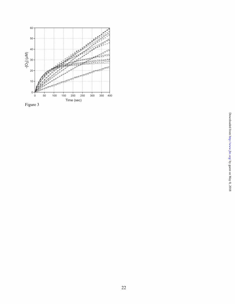
numerically integrated, and the theoretical progress curves were globally fit to the experimental data, allowing the individual elementary rate constants to be evaluated (Fig. 3). pH dependence of kinetic parameters –The steady state behavior of oxalate oxidase was examined over a range of pH (from 2.5 to 6.0) in a direct assay by recording the oxygen uptake kinetics and analyzing the progress curves as illustrated in Figs. 1&2. The results of the analysis are given in Table 2 and Fig. 2, D. The catalytic efficiency (Vmax/Km) increases continuously to low pH, rather than reaching a maximum at pH 4, as previously described for turnover at 37 °C using a peroxidase coupled assay (25). The Km evaluated from initial velocity data exhibits a strong pH dependence (Fig. 2, D), with limiting slopes (vs. pH) of 0.9 ±0.01 (below pH 4) and 1.50±0.03 (above pH 4). In contrast to the strong pH dependence of Km, Vi,max is nearly independent of pH over the range from 3 to 5. Solvent kinetic isotope effect –The effect of the isotopic composition of the solvent on the kinetic parameters was investigated by assaying oxalate oxidase in buffer prepared from H2O/D2O mixtures. Kinetic parameters were evaluated by fitting the progress curves to the burst equation (Eqn. 4) and the isotope sensitivity of the parameters was determined (Fig. 4). The initial rate shows a modest normal solvent isotope effect (kH2O/kD2O = 1.59±0.05) that appears to exhibit a linear proton inventory plot (Fig. 4, Top), consistent with a single proton being involved in the isotope-sensitive transition state. The burst rate constant is essentially insensitive to the isotopic composition of the solvent (kH2O/kD2O = 1.027±0.04) (Fig. 4, Middle). In contrast, the effects of varying the deuterium content of the solvent on the steady state rate (Vs) were relatively dramatic (Fig. 4, Bottom). The limiting values of Vs lead to an estimate of the overall solvent kinetic isotope effect kH2O/kD2O = 8.5±1.3, suggesting that full primary deuterium kinetic isotope effect is being expressed. The proton inventory is also markedly parabolic, deviating from linear solvent isotope concentration dependence. This type of curved proton inventory plot is consistent with multiple protons being involved in the transition state for the inactivation process (26), as indicated by the quality of the fit to a quadratic form (Fig. 4,
Bottom, dashed line). Alternatively, this behavior may be interpreted as evidence for isotopic fractionation. In the latter view, an equilibrium isotope effect associated with the H/D exchange reaction would lead to enrichment of D over H at a kinetically significant site in the protein. Isotope fractionation is well established and in fact forms the basis for one of the methods for D2O enrichment. The fractionation factors may be evaluated from the experimental data by fitting with a simplified form of the Gross-Beutler equation appropriate for a single exchange site that exhibits distinct fractionation factors (φR, φT) in the ground and transition states for the reaction, respectively (27) (Eqn. 6):
( ) ( )RTsns nnnnVV φφ +−+−= 1/10,, Eqn. 6
where Vs,n corresponds to the observed value of Vs at a mol fraction n of deuterium. The results of the analysis are consistent with a significant fractionation factor φR = 2.3±0.2 in the ground state changing to φT = 0.23±0.04 in the transition state. The latter is in the range of values associated with strong hydrogen-bonding sites. One caveat in evaluating this SKIE data is the possibility of interferences that introduce kinetic artifacts. This is particularly important since the commercial D2O specifications define the isotopic purity, and there is virtually no quality control for dissolved contaminants. In the oxalate oxidase studies, distinct proton inventory curves were observed when D2O from different sources or different levels of processing were used. Glass-distilled D2O gave rise to the most pronounced curvature in the Vs proton inventory, and the largest apparent kH2O/kD2O (approaching 80). In order to try to rule out potential interferences as the source of the observed SKIE results, D2O was subjected to additional purification by passage through a mixed bed ion exchanger and carbon filter to remove both ionic and dissolved organic species. The data reported in Fig. 4 was based on the purified D2O solvent, which was essentially the same as that observed for the untreated CDN Isotopes D2O sample. Turnover-based oxidative modification of oxalate oxidase –In an attempt to prepare a homogenous sample of inhibited oxalate oxidase (E†S) for spectroscopic characterization, a solution of
by guest on May 8, 2018
http://ww
w.jbc.org/
Dow
nloaded from

6
concentrated enzyme containing a saturating concentration of substrate (20 mM) was purged with pure oxygen. A yellow color rapidly developed and subsequently slowly faded during exposure to oxygen. When the reaction was repeated in D2O, a stable yellow solution was formed (Fig. 6, B). Incubation of the enzyme with dioxygen in the absence of substrate does not lead to oxidation of the metal center. B. Manganese Redox Modifications Redox modification of oxalate oxidase –The observation of transient color changes under turnover conditions suggests the possibility of oxidative modifications in the active site metal complex, which have not previously been described for oxalate oxidase. In order to more systematically investigate the redox chemistry of the active site manganese center, the as-isolated oxalate oxidase was titrated with the powerful inorganic oxidant, sodium m-periodate (NaIO4). This reagent is a powerful oxidizer (E0 = 1.6 V), and in acid solution undergoes a two-electron reduction changing formal oxidation state of I from +5 to +3 (28). Titration of oxalate oxidase with sodium periodate results in nearly stoichiometric oxidation of the enzyme to an intensely colored yellow complex (Fig. 5, Top), whose complete spectroscopic characterization leads to assignment to a superoxidized Mn(IV) complex (see below). The presence of a single tryptophan residue in the protein results in unusually low intrinsic UV absorption from the polypeptide, which allows the absorption spectrum of the complex to be recorded down to 240 nm. Sharp vibronic structure is observed in the spectra from the unique tryptophan absorption (λmax =278 nm) underlying these spectra. Similar absorption features are obtained by addition of peracetic acid to the native enzyme, although the sample is not stable and the intensity declines over time, due to the unavoidable presence of hydrogen peroxide in the reaction, which serves as a reductant for Mn(IV) OXO (data not shown). Treatment of the periodate-oxidized enzyme with ascorbate (Fig. 5, Bottom, Spectrum 2) results in a substantial decrease in absorption, forming a complex that is spectroscopically identified as a Mn(III) species (see below). Titration of periodate-oxidized oxalate oxidase with hydroxylamine (Fig. 5, Bottom, Spectrum 3; and Inset) completely eliminates the visible absorption, forming a
homogeneous Mn(II) form of the enzyme (see below). In EPR experiments, anaerobic addition of substrate to both Mn(III) and Mn(IV) OXO restores the spectra of the Mn(II) substrate complex (data not shown). Because the absorption spectra for the oxidatively modified forms of oxalate oxidase extends over the UV range, 280 nm extinction coefficients for protein quantitation were corrected for metal-centered absorption as described in Experimental Procedures. Incubation of the fully reduced enzyme with dioxygen alone does not lead to oxidation of the Mn(II) center. The redox transformations that are now known for the manganese center in oxalate oxidase are summarized in Scheme 2. Sodium periodate is a reagent commonly used in organic chemistry for oxidative cleavage of diols (including carbohydrates) forming aldehydes (28) which in principle could react with amino groups in proteins to form yellow-colored Schiff base complexes. In order to exclude the possibility of this type of interference in the periodate reaction, a non-glycosylated form of oxalate oxidase was prepared by site directed mutagenesis of the NXS glycan attachment site. S49A oxalate oxidase is expressed by Pichia pastoris without glycosylation, but the lack of glycosylation does not diminish activity (10). Treatment of Mn(II) S49A OXO generates the same yellow species as the glycosylated WT enzyme (data not shown). Mass spectra of as-isolated and periodate-treated OXO were virtually identical, demonstrating that no protein oxidation occurred (Supplementary Material). Spectroscopic characterization of oxidized oxalate oxidase –The optical absorption spectrum of the periodate-treated OXO is broad and featureless (Fig. 5, Top, Spectrum 7; Fig. 5, Inset, A), with a strong UV component that is consistent with ligand-to-metal charge transfer (LMCT) absorption within a Mn(IV) complex (29). Circular dichroism may be used to resolve components in the broad, overlapping spectra of metalloenzyme complexes on the basis of the signed intensity and the relatively large anisotropy (Δε/ε) associated with transitions that include significant magnetic dipole character, such as d→d spectra (18). The CD spectrum of periodate-treated OXO (Fig. 6, A) exhibits complex structure, and at least five transitions are resolved across the visible spectrum, with a triplet grouping
by guest on May 8, 2018
http://ww
w.jbc.org/
Dow
nloaded from

7
at 510, 460 and 395 nm. The strong CD band near 510 nm is associated with an anisotropy Δε/ε = .005. These features are consistent with spin-allowed electronic transitions from a Mn(IV) ground state to the low symmetry-split components of the orbital triplet (4T2g) excited state. The absorption and CD spectra of the turnover-generated oxidized OXO (Fig. 6, B) are nearly identical to those observed for the periodate-treated enzyme. EPR spectroscopy is specifically sensitive to paramagnetic ground states, which would include all of the accessible redox modifications of the manganese center. High spin ground states for Mn(II) (3d5, S= 5/2), Mn(III) (3d4, S=2) and Mn(IV) (3d3, S= 3/2) are all in principal detectable by EPR spectroscopy, although the sensitivity is expected to be much greater for the half-integer spin Mn(II) and Mn(IV) species. The EPR spectrum of the periodate-treated OXO (Fig. 7, A) shows relatively weak EPR absorption over the entire spectral range, with sharp features near g= 4.3 and 2 that may be assigned to very small amounts of Fe(III) and Cu(II), respectively, in the sample. Underlying these sharp spectra, and extending over the entire field range, are relatively broad, poorly resolved absorption features (see below). Treating this oxidized complex with a slight excess of ascorbate produces a distinct form (Fig. 7, B), lacking the broad underlying features but retaining the impurity Fe(III) and Cu(II) signals, which represent trace amounts (~0.01 g-atom/mol protein) of the impurities. The stability of the protein prevents these metal ions from being removed. Treating the oxidized enzyme with hydroxylamine leads to the appearance of a very strong resonance near g=2 with sextet hyperfine splitting (aMn = 95 G) characteristic of a high spin Mn(II) ion in roughly octahedral geometry (Fig. 7, C). Assuming that all of the manganese in the sample contributes to this signal, double integration of the full 7000 G wide field range provides a calibration value that may be used to quantitatively interpret the other spectra. The integrated intensity of the ascorbate-treated enzyme (Fig. 7, B) is found to represent only 4% of the full spin quantitation, when 6% of the Mn(II) signal is subtracted (note the feature near 3600 G in both B&C). While the spectra in Fig. 7, A&B look superficially quite similar, the integrated intensity of Spectrum A corresponds to more than 50% of Spectrum C, with large
contributions to intensity at low field (g=4). More accurate quantitation requires a correction for g-anisotropy (23). Using estimates of the principal g-values based on the turning points in the EPR spectrum, a corrected estimate of the spin quantitation of 82% is obtained. Thus, even though the spectrum of the periodate-treated OXO appears fairly nondescript, it represents a very substantial paramagnetic species. Subtracting the EPR spectrum of the ascorbate-reduced enzyme (Fig. 7, B) from that for the periodate-treated enzyme (Fig. 7, A) allows the spectrum of the superoxidized complex to be isolated (Fig. 8, Top). This species clearly gives rise to resonances over the entire field range, and includes significant EPR absorption extending down to the zero field limit. Low field resonances of this type are characteristic of multiplet (S > 1/2) ground states with small zero field splitting (D). Strong resonances appear near g=5 and g=2, resembling the spectrum predicted for an S=3/2 spin system in the rhombic limit (E/D = 0.33). While EPR spectra are available for Mn(IV) complexes with near-axial symmetry (30,31), data relating to lower symmetry Mn(IV) species relevant to this biological complex are just beginning to become available (32). However, it is possible to simulate the essential features of the experimental spectrum using ground state parameters that would be typical of a rhombically distorted Mn(IV) center (Fig. 8, Middle). The best agreement between experiment and theory requires a distribution in both D and E/D, suggesting disorder in the site. Analysis of the EPR spectrum of the turnover-generated superoxidized OXO is nearly identical to that found for the periodate-treated enzyme (Fig. 8, Bottom), although the features in the former spectrum appear somewhat sharper. Thus, both periodate oxidation and turnover in O2-saturated buffer lead to formation of a novel superoxidized Mn(IV) center in the protein. While Mn(IV) species have previously been proposed as components of the tetranuclear manganese water splitting active site associated with oxygenic photosynthesis (31,33,34), and a mixed valent binuclear Mn(III)Mn(IV) complex has been described in manganese catalase (35), this appears to be the first well-characterized example of a mononuclear Mn(IV) center in a protein.
by guest on May 8, 2018
http://ww
w.jbc.org/
Dow
nloaded from
8
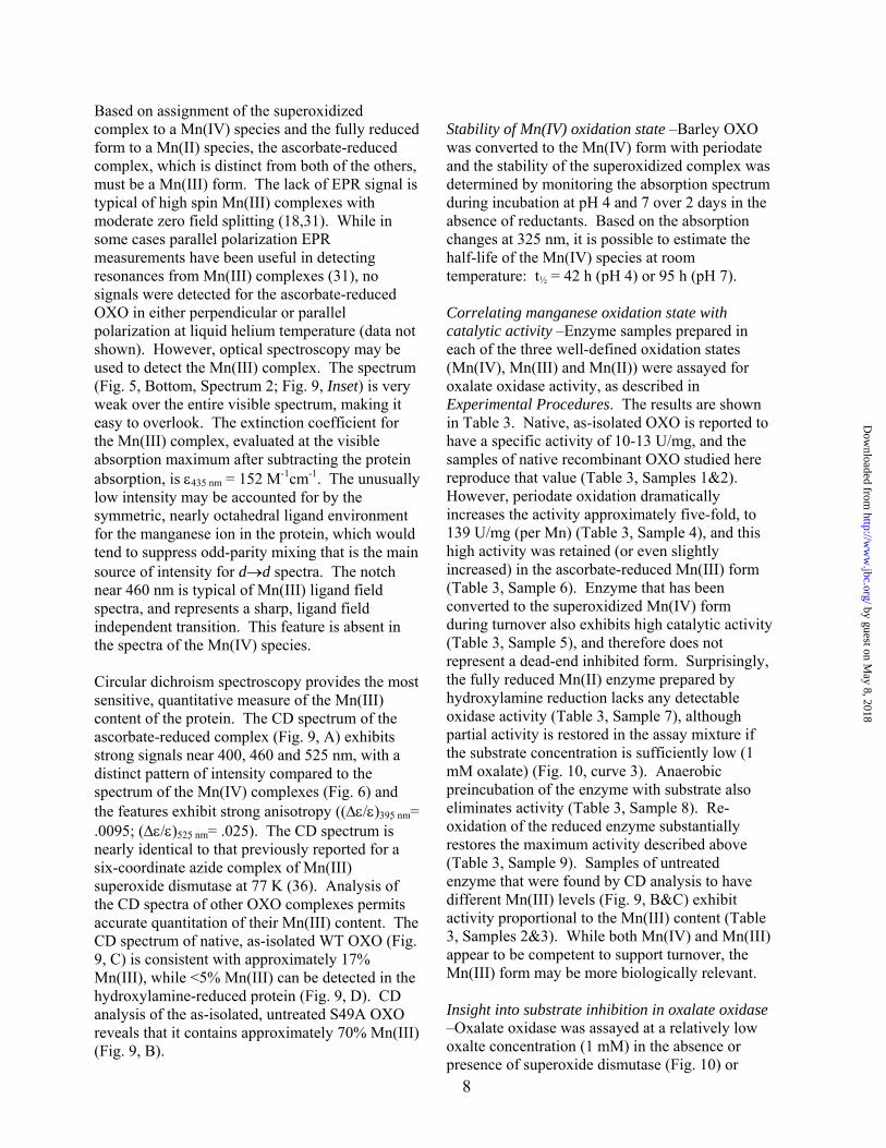
Based on assignment of the superoxidized complex to a Mn(IV) species and the fully reduced form to a Mn(II) species, the ascorbate-reduced complex, which is distinct from both of the others, must be a Mn(III) form. The lack of EPR signal is typical of high spin Mn(III) complexes with moderate zero field splitting (18,31). While in some cases parallel polarization EPR measurements have been useful in detecting resonances from Mn(III) complexes (31), no signals were detected for the ascorbate-reduced OXO in either perpendicular or parallel polarization at liquid helium temperature (data not shown). However, optical spectroscopy may be used to detect the Mn(III) complex. The spectrum (Fig. 5, Bottom, Spectrum 2; Fig. 9, Inset) is very weak over the entire visible spectrum, making it easy to overlook. The extinction coefficient for the Mn(III) complex, evaluated at the visible absorption maximum after subtracting the protein absorption, is ε435 nm = 152 M-1cm-1. The unusually low intensity may be accounted for by the symmetric, nearly octahedral ligand environment for the manganese ion in the protein, which would tend to suppress odd-parity mixing that is the main source of intensity for d→d spectra. The notch near 460 nm is typical of Mn(III) ligand field spectra, and represents a sharp, ligand field independent transition. This feature is absent in the spectra of the Mn(IV) species. Circular dichroism spectroscopy provides the most sensitive, quantitative measure of the Mn(III) content of the protein. The CD spectrum of the ascorbate-reduced complex (Fig. 9, A) exhibits strong signals near 400, 460 and 525 nm, with a distinct pattern of intensity compared to the spectrum of the Mn(IV) complexes (Fig. 6) and the features exhibit strong anisotropy ((Δε/ε)395 nm= .0095; (Δε/ε)525 nm= .025). The CD spectrum is nearly identical to that previously reported for a six-coordinate azide complex of Mn(III) superoxide dismutase at 77 K (36). Analysis of the CD spectra of other OXO complexes permits accurate quantitation of their Mn(III) content. The CD spectrum of native, as-isolated WT OXO (Fig. 9, C) is consistent with approximately 17% Mn(III), while <5% Mn(III) can be detected in the hydroxylamine-reduced protein (Fig. 9, D). CD analysis of the as-isolated, untreated S49A OXO reveals that it contains approximately 70% Mn(III) (Fig. 9, B).
Stability of Mn(IV) oxidation state –Barley OXO was converted to the Mn(IV) form with periodate and the stability of the superoxidized complex was determined by monitoring the absorption spectrum during incubation at pH 4 and 7 over 2 days in the absence of reductants. Based on the absorption changes at 325 nm, it is possible to estimate the half-life of the Mn(IV) species at room temperature: t½ = 42 h (pH 4) or 95 h (pH 7). Correlating manganese oxidation state with catalytic activity –Enzyme samples prepared in each of the three well-defined oxidation states (Mn(IV), Mn(III) and Mn(II)) were assayed for oxalate oxidase activity, as described in Experimental Procedures. The results are shown in Table 3. Native, as-isolated OXO is reported to have a specific activity of 10-13 U/mg, and the samples of native recombinant OXO studied here reproduce that value (Table 3, Samples 1&2). However, periodate oxidation dramatically increases the activity approximately five-fold, to 139 U/mg (per Mn) (Table 3, Sample 4), and this high activity was retained (or even slightly increased) in the ascorbate-reduced Mn(III) form (Table 3, Sample 6). Enzyme that has been converted to the superoxidized Mn(IV) form during turnover also exhibits high catalytic activity (Table 3, Sample 5), and therefore does not represent a dead-end inhibited form. Surprisingly, the fully reduced Mn(II) enzyme prepared by hydroxylamine reduction lacks any detectable oxidase activity (Table 3, Sample 7), although partial activity is restored in the assay mixture if the substrate concentration is sufficiently low (1 mM oxalate) (Fig. 10, curve 3). Anaerobic preincubation of the enzyme with substrate also eliminates activity (Table 3, Sample 8). Re-oxidation of the reduced enzyme substantially restores the maximum activity described above (Table 3, Sample 9). Samples of untreated enzyme that were found by CD analysis to have different Mn(III) levels (Fig. 9, B&C) exhibit activity proportional to the Mn(III) content (Table 3, Samples 2&3). While both Mn(IV) and Mn(III) appear to be competent to support turnover, the Mn(III) form may be more biologically relevant. Insight into substrate inhibition in oxalate oxidase –Oxalate oxidase was assayed at a relatively low oxalte concentration (1 mM) in the absence or presence of superoxide dismutase (Fig. 10) or
by guest on May 8, 2018
http://ww
w.jbc.org/
Dow
nloaded from

9
manganese catalase (data not shown). The presence of either enzyme in the assay mixture dramatically accelerated turnover inactivation and resulted in a vanishingly small Vs value in the steady state.
DISCUSSION Recent advances in X-ray structures of oxalate oxidase (9,16) and the availability of recombinant enzyme provides a foundation for detailed mechanistic studies on this interesting enzyme. In the present work, we have observed unusual non-stoichiometric burst kinetics which has led to a clearer understanding of the role of the manganese ion in the catalytic reaction. Kinetic bursts are well-known features in the pre-steady state reactions of many hydrolases (e.g., serine proteases) which form an obligate covalent intermediate during turnover (37). However, in those cases the amplitude of the burst phase is precisely stoichiometric with the amount of enzyme in the reaction mixture. Non-stoichiometric burst kinetics are relatively rare, but have been reported for certain enzymes, including β-lactamases which undergo slow, reversible inactivation during turnover (24,38,39). The burst behavior observed for OXO turnover (Fig. 1) is distinct because of a unique dependence on the substrate concentration. This behavior appears as substrate inhibition on the steady state velocity (Fig. 2B), rather than the initial velocity, which is the typical substrate inhibition pattern. An earlier study anecdotally described substrate inhibition at oxalate concentrations above 4 mM (25). However, in subsequent work, no substrate inhibition was detected on the initial velocity (Vi) (8). Our results confirm that substrate inhibition is present, but is only expressed at high substrate concentrations and after a significant reaction time (> 1 min). The substrate sensitivity of Vs that is expressed in the kinetic data (Fig. 1) requires that substrate is involved in an inhibitory process in addition to the turnover reaction. On the other hand, the lack of substrate inhibition on Vi implies that the initial phase kinetics are independent of the second substrate interactions. The substrate independence of the burst rate constant (k) is also an important clue, requiring that the rate law for the transition
does not contain the substrate concentration. This behavior is reproduced by a simple model for turnover-based reversible inactivation of the enzyme (Scheme 1). In this model, oxalate binding produces a substrate complex (ES) which lies at a branch point in the turnover process. Reaction of the ES complex with dioxygen leads to product formation and release, resulting in a normal turnover cycle and giving rise to the kinetic behavior in the initial phase. The model indicates that the substrate complex is able to undergo a reversible modification, forming an isomeric complex (E†) that is no longer competent for turnover. Formation of the E† complex does not directly involve a second substrate molecule, so the burst rate constant (k) is independent of substrate concentration. The E† complex is subsequently trapped by binding a second molecule of substrate to form the dead-end species E†S. This model does not require two molecules of substrate in the E†S complex (a distinction from the conventional substrate inhibition pattern, in which an ESS complex is formed), only that a second molecule of substrate interacts with a reversibly modified form of the enzyme. It also makes no specific predictions regarding the nature of E†, which might correspond, for example, to a change in protonation state, a structural isomerization, or a redox transformation (see below). The quality of the fit that may be obtained using this model, indicated in Fig. 3, supports the basic description of the turnover inactivation process shown in Scheme 1. The rate constants that solve the full set of differential equation are given in the legend to Fig. 3. Several observations that may be made from these results: (1) The experimental initial velocity Km value for oxalate turnover is close to that predicted from the elementary rate constants (Km = (k1r + k2)/k1f = 0.75 mM). (2) The model suggests that the reaction of the ES complex with dioxygen is overall rate limiting for turnover, with k2 nearly equal to kcat. (3) Conversion of ES into E† involves a slightly unfavorable equilibrium (Keq = (k4r/k4f) = 1.9) and the slow inactivation rate constant (k4f = .03 s-1) defines the time constant for depletion of active enzyme (t1/2 = 0.693/k4f = 26 s), roughly correlating with the timescale of the experimentally observed burst phase. Subsequent reversible tight binding of a second molecule of substrate (Kd = (k5r/k5f) = 0.18 mM) kinetically
by guest on May 8, 2018
http://ww
w.jbc.org/
Dow
nloaded from

10
traps the off-path complex and gives rise to the substrate-dependence of Vs. The observed substrate-dependence on Vs will be the product of the equilibrium constants for the off-path processes (Keq Kd). This observation allows the kinetic parameters evaluated from the experimental progress curves (Fig. 2) to be interpreted. Thus, the KI found for analysis of Vs curves (0.2±0.1 mM) (Fig. 2, B) would correspond to the product of substrate dissociation constant and an unfavorable isomerization equilibrium constant. Note that because the bimolecular rate constants for substrate association (k1f, k5f) were fixed in this analysis, the ratios k1r/k1f (corresponding to KS, the substrate binding constant) and k5r/k5f (the substrate inhibition constant) are actually determined, rather than the individual rate constants, k1r and k5r. The pH dependence of Km (Table 2; Fig. 2D) is dramatic, with Km values ranging over nearly 5 orders of magnitude between pH 2.5 and 6. The strong log-linear correlation between Km and pH (Fig. 2D) indicates that substrate binding is coupled to proton uptake. Two distinct slopes are evident in this plot, with a break point close to the second pKa for oxalic acid dissociation (pKa,1 = 1.23, pKa,2 = 4.19). The limiting slopes are consistent with two protons being taken up above pH 4, and a single proton taken up with substrate below pH 4. This implies that the catalytic complex contains a species equivalent to OxH2 (free acid). The realization that oxalate oxidase requires an oxidized Mn center for catalysis suggests that redox modifications may modulate catalytic activity during turnover, and might contribute to the substrate-dependent inhibition pattern for oxalate oxidase turnover (see above). If the manganese center becomes reversibly reduced during turnover, escape of free radical intermediates could lead to inactivation (E†). Reactivation could be the result of interaction with oxidizing species (product peroxide or peroxycarbonate, or superoxide formed by collisional reaction of O2 with escaped reactive intermediates). Formation of a tightly bound oxalate complex with the reduced, Mn(II) form of the enzyme (E†S) might block re-oxidation and account for the substrate-dependence of the turnover inactivation. The sensitivity of enzyme activity to the presence of other enzymes
(superoxide dismutase or catalase) in the assay mixture requires that the reactivation process involves a diffusible species, such as superoxide, or a molecule that equilibrates with superoxide in solution. Overall, these observations suggest a modification of the turnover inactivation process described in Scheme 1, where the first closed equilibrium step in the inactivation branch is replaced by an open equilibrium involving dissociation of a reactive species (S')(Scheme 3). This would not fundamentally change the behavior of the kinetic model and the theoretical fit obtained using the open model is essentially the same as that found for the simpler, closed scheme (Scheme 1). The use of periodate for preparation of the Mn(IV) OXO complex deserves some comment. Periodate is most often used in biochemistry for specific oxidation of glycoproteins, and, even though it is a high potential oxidant, amino acids do not appear to be particularly susceptible to oxidation. Mass analysis shows that no significant protein damage is observed when OXO is oxidized with periodate under our conditions (Supplementary Data, Fig. S1). Further, periodate has been described as a simple competitive inhibitor of alkaline phosphatase (40). The specific requirement for periodate or peracids for OXO oxidation may relate to their character as mono-anionic (or neutral) oxidants. Previous attempts to change the Mn oxidation state using common one-electron oxidants (K3Fe(CN)6, Na2IrCl6, K4Mo(CN)8 (10)) have failed. However, those reagents are all polyanions, and the strong pH-dependence of the oxalate Km suggests that dianions may be excluded from access to the active site. The higher oxidation state Mn(IV) complex is quite stable at room temperature in the absence of exogenous reductants, permitting convenient handling for sample preparation and analysis. Ascorbate reduces the complex to the Mn(III) state, which appears to be the biologically relevant active form of the enzyme. A modified turnover reaction may be written (Eqn. 7) to include the requirement for Mn(III) in the active site: [Mn(III)(OH)2] + C2O4
2- + 2H+ → [Mn(II)(OH2)2] + CO2 + CO2
– Eqn. 7 Association of the Mn(III) ion with two hydroxide ligands (together with Glu95) would produce a
by guest on May 8, 2018
http://ww
w.jbc.org/
Dow
nloaded from

11
neutral (uncharged) metal center, which is expected to be most favorable for a buried metal complex. The driving force for the C-C bond cleavage would be the reduction and protonation of the Mn center. Although no experimental data is currently available for the redox potential of the active site Mn complex, the free energy change for the oxidative cleavage half-reaction (ΔΔGf°C2O42-
,CO2+CO2-.) can be calculated, based on standard formation free energies for the organic species (Table 4). The Mn(III)/Mn(II) reduction potential must be greater than ΔΔGf°/F in order for the overall reaction to be thermodynamically favorable (ΔG < 0). Depending on the value of the electrochemical potential used for the CO2/ CO2
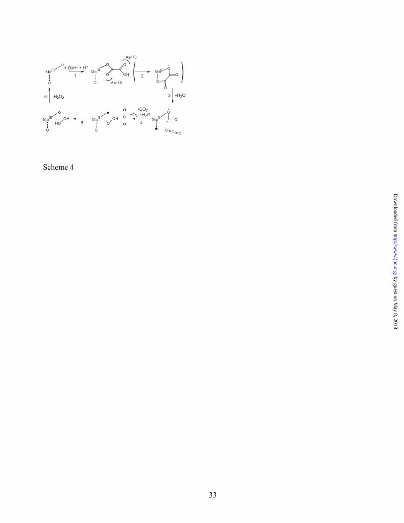
– redox couple (-1.4 V vs. NHE (in protic solvent (41)) – -2.0 V vs. NHE (in aprotic solvent (42))) ΔΔGf° will equal 37 or 93 kJ/mol, respectively, and so the protein Mn(III)/Mn(II) reduction potential must lie in the range +0.4 – +1.0, which is biologically reasonable and is less than E° for the free metal ion (+1.5 V), demonstrating the plausibility of the reaction shown (Eqn. 7). Mechanistic proposal –Based on the information on the active site interactions revealed by these investigations, we propose a new turnover mechanism for oxalate oxidase, shown in Scheme 4. In the first step (Scheme 4, Step 1), the active, resting Mn(III) enzyme binds substrate (as the monoanion) to form a Michaelis complex. Substrate is shown with monodentate carboxylate coordination, consistent with recent X-ray structural studies on a substrate analog (glycolate) complex, which also identifies a role for Asn75 and Asn85 in hydrogen-bond stabilization of the complex (16). Under anaerobic conditions, oxalate has been shown to reduce the Mn(III) form of the enzyme (10) (Scheme 4, Step 2). Reduction of Mn(III) to Mn(II) is associated with formation of an oxalyl free radical, with Scheme 4 illustrating a possible bidentate coordination mode. The oxalyl radical is very unstable and is known to undergo rapid C-C bond fission non-enzymatically in aqueous solution (k = 2×10⁶ s-1) producing a molecule of carbon dioxide and a carbon dioxide radical anion (43). The same chemistry is likely to occur in the enzyme active site (Scheme 4, Step 3), resulting in release of the first molecule of carbon dioxide and leaving the carbon dioxide radical anion bound to Mn(II). In solution, carbon dioxide radical anion undergoes
diffusion-controlled electron transfer to dioxygen (k = 2.4×10⁹ s-1) yielding superoxide and carbon dioxide products (43). The interception of a carbon dioxide radical anion intermediate by dioxygen during OXO turnover would generate a second molecule of carbon dioxide and superoxide (Scheme 4, Step 4), shown in the protonated, hydroperoxyl radical form, consistent with its pKa =4.88. Subsequent electron transfer oxidation of Mn(II) by the hydroperoxyl radical (Scheme 4, Step 5) could in principle occur through either inner sphere (by direct coordination) or outer sphere (e.g., H-atom transfer from water) pathways to generate a molecule of hydrogen peroxide. Note that the oxidation of Mn(II) OXO by superoxide (or hydroperoxyl radical) appears to be important in the steady state reactivation of OXO during turnover, based on the accelerating effect of superoxide dismutase on turnover inactivation (Fig. 10, curve 2). This Scheme predicts that one proton is consumed per turnover cycle, and that peroxycarbonate is not formed as a primary product, although the presence of both peroxide nucleophile and carbon dioxide electrophile in the product mixture makes it likely that peroxymonocarbonate (a peracid) will be produced as a secondary product in solution. Formation of peroxymonocarbonate would account for the oxidation of Mn(II) OXO to Mn(IV) OXO in the turnover-based redox modification of the enzyme, consistent with the oxidation of Mn(II) OXO by peracetic acid. The role of the metal ion in this mechanism is oxalate activation through one-electron oxidation of bound substrate by the active site Mn(III) center. In previous proposals for the OXO turnover mechanism (8,10), which were based on the assumption that Mn(II) was the catalytically active metal species, the metal ion played a very different role: the reductive activation of O2. The evidence presented in the present work for a specific requirement for the oxidized metal center in OXO supports the oxidative activation mechanism shown here. The well-established reactivity of higher oxidation state metallo-oxalate complexes towards thermal decomposition (43,44) suggests that the enzyme facilitates C-C bond cleavage through formation of a free radical form of the substrate. The current mechanistic scheme emphasizes the close parallels between oxalate oxidase and the
by guest on May 8, 2018
http://ww
w.jbc.org/
Dow
nloaded from

12
related enzyme, oxalate decarboxylase (3,14,45). Recent studies have demonstrated that the Mn(III) form of OXD is the catalytically active species , and a role for dioxygen activation of the Mn(II) enzyme has been proposed . Based on the currently available data it appears likely that both enzymes pass through a carbon dioxide radical anion (CO2
– ) intermediate. The fate of this intermediate appears to be the distinguishing factor between OXO and OXD: in the former, the reactive intermediate is intercepted by dioxygen either in the enzyme active site or in solution and oxidized, producing carbon dioxide and hydroperoxyl radical. In the decarboxylase turnover, the carbon dioxide radical anion is protected from reaction with O2, allowing it to undergo rebound electron transfer reduction by Mn(II) and protonation to yield formate as the second product. In conclusion, the Mn(III) form of oxalate oxidase is the catalytically active state of the enzyme, and the Mn(II) form, which represents the majority of the as-isolated native enzyme, is catalytically inactive. The requirement for an oxidized metal center for catalytic activity has been demonstrated by preparation of homogeneous oxidation states of the enzyme for the first time. OXO undergoes reversible inactivation during turnover, resulting in burst kinetics with the steady-state rate being a dynamic balance between inactivation and reactivation processes. In O2 saturated buffer, a turnover-based redox modification of the enzyme forms a novel superoxidized mononuclear Mn(IV) biological complex.
REFERENCES
1. Sugiura, M., Yamamura, H., Hirano, K., Sasaki, M., Morikawa, M., and Tsuboi, M. (1979) Chem. Pharm. Bull. 27, 2003-2007.
2. Lane, B.G. (1994) FASEB J. 8, 294-301. 3. Svedružić D., Jónsson S., Toyota C.G.,
Reinhardt L.A., Ricagno S., Lindqvist Y., and Richards N.G. (2005) Arch. Biochem. Biophys. 433, 176-192.
4. Koyama, H. (1988) Agric. Biol. Chem. 52, 743-748.
5. Escutia, M.R., Bowater, L., Edwards, A., Bottrill, A.R., Burrell, M.R., Polanco, R., Vicuña, R., and Bornemann, S. (2005) Appl. Environ. Microbiol. 71, 3608-3616.
6. Bernier, F., and Berna, A. (2001) Plant. Physiol. Biochem. 39, 545-554.
7. Zhou, F., Zhang, Z., Gregersen, P.L., Mikkelsen, J.D., Neurgaard, E., Collinge, D.B., and Thordahl-Christensen, H. (1998) Plant Physiol. 117, 33-41.
8. Requena, L., and Bornemann, S. (1999) Biochem. J. 343, 185-190.
9. Woo, E.J., Dunwell, J.M., Goodenough, P.W., and Pickersgill, R.W. (1998) FEBS Lett. 437, 87-90.
10. Whittaker, M.M., and Whittaker, J.W. (2002) J. Biol. Inorg. Chem. 7, 136-145.
11. Tanner, A., Bowater, L., Fairhurst, S. A., and Bornemann, S. (2001) J. Biol. Chem. 276, 43627-43634.
12. Anand, R., Dorrestein, P. C., Kinsland, C., Begley, T. P., and Ealick, S. E. (2002) Biochemistry 41, 7659-7669
13. Just, V. J., Stevenson, C. E. M., Bowater, L., Tanner, A., Lawson, D. M., and Bornemann, S. (2004) J. Biol. Chem. 279, 19867-19874.
14. Muthusamy, M., Burrell, M.R., Thorneley, R.N.F., and Bournemann, S. (2006) Biochemistry 45, 10667-10673.
15. Woo, E.J., Dunwell, J.M., Goodenough, P.W., Marvier, A.C., and Pickersgill, R.W. (2000) Nat. Struct. Biol. 7, 1036-1040.
16. Opaleye, O., Rose, R.-S., Whittaker, M.M., Woo, E.-J., Whittaker, J.W., and Pickersgill, R.W. (2006) J. Biol. Chem. 281, 6428-6433.
17. Whittaker, J.W., Lipscomb, J.D., Kent, T.A., and Munck, E. (1984) J. Biol. Chem. 259, 4466-4475.
18. Whittaker, J.W., and Whittaker, M.M. (1991) J. Am. Chem. Soc. 113, 5528-5540.
19. Whittaker, M.M., Barynin, V.V., Igarashi, T., and Whittaker, J.W. (2003) Eur. J. Biochem. 270, 1102-1116.
20. Lowry, O.H., Rosebrough, N.J., Farr, A.L., and Randall, R.J. (1951) J. Biol. Chem. 193, 265-275.
21. Whittaker, M.M., Ballou, D.P., and Whittaker, J.W. (1998) Biochemistry 37, 8426-8436.
22. Segel, I. H. (1975) Enzyme Kinetics Wiley-Interscience, New York, Chapter 2, 28-29.
23. Aasa, R., and Vänngård, T. (1975) J. Magn. Res.19, 308-315.
24. Waley, S.G. (1991) Biochem. J. 279, 87-94. 25. Kotsira, V.P., and Clonis, Y.D. (1997) Arch.
Biochem. Biophys. 340, 239-249. 26. Venkatasubban, K.S., and Schowen, R.L.
(1984) CRC Crit. Rev. Biochem. 17, 1-44.
by guest on May 8, 2018
http://ww
w.jbc.org/
Dow
nloaded from

13
27. Quinn, D.M., and Sutton, L.D. (1991) In Enzyme Mechanism from Isotope Effects (Cook, P.F., Ed.) CRC Press, Boca Raton, FL.
28. Greenwood, N.N., and Earnshaw, A. (1984) Chemistry of the Elements Pergamon Press, Paris, 17.2.7-17.2.8.
29. Reisfeld, M.J., Matwiyoff, N.A., and Asprey, L.B. (1971) J. Mol. Spectroscopy 39, 8-20.
30. Kessissoglou, D.P., Li, X., Butler, W.M., and Pecoraro, V.L. (1987) Inorg. Chem. 26, 2487-2492.
31. Campbell, K.A., Force, D.A., Nixon, P.J., Dole, F., Diner, B.A., and Britt, R.D. (2000) J. Am. Chem. Soc. 122, 3754-3761.
32. Parsell, T.H., Behan, R.K., Green, M.T., Hendrich, M.P., and Borovick, A.S. (2006) J. Am. Chem. Soc. 128, 8728-8729.
33. Messinger, J. (2000) Biochim. Biophys. Acta 1459, 481-488.
34. McElvoy, J.P., and Brudvig, G.W. (2006) Chem. Revs. 106, 4455-4483.
35. Ivancich, A., Barynin, V.V., and Zimmerman, J.L. (1995) Biochemistry 34, 6628-2239.
36. Whittaker, M. M., and Whittaker, J. W. (1996) Biochemistry 35, 6762-6770.
37. Vineyard, D., Zhang, X., and Lee, I. (2006) Biochemistry 45, 11432-11443.
38. Page, M.G.P. (1993) Biochem. J. 295, 295-304.
39. Badarau, A., and Page, M.I. (2006) Biochemistry 45, 11012-11020.
40. Ohlsson, J.T., and Wilson, I.B. (1974) Biochim. Biophys. Acta 350, 48-53.
41. Haynes, L.V., and Sawyer, D.T. (1967) Anal. Chem. 39, 332-338.
42. Gennaro, A., Isse, A.A., Savéant, J.-M., Severin, M.-G., and Vianello, E. (1996) J. Am. Chem. Soc. 118, 7190-7196.
43. Hislop, K.A., and Bolton, J.R. (1999) Environ. Sci. Technol. 33, 3119-3126.
44. Cho, M., Lee, Y., Chung, H., and Yoon, J. (2004) Appl. Environ. Microbiol. 70, 1129-1134.
45. Chang, C.H., Svedružić, D., Ozarowski, A., Walker, L., Yeagle, G., Britt, R.D., Angerhofer, A., and Richards, N.G.J. (2004) J. Biol. Chem. 279, 52840-52849.
46. Wagman, D.D., Evans, W.H., Parker, V.B., Schumm, R.H., Halow, I., Bailey, S.M., Churney, K.L., and Nuttall, R.L. (1982) J. Phys. Chem. Ref. Data 11, Suppl. 2.
47. Alberty, R.A. (1995) J. Phys. Chem. 99, 11028-11034.
FOOTNOTES
* This work was supported by a grant from the National Institutes of Health (GM42680, to J.W.W.).
1The abbreviations used are: OXO, oxalate
oxidase; OXD, oxalate decarboxylase; ODE, ordinary differential equation; NHE, normal hydrogen electrode, F, Faraday electrochemical equivalent. .
FIGURE LEGENDS
Figure 1. Steady state burst kinetics of purified oxalate oxidase. Individual time courses for oxygen uptake during oxidation of oxalate by oxalate oxidase were digitally recorded as described in the Experimental Procedures. Each progress curve is the average of three independent experiments at specified oxalate concentrations in a reaction mixture containing 10 μg of enzyme in 50 mM sodium succinate buffer, pH 4. Curves 1-7, dashed lines; curves 8-20, solid lines. Oxalate concentrations (in mM): (1) 0.1; (2) 0.2; (3) 0.3; (4) 0.4; (5) 0.5; (6), 0.6; (7) 0.7; (8) 1.0; (9) 1.2; (10) 1.4; (11) 1.6; (12) 1.8; (13) 2.0; (14) 2.5; (15) 3.0; (16) 5.0; (17) 7.5; (18) 10; (19) 20; (20) 40. Figure 2. Steady state parameter analysis for oxalate oxidase burst kinetics. The progress curves (Fig. 1) were individually fit to the burst equation (Eqn. 4) to evaluate initial velocity (Vi), steady-state velocity (Vs) and the burst rate constant (k). The average value for each data point and the standard deviation for the triplicate measurements are shown. (A) Substrate concentration dependence of Vi. The solid line represents a fit to the Michaelis-Menton equation with Km = 0.78±0.03 mM; Vi,max = 37.7±0.5 μM/min. (B) Substrate concentration dependence of Vs. The solid line represents a non-linear least-squares fit to the substrate inhibition equation (Eqn. 5) with Km = 1.4 ± 0.9; KI = 0.2±0.1 mM; Vmax = 44.3±20 μM/min. (C) The substrate concentration dependence of the burst rate constant, k. The solid line shows the average value for the data points for oxalate concentrations above 0.5 mM. (D) pH dependence of oxalate Km. A log-log plot of Km values (evaluated as described in Experimental Procedures) is shown
by guest on May 8, 2018
http://ww
w.jbc.org/
Dow
nloaded from

14
as a function of pH. The dashed lines represent linear least squares fits to the data above (1) and below (2) pH 4. Figure 3. Evaluation of a kinetic model for oxalate oxidase burst kinetics. Differential equations describing a burst kinetics model for oxalate oxidase (Scheme 1) were numerically integrated and globally fit to experimental progress curves spanning ten different substrate concentrations as described in the Experimental Procedures. The rate constants corresponding to substrate binding steps (k1f and k5f) were both fixed at a value of 1×10⁶ M-1s-1 to reduce the number of variable in the fitting process. The best fit set of rate constants is: k1r= 743 s-1; k2[O2] = 9.7 s-1; k3 = 1000 s-1; k4f = .026 s-1; k4r = 0.048 s-1; k5r = 179 s-1. The theoretical progress curves corresponding to these values are shown as solid lines in the figures. Key for oxalate concentrations (in mM): (□) 0.1; (○) 0.2; (◊) 0.3; ( ) 0.4; ( ) 0.6; ( ) 1.2; ( ) 1.8; ( ) 5; ( ) 10; ( ) 20. Figure 4. Solvent kinetic isotope measurements on oxalate oxidase burst kinetics. Proton inventory experiments were performed in H2O/D2O mixtures at pL = 4. Progress curves were performed in triplicate for each solvent composition and each set was individually analyzed by nonlinear least squares regression fitting to the burst kinetics equation (Eqn. 4). Average values and standard errors are shown for each result. (Top) Vi dependence on solvent composition. The solid line is a linear regression fit to the data yielding a kH2O/kD2O = 1.59±0.005 on Vi. (Middle) Burst rate constant dependence on solvent composition. The solid line is the linear regression fit to the data, with yielding a kH2O/kD2O = 1.027±0.04 on k. (Bottom) Vs dependence on the solvent isotopic composition. The limiting values of Vs at low and high deuterium content yield kH2O/kD2O = 8.5±1.3 on Vs. The dashed line represents a quadratic fit to the data. The solid line shows a nonlinear regression fit to the single-site Gross-Beutler equation (Eqn. 6) with fractionation factors φT = 0.23±0.04 and φR = 2.3±0.2. Figure 5. Redox titrations of oxalate oxidase. (Top) Titration of oxalate oxidase with sodium periodate. Spectrum 1: Native Mn(II) oxalate oxidase (3 mg/mL, 37 μM Mn) in 50 mM sodium succinate buffer, pH 4. Spectra 2-7: following
sequential additions of 2 μL aliquots of 4 mM NaIO4 solution to 1 mL enzyme. Inset: Plot of absorption at 280 nm versus NaIO4 concentration. Saturation occurs at 37 μM NaIO4 (Bottom) Optical absorption spectra of oxalate oxidase (3 mg/mL protein in 50 mM sodium succinate buffer, pH 4) in different oxidation states. Spectrum 1: Sodium periodate treated (superoxidized) enzyme following desalting by gel filtration. Spectrum 2: Product of ascorbate reduction of superoxidized oxalate oxidase following desalting by gel filtration. Spectrum 3: Product of hydroxylamine reduction of superoxidized oxalate oxidase. Inset: Reduction of superoxidized oxalate oxidase by sequential addition of 2 uL aliquots of 3 mM (NH2OH)2 H2SO4 (6 mM hydroxylamine) to sample for Spectrum 1. The amplitude of the reduction step corresponds to 60 μM NH2OH. Figure 6. Circular dichroism spectra of superoxidized oxalate oxidase. (A) Superoxidized Mn(IV) OXO prepared by periodate oxidation and desalting (1 mM active sites, 0.26 mM Mn) in 50 mM sodium succinate buffer, pH 4. The solid line is the experimental CD spectrum; the dashed lines are the Gaussian deconvolution. (B) The superoxidized oxalate oxidase prepared by turnover in oxygen-saturated buffer (1 mM active sites, 0.22 mM Mn) in 50 mM sodium succinate buffer, pH 4. Inset: Optical absorption difference spectra for these species. (A) (Fig. 5, Bottom, Spectrum 1) – (Fig. 5, Bottom, Spectrum 3). (B) (Initial – Final) difference spectrum for oxalate oxidase (3 mg/mL in 50 mM sodium succinate buffer, pH 4 in D2O with 10 mM oxalate) before and after purging the anaerobic complex with pure oxygen for 5 min. Figure 7. EPR Spectra for oxalate oxidase complexes. Oxalate oxidase (2 mM active sites, 0.37 mM Mn) in 20 mM potassium phosphate buffer, pH 7. (A) Superoxidized enzyme formed by addition of 1 mM sodium periodate; (B) following addition of 2 mM ascorbic acid to a sample of superoxidized enzyme; (C) following addition of 2 mM (NH2OH)2 H2SO4 to a sample of superoxidized enzyme. Instrumental parameters: Temperature, 110 K; microwave frequency, 9.39 GHz; microwave power, 20 mW; modulation amplitude, 10 G.
by guest on May 8, 2018
http://ww
w.jbc.org/
Dow
nloaded from
15
Figure 8. EPR spectra for superoxidized (Mn(IV)) oxalate oxidase complexes. (Top) EPR difference spectrum (periodate oxidized – ascorbate reduced) (Fig. 7, Spectrum A – Fig. 7, Spectrum B). (Middle) Spectral simulation based on spin Hamiltonian parameters: S= 3/2 ground state, D= 0.115 cm-1, E/D = 0.33, σD = 0.15 cm-1, σE/D = 0.2, Γ = 300 G. (Bottom) EPR difference spectrum (turnover modified – ascorbate reduced) for superoxidized oxalate oxidase prepared by oxalate turnover in oxygen-saturated buffer as described in Experimental Procedures. Figure 9. CD spectra for Mn(III) oxalate oxidase. The solid line is the experimental data, the dashed lines the Gaussian deconvolution. (A) Periodate-oxidized, ascorbate-reduced oxalate oxidase (1 mM active sites, 0.23 mM Mn). Inset: Optical absorption spectrum for this sample. (B) As-isolated S49A OXO (1 mM active sites, 0.26 mM Mn). (C) As-isolated, native oxalate oxidase (1 mM active sites, 0.26 mM Mn). (D) Hydroxylamine-reduced native enzyme (1 mM active sites, 0.22 mM Mn). Figure 10. Inactivation and reactivation of oxalate oxidase under assay conditions. Oxalate oxidase was assayed in a Clark oxygen electrode with 10 μg native OXO and 1 mM oxalate as described in the Experimental Procedures: (1) Control reaction; (2) plus 25 μg manganese superoxide dismutase; (3) with 10 μg hydroxylamine-reduced, desalted OXO replacing native OXO, without superoxide dismutase.
by guest on May 8, 2018
http://ww
w.jbc.org/
Dow
nloaded from
16
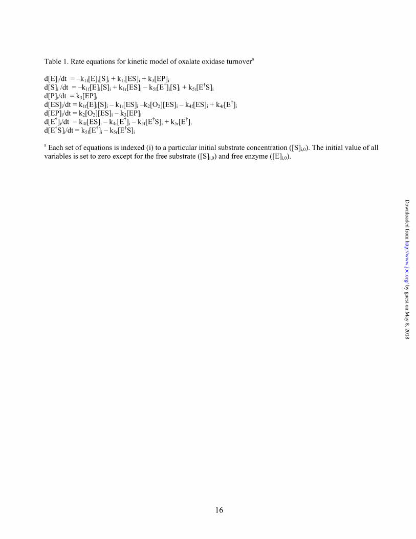
Table 1. Rate equations for kinetic model of oxalate oxidase turnovera d[E]i/dt = –k1f[E]i[S]i + k1r[ES]i + k3[EP]i d[S]i /dt = –k1f[E]i[S]i + k1r[ES]i – k5f[E†]i[S]i + k5r[E†S]i d[P]i/dt = k3[EP]i d[ES]i/dt = k1f[E]i[S]i – k1r[ES]i –k2[O2][ES]i – k4f[ES]i + k4r[E†]i d[EP]i/dt = k2[O2][ES]i – k3[EP]i d[E†]i/dt = k4f[ES]i – k4r[E†]i – k5f[E†S]i + k5r[E†]i d[E†S]i/dt = k5f[E†]i – k5r[E†S]i a Each set of equations is indexed (i) to a particular initial substrate concentration ([S]i,0). The initial value of all variables is set to zero except for the free substrate ([S]i,0) and free enzyme ([E]i,0).
by guest on May 8, 2018
http://ww
w.jbc.org/
Dow
nloaded from

17
Table 2. pH dependence of the steady state kinetic parameters for oxalate oxidase. pHa Km (mM) Vi,max (μM/min) 2.5 0.020±0.003 21±0.5 3.0 0.056±.007 36.1±1.8 3.5 0.170±.004 35.2±3.1 4.0 0.78±0.03 37.7±0.5 4.5 2.9±0.8 35.5±0.4 5.0 18±3 43.5±0.8 5.5 88±5 22±0.5 6.0 522±29 b -b a Oxalate oxidase activity was assayed with a Clark oxygen electrode in air saturated 50 mM sodium succinate or 50 mM sodium phosphate buffer. The pH of the buffer and oxalate stock solution were adjusted as described in the Experimental Procedures b The limited solubility of the substrate at the higher pH range prevented a complete saturation profile from being obtained at pH 6. In order to estimate the Km value, an average value of the asymptotic limit (35 μM/min) was used to constrain the analysis.
by guest on May 8, 2018
http://ww
w.jbc.org/
Dow
nloaded from

18
Table 3. Correlation between metal oxidation state and oxalate oxidase activity
Sample Mn(III) content (%)a
Mn(IV) content (%)a
Specific Activity (U/mg/Mn)b
Specific Activity (U/mg/Mn(ox)) b
1. Native WT OXO (A) 17 – 21.9±1.9 129±11 2. Native WT OXO (B) 26 – 33.5±0.6 129±2 3. Native S49A OXO 70 – 94±1 134±1.5 4. Periodate-oxidized – 100 139±1.5 139±1.5 5. Turnover-modifiedc – – 107±1 – 6. Ascorbate-reduced >95 – 156±6 156±6 7. NH2OH-reduced 0 – 0.03±0.04 – 8. Substrate-reducedd – – 0.19±0.08 – 9. Reduced/reoxidizede – – 85±0.5 –
a Based on spectroscopic analysis. b Measured by oxygen uptake kinetics in Clark oxygen electrode as described in Experimental Procedures. c Enzyme reacted with 20 mM oxalate in assay buffer with O2 purge for 5 min. d Prepared by anaerobic preincubation of native WT OXO with 20 mM oxalate in assay buffer for 30 min. e Sample (4) treated with NH2OH and reoxidized with NaIO4.
by guest on May 8, 2018
http://ww
w.jbc.org/
Dow
nloaded from

19
Table 4. Thermodynamic analysis of the oxalate cleavage reaction Species ΔGf° (kJ/mol) oxalate (C2O4
2-) -673.9 a carbon dioxide (CO2,(aq)) -385.97 b carbon dioxide radical anion (CO2
-.) -250 – -195c a Ref. 46. b Ref. 47. c ΔGf°CO2-. = ΔGf°CO2 + ΔG°CO2/CO2-. = ΔGf°CO2 – nFE°CO2/CO2-.; (F = 96.485 kC/mol).
by guest on May 8, 2018
http://ww
w.jbc.org/
Dow
nloaded from


Mei M Whittaker, Heng-Yen Pan, Erik T Yukl and James W Whittakeroxidase: Implications for the catalytic mechanism
Burst kinetics and redox transformations of the active site manganese ion in oxalate
published online January 8, 2007J. Biol. Chem.
10.1074/jbc.M609374200Access the most updated version of this article at doi:
Alerts:
When a correction for this article is posted•
When this article is cited•
to choose from all of JBC's e-mail alertsClick here
Supplemental material:
http://www.jbc.org/content/suppl/2007/01/09/M609374200.DC1
by guest on May 8, 2018
http://ww
w.jbc.org/
Dow
nloaded from