c ell d ivision c ycle kanokporn boonsirichai. t wo t ypes of c ell d ivision mitosis chromosome...
TRANSCRIPT

CELL DIVISION CYCLE Kanokporn Boonsirichai

TWO TYPES OF CELL DIVISION
Mitosis Chromosome number is preserved.
Meiosis Chromosome number is reduced by half.

MITOSIS

MEIOSIS
Crossing over
Meiosis does two things -
1) Meiosis takes a cell with two copies of every chromosome (diploid) and makes cells with a single copy of every chromosome (haploid).
2) Meiosis scrambles the specific forms of each gene that each sex cell (egg or sperm) receives.

Crossing over
Independent assortment

CONTROL OF CELL CYCLE
Figure 18-1 Essential Cell Biology (© Garland Science 2010)

Prokaryote: Escherichia coli ~ 20 minutes

Figure 18-2 Essential Cell Biology (© Garland Science 2010)
PHASES OF THE CELL CYCLE

CELL CYCLE CONTROL SYSTEM
A timer/clock: when and how long
A play list: ordering of event
Preventions of repeats On/off switches Backup mechanisms Adaptibility/sensors

CELL CYCLE CHECKPOINTS
Cause the cell to become arrested at a specific point in the cell cycle, if previous events have not been completed
Utilize negative signals

What do you think might be the nature of the cell cycle control system?

CYCLIN-CDK COMPLEXES
Cdk (cyclin-dependent kinase): cyclically activated protein kinase
Cyclin: switches Cdk on and off
Figure 18-5 Essential Cell Biology (© Garland Science 2010)

CYCLIN-CDK ACTIVITY AND CELL CYCLE

FOUR CLASSES OF CYCLINS
G1 cyclins promote the cell through “Start” or restriction point in late G1
G1/S-cyclins bind Cdks at the end of G1 and commit the cell to DNA replication
S-cyclins bind Cdks during S phase and are required for initiation of DNA replication
M-cyclins promote the events of mitosis
wikipedia

MAJOR CYCLINS AND CDKS
Cyclin-Cdk complex
Vertbrate Budding yeast
CyclinCyclin CdkCdk CyclinCyclin CdkCdk
G1-Cdk cyclin D Cdk4, 6
Cln3 Cdk1
G1/S-Cdk
cyclin E Cdk2 Cln1, 2 Cdk1
S-Cdk cyclin A Cdk2 Cln5, 6 Cdk1
M-Cdk cyclin B Cdk1 Cln1, 2, 3, 4
Cdk1

Figure 18-10 Essential Cell Biology (© Garland Science 2010)

ACTIVATION OF CDKS
Binding to cyclins Phosphorylation by Cdk-activating kinase
(CAK) near the entrance of the active site
Figure 18-9 Essential Cell Biology (© Garland Science 2010)

INHIBITION OF CDK ACTIVITY
Binding of cyclin-Cdk complexes by Cdk inhibitor proteins (CKI)
Proteolysis of cyclin

Figure 18-12 Essential Cell Biology (© Garland Science 2010)
CELL CYCLE CHECKPOINTS At G1 checkpoint, cell
decides whether to commit to another cell cycle.
At each checkpoint, cell may be arrested if conditions are not favorable.

THE S PHASE
Active S-Cdk
Initiation of DNA replication
Prevention of rereplication

INITIATION OF DNA REPLICATION

REGULATION AT THE ORIGIN OF DNA REPLICATION
S-Cdk allows DNA replication to initiate.

Mcm
Mcm
Export from the nucleus
S-CdkM-Cdk
Ubiquitylation by SCF
S-CdkM-Cdk
PREVENTION OF RE-REPLICATION

THE M PHASE
Chromosome separation
Figure 18-17 Essential Cell Biology (© Garland Science 2010)

ROLES OF M-CDK IN MITOSIS
Induces the assembly of the mitotic spindle
Ensures that replicated chromosomes are attached to the spindle
Triggers chromosome condensation, nuclear envelope breakdown, actin rearrangement, reorganization of Golgi apparatus and ER

PO
SIT
IVE F
EED
BA
CK
LO
OP
Positive feedback loop allows for commitment to a cell cycle event.
Figure 18-18 Essential Cell Biology (© Garland Science 2010)

SPINDLE-ATTACHMENT CHECKPOINT
Ensures that all chromosomes are properly attached to the mitotic spindle before sister-chromatid separation occurs
Unattached kinetochores send out a negative signal that blocks activation of Cdc20-APC complex
Binding of Mad2 to unattached kinetochore, leading to inhibition of Cdc20-APC and securin degradation

Exit from Mitosis
Figure 18-29 Essential Cell Biology (© Garland Science 2010)

CREATION OF G1 PHASE
Destruction of M-cyclin at the end of mitosis leads to:
inactivation of Cdc20-APC
activation of Hct1-APC
activation of Sic1 CKI
decrease in the transcription of M cyclin gene

TRANSITION THROUGH START Extracellular signals cause an accumulation of G1
cyclin (not sensitive to Hct1-APC and Sic1) G1-Cdk stimulates transcription of G1/S cyclin gene G1/S-Cdk stimulates transcription of S-cyclin gene

CONTROL OF S-PHASE INITIATION
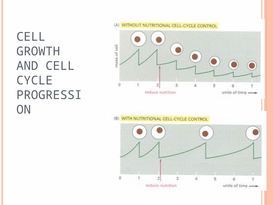
CELL GROWTH AND CELL CYCLE PROGRESSION

MONITORING OF CELL CYCLE PROGRESSION
Cln3, the budding yeast G1 cyclin, is synthesized in parallel to cell growth
Cells may inherit a fixed amount of inhibitor that binds Cln3

DNA DAMAGE CHECKPOINTS
Checkpoint in late G1 prevents entry into S phase
Checkpoint in late G2 prevents entry into mitosis

G1 CHECKPOINT
DNA damage leads to the activation of p53, a gene regulatory protein
p53 stimulates expression of many genes including a CKI called p21, which binds G1/S-Cdk and S-Cdk

G2 CHECKPOINT
Damaged DNA sends signals to inactivate Cdc25.
Figure 18-17 Essential Cell Biology (© Garland Science 2010)
XX XX

CONTROL OF THE CELL CYCLE

PROGRAMMED CELL DEATH

FUNCTION OF APOPTOSIS
To eliminate damaged cells Is an essential part of development in
multicellular organisms Balances cell division to regulate
tissue/organ size
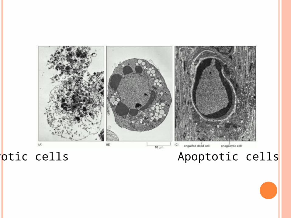
Necrotic cells Apoptotic cells

CASPASE CASCADE
Caspase is a class of proteases
Contain cysteine at their active site
Cleave at specific aspartic acid residue


EXTRACELLULAR CONTROL OF CELL DIVISION

EXTRACELLULAR SIGNALS Mitogens: stimulates cell division by relieving
intracellular negative control
Growth factors: stimulates cell growth by promoting synthesis of proteins and other macromolecules and inhibiting their degradation
Survival factors: promotes cell survival by suppressing apoptosis

PDGF: PLATELET-DERIVED GROWTH FACTOR
Functions as a mitogen
Secreted by platelet cells to stimulate cell division during wound healing
Can act on multiple cell types: fibroblasts, neuroglial cells, smooth muscle cells

MITOGEN SIGNALING PATHWAY(THROUGH GTPASE RAS AND MAP KINASES)

OVERACTIVE MITOGENIC SIGNAL RESULTS IN CELL CYCLE ARREST OR APOPTOSIS

REPLICATIVE CELL SENESCENCEFibroblasts from normal human
tissues can only go through 25-50 population doubling when cultured in standard mitogenic medium

GROWTH FACTOR SIGNALING PATHWAY
PI 3-kinase phosphorylates inositol phospholipid in the membrane activating S6 kinase which activates components of translational machinery
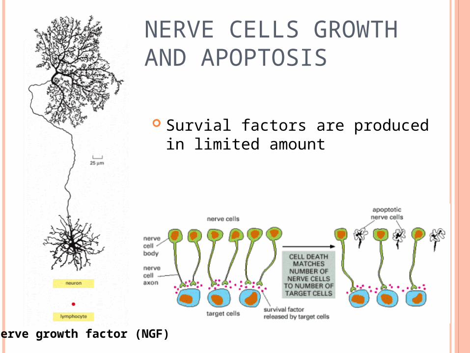
NERVE CELLS GROWTH AND APOPTOSIS
Survial factors are produced in limited amount
Nerve growth factor (NGF)

SURVIVAL FACTOR SIGNALING PATHWAY

ANCHORAGE-DEPENDENT CELL DIVISION
Cells are growth over non-adhesive substratum with or without a patch of adhesive palladium, fed with 3H-thymidine and autoradiographed.

Integrins (cell surface matrix receptors) interact with laminin and/or fibronectin (extracellular matrix molecules, leading to activation of FAK (focal adhesion kinase) and signaling pathways that promote cell survival, growth and division
Actin is labeled in green and proteins with phosphotyrosines are labeled in red

EXTRACELLULAR NEGATIVE SIGNAL PROTEINS TGF- signal proteins inhibit the proliferation
of many cell types (blocking progression through G1 or stimulating apoptosis)
BMP (bone morphogenetic protein) triggers apoptosis of cells between developing digits of a mouse paw
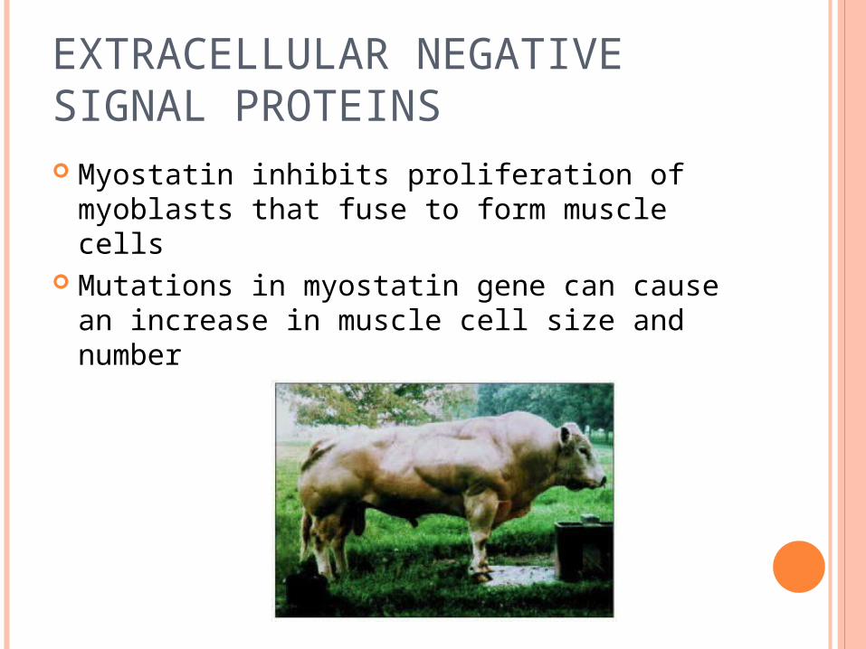
EXTRACELLULAR NEGATIVE SIGNAL PROTEINS Myostatin inhibits proliferation of myoblasts
that fuse to form muscle cells Mutations in myostatin gene can cause an
increase in muscle cell size and number

CONTROL OF BODY SIZE
Through the control of total cell mass
Salamanders of different ploidy levels are of the same body size
But their cell size and cell number are differentKidney tubules
hindbrain
Haploid
Tetraploid

QUESTION
Describe a mechanism by which Cdk is activated during the cell cycle.