cactus protein degradation mediates drosophila dorsal...
TRANSCRIPT
Cactus protein degradation mediates Drosophila dorsal-ventral signaling Marcia P. Belvin, Yishi Jin, ~ and Kathryn V. Anderson 2
Division of Genetics, Department of Molecular and Cell Biology, University of California at Berkeley, Berkeley, California 94720 USA
Dorsal-ventral patterning in the Drosophila embryo relies on a signal transduction pathway that is similar to a signaling pathway leading to the activation of the mammalian transcription factor NF-KB. Stimulation of this Drosophila pathway on the ventral side of the embryo causes the nuclear translocation of Dorsal, the Drosophila NF-~B homolog. Cactus, like its mammalian homolog IKB, inhibits nuclear translocation by binding Dorsal and retaining it in the cytoplasm. We show that Cactus, like IKB, is rapidly degraded in response to signaling. More importantly, signal-dependent degradation of Cactus does not require the presence of Dorsal, indicating that Cactus degradation is a direct response to signaling, and that disruption of the Dorsal/Cactus complex is a secondary result of Cactus degradation. Mutant alleles of cactus that encode more stable forms of the protein block signaling, showing that efficient degradation is necessary for signaling. We find that Cactus protein stability is regulated by two independent processes that rely on different regions within the protein: signal-dependent degradation requires sequences in the amino terminus or ankyrin repeats, whereas signal-independent degradation of free Cactus requires the carboxy-terminal region of the protein that includes a PEST sequence.
[Key Words: Cactus; IKB; NF-KB; protein degradation; dorsal-ventral patterning]
Received January 17, 1995; revised version accepted February 27, 1995.
Signal transduction pathways that mediate the responses of eukaryotic cells to extracellular signaling molecules generally involve covalent modifications of cytoplasmic proteins. Protein phosphorylation, mediated either by re- ceptor tyrosine kinases or by cytoplasmic kinases that associate with activated receptors, is the best understood of these signal-induced modifications. Recent data have suggested that a different kind of covalent change, pro- tein degradation, may play an important role in at least one kind of signal transduction pathway, the pathway that leads to the activation of the transcription factor NF-KB.
NF-KB, a mammalian transcription factor originally identified in B cells, but subsequently found in several nonimmune cell types, is activated rapidly by a post- translational mechanism in response to a variety of ex- tracellular stimuli (Blank et al. 1992; Gri!li et al. 1993). Inactive NF-KB is retained in the cytoplasm by its inhib- itor, IKB, and migrates into the nucleus when the NF- KB/IKB complex dissociates in response to several cytok- ines, including interleukin-1 (IL-1) and tumor necrosis factor (TNFR) (Sen and Baltimore 1986; Bauerle and Bal- imore 1988; Beg et al. 1993). IKB family members share a block of ankyrin repeats that mediate binding to NF-KB
1Present address: Department of Biology, Massachusetts Institute of Technology, Cambridge, Massachusetts 02139 USA. 2Corresponding author.
and mask the NF-KB nuclear localization signal (Ganchi et al. 1992; Henkel et al. 1992; Beg and Baldwin 1993), providing an explanation for why NF-KB/IKB complexes remain cytoplasmic while free NF-KB is nuclear.
Recent work using mammalian tissue culture cells has shown that IKB is rapidly degraded in response to signal- ing, and that this degradation correlates with NF-KB ac- tivation (Beg et al. 1993; Henkel et al. 1993; Miyamoto et al. 1994; Palombella et al. 1994; Traenckner et al. 1994). Several calpain and proteasome inhibitors stabilize IKB and block NF-KB activation, indicating that IKB degrada- tion is necessary for activation of NF-KB (Miyamoto et al. 1994; Traenckner et al. 1994; Lin et al. 1995). In some of these experiments, the stabilized form of IKB is a more highly phosphorylated form, suggesting that phosphory- lation of IKB in the complex in response to signaling precedes its degradation.
The Drosophila dorsal-ventral signal transduction pathway is comprised of 12 known maternal effect genes. The end result of the activity of this pathway is the nuclear localization of the transcription factor Dor- sal on the ventral side of the embryo. Three components of this pathway, Toll, Cactus, and Dorsal, are homolo- gous to members of the IL-1/NF-KB pathway. The cyto- plasmic domain of Toll, a transmembrane receptor pro- tein, is homologous to the cytoplasmic domain of the mammalian IL-1 receptor (Hashimoto et al. 1988; Schneider et al. 1991). Dorsal and Cactus are homolo-
GENES & DEVELOPMENT 9:783-793 �9 1995 by Cold Spring Harbor Laboratory Press ISSN 0890-9369/95 $5.00 783
Cold Spring Harbor Laboratory Press on May 25, 2018 - Published by genesdev.cshlp.orgDownloaded from

Belvin et al.
gous to NF-KB (Steward 1987) and IKB (Geisler et al. 1992; Kidd 1992), respectively. Localized ventral activa- tion of the Toll receptor causes disruption of the Dorsal/ Cactus complex and the subsequent nuclear localization of Dorsal, just as activation of the IL-1 receptor leads to the disruption of the NF-KB/IKB complex and the nuclear localization of NF-KB. Neither signal-induced phosphor- ylation nor signal-dependent degradation of the Droso- phila IKB homolog Cactus has been observed previously (Whalen and Steward 1993). However, given the homol- ogy that exists between the Drosophila and mammalian pathways, it seems likely that degradation of Cactus could also be linked to the activation of Dorsal.
The early Drosophila embryo provides a unique oppor- tunity to dissect the in vivo response of Cactus and Dor- sal to signaling. By activating the pathway at a precise time by microinjection of the activating extracellular ligand, Sp~itzle, we show that Cactus, like IKB, is rapidly degraded in vivo in response to signaling. Taking advan- tage of Drosophila mutants, we show that signal-in- duced Cactus degradation can occur in the absence of Dorsal, indicating that degradation of the Cactus protein is a direct result of signaling, rather than a secondary consequence of separation of the Dorsal/Cactus com- plex. We find that mutant alleles of Cactus that increase its stability block signaling, confirming that destruction of Cactus is necessary for signaling. These mutant alleles also reveal that Cactus protein turnover is regulated by two independent processes: signal-independent degrada- tion of free Cactus, which requires the carboxy-terminal PEST region of the protein, and signal-dependent degra- dation, which is mediated by more amino-terminal re- gions of the protein.
R e s u l t s
Signaling causes rapid degradation of wild-type Cactus
To test whether Cactus, like IKB, is degraded in response to signaling, we activated the dorsal-ventral signaling pathway at a controlled t ime by injecting embryos with processed Sp/itzle, the putative Toll ligand (Morisato and Anderson 1994; Schneider et al. 1994; Fig. 1C). An acti- vated, processed form of the Sp~itzle protein can activate Toll when it is injected into the extracellular (perivi- telline) space of early embryos (Schneider et al. 1994). Therefore, to stimulate the pathway, we injected acti- vated Spfitzle protein into the perivitelline space of syn- cytial blastoderm embryos. In the 1- to 1.5-hr embryos used, all of the components required for signaling are present, and localized endogenous signaling is just begin- ning. After a short incubation, the embryonic cytoplasm was harvested and the Cactus protein was analyzed by Western blot. We observed rapid degradation of Cactus in response to injection of activated Sp~itzle (Fig. 1A); >75% of the Cactus protein was degraded in the first 15 min after injection. In control injections with water, Cactus was not detectably degraded in the 30 min fol- lowing injection (data not shown).
To test whether the Cactus degradation that we ob- served was specific to the dorsal-ventral pathway, we repeated these injections into embryos from Toll- (T1-) females. Because activated Spfitzle is postulated to bind and activate the Toll receptor, it should not stimulate signaling in an embryo lacking Toll. Figure 1B shows that injection of activated Sp~itzle into T1- embryos did not cause degradation of Cactus. Therefore, the signal- dependent degradation of Cactus that we observe in
Figure 1. Degradation of Cactus in re- sponse to signaling. Response of 1.5- to 2-hr-old embryos from wild-type (A) or TI-, tub-, and pll- females (B). {C)Sche- matic diagram of the order of action of genes in the pathway. For A and B, em- bryos were injected in the perivitelline space with an activated Spfitzle prepara- tion. Embryonic cytoplasm was collected 5 or 15 min after injection and analyzed by Westem blot, using an anti-Cactus anti- body. The lanes labeled Uninj contain cy- toplasm from uninjected embryos; the lanes marked 5' and 15' contain cytoplasm collected 5 and 15 min after injection of Sp/itzle protein. The arrows point to the Cactus protein. In wild-type embryos, Cac- tus protein disappeared rapidly after Sp~it- zle injection (A}. In mutant embryos that lack Toll, Tube, or Pelle, there was no deg- radation of Cactus in response to Spfitzle injection (B). The high molecular weight- band in each lane is a crossreacting band that can be used as an internal loading control. Control injections with water caused no degradation of Cactus (data not shown). The blot in A resolves the two forms of Cactus: the predominant phosphorylated form (arrow), and the less abundant, more rapidly migrating unphosphorylated form. The upper band collapses to the lower band after treatment with calf intestinal phosphatase (Whalen and Steward 1993).
784 GENES & DEVELOPMENT
Cold Spring Harbor Laboratory Press on May 25, 2018 - Published by genesdev.cshlp.orgDownloaded from

Cactus protein degradation
wild-type embryos is specific to the dorsal-ventral path- way. We also tested whether this degradation required the dorsal group genes tube and pelle, which act down- stream of Toll and upstream of dorsal and cactus (Fig. 1C). The sequence of tube does not suggest a biochemi- cal function (Letsou et al. 1991); pelle appears to encode a cytoplasmic serine/threonine kinase (Shelton and Wasserman 1993). We found that in embryos from t u b e - ( tub-) and pe l l e - (pll-) females, Cactus does not de- grade in response to signaling (Fig. 1B). Therefore, signal- dependent degradation of Cactus is mediated by all of the genetically defined components of the pathway that act between Spdtzle and dorsal~cactus in the pathway.
Cactus is rapidly degraded in response to signal even in the absence of Dorsal
When the rapid degradation of IKB in response to TNFa is blocked by calpain inhibitors, IKB becomes phosphory- lated but does not dissociate from NF-KB (Miyamoto et al. 1994). One interpretation of this result is that the degradation of IKB in response to signal takes place when it is in the complex. However, in the absence of inhibi- tors, it has not been shown whether degradation of IKB is a direct response to in vivo signaling or occurs as a sec- ondary consequence of IKB becoming separated from NF- KB. To differentiate between these two possibilities for Cactus and Dorsal, we wanted to assay the response of Cactus to injection of activated Sp~itzle into embryos lacking Dorsal protein. If Cactus is a direct target for signaling, then activation of the signaling pathway should cause rapid degradation of Cactus even in the absence of Dorsal. If Cactus is degraded only as a conse- quence of being separated from Dorsal, then the level of Cactus protein in the absence of Dorsal should be unre- sponsive to signaling.
A complication of this experiment is that that there is no detectable maternal Cactus protein in embryos or ovaries lacking Dorsal (Fig. 2, lane 1; Whalen and Stew- ard 1993; Kidd 1992), suggesting that Cactus is unstable when not bound to Dorsal even prior to signaling. We found, however, that it is possible to produce Cactus protein in embryos lacking Dorsal protein, that is, em- bryos laid by dorsal- (dl - ) females, by injecting in vitro- synthesized cactus RNA into the cytoplasm of these em- bryos and allowing translation in vivo (Fig. 2, lanes 2,4). To test the effect of signaling, activated Sp~itzle was in- jected into the perivitelline space of the embryos after translation of Cactus had begun (Fig. 2, lanes 3,5). We observed that Cactus was degraded in response to signal- ing at roughly the same rate in the embryos from d l - females as in embryos from wild-type females (cf. Fig. 2, lanes 2-5 with Fig. 1A), whereas it continued to accu- mulate in the absence of signaling (Fig. 2, lanes 2,4). We conclude that free Cactus is as effective a substrate for signal-dependent degradation as Cactus in the Dorsal- Cactus complex, indicating that Cactus degradation is a direct consequence of signaling.
This experiment also revealed that two kinds of deg- radation control the level of Cactus protein in the era-
Figure 2. Degradation of Cactus in response to signaling in the absence of Dorsal. To test the effect of signaling on Cactus protein in the absence of Dorsal, 1.5- to 2-hr-old embryos laid by dl- females were injected with cactus transcripts. (Lane 1) Un- injected dl- embryos contain no detectable Cactus protein. Fol- lowing a 5-min incubation to allow translation of the injected RNA, activated Sp/itzle was injected into the perivitelline space of the experimental embryos (lanes 3,5) but not the controls (lanes 2,4). After an additional 5 or 15 min with or without injected Spgtzle, cytoplasm was collected and assayed for Cac- tus protein as described in Fig. 1. In the absence of Sp~itzle (lanes 2,4) Cactus protein accumulated, whereas in embryos exposed to Sp/itzle for 5 or 15 rain (lanes 3,5), the newly synthesized Cactus protein was degraded. The arrow points to the Cactus protein; the high molecular weight band was used as a loading control.
bryo: signal-dependent and signal-independent degrada- tion. Signal-independent degradation eliminates free Cactus, so that essentially all of the Cactus in the em- bryo is complexed with Dorsal. Signal-dependent degra- dation destroys. Cactus, whether or not it is complexed with Dorsal. Signal-dependent degradation of Cactus must be more rapid than signal-independent degrada- tion, because we observed that the amount of Cactus in embryos lacking Dorsal was decreased dramatically by exposure to signal, even though both injected and con- trol embryos were subject to signal-independent degra- dation (e.g., cf. Fig. 2, lanes 4 and 5).
A more stable m u t a n t form of Cactus inhibits signaling
These experiments showed that Cactus protein stability is controlled by both signal-dependent and signal-inde- pendent degradation but did not test whether degrada- tion of Cactus is necessary for signaling. We isolated an unusual allele of cactus, cact su, on the basis of its ability to block signaling. This mutant allowed us to investigate the importance of Cactus stability in proper signaling.
In a large-scale genetic screen for suppressors of a dom- inant, ventralizing allele of easter, ea 831, one suppressor mutation was isolated, easter acts genetically upstream of Spiitzle and is homologous to serine proteases (Chasan and Anderson 1989; Morisato and Anderson 1994). All of the embryos (100%} laid by ea831/+ females are moder- ately ventralized (Jin and Anderson 1990), whereas Su /+ ; ea83~/+ females produced 50% wild-type, hatching lar-
GENES & DEVELOPMENT 785
Cold Spring Harbor Laboratory Press on May 25, 2018 - Published by genesdev.cshlp.orgDownloaded from

Belvin et al.
vae and 50% unhatched embryos with an apparently wild-type cuticular phenotype (Fig. 3A). The Su muta- tion also suppresses ventralizing mutations of several other dorsal group genes, including Tol l (Fig. 3B), spdtz le , and cac tu s (data not shown), demonstrating that the sup- pressor is not specific to eas ter but, instead, inhibits the activity of the pathway.
The Su mutation mapped by recombination to the sec- ond chromosome between b and pr, a region including the dorsal and c a c t u s genes. However, the Su mutation complemented both dorsal and cac tus , as well as all available deficiencies in this interval, suggesting that it could be a gain-of-function allele. To obtain loss-of-func- tion alleles at this locus we carried out a genetic screen to revert the suppressing effect of the mutation (Materi- als and methods). We obtained four revertants that were no longer able to suppress e a 831 and mapped to the same site as Su. Three of these revertants failed to comple- ment loss-of-function c a c t u s alleles. We therefore con- cluded that the Su mutation was a gain-of-function allele of cac tus and renamed it cac t su.
Despite the ability of cac t su to act as a strong domi- nant suppressor, it had no phenotype on its own: cactSu/ cac t su flies are fully viable, and females produce wildo type embryos. The lack of an obvious effect on dorsal- ventral patterning by cac t su was perplexing, given its strong suppression of ventralization. To look for more subtle effects of the mutation, we examined the Dorsal gradient in embryos from cactSU/cact su females by ex- amining sections stained with an anti-Dorsal antibody. Figure 4 shows that there is a reduction in the extent of the dorsal gradient in cac tSu /cac t su embryos compared with wild type. In wild-type embryos, 27% of the nuclei
in the embryo circumference had high levels of nuclear Dorsal, whereas in cac tSu /cac t su embryos this was re- duced to 20%. The absence of a later embryonic pheno- type indicates that there is enough regulative capacity in the patterning process to compensate for this reduction in the number of Dorsal-containing nuclei.
Cact su encodes a s l i gh t l y t r u n c a t e d pro te in t ha t appears to be m o r e s tab le than w i l d - t y p e C a c t u s
To determine the molecular basis of the cact su activity, we used reverse transcription-PCR to amplify the cac tus mRNA from ovarian RNA of cac t su females. The se- quence revealed that the cac t su allele has a single base pair change in the 5' splice donor of intron six, which should render this splice site defective. The predicted protein lacks the seventh exon of the maternal cac tus mRNA, which encodes 23 amino acids, and in its place has 5 amino acids encoded by intron 6 (Fig. 5A). This mutant protein is almost identical to the zygotic form of Cactus, a naturally occurring splicing variant expressed later in development {Fig. 5A; Kidd 1992).
To characterize the Cact su protein, we performed Western blots of 0- to 2-hr embryo extracts from em- bryos laid by wild-type, c a c t S u / + , and cac tSu /cac t su fe- males. As predicted from the mRNA sequence, the Cact su protein is smaller than the wild-type maternal protein (Fig. 5B). Unexpectedly, we found that in em- bryos from c a c t S ~ / + heterozygous females, there was 10 times as much Cact su protein as wild-type protein (Fig. 5B, lane 2), even though the amounts of wild-type and cac t su RNAs in these embryos were equal (data not
A
ea831 Su ea 83I + + I +
Figure 3. T h e S u m u t a t i o n s u p p r e s s e s v e n -
tralizing alleles of easter and Toll. Su was recovered in a screen for suppressors of a dominant ventralizing allele of easter, ea 831. Embryos produced by eaS31/+ females are moderately ventralized, whereas those pro- duced by Su /+ ;ea831/+ females either hatched or appeared wild type (A). Su also suppressed the dominant phenotype of T1 s4r {B), as well as ventralizing alleles of spdtzle and cactus (data not shown).
B
Tt84 c Su Tl84C
786 GENES & DEVELOPMENT
Cold Spring Harbor Laboratory Press on May 25, 2018 - Published by genesdev.cshlp.orgDownloaded from

Cactus protein degradation
Wild Type
D o r s a l - s t a i n e d 27% n u c l e i
cact Su / c a c t Su
20%
Figure 4. cac t su narrows the Dorsal gra- dient. Embryos {2.5-3.5 hr) laid by wild- type or cac tS~ /cac t s~ females were stained with Dorsal antibody and then sectioned. The extent of the Dorsal gradient {marked by arrows) was calculated as the percent- age of nuclei around the embryonic cir- cumference that stained strongly for nu- clear Dorsal. In the wild-type embryos, 27---2% of the nuclei (24 of 91 in this sec- tion) contained Dorsal, whereas in em- bryos from c a c t S u / c a c t s'' females only 20+--2% of the nuclei (17 of 94 in this sec- tion) contained Dorsal. Four to six sec- tions of each embryo were counted. Eight wild-type embryos and five cac tS~ / cac t su
embryos were scored, and the results of all sections were averaged for each genotype.
shown}. Based on the observat ion tha t Cactus is unsta- ble, the greater a c c u m u l a t i o n of Cact su pro te in could reflect its greater s tabi l i ty. Mate rna l Cac tus is not de- tec ted in Wes te rn blots f rom d / - ovar ian extracts (Kidd 1992; Wha len and Steward 1993; see also Fig. 2), presum- ably ref lect ing the ins tab i l i ty of Cactus w h e n no t com-
plexed w i t h Dorsal. If there is n o r m a l l y an equ i l i b r ium be tween the D o r s a l / C a c t u s complex and free subuni ts , t hen any free Cactus pro te in wou ld be a target for deg- radat ion and newly syn thes ized Cactus could take i ts place in the complex. If Cact su represents a more s table pro te in than wi ld type, i t shou ld e v e n t u a l l y replace wi ld
Figure 5. Molecular characterization of cac t su and cac t avEsT. (A} Schematic dia- gram of the wild-type maternal Cactus, zygotic Cactus, Cact su, and Cact avEsx proteins {drawing is not to scale). The ANKYRIN domain is a set of five con- served ankyrin repeats. The PEST do- main is a region rich in Pro, Glu, Asp, Ser and Thr. The first residue after the PEST sequence in Cact su is Glu rather than Val in zygotic Cactus because of the EMS-induced base change {T ~ A at the second nucleotide in intron 6) at the splice junction. Cact avEsT lacks the en- tire PEST sequence, as well as the car- boxy-terminal region deleted in Cact su. {B) Cactus Western blot of early embryo extracts from 0- to 2-hr embryos pro- duced by wild-type, c a c t S u / + , or cactSu/
cac t s~ females. Arrows point to the wild-type Cactus (lane 1) and Cact su {lane 3) proteins. (Lane 2) In embryos from c a c t S u / + heterozygotes, there was -10 times more Cact su protein than wild-type Cactus protein. {C) Cactus Western blot of cytoplasm from wild- type embryos injected with identical concentrations of cac t +, cac t su, and cac t avEsT RNA (Materials and methods), followed by a 20-rain incubation to al- low translation of the transcripts. The
left-hand arrow points to the endogenous wild-type Cactus; the right-hand arrows point to the Cact s~ proteins in lane 2 {large arrow) and the Cact avEsT proteins in lane 3. After 20 min of translation there is approximately three times m o r e C a c t apEsT protein than wild-type Cactus present as calculated by densitometry. Note that both CactS~and Cact avEsT accurnulate phosphorylated and un- phosphorylated forms, indicating that this phosphorylation site{s) is not located in the carboxyl terminus.
GENES & DEVELOPMENT 787
Cold Spring Harbor Laboratory Press on May 25, 2018 - Published by genesdev.cshlp.orgDownloaded from

Belvin et al.
type Cactus, yielding a ratio of CactSU/wild-type Cactus similar to what we observed.
Dele t ing the PEST sequence s tabi l i zes Cactus and s trongly i nh ib i t s signaling
The amino acids deleted in Cact su lie just carboxy-ter- minal to the PEST region of the protein (Fig. 5A; Geisler et al. 1992; Kidd 1992). PEST sequences are rich in Pro, Glu, Asp, Set, and Thr and are thought to be associated with rapid protein turnover (Rogers et al. 1986; Rech- steiner 1990). One possible explanation for the appar- ently increased stability of Cact s~ is that the lack of sequences immediately adjacent to the PEST region in- terferes with the destabilizing function of the PEST se- quence. In this view, removing the PEST sequence en- tirely might result in an even more stable protein.
To test whether the PEST sequence does affect Cactus stability, we constructed a mutant form of cactus, cact aPEsT, in which both the seventh exon and the entire PEST sequence were deleted. The ankyrin repeat domain remained intact in this mutant (Fig. 5A). To compare the stability of the different Cactus proteins, we injected in vitro-synthesized trancripts of cact+, cact s'', and c a c t a P E s T into wild-type embryos and allowed them to be translated in vivo. We then collected the cytoplasm from these embryos and analyzed it by Western blot. After 20 min of translation, there was m o r e C a c t APEsT
and Cact s~ than wild-type Cactus (Fig. 5C), even though equivalent concentrations of RNA were injected (Mate- rials and methods). This suggested that C a c t APEsT and Cact s~ are both more stable than wild- type Cactus.
To test whether cact APEsT could block signaling like cact s~, we injected t a c t a p E s T RNA transcripts into wild- type embryos. This resulted in weakly dorsalized em- bryos (Fig. 6), suggesting that the C a c t aPEsT protein in- terfered with signaling. A stronger effect was seen when t a c t a P E s T RNA was injected into cactSU/cact su embryos, where 15% of the embryos were completely dorsalized (Fig. 6). This strong dorsalization was never observed with injection of cact + or cact su transcripts (data not shownl. Therefore, C a c t APEsT is more effective at block- ing signaling than Cact su and is capable of completely blocking all signaling in embryos.
Cact su and Cact aP~sT also degrade rapid ly
in response to signal
The above experiments showed that the more stable forms of Cactus, Cact s~ and C a c t aPEsT, c a n block signal- ing. In principle, the ability to block signaling could re- flect resistance to either signal-dependent or signal-inde- pendent degradation of Cactus. To determine whether Cact su and C a c t aPEsT w e r e more resistant to signal-de- pendent degradation than wild-type Cactus, we injected activated Sp/itzle into embryos laid by females carrying these mutants. Activated Sp~itzle elicited the rapid deg- radation of the Cact su protein at a similar rate as the wild-type protein (cf. Figs. 7A and 1A). There was more Cact su than wild-type Cactus per embryo at all time
cac t A P E S T ~ W i l d T y p e
cac tSu c a c t A P E S T > cac t Su
Figure 6. t a c t ApEsT c a n block signaling. In vitro-synthesized cact apEsT RNA was injected into embryos from wild-type or cactSU/cact su females. When injected into wild-type embryos, cact apEsT RNA causes weak dorsalization (top). When cact aPEsT was injected into embryos produced by cactSu/cact su females, it caused complete dorsalization in 15% of the embryos (bottom). This strongly dorsalized embryo lacks all ventral and lateral structures and is composed of a hollow tube of dorsal cuticle. Weaker dorsalization was observed in the other 85% of injected embryos.
points, but the rate of degradation was approximately the same. Therefore, the Cact su protein, while encoding a more stable form of Cactus, is not resistant to signal- dependent degradation.
To test the response of C a c t avEsT to signaling, we made stable transformants of c a c t ApEST (Materials and methods). When activated Sp/itzle was injected into em- bryos from transformant females carrying one copy of t h e c a c t a P E s T transgene and two wild-type copies of cac- tus, both C a c t aPEsT and wild-type Cactus were degraded rapidly (Fig. 7B). Therefore, both Cact su and C a c t aPEsT
are degraded rapidly in response to signal.
Cact su and Cact aP~sT are res i s tan t to signal- i n d e p e n d e n t degradation
The previous experiments showed that Cact su and C a c t apEsT undergo normal rates of signal-dependent deg- radation. To test whether their greater stability might be caused by greater resistance to signal-independent deg- radation of free Cactus, we compared the level of protein present after injection of cact +, cact su, and c a c t a P E s T
RNAs into embryos from d l - females prior to the nor- mal time of signaling (Fig. 8). Because the mutant pro- teins were synthesized in the absence of Dorsal, and be- cause no activated Sp~itzle was injected, the proteins were subject to signal-independent but not signal-depen- dent degradation. Thirty minutes after injection, there
788 GENES & DEVELOPMENT
Cold Spring Harbor Laboratory Press on May 25, 2018 - Published by genesdev.cshlp.orgDownloaded from

Figure 7. Cact su and C a c t avEsT a r e degraded in response to signaling. Activated Spatzle was injected into embryos pro- duced by cactSu/cact su females (A) or P[w +, cact apEsT] females (B). The arrow in A points to the Cact su protein. Cact su degrades rapidly in response to signaling: 15 min following injection there is still some detectable Cact su protein visible (lane 3), but its level is greatly reduced compared with uninjected embryos (lane 1). In B, embryos laid by transformant females carrying one copy of cact aPFsT and two copies of wild-type cactus were in- jected with activated Sp~itzle. This transformant line produces substantially less C a c t avEsT protein than wild-type Cactus. The arrow points to the wild-type Cactus protein; the open arrow- head points to the Cact aPEsT protein. Lane 1 contains cytoplasm from uninjected embryos. Five minutes after injection of acti- vated Sp~itzle (lane 2), the levels of both wild-type Cactus and C a c t aPnsT a r e greatly reduced.
was m u c h more Cact su and Cact aPEsT protein present than wild-type Cactus (Fig. 8, lanes 3,6,9) even though equivalent amounts of RNA were injected (Materials and methods). Because cact su contains the entire wild-type 5'- and 3 ' -untranslated regions, the observed difference in protein levels is most l ikely the result of the greater stabili ty of the Cact su and C a c t a P E s T proteins rather than differential translation. This result suggests that the Cact su and Cact avEsT proteins are relatively insensi- tive to signal- independent degradation and that it is the resultant increase in free Cactus in the embryo that in- hibi ts signaling.
Cactus protein degradation
Dorsal, we conclude that Cactus degradation is a direct target of signaling. We infer that in the wild-type em- bryo, Cactus in the complex is degraded and that this degradation releases Dorsal, al lowing it to move to the nucleus.
All of the genetically defined components that act up- stream of cactus in the pathway, Toil, tube, and pelle, are required for signal-dependent degradation of Cactus, confirming that Cactus degradation is an integral step in this signaling pathway. However, the nature of these components does not clarify how Cactus protein is de- graded in response to the signal. Genetic analysis has shown that tube acts downstream of Toll (Hecht and Anderson 1993) and pelle acts downstream of tube (Grosshans et el. 1994). The pelle gene appears to encode a ser ine- threonine kinase (Shelton and Wasserman 1993), and because Cactus is a phosphoprotein, it could be a substrate for Pelle.
Rapid phosphorylat ion of IKB in response to signaling and prior to its degradation has been observed in re- sponse to IL-1 and TNF-a (Beg et el. 1993; Miyamoto et el. 1994; Traenckner et el. 1994). Al though we found that Pelle activity is required in vivo for Cactus degra- dation, we did not observe phosphorylat ion of Cactus in response to signaling. It is possible that such a phosphor- ylated form exists but was not detected by our methods because of its short half-life or because it did not cause a detectable mobi l i ty shift. Alternatively, Pelle might con- trol Cactus degradation indirectly, phosphorylat ing an unknown intermediate in the pathway.
Our findings that the Cact su and Cact aPEsx mutan t s degrade rapidly in response to signaling demonstrate that the PEST sequence is not required for signal-dependent degradation of Cactus. This impl ies that there is another region of Cactus, located either in the amino te rminus or
D i s c u s s i o n
Signal -dependent degradat ion of Cactus
We have shown that Cactus is rapidly degraded in re- sponse to s t imula t ion of the Drosophi la dorsal-ventral signaling pathway. The m a m m a l i a n homolog of Cactus, IKB, is degraded in response to signaling in tissue culture cells (Beg et el. 1993; Henkel et el. 1993; Palombella et el. 1994; Traenckner et al. 1994). Our experiments in whole embryos demonstrate that rapid signal-dependent degradation of Cactus takes place in the intact organism.
Two plausible models could explain how Cactus is degraded in response to the signal. One model is that signaling causes dissociation of the Dorsal /Cactus com- plex by modifying either Dorsal or Cactus, and the re- sulting free Cactus is degraded by signal-independent degradation (see below). The other model is that Cactus is degraded directly in the complex. Because Cactus is degraded in response to signal even in the absence of
Figure 8. Cact su and Cact avEsT are relatively resistant to sig- nal-independent degradation of free Cactus in the absence of Dorsal. In vitro-synthesized transcripts of wild type cactus, cact su, and cact AvEsT were injected into embryos from d/- fe- males. Cytoplasm was collected 10, 20, and 30 min after injec- tion and analyzed by a Cactus Western blot. Identical concen- trations of the three RNAs were injected (Materials and meth- ods}. The level of Cact AvEsT protein that accumulates after 30 min is greater than that of Cact su and much greater than that of wild-type Cactus (cf. lanes 3, 6, and 9). The arrows point to the Cactus proteins; the more slowly migrating band is a cross- reacting band.
GENES & DEVELOPMENT 789
Cold Spring Harbor Laboratory Press on May 25, 2018 - Published by genesdev.cshlp.orgDownloaded from
Belvin et al.
in the ankyr in repeats, that is required for rapid degra- dation in response to signaling.
Two kinds of degradation control Cactus protein levels
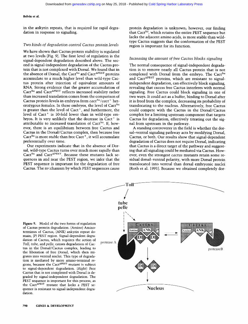
We have shown that Cactus protein stabil i ty is regulated at two levels (Fig. 9). The first level of regulation is the signal-dependent degradation described above. The sec- ond is signal-independent degradation of the Cactus pro- tein that is not complexed wi th Dorsal. We found that in the absence of Dorsal, the Cact su and C a c t APEsT proteins accumulate to a m u c h higher level than wild-type Cac- tus protein after injection of equivalent amounts of RNA. Strong evidence that the greater accumulat ion of Cact su and Cact APEsT reflects increased stabil i ty rather than increased translat ion comes from the comparison of Cactus protein levels in embryos from cactSu/cact + het- erozygous females. In these embryos, the level of Cact su is greater than the level of Cact +, and furthermore, the level of Cact + is 10-fold lower than in wild-type em- bryos. It is very unl ike ly that the decrease in Cact + is attributable to increased translat ion of Cact su. If, how- ever, there is an equi l ibr ium between free Cactus and Cactus in the Dorsal /Cactus complex, then because free Cact su is more stable than free Cact +, it wil l accumulate preferentially over time.
Our experiments indicate that in the absence of Dor- sal, wild-type Cactus turns over much more rapidly than Cact su and C a c t AvEsT. Because these mutan ts lack se- quences in and near the PEST region, we infer that the PEST sequence is important for the degradation of free Cactus. The m~;chanism by which PEST sequences cause
protein degradation is unknown; however, our finding that Cact su, which retains the entire PEST sequence but lacks the adjacent amino acids, is more stable than wild- type Cactus suggests that the conformation of the PEST region is important for its function.
Increasing the amount of free Cactus blocks signaling
The normal consequence of signal-independent degrada- tion is to remove nearly all Cactus protein that is not complexed wi th Dorsal from the embryo. The Cact su a n d C a c t APEsT proteins, which are resistant to signal- independent degradation, can effectively block signaling, revealing that excess free Cactus interferes wi th normal signaling. Free Cactus could block signaling in one of two ways. It could act as a buffer, binding to Dorsal after it is freed from the complex, decreasing its probabili ty of translocating to the nucleus. Alternatively, free Cactus could compete wi th the Cactus in the Dorsal /Cactus complex for a l imi t ing upstream component that targets Cactus for degradation, effectively titrating out the sig- nal from upstream in the pathway.
A standing controversy in the field is whether the dor- sal-ventral signaling pathway acts by modifying Dorsal, Cactus, or both. Our results show that signal-dependent degradation of Cactus does not require Dorsal, indicating that Cactus is a direct target of the pathway and suggest- ing that all signaling could be mediated via Cactus. How- ever, even the strongest cactus mutants retain some re- sidual dorsal-ventral polarity, wi th more Dorsal protein translocated into ventral than dorsal embryonic nuclei (Roth et al. 1991). Because we obtained completely dot-
Figure 9. Model of the two forms of regulation of Cactus protein degradation. {Aminol Amino- terminus of Cactus; lANK) ankyrin repeat do- main; {P) PEST region. Signal-dependent degra- dation of Cactus, which requires the action of Toil, tube, and pelle, causes degradation of Cac- tus in the Dorsal/Cactus complex, leading to the liberation of free Dorsal, which then mi- grates into ventral nuclei. This type of degrada- tion is mediated by more amino-terminal re- gions, because the Cact aPEsT mutant is subject to signal-dependent degradation. (Right) Free Cactus that is not complexed with Dorsal is de- graded by signal-independent degradation. The PEST sequence is important for this process, as t h e C a c t aPnsT mutant that lacks a PEST se- quence is resistant to signal-independent degra- dation.
790 GENES & DEVELOPMENT
Cold Spring Harbor Laboratory Press on May 25, 2018 - Published by genesdev.cshlp.orgDownloaded from

Cactus protein degradation
salized embryos w i t h the s tabi l ized forms of Cac tus pro- tein, th is demons t r a t e s tha t excess Cac tus can effec- t ive ly block all s ignal ing. We therefore conclude tha t degradat ion of Cac tus is necessary for any nuclear trans- loca t ion of Dorsal . Once Cac tus is degraded, the addi- t ional modi f i ca t ion of free Dorsa l t ha t has been observed (Gillespie and W a s s e r m a n 1994) m a y be required for Dor- sal to achieve the h ighes t nuc lear concent ra t ions .
The observa t ion tha t Cactus, l ike IKB, is rapidly de- graded in response to s ignal ing s t r eng thens the parallels be tween the m a m m a l i a n and D r o s o p h i l a s ignal ing path- ways. Given the s imi la r i t i e s tha t exis t be tween the two systems, our f indings on Cac tus m a y also apply to IKB. It should n o w be possible to tes t w h e t h e r the level of IKB protein, l ike Cactus, is cont ro l led by both signal-depen- dent and s igna l - independen t processes, w h e t h e r no rma l s ignal ing targets IKB di rec t ly for degradation, and w h e t h e r there is a specific k inase required for IKB degra- dation. We plan to c o n t i n u e to exploi t the genet ic inter- ac t ions in D r o s o p h i l a to e luc idate the m e c h a n i s m s of s ignal -dependent p ro te in degradat ion.
Mater ia l s and m e t h o d s
Fly strains and mutagenesis
Oregon-R was used as the wild-type stock. The easter alleles are described in Chasan and Anderson (1989) and Jin and Ander- son (1990). For TI- , t ub - , p l l - , and d l - females: TISBR~Q/ T19QRa (Anderson et al. 1985), tubRS6/Df(3R)XM3, p1174/ Df(3R)Ser + R82~ (Hecht and Anderson 1993), and dlS/Df(2L)l 19 (Roth et al. 1989) were used. Ethylmethane sulfonate (EMS) mu- tagenesis was carried out as described (Lewis and Bacher 1968). To isolate suppressors of ea 831, mutagenized ea 831, knirps /TM3, tld l~ males were crossed to ea I, t ldl~ knirps females. Balancers carrying the zygotic lethal mutations had been iso- lated previously (Ferguson and Anderson 1992). The only viable progeny from this cross were ea ], tldl~ 83j, knirps males and females, which were placed in bottles and checked for fertility. The presence of ea ~ in these flies ensured that intragenic sup- pressors would not be recovered, as ea l / ea - females are sterile. Of 75,000 F 1 females, one was fertile, which proved to be cact su. In the screen to generate revertants of cact su, a transformant line was used that contained a dominant allele of easter, ea 831 125.3 inserted on the second chromosome (Jin 1991 ). cactSu/CyO males were mutagenized with EMS and crossed to + /CyO fe- males. Four thousand single F 1 females of the genotype cactSu*/ CyO were crossed to ea831-12s3/CyO males. The embryos laid by cactSU/ea 831-12s3 F2 females were scored for ventralization. Four revertants were recovered; all four mapped to cact su, and three of these were ventralized over cact 99 (Roth et al. 1991).
Cuticle preparations
Embryos were collected for 24 hr and aged for another 24 hr to allow completion of embryonic development. The embryos were then dechorionated in bleach, dissected out of their vi- telline membranes, fixed in 1:4 glycerol/acetic acid, and mounted in Hoyer's medium diluted 1:1 with lactic acid. In- jected embryos were allowed to develop for 36 hr after injection before cuticle preparations were made.
Embryo injections
Synthetic SP6 RNA transcripts were generated by in vitro tran-
scription carried out in the presence of radioactively labeled ATP as described (Schneider et al. 1991) with the modification that two sequential 5 M ammonium acetate/isopropanol precip- itations were carried out followed by Cerenkov counting. The transcripts were diluted as necessary to ensure equivalent con- centrations. For RNA injections, transcripts were injected into 1- to 1.5-hr embryos (Chasan and Anderson 1989). Embryos were allowed to develop for cuticle preparations, or the cyto- plasm was collected for Western analysis. Cytoplasm was col- lected by aspirating the contents of 15 embryos into the injec- tion needle, avoiding any cytoplasm that had leaked out of the embryo after injection. The cytoplasm was deposited into the injection oil where it was recovered with a pipette and placed immediately in buffer on ice. The buffer was a 1:1 dilution of 20 mM HEPES (pH 7.5), 20 mM NaC1, 1 mg/ml of aprotinin, 1 mg/ml of leupeptin, 1 mg/ml of antipain, in 2x protein sample buffer (Ausubel et al. 1991} containing 10% 2-mercaptoethanol. A crude activated Sp/itzle preparation was made (Schneider et al. 1994), and a concentration equivalent to 150 U/ml, as de- scribed in Schneider et al. (19941 was used. The activated Sp/it- zle injections were carried out in 2- to 2.5-hr embryos. For the sequential RNA and activated Sp~itzle injections, RNA was first injected into the cytoplasm as described above. The activated Sp/itzle was then injected into the perivitelline space on the dorsal side of the embryo as described (Schneider et al. 1994) using the same point of entry as for the RNA. Cytoplasm was collected for Western analysis as described above.
Ant ibody staining of embryos
Embryos (2.5- to 3.5-hr) were prepared and stained as described (Roth et al. 1989} using a 1:1000 dilution of rat anti-Dorsal antibody received from R. Steward (Rutgers University, Piscat- away, NJ). A goat anti-rat-horseradish peroxidase (HRP) second- ary antibody was used; staining was detected with DAB. Em- bryos were embedded in Durcupan (Fluka) resin and sectioned (10 p~m).
Cactus constructs and transformants
To sequence cact su, ovarian RNA was isolated (Ashburner et al. 1989a) from cactSU/cact su females. An oligonucleotide (GAT CGA ATT CTT CTG CAT CCT TGT ATG CTT TA) directed against the 3' end of the cactus eDNA was used as a primer for reverse transcriptase, and RT-PCR was carried out (Ausubel et al. 1991) using this primer as the 3' primer and GAT CGG ATC CAT TCG CTA TCG AAA CGT G as the 5' primer. The 5' primer contained a BamHI site, and the 3' primer contained an EcoRI site for cloning into pSP64Poly(A) (Promega). Sequencing was performed according to Ausubel et al. (1991.) Cact aP~sT was constructed by PCR mutagenesis using TTT GCC GTG CCA AAC GAA as the 5' primer and ACT GTC CGG ATC ATC ATA CGG TCTC as the 3' primer. This inserted two tandem stop codons just prior to the PEST sequence. AgeI, a site just downstream of the stop codons, and BspEI, an upstream site, were used to clone the mutant fragment into the wild-type vec- tor. CaSpeR and p~25.7wc were used to generate stable trans- formants (Ashburner 1989b).
Western blots
Samples were boiled for 5 min, spun at 13,000 rpm for 2 min, and run on a 7% polyacrylamide gel. The proteins were trans- ferred onto nitrocellulose (Sigma) and probed with 1:1000 mouse anti-Cactus antibody from S. Kidd as described (Ausubel et al. 1991). A goat anti-mouse HRP-conjugated antibody (Bio-
GENES & DEVELOPMENT 791
Cold Spring Harbor Laboratory Press on May 25, 2018 - Published by genesdev.cshlp.orgDownloaded from
Belvin et al.
Rad) was used as the secondary antibody, and ECL (Amersham) was used for detection. Scanning densitometry was used to mea- sure band densities�9
A c k n o w l e d g m e n t s
We are very grateful to Simon Kidd for providing Cactus anti- body. We thank Ruth Steward for providing Dorsal antibody and Robert Geisler for sending the sequence of Cactus prior to its publication. We thank Tanya Wolff, Don Rio, and members of the Anderson laboratory, especially Sima Misra, for helpful comments on the manuscript. This work was supported by an American Cancer Society grant (DB-42D) to K.V.A. and a Na- tional Institutes of Heath predoctoral training grant to M.P.B.
The publication costs of this article were defrayed in part by payment of page charges. This article must therefore be hereby marked "advertisement" in accordance with 18 USC section 1734 solely to indicate this fact.
R e f e r e n c e s
Anderson, K.V., G. Jfirgens, and C. Nfisslein-Volhard. 1985. Es- tablishment of dorsal-ventral polarity in the Drosophila em- bryo: Genetic studies on the role of the Toll gene product. Cell 42: 779-789.
Ashbumer, M. 1989a. Drosophila: A laboratory manual. Cold Spring Harbor Laboratory Press, Cold Spring Harbor, New York.
�9 1989b. Drosophila: A laboratory handbook. Cold Spring Harbor Laboratory Press, Cold Spring Harbor, New York.
Ausubel, F.M., R. Brent, R.E. Kingston, D.D. Moore, J.G. Seid- man, J.A. Smith, and K. Struhl. 1991. Current protocols in molecular biology. John Wiley and Sons/Greene, New York.
Baeuerle, P.A. and D. Baltimore. 1988. Activation of DNA-bind- ing activity in an apparently cytoplasmic precursor of the NF-KB transcription factor. Cell 53: 211-217.
Beg, A.A. and A.S. Baldwin Jr. 1993. The IKB proteins: Multi- functional regulators of Rel/NF-KB transcription factors. Genes & Dev. 7: 2064-2070.
Beg, A.A., T.S. Finco, P.V. Nantermet, and A.S. Baldwin Jr. 1993. Tumor necrosis factor and interleukin-1 lead to phosphory- lation and loss of IKBa: A mechanism for NF-KB Activation. Mol. Cell Biol. 13: 3301-3310.
Blank, V., P. Kourilsky, and A. Isra61. 1992. NF-KB and related proteins: Rel/dorsal homologies meet ankyrin-like repeats. Trends Biochem. Sci. 17: 135-140.
Chasan, R. and K.V. Anderson. 1989. The role of easter, an apparent serine protease, in organizing the dorsal-ventral pattern of the Drosophila embryo. Cell 56: 391-400.
Ferguson, E.L. and K.V. Anderson. 1992. Localized enhance- ment and repression of the activity of the TGF-I3 family member, decapentaplegic, is necessary for dorsal- ventral pattem formation in the Drosophila embryo. Development 114: 583-597.
Ganchi, P.A., S.C. Sun, W.C. Greene, and D.W. Ballard. 1992. IKB/MAD-3 masks the nuclear localization signal of NF-KB p65 and requires the transactivation domain to inhibit NF- KB p65 DNA binding�9 Mol. Biol. Cell 3: 1339-1352.
Geisler, R., A. Bergmann, Y. Hiromi, and C. Niisslein-Volhard. 1992. cactus, a gene involved in dorsoventral pattern forma- tion of Drosophila, is related to the IKB gene family of ver- tebrates. Cell 71: 613-621.
Gillespie, S.K. and S.A. Wasserman. 1994�9 dorsal, a Drosophila Rel-like protein, is phosphorylated upon activation of the transmembrane protein Toll�9 Mol. Cell. Biol. 14: 3559-3568.
Grilli, M., J.J.-S. Chiu, and M.J. Lenardo. 1993. NF-KB and Rel: Participants in a multiform transcriptional regulatory sys- tem. Int. Rev. Cytol. 143: 1-62.
Grosshans, J, A. Bergmann, P. Haffter, and C. Nfisslein-Volhard. 1994. Activation of the kinase Pelle by Tube in the dorso- ventral signal transduction pathway of Drosophila embryo. Nature 372: 563-566.
Hashimoto, C., K.L. Hudson, and K.V. Anderson. 1988. The Toll gene of Drosophila, required for dorsal-ventral embryonic polarity, appears to enode a transmembrane protein. Cell 52: 269-279.
Hecht, P.M. and K.V. Anderson. 1993. Genetic characterization of tube and pelle, genes required for signaling between Toll and dorsal in the specification of the dorsal-ventral pattern of the Drosophila embryo. Genetics 135:405-417.
Henkel, T., U. Zabel, K. van Zee, J.M. Miiller, E. Fanning, and P.A. Baeuerle. 1992. Intramolecular masking of the nuclear location signal and dimerization domain in the precursor for the p50 NF-KB subunit. Cell 68: 1121-1133.
Henkel, T., T. Machleidt, I. Alkalay, M. Kr6nke, Y�9 Ben-Neriah, and P.A. Baeuerle. 1993. Rapid proteolysis of IKB-a is neces- sary for activation of transcription factor NF-KB. Nature 365: 182-185.
Jin, Y. 1991. "Generating dorsal-ventral asymmetry in the Drosophila embryo: Studies on the easter gene and the po- larizing activity." Ph.D. thesis, University of Calfornia at Berkeley, Berkeley, CA..
Jin, Y. and K�9 Anderson. 1990. Dominant and recessive alleles of the Drosophila easter gene are point mutations at con- served sites in the serine protease catalytic domain. Cell 60:873-881.
Kidd, S. 1992. Characterization of the Drosophila cactus locus and analysis of interactions between cactus and dorsal Pro- teins. Cell 71: 623-635.
Letsou, A., S. Alexander, K. Orth, and S.A. Wasserman. 1991. Genetic and molecular characterization of tube, a Droso- phila gene maternally required for embryonic dorsoventral polarity. Proc. Natl. Acad. Sci. 88: 810-814.
Lewis, E.B. and F. Bacher. 1968. Methods of feeding ethyl meth- ane-sulfonate (EMS) to Drosophila males. Dros. Inf. Serv. 43: 193.
Lin, Y.-C., K. Brown, and U. Siebenlist. 1995. Activation of NF- KB requires proteolysis of the inhibitor IKB-a: Signal-induced phosphorylation of IKB-a alone does not release active NF- KB. Froc. Natl. Acad. Sci. 92: 552-556.
Miyamoto, S., M. Maki, M.J. Schmitt, M. Hatanaka, and I.M. Verma. 1994. Tumor necrosis factor a-induced phosphoryla- tion of IKBa is a signal for its degradation but not dissocia- tion from NF-KB. Pro& Natl. Acad. Sci. 91: 12740-12744.
Morisato, D. and K.V. Anderson. 1994. The spdtzle gene en- codes a component of the extracellular signaling pathway establishing the dorsal-ventral pattern of the Drosophila em- bryo. Cell 76: 677-688.
Palombella, V.J., O.J. Rando, A.L. Goldberg, and T. Maniatis. 1994. The ubiquitin- proteasome pathway is required for processing the NF-KB1 precursor protein and the activation of NF-KB. Cell 78: 773-785.
Rechsteiner, M. 1990. PEST sequences are signals for rapid in- tracellular proteolysis. Semin. Cell Biol. 6: 433-440.
Rogers, S., R. Wells, and M Rechsteiner. 1986. Amino acid se- quences common to rapidy degraded proteins: The PEST hy- pothesis. Science 234: 364-368.
Roth, S., D. Stein, and C. Niisslein-Volhard. 1989. A gradient of nuclear localization of the dorsal protein determines dorso- ventral pattern in the Drosophila embryo. Cell 59: 1189- 1202.
792 GENES & DEVELOPMENT
Cold Spring Harbor Laboratory Press on May 25, 2018 - Published by genesdev.cshlp.orgDownloaded from

Cactus protein degradation
Roth, S., Y. Hiromi, D. Godt, and C. Nfisslein-Volhard. 1991. cactus, a maternal gene required for proper formation of the dorsoventral morphogen gradient in Drosophila embryos. Development 112: 371-388.
Schneider, D.S., K.L. Hudson, T.Y. Lin, and K.V. Anderson. 1991. Dominant and recessive mutations define functional domains of Toll, a transmembrane protein required for dor- sal-ventral polarity in the Drosophila embryo. Genes & Dev. 5: 797-807.
Schneider, D.S., Y. Jin, D. Morisato, and K.V. Anderson. 1994. A processed form of the Spfitzle protein defines dorsal-ventral polarity in the Drosophila embryo. Development 120: 1243- 1250.
Sen, R. and D. Baltimore. 1986. Inducibility of K immunoglob- ulin enhancer-binding protein NF-KB by a posttranslational mechanism. Cell 4 7 : 9 2 1 - 9 2 8 .
Shelton, C.A. and S.A. Wasserman. 1993. peIIe encodes a pro- tein kinase required to establish dorsoventral polarity in the Drosophila embryo. Ceil 72: 515-525.
Steward, R. 1987. Dorsal, an embryonic polarity gene in Droso- phila, is homologous to the vertebrate proto-oncogene, c-reI. Science 238: 692-694.
Traenckner, E.B.-M., S. Wilk, and P.A. Baeuerle. 1994. A pro- teasome inhibitor prevents activation of NF-KB and stabi- lizes a newly phosphorylated form of IKB-a that is still bound to NF-KB. EMBO ]. 13: 5433-5441.
Whalen, A.M. and R. Steward. 1993. Dissociation of the Dorsal- Cactus complex and phosphorylation of the Dorsal protein correlate with the nuclear localization of Dorsal. I. Cell Biol. 123: 523-534.
GENES & DEVELOPMENT 793
Cold Spring Harbor Laboratory Press on May 25, 2018 - Published by genesdev.cshlp.orgDownloaded from
10.1101/gad.9.7.783Access the most recent version at doi: 9:1995, Genes Dev.
M P Belvin, Y Jin and K V Anderson signaling.Cactus protein degradation mediates Drosophila dorsal-ventral
References
http://genesdev.cshlp.org/content/9/7/783.full.html#ref-list-1
This article cites 35 articles, 15 of which can be accessed free at:
License
ServiceEmail Alerting
click here.right corner of the article or
Receive free email alerts when new articles cite this article - sign up in the box at the top
Copyright © Cold Spring Harbor Laboratory Press
Cold Spring Harbor Laboratory Press on May 25, 2018 - Published by genesdev.cshlp.orgDownloaded from