carcinogen-binding proteins in the rat ventral - cancer research
TRANSCRIPT

[CANCER RESEARCH 46, 651-657, February 1986]
Carcinogen-binding Proteins in the Rat Ventral Prostate: Specific
and Nonspecific High-Affinity Binding Sites for Benzo(a)pyrene,3-Methylcholanthrene, and 2,3,7,8-Tetrachlorodibenzo-p-dioxin1
Peter Soderkvist,2 Lorenz Poellinger, and Jan-Âke Gustafsson
Department of Medical Nutrition, Karolinska Institutet, Huddinge University Hospital F-69, S-141 86 Huddinge, Sweden
ABSTRACT
The polychlorinated dibenzodioxin [3H]-2,3,7,8-tetrachlorodi-benzo-p-dioxin (TCDD) and the carcinogens [3H]benzo(a)pyreneand [3H]-3-methylcholanthrene bound to saturable binding sites
in cytosol from the rat ventral prostate. Analysis of equilibriumbinding parameters in diluted cytosol preparations indicated anapparent Ka of ~2 nw and a binding capacity of approximately 1nmol/mg cytosolic protein, corresponding to ~5% of the total
protein content. However, gel permeation chromatography analysis as well as velocity sedimentation analysis on sucrose gradients of [3H]TCDD-labeled rat prostatic cytosol indicated bindingof [3H]TCDD to two discrete species. These analyses indicateda sedimentation coefficient of 3.6-3.8S, a Stokes radius of 25-28 A, and a calculated relative molecular weight of 42,000-
45,000 for the most abundant binding species. The other bindingspecies sedimented at 4-5S under high ionic strength conditionsand at 8-1 OS under low ionic strength conditions and had a
Stokes radius of approximately 60 A, a relative molecular weightof ~100,000, and an estimated concentration of 5-20 fmol/mgcytosolic protein. Binding of [3H]TCDD to this species was
displaceable by a 200-fold M excess of 2,3,7,8-tetrachlorodiben-
zofuran. Therefore, this species was tentatively identified as theTCDD receptor. The properties of the high-capacity binder of[3H]TCDD were found to be similar to the characteristics of a
protein previously purified from the rat ventral prostate, prostaticsecretory protein, which binds androgens as well as estramus-
tine, a nitrogen mustard derivative of estradiol. The binding ofestramustine to diluted prostatic cytosol was shown to be competitively inhibited by 2,3,7,8-tetrachlorodibenzofuran. Moreover,purified prostatic secretory protein bound [3H]TCDD, [3H]-benzo(a)pyrene, as well as [3H]-3-methylcholanthrene. It is sug
gested that binding to this protein is responsible for the high-
binding capacity of carcinogens in cytosol from the rat ventralprostate.
INTRODUCTION
As a chemical etiology of prostatic cancer recently has beensuggested (1, 2), several studies have addressed the issuewhether the prostatic gland possesses enzymes necessary forthe metabolic activation of chemical carcinogens. In the ratventral prostate of untreated rats, it has been shown that the
Received 4/24/85; revised 10/15/85; accepted 10/16/85.1Supported by grants from the Swedish Medical Research Council (Grant 03X-
6807), the Swedish Cancer Society, and the Swedish Board for Planning andCoordination of Research.
2To whom requests for reprints should be addressed.
cytochrome P-450-dependent microsomal enzyme activity AHH3
as well as ¡mmunodetectable amounts of the major 0-naphtho-flavone-inducible isozyme of cytochrome P-450, cytochrome P-450c (the form mainly responsible for AHH-activity) are very low.Following treatment with /3-naphthoflavone or TCDD, an approximately 500-fold induction of cytochrome P-450c is detectable
by both immunochemical techniques and AHH activity determinations (3-7). Moreover, this induction in the prostate has been
shown to be accompanied by an increased capability to formmutagenic metabolites from the promutagens 2-aminofluoreneand B(a)P as determined by the Ames' Sa/mone//a/microsome
assay (6).A substantial amount of evidence indicates that the induction
of cytochrome P-450c in several tissues is mediated by binding
of the inducing agent to an intracellular, soluble receptor protein,which in a poorly understood process leads to an increasedaffinity of the ligand-receptor complex for target sites within the
cell nucleus (8). TCDD is one of the most potent agonists knowntoday for both the enzyme-induction response (9) and receptorbinding (10). Other agonists include such well-known carcinogens as 3-MC and B(a)P (10). The TCDD receptor has been
demonstrated in several extrahepatic tissues in both rats andmice (11,12). Surprisingly, no TCDD receptor has been reportedfor the rat ventral prostate, possibly indicating a nonreceptormediated induction process of cytochrome P-450c in this tissue.
However, a very high degree of nonspecific (nonreceptor) bindingof [3H]TCDD to an unidentified component in undiluted cytosol
from the rat prostate has been reported (11,12). It has also beenreported that [3H]B(a)P readily interacts with components in rat
prostatic cytosol (13). In this paper, we characterize the bindingof various radioligands ([3H]TCDD, [3H]-3-MC, and [3H]B(a)P) to
ventral prostatic cytosol and fractions thereof.
MATERIALS AND METHODS
Chemicals. [1,6-3H]TCDD (28 Ci/mmol) was a generous gift from Dr.A. Poland (Madison, Wl). [G-3H]3-MC (27 Ci/mmol) and [G-3H]B(a)P (25
Ci/mmol) were from the Radiochemical Centre, Amersham, England. [1-14C]-Glucose (8 Ci/mol) was from New England Nuclear Corp. (Boston,
MA). Unlabeled TCDF was kindly supplied by Dr. C. Rappe (Umeà ,Sweden). Estramustine and [2,4,6,7-3H]estramustine (107 Ci/mmol) were
generously provided by Leo Pharmaceuticals (Helsingborg, Sweden).Activated charcoal (Norit A), cytochrome c (equine heart), albumin (bovineserum fraction V), rabbit IgG, and catalase (bovine liver) were purchased
3The abbreviations used are: AHH, aryl hydrocarbon hydroxylase; cytochromeP-450C, the major form of cytochrome P-450 isolated from rat liver microsomes of0-naphthoflavone-(5,6-benzoflavone}-treated rats; TCDD, 2,3,7,8-tetrachlorodi-benzo-p-dioxin; 3-MC, 3-methylcholanthrene; B(a)P, benzo(a)pyrene; TCDF,2,3,7,8-tetrachlorodibenzofuran; estramustine, 1,3,5(10)-estratriene-3,17/i-diol-3-N-bis(2-chloroethyl)carbamate; DCC, dextran-coated charcoal; PSP, prostatic steroid binding protein; PAH, polycyclic aromatic hydrocarbon.
CANCER RESEARCH VOL. 46 FEBRUARY 1986
651
on April 5, 2019. © 1986 American Association for Cancer Research. cancerres.aacrjournals.org Downloaded from

CARCINOGEN-BINDING PROTEINS IN THE RAT PROSTATE
from Sigma Chemical Co. (St. Louis, MO). Sephacryl S-300, SephadexG-25 and G-100 fine, DEAE-Sepharose, heparin-Sepharose, blue dextran2000, and dextran T-70 were from Pharmacia Fine Chemicals (Uppsala,
Sweden). All other chemicals were analytical grade products from eitherSigma or Merck AG (Darmstadt, Federal Republic of Germany).
Animals. Male Sprague-Dawley rats weighing 200-300 g were used
throughout this study.Buffer. The following buffer was routinely used: 20 rnw potassium
phosphate-1 HIM EDTA-2 rriM 2-hydroxyethylmercaptan-10% (w/v) glyc-
erol, pH 7.2.Preparation of Cytosol. Rats were killed by cervical dislocation and
the two ventral prostate lobes were immediately excised and removedonto ice. All successive work was carried out at 0-4°C unless indicated
differently. The prostates were finely minced with a pair of scissors inapproximately 5-10 vol of buffer and thereafter homogenized in 2 vol ofbuffer in a teflon/glass Potter-Elvehjem homogenizer. The homogenate
was then centrifuged at 140,000 x gav for 45 min. The supernatant,hereafter termed cytosol, was removed and care was taken to avoid thefloating lipid layer.
Purification of PSP. The mincing procedure of the prostatic glandresulted in release of secretory fluid from the ductules and lumina (14)which subsequently together with the cytosol was used for purificationof PSP by anion-exchange and gel permeation chromatography as
described by Heyns et al. (15). Purified PSP was quickly frozen in aliquotsand stored at -70°C. In some cases, PSP was delipidated by precipi
tation in acetone (16,17) before assessment of ligand binding.In Vitro Labeling Conditions. For determination of TCDD receptor
binding, crude cytosol or various Chromatographie fractions of cytosolwere labeled with 3-5 nw [3H]TCDD at the indicated protein concentrations for 2-18 h at 0-4°C, usually in the absence or presence of a 200-
fold M excess of unlabeled TCDF. TCDF was used instead of unlabeledTCDD to determine specific binding in view of its greater solubility inwater and similar affinity for the receptor (10). Bound and free ligandwere usually separated by DCC adsorption [1.9% (w/v) charcoal and0.19% (w/v) dextran T-70, final concentrations] as previously described(18). In some cases, a hydroxylapatite-based assay for receptor binding
of TCDD (19) was used to separate free as well as specifically andnonspecifically bound [3H]TCDD. PSP was labeled with the indicatedconcentrations of radioligand at 37°C for 30 min, whereafter DCCadsorption was performed at 0-4°C (0.1-1% charcoal, final concentra
tions, as indicated). All receptor or PSP ligands were dissolved andadded to the incubations in dioxane, the concentration of which neverexceeded 1%(v/v).
Velocity Sedimentation Analysis. Labeled samples (200 n\) werelayered onto linear 5-20% (w/v) or 10-40% (w/v) sucrose gradients in
buffer (containing 0.4 M KCI in indicated cases), followed by centrifugationin an SW 60ri rotor in a Beckman L8-M centrifuge at 260,000-320,000x gavfor 14-18 h to a preset cumulative centrifugal effect (w^) of 1.7 x10¹² rads2/s for TCDD receptor analysis on 10-40% (w/v) sucrosegradients (17) and 2.15 x 10¹² rads2/s for PSP-binding analysis on 5-
20% (w/v) sucrose gradients. Fractions (s200 Ml/fraction) were collected
from the bottom of the centrifuge tubes by gravity flow. Cytochrome c(1.7S), bovine serum albumin (4.4S), -y-globulin (6.6S), and catalase(11.35) were 14C-labeled (20) and used as sedimentation markers. Sedi
mentation velocities of the receptor peaks were calculated according toMartin and Ames (21).
Gel Permeation Chromatography. Analytical gel permeation chromatography on 1- x 115-cm columns of Sephacryl S-300 was performedat a flow rate of 5 ml/cm2/h as previously described (18). The columns
were equilibrated in buffer containing 0.15 M NaCI and 0.02% (w/v)sodium azide. Sephadex G-100 was equilibrated in buffer containing
0.02% (w/v) sodium azide and packed into a siliconized column (1.5 x87 cm). The flow rate was 14 ml/cm2/h and the sample volumes were
approximately 1% of the total column volume. The columns were calibrated with the following standard proteins: thyroglobulin (86.1 A); ferritin
(61.5 A); catalase (51.3 A); bovine serum albumin (35.9 A); and cytc-chromec(17.9A).
Calculation of Hydrodynamic Properties. Relative molecularweights, and frictional ratios due to shape, f/f0, were calculated from theStokes radius, R5, and sedimentation coefficient as described by Siegeland Monty (22), assuming a partial specific volume of 0.725 cm3/g and
a solvation factor of 0.2 g/g of solute (18). Axial ratios, a/b, for hydro-dynamically equivalent prolate ellipsoids were derived from the valueslisted by Schachman (23). S and fls values for the protein standardswere obtained from the literature (24, 25).
Heparin-Sepharose Chromatography. Heparin-Sepharose was equil
ibrated in buffer and packed into a siliconized column (2.6 x 8 cm).Nonlabeled cytosol was applied to the column which was then washedwith 2-3 column vol of buffer, whereafter retained material was eluted
with 1 column vol of buffer containing 0.5 M NaCI at a flow rate of 4 ml/cm2/h.
Saturation Studies. Saturation studies were performed in siliconizedglass tubes at cytosolic protein concentrations of approximately 1 ^g/mlbuffer (26, 27) containing 0.1% (w/v) gelatin (26). Gelatin was includedin the incubation mixture in order to avoid adsorption of ligand or proteinto the wall of the incubation tube. Under these conditions, up to 10 nw[3H]TCDD and approximately 25 nw [3H]B(a)P and [3H]-3-MC, respec
tively, could be dissolved. Diluted cytosol or purified PSP were incubatedin 0.5-ml aliquots with varying concentrations of ligand at 37°C for 30
min. The tubes were removed onto ice, and unbound ligand was adsorbed by the addition of 0.5 ml of an ice-cold DCC suspension [1.5%(w/v) charcoal-0.05% (w/v) dextran T-70-0.1% (w/v) gelatin in buffer].
The incubations were agitated and left on ice for 10 min. The charcoalwas then pelleted by centrifugation at 1500 x g for 10 min, whereafterthe supernatant was assayed for radioactivity. To determine the totalradioactivity added, aliquots (generally, 20 ><l)of the incubations wereremoved shortly before the addition of the DCC. Binding data wereanalyzed by Scatchard plots (28) or double-reciprocal plots using linear
regression analysis to determine apparent equilibrium parameters ofbinding.
General Methods. Protein concentrations were determined using themethod of Lowry ef al. (29), with bovine serum albumin as the standard.Radioactivity was measured in a Scintillator 299 from Packard (DownersGrove, IL) in an LKB-Wallac Rackbeta II scintillation spectrometer (Stock
holm, Sweden) with an average counting efficiency of 40%.Safety Precautions. Since TCDD and TCDF are extremely toxic
compounds (8), their use necessitated special handling and disposalprocedures as outlined by Poland ef al. (10).
RESULTS
By means of gel permeation chromatography of [3H]TCDD-
labeled crude rat ventral prostatic cytosol on Sephacryl S-300 itwas possible to distinguish between two discrete [3H]TCDD-
binding species which both were included into the gel (Fig. 1).The first peak of radioactivity with an f?s of approximately 60 Awas nearly completely abolished by labeling of the crude cytosolin the presence of a 200-fold M excess of TCDF. The 60-Õ
binding entity was, however, partly obscured by high levels of amajor [3H]TCDD-binding species eluting in the 25- to 28-Õ region
of the column. Under these experimental conditions, binding of[3H]TCDD to this entity was nondisplaceable (nonspecific) anddisturbed the further characterization of the 60-Â [3H]TCDD-
binding species. It was therefore investigated whether heparin-Sepharose chromatography of nonlabeled cytosol could removeany interfering nonspecific binding. It has previously been established that the TCDD receptor readily interacts with heparin-Sepharose both in the presence or absence of ligand (18). It waspossible to label the material eluted from heparin-Sepharose with
CANCER RESEARCH VOL. 46 FEBRUARY 1986
652
on April 5, 2019. © 1986 American Association for Cancer Research. cancerres.aacrjournals.org Downloaded from

CARCINOGEN-BINDING PROTEINS IN THE RAT PROSTATE
-10 -
20 40 60 80 100
ELUTION VOLUME Imi!
Fig. 1. Gel permeation chromatography of rat prostatic cytosol labeled with[3H]TCDD.Prostatic cytosol (=10 mg protein/ml) was incubated with 5 nM [3H]TCDD for 2 h at 0-4°Cin the absence (O) or presence (•)of a 200-fold Mexcessof unlabeled TCDF. Bound and free ligand were separated by adsorption toprepelletedDCC [final concentration of charcoal, 1.9% (w/v)]. A sample volume of1 ml was then chromatographed as described under "Materials and Methods."
Fractions (2.0 ml) were collected and assayed for radioactivity. The columns werecalibrated with blue dextran 2000 (V0),[uC]glucose (V,)and the following standardproteins: ?, thyroglobulin (86.1 A); 2, ferritin (61.5 À);_3,catalase(51.3 À);4, bovineserum albumin (35.9 A); and 5, cytochrome c (17.9 A).
2.0
>•1.0
0.5
3 2 1
I I I
SB*
3 2 1
10 20
FRACTION
20top
Fig. 2. Velocity sedimentationanalysisof [3H]TCDD-labeledheparin-Sepharoseeluates. Nonlabeled prostatic cytosol (5 ml; =50 mg protein) was applied to aheparin-Sepharose column and then washed with 2-3 column vol of buffer.Retained material was eluted with 1 column vol of buffer containing 0.5 M NaCIand desalted on Sephadex PD-10 columns. The eluates were incubated with 3 nu[3H]TCDDfor 2 h at 0-4°C,in the absence (O) or presence (•)of a 200-fold Mexcess of unlabeled TCDF. Bound and free ligand were separated by DCCadsorption [final concentration of charcoal, 1.9% (w/v)]. Aliquots (200 >il)were thenlayered onto 10-40% (w/v) sucrose gradients prepared in buffer (A) or in buffercontaining 0.4 M KCI (B). The gradients were centrifugea and fractions werecollected as described under "Materials and Methods." Sedimentation markers
were: 1, cytochrome c (1.7S); 2, bovine serum albumin (4.4S); 3, IgG (6.6S); and4, catalase (11.3S).
[3H]TCDD and detect specifically bound radioactivity by Sephac-
ryl S-300 gel permeation chromatography (data not shown) andsucrose gradient analysis (Fig. 2). The velocity sedimentation ofthe specific [3H]TCDD-binding entity varied as a function of the
ionic strength. At low ionic strength, it sedimented as an 8-1 OS
entity (Fig. 24), whereas at high ionic strength (0.4 M KCI), itsedimented as a 4-5S entity (Fig. 2B). In the heparin-Sepharose
eluates (Fig. 2) as well as in crude cytosol (data not shown),nonspecific [3H]TCDD binding was detectable as a slower sedi-menting entity of 3.6-3.8S under both high- and low-salt conditions. The fls value of the specific [3H]TCDD-binding entity in
prostatic cytosol and its sedimentation coefficient of 4-5S indi
cates a highly assymetrical molecule with a molecular weight ofapproximately 100,000 an f/f0 of 1.7-1.8, and an a/b of 12-15.
All of these hydrodynamic properties are indistinguishable fromthe physicochemical characteristics of the TCDD receptor present in other tissues of the rat (18, 30-32). The concentrations ofthe specific 60-Â [3H]TCDD-binding entity varied between 5-20
fmol/mg of cytosolic protein, as estimated by high-resolution gelpermeation chromatography on Sephacryl S-300. These observations were confirmed by analysis of [3H]TCDD-labeled ventral
prostatic cytosols by a hydroxylapatite assay (19) which indicated TCDD receptor concentrations of approximately 10-12
fmol/mg protein. In this assay, 175 HIM phosphate was sufficientto extract all the specifically bound [3H]TCDD which initially had
been immobilized on hydroxylapatite (data not shown). This is inagreement with the behavior of the TCDD receptor on hydroxylapatite in other tissues, where the TCDD receptor élûtesfromhydroxylapatite at 150-175 rriM phosphate (18,19). Quantitationof specifically bound [3H]TCDD in labeled heparin-Sepharoseeluates yielded concentrations of the specific [3H]TCDD binding
species of -15-20 fmol/mg protein. However, this value might
be artificially low due to, e.g., low recovery of the binding entityduring the heparin-Sepharose chromatography or difficulties in
labeling partially purified TCDD receptor preparations (33).The hydrodynamic properties of the major [3H]TCDD-binding
entity in undiluted cytosol from the rat ventral prostate (fls ~25-28 A; 3.6-3.8S; cf. above) indicate an apparent molecular weightof 40,000-45,000, an f/f0 of 1.0-1.1, and an a/b of 2-5 which is
characteristic of a nearly symmetrical molecule. It has also beenshown that [3H]TCDD interacts with a binding species in rat
ventral prostatic cytosol which focuses at a pH of about 4.7-5
during isoelectric focusing in polyacrylamide gels and which isdistinct from or obscures the TCDD receptor (11). All of thesedata (summarized in Table 1) closely resemble the physicochem-
Table 1Comparisonof physicochemical properties of the major pHJTCDD-binding
species in rat prostatic cytosol and PSPPhysicochemicalcharacteristics of the major rat prostatic [3H)TCDD binding
species were experimentally determined or calculated as described in "Materialsand Methods." Properties of PSP were from the literature or in given cases
calculated by the equations used in the text.
ParameterS(s-")
M, .fl.(A)fifaAxial ratioIsoelectricpointMajor
[3H]TCDD-bindingspecies3.6-3.8
40,000-45,00025-281.1-1.22-54.7-5.0"PSP3.7"
38,000i>-46,000c29"1.3*5-8"4.6-4.9'
" Heyns and De Moor (17).6 Calculatedsum of the molecularweight of the different PSPsubunits (39).c Forsgrener al. (37)." Calculatedvaluesfrom the sedimentationcoefficient (17)and molecularweight
(37) (cf. "Materials and Methods").eCartstedt-Duke(11).' Heynsef al. (36).
CANCER RESEARCH VOL. 46 FEBRUARY 1986
653
on April 5, 2019. © 1986 American Association for Cancer Research. cancerres.aacrjournals.org Downloaded from

CARCINOGEN-BINDING PROTEINS IN THE RAT PROSTATE
¡calcharacteristics of a major secretory protein in the rat ventralprostate, often referred to as PSP (17, 34). In view of these datawe examined the binding of various TCDD receptor ligands tocrude cytosol as well as to purified PSP.
When crude prostatic cytosol or purified PSP was labeled withsubsaturating concentrations of [3H]TCDD at 37°C, a rapid
association of ligand could be observed, as determined by DCCadsorption. After 30 min, maximal association was reached andrepresented approximately 85% of the total radioactivity. Thereafter, charcoal-resistent binding slowly declined, so that after 24h approximately 10% of the maximal binding remained. At 0°C,
the association rate was slow; first after 24 h, a plateau wasreached both in crude cytosol and incubations using pure preparations of PSP. However, in both cases this binding representedonly approximately 30-40% of the maximal binding observed at37°C(data not shown). Analysis of the purified PSP preparations
on 5-20% (w/v) sucrose gradients revealed a single symmetricalpeak of radioactivity in the same region of the gradients (3.5-4S) (Fig. 3A) as the major [3H]TCDD-binding entity in crude
cytosol (cf. above). The radioactivity recovered under this peakaccounted for approximately 78% of the total radioactivity. Itwas also possible to label purified PSP with [3H]-3-MC or [3H]
B(a)P, as determined by velocity sedimentation (Fig. 36). In thecase of [3H]-3-MC and [3H]B(a)P, 65 and 46% of the total
radioactivity were recovered, respectively, on the sucrose gradients.
Saturation of binding of [3H]TCDD and [3H]estramustine to the
major binding species was obtained only when cytosol wasdiluted to a protein concentration of approximately 1 ¿ig/mlinthe presence of 0.1% (w/v) gelatin. This binding was furtherevaluated by Scatchard analysis (28). Linear Scatchard plots (ra 0.9) were obtained for the binding of both compounds. This isin agreement with a single class of binding sites with uniformaffinity for the tested ligand. Typical plots are shown in Fig. 4.Both [3H]TCDD and [3H]estramustine bound to the same maximal number of sites (~1 nmol/mg cytosolic protein; s5% of the
10 20top
20
FRACTION
Fig. 3. Velocity sedimentation analysis of purified PSP labeled with [3H]TCDD(A), or [3H]-3-MC (O) and [3H]B(a)P (•)(B). Purified PSP (a1 mg protein/ml) wasincubated with 5 nM [3H]TCDD, 5 nw [3H]-3-MC, or 5 nw [3H]B(a)P at 37°Cfor 30
min. Bound and free ligand were separated by DCC adsorption [final concentrationof charcoal, 1% (w/v)]. Aliquots (200 pi) were then layered onto 5-20% (w/v)sucrose gradients prepared in buffer. The gradients were centrifuged and fractionswere collected as described under "Materials and Methods". Sedimentation mark
ers were: 1, myogtobin (2.0S); 2, ovalbumin (3.5S); and 3, bovine serum albumin(4.4S).
<U
0.2
0.5
BOUND
1.0
I nM 1Fig. 4. Representative Scatchard plots comparing binding of [3H]estramustine
and [3H]TCDD to rat ventral prostatic cytosol. Cytosol (s1.3 pg protein/ml) wasincubated in the presence of 1% (w/v) gelatin at 37°Cfor 30 min with increasingconcentrations of [3H]estramustine, 0-10 nu (O) or [3H]TCDD, 0-5 nM (D)- Theincubations were terminated by the addition of 0.5 ml of an ice-cold DCC suspensionyielding a final concentration of charcoal of 1% (w/v). Subsequent to DCC adsorption (see "Materials and Methods"), free ligand was calculated by subtracting bound
ligand from the total concentration of ligand present in the incubation. From thesedata, Scatchard plots (25) were constructed by least squares linear regressionanalysis.
4.0
3.0o *-Z 2-
2.0
0.5 1.0{UNBOUND)'1
1.5l n M'
Fig. 5. Competitive binding of TCDF to [3H]estramustine-binding sites in prostatic cytosol. Cytosol (=1.3 tig protein/ml buffer containing 0.1% gelatin) wasincubated at 37°Cfor 30 min with 0.5-1.0 nM [3H]estramustin in the absence (O)
or presence of 5 (•)or 10 (D) nw unlabeled TCDF. Specific binding was determinedby DCC adsorption [final concentration of charcoal. 1% (w/v)] as described under"Materials and Methods," and the obtained data were plotted according to Line-
weaver and Burk (35) using linear regression analysis. Points, mean of duplicatedeterminations.
total cytosolic protein content), suggesting binding of the twoligands to the same site in cytosol. The apparent Ka in thisparticular experiment was 4.1 nM for [3H]estramustine and 1.9nw for [3H]TCDD.
To further investigate if halogenated PAH interacted with thesame class of binding sites in diluted prostatic cytosol as did[3H]estramustine, binding of various concentrations of [3H]estra-
mustine was determined in the absence or presence of 5 or 10nM TCOF, respectively. TCDF was chosen as a structural analogue of TCDD because of its more pronounced solubility inwater (10). The binding data obtained were analyzed accordingto Lineweaver and Burk (35) and were found to be compatiblewith a competitive inhibition of [3H]estramustine binding by TCDF(Fig. 5). An apparent Ka of 4.6 nM could be calculated for [3H]estramustine from the double-reciprocal plot in Fig. 5, which is
in good agreement with the value determined by Scatchard plots(cf. above). Moreover, the number of binding sites determinedby double-reciprocal plot analysis in this experiment was approx
imately 0.7 nmol/mg cytosolic protein.
CANCER RESEARCH VOL. 46 FEBRUARY 1986
654
on April 5, 2019. © 1986 American Association for Cancer Research. cancerres.aacrjournals.org Downloaded from
CARCINOGEN-BINDING PROTEINS IN THE RAT PROSTATE
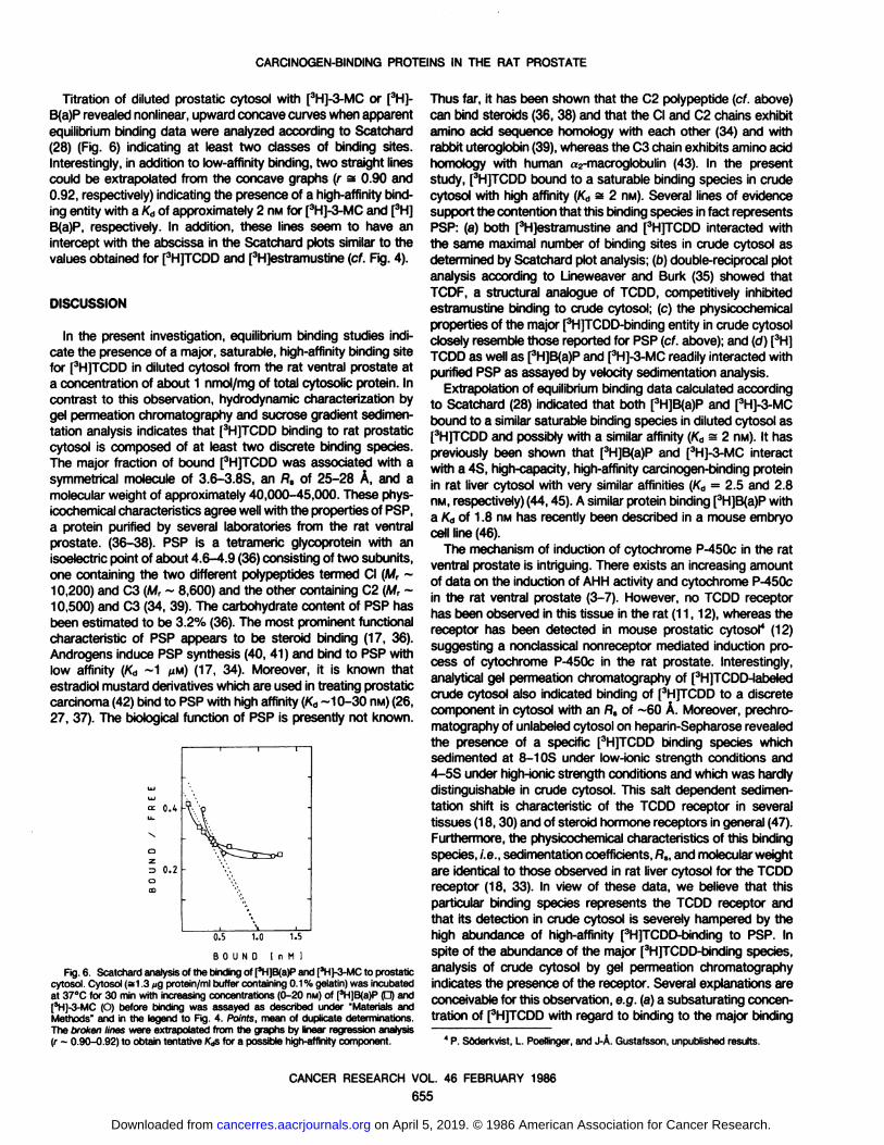
Titration of diluted prostatic cytosol with [3H]-3-MC or [3H]-
B(a)P revealed nonlinear, upward concave curves when apparentequilibrium binding data were analyzed according to Scatchard(28) (Fig. 6) indicating at least two classes of binding sites.Interestingly, in addition to low-affinity binding, two straight linescould be extrapolated from the concave graphs (r s 0.90 and0.92, respectively) indicating the presence of a high-affinity binding entity with a Ka of approximately 2 nw for [3H]-3-MC and [3H]
B(a)P, respectively. In addition, these lines seem to have anintercept with the abscissa in the Scatchard plots similar to thevalues obtained for [3H]TCDD and [3H]estramustine (cf. Fig. 4).
DISCUSSION
In the present investigation, equilibrium binding studies indicate the presence of a major, saturable, high-affinity binding sitefor [3H]TCDD in diluted cytosol from the rat ventral prostate at
a concentration of about 1 nmol/mg of total cytosolic protein. Incontrast to this observation, hydrodynamic characterization bygel permeation chromatography and sucrose gradient sedimentation analysis indicates that [3H]TCDD binding to rat prostatic
cytosol is composed of at least two discrete binding species.The major fraction of bound [3H]TCDD was associated with a
symmetrical molecule of 3.6-3.8S, an Rs of 25-28 A, and amolecular weight of approximately 40,000-45,000. These phys-
icochemical characteristics agree well with the properties of PSP,a protein purified by several laboratories from the rat ventralprostate. (36-38). PSP is a tetrameric glycoprotein with anisoelectric point of about 4.6-4.9 (36) consisting of two subunits,one containing the two different polypeptides termed CI (M, ~10,200) and C3 (M, ~ 8,600) and the other containing C2 (M, ~
10,500) and C3 (34, 39). The carbohydrate content of PSP hasbeen estimated to be 3.2% (36). The most prominent functionalcharacteristic of PSP appears to be steroid binding (17, 36).Androgens induce PSP synthesis (40, 41) and bind to PSP withlow affinity (Ka ~1 pM) (17, 34). Moreover, it is known that
estradici mustard derivatives which are used in treating prostaticcarcinoma (42) bind to PSP with high affinity (Ka~10-30 nw) (26,
27, 37). The biological function of PSP is presently not known.
CL O.It -
0.2 -
0.5 1.0 1.5
BOUND I n M I
Fig.6. Scatchardanalysisof the bindingof [3H]B(a)Pand [3H]-3-MCto prostaticcytosol. Cytosol (si .3 »igprotein/ml buffer containing0.1% gelatin)was incubatedat 37°Cfor 30 min with increasingconcentrations (0-20 nM)of [3H]B(a)P(D) and|3H|-3-MC (O) before binding was assayed as described under "Materials andMethods" and in the legend to Fig. 4. Points, mean of duplicate determinations.
The broken lines were extrapolated from the graphs by linear regression analysis(r ~ 0.90-0.92) to obtain tentative Kasfor a possible high-affinitycomponent.
Thus far, it has been shown that the C2 polypeptide (cf. above)can bind steroids (36, 38) and that the CI and C2 chains exhibitamino acid sequence homology with each other (34) and withrabbit uteroglobin (39), whereas the C3 chain exhibits amino acidhomology with human a2-macroglobulin (43). In the presentstudy, [3H]TCDD bound to a saturable binding species in crude
cytosol with high affinity (K„s 2 nM). Several lines of evidencesupport the contention that this binding species in fact representsPSP: (a) both [3H]estramustine and [3H]TCDD interacted with
the same maximal number of binding sites in crude cytosol asdetermined by Scatchard plot analysis; (D)double-reciprocal plot
analysis according to Lineweaver and Burk (35) showed thatTCDF, a structural analogue of TCDD, competitively inhibitedestramustine binding to crude cytosol; (c) the physicochemicalproperties of the major [3H]TCDD-binding entity in crude cytosolclosely resemble those reported for PSP (cf. above); and (d) [3H]TCDD as well as [3H]B(a)P and [3H]-3-MC readily interacted with
purified PSP as assayed by velocity sedimentation analysis.Extrapolation of equilibrium binding data calculated according
to Scatchard (28) indicated that both [3H]B(a)P and [3H]-3-MC
bound to a similar saturable binding species in diluted cytosol as[3H]TCDD and possibly with a similar affinity (Ka s 2 nM). It haspreviously been shown that [3H]B(a)P and [3H]-3-MC interact
with a 4S, high-capacity, high-affinity carcinogen-binding proteinin rat liver cytosol with very similar affinities (Ka = 2.5 and 2.8nM, respectively) (44,45). A similar protein binding [3H]B(a)P with
a Ka of 1.8 nM has recently been described in a mouse embryocell line (46).
The mechanism of induction of cytochrome P-450c in the rat
ventral prostate is intriguing. There exists an increasing amountof data on the induction of AH H activity and cytochrome P-450cin the rat ventral prostate (3-7). However, no TCDD receptor
has been observed in this tissue in the rat (11,12), whereas thereceptor has been detected in mouse prostatic cytosol4 (12)
suggesting a nonclassical nonreceptor mediated induction process of cytochrome P-450c in the rat prostate. Interestingly,analytical gel permeation chromatography of [3H]TCDD-labeledcrude cytosol also indicated binding of [3H]TCDD to a discrete
component in cytosol with an fls of ~60 A. Moreover, prechro-matography of unlabeled cytosol on heparin-Sepharose revealedthe presence of a specific [3H]TCDD binding species which
sedimented at 8-1 OS under low-ionic strength conditions and4-5S under high-ionic strength conditions and which was hardly
distinguishable in crude cytosol. This salt dependent sedimentation shift is characteristic of the TCDD receptor ¡nseveraltissues (18,30) and of steroid hormone receptors in general (47).Furthermore, the physicochemical characteristics of this bindingspecies, i.e., sedimentation coefficients, fls, and molecular weightare identical to those observed in rat liver cytosol for the TCDDreceptor (18, 33). In view of these data, we believe that thisparticular binding species represents the TCDD receptor andthat its detection in crude cytosol is severely hampered by thehigh abundance of high-affinity [3H]TCDD-binding to PSP. Inspite of the abundance of the major [3H]TCDD-binding species,
analysis of crude cytosol by gel permeation chromatographyindicates the presence of the receptor. Several explanations areconceivable for this observation, e.g. (a) a subsaturating concentration of [3H]TCDD with regard to binding to the major binding
4P. Soderkvist, L. Poellinger,and J-Õ.Gustafsson, unpublishedresults.
CANCER RESEARCH VOL. 46 FEBRUARY 1986
655
on April 5, 2019. © 1986 American Association for Cancer Research. cancerres.aacrjournals.org Downloaded from

CARCINOGEN-BINDING PROTEINS IN THE RAT PROSTATE
species and (b) differences in affinities of the TCDD receptor andthe major binding species, respectively. For instance, the rathepatic TCDD receptor has a Ka for [3H]TCDD of 0.2-0.8 nM
(19). Moreover, no information is available at the present on thedissociation rates of [3H]TCDD from either the major binding
species or the TCDD receptor. Gel permeation chromatographyanalysis on Sephacryl S-300 was carried out during approxi
mately 24 h. Therefore, the loss of bound ligand due to dissociation and association to the gel matrix might be considerable.
The pronounced abundance of PSP in prostatic epithelial cells(S20% of the total protein content in cytosol) and in seminal fluid(17) confers a potentially high binding capacity for chemicalcarcinogens like B(a)P and 3-MC as well as for toxic substances
such as chlorinated dibenzodioxins and dibenzofurans and mightcause the accumulation of these and possibly related substancesin the prostate. High-capacity binding of the chlorinated hydrocarbon insecticides dieldrin and o,p'-dichlorodiphenyltrichloro-
ethane to a 3.5S androgen-binding protein in cytosol from the
rat ventral prostate has previously been described (48). Theproperties of this protein are similar to those of PSP (cf. Table1). In vivo studies have shown that o,p'-dichlorodiphenyltri-
chloroethane accumulates in the mouse prostate (49). A 3-4Scarcinogen- and TCDD-binding protein has been observed in
mouse prostatic cytosol, albeit at lower concentrations as compared to in rat prostatic cytosol4 (12). Furthermore, in vivo uptake
and secretion of 3-MC by the rat and dog prostates has been
demonstrated (50). Thus, it is conceivable that PSP is of relevance for the tissue-specific accumulation and secretion of struc
turally related PAH.It is interesting that a secretory protein exhibiting amino acid
homology with PSP, uteroglobin (39) (cf. above), also bindssteroids with a similarly low affinity as compared to PSP (Ka s 1nM) and polychlorinated biphenyl congeners such as 4,4'-bis(methylsulfonyl)-2,2',5,5'-tetrachlorobiphenyl with high affin
ity (Ka = 2.5-15 nM) (51). The physiological roles of either PSPor uteroglobin or the toxicological implications of the PAH-protein
interaction are presently not understood. Furthermore, it is notknown if high-capacity binding of PAH by e.g. PSP is of importance for the mechanism of induction of cytochrome P-450c.However, it has been suggested that carcinogen-binding mole
cules in rat liver may participate in the microsomal metabolismof PAH as intracellular transport proteins (52, 53). Unfortunately,these molecules have not yet been identified or purified. It isconceivable that PSP might be functionally similar to such amolecule in rat liver. Therefore, the rat ventral prostate mightpresent an attractive model for further studies of the mechanismof induction of cytochrome P-450c and the microsomal metab
olism of PAH.
ACKNOWLEDGMENTS
We thank Dr. A. Poland (University of Wisconsin, Madison, Wl) for providing[3H]TCDD, Dr. C. Rappe (Univerity of Umeà ,Sweden) for unlabeled TCDF and Dr.B. Forsgren (AB Leo Research Laboratories, Helsingborg, Sweden) for [3H]estra-
mustine. Dr. J. Lund (Karolinska Institute, Stockholm, Sweden) is gratefully acknowledged for fruitful discussions throughout the study.
REFERENCES
1. Goldsmith, D. F., Smith, A. H., and McMichael, A. J. A case-control study ofprostate cancer within a cohort of rubber and tire workers. J. Occup. Med.,22:533-541,1980.
2. Lernen, R. A., Lee, J. S., Wagoner, J. K., and Bleijer, H. P. Cancer mortalityamong cadmium production workers. Ann. NY Acad. Sci., 277: 273-279,1976.
3. Söderkvist, P., Toftgârd, R., and Gustafsson, J-À.Induction of cytochrome P-450 related metabolic activities in the rat ventral prostate. Toxico). Lett (Amst.),70:61-69,1982.
4. Haaparanta, T., Glaumann, H., and Gustafsson, J-Â. Induction of cytochromeP-450 dependent reactions in the rat ventral prostate by /3-naphthoflavone and2,3,7,8-tetrachlorodibenzo-p-dioxin. Toxicology, 29:61-75,1983.
5. Haaparanta, T., Halpert, J., Glaumann, H., and Gustafsson, J-À.Immunochem-ical detection and quantification of microsomal cytochrome P-450 and reducednicotinamide adenine dinucleotide phosphatercytochrome P-450 reducÃaseinthe rat ventral prostate. Cancer Res., 43: 5131-5137,1983.
6. Söderkvist, P., Busk, L., Toftgârd,R., and Gustafsson, J-Â. Mtabolic activationof promutagens, detectable in Ames' Salmonella assay, by 5000 x g super
natant of rat ventral prostate. Chem.-Biol. Interact., 46: 151-163,1983.7. Haaparanta, T., Norgà rd, M., Haglund, L., Glaumann, H., and Gustafsson, J-
Â. Immunohistochemical localization of cytochrome P-450 and reduced nicotinamide adenine dinucleotide phosphate:cytochrome P-450 reducÃase in theral ventral prostate. Cancer Res., 45: 1259-1262,1985.
8. Poland, A., and Knutson, J. C. 2,3,7,8-Tetrachlorodibenzo-p-dioxinand relatedhalogenated hydrocarbons: examination of the mechanism of toxicity. Annu.Rev. Pharamcol. Toxico).. 22: 517-554,1982.
9. Poland, A., and Glover, E. Comparison of 2,3,7,8-tetrachlorodibenzo-p-dioxin,a potent inducer of aryl hydrocarbon hydroxylase, with 3-methylcholanthrene.Mol. Pharmacol., 70: 349-359, 1974.
10. Poland, A., Glover, E., and Kende, A. S. Stereospecific high-affinity binding of2,3,7,8-tetrachlorodioenzo-p-dioxin by hepatic cytosol. Evidence that the bind
ing species is a receptor for induction of aryl hydrocarbon hydroxylase. J. Biol.Chem. 251: 4936-4946,1976.
11. Carlstedt-Duke, J. Tissue distribution of the receptor for 2,3,7,8-tetrachloro-dibenzo-p-dioxin in the rat. Cancer Res., 39: 3172-3176,1979.
12. Mason, M. E., and Okey, A. B. Cytosolic and nuclear binding of 2,3,7,8-tetrachlorodibenzo-p-dioxin to the Ah receptor in extrahepatic tissues of ratsand mice. Eur. J. Biochem., 723: 209-215,1982.
13. McKeehan, W. L., and Fast, D. The major androgen-dependent protein in ratventral prostate binds polycyclic aromatic hydrocarbons. Cell. Biol. Int. Rep.,5:2,1981.
14. Haaparanta, T., Pousette, A, Hogberg, B., Gustafsson, J-À.,and Glaumann,
H. Fractionation of the rat ventral prostate with respect to isolation andexocytosis of the prostatic secretion protein. Biochim. Biophys. Acta, 776: 79-93,1982.
15. Heyns, W., Bossyns, D., Peelers, B., and Rombauts, W. Study of a proline-rich polypeptide bound to the prostatic binding protein of rat ventral prostate.J. Biol. Chem., 257: 7407-7413,1982.
16. Ichii, S. 5a-Dihydrotestosterone binding protein in rat ventral prostate: purifi
cation, nuclear incorporation and subnuclear localization. Endocrino). Jpn., 22:433-477, 1975.
17. Heyns, W., and De Moor, P. Prostatic binding protein: a steroid-binding proteinsecreted by rat prostate. Eur. J. Biochem., 78: 221-230,1977.
18. Poellinger, L., Lund, J. Gillner, M., Hansson, L-A., and Gustafsson, J-À.Physicochemical characterization of specific and nonspecific polycyclic hydrocarbon binders in rat and mouse liver cytosol. J. Biol. Chem, 258: 13535-13542,1983.
19. Poellinger, L., Lund, J., Dahlberg, E., and Gustafsson, J-À.A hydroxylapatitemicroassay for receptor binding of 2,3,7,8-tetrachlorodibenzo-p-dioxin and 3-methylcholanthrene in various target tissues. Anal. Biochem., 744: 371-384,1985.
20. Rice, R. H., and Means, G. E. Radioactive labeling of proteins in vitro. J. Biol.Chem., 246: 831-832,1971.
21. Martin, R. G., and Ames, B. N. A method for determining the sedimentationbehavior or enzymes: application to protein mixtures. J. Biol. Chem., 236:1372-1379,1961.
22. Siegel, L. M., and Monty, K. J. Determinations of molecular weights andfrictional ratios of proteins in impure systems by use of gel filtration and densitygradient centrifugation. Application to crude preparations of sulfite and hy-droxylamine reducÃase.Biochim. Biophys. Acta, 772: 346-362,1966.
23. Schachman, H. K. Ultracentrifugation in Biochemistry, p. 239. New York:Academic Press, Inc., 1959.
24. Sherman, M. R., Tuazon, F. B., and Miller, L. K. Estrogen receptor cleavageand plasminogen activation by enzymes in human breast tumor cytosol.Endocrinology, 706:1715-1727,1980.
25. Edelnoch, H. The properties of thyroglobulin. I. The effects of alkali. J. Biol.Chem., 235: 1326-1334,1960.
26. Forsgren, B., Gustafsson, J-À., Pousette A., and Hogberg, B. Binding characteristics of a major protein in rat ventral prostate cytosol that interacts withestramustine, a nitrogen derivative of 17/3-estradiol. Cancer Res., 39: 5155-
5164, 1979.27. Heyns, W., and Bossyns, D. A. Comparative study of estramustine and
pregnenolone binding to prostatic binding protein: evidence for subunit coop-erativity. J. Steroid Biochem., 79: 1689-1694,1983.
CANCER RESEARCH VOL. 46 FEBRUARY 1986
656
, i.Jfon April 5, 2019. © 1986 American Association for Cancer Research. cancerres.aacrjournals.org Downloaded from

CARCINOGEN-BINDING PROTEINS IN THE RAT PROSTATE
28. Scatchard, G. The attraction of proteins for small moleculesand ions. Ann. NYAcad. Sci., 5/: 660-672,1949.
29. Lowry, O. H., Rosebrough, N. J., Fair, A. L, and Randall, R. J. Proteinmeasurementwith folin phenol reagent. J. Biol. Chem., 793: 265-275,1951.
30. Lund, J., Kurt, R. N., Poellinger,L., andJ-À.Gustafsson.Cytosolic and nuclearbinding proteins for 2,3,7,8-tetrachlorodibenzo-p-dioxin in the rat thymus.Biochim. Biophys. Acta, 776: 16-23,1982.
31. Poellinger,L., Kurt, R. N., Lund, J., Gillner, M., Cartstedt-Duke,J., Hogberg,B., and Gustafsson, J-À.High-affinitybinding of 2,3,7,8-tetrachlorodibenzo-p-dioxin in cell nuclei from rat liver. Biochim.Biophys.Acta, 714: 516-523,1982.
32. Poellinger, L., Lund, J., Gillner, M., and Gustafsson, J-Õ.The receptor for2,3,7,8-tetrachlorodibenzo-p-dioxin:similaritiesand dissimilaritieswith steroidhormone receptors. In: V. K. Moudgil (ed.). Molecular Mechanismof SteroidHormoneAction, pp. 755-790. New York: Walter de Gruyter, 1985.
33. Poellinger,L., and Gullberg, D. Characterizationof the hydrophobic propertiesof the receptor for 2,3,7,8-tetrachlorodibenzo-p-dioxin. Mol. Pharmacol., 27:271-276, 1985.
34. Parker,M., Needham,M., and White, R. Prostatic steroid bindingprotein: geneduplication and steroid binding. Nature (Lond)., 298: 92-94, 1982.
35. Lineweaver. H., and Burk, D. The determination of enzyme dissociation constants. J. Am. Chem. Soc., 56: 658-666, 1934.
36. Heyns,W., Peelers, B., Mous, J., Rombouts, W., and De Moor, P. Purificationand characterisation of prostatic binding protein and its subunits. Eur. J.Biochem.,09: 181-186,1978.
37. Forsgren, B., Bjork, P., Carlström,K., Gustafsson, J-Õ.,Pousette, À.,andHögberg,B. Purificationand distribution of a major protein in rat prostate thatbinds estramustine, a nitrogen mustard derivative of estradiol-17|a.Proc. Nati.Acad. Sci. USA, 76: 3149-3153, 1979.
38. Chen, C., Schilling, K., Hiipakka, A., Huang, I-Y., and Liao, S. Prostate a-protein: isolation and characterization of the polypeptide components andcholesterol binding.J. Biol. Chem., 257:116-121, 1982.
39. Baker, M. E. Amino acid homology between rat prostatic steroid bindingprotein and rabbit uteroglobin. Biochem. Biophys. Res. Commun., 774: 325-330, 1983.
40. Page, M. J., and Parker, M. G. Effect of androgen on the transcription of ratprostatic binding protein genes. Mol. Cell. Endocr., 27: 343-355,1982.
41. Page, M. J., and Parker, M. G. Androgen-regulatedexpression of a cloned rat
prostatic C3 gene transfected into mousemammarytumor cells.Cell,32:495-502,1983.
42. Jonsson, G., Hogberg,B., and Madsen,P. O. Treatment of advancedprostaticcarcinoma with estramustine phosphate. Scand. J. Urol. Nephrol., 77: 231-238, 1977.
43. Baker, M. E. Amino acid homology between the C3 chain of rat prostaticsteroid binding protein and human alphaj-macroglobulin. Biochem. Biophys.Res. Commun., 722: 662-667, 1984.
44. Holder, G. M., Tiemey, B., and Bresnick, E. Nuclear uptake and subsequentnuclearmetabolismof benzo(a)pyrenecomplexedto cytosolic proteins.CancerRes., 47. 4408-4414, 1981.
45. Tiemey, B., Weaver, D., Heintz, N. H., Schaeffer, W. I., and Bresnick, E. Theidentity and nuclear uptake of a cytosolic binding protein for 3-methylcholan-threne. Arch. Biochem. Biophys.,200: 513-523, 1980.
46. Zytkovicz, T. H. Identificationand characterization of a high-affinity saturablebinding protein for the carcinogen benzo(a)pyrene.Cancer Res., 42: 4387-4393, 1982.
47. Sherman, M. R., and Stevens, J. Structure of mammaliansteroid receptors:evolvingconcepts and methodologicaldevelopments.Annu. Rev. Physiol.46:83-105,1984.
48. Wakeling,A. E., and Visek, W. J. Insecticideinhibition of Sn-dihydrotestoster-one binding in the rat ventral prostate. Science (Wash. DC), 787: 659-661,1973.
49. Smith, M. T., Thomas, J. A., Smith, C. G., Mawhinny, M. G., and Lloyd, J. W.Effects of DDT on radioactive uptake from testosterone-1,2-[3H] by mouseprostate glands. Toxico!. Appi. Pharmacol.,23:159-164,1972.
50. Smith, E. R., and Hagopian,M. The uptake and secretion of 3-methylcholan-threne by the prostate glands of rat and dog. J. Nati. Cancer Inst. 59: 119-122,1977.
51. Lund, J., Brandt, I., Poellinger, L., Bergman, A., Klasson-Wehler, E., andGustafsson, J-À.Target cells for the polychlorinatedbiphenyl metabolite 4,4-bis(methylsulfonyl)-2,2',5,5'-tetrachlorobiphenyl:characterizationof high affinity bindingin rat and mouse lungcytosol. Mol. Pharmacol.,27:314-323,1985.
52. Hanson-Painton,0., Griffin, M. J., and Tang, J. Involvement of a cytosoliccarrier protein fraction in the microsomalmetabolism of benzo(a)pyrenein ratliver. Cancer Res., 43: 4198-4206,1983.
53. Collins,S., and Marietta, M. A. Carcinogenbindingprotein. Highaffinity bindingsites for benzo(a)pyrenein mouse liver distinct from the Ah receptor. Mol.Pharmacol.,26: 353-359, 1984.
CANCER RESEARCH VOL. 46 FEBRUARY 1986
657
on April 5, 2019. © 1986 American Association for Cancer Research. cancerres.aacrjournals.org Downloaded from

1986;46:651-657. Cancer Res Peter Söderkvist, Lorenz Poellinger and Jan-Åke Gustafsson
-dioxinp2,3,7,8-Tetrachlorodibenzo-)pyrene, 3-Methylcholanthrene, anda
Specific and Nonspecific High-Affinity Binding Sites for Benzo(Carcinogen-binding Proteins in the Rat Ventral Prostate:
Updated version
http://cancerres.aacrjournals.org/content/46/2/651
Access the most recent version of this article at:
E-mail alerts related to this article or journal.Sign up to receive free email-alerts
Subscriptions
Reprints and
To order reprints of this article or to subscribe to the journal, contact the AACR Publications
Permissions
Rightslink site. Click on "Request Permissions" which will take you to the Copyright Clearance Center's (CCC)
.http://cancerres.aacrjournals.org/content/46/2/651To request permission to re-use all or part of this article, use this link
on April 5, 2019. © 1986 American Association for Cancer Research. cancerres.aacrjournals.org Downloaded from