case presentation mr. golf unknown critical care academics 11 february 2013
TRANSCRIPT

Case presentation
Mr. Golf Unknown
Critical Care Academics 11 February 2013

• 28 Year male, community assault-pt put alight and assualted with sjambok
• Injuries include:
• 30% , 3rd degree burns to face, abdomen, chest, arms, legs + inhalation burns(soot in mouth)=total of 40%
• Bilateral mandible fractures, subdural- and subarachnoid hemorraghes and cerebral oedema
• Diffuse soft tissue injuries with rhabdomyolyis
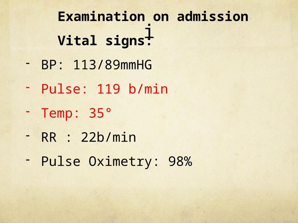
i Examination on admission
Vital signs:
- BP: 113/89mmHG
- Pulse: 119 b/min
- Temp: 35°
- RR : 22b/min
- Pulse Oximetry: 98%

• Physical examination
- GCS 7/15 (E1 M5 V2), pupils dilated
but reactive
- Abdomen: no signs of acute abdomen or injury
- Resp: no signs of resp compromise/distress (intubated due to low GCS and inhalation burns)
- 30% burns arms, legs, abdomen, face

• Biochemistry:
- Sodium: 139 mmol/l
- Potassium: 3,8 mmol/l
- Chloride: 105 mmol/l
- Urea: 5,6 mmol/l
- Creatinine: 124 mmol/l
- Myoglobin: 2322 uq/l
- CK: 4052 u/l
- CK-MB: 48,9 ug/l
- Trop I: 71ng/

- HB: 13,8 g/dl
- WCC: 10, 42
- Platelets: 404
- CRP: 138 ng/l
- INR 1,53
- PCT 7,9 ug/l

• Arterial bloodgas
- pH : 7,45
- paO2 : 133,7mmHg
- paCo2 : 40,0 mmHg
- P/F ratio : 232 mmHg
- SaO2 : 98%
- HCO3 : 20,9 mmol/l
- Lactate : 4.0 mmol/l

• Patient intubated and ventilated in casualty-SIMV (vol control)
• Myoglobin regime initiated 6 hours post injury
• FAST –no free fluid, no abdominal pathology
• Fluid resuscitation acc to Parkland formula (4ml/kg/% burns)
• Admitted to SICU 12 hours post-injury
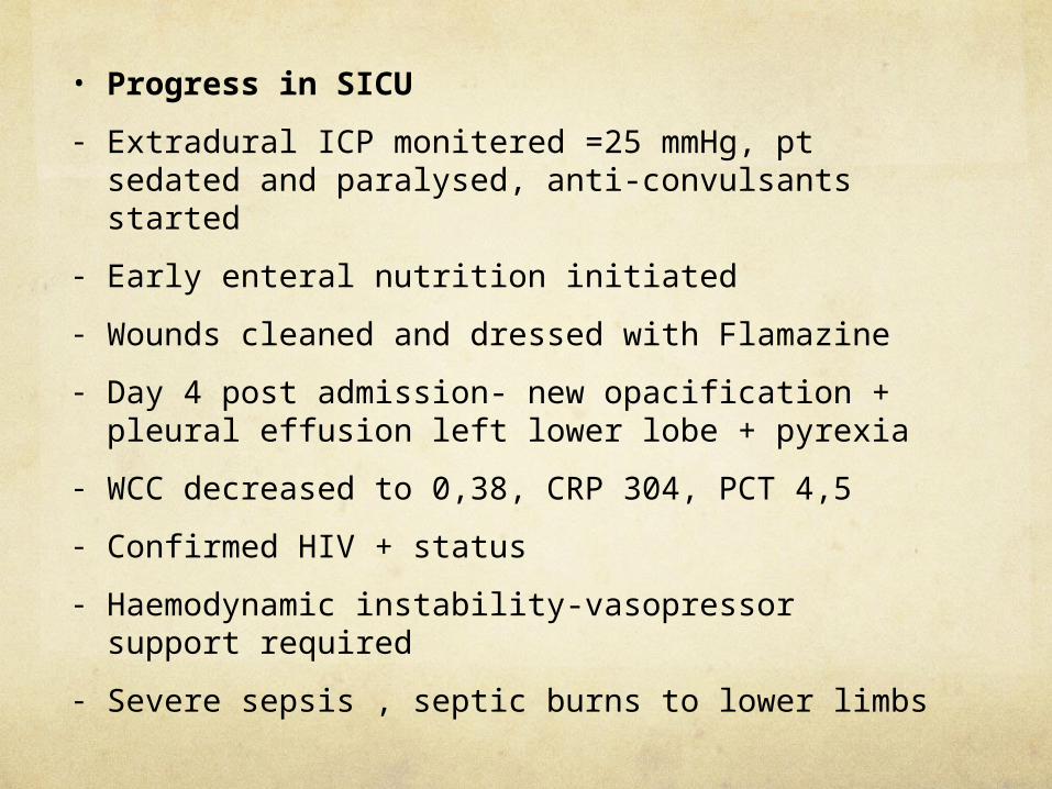
• Progress in SICU
- Extradural ICP monitered =25 mmHg, pt sedated and paralysed, anti-convulsants started
- Early enteral nutrition initiated
- Wounds cleaned and dressed with Flamazine
- Day 4 post admission- new opacification + pleural effusion left lower lobe + pyrexia
- WCC decreased to 0,38, CRP 304, PCT 4,5
- Confirmed HIV + status
- Haemodynamic instability-vasopressor support required
- Severe sepsis , septic burns to lower limbs

• Below-knee R + forefoot amputation L done to obtain source control
• Pseudomas, coagulse - Staph cultured on LUKI, yeast on CVP
• Meronem, Teicoplanin , Colistin started, Amphotericin B added later
• No response after days, still severe LRTI-necrotising pneumonitis, pneumothorax developed and ICD placed

Currently:
• Day 11 Meronem +Teioplanin +Colistin, Amphotericin B day 6, necrotising pneumonitis-minimal response, still septic, deranged LFT’s
• Minimal neurological improvement - GCS=5/10 (M4, E1)
• Still ventilated, haemodynamically unstable, pyrexial

• Arterial blood gas
- ph: 7,41
- FiO2:40%
- P/F ratio: 186
- PaO2: 74
- PaCo2:36
- HCO3: 23
- HB : 8,7
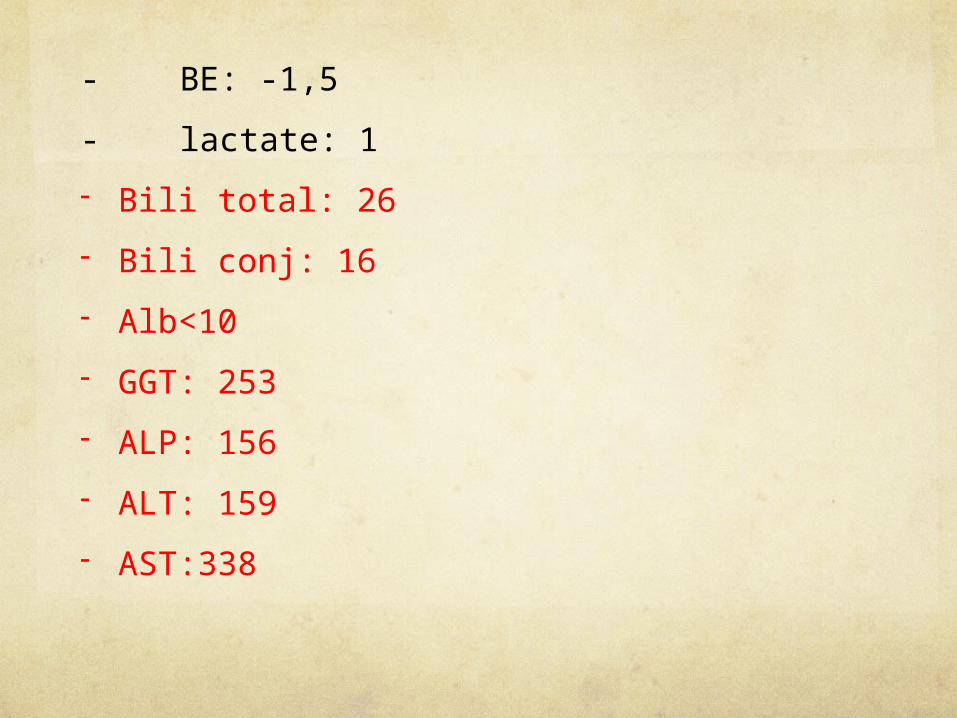
- BE: -1,5
- lactate: 1
- Bili total: 26
- Bili conj: 16
- Alb<10
- GGT: 253
- ALP: 156
- ALT: 159
- AST:338

• Biochemistry
- Na: 137
- K: 4,2
- Cl: 106
- Urea: 5,2
- Creatinine: 49
- WCC 5,36
- PCT 3,6
- CRP 217
- INR 1,49

• Endpoints of discussion
- Fluid resuscitation/ management of burns patient
- Pathophysiology of rhabdomyolysis
- Myoglobin regime
- Management of severe head injury
- Nutrition in critical care- severe head injury and burns
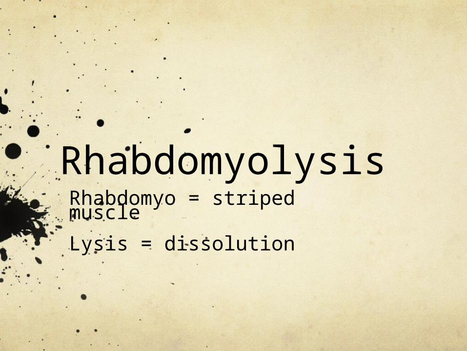
RhabdomyolysisRhabdomyo = striped muscle
Lysis = dissolution

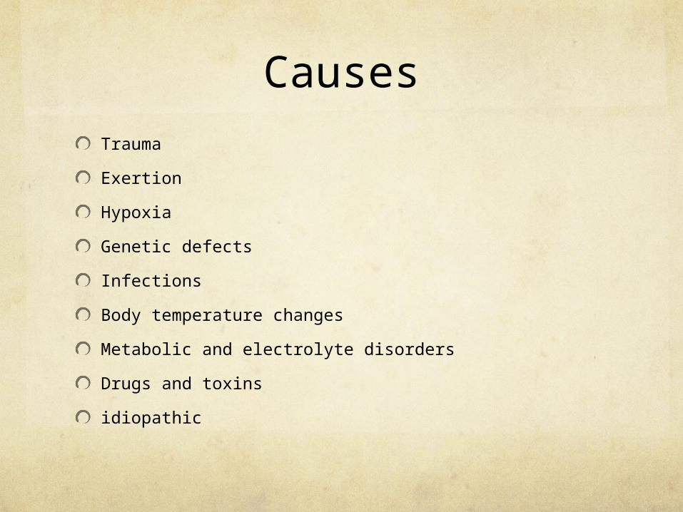
CausesTrauma
Exertion
Hypoxia
Genetic defects
Infections
Body temperature changes
Metabolic and electrolyte disorders
Drugs and toxins
Idiopathic


RhabdomyolysisRhabdomyo = striped muscleLysis = dissolution
CausesTrauma
Exertion
Hypoxia
Genetic defects
Infections
Body temperature changes
Metabolic and electrolyte disorders
Drugs and toxins
idiopathic

CausesTrauma
Exertion
Hypoxia
Genetic defects
Infections
Body temperature changes
Metabolic and electrolyte disorders
Drugs and toxins
idiopathic
Burns

Most serious complication : AKI
Rhabdomyolysis + AKI : 59 percent †Rhabdomyolysis – AKI : 22 percent †
AKI KILLS

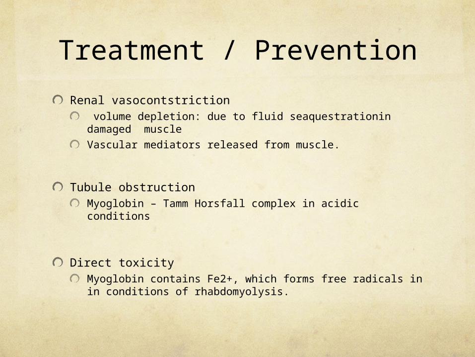
Pathogenesis of AKI in rhabdmyolysis
Renal vasocontstriction volume depletion: due to fluid sequestration damaged muscle
Vascular mediators released from muscle.
Tubule obstructionMyoglobin – Tamm Horsfall complex in acidic conditions
Direct toxicityMyoglobin contains Fe2+, which forms free radicals in in conditions of rhabdomyolysis.
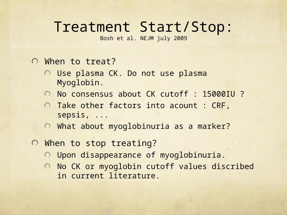
Treatment Start/Stop:Bosh et al. NEJM july 2009
When to treat?Use plasma CK. Do not use plasma Myoglobin.
No consensus about CK cutoff : 15000IU ?
Take other factors into acount : CRF, sepsis, ...
What about myoglobinuria as a marker?
When to stop treating? Upon disappearance of myoglobinuria.
No CK or myoglobin cutoff values discribed in current literature.
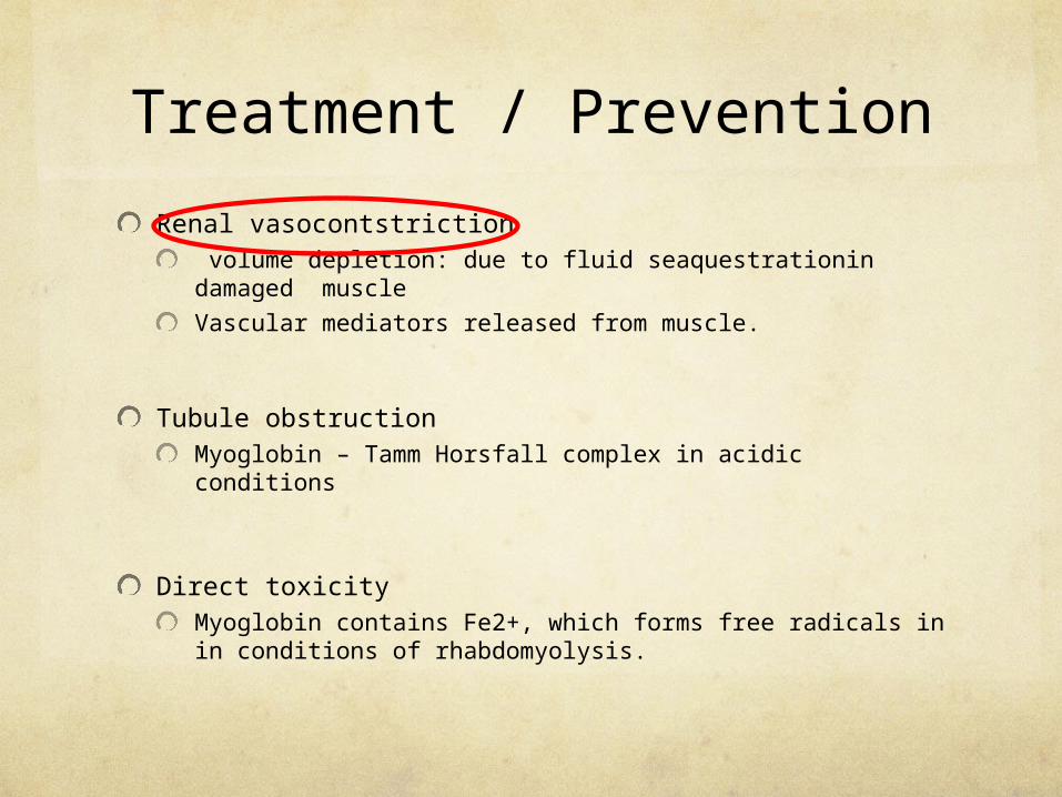
Treatment / Prevention
Renal vasocontstriction volume depletion: due to fluid seaquestrationin damaged muscle
Vascular mediators released from muscle.
Tubule obstructionMyoglobin – Tamm Horsfall complex in acidic conditions
Direct toxicityMyoglobin contains Fe2+, which forms free radicals in in conditions of rhabdomyolysis.

Treatment of volume depletion
Early and agressive volume replacementOnly retrospective data, Shimazu et al. , Gunal et al.
... but expert consensus.
What fluid? Studies only done with saline and ringers.
Ringers group needed more HCO3- (Cho et al.)
consensus to use saline
• What is agressive?
Very vague recommendations (NEJM july 2009) : On average 400cc / h
200 – 1000cc / h depending on setting and severity
While monitoring hemodynamic status
Target urine output of 3 ml / kg

Treatment / Prevention
Renal vasocontstriction volume depletion: due to fluid seaquestrationin damaged muscle
Vascular mediators released from muscle.
Tubule obstructionMyoglobin – Tamm Horsfall complex in acidic conditions
Direct toxicityMyoglobin contains Fe2+, which forms free radicals in in conditions of rhabdomyolysis.

Treatment of acidosis: NaHCO3+?
No Evidence based proof for this practice (Homsi et al., Brown et al.)
... But attractive rationaleNo Tamm Horsfall – myoglobin complex formation
Prevents redox reactions
Prevents vasoconstriction due to methemoglobin
Counteracts saline hyperinfusion acidosis
Recommendation : (NEJM 2009)If urinary pH < 6.5 alternate saline with saline + 100mmol NaHCO3+
Stop if no effect on urinary pH in 4-6h

Treatment / Prevention
Renal vasocontstriction volume depletion: due to fluid seaquestrationin damaged muscle
Vascular mediators released from muscle.
Tubule obstructionMyoglobin – Tamm Horsfall complex in acidic conditions
Direct toxicityMyoglobin contains Fe2+, which forms free radicals in in conditions of rhabdomyolysis.

Flushing away the obstruction?Volume repletion !!
Diuretics
Mannitol :
No evidence based proof (Homsi et al, Brown et al.)
... But attractive rationale
Prevents hypovolemia (‘draws sequestrated fluid back in circulation)
Free radical scavenging effects
Flushing effect
Recomendation (NEJM 2009): consider mannitol
Lasix:
Only flushing effect
No evidence based proof
Recommendation: (NEJM 2009)
use only if indicated due to other reasons than rhabdomyolysis.

Treatment / Prevention
Renal vasocontstriction volume depletion: due to fluid seaquestrationin damaged muscle
Vascular mediators released from muscle.
Tubule obstructionMyoglobin – Tamm Horsfall complex in acidic conditions
Direct toxicityMyoglobin contains Fe2+, which forms free radicals in in conditions of rhabdomyolysis.

Antioxidants
Pentoxyifilline, Vitamin E, Vitamin C
Case reports, case series, in vitro studies
May be justified, but not enough evidence. (Vanholder et al, Huerta et al)

Treatment overview
Early and agressive volume replacement with saline.
Average 400cc/h
Target urinary output : 3cc/kg/h
Monitor hemodynamic status
Add HCO3- if urinary pH < 6.5
Consider mannitol
In case of Hyperkalemia, oliguria, volume overload, metabolic acidosis dialyse (with high flux filter?)

Treatment pitfalls
Late treatment, underresuscitation
Overresuscitation , especially in anuric/oliguric patients.
Hyperkalemia
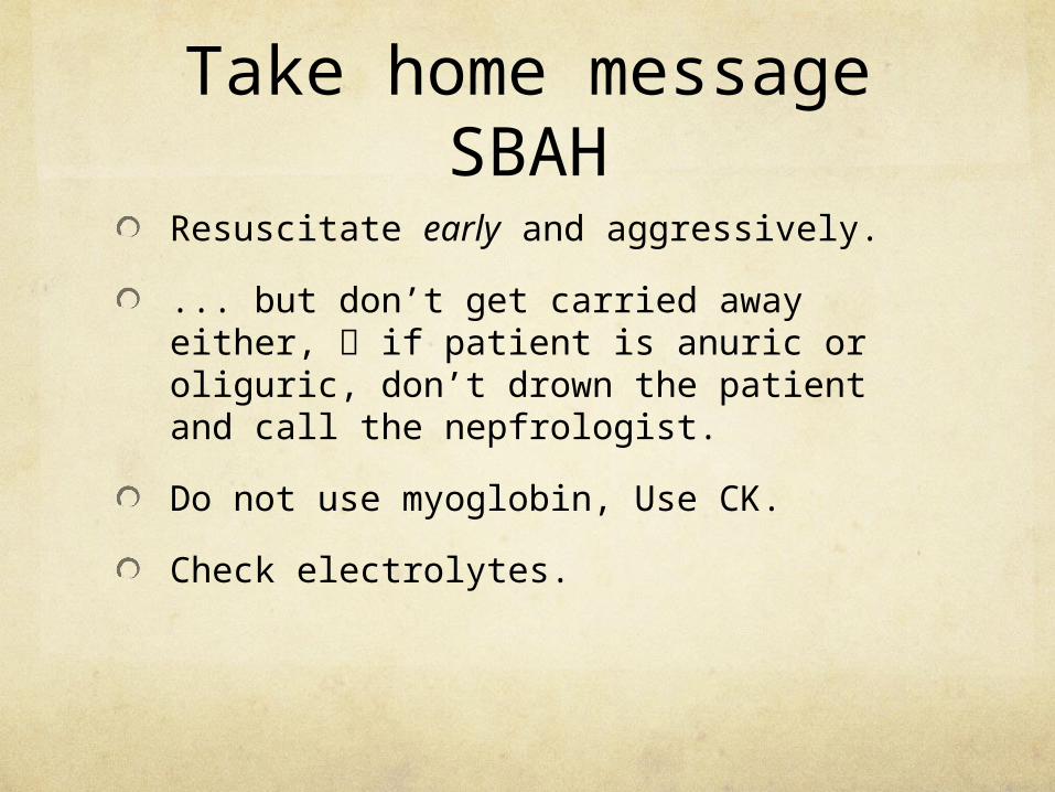
Take home message SBAH
Resuscitate early and aggressively.
... but don’t get carried away either, if patient is anuric or oliguric, don’t drown the patient and call the nepfrologist.
Do not use myoglobin, Use CK.
Check electrolytes.

Resuscitation of the Burns Patient
Initial Assessment and TreatmentAirway Management
Fluid Resuscitation
Wound Management
Secondary Survey
Complications

Initial assessment and treatment
Initial treatment and assessment occurs simultaneously with resuscitation.
Primary management includes stabilizing airway +C-spine, assess for inhalation burns, ?resp distress, ?intubation , breathing, circulation (ABCDE).
Add algorithm

Fluid resuscitation
Rapid, aggressive fluid resuscitation to
-reconstitute intravascular volume
-maintain end-organ perfusion
Determine fluid requirements by considering following:
Age
Severity of burns-depth and percentage burns
Co-morbidities
Associated injuries
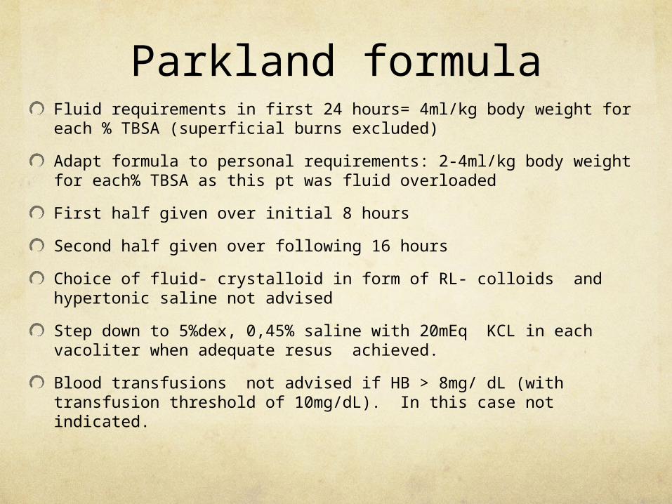
Parkland formulaFluid requirements in first 24 hours= 4ml/kg body weight for each % TBSA (superficial burns excluded)
Adapt formula to personal requirements: 2-4ml/kg body weight for each% TBSA as this pt was fluid overloaded
First half given over initial 8 hours
Second half given over following 16 hours
Choice of fluid- crystalloid in form of RL- colloids and hypertonic saline not advised
Step down to 5%dex, 0,45% saline with 20mEq KCL in each vacoliter when adequate resus achieved.
Blood transfusions not advised if HB > 8mg/ dL (with transfusion threshold of 10mg/dL). In this case not indicated.

Monitor fluid statusNB!!!! Confirming adequate resus response, more important than adherence to Parkland formula( in this case myglobin regime +fluid resus should’ve been adapted to pt’s fluid requirements)
Maintain urine output at 0,5ml/kg/hour
Clinical signs of volume status
-pulse rate <110b/min (normovolemia), > 120b/min (hypovolemia)
-distal pulses
-pulse pressure
-straight-leg-rise test etc.
-vigileo, venous sats, serum lactate levels
Monitor hourly for first 24 hrs and ADJUST ACCORDINGLY!!!
PREVENT OVER-RESUSCITION!!!!!! increased incidence of compartment syndromes, pulmonary oedema etc.

Immediate wound care and cooling
Pain management
Intravenous morphine advised
Consider benzodiazepines in severe anxiety

Second survey and managementLaboratories- FBC,U+E, glucose, venous blood gas, ABG, CXR, Myoglobin levels, CK, serum lactate levels
Tetanus immunization
Topical antibiotics-silver sulfadiadize most commonly used
Wound management-irrigation soap + water, debridement,
Metabolism/nutrition
-Start feeding as soon as resus underway (increased wound healing and shorter hospitalization)
-Glucose monitoring (strict glucose control @ 8-10mmol/l)
-Anabolic steroids
Modulating catabolic responseGlycemic control( 8-10mmol/l)
Beta blockers
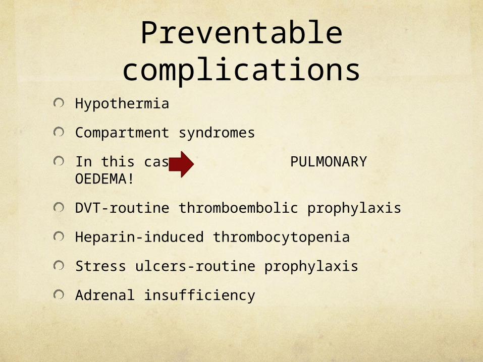
Preventable complications
Hypothermia
Compartment syndromes
In this case PULMONARY OEDEMA!
DVT-routine thromboembolic prophylaxis
Heparin-induced thrombocytopenia
Stress ulcers-routine prophylaxis
Adrenal insufficiency