ch 13: central nervous system part 1: the...
TRANSCRIPT
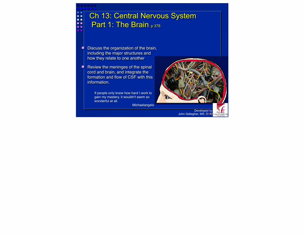
Ch 13: Central Nervous System Part 1: The Brain p 378
Discuss the organization of the brain, including the major structures and how they relate to one another
Review the meninges of the spinal cord and brain, and integrate the formation and flow of CSF with this information.
Developed byJohn Gallagher, MS, DVM
If people only knew how hard I work to gain my mastery, it wouldn't seem so wonderful at all.
Michaelangelo
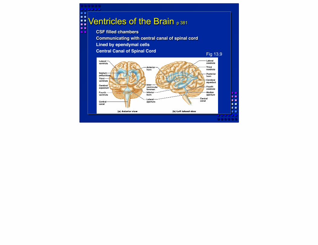
Ventricles of the Brain p 381
CSF filled chambersCommunicating with central canal of spinal cordLined by ependymal cellsCentral Canal of Spinal Cord
Fig 13.9

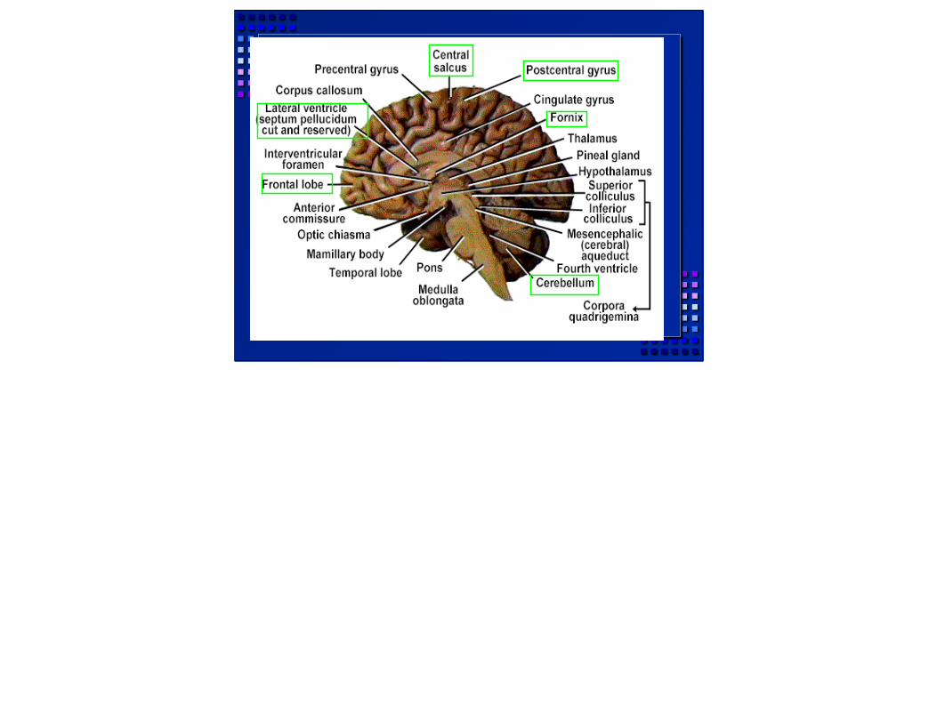
Four Major Brain Subdivisions
1. Brain Stema. Medulla oblongatab. Ponsc. Midbrain
2. Cerebellum3. Diencephalon
a. Hypothalamusb. Thalamus c. Epithalamus
4. Cerebral HemispheresAKA Cerebrum
Fig 13.11
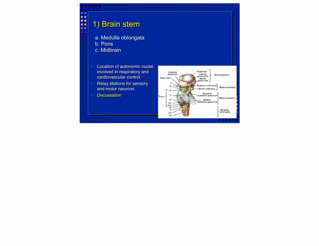
1) Brain stem
• Location of autonomic nuclei involved in respiratory and cardiovascular control
• Relay stations for sensory and motor neurons
• Decussation
a. Medulla oblongatab. Ponsc. Midbrain

1a) Medulla oblongata
Pyramids– Motor output to spinal
cord– Decussation
Reticular formation– Lower functions– Respiration, sleep, etc.
Cranial nerves– VIII, IX, X, XI, XII

1b) Pons
Pons = bridge Connects to cerebellum
– Via cerebellar peduncles Cranial Nerves
– V, VI, VII

1c) Midbrain
Cerebral aqueduct– Old term: Aqueduct of
Sylvius Several nuclei (ganglia)
– Substantia nigra Sensory reflexes
– Aural, visual

2) Cerebellum
Dorsal to the Pons Two hemispheres
– Connected by the vermis Maintains posture and equilibrium
– Smooths motor activities– Some cognitive function
Cortex - gray surface – Purkinje cells (p 354), axons of which
become arbor vitae (white matter) in center
» Large cell bodies visible in gray matter of cerebellum
» Use GABA as NT» Motor output from cerebellum
White matter: Arbor Vitae

3) Diencephalon p 390
3a. Hypothalamus
3b. Thalamus
3c. Epithalamus
Thalamus = 80% of diencephalon. Gateway (relay station) to the cerebral cortex not just for sensory input but for all info. Processing and editing also takes place. About 12 nucleiHypothalamus also about 12 nuclei. Main visceral control center of the body

3a. Hypothalamus
Just superior to optic chiasma Infundibulum - connects to pituitary
gland Some functions:
– Control of autonomic nervous system – Coordination of nervous and endocrine
systems – Manufacture of hormones - ADH and
oxytocin (Ch 17)

3b. Thalamus
(80% of diencephalon) Next to 3rd ventricle Communication with hemispheres
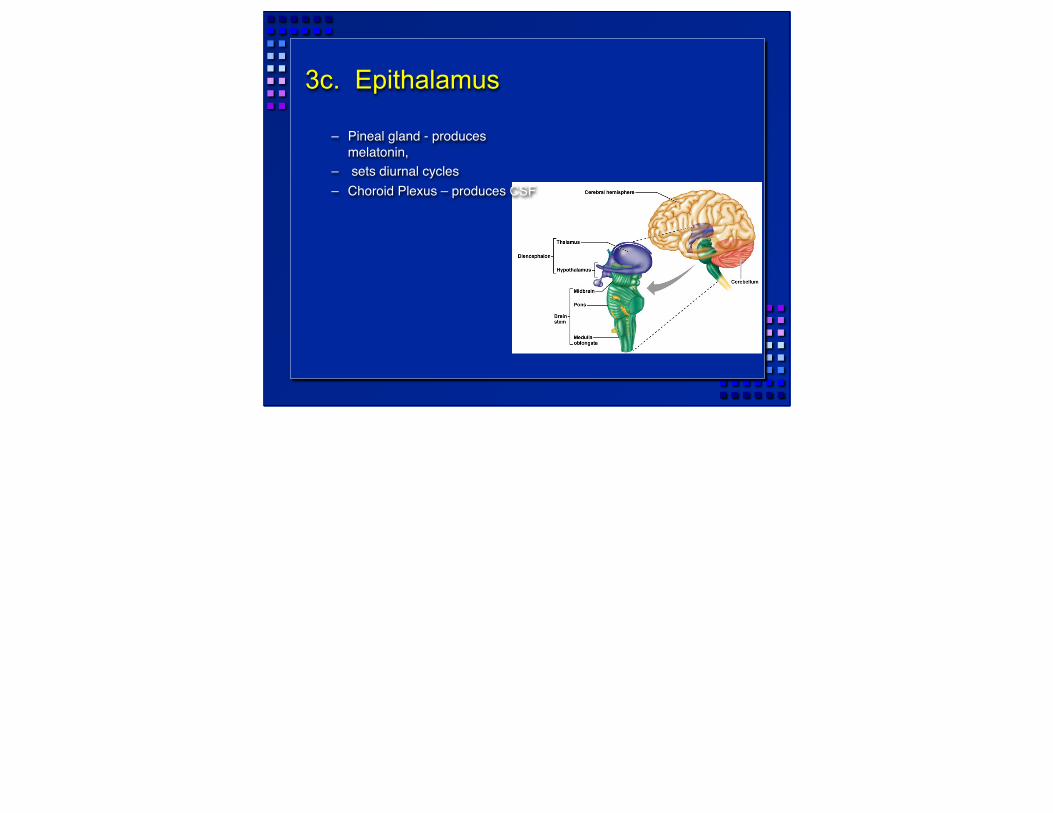
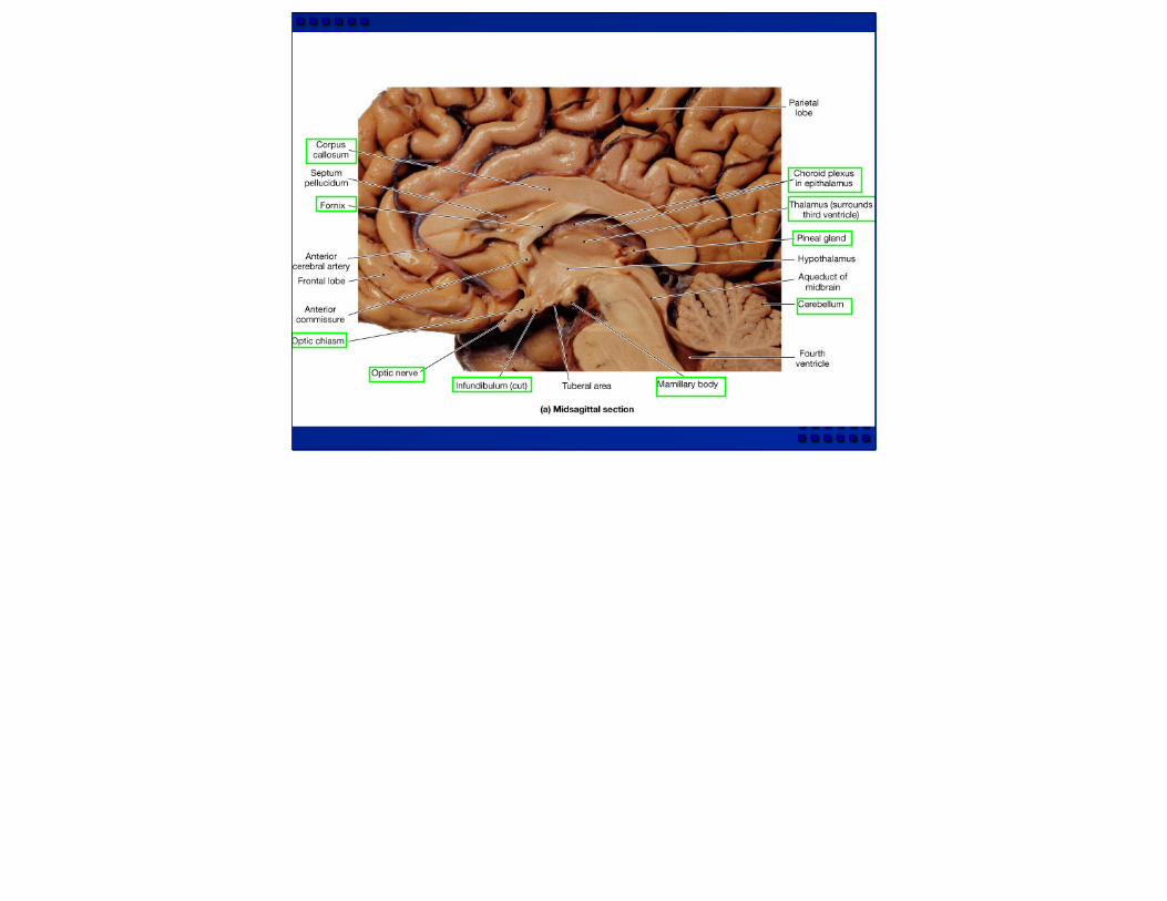
3c. Epithalamus
– Pineal gland - produces melatonin,
– sets diurnal cycles– Choroid Plexus – produces CSF
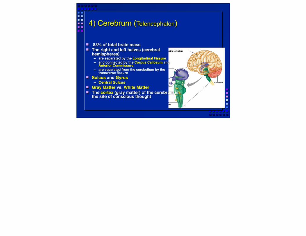
4) Cerebrum (Telencephalon)
83% of total brain mass The right and left halves (cerebral
hemispheres) – are separated by the Longitudinal Fissure– and connected by the Corpus Callosum and
Anterior Commissure– are separated from the cerebellum by the
transverse fissure Sulcus and Gyrus
– Central Sulcus Gray Matter vs. White Matter The cortex (gray matter) of the cerebrum is
the site of conscious thought
In 90-95% of people left side has more control over language, math and logic. Right side is more involved withy visual-spatial skills, intuition, emotion, artistic and musical skill.
4) Cerebral Hemispheres, cont’d
. . have functional regions – Sensory and motor areas
» e.g. Broca’s area (speech)– Prefrontal Cortex (Cognitive functions)
. . . have some functional differences (in spite of anatomical resemblance) → Lateralization of cortical functioning– Right brain: artistic skill– Left Brain: math, logic
. . . receive sensory information and generate commands for opposite side of body– Decussation of sensory input is in the spinal cord– Decussation of motor output is in the pyramids
In 90-95% of people left side has more control over language, math and logic. Right side is more involved withy visual-spatial skills, intuition, emotion, artistic and musical skill.
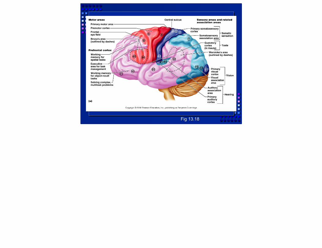
Fig 13.18
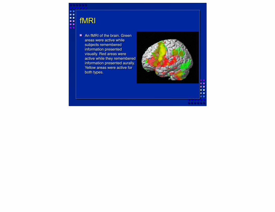
fMRI
An fMRI of the brain. Green areas were active while subjects remembered information presented visually. Red areas were active while they remembered information presented aurally. Yellow areas were active for both types.
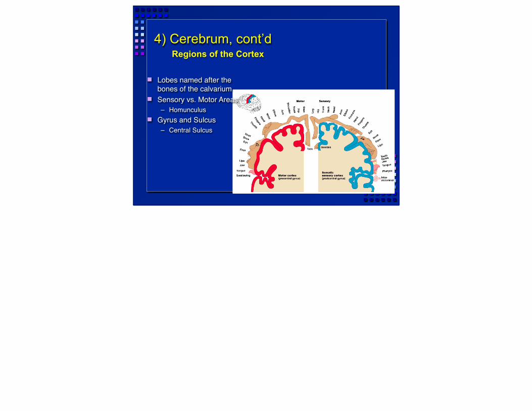
4) Cerebrum, cont’dRegions of the Cortex
Lobes named after the bones of the calvarium
Sensory vs. Motor Areas– Homunculus
Gyrus and Sulcus– Central Sulcus
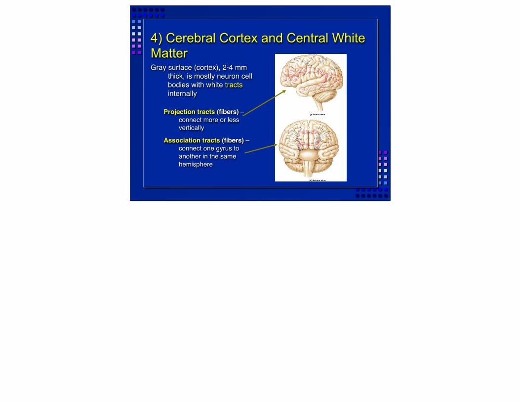
4) Cerebral Cortex and Central White MatterGray surface (cortex), 2-4 mm
thick, is mostly neuron cell bodies with white tracts internally
Projection tracts (fibers) – connect more or less vertically
Association tracts (fibers) – connect one gyrus to another in the same hemisphere
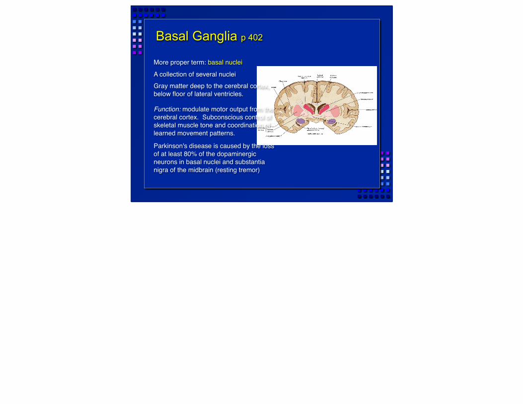
Basal Ganglia p 402
More proper term: basal nuclei
A collection of several nucleiGray matter deep to the cerebral cortex, below floor of lateral ventricles.
Function: modulate motor output from the cerebral cortex. Subconscious control of skeletal muscle tone and coordination of learned movement patterns.
Parkinson's disease is caused by the loss of at least 80% of the dopaminergic neurons in basal nuclei and substantia nigra of the midbrain (resting tremor)
Substantia nigra is in mesencephalon

Gray & White Matter Organization
In brain stem similar to spinal cord (nuclei around ventricles, tracts on outside)
In cerebrum and cerebellum: white matter covered with layer of neural cortex (grey)
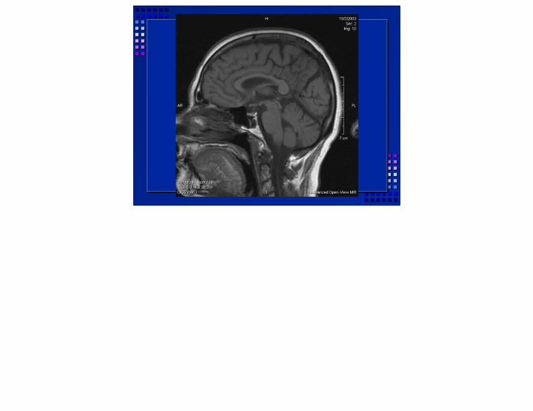
sagittal image showing brain and normal pituitary with bright spot in posterior, neurohypophysis, which is secretory granules which are made in base of brain and transported down infundibulum or pituitary stalk.
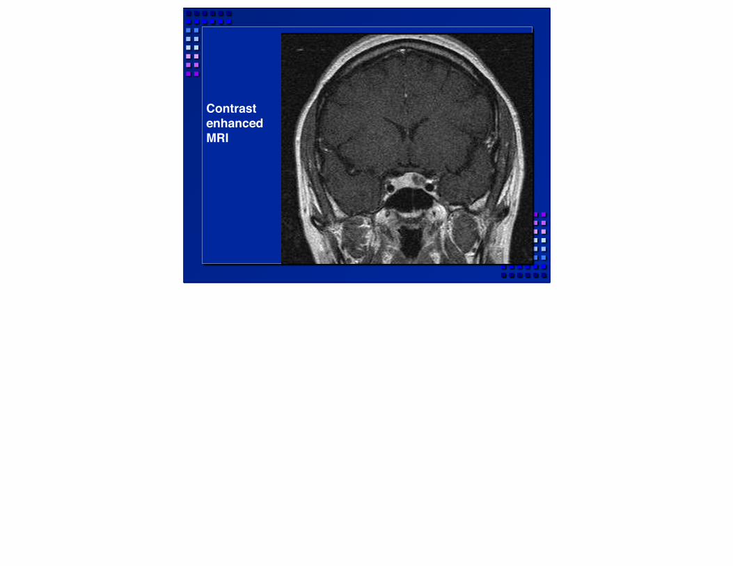
Contrast enhanced MRI
contrast enhances pituitary because of no blood brain barrier, the adenoma has less blood supply and is therefore less enhanced. The Pit. is an endocrine organ so it is highly vascular to release various endocrine hormones into the circulation quickly.