ch11 tumor angiogenesis - mitweb.mit.edu/hst527/www/readings/lecture 19/cancer... · angiogenesis,...
TRANSCRIPT

Angiogenesis, the growth of new capillary bloodvessels, is central to the growth of cancer. Anunderstanding of the cellular and molecular basisof tumor angiogenesis is therefore important forclinicians who diagnose and treat cancer bywhatever modalities. This chapter is focused oncertain general principles of tumor angiogenesisthat are intrinsic to the behavior of human can-cer. Almost every week, the biomedical literaturecontains elegant reports on the cause and devel-opment of cancer at the cellular and molecularlevels. This rapid progress in understanding themolecular and genetic events that underlie thetransformation of a normal cell to a cancer cell isreflected in many chapters in this book. Thesestudies provide strong evidence for the hope that,eventually, there will be molecular solutions tothe cancer problem. However, in virtually all sce-narios of current or future therapy, the target isthe cancer cell.
Experimental evidence indicates that it is prudent to develop cancer therapies againstanother target, the microvascular endothelialcell, without implying that the two targets aremutually exclusive.
Consider a cancer cell that has progressedthrough a series of mutations so that by activationof certain oncogenes and by loss of specific sup-pressor genes, it has become self-sufficient ingrowth signals, insensitive to antigrowth signals,unresponsive to apoptotic signals, capable of lim-itless replicative potential, and tumorigenic.1 Arethese neoplastic properties necessary and suffi-cient for such a cell to expand into a populationthat is clinically detectable, symptomatic, orlethal? Current evidence argues that these neo-plastic properties may only be necessary but notsufficient for the cancer cell to become metastaticand lethal. The reported studies suggest that themicrovascular endothelial cell dictates to a cancercell whether it can grow a tumor to a clinicallydetectable size, metastasize, or kill its host. For atumor to develop a metastatic or a lethal pheno-type, it must first recruit and sustain its own pri-vate blood supply, a process called tumor angio-genesis.1 Tumors unable to induce angiogenesisremain dormant at a microscopic in situ size.Such nonangiogenic lesions are usually notdetectable unless they are on external surfacessuch as skin, oral mucosa, or cervix. Inimmunodeficient mouse models, heterotrans-planted malignant cells sometimes fail to formgrossly-identifiable tumor nodules but nonethe-
less persist as small nonangiogenic tumors called“no-takes.”2
Angiogenesis is fundamental to reproduction,development, and repair. In the adult, repair andreproductive angiogenesis occur mainly as briefbursts of capillary blood vessel growth that usuallylast only days or weeks. Such physiologic angio-genesis, including neovascularization in exercisedmuscle, is tightly regulated.3–5
A variety of circulating and sequesteredinhibitors suppress proliferation of vascularendothelium under normal conditions. As a result,endothelial cells are among the most quiescentcells of the body. Turnover times of endothelialcells are measured in hundreds of days in contrastto bone marrow cells, which maintain an averageturnover time of 5 days and proliferate at approxi-mately 6 billion cell divisions per hour. Duringangiogenesis, microvascular endothelial cells canproliferate as rapidly as bone marrow cells. Fur-thermore, endothelial proliferation is not the onlyevent necessary for development of a new capil-lary blood vessel. Endothelial cells must degradetheir own basement membrane, develop sproutsfrom preexisting microvessels, invade the extracel-lular matrix, form tubes, and connect the tips ofthese tubes to create loops capable of handlingblood flow.6,7 Even in the absence of endothelialDNA synthesis in tissue that has been heavily irra-diated, new capillary blood vessels and theirbranches still develop for a few days.8
A hallmark of pathologic angiogenesis is per-sistent growth of blood vessels (ie, sustained neo-vascularization). Angiogenesis that continues formonths or years supports the progression of manyneoplastic and nonneoplastic diseases.9,10 How-ever, both physiologic and pathologic angiogenesisare usually focal. An angiogenic focus appears asonly a tiny fraction or a small “hot spot” of prolif-erating and migrating endothelial cells that arisefrom a monolayer of resting endothelium ofapproximately 1000 m2, an area the size of a tenniscourt. A cubic millimeter of human cardiac musclecontains approximately 2,500 millimeters of capil-lary blood vessels, (as determined by stereologicmethods).4 The fundamental objective of allantiangiogenic therapy is to return a pathologicneovascular focus to its normal resting state or toprevent its initiation.
HISTORIC BACKGROUND
For more than 100 years, tumors had beenobserved to be more vascular than normal tis-
sues.11 This tumor hyperemia observed duringsurgery was explained by simple dilation of exist-ing host blood vessels.12 Vasodilation was gener-ally thought to be a side effect of metabolites or ofnecrotic tumor products escaping from the tumor.Three reports, although largely overlooked, sug-gested that tumor hyperemia could be related tonew blood vessel growth; that is, to neovascular-ization and not solely to vasodilation. A 1939paper showed that whereas neovascularization of awound in a transparent chamber in a rabbit earregressed completely after the wound healed,13 atumor implant in the chamber was associated withaccelerated growth of new capillary blood vessels.The other two reports, in 1945 and 1947, demon-strated that new vessels in the neighborhood of atumor implant arose from host vessels and notfrom the tumor itself.14,15 These papers not with-standing, debate continued in the literature for twomore decades about whether a tumor could expandto a large size (centimeters) by simply living onpreexisting vessels.16 Even among the few investi-gators who accepted the concept of tumor-inducedneovascularization, it was generally assumed thatthis vascular response was an inflammatory reac-tion, a side effect of tumor growth, not a require-ment for tumor growth.17
BEGINNING OF ANGIOGENESIS RESEARCH
HYPOTHESIS: TUMOR GROWTH DEPENDS ON
ANGIOGENESIS In 1971, Folkman proposed anew view of the role of blood vessels in tumorgrowth in the form of a hypothesis that tumorgrowth is angiogenesis dependent.18 This hypothe-sis suggested that tumor cells and vascularendothelial cells within a neoplasm may constitutea highly integrated ecosystem and that endothelialcells may be switched from a resting state to arapid growth phase by a “diffusible” chemical sig-nal from tumor cells. An additional speculationwas that angiogenesis could be a relevant target fortumor therapy (ie, antiangiogenic therapy). Folk-man proposed these ideas from experiments heperformed with Frederick Becker in the early1960s, which revealed that tumor growth in iso-lated perfused organs was severely restricted in theabsence of vascularization of the tumors.19–24
These concepts were not accepted at the time.Although a few investigators in the early 1970sperceived that tumors could actually induce neo-vascularization, the belief persisted that suchneovascularization was an inflammatory host
Tumor AngiogenesisJudah Folkman, MD Raghu Kalluri, PhD
11

162 SECTION 1 / Cancer Biology
response to necrotic tumor cells or possibly a hostdefense detrimental to the tumor. Another obstacleto research on tumor angiogenesis was the conven-tional wisdom at that time that any new vesselsinduced by a tumor, like new vessels in a wound,would become established and thus could not bemade to involute. From this assumption, scientistsconcluded that antiangiogenic therapy could neverregress a tumor; therefore, it would be fruitless totry to discover angiogenesis inhibitors. In this pes-simistic atmosphere, it was not an easy task to pro-duce compelling evidence that tumor growthdepended on neovascularization. Eventual accep-tance of the 1971 hypothesis was slow because itwould be 2 more years before the first vascularendothelial cells were successfully cultured invitro, 8 years before it was possible to grow capil-lary endothelial cells in vitro, 11 years before thediscovery of the first angiogenesis inhibitor, and13 years before the purification of the first angio-genic protein.25–29
Throughout the 1970s, laboratory studies weredevoted to demonstrating that: tumor vessels werenew proliferating capillaries; the sequential stepsof the angiogenic process could be identified;qualitative and quantitative bioassays for angio-genesis could be developed; viable tumor cellsreleased diffusible angiogenic factors that stimu-lated new capillary growth and endothelial mitosisin vivo, despite the arrest of tumor cell prolifera-tion by irradiation; necrotic tumor products werenot angiogenic per se (reviewed in Folkman andCotran); and that angiogenesis itself could beinhibited.30–35 Because of these efforts to providesupporting evidence that tumor growth was angio-genesis dependent, the field of angiogenesisresearch began. Today the field has broadened toinclude a wide spectrum of basic science disci-plines, from developmental biology to moleculargenetics, as well as a variety of clinical specialties,which include in addition to oncology, cardiology,dermatology, gynecology, ophthalmology, andrheumatology. Approximately 40 publicationswith angiogenesis in the title, appear every week.
EXPERIMENTAL EVIDENCE By the mid-1980s,considerable experimental evidence had beenassembled to support the hypothesis that tumorgrowth is angiogenesis dependent. The idea couldnow be stated in its simplest terms: “Once tumortake has occurred, every further increase in tumorcell population must be preceded by an increase innew capillaries that converge upon the tumor.”32
The hypothesis predicted that if angiogenesiscould be completely inhibited, tumors wouldbecome dormant at a small, possibly microscopic,size.22 It forecast that whereas the presence of neo-vascularization would be necessary, but not suffi-cient for expansion of a tumor, the absence of neo-vascularization would prevent expansion of aprimary tumor mass beyond 1 to 2 mm3 andrestrict a metastasis to a microscopic dormantlesion. Most nonneovascularized tumors are notclinically detectable, with the exception of surface lesions on the skin or the externalmucous membranes.
The hypothesis that tumors are angiogenesisdependent is supported by biologic and pharmaco-logic evidence and proved by genetic evidence.Both types of evidence are summarized belowbecause they provide a scientific basis for currentclinical trials of angiogenesis inhibitors.36,37
Biologic evidence1. In two-dimensional flat cultures, a population
of tumor cells expands indefinitely as long asfresh medium is added and unlimited cell-freesurface is provided ( ie, passage of cells to anew flask). In contrast, three-dimensionalspheroids of the same cells, suspended in softagar or methylcellulose, stop enlarging at adiameter of a few millimeters, despiterepeated passage of the spheroids to freshmedia.38 In these “steady-state” spheroids,cell proliferation is balanced by celldeath.39–41 This in vitro model is analogousto a dormant micrometastasis in which angio-genesis is blocked.42
2. Tumors implanted into subcutaneous transparent chambers grow slowly beforevascularization, and tumor volume increaseslinearly. After vascularization, tumor growthis rapid and tumor volume may increaseexponentially.15,43
3. Tumor growth in the avascular rabbit corneaproceeds slowly at a linear rate, but converts toexponential growth after neovascularization.44
4. Tumors suspended in the aqueous fluid of theanterior chamber of the rabbit eye remain ina dormant state: viable, avascular, and lim-ited in size (<1 mm3). These tumors induceneovascularization of iris vessels, but the newvessels are out of reach of the tumors floatingin the aqueous fluid. Once a tumor spheroidis apposed to the proliferating iris vessels, thetumor becomes neovascularized and canenlarge up to 16,000 times its original vol-ume within 2 weeks.45 A current interpreta-tion of this experiment is that the tumorspheroid had already switched to the angio-genic phenotype, but that angiogenesis wasblocked by separation of the floating tumorfrom the nearest vascular bed.46
5. Tumors grown in the vitreous of the rabbit eyeremain viable but are restricted to diameters ofless than 0.50 mm for as long as 100 days.Once such a tumor reaches the retinal surface,it becomes neovascularized and within 2 weekscan undergo a 19,000-fold increase in volumeover the avascular tumor.47 Cross-sectional his-tology of the avascular tumors reveals prolifer-ating cells at the periphery of the tumor andnecrotic tumor cells in its center.
6. Human retinoblastomas that have metasta-sized to the vitreous are viable and avascularand tumor expansion is restricted.48
7. Within a solid tumor, the [H3]thymidinelabeling index of tumor cells decreases withincreasing distance from the nearest opencapillary vessel.49 The tissue oxygen tensionalso decreases with increasing distance of atumor cell from the center of a capillary ves-
sel.50 Because the limit of oxygen diffusion is100 to 200 µm, tumor cells that exceed thesedistances from a capillary vessel becomeanoxic (as determined by infrared spec-troscopy of tumors in transparent skin cham-bers in mice).51
8. Tumors implanted into the chorioallantoicmembrane of the chick embryo remainrestricted in growth during the avascularphase, but enlarge rapidly once they are vascularized.52
9. Vascular casts of metastases in the rabbitliver reveal that tumors of up to 1 mm indiameter are usually avascular, but beyondthat size are vascularized.53
10. Human ovarian carcinomas metastasize tothe peritoneal membrane as tiny avascularseeds. These implants rarely grow beyond alimited diameter of a few millimeters, untilafter vascularization. This “avascular” stateof peritoneal implants has also been demon-strated in mice with four different tumortypes and is independent of whether the miceare immunocompetent or immunodeficient.The avascular peritoneal implants are lessthan 0.5 mm diameter and are of uniform size(C. Chen, personal communication, 1998).
11. In transgenic mice that develop carcinomasof the beta cells in the pancreatic islets, largetumors arise from a subset of preneoplastichyperplastic islets, but only after they havebecome vascularized.54
12. Neoplastic cells injected subcutaneouslydevelop into tumors that become vascular-ized at about 0.4 mm3.55 As tumor sizeincreases, blood vessels continue to prolifer-ate and are enveloped by tumor cells thatappear to grow toward and around the newvessels. The vessels eventually occupy up to1.5% of the tumor volume. This is a 400%increase in vascular density over normal subcutaneous tissue.55 In this model, newmicrovascular sprouts that converge on atumor are enveloped by tumor cells, givingthe appearance that the tumor has been penetrated by vessels.
13. In colon carcinomas arising in rats after car-cinogen exposure, there is an early phase(tumor diameter <3.5 mm) during which thetumor is temporarily supplied by preexistinghost microvessels that are dilated andwidened.56 This stage is similar to “coop-tion” of blood vessels recently reported.57
Subsequently, new capillary vessels sproutand proliferate (angiogenesis), which leads toincreasing microvessel density and rapidtumor growth.
Pharmacologic evidence By the late1980s it became possible to inhibit tumorangiogenesis by biochemical and molecularmethods. These experiments were based on(1) administration of molecules that inhibitedangiogenesis specif ically or selectively, (2)blockade of tumor-derived angiogenic factors,and (3) the development of spontaneous

CHAPTER 11 / Tumor Angiogenesis 163
tumors in transgenic mice. Some examples ofsuch are listed below.
1. An angiogenesis inhibitor, TNP-470 (AGM-1470), a synthetic analogue of fumagillin,selectively inhibited proliferating endothelialcells in vitro and in vivo.59 It potently inhib-ited tumor growth in vivo but did not inhibittumor cells in vitro. In pre-clinical studies ithas a broader spectrum of antitumor activitythan any known anticancer drug. There aremore than 60 reports from different laborato-ries of inhibition of 33 different types ofhuman, mouse, rat, hamster, and rabbit pri-mary tumors in animals and of 23 differenttypes of metastatic tumors including humantumor metastases in mice and rat and hamstertumor metastases. Tumor growth was inhib-ited at an average of 65% with a range of 43to 100% (ie, complete regression). All ofthese reports employed the same dose, 30mg/kg every other day. Complete regressionwas observed in seven tumor types in mice,including neurofibrosarcoma, neurofibroma,breast cancer, gastric cancer, and choriocarci-noma, as well as mouse reticulum sarcomaand gastric carcinoma.60 Because TNP-470inhibits microvascular endothelial cells at aconcentration 3 logs lower than tumor cells,this angiogenesis inhibitor is virtually a spe-cific inhibitor of endothelial proliferationand migration. These studies provided thefirst clue that angiogenesis inhibitors couldbe broad spectrum anticancer agents that arerelatively independent of tumor type. Theappearance of different therapeutic responsesby different tumor types (eg, 43 to 100%) wasmore adequately explained by the total angio-genic output of each tumor type matchedagainst a fixed dose of angiogenesisinhibitor. (See below for improvements inTNP-470.)
2. In another experiment, antibody againstbFGF (basic fibroblast growth factor)secreted by a tumor administered to micebearing the tumor, resulted in dramaticreduction in neovascularization and in tumorvolume.61 To engineer bFGF that wassecreted from the tumor, the cDNA forhuman bFGF hybridized to a signal sequencewas transfected into normal mouse fibrob-lasts.61 Furthermore, the structure of thebFGF was modified by site-specific mutage-nesis in which two serines were substitutedfor cysteines. Thus, the bFGF released by thetumor could be neutralized by a specific anti-body that had no effect on natural bFGF. Thetransfected fibroblasts became tumorigenic,exported bFGF, and were highly angiogenic.They formed large lethal tumors whenimplanted into mice. Tumor angiogenesiswas mediated solely by bFGF released fromthese tumors.
3. In an analogous experiment, a neutralizingantibody to another angiogenic protein, vas-cular endothelial growth factor (VEGF), was
administered to mice bearing tumors thatinduced angiogenesis solely by VEGF.62
Tumor growth was inhibited by more than90%. The antibody had no effect on the tumorcells in vitro. With but few exceptions, VEGFis considered to be a specific mitogen for vas-cular endothelial cells.63
4. Specific immunologic inhibition of overexpres-sion of the integrin αVβ3 on capillary endothe-lial cells resulted in apoptosis of proliferatingendothelial cells, blocked neovascularization,and induced tumor regression.64
5. Endogenous specific inhibitors of endothelialproliferation and of angiogenesis include:angiostatin, a 38 kDa internal fragment of plasminogen (generated by Lewis lung carci-noma), endostatin, a 20 kDa internal fragmentof collagen XVIII (generated from a murinehemangioendothelioma), a 53 kDa conforma-tionally changed fragment of antithrombin III(generated from human small cell lung cancer),and tumstatin, a 28 kDa internal fragment ofcollagen IV (α 3NC1 domain).65–71 They donot inhibit proliferation of resting confluentendothelial cells, epithelial cells, smooth-muscle cells, fibroblasts, or tumor cells in vitro.These proteins inhibit angiogenesis in thechick chorioallantoic membrane or in themouse cornea. Both primary and metastatictumors are markedly inhibited and tumorregression has been achieved without toxicityor drug resistance.69
6. In SCID mice bearing human pancreatic can-cer, human recombinant endostatin wasinjected into the peritoneal cavity once daily,or delivered continuously by an implantedmicro-osmotic pump that released 1 micro-liter/hour.72 Continuous dosing inhibitedtumor growth 10-fold more effectively thanonce a day bolus dosing and also inducedtumor regression, whereas bolus dosing didnot (Figure 11-1).
7. An antibody to two receptors of VEGF calledVEGF-Trap, inhibits growth of VEGF pro-ducing tumors in mice.73
Genetic evidence Genetic evidence thattumors are angiogenesis dependent began to bereported by the mid-1990s and provides the mostcompelling proof that tumors and their metas-tases cannot not grow beyond a microscopic insitu size without recruiting new microvessels.These experiments are based on (1) transfectionof dominant-negative receptors for a proangio-genic protein into endothelial cells in the tumorbed, (2) transfection of genes for antiangiogenicproteins into tumor cells, (3) regulation of theexpression of angiogenic oncogenes, and (4)manipulation of developmental genes. Someexamples are listed below.
1. The growth of brain tumors in nude mice wassignificantly inhibited or prevented whentumor angiogenesis was suppressed by a
Figure 11-1 Continuous administration of endostatin is ten times more effective than the same dose given once a dayas a bolus. Continuous dosing can regress tumors whereas bolus dosing does not. In this paper the pharmacokineticsshow that bolus dosing gives a high peak of endostatin with a rapid fall below effective theraputic levels. In contrast, con-tinuous dosing provides constant therapeutic levels of 200 ng/ml up to 250 ng/ml. The implication of this study is that inorder to stop tumor growth or to induce tumor regression, endothelial cells in the tumor bed must be continuouslyexposed to therapeutic levels of inhibitor to counteract stimulators of endothelial growth which constantly bathe theendothelium.72 (Four-color version of figure on CD-ROM)

164 SECTION 1 / Cancer Biology
strategy in which a dominant-negativemutant of the receptor (Flk-1) for the angio-genic protein VEGF was introduced into hostendothelial cells (carried by a retrovirus).This signaling-defective receptor mutantformed an inactive dimer with the native Flk-1 receptor on endothelial cells and preventedthe formation of new capillary blood vesselsin response to VEGF released by the tumor.74
2. Transformed cells were not tumorigenic untilafter they had become angiogenic by down-regulating expression of thrombospondin-1 aprocess that was p53-dependent.75
3. Transfection of normal human cells with theSV40 large T oncogene, generated immortal-ized cells in vitro. However, when these cellswere inoculated into mice they formedmicroscopic sized nonangiogenic tumors thatdid not grow beyond approximately 1 mmdiameter.76 A subsequent transfection withthe ras oncogene switched the cells to theangiogenic phenotype, upregulated VEGFproduction, downregulated production of tis-sue inhibitors of metalloproteinases, andgenerated highly neovascularized, rapidlygrowing tumors.
4. Downregulation of the ras oncogene in amelanoma driven by doxycycline inducibleras, led to massive apoptosis of microvascu-lar endothelium in the tumor bed startingwithin 6 hours. Tumor cells began to die dayslater, and large tumors had completely disap-peared by 12 days.77
5. When tumor cells were transfected with thecDNA for angiostatin and implanted intomice, the higher the secretion of angiostatin,the slower the growth of tumors. However,tumor cell proliferation in vitro was notaffected by angiostatin transfection.78
6. When tumors were transfected with the secretable antiangiogenic protein thrombo-spondin-1 (TSP-1) and/or thrombospondin-2(TSP-2), tumor growth was directly propor-tional to suppression of angiogenesis.Tumor growth was inhibited up to 100% ( ie,microscopic dormant tumor or no tumor)when angiogenesis was suppressed completely. Tumor cell proliferation wasindependent of level of TSP-1 or TSP-2 production.79
7. Id1 and Id3 are helix-loop-helix proteinsthat may control differentiation by interfer-ing with DNA binding of transcription fac-tors. After targeted disruption of one alleleof Id1 and two alleles of Id3, three differenttypes of implanted tumors failed to induceangiogenesis, their growth was severelyrestricted, and they did not metastasize.80
However, when these mice were injectedwith bone marrow from wild-type mice containing normal Id1 and Id3 markers, progenitor endothelial cells expressing Id1and Id3 circulated from the bone marrow tothe tumor vascular bed and permitted thetumors to undergo neovascularization andto grow.81
METASTASIS IS ALSO ANGIOGENESIS DEPENDENT
Experimental and clinical evidence suggests thatthe process of metastasis is also angiogenesisdependent. For a tumor cell to metastasize suc-cessfully, it must breach several barriers andrespond to specific growth factors.82–86 Thus,tumor cells must gain access to the vasculature inthe primary tumor, survive the circulation, arrest inthe microvasculature of the target organ exit fromthis vasculature, grow in the target organ, andinduce angiogenesis.84–90 Therefore, angiogenesisappears to be necessary at the beginning as well asat the completion of the metastatic cascade.
In experimental animals, tumor cells arerarely shed into the circulation before a primarytumor is vascularized, but they can appear in thecirculation continuously after neovasculariza-tion.91,92 The number of cells shed from the pri-mary tumor appears to correlate with the densityof tumor blood vessels as well as with the num-ber of lung metastases observed later. Tumorcells can enter the circulation by penetratingthrough proliferating capillaries that have frag-mented basement membranes and are leaky.92,93
Further, angiogenic factors from tumors such asbFGF and VPF/VEGF induce increased produc-tion of plasminogen activator and collagenasesin proliferating endothelial cells, thus furthercontributing to degradation of basement mem-branes.94–96 These degradative enzymes mayfacilitate the entry of tumor cells into the circu-lation. Tumor cells may not immediately becomeneovascularized after reaching the target organ.Such a metastasis lacking angiogenic activity fora variety of reasons may remain as a microscopictumor of 100 to 200 µm diameter indefi-nitely.42,97,98 It has been the conventional wis-dom that in human dormant micrometastases(eg, metastases appearing 5 to 10 years afterremoval of a breast cancer), tumor cells are notcycling or are in G0. However, increasing exper-imental evidence, indicates that micrometastasescan be held dormant by blocked angiogenesisthat results in a balance of tumor cell pro-liferation and apoptosis.42,97 Finally, experimen-
tal metastases are as susceptible as primarytumors to control by specific angiogenesisinhibitors.65,66 However, the relative efficacy ofa given angiogenesis inhibitor can be affected bydifferences in metastatic sites and by the pres-ence or absence of the primary tumor.65
PREVASCULAR PHASE LIMITS TUMOR EXPANSION
During the prevascular phase, when angiogenicactivity is absent or insufficient, tumors remainsmall, with volumes measured in a few cubicmillimeters. Growth of the whole tumor is slow,and doubling times for the whole tumor may beyears. However, this does not mean that thetumor cells are proliferating slowly. Experimen-tal studies show that tumor cells in a prevascularneoplasm may have a [3H]thymidine labelingindex as high as that of a large vascularizedtumor. The difference between the two is that theprevascular tumor reaches a steady state in whichgeneration of new tumor cells is balanced bytumor cell apoptosis.99
When the prevascular phase of bladder can-cer, cervical cancer, or cutaneous melanoma isfirst detected, these lesions are usually thin,slowly growing, stable for months to years,asymptomatic, and rarely metastatic.100–103 Forthe majority of tumors, however, the prevascularstage is clinically undetectable and can only beobserved microscopically. For example, in breastand prostate cancer, carcinomas in situ can beobserved before and after neovascularization inthe same specimen.89,104,105
The size limits of experimental tumors whenangiogenesis is blocked or absent are betweenapproximately 0.2 mm diameter (eg, for lungmetastases in mice) and 2 mm (eg, for chon-drosarcomas in rats,106 having a tumor popula-tion of 105–106 cells).42,106 The differences insize of prevascular (nonangiogenic) tumors maybe due in part to the capacity of tumor cells to sur-vive under differing degrees of hypoxia (Figure11-2).51,107 However, if cancer cells are alreadyangiogenic at the time of implantation, they caninitiate angiogenesis before the tumor population
Figure 11-2 Left Panel: A cuff of live tumor cells around a microvessel in a human melanoma growing in a SCIDmouse has an average radius of 85 microns. The appearance of an ellipsoid is due to the way the section is cut. RightPanel: A cuff of rat prostate cancer cells around a microvessel has an average radius of 110 microns.37 (Four-color ver-sion of figure on CD-ROM)

molecular barrier to migrating endothelial cells.For example, tumstatin, a potent inhibitor ofendothelial cell migration and proliferation isfound in collagen IV.71 After the basement mem-brane has been breached by new vessel sprouts,tumor cells form multiple cell layers around eachnew capillary blood vessel.122 The radius ofthese microcylinders is restricted to the oxygendiffusion limit for a given tumor type as origi-nally defined by Thomlinson and Gray.37,50 Forexample, for a human melanoma, the oxygen dif-fusion limit is approximately 85 µm. Beyond thatdistance from a capillary blood vessel, virtuallyall tumor cells are apoptotic (or necrotic). Withinthat radius, most tumor cells are viable (see Fig-ure 11-2). For a prostate carcinoma the oxygendiffusion limit is approximately 110 µm, andit may be greater for certain tumors, such asfor a chondrosarcoma or a tumor in which p53is mutated or absent, but rarely would 200 µmbe exceeded.
Circulating endothelial stem cells in tumorangiogenesis Recent experimental and clinicalevidence reveals that circulating endothelial pro-genitor cells derived from stem cell reservoirs inthe postnatal bone marrow can be recruited to thevascular bed of tumors and contribute to tumorgrowth.81,123–135 Functional vascular endothelialgrowth factor receptor-1 (VEGFR-1) isexpressed on hematopoietic stem cells (CD34+
in humans and Lin-Sca-1+c-Kit+ in mice).133
Functional VEGFR-2 is expressed on CD34+
AC133+ progenitor human endothelial cells.VEGF elaborated by a variety of tumor signalsthrough both VEGFR-1 (Flt-1) and VEGFR-2(Flk-1), can mobilize progenitor endothelialcells into the circulation where they are recruitedinto the vascular bed of certain tumor types, butnot others.81
Before endothelial progenitor cells can leavethe bone marrow, they must transfer (along withhematopoietic stem cells) from the quiescent tothe proliferative niche within the marrow. Mobi-lization of hematopoietic stem cells and progen-itor endothelial cells necessary for their transferto the proliferative niche is dependent uponrelease of soluble Kit-ligand, which itselfdepends upon upregulation of metallopro-teinase-9.134 Thus, the microvascular endothelialcells in the vascular bed of a tumor may be
CHAPTER 11 / Tumor Angiogenesis 165
would have reached the limiting size of a non-neovascularized tumor (ie, 0.2 to 2 mm). The evi-dence for this conclusion is from an experimentin which only 20 to 30 angiogenic mammarytumor cells were implanted into a transparentskin chamber in a mouse.108,109 The tumor cellselongated by day 2, migrated toward the nearestmicrovessels in a parallel orientation by day 4,divided undirectionally, induced vascular dilationand tortuosity in the neighboring microvessels byday 6, and stimulated new vascular sprouts andloops with intermittent blood flow by day 8. Bythis time, a microscopic colony of up to 300 to400 cells was visible. Tumor cells preferentiallygrew contiguous to the microvessel sprouts, andby day 20 the tumor was filled with a newlyformed vasculature. When angiogenesis was pre-vented by injection into the chamber of a trun-cated soluble receptor (Flk 1) for vascularendothelial growth factor (VEGF), some tumorcells underwent apoptosis and microscopictumors regressed or became dormant within 5days (ie, before the appearance of neovascularsprouts). The soluble receptor potently inhibitedendothelial cell proliferation in vitro, but had noeffect on tumor cells. This result provides evi-dence for the operation of a two-way paracrineexchange of growth factors and survival factorsbetween tumor cells and neighboring vascularendothelial cells (see below). In this model,tumor cells secrete angiogenic proteins that acti-vate endothelial cells to elaborate chemoattrac-tants for tumor cells. Simultaneously, the acti-vated endothelium forms new vascular sproutsdirected toward the tumor. Vascular endothelialcells can produce at least 20 mitogens and anti-apoptotic factors.110 Many of these proteins, suchas basic fibroblast growth factor (bFGF) and hep-arin-binding epithelial growth factor (HB-EGF),are stored in the extracellular matrix and could bemobilized by VEGF stimulation of endothelialcells to secrete proteases.106
It is also possible that VEGF elaborated fromtumor cells increases the permeability of localmicrovessels so that the tumor microcolony isbathed in nutrients even before neovasculariza-tion begins. The diffusion of nutrients (and oxy-gen) into the tumor bed would also be facilitatedby loss of pericytes from microvessels due toendothelial elaboration of angiopoietin-2.111
Endothelial cells up-regulate expression ofangiopoietin-2 in the presence of tumor cells.Further, plasma and fibrin leakage from themicrovessels could facilitate chemotaxis andalignment of tumor cells, as well as subsequentmigration of endothelial cells.112,113 VEGF alsoup-regulates synthesis of nitric oxide in vascularendothelium.114–116 Vasodilation and vessel tor-tuosity permit endothelial elongation and thusmay be a prerequisite for endothelial cells toundergo mitosis and migration in response toangiogenic factors like VEGF.117 Even highlytransformed neoplastic cells respond more effi-ciently to mitogens when the cells are elongatedor spread than when they are rounded.118,119 Fur-thermore, when well-vascularized tumors in
mice were treated with antiVEGF monoclonalantibodies, there was a dramatic reduction indiameter, tortuosity, and vascular permeability inthe tumor vessels.58
In summary, when a microscopic populationof tumor cells emerges in avascular epidermis ormucosa–before becoming angiogenic–it mayremain dormant as an in situ carcinoma sepa-rated from its vascular bed by a basement mem-brane.46 If a nonangiogenic tumor emerges invascularized tissue (ie, an islet cell carcinoma) oras a micrometastasis, it may form a microcylin-der of tumor cells around preexisting capillaryvessels (also called cooption).42,46,57,58,98 In allof these situations, the pre-angiogenic tumor isrestricted in size to a range of approximately 0.2mm to 2 mm diameter.42,106 In contrast, a micro-scopic population of angiogenic tumor cells maybegin to modify microvessels in the neighbor-hood by inducing an increase in vascular perme-ability, dilation, and tortuosity even before theinduction of new vascular sprouts.56
HUMAN TUMORS AND SPONTANEOUS ANIMAL
TUMORS SWITCH TO AN ANGIOGENIC PHENOTYPE
Mechanisms of the angiogenic switch Nor-mal cells that have been transformed to neoplas-tic cells are not usually angiogenic at the outset.Experimental studies of spontaneous tumors intransgenic mice reveal that the angiogenic switchis a discrete event that develops during progres-sive stages of tumorigenesis, beginning with thepremalignant stage in these mouse mod-els.46,120,121 By the time most human tumors aredetected, for example, by a positive mammo-gram, neovascularization has usually occurred.However, most human tumors arise withoutangiogenic activity, exist in situ without neovas-cularization for months to years, and then switchto an angiogenic phenotype.121 Therefore, theangiogenic phenotype appears after the expres-sion of the malignant phenotype in the majorityof primary tumors. However, for certain humantumors such as carcinoma of the cervix, the pre-neoplastic stage of dysplasia becomes neovascu-larized before the malignant tumor appears.102
This sequence of events also occurs in certainspontaneously arising tumors in animals.
At least four mechanisms of the angiogenicswitch have been identified in both human tumorsand spontaneous tumors in mice (Table 11-1).
Prevascular tumors recruit their own bloodsupply This is the most common mechanism ofthe angiogenic switch. Approximately 95% ofhuman cancers are carcinomas that originate asmicroscopic in situ lesions in an avascularepithelial layer separated by a basement mem-brane from underlying vasculature in the dermisor submucosa, respectively. In microscopic duc-tules of breast or prostate containing in situ car-cinoma that has switched to the angiogenic phe-notype, one can observe a ring of newmicrovessels encircling the ductule. However,these vessels remain temporarily separated fromthe tumor cells by intact basement membrane.The basement membrane is a physical and
Table 11-1 Metastatic Patterns in Cancer Patients
At first diagnosis
Primary Recurrence of
Tumor Metastases Metastases
I + 0 monthsII + +III 0 +IV + 0 yearsV + +Metastases regress when primary tumor removed;
renal carcinoma, rare.
Clinical patterns of metastasis may be governed by angiogenic
mechanisms. Roman numerals define a patient class.

166 SECTION 1 / Cancer Biology
recruited both from the local neighborhood aswell as from the bone marrow. Rafii has shownthat the ratio of these endothelial cells from dif-ferent sources may differ by tumor type (S. Rafii,personal communication,). For example, in lym-phomas the majority (>90%) of endothelial cellsare derived from the bone marrow. In breast can-cer, bone marrow-derived endothelial cells mayapproximate the number of endothelial cellsfrom the local neighborhood. In contrast, inprostate cancer the majority of endothelial cellsare recruited locally and very few come from thebone marrow. At this writing, the ratios of bonemarrow-derived endothelium and local endothe-lium have not been elucidated for a wide varietyof tumors. Nevertheless, the possible implica-tions of these studies are provocative.
First, the efficacy of conventional chemo-therapy may turn out to correlate with the percentof bone marrow-derived endothelial cells in atumor. Second, during conventional chemother-apy, the off therapy intervals necessary to rescuebone marrow may result in a surge of progenitorendothelial cells that could traffic to the tumor (S. Rafii, personal communication, 2002). Third,certain angiogenesis inhibitors may suppress therelease of bone marrow-derived progenitorendothelial cells. Angiostatin targets progenitorendothelial cells; endostatin induces apoptosis incirculating endothelial cells in tumor-bearingmice, and thalidomide decreases circulatingendothelial cells by 10-fold in multiplemyeloma.136–138 Endothelial cells (P1H1-12) inthe circulation may be shed from the vascularbed of a tumor, especially after therapy whenapoptotic endothelial cells may appear in the cir-culation. Some of these cells may be apoptotic (J. Heymach and S. Soker, unpublished data).Quantification of these two populations of circu-lating endothelial cells, bone marrow-derivedprogenitor endothelial cells and endothelial cellsshed from the vascular bed of a tumor, is cur-rently being evaluated as a potential surrogatemarker for efficacy of antiangiogenic therapy, orperhaps for chemotherapy.
Circulating VEGF may be one of the angio-genic signals by which tumors can recruitendothelial cells from bone marrow. Subcuta-neously implanted collagen gels embedded withVEGF are invaded by endothelial cells frombone marrow as well as from the local neighbor-hood.81 VEGF serum concentrations closely cor-relate with platelet counts in cancer patients.VEGF is stored, transported, and released fromplatelets.355,356 Furthermore, Pinedo and col-leagues report that platelet counts have prognos-tic significance for cancer patients: higherplatelet counts correlate with a worse progno-sis.357–359 Therefore, it is possible that for thosetypes of tumors that recruit bone marrow-derived endothelial cells, communication fromtumor to bone marrow may be mediated in partby the VEGF in circulating platelets. At this writ-ing, these results have as yet no direct impact onclinical cancer therapy. However, oncologistsshould be aware of a potential role for platelets in
cancer growth, and in tumor angiogenesis. Forexample, bone marrow-supportive agents, cur-rently used in high-dose chemotherapy, con-tribute to platelet production and thereby mayinfluence response to therapy.357
Nonendothelial host cells may amplifytumor angiogenesis In addition to recruitingvascular endothelium from the host, certaintumors may also attract mast cells, macrophages,and inflammatory cells.139–142 These cells canamplify tumor angiogenesis by releasing proan-giogenic molecules such as bFGF, or by releasingmetalloproteinases that can mobilize VEGF andother angiogenic proteins.106,113,139,143 Tumorangiogenesis and tumor growth are significantlydiminished in mice deficient in metallopro-teinase-9.141 Certain tumor cells may also triggerhost stromal cells in the tumor bed to overexpressthe angiogenic protein VEGF.144 This is anothermechanism of amplification of the angiogenicphenotype once it has been initiated.
Vessel cooption In certain metastases (eg,the mouse brain), it has been shown that tumorcells exit from microvessels in the target organ,begin to grow around these vessels, cause theendothelial cells to undergo apoptosis, andfinally induce neovascular sprouts from neigh-boring vessels. This process, called “cooption,”may represent an intermediate or alternative stepin the switch to the angiogenic phenotype.57,58
The general hypothesis that tumors areangiogenesis dependent and that antiangiogenictherapy is a method of controlling tumor growthoperates for any or all of these mechanisms ofangiogenic switching.
Persistence of nonangiogenic tumor cells inthe vascularized tumor After a nonangiogenicmicroscopic in situ carcinoma has become neovas-cularized, a significant percentage of tumor cellsremain nonangiogenic.2 The vascularized humanprimary tumors that we have studied in the Folk-man lab so far, generally appear to contain a mix-ture of angiogenic and nonangiogenic tumor cells.The nonangiogenic tumor cells can be isolatedfrom a human tumor removed at surgery byimplanting numerous tiny pieces (1 mm3) of tumorinto the subcutaneous dorsum of immunodeficientSCID mice, or by implanting cultured humantumor cells.2 While a few tumors grow to a palpa-ble and visible size (100 mm3 to >1,000 mm3)within a few weeks, and a few others appear afterseveral months, most human tumor transplantsremain invisible in the majority of mice, a phe-nomenon called “no take.” However, upon openingthe skin of these mice, one invariably finds a tiny(<0.5 to 1 mm3) whitish avascular or poorly vascu-larized tumor that is transplantable, contains pro-liferating and apoptotic tumor cells, but does notexpand nor metastasize. A few of these nonangio-genic dormant tumors have spontaneously becomeangiogenic after 1 or more months, depending onthe tumor type and have then grown. Others (eg,osteosarcoma) have remained nonangiogenic anddormant up to 3 years after being transplanted tonew mice every 8 months (Figure 11-3).2,99 Sometumor types that remain nonangiogenic can be
rapidly switched to the angiogenic phenotypeby transfection with the ras oncogene, whichsignificantly increases tumor cell productionof VEGF and decreases thrombosponsin-1 pro-duction.99 Labeling the nonangiogenic tumorcells by transfection with green fluorescentprotein (GFP), permits them to be visualizedbeneath the skin. When the green nonangio-genic tumor cells are mixed 1:1 with angio-genic tumor cells that are not labeled, or mixedwith angiogenic tumor cells transfected withred fluorescent protein (T. Udagawa et alunpublished data), the resulting large neovas-cularized tumor, which grows up rapidly, con-tains tiny (<0.5 mm) green nonangiogeniccolonies dispersed throughout.99 When thenonangiogenic green tumor cells are mixedwith a decreasing fraction of angiogenic tumorcells, there is a long latent period of one ormore months before neovascularized tumorsarise. The latent period appears to be the timerequired for the angiogenic tumor cells to accumulate to a threshold population sufficientto recruit new blood vessels for the wholetumor. Once neovascularization has occurred,spontaneous metastases may appear in the lung(depending on tumor type). Non-angiogenictumor cells form microscopic nonangiogenicmetastases (T. Udagawa et al, unpublished personal communication). These studies indi-cate that a nonangiogenic, dormant primarytumor may not shed tumor cells into the circu-lation. Once it switches to the angiogenicphenotype, it may then shed both angiogenicand nonangiogenic tumor cells into the circula-tion. The nonangiogenic tumor cells may be thesource of microscopic metastases that arecapable of remaining dormant for prolongedperiods. It has also been reported that in someinstances, tumor cells may remain dormant assolitary cells.145,146
600
400
200
00
a
b
40 80 120 160
Days post transplantation
Tum
or v
olum
e (m
m3 )
1000
800
Figure 11-3 Tumor growth curves of subcutaneousxenotransplants of human liposarcoma in severe com-bined immunodeficient (SCID mice). Tumor cells weretransplanted into groups of 28 mice. A majority of tumorsremained as non-angiogenic in situ tumors of less thanone cubic millimeter beyond 150 days. Some tumorsswitched to the angiogenic phenotype by approximately30 days, while others did not become angiogenic until 100days or more. Once the tumors were angiogenic, thegrowth rates of the angiogenic tumors were similar toeach other. This experiment reveals that human tumorsthat are angiogenic contain subclones of non-angiogenictumor cells mixed with angiogenic tumor cells.2

CHAPTER 11 / Tumor Angiogenesis 167
Molecular components of the angiogenicswitch The first proteins that regulate angio-genesis were discovered in the early 1980s.
Promoters of angiogenesis The observa-tion in the 1970s that tumors implanted into theavascular cornea or onto the vascularized chickchorioallantoic membrane induced an ingrowthof new capillaries indicated that tumors releaseddiffusible angiogenic factors.46 This result moti-vated the development of in vitro and in vivobioassays to guide the search for tumor-derivedangiogenic factors.147–149
Fibroblast growth factors Basic fibroblastgrowth factor (bFGF or FGF-2) was the firstangiogenic protein to be isolated and purifiedfrom a tumor (1982), followed shortly by acidicFGF (aFGF or FGF-1).29,150–152 There is exten-sive literature on the FGFs and their receptors(reviewed in Amalric and colleagues).153 Acidicand bFGFs stimulate endothelial cell mitosis andmigration in vitro and are among the most potentangiogenic proteins in vivo. They have highaffinity for heparin and heparan sulfate, arestored in extracellular matrix, but lack a signalsequence for secretion.154 The expression ofbFGF receptors is very low. Although many dif-ferent cells synthesize bFGF, including tumorcells of the central nervous system, sarcomas,genitourinary tumors, and even endothelial cellsin the tumor vasculature, it is not clear howbFGFs, in the absence of a signal sequence, areexported from tumors, unless proteinases or hep-aranases mediate release of FGF from extracel-lular matrix.143,154–158 Recent work on identifi-cation of an FGF-binding protein secreted bytumors into the extracellular matrix may illumi-nate a mechanism of tumor mobilization ofstored FGFs.159,160,161 Furthermore, sometumors recruit macrophages140 and activate themto secrete bFGF,143 whereas others attract mastcells, which, because of their high content ofheparin, could sequester bFGF. In spontaneoustumors that arise in transgenic mice, aFGF andbFGF are exported into conditioned medium by
angiogenic tumor cells but not by pre-angiogeniccells in earlier stages of tumor progression.54,120
bFGF is not a specific endothelial mitogen, buthas several cell targets including fibroblasts,smooth-muscle cells, and neurons. Therefore, itis puzzling why experimental tumors transfectedwith bFGF containing an engineered signalsequence stimulate mainly endothelial prolifera-tion almost to the exclusion of smooth muscleand fibroblast proliferation.61,163 However, theability of endothelial-derived angiopoietin-2 torepel smooth muscle or to prevent smooth mus-cle or pericytes from intimate contact withendothelial cells may explain this phenomenonin part (see Angiopoietins below).164–166
bFGF interferes with adhesion of leukocytesto endothelium, and it has been suggested thattumors that elaborate bFGF may produce a formof local immunologic tolerance.167–169
Abnormally elevated levels of bFGF arefound in the serum and urine of cancer patientsand in the cerebrospinal fluid of patients withdifferent types of brain tumors.170,171 HighbFGF levels in renal carcinoma correlate with apoor outcome.172 Also, bFGF levels in the urineof children with Wilms’ tumor correlate withstage of disease and tumor grade.173
VEGF/VPF Dvorak first proposed thattumor angiogenesis is associated with increasedmicrovascular permeability.174 This led to theidentification of vascular permeability factor(VPF).175–177 VPF was subsequently sequencedby Ferrara and shown to be a specific inducer ofangiogenesis called vascular endothelial growthfactor (VEGF).176–179 Since then, more than 15angiogenic inducers have been identified, mostof them as tumor products (Table 11-2).180
VEGF is an endothelial cell mitogen andmotogen that is angiogenic in vivo.181–183 Itsexpression correlates with blood vessel growthduring embryogenesis and is essential for devel-opment of the embryonic vascular system.184–186
VEGF expression also correlates with angiogene-sis in the female reproductive tract, and in
tumors.187–190 VEGF is a 40 to 45 kDa homodi-meric protein with a signal sequence secreted by awide variety of cells and the majority of tumorcells. VEGF exists as five different isoforms of121, 145, 165, 189, and 206 amino acids, of which(VEGF165 is the predominant molecular speciesproduced by a variety of normal and neoplasticcells). Two receptors for VEGF are found mainlyon vascular endothelial cells, the 180 kDa fms-like tyrosine kinase (Flt-1)191 and the 200 kDahuman kinase insert domain-containing receptor(KDR) and its mouse homolog, Flk-1.192 VEGFbinds to both receptors, but KDR/Flk-1 transduc-ers the signals for endothelial proliferation andchemotaxis .7,193–196 Other structural homologuesof the VEGF family have recently been identified,including VEGF-B, VEGF-C, VEGF-D, andVEGF-E.197,198 VEGF-C binds to Flt-4, which ispreferentially expressed on lymphatic endothe-lium.199,200 Neuropilin-1, a neuronal guidancemolecule, is a recently discovered receptor forVEGF165, but not for VEGF121.201 Neuropilin isnot a tyrosine kinase receptor and is expressed onnonendothelial cells, including tumor cells. Thisallows VEGF that is synthesized by tumor cells tobind to their surface. Surface-bound VEGF couldmake endothelial cells chemotactic to tumor cells,or it could act in a juxtacrine manner to mediatecooption of microvessels by tumor cells. Neuropilin also binds placenta growth factor-2(PlGF-2) and heparin is essential for the bindingof VEGF165 and PlGF-2 to neuropilin-1.57,202 Thenatural cell surface polysaccharide in vivo is hep-aran sulfate, not heparin. Heparan sulfate may actas a template to accelerate the interaction ofVEGF or PLGF-2 with VEGF. VEGF expressionby tumors is up regulated by hypoxia and is oftenelevated near areas of tumor necrosis.203–207
Hypoxia activates a hypoxia inducible factor-1(HIF-1)-binding sequence in the VEGF promoter,which leads to VEGF mRNA transcription andstability.7,205
VEGF expression is also upregulated by theras oncogene. The farnesyl transferase inhibitorsinhibit ras expression. It has been suggested thatat least one mechanism of their antitumor effectmay be to inhibit angiogenesis by inhibitingVEGF expression.208
VEGF expression is inhibited by the vonHippel Lindau (VHL) protein.209 The VHLtumor suppressor gene is inactivated in patientswith VHL disease and in most sporadic clear-cellrenal carcinomas.210,211 The VHL gene nega-tively regulates a series of hypoxia-induciblegenes, including the VEGF, platelet-derivedgrowth factor (PDGF) B, and the glucose trans-porter GLUT1 genes.209,212 When VHL ismutated or deleted, these genes are overex-pressed even under normoxic conditions. Arecent important therapeutic advance employedan inhibitor (SU5416) of the VEGF receptor(VEGFR2) to restore sight in a patient with vonHippel Lindau syndrome who had a heman-gioblastoma in the retina of her only eye.213
VEGF also induces fenestrations in endothe-lium of small venules and capillaries and even in
Table 11-2 Endogenous Angiogenesis Promoters
Protein Molecular Mass (kDa) Year
FGF-β 18 1984FGF-α 16.4 1984Angiogenin 14.1 1985Transforming growth factor-α 5.5 1986Transforming growth factor-β 25 1986Tumor necrosis factor-α 17 1987Vascular endothelial growth factor 40–45 1983(VPF/VEGF) 1989Platelet-derived endothelial growth factor 45 1989Granulocyte colony-stimulating factor 17 1991Placental growth factor 25 1991Interleukin-8 40 1992Hepatocyte growth factor 92 1993Proliferin 35 1994Angiopoietin-1 70 1996Leptin 16 1998
FGF-β = basic fibroblast growth factor; FGF-α = acidic FGF; VPF = vascular permeability factor.

168 SECTION 1 / Cancer Biology
tissues where microvessels are not normally fen-estrated.212,214 The increased permeability oftumor vessels as revealed, for example, by theedema of brain tumors, may be partly mediatedby VEGF-induced fenestrations.215
It is possible that certain other positive reg-ulators of angiogenesis may operate throughVEGF or be VEGF-dependent. It remains to beseen whether the high angiogenic activity ofbFGF is somehow acting indirectly throughanother endothelial mitogen such as VEGF(see below), because of the following observa-tions. First, bFGF induces the expression ofVEGF.216 Second, the two endothelial mito-gens act synergistically216,217,218,219 to stimu-late capillary tube formation in vitro. Third,systemic administration of a soluble receptorfor VEGF (Flk-1) partially blocks corneaangiogenesis induced by implanted bFGF (R.D’Amato and C. Kuo, unpublished data).Another positive regulator of angiogenesis,transforming growth factor (TGF)-β, may alsobe dependent on VEGF. VEGF mRNA and pro-tein are induced in fibroblasts and epithelialcells by TGF-β.220 In contrast, mRNA for pla-cental growth factor, an angiogenic proteinrelated to VEGF, is not induced by TGF-β.
These recent f indings suggest that theangiogenic effect of TGF-β is mediated partlyby its induction of VEGF in tissues. A clinicalimplication of these studies is that angiogene-sis inhibitors that block VEGF may inhibitother angiogenic promoters as well.
Angiopoietins Angiopoietin-1 is a 70kDa ligand that binds to a specific tyrosinekinase expressed only on endothelial cells,called Tie2 (also called Tek). A ligand for Tie1has not been elucidated.221–225 Like VEGF,angiopoietin-1 is an endothelial cell specific
growth factor. Angiopoietin-1, however, is nota direct endothelial mitogen in vitro. Rather, itinduces endothelial cells to recruit pericytesand smooth muscle cells to become incorpo-rated in the vessel wall. Pericyte and smoothmuscle recruitment are mediated by endothe-lial production of PDGF-BB (and probablyother factors) when Tie2 is activated byangiopoietin-1.226 There is increased vascular-ization in mice that overexpress angiopoietin-1in the skin.224 The vessels are signif icantlylarger than normal and the skin is reddened.The vessels are not leaky and there is no skinedema, in contrast to dermal vessels of miceoverexpressing VEGF. In double transgenicmice expressing both angiopoietin-1 andVEGF in the skin, dermal angiogenesis isincreased in an additive manner, but the vesselsdo not leak.227 This model closely approxi-mates angiogenesis in healing wounds (ie, rel-atively nonleaky vessels with pericytes andsome perivascular smooth-muscle cells con-tained in the vascular wall). In contrast, tumorvessels are leaky and thin-walled with apaucity of pericytes. Angiopoietin-2, producedby vascular endothelium in a tumor bed, blocksthe Tie-2 receptor and acts to repel pericytesand smooth muscle.225 Nevertheless, tumorvessels remain thin “endothelium-lined tubes”even though some of these microvessels reachthe diameter of venules (Figure 11-4). A keypoint is that angiopoietin-2 and VEGF togetherincrease angiogenesis. However, if VEGF isantagonized or withdrawn at this point,endothelial cells may undergo apoptosis andnew microvessels can regress.227
It is useful for clinicians to think of twoclasses of angiogenesis inhibitors: direct andindirect (Figure 11-5).37
Indirect angiogenesis inhibitors Theseinhibitors generally decrease or block expressionof a tumor cell product, neutralize the tumorproduct itself, or block its receptor on endothe-lial cells (Figure 11-6). Many of these tumor cellproteins are the products of oncogenes that drivethe angiogenic switch. Rak and Kerbel reviewedthe impact of 15 different oncogenes on tumorangiogenesis (Table 11-3).37,228–230 In vitroassays were originally employed to elucidate theactivities of oncogene and tumor suppressorgene products, including, proliferation of cancercells, resistance to apoptosis, immortalization,and anchorage independence.228,229 In these invitro assays, increased cancer cell proliferationand decreased apoptosis associated with onco-gene mutations correlated so well with tumorgrowth in vivo, there was at first no indicationthat oncogenes could also upregulate angiogenicactivity in tumor cells.231 Nor was there anybasis to predict that novel anticancer drugs,which inhibit oncogene activity by inhibition ofits signal transduction, (eg, signal transductioninhibitors such as Herceptin) could also blockproangiogenic products of tumor cells.231 Onlyrecently has it been recognized that many onco-genes are proangiogenic, and that this may be amajor component of their tumorigenic activity.The proangiogenic oncogenes (in addition to bcl-2) listed in Table 11-3 upregulate expression bytumor cells of angiogenic proteins and/or down-regulate inhibitors of angiogenesis.228,232
Other experimental studies in which transfec-tion with oncogenes increase tumor cell outputof proangiogenic proteins and/or decreaseexpression of negative regulators of angiogene-sis, support this concept.76,77,99,230 For example,when the bcl-2 oncogene was transfected intoprostate cancer cells that were already capable ofdeveloping large neovascularized tumors, VEGFexpression and microvessel density bothincreased significantly.232 Therefore, drugs thattarget oncogenes, or their products, or the recep-tors of those products, not only inhibit prolifera-tion of tumor cells and increase their susceptibil-ity to apoptosis, but, most importantly, thesedrugs disrupt production of tumor angiogenesis.Even re-activation of the tumor suppressor p53,can inhibit angiogenesis by at least four differentmechanisms discussed below.75,161,233,234 It isnot surprising then that certain anticancer drugsdeveloped for their capacity to block an onco-gene product (for example, inhibitors of the EGFreceptor tyrosine kinase), may have significantantiangiogenic activity, and that it will be “indi-rect” (Tables 11-3 and 11-5).228,231,235 For exam-ple, ras farnesyl transferase inhibitors blockoncogene signaling pathways which upregulatetumor cell production of VEGF and downregu-late production of the angiogenesis inhibitor,thrombospondin-1.208
Why emphasize in this chapter that anti-cancer drugs that target an oncogene productmay inhibit tumor angiogenesis? Because a clin-ician armed with this knowledge should be bet-ter able to optimize the dose and schedule of an
Figure 11-4 Blood vessels in a MCa-IV breast cancer. Tumors were transplanted into mice and subsequentlythe tumors were perfused with fixative so that the microvessels would not be compressed. From ProfessorDonald McDonald, University of California, San Francisco.122 (Four-color version of figure on CD-ROM)

CHAPTER 11 / Tumor Angiogenesis 169
“antioncogene drug” or a “signal transductioninhibitor” by following antiangiogenic guide-lines.37 For example, if mutant tumor cells thatexpress an angiogenic protein different from theone blocked by an “indirect” angiogenesisinhibitor arise, the indirect angiogenesisinhibitor may be discontinued prematurely, onthe assumption that the tumor had become drug
resistant, and because conventional chemother-apy is traditionally discontinued in the presenceof acquired drug resistance (see Figure11-6).However, it may be prudent to continue a drugwith significant antiangiogenic activity, espe-cially if it is not toxic, and add a second angio-genesis inhibitor. For example, trastuzumab(Herceptin), an antibody that blocks HER-2/neureceptor tyrosine kinase signaling, suppressescancer cell production of angiogenic factors suchas TGF-β, angiopoietin-1, and plasminogen acti-vator inhibitor-1 (PAI-1) and possiblyVEGF.231,235 Herceptin also up-regulates expres-sion of an endogenous angiogenesis inhibitor,thrombospondin, which may be the importantbasis of its antiangiogenic activity.235 If tumor cellsbegin to express a different angiogenic proteinsuch as bFGF, or interleukin-8 the tumor undertreatment may appear to have become “resistant”to Herceptin.236,237 A similar speculation mayapply to certain epidermal growth factor (EGF)receptor inhibitors such as Iressa (gefitinib; ZD1839), or to other tumor cell products which regu-late angiogenesis (see Table 11-3).237,238
Of interest is that certain tumors such as giantcell bone tumors and angioblastomas, generateonly or mainly bFGF, without usually expressingother angiogenic proteins. When patients withthese tumors were treated with interferon-α atlow daily doses, drug resistance was notobserved with therapy of 1 to 3.5 years dura-tion.239–242 Interferon-α at low doses inhibitsbFGF production by tumor cells and wouldtherefore be considered an indirect angiogenesis
inhibitor.243 However, interferon also inhibitsendothelial cell motility directly, and thereforecan be considered to have both direct and indi-rect antiangiogenic activity.244
Direct angiogenesis inhibitors Theseinhibitors directly block vascular endothelialcells from proliferating, migrating or increasingtheir survival in response to a spectrum of proan-giogenic proteins, including VEGF, bFGF, IL-8,PDGF, and PD-ECGF and others (Table 11-4).Direct angiogenesis inhibitors are less likely toinduce acquired drug resistance, because theytarget genetically stable endothelial cells, ratherthan unstable mutating tumor cells.245 Tumors inmice treated with antiangiogenic therapy did notdevelop drug resistance.69 This does not meanthat angiogenesis inhibitors are less susceptibleto metabolic degradation, increased clearancefrom the blood stream, or development of neu-tralizing antibodies, than any other drug that isadministered systemically.
Direct angiogenesis inhibitors include (1)certain synthetic inhibitors or peptidesdesigned to interfere with various steps in theangiogenic process (eg, antagonists of the inte-grin αvβ3); or inhibitors of metalloproteinases;(2) low molecular weight compounds (eg,TNP-470), thalidomide, angiostatic steroidssuch as tetrahydrocortisol, 2-methoxyestradiol,and squalamine; and (3) endogenous (natural)proteins, which generally prevent vascularendothelial cells from responding to a widespectrum of angiogenic promoters (eg, inter-feron-α, interleukin-12, platelet factor 4,thrombospondin-1, angiostatin, endostatin,arresten, canstatin, tumstatin, PEX, pigmentepithelium-derived factor, and antiangiogenicantithrombin III (an internal fragment ofantithrombin III, named aaAT).28,59,64,65,67,
70,246,247–266
Tumor
Blocks receptorfor VEGF (and otherangiogenic stimulators)
Iressa
Endothelial cells
Blocks productionof VEGF (and otherangiogenic stimulators)
Neutralizes VEGF
Avastin
VEGF
SU 11248
Figure 11-6 Examples of indirect angiogenesisinhibitors that can block production of a tumor cell angio-genic protein (Iressa), or neutralize a pro-angiogenic fac-tor in the blood (Avastin), or block a receptor for a tumorcell produced angiogenic factor (SU11248).
Table 11-3 Impact of Oncogenes or Potential Oncogenes on Tumor Angiogenesis
Oncogene Implicated Proangiogenic Activity
K-ras, H-ras VEGF upregulation, TSP-1 downregulation
v-src VEGF upregulation, TSP-1 downregulation
c-myb TSP-2 downregulationN-myc angiogenic properties in neuroblastomac-myc angiogenic properties in epidermisHER-2 VEGF upregulationEGFR VEGF, bFGF, IL-8 upregulationPyMT TSP-1 downregulationc-fos VEGF expressiontrkB VEGF downregulationHPV-16 secretion of VEGF and IFN-αv-p3k VEGF production and angiogenesisODC novel angiogenic factorPTTG1 VEGF and bFGF upregulationE2a-Pbx1 induction of mouse angiogenin-3bcl-2 VEGF upregulation
bFGF = basic fibroblast growth factor; IFN = interferon; TSP =
thrombospondin; VEGF = vascular endothelial growth factor.
Types of angiogenesis inhibitors
Inhibitor
Indirect
Iressa
Endothelial cells
Inhibits synthesis by tumor cells of angiogenic proteins: bFGF, VEGF, and TGF-α
Inhibits endothelial cells from responding to multiple angiogenicproteins, e.g., bFGF, VEGF, IL-8, PDGF
Endostatin
direct
Mechanism
bFGFVEGFTGF-α
Tumor
Figure 11-5 Direct angiogenesis inhibitors are less likely to induce acquired drug resistancebecause they inhibit endothelial cells in the tumor bed from responding to a wide spectrum ofpro-angiogenic proteins from tumor or from stroma. In contrast, indirect angiogenesisinhibitors, block an oncogene expressed by the tumor, or a tumor cell product, or the receptorfor that product. This permits the emergence of mutants producing pro-angiogenic proteins thatmight not be antagonized by the indirect angiogenesis inhibitor.

170 SECTION 1 / Cancer Biology
Endogenous Inhibitors of AngiogenesisAngiostatin, endostatin, tumstatin, arresten,canstatin and aaAT are specific angiogenesisinhibitors that are endogenous. The rationale thatled to their discovery is discussed here in detailbecause (1) these proteins have profoundlychanged our thinking about how primary tumorsand metastases regulate their own growth; (2) themethod of their discovery provides a uniquestrategy that is leading to the elucidation of anenlarging family of endothelial inhibitor proteinswhich, under physiologic conditions, may act tolimit or prevent angiogenesis; and (3) endostatinand angiostatin are currently in Phase II clinical
trials.267 Tumstatin is a candidate for future clin-ical trials (see Clinical Trials below).
The first clue to the existence of endoge-nous angiogenesis inhibitors came from the dis-covery that interferon-α/-β inhibited endothe-lial cell migration and that platelet factor-4inhibited endothelial proliferation.244,260,268
Both were subsequently shown to inhibit angio-genesis.244,260,268–270
However, Rastinejad and colleagues were thefirst to show that a tumor could generate anangiogenesis inhibitor. They subsequently pro-posed that the angiogenic phenotype was theresult of a net balance of endogenous inhibitors
and stimulators of angiogenesis.271 A nontumori-genic hamster cell line became tumorigenic inassociation with the loss of a suppressor gene andconcomitant with the onset of angiogenic activity.The nontumorigenic line secreted high levels ofan angiogenesis inhibitor, a truncated form ofthrombospondin-1 (TSP-1), that decreased byabout 96% in the tumorigenic cells.261 TSP-1 wasshown to be regulated by the wild-type tumor sup-pressor p53 in fibroblasts and in mammaryepithelial cells.75,272 Loss of p53 function in thetransformed derivatives of these cells dramaticallydecreased the level of angiogenesis inhibitor.Restoration of p53 upregulated TSP-1 and raisedthe antiangiogenic activity of the tumor cells.Deletion of thrombospondin-1 leads to acceler-ated growth of breast cancers that arise sponta-neously in neu-transgenic mice.273 The demon-stration by Rastinejad and coworkers that theswitch to an angiogenic phenotype involved anegative regulator of angiogenesis generated bythe tumor per se suggested to Folkman a unifyingangiogenic mechanism to explain a well-recog-nized but previously unsolved clinical and experi-mental phenomenon: the inhibition of tumorgrowth by tumor mass. A description of this phe-nomenon is quoted from the introduction of Folk-man’s paper.65 “The removal of certain tumors, eg,breast carcinomas, colon carcinomas, andosteogenic sarcomas can be followed by rapidgrowth of distant metastases.”274–277 Postopera-tive chemotherapy was introduced mainly to pre-vent or delay the growth of secondary metastases.Several studies in terminally ill patients demon-strate the suppression of a secondary tumor by aprimary tumor.278,279 A primary tumor can sup-press metastases originating from a different typeof tumor (eg, a breast cancer can inhibitmelanoma metastases). In melanoma, partialspontaneous regression of the primary tumor maybe followed by rapid growth of metastases. Whenionizing radiation is employed to regress a small-cell lung cancer, distant metastases may undergorapid growth.70,280 If one portion of a primarytumor is removed (eg, cytoreductive surgery fortesticular cancer), the residual tumor increases itsrate of expansion.281 The same phenomenon isobserved in several different animal tumors, that is,that some primary tumors may inhibit the growthbut not the number of their metastases.282–286 Par-tial removal of a primary tumor increases growthrate of the residual tumors similar to humans.287,288
Furthermore, metastatic growth can suppress thegrowth of a primary tumor (analogous to the occultprimary in a cancer patient).289 Many primarytumors can suppress the growth of a second tumorinoculation.290–297 This “resistance” to a secondtumor challenge is inversely proportional to the sizeof the tumor inoculum and directly proportional tothe size of the first tumor. A threshold size is nec-essary for the inhibitory effect to occur, and someprimary tumors can inhibit a secondary tumor of adifferent type.295,297
At least three hypotheses have been advancedto explain these diverse observations and experi-ments: (1) “concomitant immunity,” in which a
Table 11-5 Antiangiogenic Activity of Drugs Targeting an Oncogene, Its Product, Or Product Receptor
Oncogene Product or Its Receptor Proangiogenic Protein(s) Inhibited By:
EGF tyrosine kinase receptor VEGF, bFGF, TGF-α ZD 1839 (Iressa), ZD 6474, OSI 774 (Tarceva), CI 1033, PKI 1666, IMC 225 (Erbitux)
VEGF receptor VEGF receptor on endothelium PTK 787, ZD 6474, SU 5416, SU 6668, SU 11248
VEGF VEGF AvastinPDGF receptor PDGF receptor PTK 787, SU 11248, STI 571 (Gleevec)HER-2 receptor VEGF, Angiopoietin-1, TGF-α, Herceptin
PAI-1, (Thrombospondin = endothelial inhibitor)Upregulated by Herceptin
bFGF bFGF Interferon-α
EGF = epidermal growth factor; PDGF = platelet-derived growth factor.
Table 11-4 Direct Angiogenesis Inhibitors
Drug Endothelial Cell Target Clinical Trial
Angiostatin Binds to ATP synthase, angiomotin, and annexin II on endothelial Phase Icells to inhibit endothelial cell proliferation and migration
Bevacizumab (Avastin) Recombinant humanized monoclonal antibody against VEGF Phase II and IIIArresten Believed to bind integrin-α1β1 to inhibit endothelial cell No
proliferation, migration, tube formation, and neovascularizationCanstatin Believed to bind integrin-αVβ3 to inhibit endothelial cell No
proliferation, migration, and tube formationCombretastatin Microtubules: induces reorganization of the actin cytoskeleton Completed phase I
and early membrane blebbing in human endothelial cellsEndostatin Inhibits VEGF receptor. Binds and inhibits MMP2. Phase I and II
Blocks endothelial motility by binding to α5 and αv integrins. Interferes with phosphorylation of eNOS at Ser 1177. Inhibits cyclin D1 in endothelial cells. Prevents bFGF-induced
and VEGF-induced loss of adhesion of endothelial cells to basement membrane
NM-3 An isocoumarin small-molecule that selectively inhibits endothelial Phase Icell proliferation, sprouting, and tube formation in vitro
Thalidomide Inhibits bFGF-induced angiogenesis. Decreases VEGF and bFGF Phase IIin plasma of multiple myeloma patients.
Decreases bone marrow-derived circulating endothelial cells by 10-fold toward normal in multiple myeloma.
Its antiangiogenic activity is not its relatively weak TNF-α activity or its very weak immunosuppressive activity.
Thrombospondin Blocks endothelial cell migration and neovascularization in the Nocornea, but might not be specific for endothelial cells
Tumstatin Binds to integrin-αvβ3 on endothelial cells; inhibits endothelial Nocell proliferation and neovascularization
2-Methoxyestradiol Inhibits microtubule function in proliferating endothelial cells, Phase I and IIresulting in endothelial cell apoptosis
Vitaxin A humanized monoclonal antibody against αvβ3 Phase I and II
eNOS= endothelial nitric oxide synthase; MMP = matrix metalloproteinase; TNF = tumor necrosis factor.
CHAPTER 11 / Tumor Angiogenesis 171
primary tumor induces an immunologicresponse against a secondary tumor or a metas-tasis in the same host, (2) depletion of nutrientsby the primary tumor, or (3) production of anti-mitotic factors from the primary tumor thatdirectly inhibit the proliferation of the secondarytumor.291,295,298–300 None of these concepts,however, offered a molecular mechanism toexplain all of the experiments cited above, inwhich tumor growth is suppressed by tumormass. Once it was demonstrated that a tumorcould generate a negative regulator of angiogen-esis, then it became clear that a primary tumor,while stimulating angiogenesis in its own vascu-lar bed, could possibly inhibit angiogenesis in thevascular bed of a distant metastasis.261 However,at least two conditions would be necessary: first,the primary tumor (ie, the first tumor to grow)would need to generate an angiogenic promoterin excess of an inhibitor in its own vascular bedand, second, the putative inhibitor would need tohave a longer half-life in the circulation than theangiogenic promoter. After arriving in my labo-ratory in the summer of 1991, Michael O’Reillyvalidated this hypothesis by discovering angio-statin, endostatin, and antiangiogenic antithrom-bin over the next 8 years.65–67,69,70,301
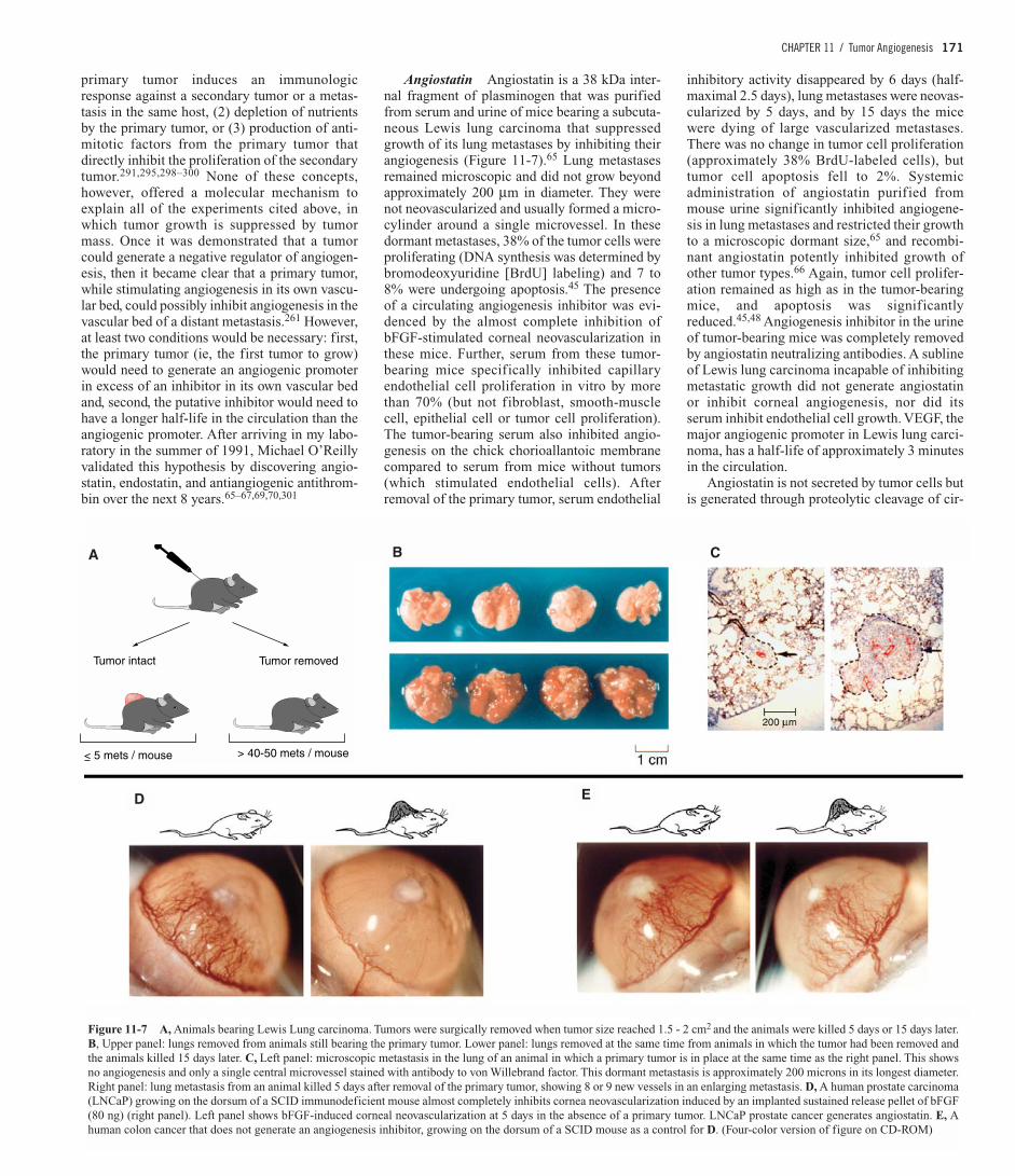
Angiostatin Angiostatin is a 38 kDa inter-nal fragment of plasminogen that was purifiedfrom serum and urine of mice bearing a subcuta-neous Lewis lung carcinoma that suppressedgrowth of its lung metastases by inhibiting theirangiogenesis (Figure 11-7).65 Lung metastasesremained microscopic and did not grow beyondapproximately 200 µm in diameter. They werenot neovascularized and usually formed a micro-cylinder around a single microvessel. In thesedormant metastases, 38% of the tumor cells wereproliferating (DNA synthesis was determined bybromodeoxyuridine [BrdU] labeling) and 7 to8% were undergoing apoptosis.45 The presenceof a circulating angiogenesis inhibitor was evi-denced by the almost complete inhibition ofbFGF-stimulated corneal neovascularization inthese mice. Further, serum from these tumor-bearing mice specifically inhibited capillaryendothelial cell proliferation in vitro by morethan 70% (but not fibroblast, smooth-musclecell, epithelial cell or tumor cell proliferation).The tumor-bearing serum also inhibited angio-genesis on the chick chorioallantoic membranecompared to serum from mice without tumors(which stimulated endothelial cells). Afterremoval of the primary tumor, serum endothelial
inhibitory activity disappeared by 6 days (half-maximal 2.5 days), lung metastases were neovas-cularized by 5 days, and by 15 days the micewere dying of large vascularized metastases.There was no change in tumor cell proliferation(approximately 38% BrdU-labeled cells), buttumor cell apoptosis fell to 2%. Systemicadministration of angiostatin purified frommouse urine significantly inhibited angiogene-sis in lung metastases and restricted their growthto a microscopic dormant size,65 and recombi-nant angiostatin potently inhibited growth ofother tumor types.66 Again, tumor cell prolifer-ation remained as high as in the tumor-bearingmice, and apoptosis was significantlyreduced.45,48 Angiogenesis inhibitor in the urineof tumor-bearing mice was completely removedby angiostatin neutralizing antibodies. A sublineof Lewis lung carcinoma incapable of inhibitingmetastatic growth did not generate angiostatinor inhibit corneal angiogenesis, nor did itsserum inhibit endothelial cell growth. VEGF, themajor angiogenic promoter in Lewis lung carci-noma, has a half-life of approximately 3 minutesin the circulation.
Angiostatin is not secreted by tumor cells butis generated through proteolytic cleavage of cir-
Figure 11-7 A, Animals bearing Lewis Lung carcinoma. Tumors were surgically removed when tumor size reached 1.5 - 2 cm2 and the animals were killed 5 days or 15 days later.B, Upper panel: lungs removed from animals still bearing the primary tumor. Lower panel: lungs removed at the same time from animals in which the tumor had been removed andthe animals killed 15 days later. C, Left panel: microscopic metastasis in the lung of an animal in which a primary tumor is in place at the same time as the right panel. This showsno angiogenesis and only a single central microvessel stained with antibody to von Willebrand factor. This dormant metastasis is approximately 200 microns in its longest diameter.Right panel: lung metastasis from an animal killed 5 days after removal of the primary tumor, showing 8 or 9 new vessels in an enlarging metastasis. D, A human prostate carcinoma(LNCaP) growing on the dorsum of a SCID immunodeficient mouse almost completely inhibits cornea neovascularization induced by an implanted sustained release pellet of bFGF(80 ng) (right panel). Left panel shows bFGF-induced corneal neovascularization at 5 days in the absence of a primary tumor. LNCaP prostate cancer generates angiostatin. E, Ahuman colon cancer that does not generate an angiogenesis inhibitor, growing on the dorsum of a SCID mouse as a control for D. (Four-color version of figure on CD-ROM)
Tumor intact Tumor removed
< 5 mets / mouse > 40-50 mets / mouse
A B C
D E

172 SECTION 1 / Cancer Biology
culating plasminogen by a series of enzymesreleased from the tumor cells. At least one ofthese tumor-derived enzymes, urokinase plas-minogen activator (uPA), converts plasminogento plasmin, while a phosphoglycerate kinasefrom hypoxic tumor cells then reduces the plas-min so that it can be converted to angiostatin byone of several different metalloproteinases (Fig-ure 11-8).302 Other types of tumors have sincebeen reported to generate angiostatin (eg, humanprostate cancer).303–305 Human prostate carci-noma cells express enzymatic activity that con-verts human plasminogen to the angiogenesisinhibitor angiostatin.302 Prostate-specific anti-gen generates angiostatin-like fragments fromplasminogen.307
Tranfection of Angiostatin. Furthermore,when murine fibrosarcoma cells were trans-fected with angiostatin, primary subcutaneoustumors formed whose growth was slowed in pro-portion to increased levels of angiostatin produc-tion by the tumor cells. In these tumors, the totalangiogenic output of the primary tumor wasdecreased by transfected angiostatin, whichopposed in a dose-dependent manner the activityof the tumor’s secreted angiogenic promoter, butnever completely counteracted it.78 It should beemphasized that the rate of tumor growth(expansion of tumor mass) was directly propor-tional to total angiogenic output of the tumor,inversely proportional to angiostatin productionand to tumor cell apoptosis, and virtually inde-pendent of tumor cell proliferation. This resultwas confirmed in a different tumor model,Kaposi’s sarcoma subjected to angiostatin genetransfer, which resulted in sustained angiostatinexpression, delayed tumor growth and reducedtumor vascularization.360 Gene transfer with adifferent angiogenesis inhibitor, throm-bospondin, gave similar results. Streit and col-
leagues reported (in an experiment discussedabove) that when tumor cells were transfectedwith the angiogenesis inhibitors throm-bospondin-1 and/or thrombospondin-2, prolifer-ation rate (as determined by staining for the pro-liferating cell nuclear antigen [PCNA]) (seeFigure 11-9), also remained relatively constant,despite the decreased angiogenesis and sup-pressed tumor growth.79
Mechanisms of Action. Angiostatin andits isoforms induce cell arrest and apoptosis ofendothelial cells and inhibit endothelialmigration, angiogenesis in vitro, and angio-genesis in the quail chorioallantoic membrane,which provides a quantitative bioassay.308–317
They can also be generated by differentenzymes and by other cell types; can inhibitother tumor types; decrease activity of themitogen-activated protein kinase ERK-1 andERK-2 in endothelial cells; up-regulate E-selectin in proliferating endothelial cells; andcan be delivered in vivo by gene ther-apy.302–304,313–333 In addition, they bindspecif ically to ATP-synthase, a transmem-brane protein expressed by vascular endothe-lial cells; bind to a fragment of vitronectin;and can potentiate radiation therapy of experi-mental tumors.334–337
A provocative recent finding is that prolifera-tion of circulating precursor endothelial cellsderived from bone marrow is inhibited at signifi-cantly lower concentrations of angiostatin than isproliferation of endothelial cells isolated from tis-sues.136 Because precursor endothelial cells frombone marrow can home to angiogenic sites andparticipate in new vessel formation in a tumor (seeabove), it has been speculated that at least onemechanism of angiostatin is to inhibit this sub-population of endothelium, and that these cellsmay be employed as a sensitive bioassay for iden-
tification of novel antiangiogenic molecules.136
Angiostatin effectively blocks endothelial cellmigration induced by plasmin binding to αvβ3integrin.346,362 Angiostatin may act at least in partby transiently increasing ceramide, a sphingolipidsecond messenger implicated in a proapoptoticpathway, and by increasing RhoA, an effector ofcytoskeletal structure.361
Numerous additional recent studies havebeen reported on the mechanism of angiostatinand on its biological and pharmacological activ-ities. A few of these new findings are discussedhere. Angiostatin, but not plasminogen, specifi-cally binds to tyrosine kinase substrate annexinII through the lysine-binding domain in endothe-lial cells.338 This affinity appears to be specificfor endothelial cells and does not occur withfibroblasts. Annexin II on endothelial cells orga-nizes assembly of plasminogen on its surface andproduces plasmin at maximum efficiency in thevascular bed.338–341 Plasmin is a serine proteasethat degrades fibrin and several endothelial cellmatrix proteins, such as laminin, throm-bospondin, and collagens, and facilitates capil-lary sprouting during angiogenesis as well asendothelial migration.338,346,362 In fact, angio-statin has been shown to inhibit endothelial andmelanoma cellular invasion by blocking matrix-enhanced plasminogen activation (see Figure 11-8).326 However, because hypoxic tumor cellsproduce a phosphoglycerate kinase that enzy-matically reduces plasmin, this mechanism con-tributes to the generation of angiostatin, whichcan then block plasmin organization on theendothelial surface and interfere with angiogen-esis.302,363 The gene for annexin II is also upreg-ulated in endothelial cells during angiogenesis.Annexin II is a central molecule in signal trans-duction mechanisms, and disruption of its func-tion affects calcium signaling, tyrosine phospho-rylation, cell proliferation and apoptosis.342–344
Angiostatin mediation of endothelial cell deathappears to be through a calcium signaling mechanism.345 Therefore, annexin II may act as
2000
1000
0
3
38 + 12%-
-
PCNA
41 + 6.9%
Controls
TSP-1
TSP-2TSP-1 + TSP-2
1 2
Time (weeks)
Tum
or v
olum
e (m
m3 )
3000
Figure 11-9 Squamous cell carcinoma cells trans-fected with the angiogenesis inhibitor thrombospondin-1 and or thrombospondin-2. Tumors producing the mostantiangiogenic activity (TSP-1 and TSP-2) have the low-est growth rate and remained dormant. (Modified fromStreit et al.79) Tumors with low or no release of antian-giogenic activity have the highest growth rate and pro-duce large tumors. The proliferation rate of tumor cells(PCNA) is not significantly different between large andsmall tumors.
Tumor
uPAPhosphoglycerate
kinaseSerineproteinase
MMP-12MMP-9MMP-3MMP-7
MMP-2MMP-12
or PlasminPlasminogen Plasmin K1-4.5
K1-4
K1-3
angiostatin S. Gately et al., Cancer Res. 56: 4887, 1996P. Stathakis et al., (P. Hogg) J. Biol. Chem. 272: 20641, 1997Z. Dong et al., (I. Fidler) Cell 88: 801, 1997M.S. O'Reilly et al., (M. Moses) J. Biol. Chem. 274: 29568, 1999B. C. Patterson and Q. A. Sang, J. Biol. Chem. 272: 28823, 1997
Reducedplasmin
TumorHypoxictumor
Figure 11-8 Model of angiostatin formation. Urokinase plasminogen activator from tumors converts plasminogen toplasmin. A phosphoglycerate kinase from hypoxic tumors converts plasmin to reduced plasmin which is then suscepti-ble to further cleavage by a serine proteinase from tumors or by plasmin itself, to kringle fragments 1-4.5 or lowerdepending upon which metalloproteinase is present. From Stathakis et al.302

CHAPTER 11 / Tumor Angiogenesis 173
an endogenous angiogenic switch, and itsblockade may be at least one potential mecha-nism for angiostatin.
Binding proteins for angiostatin. Threeother binding proteins have been suggested asputative receptors for angiostatin: (1) ATP syn-thase on the endothelial surface, (2) aVb3, and(3) angiomotin, which belong to a novel proteinfamily with conserved coiled-coil and PDZ bind-ing domains.334,346–348 Taken together withannexin II, Tuszynski and colleagues suggestthat angiostatin may bind to multiple receptorsand could mediate a cascade of events leading toinhibition of angiogenesis.338 Angiostatin alsoinhibits signaling of hepatocyte growth factor(HGF) in endothelial cells (and in smooth mus-cle cells), by blocking HGF-induced signaling ofc-met, Akt, and ERK1/2.349 However, angio-statin does not block VEGF or bFGF signalingevents in endothelial cells. Angiostatin alsoinduces intracellular acidosis in endothelial cellsand anoikis (loss of cell attachment) in endothe-lial cells that are incubated under conditionsresembling the low pH in a tumor.350 Angiostatindownregulates expression of VEGF expressionin tumor cells.351 This implies that angiostatinmay act not only as a direct inhibitor of angio-genesis, but also as an indirect angiogenesisinhibitor. Analysis of the x-ray crystallographicstructure of angiostatin reveals that kringles 1, 2,and 3 produce a central cavity suggesting aunique domain where they may function in concert.352,364 The enzymatic mobilization ofangiostatin is facilitated by tumor-derived matrix-metalloproteinase.353 Otherlabs confirm the report that circulating angio-statin generated by a primary tumor decreasessignif icantly after the primary tumor isremoved and that this is followed by growth ofremote metastases.35,65
Angiostatin gene therapy. Angiostatin genetherapy shows efficacy in tumor-bearing miceagainst a variety of tumors and their metastasesincluding, breast cancer, renal cell cancer,gliomas, melanoma, Kaposi’s sarcoma, squamouscell carcinoma, and leukemia.310,360,365–375 Ofinterest was that viral vector-mediated antiangio-genic therapy using an angiostatin-endostatinfusion protein demonstrated significant downreg-ulation of ascites, tumor growth, vascularity andprolongation of animal survival in three differentascites tumor models.376 Angiostatin has beentransfected into canine eyes as a preclinical modelfor the treatment of human uveal melanoma.377
Long-term expression of angiostatin and endo-statin from lentivirus-transduced human bladdercarcinoma cells inhibited proliferation ofendothelial cells co-cultured with the tumor cells,the first use of a lentiviral vector for antiangio-genic gene delivery.378 Angiostatin gene therapyhas also shown efficacy in nonneoplastic mousemodels of disease such as arthritis, retinal neovas-cularization, choroidal neovascularization, andendometriosis.379–382
Angiostatin protein therapy. Angiostatinprotein therapy has shown significant efficacy
when delivered systemically to mice bearing avariety of different tumors, and also in combina-tion with endostatin where it has shown a syner-gistic effect or as a fusion protein of angiostatinand endostatin, or in combination with inter-leukin-12.351,383–387 When three different types ofmurine tumors were engineered to produce gran-ulocyte colony-stimulating factor, (GM-CSF), thelevel of angiostatin in the serum increased 4-foldabove controls and directly correlated with GM-CSF production.388 Angiostatin levels directlycorrelated with macrophage metalloelastase pro-duction, which appeared to mediate cleavage ofangiostatin from plasminogen. Metastases weresuppressed in all three tumor systems.
Replacement of angiostatin after radiotherapy.Radiotherapy of primary Lewis lung carcinomasin mice induced regression of the primary tumor,but led to disappearance of angiostatin from theblood and rapid growth of lung metastases.280
Angiostatin replacement therapy preventedgrowth of lung metastases after regression of theprimary tumor by radiotherapy. In some patientssuccessful radiotherapy of a primary tumor is fol-lowed by growth of distant metastases, for exam-ple, nonsmall cell lung cancer. It may eventuallybe possible to determine if a tumor is generatingan endogenous angiogenesis inhibitor prior toradiotherapy and then administer that inhibitor incombination with the radiotherapy.
Angiostatin protein also inhibits angiogene-sis in nonneoplastic states. For example, angio-statin inhibits corneal neovascularization, corpusluteum development in the pre-ovulatory folli-cle, and as pathological retinal angiogenesis, butnot physiological retinal neovasculariza-tion.389–392 Although the original report of 38kDa angiostatin included kringles 1-4 of plas-minogen, a recent report reveals the generationof a novel plasminogen fragment of 22 kDa gen-erated by enzymatic activity from tumorcells.65,393 The p22 plasminogen fragmentincludes kringle 1 plus additional N- and C- ter-minal residues, but circular dichroism and intrin-sic fluorescence analysis define structural differ-ences between it and recombinant kringle 1.Angiogenesis and tumor growth are potentlyinhibited by p22.
When a strategy similar to the one thatuncovered angiostatin (eg, suppression of atumor growth by tumor mass) was employed byO’Reilly and colleagues with murine heman-gioendothelioma and human small-cell lungcancer, endostatin and antiangiogenicantithrombin (aaAT) were discovered.67,69,70
Both endostatin and aaAT are generated fromlarger parent proteins by enzymes released bythe tumor cells. Angiostatin is currently in phaseI/II clinical trials.
Endostatin Endostatin is a 20-22 kDainternal fragment of collagen XVIII.67,69,394,395
It is the first of a group of endogenous angiogen-esis inhibitors which are predominately extracel-lular proteins, which generally require prote-olytic processing to become active.396,397 Morethan 350 reports on endostatin have been pub-
lished in the 5 years since its isolation and iden-tification as an angiogenesis inhibitor.67 Theserange from mechanistic studies to experimentaltherapeutics. A variety of new findings havebeen reported on mechanism of action, physio-logic function, structure-activity relationships,experimental therapeutics including gene ther-apy, and early clinical studies. A few of thesestudies are reviewed here.
Structure-activity relationships. Endostatinwas first isolated and sequenced from condi-tioned medium of murine hemangioendothe-lioma based on the same strategy employed forthe discovery of angiostatin (ie, hemangioen-dothelioma suppressed secondary tumors).398 Itis a specific inhibitor of endothelial cell prolifer-ation and migration like angiostatin. High-reso-lution radiographic structures have been deter-mined for mouse and for human endostatin(Figure 11-10).399,400 At least two enzymes pro-duced by tumor cells are necessary to cleaveendostatin from collagen XVIII, an elastase anda cathepsin.401,402 Endostatin is derived from thenontriple helical C-terminal NCI domain of col-lagen XVIII. Subsequently collagen XV has alsobeen found to share a C-terminal NC1 domaincontaining the endostatin molecule, which hasnot been found to date in any other proteins.403
Endostatin is released proetolytically in trimericform and further converted to monomeric formsof about 20 kDa. Both endostatin isoforms sharea compact globular fold, but differ in bindingproperties for proteins and cells.403 Tissue distri-butions are also different. Differences in activityhave been found between NCI domains and
A
B
N-terminus of 184 aa
Endostatin
I
H
A B
P
F
E
J
CN
G D KL
O
M
Collagen XVIII
HTHQDFQPVLHLVALNTPLS...
Figure 11-10 A, Amino acid sequence of human endo-statin.67 B, Crystal structure of human endostatinresolved to 3.5 angstroms. 400

174 SECTION 1 / Cancer Biology
endostatin activity may be related to itsoligomerization state.
Endostatin in basement membrane. Endo-statin is present in basement membranes and ves-sel walls and is especially rich in elastic fibers ofthe aorta and sparse elastic fibers of veins.404
Some, but not all, capillaries or arterioles showweak labeling for endostatin. Within the elasticfibers, endostatin is co-localized and has beenreported to bind to fibulin-1 and -2, nidogen-1and -2, laminin-1 and perlecan.405 Recombinantmouse endostatin binds to heparin with a Kd of0.3 µM.406 A major site of four clusteredarginines, 155, 158, 184 and 270 and a secondsite (R193, R 194) is essential for binding to hep-arin as well as to heparan sulfate and sulfatides,but not to fibulin-1 and fibulin-2. A minimumheparin size of 12-mer (dodecasaccharide) isnecessary for efficient binding and there is a cru-cial role for 2-O and 6-O sulfation. A syntheticarginine-rich dendrimer that mimicked the sur-face of endostatin and had high affinity for hep-arin, had similar antiangiogenic activity in thechick embryo. This experiment demonstrates theimportant role of heparin affinity for theinhibitory activity of endostatin.407 A decreasedinhibitory activity after mutation of the zinc-binding site was reported for insoluble recombi-nant endostatin from E. coli,408 but eliminationof zinc binding had no effect on antiangiogenicactivity in mouse soluble recombinant endo-statin.406 Also, zinc ligand-disrupted recombi-nant soluble human endostatin from yeastshowed potent antitumor activity in mice.409 It isunclear why insoluble recombinant endostatinrequired zinc for inhibitory activity,408 but thatsoluble endostatin did not. However, it shouldnot be overlooked, that the original insolublerecombinant zinc-containing endostatin inducedmore rapid tumor regression in different tumortypes than has been obtained to date with solubleforms with or without zinc.67,69 One possibilityis that zinc is required to maintain folding of theinsoluble protein.403 Alternatively we have pre-liminary data that peptides of endostatin releasedfrom insoluble endostatin inhibit angiogenesis(K. Javaherian, personal communication). Fur-thermore, under physiological conditions endo-statin exists mainly as a fragment of an insolublematrix protein, collagen XVIII. It appears thatonly a small fraction of endostatin circulates inthe soluble form (ie, in the range of 30 ng/mL).
Of the 20 collagen isoforms that have beenidentified so far in mammalian species, colla-gens IV, XV and XVIII (which possess C-termi-nal globular domains) have been implicated inregulation of angiogenesis.410 Of interest is thathomologues of these three collagens have alsobeen identified in the worm C. elegans, wherethe endostatin domain of type XVIII collagencontrols migration of neuronal cells and axonguidance instead of vascular endothelial cells.411
The evolutionary age of endostatin of approxi-mately 600 million years, may be responsible inpart, for its lack of toxicity in animals andpatients.67,69,267 It is also fascinating from an
evolutionary perspective that certain regulatorsof endothelial cell migration have (or had) con-trol of axon migration, eg, endostatin, neuropilinsemaphorins and ephrins, among others.412–415
This relationship provides insight into howdevelopment of nerves and blood vessels may becoordinated.
In the chick embryo angiogenesis induced bybFGF was inhibited by endostatin, but not by anendostatin mutant that does not bind to heparin.
Mechanisms of action. Novel mechanismshave recently been reported for the antiangio-genic action of endostatin, although we are along way from a complete picture.
Inhibition of VEGF receptor. Endostatinblocks the binding of VEGF121 and VEGF165 tothe KDR/Flk-1 receptor, blocks tyrosine phos-phorylation of this receptor, and blocks activa-tion of its intracellular signaling events, ERK,p38 MAPK and p125FAK.416 This receptormediates endothelial cell motility and prolifera-tion. Endostatin also blocks VEGF.
Downregulation of VEGF expression.While endostatin does not bind to VEGF,416 itdoes downregulate VEGF expression in tumorcells (similar to the effect of angiostatin).351
Endostatin can therefore be considered to act asboth a ‘direct’ and an ‘indirect’ angiogenesisinhibitor. In bFGF-treated endothelial cells,endostatin induces endothelial cell apoptosis, inpart by activating tyrosine kinase signaling of theShb adaptor protein.417 However, endostatindoes not compete with binding of bFGF to tis-sues, and it does not affect bFGF receptor sig-naling.418 This result is consistent with the obser-vation that bFGF (FGF-2) stimulation ofendothelial cell proliferation in vitro and angio-genesis in vivo is mediated by VEGF.419
Prevention of bFGF-induced or VEGF-induced loss of endothelial cell–cell adhesion orendothelial cell adhesion to basement mem-brane. Endothelial cell migration, which iscritical for new sprout formation during angio-genesis, requires continuous turnover of cell-cellinteractions and of cell-matrix interactions.420
Cell-cell interactions between endothelial cellsare maintained in part by β-catenin localized atcell-cell junctions in cadherin complexes of cap-illary endothelial cells in vitro. Treatment ofthese cells with bFGF shifts β-catenin to thecytoplasm, which loosens cell-cell adhesion andfacilitates endothelial migration.421 However,co-treatment with endostatin transiently blocksthis shift.421 Cell-matrix interactions are main-tained in part by formation of focal adhesionsand actin stress fibers. Either bFGF or endostatinalone, induced tyrosine phosphorylation of focaladhesion kinase and paxillin, which promotedformation of focal adhesions and actin stressfibers. However, when microvascular endothelialcells were co-treated with bFGF and endostatin,focal adhesion and actin stress fibers weredecreased, which decreased migration ofendothelial cells. In an in vivo situation, theentire vascular bed would be exposed to circulat-ing endostatin. But its antiangiogenic effect
would only become evident in areas of highgrowth factor stimulation, such as those presentin the tumor endothelium.421 Taken together,these results provide a plausible mechanism toexplain clinical trial data (see below) in whichantiangiogenic effects of endostatin are observedin the absence of systemic toxicity.421
Vessel stabilization: reversal of bFGF-induced or VEGF-induced loss of endothelialcell–cell adhesion or endothelial cell adhesion tobasement membrane. Another antiangiogenicmechanism of endostatin in addition to itsantiproliferative and antimotility effect on angio-genic endothelial cells is the stabilization ofnewly formed endothelial tubes. Endostatindecreases formation of VEGF-induced microves-sels sprouting from aortic rings in vitro. Byimmunohistochemistry, administered endostatinlocalizes to endothelial cell-cell junctions as wellas to adhesion sites between endothelial cells andtheir basement membrane, thus anchoringendothelial cells to each other and to their base-ment membrane (Figure 11-11).422 Endostatindoes not localize to quiescent vasculature. Highconcentrations of endostatin were detected byimmunohistology in rat and human brain tumorsin and around endothelial cells, in tumors cells,and in macrophages and leukocytes in thetumor’s vascular bed, but normal brain showed noendostatin reactivity.447 This unique localizationof endostatin only in newly formed angiogenicmicrovessels, appears to reverse the loosening ofendothelial cells to each other and to their base-ment membrane, which was necessary for themto form angiogenic sprouts in the first place. Sim-ilar “stabilizing” effects of endostatin wereobserved in tumors in vivo, where administeredendostatin was more abundant in association withangiogenic vessels undergoing maturation,mainly at the tumor marginal zone where angio-genesis was highly active. Endostatin immunos-taining is present during vascular formation up tothe stage exhibiting one layer of peri-endothelialcells, but disappears in further stabilized vesselsthat have more than one layer of these cells intheir wall.422 This suggests that the vascular sta-bilizing effect of endostatin occurs earlier thanthe integration of peri-endothelial cells into thevascular wall. A subsequent step of vascular sta-bilization by recruitment and integration of peri-endothelial cells (pericytes and smooth musclecells) is mediated by angiopoietin-1, TGF-β, andPDGF.226 Of interest is that during endothelialtube formation induced by VEGF, endostatin plusVEGF-treated tubes became so stable that aftercessation of endostatin, the survival time of thesetubes was approximately doubled in comparisonto VEGF alone. Furthermore, incubation ofendothelial cells with an antibody to endostatinprevented formation of stable VEGF-inducedtubes and disrupted existing tubes. The role ofendostatin in modulating endothelial cell interac-tion with extracellular matrix is also revealed byexperiments in which oligomeric endostatin(from the NC1 domain of collagen XVIII),induces motility and morphogenesis of tube

CHAPTER 11 / Tumor Angiogenesis 175
forming endothelial cells in Matrigel, whereasmonomeric endostatin inhibits these effects.423
Endostatin downregulates the uPA system on theendothelial cell surface.424 Pro-angiogenic pro-teins such as bFGF increase the secretion of uPA)and plasminogen activator inhibitor (PAI-1) lead-ing to an overall increase in proteolytic activityprerequisite for endothelial cell sprouting andtube formation. Endostatin treatment of endothe-lial cells markedly decreases their production ofuPA and PAI-1.
Inhibition of metalloproteinase. Endostatinalso binds and inhibits the catalytic activity ofMMP-2.425,426 As a result, endothelial cell inva-sion is inhibited and tumor cell invasion may also
be decreased. Tumor progression and angiogene-sis are reduced in MMP-2 deficient mice andMMP-2 is required for the switch to the angio-genic phenotype.106,427
Binding of endostatin to endothelial cell sur-face integrins and other proteins that modulatemotility and morphogenesis. Endostatin alsoinhibits integrin-dependent endothelial cellmigration because it binds to α5- and αv-inte-grins on the endothelial cell surface, in particularα5β1.428 Kalluri and colleagues have also pro-posed that α5β1-integrin may be a functionalreceptor for endostatin (R. Kalluri, unpublisheddata).37 High concentrations of endostatin alsointeract with tropomyosin to interrupt microfila-ment integrity, which inhibits endothelial motil-ity and may participate in part in endothelialapoptosis.429 Endostatin also binds to glypicanson the endothelial cell surface at concentrationswhich suggest that glypicans may be low affinityendostatin receptors.430
Inhibition of cyclin D1 causing G1 arrest ofendothelial cells. Endostatin causes G1 arrestin endothelial cells, by decreasing the hyper-phosphorylated retinoblastoma gene product anddownregulating cyclin D1 mRNA and pro-tein.431 Transcription through the LEF1 site inthe cyclin D1 promoter is essential for endo-statin’s inhibitory activity. Others report thatendostatin arrests proliferation and causes apop-tosis of endothelial cells.432,433
Dephosphorylation of endothelial nitricoxide. Human recombinant endostatin dose-dependently blocked VEGF-induced endothelialcell migration, with complete inhibition at 100ng/mL.434 VEGF stimulates synthesis ofendothelial nitric oxide (eNOS). In endothelialcells sphingosine-1 phosphate activates the ser-ine/threonine kinase Akt and stimulates phos-phorylation of eNOS to release nitric oxide.435
VEGF-stimulated endothelial nitric oxide syn-thesis (eNOS) was abolished by endostatin,(although VEGF-induced formation of prostacy-clin was not significantly affected by endo-statin). Endostatin interfered with activation ofeNOS downstream of Akt by specifically inhibit-ing phosphorylation of eNOS at Ser 1177 bystimulating activation of the phosphatase PP2A(Figure 11-12).434 Nitric oxide synthesis alsoappears to be critical for the mitogenic effect ofVEGF on endothelial cells.114–116 Inhibition ofnitric oxide synthesis also blocks tumor angio-genesis.436 We have previously proposed anotherrole for nitric oxide synthesis in angiogenesis:early vasodilation of pre-existing quiescent ves-sels as a prerequisite for sprout formation.116
Hypoxic downregulation of endostatin pro-duction by endothelial cells and pericytes. Col-lagen XVIII is converted to endostatin, which issecreted from endothelial cells and pericytes invitro at a constant rate.437 The rate of conversionis higher in pericytes. During hypoxia, produc-tion of endostatin is significantly decreased. (30-40% of this hypoxia-induced 43% degradation ofendostatin occurs in pericytes and approximately10% in endothelial cells.) Exogenous endostatin
is more rapidly degraded when incubated withhypoxic conditioned media of endothelial cells orpericytes. The mechanism of endostatin degrada-tion by the conditioned medium is unknown.These results imply that: (1) decreased endostatinproduction by hypoxic pericytes and/or endothe-lial cells may be another mechanism to facilitatethe angiogenic switch in a tumor; (2) pericytesmay employ endostatin as one of the strategies toinhibit endothelial proliferation under normalconditions; and (3) the treatment of a patient witha large tumor burden or with a tumor with a largehypoxic center (eg, glioblastoma), may requirehigh doses of endostatin at the beginning of ther-apy, because of endostatin degradation.
Physiologic Functions Physiologic func-tions for endostatin are gradually being elucidated.
Regulation of development of retinal vascu-lature. Endogenous endostatin in the eye maycontribute in part to the normal development ofvasculature in the retina. During development ofthe eye, hyaloid vessels supply the vitreous andthe lens. Subsequently, these vessels regressleaving a transparent vitreous and lens. Endo-statin is thought to play a key role in regressionof the hyaloid vessels because they fail to regressin endostatin’s absence. In a rare disease calledKnobloch syndrome, the hyaloid vessels fail toregress, retinal vasculature fails to develop, andpatients suffer retinal degeneration and blind-ness, among other abnormalities.438,439 In thissyndrome, a splice mutation in human collagenXVIII leads to a truncated protein lacking theendostatin fragment. A proposed mechanism ofretinal degeneration is that persistent hyaloidvessels increase oxygen in the retina, which sup-presses hypoxia-inducible factor-1 (HIF-1α) andVEGF, both of which drive normal vasculargrowth in the normal developing retina. Retinalvasculature also fails to develop in a collagenXVIII-deficient mouse model.440 Thus, a patho-logical deficiency of endostatin in humans andmice reveals that endostatin is an endogenous
A B
C
New sprout (non-stabilized)
Endostatin therapy ( )
Maturing vessel at stage before pericytes
Maturing vessel
Endostatin Endostatin
Endostatin
Inter-cell endostatin
Basement membrane
Figure 11-11 Model for the antiangiogenic mechanismof endostatin. During vascular stabilization and matura-tion endostatin immunostaining (red dots) changes from adiffuse distribution around endothelial cells of non-stabi-lized blood vessels to a stronger staining in the inter-endothelial junctions and in the contact zone betweenendothelial cells and their basement membrane. (A) It canbe postulated that endostatin (ES) stabilizes the wall ofnewly formed blood vessels via anchoring of endothelialtubes to the basement membrane (BM) and of endothelialcells (green) to each other which leads to vascular matu-ration (B). This process converts the angiogenic blood ves-sels into the quiescent phenotype, thereby inhibiting tumorvascularization. One of the potential consequences of theangiogenic switch may be the reduction of vascular peme-ability leading to tumor regression. Beside the demon-strated anti-proliferative effect of endostatin on endothelialcells, these effects point to an additional antiangiogenicmechanism of endostatin, e.g. tumor regression via stabi-lization of newly formed blood vessels. Therapeutic strate-gies accelerating vascular stabilization and maintenancepromise to be effective in tumor treatment.422
VEGF Akt
Endostatin
-
eNOS
NO - synthesis
Endothelial cell migration angiogenesis
PhosphotasePP2A
Ser 1177 - P
Figure 11-12 Endostatin specifically interferes witheNOS phosphorylation at Ser 1177. The upstream signal-ing pathway including VEGF-induced Akt activation isnot affected. 434

176 SECTION 1 / Cancer Biology
inhibitor of angiogenesis under normal physio-logical conditions.
Endostatin in platelets. Platelets contain atleast 14 positive regulators and approximately 12negative regulators of angiogenesis.441 The posi-tive regulators include VEGF-A,VEGF-C,bFGF, HGF, angiopoietin-1, PDGF, heparanseand sphingosine-1 phosphate, among others. Thenegative regulators include endostatin, plateletfactor 4, thrombospondin, plasminogen (angio-statin), high molecular weight kininogen(domain 5), and α-2 antiplasmin, among others.These stimulators and inhibitors of endothelialcell migration and proliferation may be releasedinto a blood clot in a wound in an orchestratedsequence to regulate neovascularization.442 Forexample, thrombin induces the release of theproangiogenic protein VEGF from platelets.356
Thrombin also induces the release of endostatinfrom platelets.443
Thrombin induction of endostatin release ismediated by proteinase-activated receptor-4(PAR-4), a mechanism that is independent ofplatelet aggregation and of the action of ADP.When a clot forms in a wound, invasion bymicrovascular endothelial cells is initially inhib-ited, which avoids premature clot lysis.442 Subse-quently, endothelial cells are recruited into theclot during its neovascularization (“granulationtissue”). It is not known how the release of nega-tive and positive regulators of endothelial migra-tion from platelets is orchestrated in a wound.Recent evidence, however, reveals that if athrombus in a large vessel is recanalized, theneovasculature is made up mainly of bone mar-row-derived endothelial cells.444 This is anal-gous to certain tumors (eg, lymphomas) thatrecruit the majority of their endothelial cellsfrom bone marrow. Endostatin is also releasedfrom platelets by certain drugs, such as ticlopi-dine, which inhibits platelet aggregation byblocking the action of ADP with its receptor.445
The nonsteroidal antiinflammatory drugscelecoxib and flurbiprofen or HCT-1026 (a nitricoxide-releasing derivative of flurbiprofen), all ofwhich are cyclooxygenase inhibitors, alsorelease endostatin from platelets and elevate theplasma level of endostatin.446 From these find-ings, one can ask whether the antiangiogenicactivity of celecoxib (Celebrex) operates in partby releasing endostatin from its stores and mak-ing it available to endothelial cells in an angio-genic site. Also, future research will be neces-sary to answer other questions. What is theturnover rate of endostatin in platelets? Do can-cer patients on long-term celecoxib therapydeplete their platelet stores of endostatin? Canplatelets be loaded with endostatin that is admin-istered systemically or in vitro? Would cancerpatients treated with celecoxib benefit from peri-odic endostatin therapy? Would patients on long-term endostatin benefit from celecoxib therapy?Taken together these findings indicate that endo-statin is normally distributed among three com-partments: (1) stored as a cryptic fragment incollagen XVIII in basement membranes; (2)
stored in platelets; and (3) circulating as low lev-els of free endostatin. A fourth compartment canbe considered as the concentration of endostatinin and around angiogenic vessels in tumors, andpossibly at other angiogenic sites.
CONTROL OF VASCULAR PERMEABILITY
Endostatin decreases vascular permeability.bFGF treatment increases permeability of[3H]inulin through confluent monolayers of cap-illary endothelial cells in a transwell assay, aneffect that is counteracted by co-treatment withendostatin.421 In mice that have received anintravenous injection of Evans blue dye, a largeblue stain appears at the site of a subcutaneousinjection of VEGF or platelet-activating factor(PAF) (Miles test). Pretreatment with endostatinblocks the permeability induced by these pro-teins (S. Soker, J. Heymach, and J. Folkman,unpublished data). These results imply that sys-temic administration of endostatin to patientsmay decrease edema in brain tumors, but thisspeculation has not yet been tested.
Experimental therapeutics including genetherapy Insoluble protein administered sys-temically Systemic administration of endostatincan inhibit or regress different tumors. The firstrecombinant endostatin was from Escherichia coliin an insoluble form, because this had previouslybeen successful with angiostatin, and becauserefolding of the inclusion body to a soluble formby urea, guanidine-HCL and glutathione gave alow yield (1%).65,67,69 When this endostatinpreparation was administered subcutaneouslyevery day for prolonged periods of time in mice(185 days for Lewis lung carcinoma, 160 days forT241 fibrosarcoma, and 80 days for B-16melanoma), there was no drug resistance. Afterdiscontinuation of endostatin at these time peri-ods, tumors did not recur. They remained dormantat a microscopic size.65 This was not due to animmunologic mechanism. Wounding the tumorsite or moving the tumor to another site in thesame mouse initiated tumor neovascularizationand growth. Another possibility is preferentialstorage of endostatin in the matrix at the site of theregressed tumor.448 We have observed this samedormancy after 40 days of endostatin therapy ofrat mammary cancer induced by an oral carcino-gen.449 Other investigators have reported success-ful tumor inhibition with insoluble endostatinfrom E. coli.450 Also, soluble endostatin from E.coli has inhibited tumor growth.451
Soluble recombinant endostatin A widevariety of other tumors in many different labora-tories have been inhibited by endostatin in miceand rats without evidence of toxicity or drugresistance. These include lung adenocarcinoma,thyroid carcinoma, colon carcinoma, leukemia,human nonsmall cell lung cancer, human pancre-atic cancer, human neuroblastoma, mammarycancer (soluble endostatin from E. coli), coloncancer metastases to liver, spontaneous mousemammary carcinoma (delayed onset, decreasedtumor burden, and prolonged survival), and spon-taneous pancreatic islet carcinomas.72,452–460
Mammary carcinomas were more potently inhib-
ited by a mutated form of endostatin in which analanine residue was substituted for a proline atposition 125 (P125A), than by the wild-typeendostatin.458 Gliosarcoma both orthotopic andectopic in rats was markedly inhibited (~50%)after only 10 days of treatment by a relatively lowdose of murine endostatin (0.3 mg/kg).461 Inhibi-tion of tumor growth persisted after cessation oftherapy. Yamaguchi and colleagues reported thatendostatin administered into a tumor will inhibittumor growth at significantly lower doses thansystemic administration.461a
Bolus versus continuous administrationA general characteristic of optimal antiangio-genic therapy is that angiogenesis inhibitorsshould be at effective levels in the blood contin-uously. Endothelial cells in the vascular bed of atumor are constantly exposed to a proangiogenicstimulus from the tumor cells and local stroma.This stimulus needs to be continuously opposedby inhibitors in the blood. An analogy would betitration of blood glucose by insulin, whereendothelium is glucose. In a compelling experi-ment, 24-hour continuous parenteral administra-tion of human endostatin was 10-fold moreeffective than the same dose administered as abolus once every day, against human pancreaticcancer in SCID immunodeficient mice (se Fig-ure 11-1).72 Tumor growth was inhibited in adose-dependent manner, but only continuousadministration caused tumor regression. Itshould be noted that continuous administrationof endostatin in vitro (by a floating micro-osmotic pump in 24-well plates) inhibitedendothelial proliferation more significantly thanonce daily addition of endostatin.
Combinations of endostatin with angio-statin Reports of endostatin administered incombination with angiostatin, whether as a sepa-rate proteins, separate genes, or as fusion proteinof angiostatin and endostatin in tumor-bearinganimals, all show a synergistic increase in effi-cacy.69,386,375,462 While the mechanism of syn-ergy is unclear at this writing, one possibility isthat endostatin and angiostatin each up-regulatethrombospondin expression or protect it fromenzymatic destruction (Figure 11-13).
Combination of endostatin with chemother-apy, radiotherapy, or immunotherapy Endo-statin (and angiostatin) potentiate radiotherapy intumor-bearing mice, when the inhibitor is admin-istered at the same time as the ionizing radia-tion.463 Tumors underwent regression when treatedwith a combination of ionizing radiation and endo-statin, but not with either agent alone, even whenthe endostatin dose was lowered below the effec-tive dose and the tumor cells were radioresistant. Itcan be speculated that radiation damage to activeendothelium in a tumor bed may not be repaired inthe presence of an angiogenesis inhibitor. Thisexperiment also suggests that radiotherapy may bein part endothelial-dependent. The optimal sched-ule of radiotherapy and antiangiogenic therapy forcancer patients is unclear.
Endostatin may also potentiate immunother-apy. In an elegant experiment by Davidoff et al

CHAPTER 11 / Tumor Angiogenesis 177
the gene for the potent immunogen, green fluo-rescent protein (GFP) and the gene for endo-statin were transferred together to murine neu-roblastoma cells prior to inoculation intosyngeneic immunocompetent mice.464 Preven-tion of formation of appreciable tumors waseffected in 63% of mice, while tumor cellsexpressing endostatin alone or GFP aloneyielded only a modest delay in tumor growth.Mice treated with the combination were protected against subsequent tumor challengewith unmodified cells. Endostatin also potenti-ates conventional chemotherapy in tumor-bearing animals.465 However, it is not clear howcombinations of antiangiogenic therapy andchemotherapy at maximum tolerated dose schedules will translate to patients (see below).
Endostatin gene therapy Endostatin genetherapy in tumor-bearing mice has generally beensuccessful when administered locally, but it hasbeen problematic when administered systemically.
Local gene therapy. In an example of localgene therapy, mouse liver tumor cells transfectedwith a retroviral vector containing the murineendostatin gene were injected subcutaneously orinto the peritoneal cavity.467 After 63 days, con-trol tumors (vector without endostatin) reached2400 mm3 compared to 30 mm3 for endostatintransduced tumor cells (p < 0.001). After 123days all 16 mice given an intraperitoneal injec-tion of control tumor cells had died, compared toonly 3 of 32 mice given an injection of the endo-statin tumor cells (91% survival) (Figure 11-14).467 Increased levels of endostatin were
detected in tumor lysates, but not in serum ofmice with subcutaneous tumors. Other reports oflocal gene therapy with endostatin show effec-tive tumor inhibition in animals. Examples areinhibition of human liver carcinoma, renal cellcarcinoma, mouse melanoma.468–471 A humanbreast carcinoma, MDA-MB435, was markedlyinhibited when the cells were transduced withendostatin, but another breast cancer MCF7 wasnot inhibited.365 (However, angiostatin genetransfer inhibited MCF7, but not MDAMB435).365 Gliomas were inhibited by genetransfers to tumor cells that were then implantedinto rat brains.472 Gliomas were also inhibited byintratumoral implantation of alginate-encapsu-lated beads containing packaging cells trans-fected with endostatin.473–475
Of interest is that intratumoral gene transferof endostatin inhibited mouse mammary tumors(MidT2-1), but had a minimal effect in humanbreast MDA-MD-231 in SCID mice.476 Despitemany successes of endostatin gene transfer intotumor cells, unsolved problems remain. The factthat one author can report marked and sustainedtumor inhibition with endostatin transduction ofa mouse breast cancer, but minimal effect in ahuman breast cancer MDA-MD-231, highlightsthe problem.476 A third author reports markedinhibition by endostatin gene transfer into ahuman breast cancer MDA-MBA335, but mini-mal effect in MCF7, yet angiostatin gene transferinto MCF7 was very inhibitory, but had onlyminimal effect on MDA-MB435 breast cancercells.365 For unknown reasons, the inhibitor maylose activity when produced by one type oftumor cell, but not by another. Whether this is aprotein-folding problem, or results from excesslocal levels of endostatin at the endothelialreceptor site is not clear (O. Kisker, et al., unpub-lished data). A fourth author reports that breastcancer (MCA-4) transduced with endostatin issignificantly inhibited.477
Systemically administered gene therapy.Systemically administered gene therapy (by anintravenous or intramuscular vector in mice orrats) significantly inhibited tumor growth in
Regulation of the endogenous angiogenesis inhibitor, thrombospondin-1
Id1(tumor cells)
Id1Endostatin
Annexin II(angiogenic endothelium)
Binds toAnnexin II
p53(normal cells)
Angiostatin Plasmin
Plasmin
Thrombospondin-1
Thrombospondin-1
Thrombospondin-1
Thrombospondin-1
Thrombospondin-1
X
Figure 11-13 Endostatin, angiostatin, and wild type p53 all act to up-regulate throm-bospondin-1, a potent endogenous angiogenesis inhibitor. Endostatin down-regulates Id1which leads to the up-regulation of thrombospondin-1. Angiogenic endothelium overex-presses annexin II which mobilizes plasmin at the surface of endothelial cells. Plasmindegrades thrombospondin-1. Angiostatin binds to annexin II to decrease plamin which canincrease available thrombospondin-1.
0
0.0
10 20 70 80 90 100 110 120 130
0.6
0.5
0.4
0.3
0.2
0.1
0.8
0.7
0.91.0
30 40 60
NMuli
NEF - null
NEF - Endo 2
NEF - Endo 1
NEF - Endo 3,4
50Days
Pro
port
ion
surv
ivin
g
0
0
10 20
1000
1500
2000
2500
3000
3500
500
30 40 6050Days after injection
Parental cells
NEF - null cells
NEF - endostatinClones
Tum
or v
olum
e (m
m3 )
Figure 11-14 Left panel: Mouse survival after intraperitoneal inoculation of parental tumor cells and tumor cells transduced with endostatin. n = 8.Controls are NEF-null and NMmuLi. Mice bearing NEF-endostatin transduced tumor cells survive up to 120 days. Right panel: Growth curves of endostatintransduced tumor cells which remain dormant and microscopic vs. control tumor cells which grow to over 2 grams by 60 days.467

178 SECTION 1 / Cancer Biology
renal cell carcinoma, Lewis lung carcinoma ade-nocarcinoma MC38, hemangioendothelioma,spontaneous breast cancer and its metastases,and colon cancer (when the gene was injectedintramuscularly and endostatin serum levelswere only moderately elevated, [35-40ng/mL]).367,469,478–481 Adenoviral gene transferof endostatin delayed growth of breast cancerimplants and retarded their growth by 67% afterthe tumors appeared.478a
Problems with systemic gene therapy.However, other reports of systemically adminis-tered endostatin gene therapy failed to inhibittumor growth. For example, either local or sys-temic endostatin gene therapy failed to inhibit rattumors despite the achievement of serum levelsmore than 10 to 200 times above normal levels ofendostatin (eg, 500 ng/mL up to 10 µm/mL).482
However, these tumors were significantly sup-pressed when an inhibitor of plasmin was admin-istered together with endostatin by gene therapy,but not when the plasmin inhibitor was adminis-tered alone.482 This result suggests that very highlevels of plasmin in a tumor bed may interferewith endostatin activity. It is interesting thatangiostatin, by binding to annexin II when it isoverexpressed by endothelial cells in an angio-genic site, decreases plasmin production on theendothelial surface.326,338–341
It remains to be demonstrated if this mecha-nism plays a role in the synergy of angio-statin–endostatin combinations.69,375,386,462 Wealso were unable to demonstrate significant inhi-bition of tumor growth and only 23% to 33% (p< 0.002 and 0.0001 respectively) inhibition ofcorneal angiogenesis, when viruses encodingangiostatin or endostatin were administered as asingle intravenous injection, despite the fact thatpeak serum levels of endostatin reached 400 to599 times above the normal level.485 In contrast,viruses encoding soluble forms of Flk1 or Flt1,the receptors for VEGF, resulted in approxi-mately 80% growth inhibition of the same tumorand strong inhibition of corneal neovasculariza-tion (74% to 80%) (p < 0.001). A strong correla-tion was observed between the effects of the dif-ferent viruses on tumor growth and the activityof the viruses in the inhibition of corneal micro-pocket angiogenesis. It is unclear why angio-statin and endostatin delivered by systemicallyadministered gene therapy should be so ineffec-tive as tumor inhibitors, when soluble VEGFreceptors administered by the same viruses andin the same tumor models should inhibit tumorgrowth so effectively. However, three specula-tions may be considered as a basis for futureresearch. Antiangiogenic and antitumor activityof endostatin are dose-dependent, but recentexperiments reveal a U-shaped curve (O. Kisker,et al., unpublished data), analogous to interferon-α and also TGF-β.485a,485b In one tumor systemhuman recombinant endostatin protein adminis-tered at 50 mg/kg/day inhibited tumor growth by47%; 100 mg/kg by 84% and 1000 mg/kg by37%. Therefore, it is possible that super high lev-els of endostatin above an optimum level lose
activity in vivo. A second speculation is thatendostatin may aggregate at very high serum lev-els, or that its folding may be affected. A thirdspeculation is that serum plasmin levels may beincreased by certain viruses or by high viraltiters. Other authors subsequently have reportedfailure of endostatin gene transfer to signifi-cantly inhibit a murine leukemia, when endo-statin was transduced into hematopoietic stemcells and serum endostatin levels of 300 ng/mLwere reached, or to inhibit metastases from afibrosarcoma when serum endostatin levels ofup to 700 ng/mL were obtained.483,484 Of inter-est is that these authors previously reported incollaboration with Folkman and O’Reilly,enhanced antitumor activity with an endostatin-angiostatin fusion protein engineered inhematopoietic stem cells.484,484a
Endostatin signaling network To deter-mine how the endothelial cell genome and pro-teome react to endostatin treatment, we used agenome-wide expression profile covering 95%of the human genome and phosphorylation anal-ysis on antibody arrays (Abdollahi A, MaerckerC, Rastert R, et al. Endostatin signaling network.[In preparation]) Briefly, we generated theHuman Unigene Chip II containing 74,834 ele-ments, which represents one of the largest genesets interrogated in array-based gene expressionstudies to date. Alteration in gene expression wasanalyzed after four-hour treatment humanmicrovascular endothelial cells with humanrecombinant endostatin (Pichia Pastoris, endo-toxin-free EntreMed/Calbiochem). Using 12reusable chips more than 1,800,000 gene expres-sion measurements in total were performed onendostatin-treated endothelium and compared tocontrol samples of untreated endothelium fromtwo different human subjects. The identities of50,000 gene clusters (spotted DNA clones) havebeen sequence verified. By these criteria, 429anigene clusters were more than 5-fold upregu-lated. In contrast, 421 anigene clusters weredownregulated by 80%.
Overexpressed genes during endostatin treat-ment included apoptosis genes, cardiovascularand stress response genes, (heat shock, etc.), andgenes associated with angiogenesis inhibition.Underexpressed genes during endostatin treat-ment included gene families associated with can-cer and angiogenesis induction (the angiogenicswitch), as well as transcripts encodingcytokines, cytokine receptors, cellular adhesionmolecules, G proteins, G protein-coupled-recep-tors and transcription factors.
Endostatin downregulated expression ofhypoxia-inducible transcription factor-1 α (HIF-1α) by 2.5-fold.365,427 Endostatin also downreg-ulated many upstream activators of HIF-lα,including c-jun/Fos, Ets, and EGFR. On theother side, HIF-1α normally helps to trigger acoordinated angiogenic response by inducing theexpression of genes involved in blood vesselgrowth including, VEGF and its receptors,VEGFR2, and neuropilin, as well as nitric oxidesynthase and cyclooxygenase-2.
Endostatin also downregulated the Id tran-scription factors. These genes are proangiogenic.Endostatin substantially downregulated MMP-2and αvβ3-integrin.425 Furthermore, Id-1 regulatesangiogenesis through transcriptional repressionof thrombospondin-1.528 Therefore, endostatin,by downregulating Id-1, up-regulates expressionof thrombospondin-1, a powerful angiogenesisinhibitor on its own (see Figure 11-13).
Ephrin signaling is downregulated by endo-statin. The ephrin/Eph family has recently beenshown to participate in the regulation of angio-genesis. Both ephrin B1 and ephrin B2 inducesprouting angiogenesis in vitro to an extent simi-lar to the angiogenic activity of VEGF orangiopoietin 1.529, 530
Endostatin inhibited tumor necrosis factor-α-induced angiogenic signaling by downregulatingthe TNF-α receptor. Additionally, NF-κB(nuclear factor of kappa light chain geneenhancer in B-cells) was downregulated byendostatin. It is thought that activation of NF-kappaB by TNF-α, ionizing radiation orchemotherapeutic agents protects cells fromapoptosis.532,533 Therefore, endostatin inhibitionof NF-kappaB in these settings should enhanceapoptosis of endothelial cells. STAT1 and STAT3were downregulated in endothelial cells treatedby endostatin. STATs (signal transducers andactivators of transcription factors) up-regulategenes that encode inhibitors of apoptosis (Bcl-family), inducers of angiogenesis (VEGF), andcell cycle regulators (cyclins D1/D2 and c-myc).534 After endostatin treatment of endothe-lial cells, most of the genes downstream of STATwere strongly downregulated. These include theapoptosis inhibitor Bcl-2, and the cell cycle reg-ulators, cyclins D1/D2 and c-myc.
Thrombin receptors (PAR-1 and PAR-2)were also significantly downregulated by endo-statin. Thrombin is a potent stimulus for releaseof VEGF by transcriptional regulation activationof HIF-1α.535 Thrombin also up-regulatesexpression of VEGF receptors on endothelialcells.536 In a positive loop VEGF acceleratesthrombin generation, another proangiogenicevent.537 This loop has implications for cancerpatients. The majority of cancer deaths aredirectly attributable to thrombosis or bleeding.This observation emphasizes that endostatindownregulates proangiogenic genes and simulta-neously up-regulates genes that lead to inhibitionof angiogenesis.
Endostatin directly downregulated Ets-1 inendothelial cells in a time-dependent manner anddecreased the mRNA level of most of the Ets-1target genes like MMP-1 and integrin-β3. Ets pos-itively regulates angiogenesis by inducing severaltarget genes including uPA, MMP-1, MMP-3,MMP-9 and integrin-β3, and the elimination ofthe Ets-1 activity by a dominant negativemolecule inhibits angiogenesis in vivo.538–542 Astriking finding is that several pathways thoughtto be distinct, in fact intercommunicate to bringabout a focused antiangiogenic response in thetumor bed, directly as a result of endostatin treat-

CHAPTER 11 / Tumor Angiogenesis 179
ment. It is not clear whether many of the smallmolecular angiogenesis inhibitors produce such aorchestrated antiangiogenic response at thegenetic level as endostatin does.
Clinical applications of endostatin Thereare several interesting characteristics of endo-statin that make it advantageous for clinical usein cancer patients. First, it is not toxic.267 At thiswriting, endostatin is in phase I/II clinical trials,and to date it has only been used in not more thanapproximately 120 patients. However, all centersreport that there are virtually no side effects,even in patients who have been on endostatin forup to one year (Endostatin Banbury Conference.A critical review of endostatin and its biology;2002 Mar 10–13; Cold Spring Harbor Laborato-ries, New York). Patients who receive the subcu-taneous preparation of endostatin take their owninjections each day at home, like insulin.
Wound Healing. Endostatin does notdelay wound healing in patients, nor does itdelay wound healing in animals.486 We haveconfirmed that wound healing in mice is notdelayed by endostatin doses sufficient to com-pletely inhibit growth of tumors in the sameanimal (J. Marler, et al., unpublished data).
Pregnancy. Endostatin has no effect onpregnancy in mice. Pregnant mice treated withtumor-regressing doses of endostatin delivernormal babies, even if the mothers bear a tumorthat is then inhibited by the endostatin (RobertD’Amato, et al, unpublished data).
Down Syndrome. Serum endostatin levelsare significantly elevated in Down syndromepatients.492 Endostatin serum levels in normalindividuals range from 4 to 40 ng/mL (20.3 ±11.5, mean and SD). For Down syndrome indi-viduals, it is 38.6 ± 20.1, a significant elevation.The collagen XVIII gene is on chromosome21q22.3. Because Down syndrome is most com-monly caused by a duplication of one of thecopies of chromosome 21, these individuals havean extra copy of collagen XVIII in which endo-statin is a cleavage product. Down syndromeindividuals have a low incidence of solid tumors.
A recent study in the United States of 17,987people with Down syndrome revealed that theyhave approximately 0.1 the incidence of the con-trol age-matched population for virtually allmalignancies except testicular cancer andleukemia.493 This low incidence of cancer is asso-ciated with a large reduction in cancer associateddeaths among Down syndrome patients. It hasbeen proposed that an increase of about one-thirdof normal endostatin serum levels may representan effective therapeutic dose to significantlyinhibit many solid tumors.492 When this observa-tion is taken together with endostatin’s lack oftoxicity, endostatin may someday be used chron-ically in a preventive mode for patients with ahigh cancer risk for breast cancer or other tumors.
Clinical Trial. A polymorphism in endo-statin predisposes for the development ofprostate cancer.494 A single missense substitu-tion of asparagine for aspartic acid at D104Nlocated in the C-terminal globular domain NCI
of collagen XVIII, yields endostatin with thismutation at N104. Men who are heterozygousfor this mutation have a 2.5 times increased riskof prostate cancer. The authors propose that (1)the DNA segment containing this residue mightcontain a novel interaction site to an as yetunknown receptor; (2) the presence of N104 mayimpair the angiogenesis inhibitory function ofendostatin; and (3) this mutation could possiblybe used for a prostate cancer screening test.494
Prostate Cancer. In a phase I trial of endo-statin 25 patients who had failed all previoustherapy were treated with endostatin.267 Theyreceived a daily intravenous bolus dose of endo-statin over a period of 20 minutes. Endostatinwas safe and had a linear pharmacokinetic pro-file up to 599 mg/m2/day. Tumors regressed intwo patients, although not sufficiently to satisfythe criteria for the terms ‘partial’ or ‘complete’response used for chemotherapy. In one patientwith metastatic melanoma there was prolongedstabilization for 426 days with a much improvedquality of life and he returned to work.
The limited antitumor effect of bolus dosingin these patients is similar to the weak antitumoreffect observed in mice that received bolus dos-ing.72 In ongoing phase II trials, patients inBoston are receiving subcutaneous injections ofa slow-release preparation of endostatin, whichprovides a steady state blood level of endostatinsimilar to that obtained by continuous intra-venous dosing in patients and continuousintraperitoneal pump dosing in mice.72 In thesepatients endostatin is showing more anticanceractivity and more prolonged stable disease up to1 year or more.
Production and manufacture of endostatinRecombinant endostatin can be produced inlarge scale in E. coli, Drosophila cells, P. pas-toris and in mammalian cells.487–491 Currentclinical trials in the United States and TheNetherlands employ recombinant human endo-statin produced in yeast (P. pastoris).
A family of angiogenesis inhibitors inmatrix proteins The discovery of endostatin, aspecific and potent angiogenesis inhibitor resid-ing as an internal fragment of collagen XVIII,further implicated the potential role of basementmembranes in the regulation of angiogenesis. In1996, three independent lines of evidence ledKalluri and his colleagues to launch a new line ofinvestigation into the role of vascular basementmembrane in the regulation of angiogenesis: (1)the observation that vascular basement mem-brane proteins are associated with resting andgrowth arrested capillary endothelial cells, whileproliferating and migrating endothelial cells areunder the influence of provisional matrixmolecules such as vitronectin and fibronectin; (2)studies by Madri and also by Furcht that suggestthat sub-domains of type IV collagen exhibit dif-ferent properties as compared to the intact full-length molecule; and (3) the demonstration byFurcht that the noncollagenous-1 domain (NC1domain) can disrupt the assembly of type IV col-lagen.498–500,670–673 Kalluri hypothesized that
during the inductive phase of angiogenesisinvolving the degradation of vascular basementmembrane, proteolytic degradation fragments ofbasement membrane are liberated with the poten-tial for positive and negative regulation of capil-lary endothelial proliferation and migration.Such a potential role for matrix fragments couldprovide the regulatory balance for an organizedformation of new capillaries.
A biochemical approach was designed toextract vascular basement membranes and per-form degradation with tumor microenvironmentassociated enzymes. The isolated vascular base-ment membrane per se, did not reveal any antian-giogenic activity in its solid state. But, degrada-tion by various basement membrane-degradingenzymes liberated cryptic domains of collagenmolecules from the vascular basement mem-brane with novel antiangiogenic activity.500
Interestingly, while these fragments are presentin the intact molecules, in this form they do notexhibit antiangiogenic activity. The initial screenof fragments from vascular basement membraneidentified three antiangiogenic fragments fromtype IV collagen, known as arresten, canstatinand tumstatin. Endostatin was also identified inthis collection of basement membrane frag-ments. The discovery of arresten, canstatin, tum-statin and α6 chain NCI domain of type IV col-lagen was later also independently published byPetitclerc and coworkers.504
Arresten In vitro antiangiogenic activityArresten (26 kDa) is the NC1 domain fragmentof α1 type IV collagen.262 Arrestin exhibitsantiangiogenic activity in various in vitro and invivo assays and is found circulating in normalindividuals. Recombinant arresten inhibited thegrowth of small and large renal cell carcinomaxenografts and prostate carcinoma-xenografts innude mice at doses as low as 4 mg/kg/day/intraperitoneally. The decrease in tumor size wasconsistent with a decrease in CD31 positive vas-culature.262
Mechanism of action. The antiangiogenicand antitumor action of arresten is potentiallymediated by heparan sulfate proteoglycan andα1β1-integrin on proliferating endothelialcells.262 Two independent studies support thecontention that arresten may function via α1β1-integrin. Senger and colleagues demonstrate thatα1-integrin-neutralizing antibodies can inhibitVEGF driven angiogenesis without detectableeffect on pre-existing vasculature, and VEGFcan induce expression of α1β1- and α2β1-integrins.501 Additionally, Pozzi and colleaguesdemonstrate that tumors grow slower in α1 inte-grin null mice, when compared to the wild-typemice.502,503 Collectively, all of these studies suggest that arresten can potentially act as anendogenous inhibitor of α1β1-induced tumorangiogenesis.
Canstatin Canstatin (24 kDa) is the NCIdomain fragment of α2 type IV collagen.263
Canstatin exhibits antiangiogenic activity in sev-eral in vitro and in vivo assays.263 Recombinantcanstatin inhibits the growth of small and large

180 SECTION 1 / Cancer Biology
renal cell carcinoma tumors by four- or three-fold with respect to placebo-treated mice. Estab-lished human prostate carcinoma (PC-3) insevere combined immunodeficient (SCID) miceor athymic (nu/nu) mice exhibited fractionaltumor volumes of about threefold less thanplacebo-treated mice when treated with 3 mg/kgcanstatin. This decrease in tumor size was con-sistent with a decrease in CD31 positive vascula-ture.263 Recent studies using syngeneic mouseLewis lung tumors (LLC) in C57BL/6 mice andorthotopic MDA-MB 435 breast tumors in nudemice also reveal significant tumor inhibitionwith recombinant canstatin. The antiangiogenicactivity of canstatin was also independently val-idated subsequently by Petitclerc and cowork-ers.504 Petitclerc and colleagues demonstrate thatrecombinant canstatin, tumstatin, and theα6(IV)NC1 domain can inhibit angiogenesis in aCAM assay. Petitclerc and colleagues also showthat canstatin can bind to αvβ3-integrin onendothelial cells.
Mechanism of action. The mechanism ofhow canstatin inhibits proliferation and migra-tion of endothelial cells was further investigated.Kamphaus and colleagues investigated the effectof canstatin on ERK activation induced by 20%fetal bovine serum and endothelial mitogens.Canstatin does not act primarily by inhibitingVEGF or bFGF activation of ERK.263 Instead,canstatin specifically induces apoptosis ofendothelial cells with insignificant effect onnonendothelial cells. Apoptosis of endothelialcells by canstatin is mediated by induction of asteady decrease in FLIP protein (an antiapoptoticprotein associated with FAS-mediated apoptosispathway) levels in the presence of 20% fetal calfserum, bFGF and VEGF. Interestingly, canstatindid not affect FLIP levels in the absence ofgrowth factors. Canstatin’s lack of effect onendothelial cells in the absence of growth factorsmay indicate that only proliferative endotheliumis targeted.262 Recent studies suggest thatcanstatin binds to αvβ3-integrin and also α3β1-integrin on proliferating endothelialcells.504 The importance of αvβ3 and α3β1-inte-grins for the antiangiogenic activity of canstatinis yet to be determined.
Tumstatin Tumstatin (28 kDa) is the NC1domain fragment of α3 collagen molecule andexhibits antiangiogenic activity in both the in vitroand in vivo assays.71,264,505,506,507 The antiangio-genic activity has been narrowed down to a 25amino acid stretch in the middle of the molecule.At equal molar concentrations, the full-lengthtumstatin exhibits similar activity as the 25 aminoacid peptide termed as T7 peptide71,264,505,506,507
Recombinant tumstatin and T7 peptide exhibitsignificant inhibition of human tumor xenograftsin nude and SCID mice, syngeneic LLC andB16F10 tumors in C57BL/6 mice, and orthotopicMDA-MB 435-breast tumor in nude mice at adosage of 1 mg/kg/day/ip. or iv.
Tumstatin (α3(IV)NC1) binds to endothelialcells via αvβ3-integrin and α6β1-inte-grin.71,264,505,506 The α3β1-binding activity for
tumstatin was also demonstrated by Petitclercand colleagues.504 We show that the binding toαvβ3 is pivotal for the antiangiogenic activityassociated with tumstatin and that the activity isrestricted to amino acids 54-132 (tum-5) withinthe 244 amino acid tumstatin using deletionmutagenesis.71,264,506 Additionally, αvβ3 bindingto tumstatin is mediated via a mechanism inde-pendent of the RGD-containing amino acidsequence and vitronectin/fibronectin binding ofproliferating endothelial cells. The antiangio-genic activity of tumstatin can be further local-ized to a 25 amino acid region within tum-5 anda synthetic peptide representing this region pre-serves ‘full’ antiangiogenic activity and binds toαvβ3-integrin.71,264,506 Cell biological experi-ments demonstrate that the antiangiogenic activ-ity of tumstatin is dependent on αvβ3-binding onproliferating endothelial cells.71,506 These exper-iments support the notion that through the actionof endogenous inhibitors such as tumstatin,αvβ3-integrin could also function as a negativeregulator of angiogenesis.71,508–511
Mechanism of Action. Tumstatin inducesapoptosis of proliferating endothelial cells.507
Maeshima and colleagues demonstrate that tum-statin is an αvβ3-integrin-dependent inhibitor ofcap-dependent translation mediated throughnegative regulation of mTOR signaling. Inessence, tumstatin is an endothelial cell specificinhibitor of cap-dependent translation. ThemTOR inhibitory property of tumstatin mimicsthat of rapamycin, except unlike rapamycin,which is pan-specific, tumstatin is specific foronly endothelial cells.507 The action on mTOR ismediated by the FAK-PI3K-Akt pathway down-stream of αvβ3-integrin.
Endogenous angiogenesis inhibitors ascryptic tumor suppressor proteins With thediscovery of angiostatin, endostatin, arresten,canstatin, and tumstatin, a new paradigm hasemerged that puts forward a novel notion thatsome proteins such as plasminogen and basementmembrane collagens, harbor unique propertiesthat are cryptic and become exposed only uponproteolytic degradation. Thus, in their intact form(full-length) plasminogen and basement mem-brane collagen do not exhibit antiangiogenicactivity or antitumor activity; but, when plas-minogen and basement membrane collagensundergo degradation, they now expose or liberatenovel cryptic fragments, which possess antian-giogenic activity.
All of these inhibitors are found in blood ofnormal individuals. A decrease in the physio-logic levels of tumstatin is speculated to result inan acceleration of tumor growth. A change inamino acid sequence of endostatin due to a polymorphism correlates with an increased susceptibility to prostate cancer. Conversely, anincrease in physiological levels of endostatinresults in less propensity for emergence of solidtumor in Down syndrome patients (due to anextra copy of type XVIII collagen) (seeabove).492 Collectively, these results argue thatangiostatin, endostatin, tumstatin, canstatin and
arresten may function as cryptic tumor suppres-sor proteins, offering an additional line ofdefense against tumor progression by blockingangiogenesis switching.
Antiangiogenic conformation of antithrom-bin III A human small-cell lung carcinomasuppressed angiogenesis and tumor growth atremote sites in immunodeficient mice. Thesecells generated an enzyme in vitro that convertedthe 58 kDa conformation of circulatingantithrombin III to a 53 kDa form of the proteinin which the externally configured stressed loopof antithrombin was retracted into the body ofthe molecule.70 The 53 kDa “cleaved” form is aspecific endothelial inhibitor and a potent angio-genesis inhibitor and has no thrombin bindingactivity. Antithrombin III has no antiendothelialor antiangiogenic activity. The enzyme(s) thatinduce this conformational change have not yetbeen elucidated. Human pancreatic cancer alsogenerates the 53 kDa cleaved antiangiogenicantithrombin.512
Tropinin I A novel protein, troponin I, waspurified from cartilage during an attempt to findthe inhibitors responsible for the avascularity ofcartilage.513 It is a 22 kDa subunit of the tro-ponin complex that, along with tropomysin, isresponsible for the calcium-dependent regula-tion of striated muscle contraction. It is a spe-cific inhibitor of endothelial cell proliferationand of angiogenesis in the mouse cornea andchick embryo. It inhibits growth of primarytumor and metastases.
Other angiogenesis inhibitors that areendogenous In addition to the specific angio-genesis inhibitors, endostatin, tumstatin,canstatin and arresten discussed above, theplasma and tissues contain other endogenousangiogenesis inhibitors that, although not spe-cific, appear to preferentially inhibit endothelialproliferation and/or migration. These inhibitorshave quite different structures. The increasingnumber of endogenous inhibitors of angiogene-sis being discovered indicates the existence of afamily of molecules that provide physiologicsuppression of angiogenesis. An analogy may bedrawn to the more than 40 proteins of the clot-ting system, many of which function to preventcoagulation under normal conditions (seebelow). Examples of endogenous angiogenesisinhibitors include interferon-β, platelet factor4, fibronectin fragments, fragments of SPARC,thrombospondin-1, tissue inhibitors of metallo-proteinases (TIMPS), certain angiostaticsteroids such as tetrahydrocortisol, a ,nonmineralocorticoid metabolite of cortisol, and 2-methoxyestradiol, and interleukin-12.243,246,247,259–261,514–525 Thrombospondinalso inhibits lumen formation, a rather uniqueproperty of an angiogenesis inhibitor.521 Inaddition to its cytotoxic properties, mediated byT-cell activation, interleukin-12 also induces theupregulation of interferon-γ, which itself up-regulates inducible protein 10 (IP-10), reportedto be an angiogenesis inhibitor.526 Certain cryp-tic antiangiogenic protein fragments are con-

CHAPTER 11 / Tumor Angiogenesis 181
tained within larger proteins of the hemostaticsystem, in addition to angiostatin and thecleaved conformation of antithrombin III. Theseinclude an internal fragment (domain 5) of highmolecular weight kininogen in plasma and thefirst kringle domain (NK1), or the first twokringle domains (NK2) of hepatocyte growthfactor (HGF), itself a stimulator of angiogenesisin platelets.527 The hemostatic system, like theextracellular matrix, also appears to store cer-tain angiogenesis inhibitors that may be neededduring wound healing angiogenesis (for reviewsee Browder, et al.).442
Synthetic angiogenesis inhibitors TNP-470 Takeda neoplastic product-470 (TNP-470)is a synthetic analogue of fumagillin, a com-pound secreted by the fungus Aspergillus fumi-gatus fresenius, which selectively inhibitsmethionine aminopeptidase-2 and blocks cdk2,inhibits cdc, and inhibits phosphorylation ofRb.59,534,544 Against animal models of human,rabbit, hamster, rat and mouse tumors, both pri-mary and metastatic, reports from more than 100laboratories show that TNP-470 has the broadestanticancer spectrum of any known agent.60 TNP-470 at 30 mg/kg subcutaneously every other dayinhibited the growth of human glioblastomas,embryonal rhabdosarcoma, meduloblastoma,meningioma, fibrosarcoma, neurofibrosarcoma,hemangioendothelioma, and gastric, colon,breast, prostate and esophageal carcinomas, aswell as murine Lewis lung carcinoma,melanoma, osteosarcoma, reticulum cell sar-coma, glioma, hepatoma, fibrosarcoma andmammary, bladder, tongue and pancreatic cancerby 50 to 95% with complete regression in a vari-ety of tumors especially those of the central nervous system.
In early clinical trials with TNP-470 used asa single agent, best results were that 18% ofpatients had a 50% regression of tumor. Further-more, 5 patients had “complete or dramatic anddurable tumor regression, despite having failedall conventional therapy,” including patients withcarcinoma of the cervix metastatic to lung, highgrade sarcoma of the kidney, renal cell carci-noma, androgen-independent prostate cancer,and Kaposi’s sarcoma.545–549 TNP-470 alsoshowed promise when used in combination withchemotherapy.550–552 However, at effective ther-apeutic doses, some patients have experiencedneurocognitive toxicity and minor seizures havebeen reported. Recently, Ronit Satchi-Fainaro inthe Folkman laboratory has determined thatTNP-470 rapidly enters the cerebrospinal fluid.She has conjugated TNP-470 to a water-solublesynthetic polymer, (2-hydroxypropyl)methacry-lamide (HPMA). Lysosomal enzymes (cathepsinB) in activated endothelial cells cleave the linkerbetween the polymer and the drug, releasing freeTNP-470. As a result, TNP-470 does not enterthe cerebrospinal fluid, the efficacy of the conju-gated HPMA-TNP-470 is significantly greaterthan free TNP-470 in tumor-bearing animals,and a maximum tolerated dose has not yet beenfound even at 7 times the previous maximum
tolerated dose (R. Satchi-Fainaro, Targetingangiogenesis with an HPMA-copolymer-TNP-470 conjugate.
Thalidomide In 1994 Robert D’Amato andcolleagues reported that the antiinflammatorydrug thalidomide could inhibit angiogenesisinduced by bFGF or VEGF in a rabbit corneamicropocket assay.249 Thalidomide inhibited newblood vessel formation in rabbits and mice inde-pendently of its ability to suppress infiltratinghost inflammatory cells.249 Thalidomide reducedthe growth of carcinomas in rabbits and Lewislung carcinoma in mice.554 In 1999, thalidomidetherapy was shown to be active in humans againstadvanced multiple myeloma.555 Thirty-two per-cent of patients treated with thalidomide had apositive response, as assessed by reduction of theserum levels of myeloma protein and urine levelsof Bence Jones protein. These findings have beenconfirmed by other studies.556 Thalidomide isnow being tested in numerous clinical trials in theUnited States (>160 clinical trials in >70 medicalcenters in the United States [Celgene Corp., War-ren, NJ, January 2001]), and also in Europe forthe treatment of a variety of solid tumors and hasbecome one of the most effective drugs for mul-tiple myeloma, either as first line therapy or forthe treatment of disease resistant to conventionalchemotherapy.
Mechanism of Action. Thalidomide treat-ment suppresses production of tumor necrosisfactor (TNF)-α, which has been reported to beangiogenic. However, other, more potentinhibitors of TNF-α such as pentoxifylline ordexamethasone have little or no activity againstcorneal angiogenesis.252 Ibuprofen, whichinhibits angiogenesis, actually increases serumlevels of TNF-α in mice.252 Furthermore, TNF-αinhibitors are not effective in animal models ofmyeloma or in patients. TNF-α suppressiontherefore does not appear to be a major part ofthalidomide’s antiangiogenic activity.
Thalidomide does have a direct antiprolifer-ative effect on multiple myeloma cells in vitro,although very high concentrations of thalido-mide (up to 100 µM) are required.556 Neverthe-less, this drug might be able to target both can-cer cells and vascular endothelial cells. Inpatients with multiple myeloma, or myelodys-plastic syndromes, plasma levels of the proan-giogenic proteins, VEGF and bFGF were signif-icantly decreased compared to pretreatmentlevels. This decrease correlated with efficacy ofthalidomide therapy.138
Surrogate markers of efficacy in patients.In the original clinical reports of the antitumoreffects of thalidomide against multiple myeloma,decreased microvessel density was not associ-ated with disease remission.555 This created aquestion of whether the clinical efficacy ofthalidomide was mediated entirely or even partlyby its antiangiogenic features.557 The problem isan assumption that because microvessel density(measured by the method of Weidner and col-leagues) can determine cancer prognosis, itshould also be useful for determining therapeutic
efficacy.558 While increased microvessel densityin bone marrow is associated with relapse inmultiple myeloma or with untreated multiplemyeloma in virtually all reports to date,microvessel density has not been an effectivemeasure of therapeutic response in manypatients with multiple myeloma. For example,some studies reported that increased microvesseldensity persists in patients who have undergonea complete response to thalidomide or to stemcell transplantation.555,559 Microvessel densityalso remains elevated in patients who haveundergone remission after chemotherapy foracute lymphoblastic leukemia.560 Therefore,microvessel density may not be a useful indica-tor of efficacy in solid tumors, even though it hascontinued to be a valid prognostic indicator forpredicting future mortality and metastasis.37,252
As a substitute for microvessel density, othersurrogate markers of efficacy of antiangiogenictherapy are being studied. Circulating bone mar-row-derived endothelial cells showed a 10-foldreduction and returned to normal values afterthalidomide treatment.138 Decreased circulatinglevels of VEGF correlated with thalidomide’stherapeutic efficacy in multiple myeloma andother hematologic diseases.138 However, serumVEGF alone has not been a predictable marker ofangiogenesis in other tumors, in part because ofthe high VEGF content of platelets, and becausecirculating angiogenesis inhibitors are not takeninto account.358
PRINCIPLES OF ANGIOGENESIS IMPORTANT FOR
CLINICAL TRANSLATION Antiangiogenicchemotherapy In some clinical trials angio-genesis inhibitors are also being administeredin combination with conventional chemother-apy. Chemotherapeutic agents have in fact beenshown to have antiangiogenic properties in ani-mal models, in addition to their ability toinduce direct cancer cell death. Paclitaxel,which inhibits microtubule polymerization,inhibits vascular endothelial cell proliferation,motility and invasiveness in a dose-dependentmanner in vitro and tumor angiogenesis invivo.562 Secondary effects such as these couldcontribute to the antitumor eff icacy ofchemotherapy in vivo and might delay or prevent the acquisition of drug-resistance bycancer cells.
Antiangiogenic chemotherapy Browderand colleagues proposed that the traditional doseschedule regimen for chemotherapeutic agentscannot, however, provide the sustained blockadeof angiogenesis achieved by angiogenesisinhibitors.563 This could be because chemother-apy is usually administered at the maximum tolerated dose, followed by a treatment-free inter-val to allow recovery of bone marrow and gas-trointestinal tract cells. During the treatment-freeinterval, microvascular endothelial cells in thetumor bed can resume their proliferation and sup-port tumor regrowth.563 Browder and colleaguesexperimented with changing scheduling anddose of a cytotoxic agent to augment its antien-

182 SECTION 1 / Cancer Biology
dothelial activity, an approach called ‘antiangio-genic chemotherapy’ (metronomic therapy).
Antiangiogenic chemotherapy was firstshown to be effective in tumor-bearing mice.563
Administration of cyclophosphamide at morefrequent intervals at an overall lower dose with abrief treatment-free interval induced sustainedapoptosis of endothelial cells in the vascular bedof the tumors, and more effectively controlledgrowth of drug-resistant tumors. This protocolalso reduced side-effects and avoided bone mar-row suppression, These results indicated thatantitumor efficacy of cytotoxic drugs may beimproved by changing the schedule and dose toprovide optimum cytotoxic targeting of themicrovascular endothelial cells in the tumor bed.
These results might also help to explain whysome patients who receive long-term maintenanceor even palliative chemotherapy have stable dis-ease beyond the time that the tumor would havebeen expected to develop drug resistance. Patientswith slow-growing cancers who are on antiangio-genic scheduling of chemotherapy involving con-tinuous infusion 5-fluorouracil, weekly paclitaxel,or daily oral etoposide have shown an improvedoutcome despite the fact that in some of thesepatients the tumors had already become drugresistant to conventional chemotherapy.564–571
Antiangiogenic chemotherapy has also beencalled ‘metronomic’ therapy, but the two termsdo not have precisely the same meaning. Antian-giogenic chemotherapy signifies that the targetof the chemotherapy is microvascular endothe-lium in the tumor bed.572 Metronomic therapyindicates that the schedule of administration is atvery regular intervals.
Combinations of antiangiogenic therapyand standard chemotherapy Clinical trials ofantiangiogenic chemotherapy are underway atpresent.573,574 These trials usually combine verylow-dose chemotherapy at frequent intervals withone or more angiogenesis inhibitors. For example,a current clinical trial at the Dana Farber CancerCenter, developed by Mark Kieran, combinescelecoxib, very low-dose cyclophosphamide, andthalidomide. Every 3 weeks, low-dose etoposideis substituted for cyclophosphamide. In other clin-ical trials, angiogenesis inhibitors have beenadded to high-dose conventional chemotherapy.Millar however, has cautioned about possible lim-itations of combining standard chemotherapywith antiangiogenic therapy or, alternatively, withan oncogene signaling inhibitor that has antian-giogenic activity.466 Unexpected toxicities mayoccur, or at the least, lack of increased efficacy.However, we and subsequently others havereported that angiogenesis inhibitors can be com-bined with chemotherapeutic drugs which areadministered on an ‘antiangiogenic’ or ‘metro-nomic’ dose and schedule; ie, that is more fre-quently than most conventional schedules, and atsignificantly lower than maximum tolerateddoses.37,563,572,575–577 Administration ofchemotherapeutic drugs in this manner isdesigned to maximize their antiangiogenic activ-ity while reducing the severe and acute toxicities
often associated with conventional regimens ofmaximum tolerated dose.578 Clinical trials ofantiangiogenic chemotherapy (metronomic) arecurrently underway.573,574
Clinical patterns of metastasis may be gov-erned by angiogenic mechanisms Metastasesmay present at least four common clinical pat-terns (see Table 11-1): (1) a primary tumor suchas a colon carcinoma is removed, but within afew months metastases appear; (2) metastasesare already present when the primary tumor isfirst detected; (3) metastases appear first, and theprimary remains occult; and (4) the primary isremoved (or treated by other therapy), andmetastases do not appear until years later (eg,5–10 years). A fifth and rare pattern is thatmetastases disappear after removal of the pri-mary tumor (eg, a few cases of renal cell carci-noma). These patterns of metastatic presentationare well recognized, but their biologic basis hasbeen poorly understood.
New experimental evidence suggests that themajority of the presenting patterns of metastasesmay be dictated by the intensity of angiogenesisin their vascular bed. The essential role thatangiogenesis plays in the metastatic cascade canbe appreciated by examining animal models thathave been developed for each of the commonpresenting patterns of metastases in cancerpatients (see Table 11-1). These five patterns canbe explained by a unifying angiogenic mecha-nism and will depend mainly upon whether ornot a primary tumor generates an angiogenesisinhibitor(s) (eg, angiostatin, endostatin), whichcirculate and suppress angiogenesis in remotemetastases.97
Hematologic malignancies are angiogenicAlthough solid tumors are known to be angio-
genesis dependent, it was assumed until 1993that leukemias and other hematologic malignan-cies did not induce angiogenesis to promotetheir own survival.579 In 1993, Brunner and col-leagues observed that bFGF was expressed byhuman bone marrow and peripheral blood cells,and the following year Nguyen and coworkersreported that bFGF was elevated in the urine ofnewly diagnosed leukemic patients to higherlevels than most other malignancies.170,580 Bonemarrow angiogenesis was subsequently found tocorrelate with multiple myeloma progression,and was also observed in lymph nodes of B cellnon-Hodgkin lymphoma patients and in bonemarrow biopsies from children with newly diag-nosed untreated acute lymphoblasticleukemia.561,581–584 By 1999, cellular levels ofanother angiogenic protein, VEGF, werereported to predict outcome of patients withmultiple myeloma (Figure 11-15).585
Angiogenesis inhibitors might therefore beuseful in treating hematologic malignancies. Arecent study reports that the retroviral gene trans-fer of a vector encoding the direct angiogenesisinhibitors angiostatin and endostatin inhibitsbone marrow angiogenesis and tumor growth ina mouse model of leukemia.375 This therapy wasshown to directly inhibit endothelial prolifera-tion in vitro, but to have no effect on leukemiacell proliferation. Mice inoculated with B-cell, T-cell or myelogenous leukemias and treated withrecombinant endostatin have also been observedto live significantly longer and experience fewertoxic side effects than with conventionalchemotherapy (T. Browder, et al., unpublishedobservation).
Host angiogenic response may be geneti-cally controlled A recent finding is that differ-
Figure 11-15 Leukemia in human bone marrow. Comparison of normal vs. leukemic bone marrow, with blood vesselsshown in red. Confocal microscopic sections of bone marrow biopsies stained with antibody to von Willebrand factor tohighlight blood vessels. In the left panel, normal bone marrow (from a child with a non-neoplastic disease) shows nor-mal microvasculature of uniform sized vessels. In the right panel, a bone marrow biopsy from a child with newly diag-nosed acute lymphoblastic leukemia reveals intense neovascularization, with microvessels of variable diameter.560 (Four-color version of figure on CD-ROM)

CHAPTER 11 / Tumor Angiogenesis 183
ent strains of inbred mice have an approximately10-fold range of response to a constant dose ofangiogenic stimulation (bFGF) in the cornealmicropocket assay.586 Furthermore, the in vitromigratory activity of endothelial cells from aor-tic rings of selected strains correlates with the invivo responsiveness. Also, high angiogenesisresponders require higher doses of an angiogen-esis inhibitor to achieve the same suppression ofangiogenesis as a low dose of inhibitor in a lowangiogenesis responder. If this early work trans-lates to humans, one can speculate that a lowangiogenic host response would decrease theprobability of in situ carcinomas switching to theangiogenic phenotype, that tumors that didbecome angiogenic would grow slowly or beindolent (eg, indolent prostate cancer) and thatrelatively low doses of angiogenesis inhibitorswould be necessary to achieve effective therapy.In contrast, a high angiogenic host responsewould predict a higher frequency of in situswitching to the angiogenic phenotype, fastergrowing tumors, and significantly higher dosesof antiangiogenic therapy would be required.
p53 normally suppresses angiogenesisp53 mutations also allow tumor cells to becomeresistant to apoptosis under conditions ofhypoxia.587 In mice, p53 mutations decrease theresponse of cancer cells to antiangiogenic thera-pies.588 Wild-type p53 normally suppressestumor angiogenesis by upregulating throm-bospondin-1, inducing degradation of HIF-1α,suppressing transcription of VEGF, and down-regulating bFGF-binding protein expres-sion.75,161,233 The increased neovascularizationthat occurs following loss of p53 function, how-ever, can be overcome by increasing the dose ofantiangiogenic therapy. For example, in micebearing a mutant p53-associated human pancre-atic tumor, there was a dose-dependent responseto a single angiogenesis inhibitor, varying from33% inhibition to 97% inhibition of tumorgrowth with tumor regression.72,589
It remains to be seen whether this result canbe translated to those patients with tumors thatpossess p53 mutations. Therefore, in contrast tothe maximum tolerated doses of chemotherapy,antiangiogenic therapy dosing may be optimizedby titrating against the total angiogenic output ofa tumor, analogous to the titration of insulinagainst blood sugar or Coumadin against pro-thrombin levels. There is currently, however, noquantitative method for determining the totalangiogenic output of a patient’s tumor burden, sosurrogate markers such as circulating progenitorendothelial cells are being studied.137
Slowly growing indolent tumors Rapidlygrowing tumors are generally more sensitive toconventional cytotoxic chemotherapy than slowlygrowing, indolent tumors. In fact, the most slowlygrowing tumors (such as neurofibromas andindolent prostate carcinomas) are virtually unre-sponsive to chemotherapy. It has been assumedby some oncologists that slowly growing tumorswould be as unresponsive to antiangiogenic ther-apy as they are to chemotherapy.590 However,
when two types of human bladder cancers, arapidly growing highly vascularized tumor and aslowly growing poorly vascularized tumor wereimplanted into immunodeficient mice, growth ofboth tumor types was inhibited by angiogenesisinhibitors. For a given dose of angiogenesisinhibitor, the slower the tumor was growing, themore effective the inhibitor.590 In carcinogen-treated mice, breast cancers that arose sponta-neously and grew very slowly were inhibitedand/or regressed by treatment with mouse endo-statin.285 Spontaneously arising carcinomas ofthe pancreatic beta cells in transgenic mice grewvery slowly compared to most transplantabletumors, but retained a high degree of sensitivityto angiogenesis inhibitors.449
Poorly vascularized tumors Anothercommon misconception about antiangiogenictherapy is that it is only effective in highly vas-cularized tumors. Some cancer patients whohave failed conventional therapy lament thattheir physician told them they were not candi-dates for antiangiogenic therapy because theirtumor was not vascularized. It is sometimesassumed by surgeons that a tumor that appears tobe white and avascular (eg, pancreatic cancer) isnot a candidate for antiangiogenic therapy. Basedon these assumptions, some angiogenesisinhibitor clinical trials currently require a pre-treatment biopsy for microvessel density toexclude patients whose tumor microvessel den-sity is too low. Another clinical trial for a newangiogenesis inhibitor is restricted to renal cellcarcinoma and other highly vascularized tumors.
There are several flaws in these assumptions.First, virtually any tumor that is large enough tobe visible or palpable has already undergoneneovascularization to attain that size. Second,intensity of neovascularization cannot be deter-mined by gross inspection of a tumor. A whiteneurofibrosarcoma of one or more cubic cen-timeters may have a microvessel density bymicroscopy that is similar to the microvesseldensity of a reddish hepatic carcinoma of similarsize, except in the whitish tumor, the vessels aremore compressed. Third, the lower the vascular-ity of a tumor, the more susceptible it appears tobe to antiangiogenic therapy.590 Highly vascular-ized tumors may require higher doses of anangiogenesis inhibitor or combinations of angio-genesis inhibitors to achieve a tumor response.
Predicting efficacy of antiangiogenic ther-apy: is microvessel density helpful? In 1972,a quantitative method for histologic grading oftumor angiogenesis was developed that corre-lated the amount of neovascularization withtumor grade in human brain tumors.591 Addi-tional methods for quantifying grade of tumorvascularization were developed, and followed bythe first report of the use of tumor vascularity asa prognostic marker for cutaneousmelanoma.103,592 In 1991, Weidner and col-leagues used antiendothelial cell antibodies toidentify tumor vasculature and showed thatmicrovessel density was a useful prognosticmarker for human breast cancer.89 Microvessel
density measures the relative intensity of angio-genesis in angiogenic clones from one tumor toanother. In the past 10 years, microvessel densityquantification has become a reproducible prog-nostic factor for risk of metastasis. The mostplausible explanation for this correlation is thatmost human tumors contain angiogenic andnonangiogenic cells, both of which enter the cir-culation, but only the angiogenic cells can formdetectable metastases.2 Although there are a fewreports in which microvessel density did not cor-relate with risk of metastasis or mortality, thiscould be due to technical or biologic causes. Forexample, if a primary tumor that was the sourceof a biopsy for quantification of microvesseldensity, generated a high level of circulatingangiogenesis inhibitor(s), which suppressedgrowth of distant metastases, the prognosticvalue of this test could be negated. For a reviewof more than 50 publications of microvessel den-sity quantified in more than 8,000 patients as aprognostic indicator of future metastasis or mor-tality, see Gasparini et al.561,558,593
Although microvessel density is a usefulprognostic marker of metastatic risk, it is not agood indicator of therapeutic efficacy for severalreasons. In normal tissues, degree of vascular-ization and oxygen/nutrient demand are tightlycoupled. In tumors, however, degree of vascular-ization and tumor growth are loosely coupled, oreven uncoupled. This may be because in tumorcells, expression of angiogenic factors, such asVEGF, are no longer regulated by oxygen con-centration. During tumor regression underantiangiogenic therapy, microvessel density maydecrease if capillary dropout exceeds tumor celldropout (autolysis), increase if tumor celldropout exceeds capillary dropout, or, remainthe same if disappearance of capillaries andtumor cells parallel each other (Figure 11-16).Figure 11-16 shows osteosarcomas taken frommice treated with endostatin until there was morethan a 50% inhibition of growth.252 Despite thefact that this drug inhibited growth of bothtreated tumors depicted, the intensity of vascu-larization after treatment differed significantlybetween the tumors. Microvascular density wasquantified over the entire histological sectionrather than over vascular hotspots, in order toavoid the effects of heterogeneity of vasculariza-tion in the tissue sample. Microvascular densitydropped sharply in one treated tumor but roseslightly in the second, yet both tumors wereequivalently reduced in size by the treatment rel-ative to control. Thus, detection of a decrease inmicrovessel density during treatment with anangiogenesis inhibitor, suggests that the agent isactive. However, the absence of a decrease inmicrovessel density does not indicate that theagent is ineffective.558
Antiangiogenic activity of interferon-a:lessons for other angiogenesis inhibitorsInterferon-α has been widely used as an antiviralagent to treat chronic hepatitis, but also as a cyto-toxic agent to treat certain leukemias and somebladder cancers.258 The first evidence that inter-

184 SECTION 1 / Cancer Biology
feron-α had antiangiogenic activity was reportedin 1980 when it was found to inhibit motility ofvascular endothelial cells in vitro in a dose-depen-dent and reversible manner, and subsequentlyfound to inhibit angiogenesis in vivo.244,269,270
Experimental studies in mice demonstrate that theantiangiogenic efficacy of interferon-α is optimalat low doses and declines at higher doses.485a Newblood vessel growth in proliferating hemangiomashas been associated with increased expression ofbFGF, and interferon-α has been shown to down-regulate bFGF expression in human cancercells.239,240,243,594,595
The first use of antiangiogenic therapy in ahuman was in 1988, when pulmonary heman-giomatosis in a 12-year old boy was successfullytreated with low dose interferon-α therapy (3 mil-lion units/m2).596,597 Subsequently, infants withlife-threatening or sight-threatening hemangiomaswere successfully treated with the same low-doseinterferon-α.598,599 Durable complete regressionshave been achieved with low-dose interferon-αtherapy in patients with solid tumors such asangioblastomas and giant cell tumors, wheretumor angiogenesis was mediated mainly bybFGF.239–241 Interferon-α at low doses has alsobeen used to successfully treat hemangioendothe-lioma with or without metastases.600–602 In arecent report, Kaban and colleagues treated recur-rent high-grade giant cell tumors of the maxilla ormandible by simple surgical enucleation to sparenerves and teeth, and after 48 hours, patients
received interferon-α, 3 million units/day subcuta-neously for up to 8 months.241,242 All 8 of 8patients had complete and durable regressionswithout recurrence for follow-up for up to 6 years.
Clinical signs and symptoms in cancerpatients that may be based on angiogenesisCertain clinical signs and symptoms from tumorneovascularization are associated with specifictumor types. For example, retinoblastomas in theposterior eye induce iris neovascularization inthe anterior chamber. Certain brain tumorsinduce angiogenesis in remote areas of the brain.Bone pain in metastatic prostate cancer may berelated in part to neovascularization. A problemin the diagnosis of a primary bone tumor is thatif the biopsy specimen contains only the neovas-cular response at the periphery of the tumor, itmay be mistaken for granulation tissue orinflammation.
A variety of cancer syndromes, such as inap-propriate hormonal activity, hypercoagulation,and cachexia, are secondary to the presence ofbiologically active peptides released into the cir-culation from vascularized tumors. Therefore, itmight be predicted that an early therapeuticeffect of antiangiogenic therapy would beincreased appetite, weight gain, and disappear-ance of certain cancer syndromes. This earlytherapeutic effect would be most apparent withthose angiogenesis inhibitors that had the leastside effects. The angiogenesis induced by cervi-cal cancer may be observed by colposcopy; the
appearance of telangiectasia or “vascular spi-ders” in a mastectomy scar may herald localrecurrence of tumor; color Doppler imaging candemonstrate neovascularization in breast cancerand other tumors; bladder carcinoma is detectedby cystoscopy based, in part, on its neovascular-ization; and mammography often reveals the vas-cularized rim of a breast tumor.92,445 In fact, awide range of radiologic signs of cancer arebased on “enhancement” of lesions byradiopaque dyes trapped in the neovasculature ofa tumor. Moreover, in some tumors, large centralareas cannot be penetrated by radiopaque dyesbecause of vascular compression, a situation thatis unusual in prevascular tumors.
Future directions There are currently somany clinical trials of angiogenesis inhibitors inthe United States and other countries that spacedoes not allow a summary of these trials. How-ever, two recent reports validate the efficacy ofantiangiogenic therapy for recurrent tumorswhen conventional therapy has failed. Kaban andcolleagues report complete and durable regres-sion of recurrent high grade giant cell tumors ofthe maxilla and mandible in adults and childrenby low-dose daily α-interferon for eight months,initiated 48 hours after simple enucleationsurgery.241 Aiello and colleagues report the firstcase of treatment of an ocular hemangioblastomawith an angiogenesis inhibitor that blocks thereceptor for VEGF.213 In this patient there wasrapid and durable return of vision. The rapidity
Figure 11-16 Microvessel density may not predict efficacy of an angiogenesis inhibitor. Human osteosarcoma xenografts were treated with the angiogenicinhibitor endostatin. Shown are tumor size, vascularization as detected by an antibody to CD34, and vascular density quantified by digitized imaging for acontrol and two endostatin-treated tumors. Endostatin significantly inhibited tumor growth in treated tumors. Despite the fact that endostatin inhibited bothtreated tumors, the post-treatment level of vascularization of the two tumors varies considerably. Entire tumor sections were scored for microvascular den-sity rather than just vascular hotspots, thereby avoiding effects of heterogeneity in vascularization over the sample. Sections were scored by imaging as manymicroscope fields at 200X as it took to cover the entire sections. Vascular density was found to drop sharply in one treated tumor (left), yet actually roseslightly in the second (right). The mechanism of this disparity, based on the relative rates of capillary dropout vs. dropout of tumor cells in the perivascularcuff, is explained in the text.252 (Four-color version of figure on CD-ROM)

CHAPTER 11 / Tumor Angiogenesis 185
of return of eyesight was at the outset due to inhi-bition of increased permeability of the tumor bythe antiVEGF regimen.
Need for surrogate markers of anitangio-genic efficacy As translation of antiangiogenictherapy for cancer continues in clinical trials,there are several important challenges ahead.There is a need for surrogate markers of antian-giogenic efficacy. Maximum tolerated dose isnot a useful indicator for antiangiogenic therapy.Administration of antiangiogenic therapy may bemore analogous to insulin therapy than tochemotherapy. However, there is as yet no way totitrate dosing of angiogenesis inhibitors to theoutput of proangiogenic activity of the patient’stumor burden. Microvessel density, althoughuseful as a prognostic indicator, is not as usefulto determine efficacy of therapy. Quantificationof proangiogenic factors such as VEGF, althoughcorrelative with prognosis in a few reports, maynot measure all of the positive and negative reg-ulators of angiogenesis generated in a tumor bed.Preliminary data indicates that quantification ofcirculating endothelial cells may become apromising surrogate marker of efficacy ofantiangiogenic therapy, but much work remainsto be done. Detection of novel circulating molec-ular markers of ongoing angiogenesis is a wideopen opportunity for molecular biologists.
Combinations of angiogenesis inhibitorsCombinations of angiogenesis inhibitors willneed to be tested in the future, as single angiogen-esis inhibitors are approved for use in cancer. Forexample, it will be important to know whether bis-phosphonates are synergistic with certain naturalangiogenesis inhibitors such as angiostatin, endo-statin, thrombospondin, or tumstatin.
Preventative antiangiogenic therapy Pre-ventive antiangiogenic therapy may also be pos-sible in the future. Because antiangiogenic ther-apy is generally less toxic and less susceptible toinduction of acquired drug resistance, it is possi-ble that angiogenesis inhibitors could be used asprophylactic therapy in patients who have a highrisk for cancer or for recurrence of cancer. Anexperimental study of spontaneous carcinogen-induced breast cancer in rats revealed that endo-statin prevented the onset of breast cancer andalso prolonged survival, compared withuntreated controls.449 Another example of pre-ventive antiangiogenic therapy would be therapyguided by a molecular marker even before atumor is visible. For example, in medullary car-cinoma of the thyroid after surgical removal ofthe primary tumor, secondary tumors sometimesappear in the mediastinum and other sites.Because tumor recurrence can be preceded byrising serum calcitonin as early as 1 year or morebefore the recurrent tumor itself is detectable, itmay in the future be feasible to give antiangio-genic therapy and follow the decline in calitoninlevel analgous to the treatment of high gradegiant cell tumors with low-dose interferon,where bFGF is the marker that is followed.239
Antiangiogenic foods It is also possible thatin the future, foods that have high levels of antian-
giogenic substances may be studied in clinical tri-als for prevention of cancer or its recurrence.Recently, there has been a summary of foodsreported to contain antiangiogenic substances.602
Antiangiogenic gene therapy Finally, inthe long-term, antiangiogenic gene therapy maybe useful, especially for the endogenous angio-genesis inhibitors that are proteins. For example,would it be possible to achieve slightly elevatedlevels of endostatin in the blood similar to the lev-els in patients with Down syndrome? The ques-tion could then be addressed as to whether thiswould benefit patients at high risk of new tumorssuch as women with the breast cancer gene, orwith a high risk of tumor recurrence after treat-ment of the initial primary tumor.
ACKNOWLEDGMENTS
This work was supported by the National Insti-tutes of Health grants P01 CA45548 and R37CA36395 and R01 CA64481, a grant from theBreast Cancer Research Foundation, a grant toChildren’s Hospital from EntreMed, Inc.,Rockville, MD, and a gift to Children’s Hospitalfrom the Sylvetsky and Goodman families to Dr.Folkman. Dr. Folkman thanks Alison Clapp,Wendy Foss, and Sarah Schmidt for administra-tive support and help with the references.
REFERENCES1. Hanahan D, Weinberg RA. The hallmarks of cancer. Cell
2000;100:57–70.2. Achilles E-G, Fernandez A, Allred EN, et al. Heterogeneity
of angiogenic activity in a human liposarcoma: a pro-posed mechanism for ‘no take’ of human tumors inmice. J Natl Cancer Inst 2001;93:1075–81.
3. Hudlická O. Growth of capillaries in skeletal and cardiacmuscle. Circ Res 1982;50:451–61.
4. Hoppeler H, Kayar SR. Capillarity and oxidative capacityof muscles. News Physiol Sci 1988;3:113–6.
5. Adair TH, Gay WJ, Montani J-P. Growth regulation of thevascular system: evidence for a metabolic hypothesis.Am J Physiol 1990;259:R393–R404.
6. Ausprunk DH, Folkman J. Migration and proliferation ofendothelial cells in preformed and newly formed bloodvessels during tumor angiogenesis. Microvasc Res1977;15:53–65.
7. Klagsbrun M, Moses M. Molecular angiogenesis. ChemBiol 1999;6:R217–R224.
8. Sholley MM, Ferguson GP, Seibel HR, et al. Mechanismsof neovascularization: vascular sprouting can occurwithout proliferation of endothelial cells. Lab Invest1984;51:624–34.
9. Folkman J, Brem H. Angiogenesis and Inflammation. In:Gallin JI, Goldstein IM, Snyderman R, editors.Inflammation: basic principles and clinical correlates.2nd ed. New York: Raven Press; 1992. p. 821–39.
10. Folkman J. Angiogenesis in arthritis. In: Smolen J, LipskyP, editors. Targeted therapies in rheumatology. London:Martin Dunitz; 2002. p. 111–31.
11. Warren BA. The vascular morphology of tumors. In: Peter-son H-I, editor. Tumor blood circulation: angiogenesis,vascular morphology and blood flow of experimentalhuman tumors. Florida: CRC Press; 1979. p. 1–47.
12. Coman DR, Sheldon WF. The significance of hyperemiaaround tumor implants. Am J Pathol 1946;22:821–31.
13. Ide AG, Baker NH, Warren SL. Vascularization of theBrown-Pearce rabbit epithelioma transplant as seen inthe transparent ear chamber. AJR Am J Roentgenol1939;42:891–9.
14. Algire GH, Legallais FY. Growth rate of transplantedtumors in relation to latent period and host vascularreaction. Cancer Res 1947;7:724.
15. Algire GH, Chalkely HW, Legallais FY, Park H. Vascularreactions of normal and malignant tumors in vivo: I.Vascular reactions of mice to wounds and to normal andneoplastic transplants. J Natl Cancer Inst 1945;6:73–85.
16. Day ED. Vascular relationships of tumor and host. ProgExp Tumor Res 1964;4:57–97.
17. Folkman J. Toward an understanding of angiogenesis:search and discovery. Perspect Biol Med 1985;29:10–36.
18. Folkman J. Tumor angiogenesis: therapeutic implications.N Engl J Med 1971;285:1182–6.
19. Folkman J, Long DM, Becker FF. Growth and metastasis oftumor in organ culture. Cancer 1963;16:453–67.
20. Folkman J, Cole P, Zimmerman S. Tumor behavior in iso-lated perfused organs: in vitro growth and metastases ofbiopsy material in rabbit thyroid and canine intestinalsegment. Ann Surg 1966;164:491–502.
21. Folkman J. Anti-angiogenesis: new concept for therapy ofsolid tumors. Ann Surg 1972;175:408–16.
22. Folkman J. The vascularization of tumors. Sci Am1976;234:58–73.
23. Folkman J, Gimbrone MA Jr. Perfusion of the thyroid. In:Karolinska Symposia on Research Methods in Repro-duction Endocrinology, 4th Symposium: PerfusionTechniques. Edited by E. Diczfalusy. Stockholm:Karolinska Institutet, 1971, p. 237–248.
24. Folkman J. The intestine as an organ culture. In: BurdetteWJ, editor. Carcinoma of the colon and antecedentepithelium. Springfield, IL: CC Thomas; 1970. p.113–27.
25. Gimbrone MA Jr, Cotran RS, Folkman J. Endothelialregeneration and turnover. Studies with human endothe-lial cell cultures. Series Haematol 1973;6:453–5.
26. Jaffe EA, Nachman RL, Becker CG, Minick CR. Culture ofhuman endothelial cells derived from umbilical veins:identification by morphologic and immunologic crite-ria. J Clin Invest 1972;52:2745–56.
27. Folkman J, Haudenschild CC, Zetter BR. Long-term cul-ture of capillary endothelial cells. Proc Natl Acad SciUSA 1979;76:5217–21.
28. Taylor S, Folkman J. Protamine is an inhibitor of angiogen-esis. Nature 1982;297:307–12.
29. Shing Y, Folkman J, Sullivan R, et al. Heparin affinity:purification of a tumor-derived capillary endothelialcell growth factor. Science 1984;223:1296–8.
30. Folkman J. Antiangiogenesis. In: DeVita VT Jr, Hellman S,Rosenberg SA. Biologic therapy of cancer. Philadel-phia: Lippincott, 1991. p. 743–53.
31. Folkman J, Merler E, Abernathy C, Williams G. Isolation ofa tumor factor responsible for angiogenesis. J Exp Med1971;133:275–88.
32. Folkman J. Angiogenesis. In: Jaffe EA, editor. Biology ofendothelial cells. Boston, MA: Nijhoff; 1984. p.412–28.
33. Klagsbrun M, Knighton D, Folkman J. Tumor angiogenesisactivity in cell grown in tissue culture. Cancer Res1976;36:110–4.
34. Auerbach R, Arensman R, Kubai L, Folkman J. Tumor-induced angiogenesis: lack of inhibitor by irradiation.Int J Cancer 1975;15:241–5.
35. Folkman J, Cotran RS. Relation of vascular proliferation totumor growth. Int Rev Exp Pathol 1976;16:207–48.
36. Folkman J. What is the evidence that tumors are angiogen-esis dependent? J Natl Cancer Inst 1990;82:4–6.
37. Kerbel R, Folkman J. Clinical translation of angiogenesisinhibitors. Nat Rev Cancer 2002;2:727–39.
38. Folkman J, Hochberg M. Self-regulation of growth in threedimensions. J Exp Med 1973;138:745–53.
39. Adam JA, Maggelakis AA. Diffusion of regulated growthcharacteristics of a spherical prevascular carcinoma.Bull Math Biol 1990;52:549–82.
40. Sutherland RM. Cell and environment interactions intumor microregions: the multicell spheroid model. Sci-ence 1988;240:177–84.
41. Sutherland RM, McCredie JA, Inch WR. Growth of multi-cell spheroids in tissue culture as a model of nodularcarcinomas. J Natl Cancer Inst 1971;46:113–20.
42. Holmgren L, O’Reilly MS, Folkman J. Dormancy ofmicrometastases: balanced proliferation and apoptosisin the presence of angiogenesis suppression. Nat Med1995;1:149–53.
43. Jain RK. Vascular and interstitial barriers to delivery oftherapeutic agents in tumors. Cancer Metastasis Rev1990;9:253–66.
44. Gimbrone MA Jr, Cotran R, Leapman S, Folkman J. Tumorgrowth and neovascularization: an experimental modelusing rabbit cornea. J Natl Cancer Inst 1974;52:413–27.
45. Gimbrone MA Jr, Leapman S, Cotran RS, Folkman J.Tumor dormancy in vivo by prevention of neovascular-ization. J Exp Med 1972;136:261–76.
46. Hanahan D, Folkman J. Patterns and emerging mechanisms

186 SECTION 1 / Cancer Biology
of the angiogenic switch during tumorigenesis. Cell1996;86:353–64.
47. Brem S, Brem H, Folkman J, et al. Prolonged tumor dor-mancy by prevention of neovascularization in the vitre-ous. Cancer Res 1976;36:2807–12.
48. Folkman J. Tumor angiogenesis factor. Cancer Res1974;34:2109–13.
49. Tannock IF. Population kinetics of carcinoma cells, capil-lary endothelial cells, and fibroblasts in a transplantedmouse mammary tumor. Cancer Res 1970;30:2470–6.
50. Thomlinson R, Gray L. The histological structure of somehuman lung cancers and the possible implications forradiotherapy. Br J Cancer 1955;9:539–49.
51. Helmlinger G, Yuan F, Dellian M, Jain RK. Interstitial pHand PO2 gradients in solid tumors in vivo: high-resolu-tion measurements reveal a lack of correlation. Nat Med1997;3:177–82.
52. Knighton D, Ausprunk D, Tapper D, Folkman J. Avascularand vascular phases of tumour growth in the chickembryo. Br J Cancer 1977;35:347–56.
53. Lien W, Ackerman NB. The blood supply of experimentalliver metastases. II. A microcirculatory study of normaland tumor vessels of the liver with the use of perfusedsilicone rubber. Surgery 1970;68:334–40.
54. Folkman J, Watson K, Ingber D, Hanahan D. Induction ofangiogenesis during the transition from hyperplasia toneoplasia. Nature 1989;339:58–61.
55. Thompson WD, Shiach KJ, Fraser RA, et al. Tumorsacquire their vasculature by vessel incorporation, notvessel ingrowth. J Pathol 1987;151:323–32.
56. Skinner SA, Tutton PJ, O’Brien PE. Microvascular archi-tecture of experimental colon tumors in the rat. CancerRes 1990;50:2411–7.
57. Holash J, Maisonpierre PC, Compton D, et al. Vessel coop-tion, regression, and growth in tumors mediated byangiopoietins and VEGF. Science 1999;284:1994–8.
58. Kim ES, Serur A, Huang J, et al. Potent VEGF blockadecauses regression of coopted vessels in a model of neurob-lastoma. Proc Natl Acad Sci USA 2002;99:11399–404.
59. Ingber DM, Fujita T, Kishimoto S, et al. Synthetic ana-logues of fumagillin that inhibit angiogenesis and sup-press tumour growth. Nature 1990;348:555–7.
60. Folkman J. Tumor angiogenesis. In: Wells SA Jr, Sharp PA.Accomplishments in cancer research. Philadelphia, PA:JB Lippincott Williams and Wilkins; 1998. p. 32–44.
61. Hori A, Sasada R, Matsutani E, et al. Suppression of solidtumor growth by immuno-neutralizing monoclonal anti-body against human basic fibroblast growth factor.Cancer Res 1991;51:6180–4.
62. Kim KJ, Li B, Winer J, et al. Inhibition of vascular endothe-lial growth factor-induced angiogenesis suppressestumour growth in vivo. Nature 1993;362:841–4.
63. Plate KH, Breier G, Millauer B, et al. Up-regulation of vas-cular endothelial growth factor and its cognate receptorsin a rat glioma model of tumor angiogenesis. CancerRes 1993;53:5822-7.
64. Brooks PC, Montgomery AMP, Rosenfeld M, et al. Inte-grin αvβ3 antagonists promote tumor regression byinducing apoptosis of angiogenic blood vessels. Cell1994;79:1157–64.
65. O’Reilly MS, Holmgren L, Shing Y, et al. Angiostatin: anovel angiogenesis inhibitor that mediates the suppres-sion of metastases by a Lewis lung carcinoma. Cell1994;79:315–28.
66. O’Reilly MS, Holmgren L, Chen C, Folkman J. Angiostatininduces and sustains dormancy of human primarytumors in mice. Nature Med 1996;2:689–92.
67. O’Reilly MS, Boehm T, Shing Y, et al. Endostatin: anendogenous inhibitor of angiogenesis and tumorgrowth. Cell 1997;88:277–85.
68. O’Reilly MS, Brem H, Folkman J. Treatment of murinehemangioendotheliomas with the angiogenesis inhibitorAGM-1470. J Pediatr Surg 1994;30:325–30.
69. Boehm T, Folkman J, Browder T, O’Reilly MS. Antiangio-genic therapy of experimental cancer does not induceacquired drug resistance. Nature 1997;390:404–7.
70. O’Reilly MS, Pirie-Shepherd S, Lane WS, Folkman J.Antiangiogenic activity of the cleaved conformation ofthe serpin antithrombin. Science 1999;285:1926–8.
71. Maeshima Y, Manfredi M, Reimer C, et al. Identification ofthe anti-angiogenic site within vascular basement mem-brane-derived tumstatin. J Biol Chem 2001;276:15240–8.
72. Kisker O, Becker CM, Prox D, et al. Continuous adminis-tration of endostatin by intraperitoneally implantedosmotic pump improves the efficacy and potency oftherapy in a mouse xenograft tumor model. Cancer Res2001;61:7669–74.
73. Holash J, Davis S, Papadopoulos N, et al. VEGF-Trap; aVEGF blocker with potent antitumor effects. Proc NatlAcad Sci USA 2002;99:11393–8.
74. Millauer B, Shawver LK, Plate KH, et al. Glioblastomagrowth inhibited in vivo by a dominant-negative Flk-1mutant. Nature 1994;367:576–9.
75. Dameron KM, Volpert OV, Tainsky MA, Bouck N. Controlof angiogenesis in fibroblasts by p53 regulation ofthrombospondin-1. Science 1994;265:1582–4.
76. Arbiser JL, Moses MA, Fernandez CA, et al. Oncogenic H-ras stimulates tumor angiogenesis by two distinct path-ways. Proc Natl Acad Sci USA 1997;94:861-6.
77. Chin L, Tam A, Pomerantz J, et al. Essential role for oncogenicRas in tumour maintenance. Nature 1999;400:468–72.
78. Cao Y, O’Reilly MS, Marshall B, et al. Expression ofangiostatin cDNA in a murine fibrosarcoma suppressesprimary tumor growth and produces long-term dor-mancy of metastases. J Clin Invest 1998;101:1055–63.
79. Streit M, Riccardi L, Velasco P, et al. Thrombospondin-2: apotent endogenous inhibitor of tumor growth and angio-genesis. Proc Natl Acad Sci USA 1999;96:14888–93.
80. Lyden D, Young AZ, Zagzag D, et al. Id1 and ID3 arerequired for neurogenesis, angiogenesis and vascular-ization of tumour xenografts. Nature 1999;401:670–7.
81. Lyden D, Hattori K, Dias S, et al. Impaired recruitment ofbone-marrow-derived endothelial and hematopoieticprecursor cells blocks tumor angiogenesis and growth.Nature Med 2001;7:1194-201.
82. Fidler IJ, Gersten DM, Hart IR. The biology of cancer inva-sion and metastasis. Adv Cancer Res 1978;28:149–250.
83. Nicolson GL. Cancer metastasis. Sci Am 1979;240:66–76.84. Nicolson GL. Organ specificity of tumor metastasis: role
of preferential adhesion, invasion and growth of malig-nant cells at specific secondary sites. Cancer MetastasisRev 1988;7:143–188.
85. Watanabe H, Hori A, Seno M, et al. A sensitive enzymeimmunoassay for human basic fibroblast growth factor.Biochem Biophys Res Commun 1991;175:229–35.
86. Zetter BR. Angiogenesis and tumor metastasis. Annu RevMed 1998;49:407–24.
87. Netland PA, Zetter BR. Organ-specific adhesion of metastatictumor cells in vitro. Science 1984;224:1113–5.
88. Boxberger HJ, Paweletz N, Spiess E, Kriehuber R. An invitro model study of BSp73 rat tumour cell invasion intoendothelial monolayer. Anticancer Res 1989;9:1777–86.
89. Weidner N, Semple JP, Welch WR, Folkman J. Tumorangiogenesis correlates with metastasis in invasivebreast carcinoma. N Engl J Med 1991;324:1–8.
90. Weinstat-Saslow D, Steeg PS. Angiogenesis and coloniza-tion in the tumor metastatic process: basic and appliedadvances. FASEB J 1994;8:401–7.
91. Liotta LA, Tryggvason K, Garbisa S, et al. Metastaticpotential correlates with enzymatic degradation of base-ment membrane collagen. Nature 1980;284:67–8.
92. Liotta LA, Saidel MG, Kleinerman J. The significance ofhematogenous tumor cell clumps in the metastatic pro-cess. Cancer Res 1976;36:889–94.
93. Dvorak HF, Nagy JA, Dvorak JT, Dvorak AM. Identifica-tion and characterization of the blood vessels of solidtumors that are leaky to circulating macromolecules.Am J Pathol 1988;133:95–109.
94. Kalebic T, Garbisa S, Glaser B, Liotta LA. Basement mem-brane collagen: degradation by migrating endothelialcells. Science 1983;221:281–3.
95. Moscatelli D, Gross JL, Rifkin DB. Angiogenic factorsstimulate plasminogen activator and collagenase pro-duction by capillary endothelial cells [abstract]. J CellBiol 1981;91:201a.
96. Nagy JA, Brown LF, Senger DR, et al. Pathogenesis oftumor stroma generation: a critical role for leaky bloodvessels and fibrin deposition. Biochem Biophys Acta1989;948:305–26.
97. Folkman J. Angiogenesis in cancer, vascular, rheumatoidand other disease. Nature Med 1995;1:27–31.
98. Chambers AF. The metastatic process: basic research andclinical implications. Oncol Res 1999;11:161–8.
99. Udagawa T, Fernandez A, Achilles E-G, et al. Persistence ofmicroscopic human cancers in mice: alterations in theangiogenic balance accompanies loss of tumor dor-mancy. FASEB J 2002;16:1361-70.
100. Hicks RM, Chowaniec J. Experimental induction, histol-ogy, and ultrastructure of hyperplasia and neoplasia ofthe urinary bladder epithelium. Int Rev Exp Pathol1978;18:199–280.
101. Sillman F, Boyce J, Fruchter R. The significance of atypi-cal vessels and neovascularization in cervical neoplasia.Am J Obstet Gynecol 1981;139:154–9.
102. Smith-McCune KK, Weidner N. Demonstration and char-acterization of the angiogenic properties of cervicaldysplasia. Cancer Res 1994;54:800–4.
103. Srivastava A, Laidler P, Davies RP, Horfan K. The prognos-tic significance of tumor vascularity in intermediate-thickness (0.76–4.0 mm thick) skin melanoma. A quan-titative histologic study. Am J Pathol 1988;133:419–23.
104. Weidner N, Folkman J, Pozza F, et al. Tumor angiogenesis:a new significant and independent prognostic indicatorin early stage breast carcinoma. J Natl Cancer Inst1992;84:1875–87.
105. Weidner N, Carroll PR, Flax J, et al. Tumor angiogenesiscorrelates with metastasis in invasive prostate carci-noma. Am J Pathol 1993;143:401–9.
106. Fang J, Shing Y, Wiederschain D, et al. Matrix metallopro-teinase-2 (MMP-2) is required for the switch to theangiogenic phenotype in a novel tumor model. Proc NatlAcad Sci USA 2000.;97:3884-9.
107. Carmeliet P, Dor Y, Herbert JM, et al. Role of HIF-1α inhypoxia-mediated apoptosis, cell proliferation andtumor angiogenesis. Nature 1998;394:485–90.
108. Li C-Y, Shan S, Huang Q, et al. Initial stages of tumor cell-induced angiogenesis: evaluation via skin windowchambers in rodent models. J Natl Cancer Inst2000;92:143–7.
109. Folkman J. Incipient angiogenesis. J Natl Cancer Inst2000;92:94–5.
110. Rak J, Filmus J, Finkenzeller G, et al. Oncogenes as induc-ers of tumor angiogenesis. Cancer Metastasis Rev1995;14:263–77.
111. Holash J, Wiegand SJ, Yancopoulos GD. New model oftumor angiogenesis: dynamic balance between vesselregression and growth mediated by angiopoietins andVEGF. Oncogene 1999;18:5356–62.
112. Senger DR, Brown LF, Claffey KP, Dvorak HF. Vascularpermeability factor, tumor angiogenesis and stromageneration. Invasion Metastasis 1994;14:385–94.
113. Kadish JL, Butterfield CE, Folkman J. The effect of fibrinon cultured vascular endothelial cells. Tissue Cell1979;11:99–108.
114. Ziche M, Morbidelli L, Choudhuri R, et al. Nitric oxidesynthase lies downstream from vascular endothelialgrowth factor-induced but not basic fibroblast growthfactor-induced angiogenesis. J Clin Invest 1997;99:2625–34.
115. Papapetropoulos A, Garcia-Cardena G, Madri JA, SessaWC. Nitric oxide contributes to the angiogenic proper-ties of vascular endothelial growth factor in humanendothelial cells. J Clin Invest 1997;100:3131–9.
116. Garcia-Cardena G, Folkman J. Is there a role for nitricoxide in tumor angiogenesis? J Natl Cancer Inst1998;90:560–1.
117. Ingber DE, Folkman J. How does the extracellular matrixcontrol capillary morphogenesis? Cell 1989;58:803–5.
118. Chen CS, Mrksich M, Huan S, et al. Geometric control ofcell life and death. Science 1997;276:1425–8.
119. Yuan F, Chen Y, Dellian M, et al. Time-dependent vascularregression and permeability changes in establishedhuman tumor xenografts induced by an anti-vascularendothelial growth factor/vascular permeability factorantibody. Proc Natl Acad Sci USA 1996;93:14765–70.
120. Kandel J, Bossy-Wetzel E, Radvanyi F, et al. Neovascular-ization is associated with a switch to the export of bFGFin the multistep development of fibrosarcoma. Cell1991;66:1095–104.
121. Hanahan D, Christofori G, Naik P, Arbeit J. Transgenicmouse models of tumor angiogenesis: the angiogenicswitch, its molecular controls, and prospects for pre-clinical therapeutic models. Eur J Cancer1996;14:2386–93.
122. McDonald D, Hashizume H, Baluk P, Morikawa S, et al.Openings between defective endothelial cells explaintumor vessel leakiness. Am J Pathol 2000;156:1363-80.
123. Asahara T, Murohara T, Sullivan A, et al. Isolation of puta-tive progenitor endothelial cells for angiogenesis. Sci-ence 1997;275:964–7.
124. Shi Q, Rafii S, Wu MH-D, et al. Evidence for circulatingbone marrow-derived endothelial cells. Blood1998;92:362–7.
125. Hatzopoulos AK, Folkman J, Vasile E, et al. Isolation andcharacterization of endothelial progenitor cells frommouse embryos. Development 1998;125:1457–68.
126. Asahara T, Takahashi T, Masuda h, et al. VEGF contributesto postnatal neovascularization by mobilizing bone mar-row-derived endothelial progenitor cells. Embo J1999;18:3964–72.
127. Murohara T, Ikeda H, Duan J, et al. Transplanted cord blood-

CHAPTER 11 / Tumor Angiogenesis 187
derived endothelial precursor cells augment postnatalneovascularization. J Clin Invest 2000; 105:1527–36.
128. Rafii S. Circulating endothelial precursors: mystery, real-ity, and promise. J Clin Invest 2000:105(1):17–9.
129. Peichev M, Naiyer AJ, Pereira D, et al. Expression ofVEGFR-2 and AC133 by circulating human CD34(+)cells identifies a population of functional endothelialprecursors. Blood 2000;95:952–8.
130. Gehling UM, Ergun S, Schumacher U, et al. In vitro differ-entiation of endothelial cells from AC133-positive pro-genitor cells. Blood 2000;95:3106–12.
131. Hattori K, Dias S, Heissig B, et al. Vascular endothelialgrowth factor and angiopoietin-1 stimulate postnatalhematopoiesis by recruitment of vasculogenic andhematopoietic stem cells. J Exp Med 2001:193:1004–14.
132. Hattori K, Heissig B, Tashiro K, et al. Plasma elevation ofstromal cell-derived factor-1 induces mobilization ofmature and immature hematopoietic progenitor andstem cells. Blood 2001;97:3354–60.
133. Hattori K, Hessig B, Wu Y, et al. Placental growth factorreconstitutes hematopoiesis by recruiting VEGFR1(+)stem cells from bone marrow microenvironment.Nature Med 2002;8:841–9.
134. Heissig B, Hattori K, Dias S, et al. Recruitment of stem andprogenitor cells from the bone marrow niche requiresMMP-9 mediated release of kit-ligand. Cell2002;109:625–37.
135. Reyes M, Dudek A, Jahagirdar B, et al. Origin of endothe-lial progenitors in human postnatal bone marrow. J ClinInvest 2002;109:337–46.
136. Ito H, Rovira II, Bloom ML, et al. Endothelial progenitorcells as putative targets for angiostatin. Cancer Res1999;59:5875–7.
137. Monestiroli S, Mancuso P, Burlini a, et al. Kinetics andviability of circulating endothelial cells as surrogateangiogenesis marker in an animal model of human lym-phoma. Cancer Res 2001;61:4341–4.
138. Bertolini F, Mingrone W, Alietti A, et al. Thalidomide inmultiple myeloma, myelodysplastic syndromes and his-tiocytosis. Analysis of clinical results and of surrogateangiogenesis markers. Ann Oncol 2001; 12:987–90.
139. Folkman J. Role of angiogenesis in tumor growth. SeminCancer Biol 1992;3:65–71.
140. Polverini P, Leibovich S. Induction of neovascularization invivo and endothelial proliferation in vitro by tumor-associated macrophages. Lab Invest 1984;51:635–42.
141. Bergers G, Brekken R, McMahon J, et al. Matrix metallo-proteinases-9 triggers the angiogenic switch during car-cinogenesis. Nature Cell Biol 2000;2:737-44.
142. Coussens LM, Tinkle CL, Hanahan D, Werb Z. MMP-9supplied by bone marrow-derived cells contributes toskin carcinogenesis. Cell 2000;103:481–90.
143. Schulze-Osthoff K, Risau W, Vollmer E, Sorg C. In situdetection of basic fibroblast growth factor by highlyspecific antibodies. Am J Pathol 1990;137:85–92.
144. Fukumura D, Xavier R, Sugiura T, et al. Tumor induction ofVEGF promotor activity in stromal cells. Cell1998;94:715–25.
145. Luzzi KJ, MacDonald IC, Schmidt EE, et al. Multistepnature of metastatic inefficiency: dormancy of solitarycells after successful extravasation and limited survivalof early micrometastases. Am J Pathol 1998;153:865–73.
146. Cameron MD, Schmidt EE, Kerkvliet N, et al. Temporalprogression of metastasis in lung: cell survival, dor-mancy, and location dependence of metastatic ineffi-ciency. Cancer Res 2000;60:2541–6.
147. Folkman J. Angiogenesis and its inhibitors. In: DeVita VTJr, Hellman S, Rosenberg SA, editors. Importantadvances in oncology. Philadelphia: JB Lippincott;1985. p. 42–62.
148. Cockerill GW, Gamble JR, Vadas MA. Angiogenesis: mod-els and modulators. Int Rev Cytol 1995;159:113–60.
149. Murray JC. Angiogenesis protocols. New Jersey: HumanaPress; 2001.
150. Christofori G. The role of fibroblast growth factors intumour progression and angiogenesis. In: Bicknell R,Lewis CE, Ferrara N. Tumour angiogenesis. Oxford:Oxford University Press; 1996. p. 201–37.
151. Friesel RE, Maciag T. Molecular mechanisms of angiogen-esis: fibroblast growth factor signal. FASEB J1995;9:919–25.
152. Folkman J, Shing Y. Angiogenesis. J Biol Chem1992;267:10931–4.
153. Almaric F, Baldin V, Bosc-Bierne I, et al. Nuclear translo-cation of basic fibroblast growth factor. Ann NY AcadSci 1991;638:127–38.
154. Folkman J, Klagsbrun M, Sasse J, et al. A heparin-bindingangiogenic protein–basic fibroblast growth factor–isstored within basement membrane. Am J Pathol1988;130:393–400.
155. Paulus W, Grothe C, Sensenbrenner M, et al. Localizationof basic fibroblast growth factor, a mitogen and angio-genic factor, in human-brain tumors. Acta Neuropathol1990;79:418–23.
156. Takahashi JA, Fukumoto M, Igarashi K, et al. Correlationof basic fibroblast growth factor expression levels withthe degree of malignancy and vascularity in humangliomas. J Neurosurg 1992;76:792–8.
157. Chodak GW, Hospelhorn V, Judge SM, et al. Increased lev-els of fibroblast growth factor-like activity in urine ofpatients with bladder or kidney cancer. Cancer Res1988;48:2083–8.
158. Vlodavsky I, Bashkin PK, Korner G, et al. Extracellularmatrix-resident growth factors and enzymes: relevanceto angiogenesis and metastasis. Proc Annu Meet AmAssoc Cancer Res 1990;31:491–3.
159. Czubayko F, Liaudet-Coopman EDE, Aigner A, et al. Asecreted FGF-binding protein can serve as the angio-genic switch in human cancer. Nat Med1997;3:1137–40.
160. Rak J, Kerbel RS. FGF-2 and tumorangiogenesis: back inthe limelight. Nature Med 1997;3:1083–4.
161. Sherif ZA, Nakai S, Pirollo KF, et al. Down-modulation ofbFGF-binding protein expression following restorationof p53 function. Cancer Gene Ther 2001;8:771–82.
162. Norrby K. Mast cells and angiogenesis. APMIS2002;110:355–71.
163. Soutter AD, Nguyen M, Watanabe H, Folkman J. Basicfibroblast growth factor secreted by an animal tumor isdetectable in urine. Cancer Res 1993;53:5297–9.
164. Klagsbrun M, D’Amore P. Regulators of angiogenesis.Annu Rev Physiol 1991;53:217–39.
165. Yancopoulos GD, Klagsbrun M, Folkman J. Vasculogenesis,angiogenesis and growth factors. Cell 1998;93:661–4.
166. Uemura A, Ogawa M, Hirashima M, et al. Recombinantangiopoietin-1 restores higher-order architecture ofgrowing blood vessels in mice in the absence of muralcells. J Clin Invest 2002;110:1619–28.
167. Melder RJ, Koenig GC, Witwer BP, et al. During angiogen-esis, vascular endothelial growth factor and basicfibroblast growth factor regulate natural killer cell adhe-sion to endothelium. Nature Med 1996;2:992–7.
168. Griffioen AW, Tromp SC, Hillen HF. Angiogenesis modu-lates the tumour immune response. Int J Exp Pathol1998;76:363–8.
169. Griffioen AW, Damen CA, Mayo KH, et al. Angiogenesisinhibitors overcome tumor induced endothelial cellanergy. Intl J Cancer 1999;80:315–9.
170. Nguyen M, Watanabe H, Budson AE, et al. Elevated levelsof an angiogenic peptide, basic fibroblast growth factor,in the urine of patients with a wide spectrum of cancers.J Natl Cancer Inst 1994;86:356–61.
171. Li VW, Folkerth RD, Watanabe H, et al. Microvessel countand cerebrospinal fluid basic fibroblast growth factor inchildren with brain tumours. Lancet 1994;344:82–6.
172. Nanus DM, Schmitz-Drager BJ, Motzer RJ, et al. Expres-sion of basic fibroblast growth factor in primary humanrenal tumors: correlation with poor survival. J Natl Can-cer Inst 1993;85:1597–9.
173. Lin RY, Argenta PA, Sullivan KM, Adzick NS. Diagnosticand prognostic role of basic fibroblast growth factor inWilm’s tumor patients. Clin Cancer Res 1995;1:327–31.
174. Dvorak HF. Tumors: wounds that do not heal. Similaritiesbetween tumor stroma generation and wound healing. NEngl J Med 1986;315:1650–9.
175. Senger DR, Galli SJ, Dvorak AM, et al. Tumor cells secretea vascular permeability factor that promotes accumula-tion of ascites fluid. Science 1983;219:983–5.
176. Ferrara N, Henzel WJ. Pituitary follicular cells secrete anovel heparin-binding growth factor specific for vascu-lar endothelial cells. Biochem Biophys Res Commun1989;161:851–5.
177. Brown LF, Detmar M, Claffey K, et al. Vascular perme-ability factor/vascular endothelial growth factor: a mul-tifunctional angiogenic cytokine. In: Goldberg ID,Rosen E, editors. Control of angiogenesis. Berlin:Birkhauser Verlag; 1997. p. 233–69.
178. Ferrara N. Vascular endothelial growth factor. Trends Car-diovasc Med 1993;3:244–50.
179. Rosenthal RA, Megyesi JF, Henzel WJ, et al. Contitionedmedium from mouse sarcoma 180 cells contains vascu-lar endothelial growth factor. Growth Factors1990;4:53–9.
180. Bouck N, Stellmach V, Hsu S. How tumors become angio-genic. Adv Cancer Res 1996;69:135–74.
181. Connolly DT. Vascular permeability factor: a unique regu-lator of blood vessel function. J Cell Biochem1991;47:219–23.
182. Dvorak HF, Sioussat TM, Brown LF, et al. Distribution ofvascular permeability factor (vascular endothelialgrowth factor) in tumors: concentration in tumor bloodvessels. J Exp Med 1991;174:1275–8.
183. Ferrara N, Houck K, Jakeman L, Leung DW. Molecular andbiological properties of the vascular endothelial growthfactor family of proteins. Endocr Rev 1992;13:18–32.
184. Breier G, Albrecht U, Sterrer S, Risau W. Expression ofvascular endothelial growth factor during embryonicangiogenesis and endothelial cell differentiation. Devel-opment 1992;114:521–32.
185. Peters KG, De Vries C, Williams LT. VEGF receptorexpression during embryogenesis and tissue repair sug-gests a role in endothelial differentiation and blood ves-sel growth. Proc Natl Acad Sci USA 1993;90:8915–9.
186. Gerber HP, Hillan KJ, Ryan AM, et al. VEGF is requiredfor growth and survival in neonatal mice. Development1999;126:1149-59.
187. Otani N, Minami S, Yamoto M, et al. The vascular endothe-lial growth factor/fms-like tyrosine kinase system inhuman ovary during the menstrual cycle and early preg-nancy. J Clin Endocrinol Metab 1999;84:3845–51.
188. Brown LF, Berse B, Jackman RW, et al. Increased expres-sion of vascular permeability factor (vascular endothe-lial growth factor) and its receptors in kidney and blad-der carcinomas. Am J Pathol 1993;143:1255–62.
189. Inoue M, Hager JH, Ferrara N, et al. VEGF-A has a critical,nonredundant role in angiogenic switching and pancreaticbeta cell carcinogenesis. Cancer Cell 2002;1:193–202.
190. Gerber HP, Kowalski J, Sherman D, et al. Complete inhibi-tion of rhabdomyosarcoma xenografts growth and neo-vascularization requires blockade of both tumor andhost vascular endothelial growth factor. Cancer Res2000;60:6253-8.
191. De Vries C, Escobedo JA, Ueno H, et al. The fms-like tyro-sine kinase, a receptor for vascular endothelial growthfactor. Science 1992;255:989–94.
192. Terman BI, Dougher-Vermazen M, Carrion ME, et al.Identification of the KDR tyrosine kinase as a receptorfor vascular endothelial cell growth factor. BiochemBiophys Res Comm 1992;187:1579–86.
193. Klagsbrun M, D’Amore P. Vascular endothelial growth fac-tor and its receptors. Cytokine Growth Factor Rev1996;7:259–70.
194. Neufeld G, Cohen T, Gengrinovitch S, Poltorak Z. Vascularendothelial growth factor (VEGF) and its receptors.FASEB J 1999;13:9–22.
195. Dvorak HF, Nagy JA, Feng D, et al. Vascular permeabilityfactor/vascular endothelial growth factor and the signif-icance of microvascular hyperpermeability in angiogen-esis. Curr Top Microbiol Immunol 1999;237:97–132.
196. Ferrara N. VEGF and the quest for tumour angiogenesisfactors. Nat Rev Cancer 2002;2:795–803.
197. Ferrara N. Vascular endothelial growth factor: molecularand biological aspects. Curr Top Microbiol Immunol1999;237:1–30.
198. Korpelainen EI, Alitalo K. Signaling angiogenesis andlymphangiogenesis. Curr Opin Cell Biol 1998;10:159–64.
199. Kaipainen A, Korhonen J, Mustonen T, et al. Expression ofthe fms-like tyrosine kinase 4 gene becomes restrictedto lymphatic endothelium during development. ProcNatl Acad Sci USA 1995;92:3566–70.
200. Chang L, Kaipainen A, Folkman J. Lymphangiogenesis.New mechanisms. Ann NY Acad Sci 2002;979:111–9.
201. Soker S, Takashima S, Miao HQ, et al. Neuropilin-1 isexpressed by endothelial and tumor cells as an isoform-specific receptor for vascular endothelial growth factor.Cell 1998;92:735–45.
202. Mamluk R, Gechtman Z, Kutcher ME, et al. Neuropilin-1binds vascular endothelial growth factor 165, placentagrowth factor-2, and heparin via its blb2 domain. J BiolChem 2002;277:24818–25.
203. Plate K, Breier G, Weich HA, Risau W. Vascular endothe-lial growth factor is a potential tumour angiogenesis fac-tor in human gliomas in vivo. Nature 1992;359:845–8.
204. Shweiki D, Itin A, Soffer D, Keshet E. Vascular endothelialgrowth factor induced by hypoxia may mediate hypoxia-initiated angiogenesis. Nature 1992;359:843–5.
205. Levy AP, Levy NS, Goldberg MA. Post-transcriptional reg-ulation of vascular endothelial growth factor byhypoxia. J Biol Chem 1996;271:2746–53.

188 SECTION 1 / Cancer Biology
206. Dor Y, Keshet E. Ischemia-driven angiogenesis. TrendsCardiovasc Med 1997;7:289–94.
207. Shima DT, Adamis AP, Ferrara N, et al. Hypoxic inductionof endothelial cell growth factors in retinal cells: identi-fication and characterization of vascular endothelialgrowth factor (VEGF) as the mitogen. Mol Med1995;1:182–93.
208. Okada F, Rak J, St. Croix B, et al. Impact of oncogenes ontumor angiogenesis: mutant K-ras upregulation ofVEGF/VPF is necessary but not sufficient for tumori-genicity of human colorectal carcinoma cells. Proc NatlAcad Sci USA 1998;95:3609–14.
209. Iliopoulos O, Levy AP, Jiang C, et al. Negative regulationof hypoxia-inducible genes by the von Hippel-Lindauprotein. Proc Natl Acad Sci USA 1996;93:10595–9.
210. Siemeister G, Weindel K, Mohrs K, et al. Reversion of dereg-ulated expression of vascular endothelial growth factor inhuman renal carcinoma cells by von Hippel-Lindau tumorsuppressor protein. Cancer Res 1996;56:2299–301.
211. Gnarra JR, Zhou S, Merrill MJ, et al. Post-transcriptionalregulation of vascular endothelial growth factor mRNAby the product of the VHL tumor suppressor gene. ProcNatl Acad Sci USA 1996;93:10589–94.
212. Ferrara N. Role of vascular endothelial growth factor inregulation of angiogenesis. In: Teicher BA, editor.Antiangiogenic agents in cancer therapy. New Jersey:Humana Press; 1999. p. 119–42.
213. Aiello LP, George DJ, Cahill MT, et al. Rapid and durablerecovery of visual function in a patient with von Hippel-Lindau syndrome after systemic therapy with vascularendothelial growth factor receptor inhibitor SU5416.Ophthalmology 2002;109:1745–51.
214. Roberts WG, Palade GE. Neovasculature induced by vas-cular endothelial growth factor is fenestrated. CancerRes 1997;57:765–72.
215. van Bruggen N, Thibodeaux H, Palmer JT, et al. VEGFantagonism reduces edema formation and tissue dam-age after ischemia/reperfusion injury in the mousebrain. J Clin Invest 1999; 104:1613–20.
216. Pepper MS, Ferrara N, Orci L, Montesano R. Potent syner-gism between vascular endothelial growth factor andbasic fibroblast growth factor in the induction of angio-genesis in vitro. Biochem Biophys Res Commun1992;89:824–31.
217. Goto F, Goto K, Weindel K, Folkman J. Synergistic effectsof vascular endothelial growth factor and basic fibrob-last growth factor on the bovine capillary endothelialcells within collagen gels. Lab Invest 1993;69:508–17.
218. Stavri GT, Zachary IC, Baskerville PA, et al. Basic fibrob-last growth factor upregulates the expression of vascu-lar endothelial growth factor in vascular smooth musclecells. Synergistic interaction with hypoxia. Circulation1995;92:11–4.
219. Asahara T, Bauters C, Zheng LP, et al. Synergistic effect ofvascular endothelial growth factor and basic fibroblastgrowth factor on angiogenesis in vivo. Circulation1995;92:II365–71.
220. Houck KA, Leung DW, Rowland AM, et al. Dual regula-tion of vascular endothelial growth factor bioavailabil-ity by genetic and proteolytic mechanisms. J Biol Chem1992;267:26031–7.
221. Davis S, Aldrich TH, Jones PF, et al. Isolation of angiopoi-etin-1, a ligand for the TIE2 receptor, by secretion-trapexpression cloning. Cell 1996;87:1161–9.
222. Sato TN, Tozawa Y, Deutsch U, et al. Distinct roles of thereceptor tyrosine kinases Tie-1 and Tie-2 in blood vesselformation. Nature 1995;376:70–4.
223. Koblizek TI, Weiss C, Yancopoulos GD, et al. Angiopoi-etin-1 induces sprouting angiogenesis in vitro. Curr Biol1998;8:529–32.
224. Suri C, McClain J, Thurston G, et al. Increased vascular-ization in mice overexpressing angiopoietin-1. Science1998;282:468–71.
225. Maisonpierre PC, Suri C, Jones PF, et al. Angiopoietin-2, anatural antagonist for Tie2 that disrupts in vivo angio-genesis. Science 1997;277:55–60.
226. Folkman J, D’Amore P. Blood vessel formation: what is itsmolecular basis? Cell 1996;87:1153–5.
227. Thurston G, Suri C, Smith K, et al. Leakage-resistant bloodvessels in mice transgenically overexpressing angiopoi-etin-1. Science 1999;286:2511–4.
228. Rak J, Yu JL, Klement G, Kerbel RS. Oncogenes andangiogenesis: signaling three-dimensional tumorgrowth. J Invest Dermatol Symp Proc 2000;5:24–33.
229. Kerbel RS, Viloria-PetitA, Okada F, Rak J. Establishing alink between oncogenes and tumor angiogenesis. MolMed 1998;4:286–95.
230. Rak J, Yu JL, Kerbel RS, Coomber BL. What do oncogenicmutations have to do with angiogenesis/vascular depen-dence of tumors? Cancer Res 2002;62:1931–4.
231. Petit AM, Rak J, Hung MC, et al. Neutralizing antibodiesagainst epidermal growth factor and ErB-2/neu receptortyrosine kinases down-regulate vascular endothelialgrowth factor production by tumor cells in vitro and invivo: angiogenic implications for signal transductiontherapy of solid tumors. Am J Pathol 1997;151:1523–30.
232. Fernandez A, Udagawa T, Schwesinger C, et al. Angiogenicpotential of prostate carcinoma cells overexpressingbcl-2. J Natl Cancer Inst 2001;93:208–13.
233. Ravi R, Mookerjee B, Bhujwalla ZM, et al. Regulation oftumor angiogenesis by p53-induced degradation ofhypoxia-inducible factor 1α. Genes Dev 2000;14:34–44.
234. Zhang L, Zu D, Hu M, et al. Wild-type p53 suppressesangiogenesis in human leiomyosarcoma and synovialsarcoma by transcriptional suppression of vascularendothelial growth factor expression. Cancer Res2000;60:3655–61.
235. Izumi Y, Xu L, di Tomaso E, et al. Tumour biology: her-ceptin acts as an anti-angiogenic cocktail. Nature2002;416:279–80.
236. Relf M, LeJeune S, Scott, PA, et al. Expression of theangiogenic factors vascular endothelial cell growth fac-tor, acidic and basic fibroblast growth factor, tumorgrowth factor β-1, platelet-derived endothelial cellgrowth factor, placenta growth factor, and pleiotrophinin human primary breast cancer and its relation toangiogenesis. Cancer Res 1997;57:963-9.
237. Viloria-Petit A, Crombet T, Jothy S, et al. Acquired resis-tance to the antitumor effect of epidermal growth factorreceptor-blocking antibodies in vivo: a role for alteredtumour angiogenesis. Cancer Res 200l;61:5090–101.
238. Ciardiello F, Caputo R, Bianco R, et al. Inhibition ofgrowth factor production and angiogenesis in humancancer cells by ZD1839 (Iressa), a selective epidermalgrowth factor receptor tyrosine kinase inhibitor. ClinCancer Res 2001;7:1459–65.
239. Kaban LB, Mulliken JB, Ezekowitz RA, et al. Antiangiogenictherapy of a recurrent giant cell tumor of the mandiblewith interferonα-2a. Pediatrics 1999;103:1145–9.
240. Marler JJ, Rubin JB, Trede NS, et al. Successful antiangio-genic therapy of giant cell angioblastoma with inter-feron alfa 2b: report of 2 cases. Pediatrics2002;109:1–5.
241. Kaban LB, Troulis MJ, Ebb D, et al. Antiangiogenic ther-apy with interferon alpha for giant cell lesions of thejaws. J Oral Maxillofac Surg 2002;60:1101-11.
242. Folkman J. Antiangiogenic therapy with interferon alphafor giant cell lesions of the jaws. J Oral Maxillofac Surg2002;60:1111–3.
243. Singh RK, Gutman M, Bucana CD, et al. Interferon-α and-β down-regulate the expression of basic fibroblastgrowth factor in human carcinomas. Proc Natl Acad SciUSA 1995;92:4562–6.
244. Brouty Boye D, Zetter BR. Inhibition of cell motility byinterferon. Science 1980;208:516–28.
245. Kerbel RS. Inhibition of tumour angiogenesis as a strategyto circumvent acquired resistance to anticancer thera-peutic agents. Bioessays 1991;13:31–6.
246. Stetler-Stevenson WG, Krutzch HC, Liotta LA. Tissueinhibitor of metalloproteinases (TIMP-2). A new mem-ber of the metalloproteinases inhibitor family. J BiolChem 1989;264:17374–8.
247. Moses MA, Sudhalter J, Langer R. Identification of aninhibitor of neovascularization from cartilage. Science1990;248:1408–10.
248. Kruger EA, Figg WD. TNP-470: an angiogenesis inhibitorin clinical development for cancer. Exp Opin InvestDrugs 2000;9:1383–96.
249. D’Amato RJ, Loughnan MS, Flynn E, Folkman J. Thalido-mide is an inhibitor of angiogenesis. Proc Natl Acad SciUSA 1994;91:4082–5.
250. Udagawa T, Verheul HMW, D’Amato RJ. Thalidomide andanalogs. In: Teicher BA, editor. Antiangiogenic agentsin cancer therapy. New Jersey: Humana Press; 1999. p.263–74.
251. Figg WD, Reed E, Green S, Pluda JM. Thalidomide. A pro-drug that inhibits angiogenesis. In: Teicher BA, editor.Antiangiogenic agents in cancer therapy. New Jersey:Humana Press; 1999. p. 407–22.
252. Folkman J. Angiogenesis-dependent diseases. SeminOncol 2001;28:536–42.
253. Hideshima T, Anderson KC. Molecular mechanisms ofnovel therapeutic approaches for multiple myeloma.Nature Reviews Cancer 2002;2:927–37.
254. Crum R, Szabo S, Folkman J. A new class of steroidsinhibits angiogenesis in the presence of heparin or aheparin fragment. Science 1985;230:1375–8.
255. D’Amato R, Lin CM, Flynn E, et al. 2-Methoxyestradiol,an endogenous mammalian metabolite inhibits tubulinpolymerization by interacting at the colchicines site.Proc Natl Acad Sci USA 1994;91:3964–8.
256. Fotsis T, Zhang Y, Pepper MS, et al. The endogenousoestrogen metabolite 2-Methoxyestradiol inhibitsangiogenesis and suppresses tumour growth. Nature1994;368:237–9.
257. Williams JI. Squalamine. A new angiostatic steroid. In:Teicher BA, editor. Antiangiogenic agents in cancertherapy. New Jersey: Humana Press; 1999. p. 153–74.
258. Folkman J, Mulliken JB, Ezekowitz RAB. Antiangiogenictherapy of haemangiomas with interferon A. In: Stuart-Harris R, Penny R, editors. The clinical applications ofthe interferons. London: Chapman & Hall Medical;1997. p. 255–65.
259. Voest EE, Kenyon BM, O’Reilly MS, et al. Inhibition ofangiogenesis in vivo by interleukin 12. J Natl CancerInst 1995;87:581–6.
260. Maione TE, Gray GS, Petro J, et al. Inhibition of angiogen-esis by recombinant human platelet factor-4 and relatedpeptides. Science 1990;247:77–9.
261. Rastinejad F, Polverini P, Bouck NP. Regulation of theactivity of a new inhibitor of angiogenesis by a cancersuppressor gene. Cell 1989;56:345–55.
262. Colorado PC, Torre A, Kamphaus G, et al. Anti-angiogeniccues from vascular basement membrane collagen. Can-cer Res 2000;60:2520–6.
263. Kamphaus GD, Colorado PC, Panka DJ, et al. Canstatin, anovel matrix-derived inhibitor of angiogenesis andtumor growth. J Biol Chem 2000:275: 1209–15.
264. Maeshima Y, Colorado PC, Torre A, et al. Distinct anti-tumor properties of a type IV collagen domain derivedfrom basement membrane. J Biol Chem 2000;275:21340–8.
265. Brooks PC, Silletti S, von Schalscha TL, et al. Disruptionof angiogenesis by PEX, a noncatalytic metallopro-teinase fragment with integrin binding activity. Cell1998;92:391–400.
266. Dawson DW, Volpert OV, Gillis P, et al. Pigment epithe-lium-derived factor: a potent inhibitor of angiogenesis.Science 1999;285:245–8.
267. Eder JP Jr., Supko JG, Clark JW, et al. Phase I clinical trialof recombinant human endostatin administered as ashort intravenous infusion repeated daily. J Clin Oncol2002; 18:3772–84.
268. Gengrinovitch S, Greenberg SM, Cohen T, et al. Plateletfactor-4 inhibits the mitogen activity of VEGF121 andVEGF165 using several concurrent mechanisms. J BiolChem 1995;270:15059–65.
269. Sidky YA, Borden EC. Inhibition of angiogenesis by inter-ferons: effects on tumor- and lymphocyte-induced vas-cular responses. Cancer Res 1987;47:5155–61.
270. Dvorak HF, Gresser I. Microvascular injury in pathogene-sis of interferon-induced necrosis of subcutaneoustumors in mice. J Natl Cancer Inst 1989;81:497–502.
271. Bouck N. Tumor angiogenesis: the role of oncogenes andtumor suppressor genes. Cancer Cells 1990;2:179–85.
272. Volpert OV, Stellmach V, Bouck N. The modulation ofthrombospondin and other naturally occurringinhibitors of angiogenesis during tumor progression. BrCancer Res Treat 1995;36:119–26.
273. Rodriguez-Manzaneque JC, Lane TF, Ortega MA, et al.Thrombospondin-1 suppresses spontaneous tumorgrowth and inhibits activation of matrix metallopro-teinase-9 and mobilization of vascular endothelialgrowth factor. Proc Natl Acad Sci USA2001;98(22):12485–90.
274. Warren BA, Chauvin WJ, Philips J. Blood-borne tumoremboli and their adherence to vessel walls. In: Day SB,Myers WPL, Stansly P, editors. Cancer invasion andmetastasis: bio mechanisms and therapy. New York:Raven; 1977. p. 185–97.
275. Sugarbaker EV, Thornthwaite J, Ketcham AS. Inhibitoryeffect of a primary tumor on metastasis. In: Day SB,Myers WPL, Stansly P, editors. Progress in cancerresearch and therapy. New York: Raven; 1977. p. 227–40.
276. Clark WH Jr, Elder DE, Guerry DIV, et al. Model predict-ing survival in stage 1 melanoma based on tumor pro-gression. J Natl Cancer Inst 1989;81:1893–904.
277. Woodruff M. The Walter Hubert lecture. Interaction of can-cer and host. Br J Cancer 1982;46:313–22.
278. Southam CM, Brunschwig A. Quantitative studies of auto-transplantation of human cancer. Cancer 1961;14:971–8.

CHAPTER 11 / Tumor Angiogenesis 189
279. Koike A, Moore GE, Mendoza CB, Watne AL. Heterolo-gous, homologous, and autologous transplantationof human tumors. Cancer 1963;16:1065–71.
280. Camphausen K, Moses MA, Beecken W-D, et al. Radiationtherapy to a primary tumor accelerates metastaticgrowth in mice. Cancer Res 2001;61:2207–11.
281. Lange PH, Hekmat K, Bosl G, et al. Accelerated growth oftesticular cancer after cytoreductive surgery. Cancer1980;45:1498–506.
282. Marie P, Clunet J. Fréquence des métastases viscérales chezles souris cancéreuses aprés ablation chirurgicale deleur tumeur. Bull Assoc Franç l’Etude Cancer1910;3:19–23.
283. Greene HSN, Harvey EK. The inhibitory influence of atransplanted hamster lymphoma on metastasis. CancerRes 1960;20:1094–103.
284. Milas L, Hunter N, Mason K, Withers HR. Immunologicalresistance to pulmonary metastases in C3Hf/Bu micebearing syngeneic fibrosarcoma of different sizes. Can-cer Res 1974;34:61–71.
285. Gorelik E, Segal S, Feldman M. Growth of a local tumorexerts a specific inhibitory effect on progression of lungmetastases. Int J Cancer 1978;21:617–25.
286. Bonfil RD, Ruggiero RA, Bustuoabad OD, et al. Role ofconcomitant resistance in the development of murinelung metastases. Int J Cancer 1988;41:415–22.
287. Simpson-Herren L, Sanford AH, Holmquist JP. Effects ofsurgery on the cell kinetics of residual tumor. CancerTreat Rep 1976;60:1749–60.
288. Fisher B, Gunduz N, Saffer EA. Influence of the intervalbetween primary tumor removal and chemotherapy onkinetics and growth of metastases. Cancer Res1983;43:1488–92.
289. Yuhas JM, Pazmino NH. Inhibition of subcutaneouslygrowing line 1 carcinoma due to metastatic spread. Can-cer Res 1974;34:2005–10.
290. Ehrlich P, Apolant H. Beobachtungen über maligne Mäuse-tumoren. Berl Klin Wochenschr 1905;42:871–4.
291. Bashford EF, Murray JA, Cramer W. The natural andinduced resistance of mice to the growth of cancer. ProcR Soc Lond Biol Sci 1907;79:164–87.
292. Gershon RK, Carter RL, Kondo K. On concomitant immu-nity in tumor-bearing hamsters. Nature 1967;213:674–6.
293. Lausch RN, Rapp F. Concomitant immunity in hamstersbearing DMBA-induced tumor transplants. Int J Cancer1969;4:226–31.
294. Gunduz N, Fisher B, Saffer EA. Effect of surgical removalon the growth and kinetics of residual tumor. CancerRes 1979;39:3861–5.
295. Gorelik E. Concomitant tumor immunity and the resistanceto a second tumor challenge. Adv Cancer Res1983;39:71–120.
296. Ruggiero RA, Bustuoabad OD, Bonfil RD, et al. “Con-comitant immunity” in murine tumors of non-detectableimmunogenicity. Br J Cancer 1985;51:37–48.
297. Himmele JC, Rabenhorst B, Werner D. Inhibition of Lewislung tumor growth and metastasis by Ehrlich ascitestumor growing in the same host. J Cancer Res ClinOncol 1986;111:160–5.
298. Prehn RT. The inhibition of tumor growth by tumor mass.Cancer Res 1991;51:2–4.
299. Mohr U, Althoff J, Kinzel V, et al. Melanoma regressioninduced by “chalone”: a new tumour inhibiting princi-ple acting in vivo. Nature 1968;220:138–9.
300. DeWys WD. Studies correlating the growth rate of a tumorand its metastases and providing evidence for tumor-related systemic growth-retarding factors. Cancer Res1972;32:374–9.
301. O’Reilly M, Rosenthal R, Sage EH, et al. The suppressionof tumor metastases by a primary tumor. Surg Forum1993;44:474–6.
302. Stathakis P, Fitzgerald M, Matthias LJ, et al. Generation ofangiostatin by reduction and proteolysis of plasmin.Catalysis by a plasmin reductase secreted by culturedcells. J Biol Chem 1997;272:20641–5.
303. Gately S, Twardowski P, Stack MS, et al. Human prostatecarcinoma cells express enzymatic activity that convertshuman plasminogen to the angiogenesis inhibitor,angiostatin. Cancer Res 1996;56:4887–90.
304. Dong Z, Kumar R, Yang X, Fidler IJ. Macrophage-derivedmetalloelastase is responsible for the generation of angio-statin in Lewis lung carcinoma. Cell 1997;88:801–10.
305. O’Reilly MS, Wiederschain D, Stetler-Stevenson WG, etal. Regulation of angiostatin production by matrix met-alloproteinase-2 in a model of concomitant resistance. JBiol Chem 1999;274:29568–71.
306. Lay AJ, Jiang X-M, Kisker O, et al. Phosphoglycerate
kinase acts in tumour angiogenesis as a disulphidereductase. Nature 2000;408:869–73.
307. Heidtmann H-H, Nettelbeck DM, Mingels A, et al. Genera-tion of angiostatin-like fragments from plasminogen byprostate-specific antigen. Br J Cancer 1999;81:1269–73.
308. Lu H, Dhanabal M, Volk R, et al. Kringle 5 causes cellcycle arrest and apoptosis of endothelial cells. BiochemBiophys Res Commun 1999;258:668–73.
309. Lucas R, Holmgren L, Garcia I, et al. Multiple forms ofangiostatin induce apoptosis in endothelial cells. Blood1998;92:4730–41.
310. Griscelli F, Li H, Bennaceur-Griscelli A, et al. Angiostatingene transfer: inhibition of tumor growth in vivo byblockage of endothelial cell proliferation associatedwith a mitosis arrest. Proc Natl Acad Sci USA1998;95:6367–72.
311. Claesson-Welsh L, Welsh M, Ito N, et al. Angiostatininduces endothelial cell apoptosis and activation offocal adhesion kinase independently of the integrin-binding motif RGD. Proc Natl Acad Sci USA1998;95:5579–83.
312. Wu Z, O’Reilly MS, Folkman J, Shing Y. Suppression oftumor growth with recombinant murine angiostatin.Biochem Biophys Res Commun 1997;236:651–4.
313. Cao Y, Chen A, An SSA, et al. Kringle 5 of plasminogen isa novel inhibitor of endothelial cell growth. J Biol Chem1997;272:22924–8.
314. Ji WR, Castellino FJ, Chang Y, et al. Characterization ofkringle domains of angiostatin as antagonists ofendothelial cell migration, an important process inangiogenesis. FASEB J 1998;12:1731–8.
315. Ji WR, Barrientos LG, Llinas M, et al. Selective inhibitionby Kringle 5 of human plasminogen on endothelial cellmigration, an important process in angiogenesis.Biochem Biophys Res Commun 1998;247:414–9.
316. Barendsz-Janson AF, Griffioen AW, Muller AD, et al. Invitro tumor angiogenesis assays: plasminogen lysinebinding site 1 inhibits in vitro tumor-induced angiogen-esis. J Vasc Res 1998;35:109–14.
317. Parsons-Wingerter P, Lwai B, Yang MC, et al. A novel assayof angiogenesis in the quail chorioallantoic membrane:stimulation by bFGF and inhibition by angiostatinaccording to fractal dimension and grid intersection.Microvasc Res 1998;55:201–14.
318. Cornelius LA, Nehring LC, Harding E, et al. Matrix metal-loproteinases generate angiostatin: effects on neovascu-larization. J Immunol 1998;161:6845–52.
319. Falcone DJ, Khan KM, Layne T, Fernandes L. Macrophageformation of angiostatin during inflammation. Abyproduct of the activation of plasminogen. J BiolChem 1998;273:31480–5.
320. O’Mahony CA, Albo D, Tuszynski GP, Berger DH. Trans-forming growth factor-β1 inhibits generation of angio-statin by human pancreatic cancer cells. Surgery1998;124:388–93.
321. O’Mahony CA, Seidel A, Albo D, et al. Angiostatin gener-ation by human pancreatic cancer. J Surg Res1998;77:55–8.
322. Lijnen HR, Ugwu F, Bini A, Collen D. Generation of anangiostatin-like fragment from plasminogen bystromelysin-1 (MMP-3). Biochem 1998;37:4699–702.
323. Patterson BC, Sang QA. Angiostatin-converting enzymeactivities of human matrilysin (MMP-7) and gelatinaseB/type IV collagenase (MMP-9). J Biol Chem1997;272:28823–5.
324. Gately S, Twardowski P, Stack MS, et al. The mechanism ofcancer-mediated conversion of plasminogen to theangiogenesis inhibitor angiostatin. Proc Natl Acad SciUSA 1997;94:10868–72.
325. Cao R, Wu HL, Veitonmaki N, et al. Suppression of angio-genesis and tumor growth by the inhibitor K1-5 gener-ated by plasmin-mediated proteolysis. Proc Natl AcadSci USA 1999;96:5728–33.
326. Stack S, Gately S, Bafetti LM, et al. Angiostatin inhibitsendothelial and melanoma cellular invasion by blockingmatrix-enhanced plasminogen activation. Biochem J1999;340(Pt 1):77–84.
327. Kirsch M, Strasser J, Allende R, et al. Angiostatin sup-presses malignant glioma growth in vivo. Cancer Res1998;58:4654–9.
328. Sim BK, O’Reilly MS, Liang H, et al. A recombinanthuman angiostatin protein inhibits experimental pri-mary and metastatic cancer. Cancer Res1997;57:1329–34.
329. Lannutti BJ, Gately ST, Quevedo ME, et al. Human angio-statin inhibits murine hemangioendothelioma tumorgrowth in vivo. Cancer Res 1997;57:5277–80.
330. Joe YA, Hong YK, Chung DS, et al. Inhibition of humanmalignant glioma growth in vivo by human recombinantplasminogen kringles 1-3. Int J Cancer 1999;82:694–9.
331. Redlitz A, Daum G, Sage EH. Angiostatin diminishes acti-vation of the mitogen-activated protein kinases ERK-1and ERK-2 in human dermal microvascular endothelialcells. J Vasc Res 1999;36:28–34.
332. Luo J, Lin J, Paranya G, Bischoff J. Angiostatin upregulatesE-selectin in proliferating endothelial cells. BiochemBiophys Res Commun 1998;245:906–11.
333. Tanaka T, Cao Y, Folkman J, Fine HA. Viral vector-targetedanti-angiogenic gene therapy utilizing an angiostatincomplementary DNA. Cancer Res 1998;58:3362–9.
334. Moser TL, Stack MS, Asplin I, et al. Angiostatin binds ATPsynthase on the surface of human endothelial cells. ProcNatl Acad Sci USA 1999;96:2811–6.
335. Kost C, Benner K, Stockmann A, et al. Limited plasminproteolysis of vitronectin. Characterization of the adhe-sion protein as morpho-regulatory and angiostatin-bind-ing factor. Eur J Biochem 1996;236:682–8.
336. Gorski DH, Mauceri HJ, Salloum RM, et al. Potentiation ofthe antitumor effect of ionizing radiation by brief con-comitant exposures to angiostatin. Cancer Res1998;58:5686–9.
337. Mauceri HJ, Hanna NN, Beckett MA, et al. Combinedeffects of angiostatin and ionizing radiation in antitu-mour therapy. Nature 1998;394:287–91.
338. Tuszynski GP, Sharma MR, Rothman VL, Sharma MC.Angiostatin binds to the tyrosine kinase substrateannexin II through the lysine-binding domain inendothelial cells. Microvasc Res 2002;64:448–62.
339. Felez J, Miles LA, Fabregas P, et al. Characterization of cel-lular binding sites and interactive regions within reac-tants required for enhancement of plasminogen activa-tion by tPA on the surface of leukocytic cells. ThrombHaemost 1996;76:577–84.
340. Kassam G, Choi KS, Ghuman J, et al. The role of annexinII tetramer in the activation of plasminogen. J BiolChem 1998;273:4790–9.
341. Hajjar KA, Jacovina AT, Chacko J. An endothelial cellreceptor for plasminogen/tissue plasminogen activator.I. Identity with annexin II. J Biol Chem 1994;269:21191–7.
342. Aitkenhead M, Wang SJ, Nakatsu MN, et al. Identificationof endothelial cell genes expressed in an in vitro modelof angiogenesis: Induction of ESM-1, (beta)ig-h3, andNrCAM. Microvasc Res 2002;63:159–71.
343. Boyko V, Mudrak O, Svetlova M. A major cellular substratefor protein kinases, annexin II, is a DNA-binding pro-tein. FEBS Lett 1994;345:139–42.
344. Chiang Y, Davis RG, Vishwanatha JK. Altered expressionof annexin II in human B-cell lymphoma cell lines.Biochim Biophys Acta 1996;1313:295–301.
345. Jiang L, Jha V, Dhanabal M, et al. Intracellular Ca(2+) sig-naling in endothelial cells by the angiogenesis inhibitorsendostatin and angiostatin. Am J Physiol2001;280:C1140–C1150.
346. Tarui T, Miles LA, Takada Y. Specific interaction of Angio-statin with integrin αvβ3 in endothelial cells. J BiolChem. 2001;276:39562-8.
347. Troyanovsky B, Levchenko T, Mansson G, et al.Angiomotin: an angiostatin binding protein that regu-lates endothelial cell migration and tube formation. JCell Biol 2001;152:1247–54.
348. Bratt A, Wilson WJ, Troyanovsky B, et al. Angiomotinbelongs to a novel protein family with conserved coiled-coil and PDZ binding domains. Gene 2002;298:69–77.
349. Wajih N, Sane DC. Angiostatin selectively inhibits signal-ing by hepatocyte growth factor in endothelial andsmooth muscle cells. Blood 2002;101:1857-63.
350. Wahl ML, Owen CS, Grant DS. Angiostatin induces intra-cellular acidosis and anoikis in endothelial cells at atumor-like low pH. Endothelium 2002;9:205–16.
351. Hajitou A, Grignet C, Devy L, et al. The antitumor effect ofendostatin and Angiostatin is associated with a down-regulation of vascular endothelial growth factor expres-sion in tumor cell. FASEB J 2002;16:1802–4.
352. Abad MC, Arni RK, Grella Dk, et al. The X-ray crystallo-graphic structure of the angiogenesis inhibitor angio-statin. J Mol Biol 2002;318:1009–17.
353. Moses MA, O’Reilly MS. Regulation of Angiostatin mobi-lization by tumor-derived matrix metalloproteinases-2.Methods Mol Med 2003;74:375–90.
354. Apte RS, Niederkorn JY, Mayhew E, Alizadeh H. Angio-statin produced by certain primary uveal melanoma celllines impedes the development of liver metastases. ArchOphthalmol 2001;119:1805–9.

190 SECTION 1 / Cancer Biology
355. Verheul HMW, Hoekman K, Luykx-de Bakker S, et al.Platelet: transporter of vascular endothelial growth fac-tor. Clin Cancer Res 1997;3(12 Pt 1):2187–90.
356. Möhle R, Green D, Moore MAS, et al. Constitutive pro-duction and thrombin-induced release of vascularendothelial growth factor by human megakaryocytesand platelets. Proc Natl Acad Sci USA1997;94:663–8.
357. Verheul HMW, Pinedo HM. Tumour growth: a putative rolefor platelets? Oncologist 1998;3:ii.
358. Pinedo HM, Verheul HMW, D’Amato RJ, Folkman J.Involvement of platelets in tumour angiogenesis? TheLancet 1998;352:1775–7.
359. Verheul HM, Jorna AS, Hoekman K, et al. Vascularendothelial growth factor-stimulated endothelial cellspromote adhesion and activation of platelets. Blood2000;96:4216–21.
360. Indraccolo S, Morini M, Gola E, et al. Effects of angio-statin gene transfer on functional properties and in vivogrowth of Kaposi’s sarcoma cells. Cancer Res2001;61:5441–6.
361. Gupta N, Nodzenski E, Khodarev NN, et al. Angiostatineffects on endothelial cells mediated by ceramide andRhoA. EMBO Rep 2001;2:536–40.
362. Tarui T, Majumdar M, Miles LA, et al. Plasmin-inducedmigration of endothelial cells. A potential target for theanti-angiogenic action of Angiostatin. J Biol Chem2002;277:33564–70.
363. Lay AJ, Jiang XM, Dal E, et al. Plasmin reduction by phos-phoglycerate kinase is a thiol-independent process. JBiol Chem 2002;277:9062–8.
364. Abad MC, Geiger J. Crystallization and preliminary X-raydiffraction studies of human Angiostatin. Acta Crystal-logr D Biol Crystallogr 2002;58(Pt 3):513–4.
365. Indraccolo S, Gola E, Rosato A, et al. Differential effects ofangiostatin, endostatin and interferon-α(1) gene trans-fer on in vivo growth of human breast cancer cells. GeneTher 2002;9:867–78.
366. Gyorffy S, Palmer K, Gauldie J. Adenoviral vector express-ing murine Angiostatin inhibits a model of breast cancermetastatic growth in the lungs of mice. Am J Pathol2001;159:1137–47.
367. Sacco MG, Cato EM, Ceruti R, et al. Systemic gene ther-apy with anti-angiogenic factors inhibits spontaneousbreast tumor growth and metastasis in MMTVneu trans-genic mice. Gene Ther 2001;8:67–70.
368. Fukumuri T, Nishitani M, Naroda T, et al. Expression ofAngiostatin cDNA in a murine renal cell carcinoma sup-presses tumor growth in vivo. Urology 2002;59:973–7.
369. Ma HI, Lin SZ, Chiang YH, et al. Intratumoral gene ther-apy of malignant brain tumor in a rat model with Angio-statin delivered by adeno-associated viral (AAV) vector.Gene Ther 2002;9:2–11.
370. Ma HI, Guo P, Li J, et al. Suppression of intracranial humanglioma growth after intramuscular administration of anadeno-associated viral vector expressing angiostatin.Cancer Res 2002;62:756–63.
371. Zhang X, Wu j, Fei Z, et al. Angiostatin K(103) gene fortreatment of human gliomas: an experimental study.Chin Med J 2000;113:996–1001.
372. Griscelli F, Li H, Cheong C, et al. Combined effects ofradiotherapy and Angiostatin gene therapy in gliomatumor model. Proc Natl Acad Sci USA2000;97:6698–703.
373. Rodolfo M, Cato EM, Soldati S, et al. Growth of humanmelanoma xenografts is suppressed by systemic angio-statin gene therapy. Cancer Gene Ther 2001;8:491–6.
374. Matsumoto G, Ohmi Y, Shindo J. Angiostatin gene therapyinhibits the growth of murine squamous cell carcinomain vivo. Oral Oncol 2001;37:368–78.
375. Scappaticci FA, Smith R, Pathak A, et al. Combinationangiostatin and endostatin gene transfer induces syner-gistic antiangiogenic activity in vitro and antitumor effi-cacy in leukemia and solid tumors in mice. Mol Ther2001;3(2):186–96.
376. Hampl M, Tanaka T, Albert PS, et al. Therapeutic effects ofviral vector-mediated antiangiogenic gene transfer inmalignant ascites. Human Gene Ther 2001;12:1713–29.
377. Andrawiss M, Maron A, Beltran W, et al. Adeno-mediatedgene transfer in canine eyes: a preclinical study for genetherapy of human uveal melanoma. J Gene Med2001;3:228–39.
378. Shichinohe T, Bochner BH, Mizutani K, et al. Develop-ment of lentiviral vectors for antiangiogenic gene deliv-ery. Cancer Gene Ther 2001;8:879–89.
379. Kim JM, Ho SH, Park EJ, et al. Angiostatin gene transfer asan effective treatment strategy in murine collagen-induced arthritis. Arthritis Rheum 2002;46:793–801.
380. Raisler BJ, Berns KI, Grant MI, et al. Adeno-associatedvirus type-2 expression of pigmented epithelium-derived factor or Kringles 1-3 of Angiostatin reduceretinal neovascularization. Proc Natl Acad Sci USA2002;99:8909–14.
381. Lai CC, Wu WC, Chen SL, et al. Suppression of choroidalneovascularization by adeno-associated virus vectorexpressing Angiostatin. Invest Ophthalmol Vis Sci2001;42:2401–7.
382. Dabrosin C, Gyorffy S, Margetts P, et al. Therapeutic effectof Angiostatin gene transfer in a murine model ofendometriosis. Am J Pathol 2002;161:909–18.
383. Mauceri HJ, Seetharam S, Beckett MA, et al. Angiostatinpotentiates cyclophosphamide treatment of metastaticdisease. Cancer Chemother Pharmacol 2002;50:412–8.
384. Sun X, Kanwar JR, Leung E, et al. Angiostatin enhancesB7.1-mediated cancer immunotherapy independentlyeffects on vascular endothelial growth factor expression.Cancer Gene Ther 2001;8:719–27.
385. te Velde EA, Vogten JM, Gebbink MF, et al. Enhanced anti-tumour efficacy by combining conventional chemother-apy with Angiostatin or endostatin in a liver metastasismodel. Br J Surg 2002;89:1302–9.
386. Scappaticci FA, Contreras A, Smith R, et al. Statin-AE: anovel angiostatin-endostatin fusion protein withenhanced antiangiogenic and antitumor activity. Angio-genesis. 2001;4(4):263–8.
387. Matsunaga T, Weihrauch DW, Moniz MC, et al. Angiostatininhibits coronary angiogenesis during impaired produc-tion of nitric oxide. Circulation 2002;105:2185–91.
388. Dong Z, Yoneda J, Kumar R, Fidler IJ. Angiostatin-mediatedsuppression of cancer metastases by primary neoplasmsengineered to produce granulocyte/macrophage colony-stimulating factor. J Exp Med 1998;188:755–63.
389. Ambati BK, Joussen AM, Ambati J, et al. Angiostatininhibits and regresses corneal neovascularization. ArchOphthalmol 2002;120:1063–8.
390. Hazzard TM, Rohan RM, Molskness TA, et al. Injection ofantiangiogenic agents into the macaque preovulatory fol-licle: disruption of corpus luteum development and func-tion. Endocrine 2002;17:199–206.
391. Drixler TA, Rinks IH, Ritchie ED, et al. Angiostatin inhibitspathological but not physiological retinal angiogenesis.Invest Ophthalmol Vis Sci 2001;42:3325–30.
392. Berns KI, Meneses P, Duvoisin R, et al. Use of recombinantangiostatin to prevent retinal neovascularization. TransAm Clin Climatol Assoc 2001;112:68–75.
393. Kwon M, Yoon CS, Fitzpatrick S, et al. p22 Is a novel plas-minogen fragment with antiangiogenic activity. Bio-chemistry 2001;40:13246–53.
394. Oh SP, Kamagata Y, Muragaki Y, et al. Isolation andsequencing of cDNAs for proteins with multipledomains of Gly-Xaa-Yaa repeats identify a distinct fam-ily of collagenous proteins. Proc Natl Acad Sci USA1994;91:4229–33.
395. Rehn M, Pihlajaniemi T. Alpha1(XVIII), a collagen chainwith frequent interruptions in the collagenous sequence,a distinct tissue distribution, and homology with typeXV collagen. Proc Natl Acad Sci USA 1994;91:4234–8.
396. Carmeliet P, Jain RK. Angiogenesis in cancer and other dis-eases. Nature 2000;407:249–57.
397. Ortega N, Werb Z. New functional roles for non-collage-nous domains of basement membrane collagens. J CellSci 2002;115:4201–14.
398. Obeso J, Weber J, Auerbach R. A hemangioendothelioma-derived cell line: its use as a model for the study ofendothelial cell biology. Lab Invest 1990;63:259–69.
399. Hohenester E, Sasaki T, Olsen BR, Timpl R. Crystal struc-ture of the angiogenesis inhibitor endostatin at 1.5. A res-olution. EMBO J 1998;17:1656–64.
400. Ding Y-H, Javaherian K, Lo K-M, et al. Zinc-dependentdimers observed in crystals of human endostatin. ProcNatl Acad Sci USA 1998;95:10443–8.
401. Wen W, Moses MA, Wiederschain D, et al. The generationof endostatin is mediated by elastase. Cancer Res1999;59:6052–6.
402. Felbor U, Dreier L, Bryant RAR, et al. Secreted cathepsin Lgenerates endostatin from collagen XVIII. EMBO J2000;19:1187–94.
403. Sasaki T, Hohenester E, Timpl R. Structure and function ofcollagen-derived endostatin inhibitors of angiogenesis.IUBMB Life. 2002;53(2):77–84.
404. Miosge N, Sasaki T, Timpl R. Angiogenesis inhibitor endo-statin is a distinct component of elastic fibers in vesselwalls. FASEB J 1999;13:1743–50.
405. Sasaki T, Larsson H, Tisi D, et al. Endostatins derived fromcollagens XV and XVIII differ in structural and binding
properties, tissue distribution and anti-angiogenic activ-ity. J Mol Biol 2000;301:1179–90.
406. Sasaki T, Larsson H, Kreuger J, et al. Structural basis andpotential role of heparin/heparan sulfate binding to theangiogenesis inhibitor endostatin. EMBO J1999;18:6240–8.
407. Kasai S, Nagasawa H, Shimamura M, et al. Design andsynthesis of antiangiogenic/heparin-binding argininedendrimer mimicking the surface of endostatin. BioorgMed Chem Lett 2002;12:951–4.
408. Boehm T, O’Reilly MS, Keough K, et al. Zinc-binding ofendostatin is essential for its antiangiogenic activity.Biochem Biophys Res Comm 1998;252:190–4.
409. Sim BKL, Fogler WE, Zhou XH, et al. Zinc ligand-dis-rupted recombinant human endostatin: potent inhibitionof tumor growth, safety and pharmacokinetic profile.Angiogenesis 1999; 3:41–51.
410. Marneros AG, Olsen BR. The role of collagen-derived pro-teolytic fragments in angiogenesis. Matrix Biol2001;20:337–45.
411. Ackley BD, Crew JR, Elamaa H, et al. The NC1/endostatindomain of Caenorhabditis elegans type XVIII collagenaffects cell migration and axon guidance. J Cell Biol.2001;152(6):1219–32.
412. Gagnon ML, Bielenberg DR, Gechtman Z, et al. Identifi-cation of a natural soluble Neuropilin-1 that binds vas-cular endothelial growth factor: in vivo expression andanti-tumor activity. Proc Natl Acad Sci USA2000;97:2573–8.
413. Miao H-Q, Lee P, Lin H, et al. Neuropilin-1 expression bytumor cells promotes tumor angiogenesis and progres-sion. FASEB J 2000;14:2532–9.
414. Shin D, Garcia-Cardena G, Hayashi S, et al. Expression ofephrinB2 identifies a stable genetic difference betweenarterial and venous vascular smooth muscle as well asendothelial cells, and marks subsets of microvessels atsites of adult neovascularization. Dev Biol2001;230:139–50.
415. Wang HU, Chen ZF, Anderson DJ. Molecular distinctionand angiogenic interaction between embryonic arteriesand veins revealed by ephrin-b2 and its receptor Eph-B4. Cell 1998;93:741–53.
416. Kim YM, Hwang S, Kim YM, et al. Endostatin blocks vas-cular endothelial growth factor-mediated signaling viadirect interaction with KDR/Flk-1. J Biol Chem.2002;277(31):27872–9.
417. Dixelius J, Larsson H, Sasaki T, et al. Endostatin-inducedtyrosine kinase signaling through the Shb adaptor pro-tein regulates endothelial cell apoptosis. Blood2000;95:3403–11.
418. Chang Z, Choon A, Friedl A. Endostatin binds to bloodvessels in situ independent of heparan sulfate and doesnot compete for fibroblast growth factor-2 binding. AmJ Pathol 1999;155:71–6.
419. Seghezzi G, Patel S, Ren CJ, et al. Fibroblast growth fac-tor-2 (FGF-2) induces vascular endothelial growth fac-tor (VEGF) expression in the endothelial cells of form-ing capillaries: an autocrine mechanism contributing toangiogenesis. J Cell Biol 1998; 141:1659–73.
420. Shuster CB, Herman IM. The mechanics of vascular cellmotility. Microcirculation 1998;5:239–57.
421. Dixelius J, Cross M, Matsumoto T, Sasaki T, Timpl R,Claesson-Welsh L. Endostatin regulates endothelial celladhesion and cytoskeletal organization. Cancer Res.2002;62(7):1944–7.
422. Ergun S, Kilic N, Wurmbach JH, et al. Endostatin inhibitsangiogenesis by stabilization of newly formed endothe-lial tubes. Angiogenesis 2001;4:193–206.
423. Kuo CJ, LaMontagne KR Jr, Garcia-Cardena G, et al.Oligomerization-dependent regulation of motility andmorphogenesis by the collagen XVIII NC1/endostatindomain. J Cell Biol. 2001;152(6):1233–46.
424. Wickström S, Veikkola T, Rehn M, et al. Endostatin-induced modulation of plasminogen activation withconcomitant loss of focal adhesions and actin stressfibers in cultured human endothelial cells. Cancer Res2001;61:6511–6.
425. Kim Y-M, Jang J-W, Lee O-H, et al. Endostatin inhibitsendothelial and tumor cellular invasion by blocking theactivation and catalytic activity of matrix metallopro-teinases 2. Cancer Res 2000;60:5410–3.
426. Lee SJ, Jang JW, Kim YM, et al. Endostatin binds to the cat-alytic domain of matrix metalloproteinase-2. FEBS Lett2002;519(1-3):147–52.
427. Itoh T, Tanioka M, Yoshida H, et al. Reduced angiogenesisand tumor progression in gelatinase a-deficient mice.Cancer Res 1998;58:1048–51.

CHAPTER 11 / Tumor Angiogenesis 191
428. Rehn M, Veikkola T, Kukk-Valdre E, et al. Interaction ofendostatin with integrins implicated in angiogenesis.Proc Natl Acad Sci USA 2001;98:1024–9.
429. MacDonald NJ, Shivers WY, Narum DL, et al. Endostatinbinds tropomyosin. A potential modulator of the antitu-mor activity of endostatin. J Biol Chem.2001;276(27):25190–6.
430. Karumanchi SA, Jha V, Ramchandran R, Karihaloo A, et al.Cell surface glypicans are low-affinity endostatin recep-tors. Mol Cell 2001;7(4):811–22.
431. Hanai J, Dhanabal M, Karumanchi SA, et al. Endostatincauses G1 arrest of endothelial cells through inhibitionof cyclin D1. J Biol Chem. 2002;277(19):16464–9.
432. Dhanabal M, Ramchandran R, Waterman MJF, et al. Endo-statin induces endothelial cell apoptosis. J Biol Chem1999;274:11721–6.
433. Xu R, Yao ZY, Xin L, et al. NC1 domain of human typeVIII collagen (alpha 1) inhibits bovine aortic endothe-lial cell proliferation and causes cell apoptosis.Biochem Biophys Res Commun. 2001;289(1):264–8.
434. Urbich C, Reissner A, Chavakis E, et al. Dephosphoryla-tion of endothelial nitric oxide synthase contributes tothe anti-angiogenic effects of endostatin. FASEB J2002;16:706–8.
435. Morales-Ruiz M, Lee M-J, Zollner S, et al. Sphingosine 1-phosphate activates akt, nitric oxide production, andchemotaxis through a Gi protein/phosphoinositde 3-kinase pathway in endothelial cells. J Biol Chem2001;276:19672–7.
436. Gallo O, Masini E, Morbidelli L, et al. Role of nitric oxidein angiogenesis and tumor progression in head and neckcancer. J Natl Cancer inst 1998;90:587–96.
437. Wu P, Yonekura H, Li H, et al. Hypoxia down-regulatesendostatin production by human microvascularendothelial cells and pericytes. Biochem Biophys ResCommun 2001;288(5):1149–54.
438. Sertie AL, Sossi V, Camargo AA, et al. Collagen XVIII,containing an endogenous inhibitor of angiogenesis andtumor growth, plays a critical role in the maintenance ofretinal structure and in neural tube closure (Knoblochsyndrome). Human Mol Genet 2000;9:2051–8.
439. Suzuki OT, Sertie AL, Der Kaloustian VM, et al. Molecu-lar analysis of collagen XVIII reveals novel mutations,presence of a third isoform, and possible genetic hetero-geneity in Knobloch syndrome. Am J Hum Genet2002;71(6):1320-1-9.
440. Fukai N, Eklund L, Marneros AG, et al. Lack of collagenXVIII/endostatin results in eye abnormalities. EMBO J2002;21:1535–44.
441. Folkman J, Browder T, Palmblad J. Angiogenesis research:guidelines for translation to clinical application.Thromb Haemost 2001;86:23–33.
442. Browder T, Folkman J, Pirie-Shepherd S. The hemostaticsystem as a regulator of angiogenesis. J Biol Chem2000;275:1521–4.
443. Ma L, Hollenberg MD, Wallace JL. Thrombin-inducedplatelet endostatin release is blocked by a proteinaseactivated receptor-4 (PAR4) antagonist. Br J Pharmacol.2001;134(4):701–4.
444. Tepper OM, Murayama T, Kearney M, et al. Therapeuticneovascularization as a novel approach to thrombusrecanalization and resolution [abstract 324]. Circulation2002;106:II-65.
445. Ma L, Elliott SN, Cirino G, et al. Platelets modulate gastriculcer healing: role of endostatin and vascular endothe-lial growth factor release. Proc Natl Acad Sci USA2001;98(11):6470–5.
446. Ma L, del Soldato P, Wallace JL. Divergent effects of newcyclooxygenase inhibitors on gastric ulcer healing:shifting the angiogenic balance. Proc Natl Acad SciUSA. 2002;;99(20):13243–7.
447. Strik HM, Schluesener HJ, Seid K, et al. Localization ofendostatin in rat and human gliomas. Cancer2001;91:10130–9.
448. Black WR, Agner RC. Tumour regression after endostatintherapy. Nature 1998;391:4509.
449. Perletti G, Concari P, Giardini R, et al. Antitumor activityof endostatin against carcinogen-induced rat primarymammary tumors. Cancer Res 2000;60(7):1793–6.
450. Huang X, Wong MK, Zhao Q, et al. Soluble recombinantendostatin purified from Escherichia coli: antiangio-genic activity and antitumor effect. Cancer Res2001;61(2):478–81.
451. He ZY, Chen ZY, Qiu CP, et al. Cloning, expression andtumor suppression of human endostatin. Sheng Wu HuaXue Yu Sheng Wu wu Li Xue Bao 2000;32:333–6.
452. Liu DL, Wen JX, Tong WC, Ben LH. Inhibition of lung ade-
nocarcinoma LA795 in mice by recombinant humanendostatin. Di Yi Jun Yi Da Xue Xue Bao 2001;21:917–9.
453. Ye C, Feng C, Wang S, et al. Antiangiogenic and antitumoreffects of endostatin of follicular thyroid carcinoma.Endocrinology 2002;143:3522–8.
454. Solaun MS, Mendoza L, De Luca M, et al. Endostatininhibits murine colon carcinoma sinusoidal-type metas-tases by preferential targeting of hepatic sinusoidalendothelium. Hepatology. 2002;35(5):1104–16.
455. Iversen PO, Sorensen DR, Benestad HB. Inhibitors ofangiogenesis selectively reduce the malignant cell loadin rodent models of human myeloid leukemias.Leukemia. 2002;16:376–81.
456. Boehle AS, Kurdow R, Schulze M, et al. Human endostatininhibits growth of human non–small-cell lung cancer ina murine xenotransplant model. Int J Cancer.2001;94(3):420–8.
457. Kuroiwa M, Ikeda H, Hongo T, Tsuchida Y, et al. Effects ofrecombinant human endostatin on a human neuroblas-toma xenograft. Int J Mol Med 2001;8:391–6.
458. Calvo A, Yokoyama Y, Smith LE, et al. Inhibition of themammary carcinoma angiogenic switch in C3(1)/SV40transgenic mice by a mutated form of human endostatin.Int J Cancer. 2002;101(3):224–34.
459. Yokoyama Y, Green JE, Sukhatme VP, Ramakrishnan S.Effect of endostatin on spontaneous tumorigenesis ofmammary adenocarcinoma in a transgenic mousemodel. Cancer Res 2000;60(16):4362–5.
460. Bergers G, Javaherian K, Lo KM, et al. Effects of angio-genesis inhibitors on multistage carcinogenesis in mice.Science. 1999;284(5415):808–12.
461. Sorensen DR, Read TA, Porwol T, et al. Endostatin reducesvascularization, blood flow, and growth in a rat gliosar-coma. Neurooncol 2002;4(1):1–8.
461a. Yamaguchi N, Anand-Apte B, Lee M, et al. Endostatininhibits VEGF-induced endothelial cell migration andtumor growth independently of zinc binding. EMBO J1999;18:4414–23.
462. Yokoyama Y, Dhanabal M, Griffioen AW, et al. Synergybetween angiostatin and endostatin: inhibition of ovar-ian cancer growth. Cancer Res 2000;60(8):2190–6.
463. Hanna NN, Seetharam S, Mauceri HJ, et al. Antitumorinteraction of short-course endostatin and ionizing radi-ation. Cancer J 2000;6:287–93.
464. Davidoff AM, Leary MA, Ng CY, et al. Autocrine expres-sion of both endostatin and green fluorescent proteinprovides a synergistic antitumor effect in a murine neu-roblastoma model. Cancer Gene Ther 2001;8(7):537–45.
465. Sorenson DR, Leirdal M, Iverson PO, Sioud M. Combina-tion of endostatin and a protein kinase C α DNAenzyme improves the survival of rats with malignantglioma. Neoplasia 2002;4:474–9.
466. Millar A. Limitations of combination anti-angiogenesisand chemotherapy. Nature Reviews Cancer 2003. Inpress.
467. Feldman AL, Alexander HR, Hewitt SM, et al. Effect ofretroviral endostatin gene transfer on subcutaneous andintraperitoneal growth of murine tumors. J Natl CancerInst 2001;93:1014–20.
468. Wang X, Liu F, Li X, et al. Inhibitory effect of endostatinmediated by retroviral gene transfer on human liver car-cinoma SMMC7721 in vivo. Zhonghua Wai Ke Za Zhi.2002;40:692–5.
469. Cichon T, Jamrozy L, Glogowska J, et al. Electrotransfer ofgene encoding endostatin into normal and neoplasticmouse tissues: inhibition of primary tumor growth andmetastatic spread. Cancer Gene Ther 2002;9(9):771–7.
470. Szary J, Szala S. Intra-tumoral administration of nakedplasmid DNA encoding mouse endostatin inhibits renalcarcinoma growth. Int J Cancer. 2001;91(6):835–9.
471. Yamanaka R, Zullo SA, Ramsey J, et al. Induction of thera-peutic antitumor antiangiogenesis by intratumoral injec-tion of genetically engineered endostatin-producing Sem-liki Forest virus. Cancer Gene Ther 2001;8(10):796–802.
472. Peroulis I, Jonas N, Saleh M. Antiangiogenic activity ofendostatin inhibits C6 glioma growth. Int J Cancer2002;97(6):839–45.
473. Read TA, Farhadi M, Bjerkvig R, et al. Intravitalmicroscopy reveals novel antivascular and antitumoreffects of endostatin delivered locally by alginate-encapsulated cells. Cancer Res 2001;61(18):6830–7.
474. Read TA, Sorensen DR, Mahesparan R, et al. Local endo-statin treatment of gliomas administered by microencap-sulated producer cells. Nat Biotechnol. 2001;19(1):29–34.
475. Joki T, Machluf M, Atala A, et al. Continuous release ofendostatin from microencapsulated engineered cells fortumor therapy. Nat Biotechnol. 2001;19(1):35–9.
476. Jin X, Bookstein R, Wills K, et al. Evaluation of endostatinantiangiogenesis gene therapy in vitro and in vivo. Can-cer Gene Ther. 2001;8(12):982–9.
477. Ding I, Sun JZ, Fenton B, et al. Intratumoral administrationof endostatin plasmid inhibits vascular growth and per-fusion in MCa-4 murine mammary carcinomas. CancerRes 2001;61(2):526–31.
478. Feldman AL, Restifo NP, Alexander HR, et al. Antiangio-genic gene therapy of cancer utilizing a recombinantadenovirus to elevate systemic endostatin levels in mice.Cancer Res 2000;60:1503–6.
478a. Sauter BV, Martinet O, Zhang WJ, et al. Adenovirus-medi-ated gene transfer of endostatin in vivo results in highlevel expression and inhibition of tumor growth andmetastases. Proc Natl Acad Sci USA 2000;97:4802–7.
479. Shi W, Teschendorf C, Muzyczka N, Siemann DW. Adeno-associated virus-mediated gene transfer of endostatininhibits angiogenesis and tumor growth in vivo. CancerGene Ther. 2002;9(6):513–21.
480. Wen XY, Bai Y, Stewart AK. Adenovirus-mediated humanendostatin gene delivery demonstrates strain-specificantitumor activity and acute dose-dependent toxicity inmice. Human Gene Ther 2001;12:347–58.
481. Blezinger P, Wang J, Gondo M, et al. systemic inhibition oftumor growth and tumor metastases by intramuscularadministration of the endostatin gene therapy. NatureBiotech 1999;17:343–8.
482. Lefesvre P, Attema J, van Bekkum D. Adenoviral genetransfer of angiostatic ATF-BPTI inhibits tumourgrowth. BMC Cancer. 2002;2(1):17.
483. Eisterer W, Jiang X, Bachelot T, et al. Unfulfilled promiseof endostatin in a gene therapy-xenotransplant model ofhuman acute lymphocytic leukemia. Mol Ther.2002;5(4):352–9.
484. Pawliuk R, Bachelot T, Zurkiya O, et al. Continuousintravascular secretion of endostatin in mice from trans-duced hematopoietic stem cells. Mol Ther. 2002;5(4):345–51.
484a. Bachelot T, Pawliuk R, Treilleux I, et al. Retrovirus-medi-ated gene transfer of an Angiostatin-endostatin fusionprotein with enhanced anti-tumor properties in vivo[abstract 1856]. Proc Am Assoc Cancer Res1998;39:271–2.
485. Kuo CJ, Farnebo F, Yu EY, et al. Comparative evaluation ofthe antitumor activity of antiangiogenic proteins deliv-ered by gene transfer. Proc Natl Acad Sci USA.2001;98(8):4605–10.
485a. Slaton JW, Perotte P, Inoue K, et al. Interferon-α-mediateddown-regulation of angiogenesis-related genes and ther-apy of bladder cancer are dependent on optimization ofbiological dose and schedule.Clin Cancer Res1999;5:2726–34.
485b. Massague J. TGF-B signaling transduction. Annu RevBiochem 1998;67:753–91.
486. Berger AC, Feldman AL, Gnant MF, et al. The angiogene-sis inhibitor, endostatin, does not affect murine cuta-neous wound healing. J Surg Res. 2000;91(1):26–31.
487. Xu R, Du P, Fan JJ, Zhang Q, et al. High-level expressionand secretion of recombinant mouse endostatin byEscherichia coli. Protein Expr Purif 2002;24:453–9.
488. Park JH, Chang KH, Lee JM, et al. Optimal production andin vitro activity of recombinant endostatin from stablytransformed Drosophila melanogaster S2 cells. In VitroCell Dev Biol Anim. 2001;37(1):5–9.
489. Trinh L, Noronha SB, Fannon M, Shiloach J. Recovery ofmouse endostatin produced by Pichia pastoris usingexpanded bed adsorption. Bioseparation 2000;9:223–30.
490. Xia H, Zhang L, Wen JX, Tong WC. Expression and purifi-cation of human endostatin in Pichia pastoris and itsinhibition on the growth of mouse pulmonary adenocar-cinoma cell line LA795. Di Yi Jun Yi Da Xue Xue Bao2002;22(5):393–6.
491. Rokstad AM, Holtan S, Strand B, et al. Microencapsulationof cells producing therapeutic proteins: optimizing cellgrowth and secretion. Cell Transplant 2002;11(4):313–24.
492. Zorick TS, Mustacchi Z, Bando SY, et al. High serumendostatin levels in Down syndrome: implications forimproved treatment and prevention of solid tumours.Eur J Hum Genet 2001;9(11):811–4.
493. Yang Q, Rasmussen SA, Friedman JM. Mortality associatedwith Down’s syndrome in the USA from 1983 to 1997: apopulation-based study. Lancet 2002;359:1019–25.
494. Iughetti P, Suzuki O, Godoi PH, et al. A polymorphism inendostatin, an angiogenesis inhibitor, predisposes forthe development of prostatic adenocarcinoma. CancerRes 2001;61(20):7375–8.
495. Feldman AL, Pak H, Yang JC, et al. Serum endostatin lev-

192 SECTION 1 / Cancer Biology
els are elevated in patients with soft tissue sarcoma.Cancer. 2001;91(8):1525–9.
496. Feldman AL, Alexander HR Jr, Yang JC, et al. Prospectiveanalysis of circulating endostatin levels in patients withrenal cell carcinoma. Cancer. 2002;95(8):1637–43.
497. Herbst RS, Hess KR, Tran HT, et al. Phase I study of recom-binant human endostatin in patients with advanced solidtumors. J Clin Oncol 2002;20(18):3792–803.
498. Madri JA. Extracellular matrix modulation of vascular cellbehaviour. Transpl Immunol 1997;5:179–83.
499. Furcht LT. Critical factors controlling angiogenesis: cellproducts, cell matrix, and growth factors. Lab Invest1986;55:505–9.
500. Kalluri R. Discovery of type IV collagen non-collagenousdomains as novel integrin ligands and endogenousinhibitors of angiogenesis. Cold Spring Harbor 67thQuantitative Symposium, New York, 2002;in press.
501. Senger DR, Claffey KP, Benes JE, et al. Angiogenesis pro-moted by vascular endothelial growth factor: regulationthrough alpha1beta1 and α2β1-integrins. Proc NatlAcad Sci USA 1997;94:13612–7.
502. Pozzi A, LeVine WF, Gardner HA. Low plasma levels ofmatrix metalloproteinase 9 permit increased tumorangiogenesis. Oncogene 2002;21:272–81.
503. Pozzi A, Moberg PE, Miles LA, et al. Elevated matrix met-alloprotease and angiostatin levels in integrin alpha 1knockout mice cause reduced tumor vascularization.Proc Natl Acad Sci USA 2000;97:2202–27.
504. Petitclerc E, Boutaud A, Prestayko A, et al. New functionsfor non-collagenous domains of human collagen typeIV. Novel integrin ligands inhibiting angiogenesis andtumor growth in vivo. J Biol Chem 2000;275:8051–61.
505. Maeshima Y, Colorado PC, Kalluri R. Two RGD-independentαvβ3-integrin binding sites on tumstatin regulate distinctanti-tumor properties. J Biol Chem 2000;275:23745–50.
506. Maeshima Y, Yerramalla UL, Dhanabal M, et al. Extracellu-lar matrix-derived peptide binds to αvβ3-integrin andinhibits angiogenesis. J Biol Chem 2001;276:31959–68.
507. Maeshima Y, Sudhakar A, Lively JC, et al. Tumstatin, anendothelial cell-specific inhibitor of protein synthesis.Science 2002;295:140–3.
508. Cheresh DA, Stupack DG. Integrin-mediated death: anexplanation of the integrin-knockout phenotype? NatMed 2002;8:193–4.
509. Hynes RO. A reevaluation of integrins as regulators ofangiogenesis. Nat Med 2002;8:918–21.
510. Stupack DG, Cheresh DA. ECM remodeling regulatesangiogenesis: endothelial integrins look for new lig-ands. Sci STKE 2002:E7.
511. Stupack DG, Puente XS, Boutsaboualoy S, et al. Apoptosisof adherent cells by recruitment of caspase-8 to unli-gated integrins. J Cell Biol 2001;155:459–70.
512. Kisker O, Onizuka S, Banyard J, et al. Generation of mul-tiple angiogenesis inhibitors by human pancreatic can-cer. Cancer Res 2001;61:7298–04.
513. Moses MA, Wiederschain D, Wu I, et al. Troponin I is pre-sent in human cartilage and inhibits angiogenesis. ProcNatl Acad Sci USA 1999;96:2645–50.
514. Maione TE, Sharpe RJ. Development of angiogenesisinhibitors for clinical applications. Trends PharmacolSci 1990;11:457–61.
515. Homandberg GA, Williams JE, Grant D, et al. Heparin-binding fragments of fibronectin are potent inhibitors ofendothelial cell growth. Am J Pathol 1985;120:327–32.
516. Funk SE, Sage EH. Differential effects of SPARC andcationic SPARC peptides on DNA synthesis by endothe-lial cells and fibroblasts. J Cell Physiol1993;154:53–63.
517. Good DJ, Polverini PJ, Rastinejad F, et al. A tumor sup-pressor-dependent inhibitor of angiogenesis is immuno-logically and functionally indistinguishable from a frag-ment of thrombospondin. Proc Natl Acad Sci USA1990;87:6624–8.
518. Tolsma SS, Volpert OV, Good DJ, et al. Peptides derivedfrom two separate domains of the matrix protein throm-bospondin-1 have antiangiogenic activity. J Cell Biol1993;122:497–511.
519. Murphy-Ullrich JE, Schultz-Cherry S, Hook M. Trans-forming growth factor-B complexes with throm-bospondin. Mol Biol Cell 1992;3:181–8.
520. Sage EH, Bornstein P. Approaches for investigating matrixcomponents produced by endothelial cells: type VIIIcollagen, SPARC, and thrombospondin. In: HaralsonMA, Hassell JR, editors. Extracellular matrixmolecules: a practical approach. Oxford: Oxford Uni-versity Press; 1995. p. 131–60.
521. Tolsma SS, Stack MS, Bouck N. Lumen formation and
other angiogenic activities of cultured capillaryendothelial cells are inhibited by thrombospondin-1.Microvasc Res 1997;54:13–26.
522. Liotta LA, Steeg PS, Stetler-Stevenson WG. Cancer metas-tasis and angiogenesis: an imbalance of positive andnegative regulation. Cell 1991;64:327–36.
523. Spurbeck WW, Ng CY, Strom TS, et al. Enforced expres-sion of tissue inhibitor of matrix metalloproteinases-3affects functional capillary morphogenesis and inhibitstumor growth in a murine tumor model. Blood2002;100:3361–8.
524. Chang C, Werb Z. The many faces of metalloproteases: cellgrowth, invasion, angiogenesis and metastasis. TrendsCell Biol 2001;11:S37–S43.
525. Fassina G, Ferrari N, Brigati C, et al. Tissue inhibitors ofmetalloproteinases: regulation and biological activities.Clin Exp Metastasis 2000;18:111–20.
526. Angiolillo AL, Sgadari C, Taub DD, et al. Human inter-feron-inducible protein 10 is a potent inhibitor of angio-genesis in vivo. J Exp Med 1995;182:155–62.
527. Colman RW, Pixley RA, Najamunnisa S, et al. Binding ofhigh molecular weight kininogen to human endothelialcells is mediated via a site within domains 2 and 3 of theurokinase receptor. J Clin Invest 1997;100:1481–7.
528. Volpert OV, Pili R, Sidker HA, et al. Id1 regulates angio-genesis through transcriptional repression of throm-bospondin-1. Cancer Cell 2002;2:473–83.
529. Brantley DM, Cheng N, Thompson EJ, et al. Soluble Eph Areceptors inhibit tumor angiogenesis and progression invivo. Oncogene 2002;21:7011–26.
530. Sarma V, Wolf FW, Marks RM, et al. Cloning of a noveltumor necrosis factor-α-inducible primary responsegene that is differentially expressed in development andcapillary tube-like formation in vitro. J Immunol1992;148:3303–12.
531. Daniel TO, Stein E, Cerretti DP, et al. ELK and LERK-2 indeveloping kidney and microvascular endothelialassembly. Kidney Int 1996;57:S73–S81.
532. Wang CY, Mayo MW, Baldwin AS Jr. TNF- and cancertherapy-induced apoptosis: potentiation by inhibition ofNF-kappaB. Science 1996;274:784–7.
533. Beg AA, Baltimore D. An essential role for NF-kappaB inpreventing TNF-α-induced cell death. Science1996;274:782–4.
534. Buettner R, Mora LB, Jove R. Activated STAT signaling inhuman tumors provides novel molecular targets for ther-apeutic intervention. Clin Cancer Res 2002;8:945–54.
535. Carmeliet P. Biomedicine. Clotting factors build blood ves-sels. Science 2001;293:1602–4.
536. Tsopanoglou NE, Maragoudakis ME. On the mechanism ofthrombin-induced angiogenesis. Potentiation of vascularendothelial growth factor activity on endothelial cells by up-regulation of its receptors. J Biol Chem1999;274:23969–76.
537. Zucker S, Mirza H, Conner CE, et al. Vascular endothelialgrowth factor induces tissue factor and matrix metallo-proteinases production in endothelial cells: conversionof prothrombin to thrombin results in progelatinase Aactivation and cell proliferation. Int J Cancer1998;75:780–6.
538. Sharrocks AD. The ETS-domain transcription factor fam-ily. Nat Rev Mol Cell Biol 2001;2:827–37.
539. Chen Z, Fisher RJ, Riggs CW, et al. Inhibition of vascularendothelial growth factor-induced endothelial cellmigration by ETS1 antisense oligonucleotides. CancerRes 1997;57:2013–9.
540. Nakano T, Abe M, Tanaka K, et al. Angiogenesis inhibitionby transdominant mutant Ets-1. J Cell Physiol 2000;184:255–62.
541. Oda N, Abe M, Sato Y. ETS-1 converts endothelial cells tothe angiogenic phenotype by inducing the expression ofmatrix metalloproteinases and integrin beta 3. J CellPhysiol 1999;178:121–32.
542. Iwasaka C, Tanaka K, Abe M, Sato Y. Ets-1 regulatesangiogenesis by inducing the expression of urokinase-type plasminogen activator and matrix metallopro-teinases-1 and the migration of vascular endothelialcells. J Cell Physiol 1996;269:522–31.
543. Griffith ED, Su Z, Turk BE, et al. Methionine aminopepti-dase (type 2) is the common target for angiogenesisinhibitors AGM-1470 and ovalicin. Chem Biol1997;4:461–71.
544. Sin N, Meng L, Want MQW, et al. The anti-angiogenicagent fumagillin covalently binds and inhibits themethionine aminopeptidase, MetAP-2. Proc Natl AcadSci USA 1997;94:6099–103.
545. Kudelka AP, Verschraegen CF, Loyer E. Complete remis-
sion of metastatic cervical cancer with the angiogenesisinhibitor TNP-470. N Engl J Med 1998;338:991–2.
546. Bhargava P, Marshall JL, Rizvi N, et al. A Phase I and phar-macokinetic study of TNP-470 administered weekly topatients with advanced cancer. Clin Cancer Res1999;5:1989–95.
547. Chien M, Astumian M, Stadler WM. Normal humanurothelial cell (HUC) response to H2O2: senescence-like growth arrest without p16INK4A expression[abstract 1762]. Proc Amer Assoc Cancer Res1998;39:257.
548. Zukiwiski A, Gutterman J, Bui C, et al. Phase I trial of theangiogenesis inhibitor TNP-470 (AGM-1470) inpatients (Pts) with androgen independent prostate can-cer (AlPCa). Proc Am Soc Clin Oncol 1994;13:252.
549. Dezube BJ, Von Roenn JH, Holden-Wiltse J, et al. Fumag-illin analog in the treatment of Kaposi’s sarcoma: aphase I AIDS clinical trial group study. AIDS clinicaltrial group no. 215 team. J Clin Oncol 1998;16:1444–9.
550. Herbst RS, Madden TL, Tran HT, et al. Safety and pharma-cokinetic effects of TNP-470, an angiogenesis inhibitor,combined with paclitaxel in patients with solid tumors:evidence for activity in non–small-cell lung cancer. JClin Oncol 2002;20:4440–7.
551. Kim ES, Herbst RS. Angiogenesis inhibitors in lung can-cer. Curr Oncol Rep 2002;4:325–33.
552. Settles B, Stevenson A, Wilson K, et al. Down-regulation ofcell adhesion molecules LFA-1 and ICAM-1 after invitro treatment with the anti-TNF-α agent thalidomide.Cell Mol Biol 2001;47:1105–14.
553. Singhal S, Mehta J, Desikan R, et al. Antitumour activity ofthalidomide in refractory multiple myeloma. N EnglMed 1999;341:1565-71.
554. Verheul HMW, Panigrahy D, Yuan J, D’Amato RJ. Combi-nation oral anti-angiogenic therapy with thalidomideand sulindac inhibits tumour growth in rabbits. Br JCancer 1999;79:114–8.
555. Bauditz J, Wedel S, Lochs H. Thalidomide reduces tumournecrosis factor-α and interleukin 12 production inpatients with chronic active Crohn’s disease. Gut2002;50:196–200.
556. D’Amato RJ, Lentzsch S, Anderson KC, Rogers MS.Mechanism of action of thalidomide and 3-aminothalidomide in multiple myeloma. Semin Oncol2001;28:597–601.
557. Raje N, Anderson K. Thalidomide–-a revival story. N EnglJ Med 1999;341:1606–9.
558. Hlatky L, Hahnfeldt P, Folkman J. Clinical application ofantiangiogenic therapy: microvessel density, what it doesand doesn’t tell us. J Natl Cancer Int 2002;94:883–93.
559. Rajkumar SV, Fonseca R, Witzig TE, et al. Bone marrowangiogenesis in patients achieving complete responseafter stem cell transplantation for multiple myeloma.Leukemia 1999;13:469–72.
560. Perez-Atayde AR, Sallan SE, Tedrow U, et al. Spectrum oftumor angiogenesis in the bone marrow of children withacute lymphoblastic leukemia. Am J Pathol1997;150:815–21.
561. Folkman J. Tumour angiogenesis. In: Holland JF, Frei E III,Bast RC Jr, et al., editors. Cancer medicine. 5th ed.Ontario: BC Decker; 2000. p. 132–52.
562. Belotti, D, Vergani V, Drudis T, et al. The microtubule-affecting drug paclitaxel has anti-angiogenic activity.Clin Cancer Res 1996;2:1843–9.
563. Browder T, Butterfield CE, Kraling BM, et al. Anti-angio-genic scheduling of chemotherapy improves efficacyagainst experimental drug-resistant cancer. Cancer Res2000;60:1878–86.
564. Meta-analysis Group in Cancer. Efficacy of intravenouscontinuous infusion of fluorouracil compared withbolus administration in advanced colorectal cancer. JClin Oncol 1998;16:301–8.
565. Gabra H, Cameron DA, Lee LE, et al. Weekly doxorubicinand continuous infusional 5-fluorouracil for advancedbreast cancer. Br J Cancer 1996;74:2008–12.
566. Hansen RM, Ryan L, Anderson T, et al. Phase III study ofbolus versus infusion fluorouracil with or without cis-platin in advanced colorectal cancer. J Natl Cancer Inst1996;88:668–74.
567. Abu-Rustum NR, Aghahanian C, Barakat RR, et al. Sal-vage weekly paclitaxel in recurrent ovarian cancer.Semin Oncol 1997;24(5 Suppl 15):S15-62–S15-67.
568. Loffler TM, Freund W, Lipke J, Hausamen TU. Schedule-and dose-intensified paclitaxel as weekly 1-hour infu-sion in pretreated solid tumours: results of a phase I/IItrial. Semin Oncol 1996;23(6 Suppl 16):32–4.
569. Kakolyris S, Samonis G, Koukourakis M, et al. Treatment

CHAPTER 11 / Tumor Angiogenesis 193
of non-small-cell lung cancer with prolonged oraletoposide. Am J Clin Oncol 1998;21:505–8.
570. Chamberlain MC. Recurrent supratentorial malignantgliomas in children. Long-term salvage therapy withoral etoposide. Arch Neurol 1997;54:554–8.
571. Neskovic-Konstantinovic ZB, Bosnjak SM, Radulovic SS,Mitrovic LB. Daily oral etoposide in metastatic breastcancer. Anticancer Drugs 1996;7:543–7.
572. Hanahan D, Bergers G, Bergsland E. Less is more, regu-larly: metronomic dosing of cytotoxic drugs can targettumour angiogenesis in mice. J Clin Invest2000;105:1045–7.
573. Colleoni M, Rocca A, Sandri MT, et al. Low-dose oralmethotrexate and clophosphamide in metastatic breastcancer: antitumor activity and correlation with vascularendothelial growth factor levels. Ann Oncol2002;13:73–80.
574. Kerbel RS, Klement G, Pritchard KI, Kamen B. Continu-ous low-dose anti-angiogenic/metronomic chemother-apy: from the research laboratory into the oncologyclinic. Ann Oncol 2002;13:12–5.
575. Klement G, Baruchel S, Rak J, et al. Continuous low-dosetherapy with vinblastine and VEGF receptor-2 antibodyinduces sustained tumour regression without overt toxi-city. J Clin Invest 2000;105:R15–R24.
576. Bello L, Carrabba G, Giussani C, et al. Low-dosechemotherapy combined with an antiangiogenic drugreduces human glioma growth in vivo. Cancer Res2001;61:7501–6.
577. Kerbel RS, Folkman J. Reply. Nature Reviews Cancer2003; in press.
578. Gately S, Kerbel R. Antiangiogenic scheduling of lowerdose cancer chemotherapy. Cancer J 2002;7:427–36.
579. Folkman J. Regulation of angiogenesis. Blood1993;82(Suppl 1):60.
580. Brunner G, Nguyen H, Gabrilove J, et al. Basic fibroblastgrowth factor expression in human bone marrow andperipheral blood cells. Blood 1993;81:631–8.
581. Vacca A, Ribatti D, Roncali L, et al. Bone marrow angio-genesis and progression in multiple myeloma. Br JHaematol 1994;87:503–8.
582. Ribatti D, Vacca A, Nico B, et al. Bone marrow angiogene-sis and mast cell density increase simultaneously withprogression of human multiple myeloma. Br J Cancer1999;79:451–5.
583. Vacca A, Ribatti D, Presta M, et al. Bone marrow neovas-cularization, plasma cell angiogenic potential, andmatrix metalloproteinase-2 secretion, parallel progres-sion of human multiple myeloma. Blood 1999;93:3064–73.
584. Vacca A, Ribatti D, Roncali L, Dammacco F. AngiogenesisB cell lymphoproliferative diseases. Biological and clin-ical studies. Leuk Lymphoma 1995;20:27–38.
585. Rak J, Mitsuhashi Y, Bayko L, et al. Mutant ras oncogenesupregulate VEGF/VPF expression: implications forinduction and inhibition of tumor angiogenesis. CancerRes 1995;55:4575–80.
586. Rohan RM, Fernandez A, Udagawa T, et al. Genetic het-erogeneity of angiogenesis in mice. FASEB J2000;14:871–6.
587. Soengas MS, Capodieci P, Polsky D, et al. Inactivation ofthe apoptosis effector Apaf-1 in malignant melanoma.Nature 2001;409:207–11.
588. Yu, JL, Rak J, Carmeliet P, Coomber BL. Heterogenousvascular dependence of tumour populations. Am J Path2001;58:1325–34.
589. Furuwatari C, Yagi A, Yamagami O, et al. A comprehensivesystem to explore p53 mutations. Am J Clin Pathol1998;110:368–73.
590. Beecken W-DC, Fernandez S, Jouddrn SM, et al. Effect ofanti-angiogenic therapy on slowly growing, poorly vas-cularized tumours in mice. J Natl Cancer Inst2001;93:382–7.
591. Brem S, Cotran R, Folkman J. Tumor angiogenesis: a quan-titative method for histologic grading. J Natl Cancer Inst1972;48:347–56.
592. Mlynek ML, van Beunigen D, Leder LD, Streffer C. Mea-surement of the grade of vascularization in histologicaltumour tissue sections. Br J Cancer 1985;52:945–8.
593. Gasparini G, Harris AL. Prognostic significance of tumorvascularity. In: Teicher BA, editor. Antiangiogenicagents in cancer therapy. New Jersey: Humana Press;1999. p. 317–39.
594. Takahashi K, Mulliken JB, Kozakewich HPW, et al. Cellu-lar markers that distinguish the phases of hemangiomaduring infancy and childhood. J Clin Invest1994;93:2357–64.
595. Bielenberg DR, Bucana CD, Sanchez R, et al. Progressivegrowth of infantile cutaneous hemangiomas is directlycorrelated with hyperplasia and angiogenesis of adja-cent epidermis and inversely correlated with expressionof the endogenous angiogenesis inhibitor, IFN-B. Intl JOncol 1999;14:401–8.
596. White C, Sondheimer HM, Crouch EC, et al. Treatment ofpulmonary hemangiomatosis with recombinant inter-feron alfa-2a. N Engl J Med 1989;320:1197–200.
597. Folkman J. Successful treatment of an angiogenic disease.N Engl J Med 1989;320:1211–2.
598. Ezekowitz RA, Mulliken JB, Folkman J. Interferon α 2Atherapy for “life-threatening” hemangiomas in infancy.N Engl J Med 1992;326:1456-1463. See also, N Engl JMed 1994;330:300; See also, N Engl J Med.1995;333:595-596.
599. Mulliken JB, Boon LM, Takahashi K, et al. Pharmacologictherapy for endangering hemangiomas. Curr OpinDerm 1995;2:109–13.
600. Palmieri G, Montella L, Martignetti A, Bianco AR. Inter-feron-α-2b at low doses as long-term anti-angiogenictreatment of a metastatic intracranial hemangioendothe-lioma: a case report. Oncol Rep 2000;7:145–9.
601. Deb G, Donfrancesco A, Ilari I, et al. Hemangioendothe-liomas: successful therapy with interferon-α. A study inassociation with the Italian Pediatric Haematology/Oncol-ogy Society (AIEOP). Med Pediatr Oncol 2002;38:118–9.
602. Losso JN. Preventing degenerative diseases by anti-angio-genic functional foods. Food Technol 2002;56:78–88.
603. Kanai T, Konno H, Tanaka T, et al. Effect of angiogenesisinhibitor TNP-470 on the progression of uman gastriccancer xenotransplanted into nude mice. Int J Cancer1997;71:838–41.
604. Kalebic T, Tsokos M, Helman LJ. Suppression of rhab-domyosarcoma growth by fumagillin analog TNP-470.Int J Cancer 1996;68:596–9.
605. Yanase T, Tamura M, Fujita K, et al. Inhibitory effect ofangiogenesis inhibitor TNP-470 on tumor growth andmetastasis of human cell lines in vitro and in vivo. Can-cer Res 1993;53:2566–70.
606. Konno H, Tanaka T, Kanai T, et al. Efficacy of an angio-genesis inhibitor, TNP-470, in xenotransplanted humancolorectal cancer with high metastatic potential. Cancer1996;77:1736–40.
607. Yazaki T, Takamiya Y, Costello PC, et al. Inhibition ofangiogenesis and growth of human non-malignant andmalignant meningiomas by TNP-470. J Neurooncol1995;23:23–9.
608. Isobe N, Uozumi T, Kurisu K, et al. Experimental studiesof the antitumor effect of TNP-470 on malignant braintumors. Antitumor effect of TNP-470 on a humanmedulloblastoma xenograft line. Neuropediatrics 1996;27:136–42.
609. Yamaoka M, Yamamoto T, Ikeyama S, et al. Angiogenesisinhibitor TNP-470 (AGM-1470) potently inhibits thetumor growth of hormone-independent human breastand prostate carcinoma cell lines. Cancer Res1993;53:5233–6.
610. Taki T, Ohnishi T, Arita N, et al. Anti-proliferative effectsof TNP-470 on human malignant glioma in vivo: potentinhibition of tumor angiogenesis. J Neurooncol1994;19:251–8.
611. McLeskey SW, Zhang L, Trock BJ, et al. Effects of AGM-1470 and pentosan polysulphate on tumorigenicity andmetastasis of FGF-transfected MCF-7 cells. Br J Cancer1996;73:1053–62.
612. Takamiya Y, Brem H, Ojeifo J, et al. AGM-1470 inhibitsthe growth of human glioblastoma cells in vitro and invivo. Neurosurgery 1994;34:869–75.
613. Kragh M, Spang-Thomsen M, Kristjansen PE. Time untilinitiation of tumor growth is an effective measure of theanti-angiogenic effect of TNP-470 on human glioblas-toma in nude mice. Oncol Rep 1999;6:759–62.
614. Singh Y, Shikata N, Kiyozuka Y,et al. Inhibition of tumorgrowth and metastasis by angiogenesis inhibitor TNP-470 on breast cancer cell lines in vitro and in vivo.Breast Cancer Res Treat 1997;45:15–27.
615. Takamiya Y, Friedlander RM, Brem H,et al. Inhibition ofangiogenesis and growth of human nerve-sheath tumorsby AGM-1470. J Neurosurg 1993;78:470–6.
616. Arbiser JL, Panigrathy D, Klauber N,et al. The antiangio-genic agents TNP-470 and 2-methoxyestradiol inhibitthe growth of angiosarcoma in mice. J Am Acad Der-matol 1999;40:925–9.
617. Katzenstein HM, Rademaker AW, Senger C, et al, CohnSL. Effectiveness of the angiogenesis inhibitor TNP-470 in reducing the growth of human neuroblastoma in
nude mice inversely correlates with tumor burden. ClinCancer Res 1999;5:4273–8.
618. Qian CN, Min HQ, Lin HL, et al. Anti-tumor effect of angio-genesis inhibitor TNP-470 on the human nasopharyngealcarcinoma cell line NPC/HK1. Oncology 1999;57:36–41.
619. Beecken WD, Fernandez A, Panigrahy D, et al. Efficacy ofantiangiogenic therapy with TNP-470 in superficial andinvasive bladder cancer models in mice. Urology2000;56:521–6.
620. Shusterman S, Grupp SA, Maris JM. Inhibition of tumorgrowth in a human neuroblastoma xenograft model withTNP-470. Med Pediatr Oncol 2000;35:673–6.
621. Takei H, Lee ES, Cisneros A, et al. Effects of angiogenesisinhibitor TNP-470 on tamoxifen-stimulated MCF-7breast tumors in nude mice. Cancer Lett 2000;155:129–35.
622. Hotz HG, Reber HA, Hotz B, et al. Angiogenesis inhibitorTNP-470 reduces human pancreatic cancer growth. JGastrointest Surg 2001;5:131–8.
623. Kato H, Ishikura H, Kawarada Y, et al. Anti-angiogenictreatment for peritoneal dissemination of pancreas ade-nocarcinoma: a study using TNP-470. Jpn J Cancer Res2001;92:67–73.
624. Katzenstein HM, Salwen HR, Nguyen NN, et al. Antian-giogenic therapy inhibits human neuroblastoma growth.Med Pediatr Oncol 2001;36:190–3.
625. Kawano T, Yanoma S, Nishimura G, et al. The inhibitoryeffects of TNP470 on tumour growth of head and neckcarcinoma cell producing interleukin-8. J Laryngol Otol2001;115:802–7.
626. Rooks V, Beecken WD, Iordanescu I, et al. Sonographicevaluation of orthotopic bladder tumors in mice treatedwith TNP-470, an angiogenic inhibitor. Acad Radiol2001;8:121–7.
627. Nagabuchi E, VanderKolk WE, Une Y, et al. TNP-470antiangiogenic therapy for advanced murine neuroblas-toma. J Pediatr Surg 1997;32:287–3.
628. Marcus D, Kim S, Brem H. The effect of an antiangiogeniccompound (AGM-1470) on transgenic murineretinoblastoma [abstract]. Invest Ophtalmol Vis Sci1993;34:226.
629. Brem H, Gresser I, Grosfeld J, et al. The combination ofantiangiogenic agents to inhibit primary tumor growthand metastasis. J Pediatr Surg 1993;28:1253–7.
630. Brem H, Gotto F, Budson A. Minimal drug resistance afterprolonged antiangiogenic therapy with AGM-1470.Surg Forum 1994;45:674–7.
631. Murata R, Nishimura Y, Hiraoka M. An antiangiogenicagent (TNP-470) inhibited reoxygenation during frac-tionated radiotherapy of murine mammary carcinoma.Int J Radiat Oncol Biol Phys 1997;37:1107–13.
632. Brem H, Folkman J. Analysis of experimental antiangio-genic therapy. J Pediatr Surg 1993;28:445–51.
633. Morita T, Shinohara N, Tokue A. Antitumour effect of asynthetic analogue of fumagillin on murine renal carci-noma. Br J Urol 1994;74:416–21.
634. Yamamoto T, Sudo K, Fujita T. Significant inhibition ofendothelial cell growth in tumor vasculature by anangiogenesis inhibitor, TNP-470 (AGM-1470). Anti-cancer Res 1994;14:1–3.
635. Yatsunami J, Tsuruta N, Fukuno Y, et al. Inhibitory effectsof roxithromycin on tumor angiogenesis, growth andmetastasis of mouse B16 melanoma cells. Clin ExpMetastasis 1999;17:119–24.
636. Ma G, Masuzawa M, Hamada Y, et al. Treatment of murineangiosarcoma with etoposide, TNP-470 and pred-nisolone. J Dermatol Sci 2000;24:126-33.
637. Yoshizawa J, Mizuno R, Yoshida T, et al. Inhibitory effectof TNP-470 on hepatic metastasis of mouse neuroblas-toma. J Surg Res 2000;93:82–7.
638. Yanai S, Okada H, Misaki M, et al. Antitumor activity of amedium-chain triglyceride solution of the angiogenesisinhibitor TNP-470 (AGM-1470) when administered via thehepatic artery to rats bearing Walker 256 carcinosarcoma inthe liver. J Pharmacol Exp Ther 1994;271:1267–73.
639. Egawa S, Tsutsumi M, Konishi Y, et al. The role of angio-genesis in the tumor growth of Syrian hamster pancre-atic cancer cell line HPD-NR. Gastroenterology1995;108:1526–33.
640. Futami H, Iseki H, Egawa S,et al. Inhibition of lymphaticmetastasis in a syngeneic rat fibrosarcoma model by anangiogenesis inhibitor, AGM-1470. Invasion Metastasis1996;16:73–82.
641. Morishita T, Mii Y, Miyauchi Y, et al. Efficacy of the angio-genesis inhibitor O-(chloroacetyl-carbamoyl)fumagillol(AGM-1470) on osteosarcoma growth and lung metas-tasis in rats. Jpn J Clin Oncol 1995;25:25–31.

642. Yanai S, Okada H, Saito K,et al. Antitumor effect of arte-rial administration of a medium-chain triglyceride solu-tion of an angiogenesis inhibitor, TNP-470, in rabbitsbearing VX-2 carcinoma. Pharm Res 1995;12:653–7.
643. Kamei S, Okada H, Inoue Y,et al. Antitumor effects of angio-genesis inhibitor TNP-470 in rabbits bearing VX-2 carci-noma by arterial administration of microspheres and oilsolution. J Pharmacol Exp Ther 1993;264:469–74.
644. Osawa S, Terashima Y, Kimura G, et al. Antitumour effectsof the angiogenesis inhibitor AGM-1470 on rat urinarybladder tumours induced by N-butyl-N-(4-hydroxy-butyl) nitrosamine. BJU Int 1999;83:123–8.
645. Landuyt W, Theys J, Nuyts S, et al. Effect of TNP-470(AGM-1470) on the growth of rat rhabdomyosarcomatumors of different sizes. Cancer Invest 2001;19:35–40.
646. Brem H, Ingber D, Blood C. Suppression of tumor metas-tasis by angiogenesis inhibition. Surg Forum1991;42:439–41.
647. Schoof DD, Obando JA, Cusack JC, Jr.,et al. The influenceof angiogenesis inhibitor AGM-1470 on immune systemstatus and tumor growth in vitro. Int J Cancer1993;55:630–5.
648. Teicher BA, Holden SA, Ara G,et al. Potentiation of cyto-toxic cancer therapies by TNP-470 alone and with otheranti-angiogenic agents. Int J Cancer 1994;57:920–5.
649. Kato T, Sato K, Kakinuma H,et al. Enhanced suppression oftumor growth by combination of angiogenesis inhibitorO-(chloroacetyl-carbamoyl)fumagillol (TNP-470) andcytotoxic agents in mice. Cancer Res 1994;54:5143–7.
650. Ahmed MH, Konno H, Nahar L, et al. The angiogenesisinhibitor TNP-470 (AGM-1470) improves long-term sur-vival of rats with liver metastasis. J Surg Res1996;64:35–41.
651. Konno H. Antitumor effect of the angiogenesis inhibitorTNP-470 on human digestive organ malignancy. CancerChemother Pharmacol 1999;43(Suppl):S85–S89.
652. Mysliwski A, Bigda J, Koszalka P, et al. Synergistic effect ofthe angiogenesis inhibitor TNP-470 and tumor necrosisfactor (TNF) on Bomirski Ab melanoma in hamsters.Anticancer Res 2000;20:4643–47.
653. Mysliwski A, Szmit E, Szatkowski D, et al. Suppression of
growth of Bomirski Ab melanoma and its metastasis inhamsters by angiogenesis inhibitor TNP-470. AnticancerRes 1998;18:441–3.
654. Xia JL, Yang BH, Tang ZY, et al. Inhibitory effect of theangiogenesis inhibitor TNP-470 on tumor growth andmetastasis in nude mice bearing human hepatocellularcarcinoma. J Cancer Res Clin Oncol 1997;123:383–7.
655. Tanaka H, Taniguchi H, Mugitani T,et al. Angiogenesisinhibitor TNP-470 prevents implanted liver metastasesafter partial hepatectomy in an experimental model with-out impairing wound healing. Br J Surg 1996;83:1444–7.
656. Matsumoto K, Ninomiya Y, Inoue M,et al. Intra-tumor injec-tion of an angiogenesis inhibitor, TNP-470, in rabbitsbearing VX2 carcinoma of the tongue. Int J Oral Max-illofac Surg 1999;28:118–24.
657. Fujioka T, Hasegawa M, Ogiu K, et al. Antitumor effects ofangiogenesis inhibitor 0-(chloroacetyl-carbamoyl)fumagillol (TNP-470) against murine renal cell carci-noma. J Urology 1996;155:1775–8.
658. Konno H, Tanaka T, Matsuda I, et al. Comparison of theinhibitory effect of the angiogenesis inhibitor, TNP-470,and mitomycin C on the growth and liver metastasis ofhuman colon cancer. Int J Cancer 1995;61:268–71.
659. Tanaka T, Konno H, Matsuda I, et al. Prevention of hepaticmetastasis of human colon cancer by angiogenesisinhibitor TNP-470. Cancer Res 1995;55:836–9.
660. Mori S, Ueda T, Kuratsu S,et al. Suppression of pulmonarymetastasis by angiogenesis inhibitor TNP-470 in murineosteosarcoma. Int J Cancer 1995;61:148–52.
661. Shishido T, Yasoshima T, Denno R, et al, Hirata K. Inhibi-tion of liver metastasis of human gastric carcinoma byangiogenesis inhibitor TNP-470. Jpn J Cancer Res1996;87:958–62.
662. Morishita T, Miyauchi Y, Mii Y, et al. Delay in administra-tion of CDDP until completion of AGM-1470 treatmentenhances antimetastatic and antitumor effects. Clin ExpMetastasis 1999;17:15–8.
663. Gervaz P, Scholl B, Padrun V, et al. Growth inhibition ofliver metastases by the anti-angiogenic drug TNP-470.Liver 2000;20:108–13.
664. Oda H, Ogata Y, Shirouzu K. The effect of angiogenesis
inhibitor TNP-470 against postoperative lung metastasisfollowing removal of orthotopic transplanted humancolon cancer: an experimental study. Kurume Med J2001;48:285–93.
665. Saito N, Mitsuhashi M, Hayashi T, et al. Inhibition of hep-atic metastasis in mice treated with cell-binding domainof human fibronectin and angiogenesis inhibitor TNP-470. Int J Clin Oncol 2001;6:215–20.
666. Offodile R, Walton T, Lee M, et al. Regression ofmetastatic breast cancer in a patient treated with theanti-angiogenic drug TNP-470. Tumori 1999;85:51–3.
667. Stadler WM, Kuzel T, Shapiro C, et al. Multi-institutionalstudy of the angiogenesis inhibitor TNP-470 in metastaticrenal carcinoma. J Clin Oncol 1999;17:2541–5.
668. Moore JD, Dezube BJ, Gill P, et al. Phase I dose escalationpharmacokinetics of O-chloroacetylcarbamoyl) fumag-illol (TNP-470) and its metabolites in AIDS patientswith Kaposi’s sarcoma. Cancer Chemother Pharmacol2000;46:173–9.
669. Kudelka AP, Levy T, Verschraegen CF, et al. A phase I studyof TNP-470 administered to patients with advancedsquamous cell cancer of the cervix. Clin Cancer Res1997;3:1501–5.
670. Madri JA and Pratt BM. Endothelial-cell matrix interac-tions: in vitro models of angiogenesis. J HistochemCytochem 1986;34:85–91.
671. Form DM, Pratt BM, Madri JA. Endothelial cell proliferationduring angiogenesis. In vitro modulation by basementmembrane components. Lab Invest 1986;55:521–30.
672. Herbst TJ, McCarthy JB, Tsilibary EC, et al. Differentialeffects of laminin, intact type IV collagen, and specificdomains of type IV collagen on endothelial cell adhe-sion and migration. J Cell Biol 1988;106:1365–73.
673. Tsilibary EC, Reger LA, Vogel AM, et al. Identification ofa multifunctional, cell-binding peptide sequence fromthe α1(NCI) of type IV collagen. J Cell Biol1990;111:1583–91.
674. Tsilibary EC, Koliakos GG, Charonis AS, et al. Heparintype IV collagen interactions: equilibrium binding andinhibition of type IV collagen self-assembly. J BiolChem 1988;263:19112–8.
194 SECTION 1 / Cancer Biology