chapter molecules and cells in animal physiology 2
TRANSCRIPT
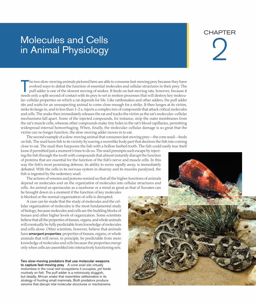
The two slow-moving animals pictured here are able to consume fast-moving prey because they have evolved ways to defeat the function of essential molecules and cellular structures in their prey. The puff adder is one of the slowest moving of snakes. It feeds on fast-moving rats, however, because it
needs only a split second of contact with its prey to set in motion processes that will destroy key molecu-lar–cellular properties on which a rat depends for life. Like rattlesnakes and other adders, the puff adder sits and waits for an unsuspecting animal to come close enough for a strike. It then lunges at its victim, sinks its fangs in, and in less than 1–2 s, injects a complex mix of compounds that attack critical molecules and cells. The snake then immediately releases the rat and tracks the victim as the rat’s molecular–cellular mechanisms fall apart. Some of the injected compounds, for instance, strip the outer membranes from the rat’s muscle cells, whereas other compounds make tiny holes in the rat’s blood capillaries, permitting widespread internal hemorrhaging. When, finally, the molecular–cellular damage is so great that the victim can no longer function, the slow-moving adder moves in to eat.
The second example of a slow-moving animal that consumes fast-moving prey—the cone snail—feeds on fish. The snail lures fish to its vicinity by waving a wormlike body part that deceives the fish into coming close to eat. The snail then harpoons the fish with a hollow barbed tooth. The fish could easily tear itself loose if permitted just a moment’s time to do so. The snail preempts such escape by inject-ing the fish through the tooth with compounds that almost instantly disrupt the function of proteins that are essential for the function of the fish’s nerve and muscle cells. In this way the fish’s most promising defense, its ability to swim rapidly away, is immediately defeated. With the cells in its nervous system in disarray and its muscles paralyzed, the fish is ingested by the sedentary snail.
The actions of venoms and poisons remind us that all the higher functions of animals depend on molecules and on the organization of molecules into cellular structures and cells. An animal as spectacular as a racehorse or a mind as great as that of Socrates can be brought down in a moment if the function of key molecules is blocked or the normal organization of cells is disrupted.
A case can be made that the study of molecules and the cel-lular organization of molecules is the most fundamental study of biology, because molecules and cells are the building blocks of tissues and other higher levels of organization. Some scientists believe that all the properties of tissues, organs, and whole animals will eventually be fully predictable from knowledge of molecules and cells alone. Other scientists, however, believe that animals have emergent properties: properties of tissues, organs, or whole animals that will never, in principle, be predictable from mere knowledge of molecules and cells because the properties emergeonly when cells are assembled into interactively functioning sets.
Molecules and Cellsin Animal Physiology
CHAPTER
2
Two slow-moving predators that use molecular weaponsto capture fast-moving prey A cone snail sits virtually motionless in the coral reef ecosystems it occupies, yet feeds routinely on fish. The puff adder is a notoriously sluggish, but deadly, African snake that resembles rattlesnakes in its strategy of hunting small mammals. Both predators produce venoms that disrupt vital molecular structures or mechanisms.
02_Hill3e_Ch02.indd 31 3/8/12 2:22 PM

32 Chapter 2
Regardless of the resolution of this important debate, molecules and cells are critically important.
The goal of this chapter is to discuss fundamental structural and functional properties of molecules and cells. Many of the properties discussed here will come up in more specific ways throughout the book. Four topics receive greatest attention: Cell membranes and intracellular membranes Epithelia—the sheets of tissue that line body cavities and
form the outer surfaces of organs Enzyme function, diversity, and evolution Mechanisms by which cells receive and act on signals
In addition we will discuss fundamental properties of proteins, the ways that proteins are repaired or destroyed, and the abilities of some cells to produce light or modify an animal’s external color.
Cell Membranes and Intracellular MembranesEach animal cell is enclosed in a cell membrane (plasma membrane). Each cell also includes many sorts of intracellular membranes (sub-cellular membranes), such as the endoplasmic reticulum, the inner and outer membranes of each mitochondrion, and the two closely associated membranes that form the nuclear envelope. These mem-branes are exceedingly thin, measuring 6–8 nanometers (nm) from one side to the other. They play vitally essential roles nonetheless. They physically compartmentalize systems in functionally essential ways; the cell membrane, for instance, separates the inside of a cell
from the cell’s surroundings, permitting the inside to have differ-ent properties from the outside. In addition, far from being inert barriers, the membranes are dynamic systems that participate in cellular and subcellular functions. For example, the cell membrane acts to receive and transmit signals that arrive at the cell surface.
The cell membrane is ordinarily composed primarily of a bilayer (double layer) of phospholipid molecules in which protein molecules are embedded (Figure 2.1). Similarly, the fundamental structure of the intracellular membranes is also a bilayer of phospholipid molecules with protein molecules embedded in it. Recognizing the ubiquity and importance of phospholipids, it is not surprising that they are targets of venoms. A principal weapon in the complex venom of a puff adder or a rattlesnake is a set of enzymes known as phospholipases, which break up phospholipids. Among other effects, these enzymes destroy the phospholipid matrix in the cell membranes of a victim’s skeletal muscle cells, thereby exposing the insides of the cells, setting membrane proteins adrift, and wreaking other havoc.
To understand the molecular logic of the structure of cell mem-branes and intracellular membranes, it is necessary to consider the polarity of molecules and the attendant attributes of hydrophilic and hydrophobic interactions. Consider vinegar-and-oil salad dressing as an everyday example of the effects of molecular polarity. Vinegar consists of acetic acid and water. Thus the dressing has three principal components: oil, acetic acid, and water. If the dressing sits still for a while, the acetic acid remains in solution in the water, but the oil forms a separate layer. This outcome occurs because the acetic acid is hydrophilic (“water-loving”), whereas the oil is hydrophobic(“water-hating”). Why do the two substances behave in these
Carbohydrate chains bonded to cell-membrane proteins (forming glycoproteins) or lipids (forming glycolipids) project into the extracellular fluid on the outside face of the cell membrane.
Cell membrane
Filaments ofcytoskeleton
Nucleus
Cytoplasm
Animal cell
Cholesterol
Extracellular fluid(aqueous solution)
Cytoplasm(aqueous solution)
Phospholipidbilayer
Carbohydratechain
Peripheral proteins are noncovalentlybonded to integral proteins or lipids but arenot within the bilayer. Some peripheralproteins help anchor the cell membrane to filaments of the cytoskeleton.
Integral proteins areembedded in the phospholipid bilayer.
Phospholipid molecule
Protein
FIGURE 2.1 The structure of a cell membrane The cell membrane consists primarily of two layers of phospholipid molecules with protein molecules embedded and attached. Intracellular membranes also have a structure based on proteins embedded in a phospho-lipid bilayer.
02_Hill3e_Ch02.indd 32 3/8/12 2:22 PM

Molecules and Cells in Animal Physiology 33
different ways? A principal reason is the polarity of the molecules. Acetic acid is polar and because of its polar nature is attracted to water. Oil is nonpolar and therefore repelled from water.
The distribution of electrons in a molecule is the property that determines whether the molecule is polar or nonpolar. Within a polar molecule, electrons are unevenly distributed; thus some regions of a polar molecule are relatively negative, whereas others are relatively positive. Water is a polar molecule. Other polar molecules, such as acetic acid—and ions—intermingle freely with polar water molecules by charge interaction, forming solutions. Within a nonpolar molecule, electrons are evenly distributed and there are no charge imbalances between different molecular regions. Nonpolar molecules, such as the oil in salad dressing, do not freely intermingle with polar water molecules. Because of this—at the molecular level—after oil is dispersed into water by violent shaking, the water molecules tend to assemble with other water molecules into arrays that surround nonpolar oil molecules. These arrays are thermodynamically less stable than a complete separation of the water and nonpolar molecules. The nonpolar molecules therefore gradually separate into a distinct layer.
As we shall shortly see, these principles help explain the structure of the phospholipid bilayer in cell membranes and intracellular mem-branes, and they also help explain the positioning of other chemical constituents within the bilayer.
The lipids of membranes are structured, diverse, fluid, and responsive to some environmental factorsPhospholipids are lipids that contain phosphate groups (Figure 2.2a). They are the principal constituents of the matrix in which proteins are embedded in cell membranes and intracellular membranes. They are amphipathic, meaning that each molecule consists of a polar part (within which there are regional differences of charge) and a nonpolar part (which lacks regional differences of charge). A membrane phospholipid consists of a polar head and two nonpolar tails (Figure 2.2b). The polar head is composed of the phosphate group, which forms a region of negative charge, bonded to another group that forms a region of positive charge, such as choline (see Figure 2.2a). Each nonpolar tail consists of a long-chain hydrocar-bon derived from a fatty acid.
CH CH2
O
C O
CH2
CH2
O
C O
CH2
CH2
CH2
CH2
CH2
CH2
CH2
CH2
CH2
CH2
CH2
CH2
CH2
CH3
CH2
CH2
CH2
CH2
CH2
CH2
CH2
CH
CH
CH2
CH2
CH2
CH2
CH2
CH3
P O–O
O
O
CH2
CH2
N (CH3)3+
(a) A phospholipid molecule (a phosphatidylcholine)
(b) Model of a phospholipid molecule
(c) Phospholipid molecules assembled into a bilayer with water on either side
Polar(hydrophilic)head
Nonpolar(hydrophobic)tails
Phospholipid moleculesare representedsymbolically like this.
When placed in water, phospholipidmolecules spontaneously assemble into a bilayer in which the nonpolar, hydrophobic tails occupy the core and the polar, hydrophilic heads occupy the two surfaces.
The bend in this tail at the double bond symbolizes that double bonds often produce bends in hydrocarbon chains.
Nonpolartails
Glycerolresidue
Phosphate
Choline
Polar head
Many different positivelycharged groups can occupythis position in the head.
The two components of the polar head of this particular phospholipid are choline (positively charged) and phosphate (negatively charged).
Each tail is a hydrocarbon chain derived from a fatty acid. Many different fatty acids can occupy each tail position.
FIGURE 2.2 The structure of membrane phospholipid molecules Phospholipid molecules are often described as having a polar head and two nonpolar tails, joined by way of ester linkages to glycerol. (a) The full chemical structure of a particular phospholipid, a phosphatidylcho-line, that is common in animal cell membranes. Because many different chemical structures can occupy the two tail positions and the labeled position in the head, hundreds of kinds of membrane phospholipid molecules are pos-sible. Any particular membrane typically consists of many different kinds of phospholipid molecules, and the kinds may change from time to time. (b) The way that a phos-pholipid molecule is usually symbolized to emphasize its polar head and lipid tails. (c) The assembly of phospho-lipid molecules into a lipid bilayer.
02_Hill3e_Ch02.indd 33 3/8/12 2:22 PM

34 Chapter 2
Whereas the polar part of a phospholipid molecule or any other amphipathic molecule is hydrophilic, the nonpolar part is hydrophobic. When phospholipid molecules are placed in a system of oil layered on water, they collect at the interface of the oil and water in a predictable way, with their polar, hydrophilic heads in the water and their nonpolar, hydrophobic tails in the oil. Of greater importance for understanding living cells is the fact that when phospholipid molecules are placed simply in an aqueous solution, they spontaneously assemble into bilayers, adopting the same bilayer conformation they take in cell membranes and intracellular membranes (Figure 2.2c). This bilayer conformation forms because it is thermodynamically stable. All the hydrophobic regions (the hydrocarbon tails) get together in the interior of the bilayer (away from the water), whereas the hydrophilic heads associate with the water on either side of the membrane. The energy barrier to mixing polar and nonpolar regions in the membrane is so great that in a cell membrane, it is nearly impossible for a phospholipid molecule to “flip” its polar head through the nonpolar interior and move from one side of the bilayer to the other (unless specifically catalyzed to do so).
A striking attribute of membrane phospholipids is their great chemical diversity. Many different types of phospholipid molecules are possible because the two tails and the positively charged part of the head, as shown in Figure 2.2a, can differ widely in their specific chemical composition. The cell membranes of human red blood cells contain more than 150 different chemical forms of phospho-lipids, and similar diversity is seen in other cell membranes. The two layers of phospholipid molecules in any particular membrane, known as the two leaflets of the membrane, typically are composed of different mixes of phospholipid molecules.
The phospholipids in a cell membrane or intracellular membrane are fluid. Individual phospholipid molecules are not covalently bound to one another. Therefore, they move relative to each other. They are able to move about rather freely by diffusion within each membrane leaflet. The rate of this diffusion is great enough that a particular phospholipid molecule is able to travel, by diffusion, around the entire circumference of a cell in a matter of minutes. The ease of motion of the phospholipid molecules in a membrane leaflet is termed their fluidity.
Fluidity depends in part on the degree of chemical saturationof the hydrocarbons that make up the phospholipid tails. What do we mean by chemical saturation? A hydrocarbon is saturatedif it contains no double bonds. It is unsaturated if it includes one or more double bonds; different degrees of unsaturation are pos-sible because the number of double bonds can be high or low. As shown in Figure 2.2a, a double bond often imparts a bend to a hydrocarbon chain. Bent tails of membrane phospholipids prevent tight, crystal-like packing of the tails in the hydrophobic interior of the membrane. This disruption of tight packing helps keep the phospholipid molecules free to move. Accordingly, a greater proportion of unsaturated fatty acids in the tails of phospholipids results in a membrane with more fluidity.
In addition to chemical composition, temperature affects the fluidity of membranes; just as butter and other household lipids stiffen when they are chilled, the phospholipids in cell membranes tend to become stiffer at lower temperatures. During evolution, one important way in which cells have become adapted to different temperatures is alteration of the numbers of double bonds (the
degree of unsaturation) in their membrane phospholipids. This is evident in fish of polar seas, for instance. The fish experience tis-sue temperatures so low that their cell membranes could be overly stiff. This problem is avoided, however, because these fish have cell membranes constructed of phospholipids that are particularly rich in double bonds; the highly unsaturated phospholipids are inher-ently quite fluid and thus less likely than other phospholipids to become detrimentally stiff at low temperatures. Recent research on the cell membranes of brain cells in fish demonstrates that the degree of phospholipid unsaturation depends in a regular way on the environmental temperatures to which various species are adapted (Figure 2.3). Tropical species of fish, which face little risk of having their membranes rendered too stiff by low tempera-tures, have evolved relatively saturated phospholipids, but as the temperature of the habitat falls, the degree of unsaturation of the phospholipids increases.
Evidence is accumulating that individual animals sometimes restructure their membrane phospholipids in response to environ-mental factors. For example, lab mice alter the mix of membrane phospholipids in their heart muscle cells after just 4–12 h of fasting and reverse the changes when fed. At least some hibernating species of mammals substantially alter the mix of phospholipids in their cell and mitochondrial membranes as they approach hibernation, in ways thought to promote the hibernating physiological state (e.g., suppression of metabolism).
5–5 15 25 35
50
30
70
90
Phosphatidylethanolamines
Tropical species
Bass
Phosphatidylcholines
Trout
Antarctic species
Brain
Typical habitat temperature (°C)
Perc
enta
ge o
f hyd
roca
rbon
tails
that
are
uns
atur
ated
FIGURE 2.3 The degree of chemical unsaturation of the hydro-carbon tails of brain phospholipids in fish varies with habitat temperature Brain synaptic membranes of 17 species of teleost (bony) fish were studied. Measurements were made of the compo-sition of the hydrocarbon tails of two categories of cell-membrane phospholipids, the phosphatidylcholines and the phosphatidyletha-nolamines, which differ in whether the group at the top of the head in Figure 2.2a is choline [—CH2—CH2—N(CH3)3] or ethanolamine (—CH2—CH2—NH2), respectively. Each plotted symbol corresponds to the average value for one species. (After Logue et al. 2000.)
02_Hill3e_Ch02.indd 34 3/8/12 2:22 PM

Molecules and Cells in Animal Physiology 35
Besides phospholipids, cell membranes and intracellular membranes contain other classes of lipids, one of which is sterols. The principal membrane sterols are cholesterol and cholesterol esters. In cell membranes, which are typically much richer in sterols than intracellular membranes are, sterols collectively occur in ratios of 1 molecule per 10 phospholipid molecules, up to 8 per 10. Cholesterol is mildly amphipathic and positioned within the phospholipid leaflets (see Figure 2.1), where it exerts complex effects on membrane fluidity.
Proteins endow membranes with numerous functional capacitiesProteins are the second major constituents of cell membranes and intracellular membranes. According to the fluid mosaic model of membranes, a membrane consists of a mosaic of protein and lipid molecules, all of which move about in directions parallel to the membrane faces because of the fluid state of the lipid matrix. As we start to discuss proteins, an important fact to recall from the study of organic chemistry is that—in terms of their chemical makeup—proteins are considered to have primary, secondary, tertiary, and sometimes quaternary structure. This aspect of protein structure is reviewed in Box 2.1.
Membrane proteins are structurally of two principal kinds: integral and peripheral. Integral membrane proteins are parts of the membrane and cannot be removed without taking the membrane apart. Most integral proteins (see Figure 2.1) span the membrane and thus are called transmembrane proteins. These molecules have both hydrophobic and hydrophilic regions. As we will see in detail shortly, each hydrophobic region typically has an amino acid composition and a molecular geometry that allow it to associate with the hydrophobic hydrocarbon tails of the membrane interior. The hydrophilic regions of transmembrane protein molecules, by contrast, typically protrude into the aqueous solutions bathing the two sides of the membrane.
Peripheral membrane proteins are associated with the membrane but can be removed without destroying the membrane. They are bonded noncovalently (i.e., by weak bonds) to membrane compo-nents (e.g., integral proteins) and are positioned on one side of the membrane or the other (see Figure 2.1). Their positioning means
that the two leaflets of a membrane differ in protein composition, as well as phospholipid composition.
The proteins of cell membranes and intracellular membranes endow the membranes with capabilities to do many things. Five functional types of membrane proteins are recognized: channels, transporters (carriers), enzymes, receptors, and structural proteins. Because these types are classified by function, the actions listed in Table 2.1 define the five types. The categories are not mutually exclusive: A membrane protein can be both a receptor and a chan-nel, or a transporter and an enzyme, for example.
The molecular structures of membrane proteins are complex and are diagrammed in several ways, depending on the degree of chemical detail to be shown. To illustrate, let’s focus on a channel, which is a type of membrane-spanning integral protein. Channels provide paths for ions or other materials in aqueous solution to pass through membranes. In our example the channel is formed by a single protein molecule, the secondary structure of which is
BOX 2.1 PROTEIN STRUCTURE AND THE BONDS THAT MAINTAIN IT
All protein molecules have primary, secondary, and tertiary structure (and some have quaternary structure). Primary structure refers to the string of covalently bonded amino
acids. As essential as primary structure is, protein function depends most directly on secondary and tertiary structure—the three-dimensional conformation of the protein molecule. Because secondary and tertiary structure are stabilized by weak chemical bonds rather than covalent bonds, the three-dimensional conformation of a protein can change and flex—a process essential for protein function. Denaturation is a disrup-tion of the correct tertiary structure; because primary structure is not altered, denaturation may be reversible (reparable). This box continues on the web at Box Extension 2.1. There you will find detailed information on—and illustrations of—all levels of protein structure, strong versus weak bonds, the types of weak bonds, denaturation, and potential repair of denaturation.
TABLE 2.1 The five functional types of membrane proteins and the functions they perform
Functional type Function performed (defining property)
Channel Permits simple or quasi-simple diffusion of solutes in aqueous solution (see page 104)—or osmosis of water (see page 121)—through a membrane. A simplified view of a channel is that it creates a direct water path from one side to the other of a membrane (i.e., an aqueous pore) through which solutes in aqueous solution may diffuse or water may undergo osmosis.
Transporter (carrier) Binds noncovalently and reversibly with specific molecules or ions to move them across a membrane intact. The transport through the membrane is active transport (see page 108) if it employs metabolic energy; it is facilitated diffusion (see page 108) if metabolic energy is not employed.
Enzyme Catalyzes a chemical reaction in which covalent bonds are made or broken (see page 41).
Receptor Binds noncovalently with specific molecules and, as a consequence of this binding, initiates a change in membrane permeability or cell metabolism. Receptor proteins mediate the responses of a cell to chemical messages (signals) arriving at the outside face of the cell membrane (see page 58).
Structural protein Attaches to other molecules (e.g., other proteins) to anchor intracellular elements (e.g., cytoskeleton filaments) to the cell membrane, creates junctions between adjacent cells (see Figure 2.7), or establishes other structural relations.
02_Hill3e_Ch02.indd 35 3/8/12 2:22 PM

36 Chapter 2
shown in Figure 2.4a (see Box 2.1 for an explanation of secondary structure).
Each cylinder in Figure 2.4a represents a sequence of amino acids that forms a helix-shaped subunit, called an α-helix (see Box 2.1), within the protein structure. The whole protein molecule exempli-fies a common property of membrane proteins, in that it consists of repeating structural patterns known as domains. To identify the domains, review the molecule from left to right. You will note five α-helices linked closely together, and then a sixth helix separated from the others by a longer string of amino acids; then you will note that this pattern of five closely spaced helices followed by a sixth more-separated helix is repeated three more times. On the basis of this repeating pattern, this molecule is said to show four domains, numbered I to IV, as illustrated in Figure 2.4a. The α-helices are predominantly hydrophobic and span the membrane by associating with the hydrophobic interior of the phospholipid bilayer. The strings of amino acids that connect successive helices are hydrophilic and protrude from the membrane into the aqueous solutions on either side. In its natural state in a membrane, this protein is believed to be shaped into a closed ring in which the four domains form cylinder-like structures surrounding a central pore, as diagrammed in Figure 2.4b.
The three additional representations of the membrane protein that are shown in Figure 2.4 are progressively simpler. The sort of representation in Figure 2.4c, which still shows that there are four domains, is a simplified way to represent the chemical structure of the molecule. The diagrammatic, semirealistic representation in Figure 2.4d, which leaves one guessing about the number of domains, is more simplified yet, and in Figure 2.4e the channel is represented schematically (without any intention of resembling the actual molecule).
The interrelations of the presentations in Figure 2.4a–e are impor-tant to note because all of these sorts of presentations are commonly used in biological literature. An important additional detail is that the major subunits of membrane proteins are not always parts of one molecule, as the four domains in our example are; sometimes the major subunits of a single channel, for example, are separate protein molecules. Moreover, the protein units that constitute the central pore-forming part of a channel may have other integral or peripheral proteins associated with them, as shown in Figure 2.4f.
Carbohydrates play important roles in membranesCell membranes and intracellular membranes also contain carbo-hydrates, which occur mostly in covalently bonded combination with lipids or proteins, or both (see Figure 2.1). Glycolipids (e.g., gan-
gliosides), glycoproteins, and proteoglycans are some of the major categories of carbohydrate-containing membrane compounds.1 Carbohydrate groups are hydrophilic and thus are associated with the mem-brane surface and adjacent aqueous solution. Carbo-hydrates reinforce the point, stressed earlier, that the two leaflets of a membrane are typically different.
(a) Hypothesized secondary structure (linear presentation)
Hydrophilicamino acidstring
Hydrophobicα-helix
Cellmembrane
Extracellular fluid
Extracellular fluid
NH2
Domain I Domain II Domain III Domain IV
Cytoplasm
Cytoplasm
COOH
This molecule consists of four domains, each of which includes six α-helices.
Each cylinder represents an α-helix (see Box 2.1).
Each black line represents a string of amino acids.
A channel protein may be associated in the membrane with other transmembrane proteins (e.g., β) or peripheral proteins (e.g., γ).
Cellmembrane
For different purposes, the protein can be represented in a variety of ways.
I III
IV
II
(b) Simplified tertiary (three- dimentional) structure enclosed in a sketch of the envelope of the molecule
(c) Stylized version of chemical structure showing domains
(d ) Semirealistic symbol
(e) Schematic symbol
( f ) Stylized version of chemical structure showing associated protein molecules
β
γ
FIGURE 2.4 The structure of a transmembrane protein—a voltage-gated Na+ channel—illustrat-ing several modes of presentation
1 The word fragment glyco refers to carbohydrates (after the Greek glykeros, “sweet”).
02_Hill3e_Ch02.indd 36 3/8/12 2:22 PM

Molecules and Cells in Animal Physiology 37
In cell membranes, for example, the carbohydrate groups always project from the outer, extracellular face, not the inner, cytoplasmic face (see Figure 2.1). These carbohydrate groups serve as attach-ment sites for extracellular proteins and as cell-recognition sites.
SUMMARY Cell Membranes and Intracellular Membranes
The matrix of a cell membrane or intracellular membrane consists of a bilayer of phospholipid molecules. The phospholipids are chemically very diverse, even within a single membrane, and in a particular cell the phospholipid composition can undergo change in response to environmental or other factors. The phospholipids are fluid, meaning that individual molecules move about relatively freely by diffusion within each membrane leaflet.
Animals exhibit adaptive trends in the phospholipid compositions of their cell membranes. Cells that function routinely at low temperatures tend to have a phospholipid composition that permits membranes to remain fluid under cold conditions (e.g., they have high proportions of double bonds in the hydrocarbon tails).
Five functional categories of proteins occur in cell and intracellular membranes: channels, transporters, enzymes, receptors, and structural proteins. A single protein may engage in more than one function.
In addition to phospholipids and proteins, which are the principal components, membranes often have other components such as cholesterol (a lipid) and glycoproteins (composed of covalently bonded carbohydrate and protein subunits).
EpitheliaAn epithelium (plural epithelia) is a sheet of cells that covers a body surface or organ, or lines a cavity. Although epithelia are radi-cally different from cell membranes and intracellular membranes, they—to some degree—perform parallel functions on a larger structural scale. Epithelia compartmentalize the body by forming boundaries between body regions. They also form a boundary between an animal and its external environment. Moreover, like cell membranes, epithelia have numerous functional capacities and play major functional roles in animal physiology.
A simple epithelium consists of a single layer of cells (Figure 2.5a). Simple epithelia are exceedingly common; in the human body, for instance, the intestines, kidney tubules, blood vessels, and sweat glands are all lined with a simple epithelium. Each cell in a simple epithelium has an apical surface (mucosal surface) facing into a cavity or open space, and a basal surface (serosal surface) facing toward the underlying tissue to which the epithelium is attached. An epithelium typically rests on a thin, permeable, noncellular, and nonliving sheet of matrix material, positioned beneath the basal cell surfaces. This sheet is called the basement membrane (basal lamina) and is composed of glycoproteins and particular types of collagen. It is secreted mostly by the epithelial cells, although the underlying cells also contribute. Simple epithelia are classified as squamous, cuboidal, or columnar, de-pending on how tall the cells are. The cells in a squamous epithelium are low and flat, whereas those in a columnar epithelium are high relative to their basal dimensions; the epithelium in Figure 2.5a is classed as cuboidal because the cells are about as tall as they are wide. Blood vessels usually do not enter epithelia. Instead, epithelial
cells exchange O2, CO2, and other materials through the underlying basement membrane with blood capillaries located on the opposite side of the basement membrane (see Figure 2.5a).
The epithelium that lines the small intestine (midgut) of a mam-mal (Figure 2.5b) is an example of a simple epithelium that will be featured prominently in this book (e.g., in Chapters 5 and 6) and that introduces additional aspects of epithelial morphology and function. The apical surfaces of the cells in this columnar epithelium face into the lumen (open central cavity) of the intestine. As digestion occurs, liberating food molecules from foods, the molecules pass through the epithelium and basement membrane to reach blood vessels and lymph passages that transport them to the rest of the body.
(a) Generalized epitheliumEpithelial cell
Intercellularspace
Nucleus
Bloodcapillary
Basementmembrane
The cell membrane of each epithelial cell has an apical region that faces out from the underlying tissue into a cavity or open space…
…and a basal region that faces toward the tissue to which the epithelium attaches.
(b) Intestinal epithelium
Brushborder
Absorptive–digestive cells Endocrine cell
Basementmembrane
Nucleus
Bloodcapillary
Basal region ofcell membrane
The epithelium includes endocrine cells that produce granules of secretory material.
The apical regions of the cell membranes of these metabolically active epithelial cells bear microvilli. The microvilli face into the intestinal lumen.
The intercellular spaces are particularly wide in this epithelium, especially toward the bases of the cells.
Microvillus
FIGURE 2.5 Simple epithelia (a) A generalized simple epithelium covering a free surface of a tissue. (b) The specific simple epithelium lining the mammalian small intestine (midgut). This epithelium consists of several cell types. Most cells are the absorptive–digestive cells emphasized here. Scattered among these cells are mucin-secreting cells (not shown) and at least ten types of endocrine or endocrine-like cells. Each endocrine cell produces granules of secretory material; the granules move to juxtapose themselves with the basal or near-basal regions of the cell membrane and then release their secretions into the spaces outside the cell, after which the secretions enter the blood for transport elsewhere. Endocrine-like cells termed paracrine cells (not shown) are also present. Paracrine cells produce secretions that affect nearby cells rather than acting on distant cells by way of the circulation (see Figure 16.1).
02_Hill3e_Ch02.indd 37 3/8/12 2:22 PM

38 Chapter 2
The intestinal epithelium illustrates that a simple epithelium can consist of two or more cell types. Whereas the epithelium is composed mostly of absorptive–digestive cells, it also includes endocrine cells (see Figure 2.5b) and additional cell types.
The intestinal epithelium also illustrates microvilli (singular microvillus), which are a common (but not universal) feature of epithelial cells. Microvilli are exceedingly fine, fingerlike projections of the apical cell membrane (see Figure 2.5b). In the intestinal epi-thelium, the microvilli greatly increase the area of contact between the epithelial cells and the contents of the gut. Microvilli are most often found in epithelia that are active in secreting or absorbing materials, such as the epithelia of certain kidney tubules and the pancreatic ducts, as well as the intestinal epithelium. Microvilli are often described collectively as a brush border because they look like the bristles on a brush when viewed microscopically.
Another significant aspect of diversity in simple epithelia arises from the geometric arrangement of the cells. Tubules or follicles (hollow globes) are often formed by the wrapping of a simple epithelium into a closed curve (Figure 2.6a) supported by the basement membrane on the outside. A tubule formed by cuboidal epithelium bearing microvilli forms the proximal region of each mammalian nephron (kidney tubule), for example (Figure 2.6b). Vertebrate blood capillaries are an especially important example. Each blood capillary consists of a single layer of highly flattened epithelial cells (lacking microvilli) supported by the epithelial basement membrane (Figure 2.6c). The basement membranes of capillaries are one of the important bio-chemical targets of the venoms of puff adders and rattlesnakes. The venoms contain enzymes (metalloproteases) that break down the basement membranes, destroying the integrity of blood capillaries. In this way the venoms cause widespread internal hemorrhaging.
Adjacent cells in an epithelium are physically joined by cell-membrane junctions of several sorts; the four most important of these are tight junctions, septate junctions, desmosomes, and gap junc-tions (Figure 2.7). In the paragraphs that follow, we look at each of these types of junction in turn.
A tight junction is a place where the cell membranes of adjacent cells are tightly joined so that there is no intercellular space between the cells; adjacent cells are perhaps 10–20 nm apart for the most part, but at tight junctions the cell membranes meet or fuse. Tight junctions typically occur between the sides of adjacent cells, just a short distance away from their apical surfaces (Figure 2.8). Any given epithelial cell has tight junctions with adjacent epithelial cells in a continuous ring around its entire perimeter. This ring of tight junctions demarcates the apical surface of the cell from its lateral and basal surfaces, giving rise to one of the most important distinctions in the physiological study of epithelia, the distinction between the apical region and the basolateral region of each cell membrane (see Figure 2.8). Many invertebrate groups have septate junctions instead of tight junctions. Septate junctions differ from tight junctions in their fine structure (see Figure 2.7), but they resemble tight junctions in
1 μm
Three cells can be seen; all three have nuclei, but because the cells are so highly flattened that they extend considerable distances along the length of the capillary, the nuclei of two of the cells are positioned outside the plane of this section.
Lumen
Nucleus of one epithelial cell
MitochondrionBasementmembrane
(a) Epithelial cells can form tubules and follicles
Tubule Follicle
(b) Proximal part of a mammalian nephron (kidney tubule) in cross section
Nucleus of oneepithelial cell
Basementmembrane
Microvilli
Lumen
Mitochondrion The cuboidal cells of this epithelium display two signature properties of high metabolic activity: microvilli on the apical surfaces, and abundant mitochondria.
10 μm
(c) Mammalian blood capillary in cross section
The flattened, squamous cells of this epithelium lack microvilli.
FIGURE 2.6 Tubules and follicles formed by simple epithelia (a) Both tubules and follicles are formed by the wrapping of simple epithelia into closed curves. Cross sections of two important tubular structures are shown in (b) and (c); in each case the basal cell sur-faces and basement membrane of the epithelium are on the outside. For historical reasons, the cells of blood capillaries are usually called endothelial cells, but they are a form of epithelium.
02_Hill3e_Ch02.indd 38 3/8/12 2:22 PM

Molecules and Cells in Animal Physiology 39
their position and in the fact that they fully encircle each cell. Tight and septate junctions are sometimes aptly called occluding junc-tions because they block or occlude the spaces between adjacent epithelial cells, preventing open passage between the fluids on either side of an epithelium.
A desmosome (see Figure 2.7) is a junction at which mutually adhering glycoprotein filaments from two adjacent cells intermingle across the space between the cells. Desmosomes are often likened to rivets or spot welds because they occur as tiny isolated spots, not continuous bands, and their principal function is believed to be to strengthen and stabilize contacts between adjacent cells.
Gap junctions (see Figure 2.7) are like desmosomes in that they occur at discrete spots, but otherwise they are very different from all the other junctions we have discussed because within a gap junc-tion there are open pores between cells. At these pores, which are formed by connexin proteins (see Figure 2.7), the two adjacent cells lack cell-membrane boundaries, and there is continuity between the cytoplasms of the cells. Molecules and ions smaller than 1000–1500 daltons (Da) in molecular mass are able to pass between cells at gap junctions, although large solutes such as proteins cannot. Gap junctions are important in cell–cell communication—including passage of intracellular signaling agents in some tissues and direct electrophysiological interactions between cells in nerve or muscle (gap junctions are treated in detail in Chapter 13 [see Figure 13.2]).
A central feature of epithelia is that each epithelial cell is functionally asymmetric. The proteins in the cell membrane of an epithelial cell are unable (for reasons only poorly known) to diffuse through tight junctions. Thus, the ring of tight junctions around each epithelial cell acts as a fence that keeps proteins from crossing between the apical and basolateral regions of the cell membrane. The two regions of the cell membrane therefore have different sets of channels, transporters, membrane enzymes, and other classes of membrane proteins, and they are functionally different in many ways. Differences also exist between the apical and basolateral regions in the membrane phospholipids composing the outer (but not inner) leaflet of the cell membrane.
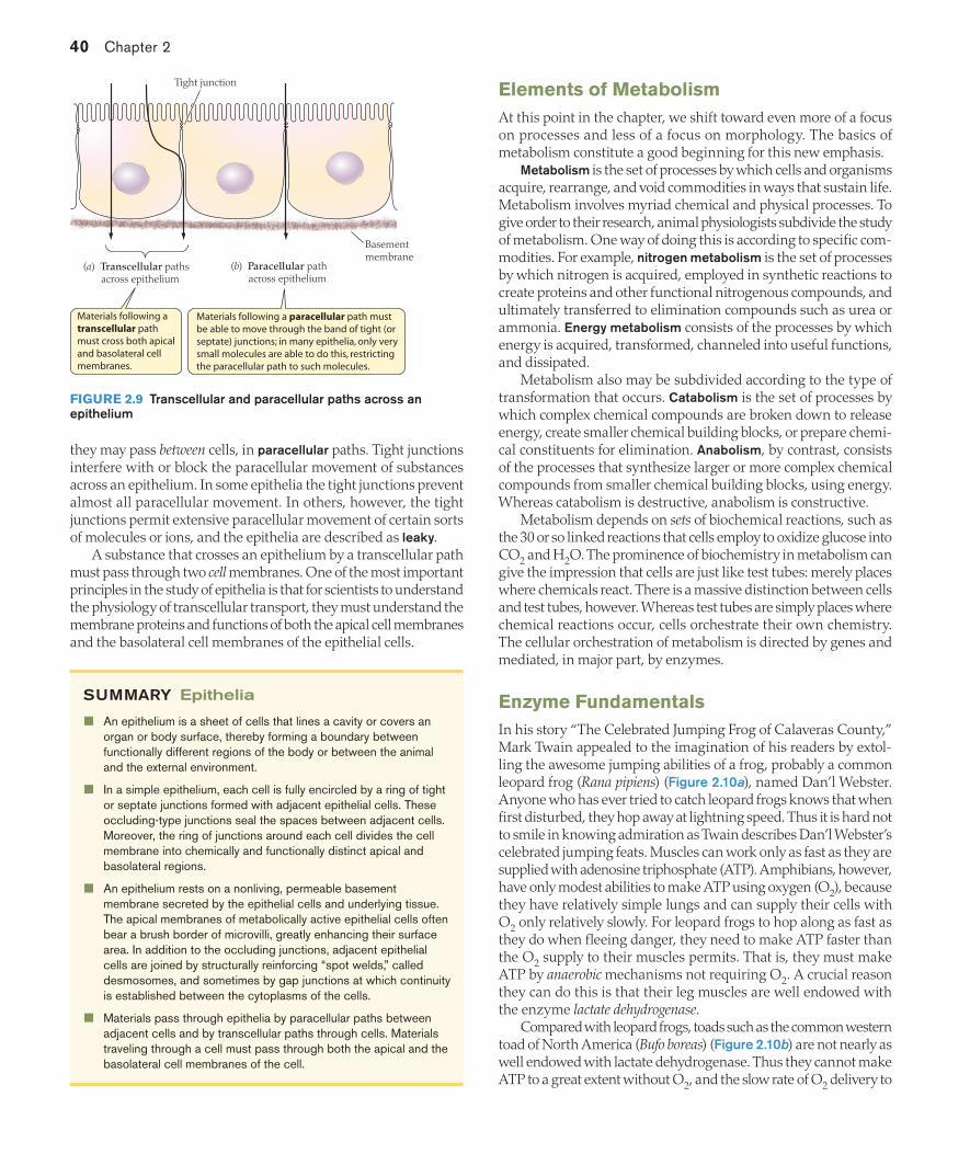
One of the important functions of an epithelium is to control and mediate the transport of substances between the apical and basal sides of the epithelium and thus between different body regions. Substances—such as ions, nutrient molecules, or water—pass through a simple epithelium by two types of paths (Figure 2.9). They may pass through cells by transcellular paths. Alternatively,
Cell A
Cell B
Cell membranes
Intercellularspace
Connexinprotein
Cytoskeletonfilament
Glycoproteinfilaments
A gap junction is a localized spotwhere the cytoplasms of two cells communicate through tiny pores, as symbolized by the double-headed arrows.
A desmosome isa localized spotwhere the contactbetween cells isstrengthened.
Tight junctions and septate junctions occlude the intercellular space between two cells because not only do the cell membranes meet or fuse at such junctions, but also the junctions form con-tinuous bands around cells. In tight junctions, the cell membranes of the two cells make contact at ridges.
Tight junction Septate junction
Occluding junctions
Desmosome(“spot weld”)
Gap junction (communicating
junction)
(a) An epithelial cell shown with one neighboring cell
(b) Schematic representation of a microvilli-bearing epithelial cell with two neighboring cells
Basolateral region ofcell membrane
Tight junction
Apical region of cell membrane
Basolateral region ofcell membrane
A band of tight junctions (septate junctions in many invertebrates) goescompletely around each cell. It acts as a fence between the apical region ofthe cell membrane and the basolateral region (which includes the lateral parts of the membrane below the tight junctions and the basal part).
The apical and basolateral regions typically differ greatly in their protein components and functional properties.
Apical region of cell membrane
FIGURE 2.7 Types of junctions between cells At a pore in a gap junction, each cell has a ring of six connexin proteins that together form the pore, and the rings of the two cells line up to create continuity between cells. Each ring of connexin proteins is called a connexon.
FIGURE 2.8 The organization of epithelial cells into apical and basolateral regions (a) The cell membrane of an epithelial cell is divided into apical (pink) and basolateral (orange) regions—which differ in their protein components and functions—by a band of tight junctions formed with adjacent epithelial cells. In this book, we will often use the schematic format shown in (b) to represent an epithelium. Microvilli do not always occur, but when they do, they are on the apical side only.
02_Hill3e_Ch02.indd 39 3/8/12 2:22 PM
40 Chapter 2
they may pass between cells, in paracellular paths. Tight junctions interfere with or block the paracellular movement of substances across an epithelium. In some epithelia the tight junctions prevent almost all paracellular movement. In others, however, the tight junctions permit extensive paracellular movement of certain sorts of molecules or ions, and the epithelia are described as leaky.
A substance that crosses an epithelium by a transcellular path must pass through two cell membranes. One of the most important principles in the study of epithelia is that for scientists to understand the physiology of transcellular transport, they must understand the membrane proteins and functions of both the apical cell membranes and the basolateral cell membranes of the epithelial cells.
SUMMARY Epithelia
An epithelium is a sheet of cells that lines a cavity or covers an organ or body surface, thereby forming a boundary between functionally different regions of the body or between the animal and the external environment.
In a simple epithelium, each cell is fully encircled by a ring of tight or septate junctions formed with adjacent epithelial cells. These occluding-type junctions seal the spaces between adjacent cells. Moreover, the ring of junctions around each cell divides the cell membrane into chemically and functionally distinct apical and basolateral regions.
An epithelium rests on a nonliving, permeable basement membrane secreted by the epithelial cells and underlying tissue. The apical membranes of metabolically active epithelial cells often bear a brush border of microvilli, greatly enhancing their surface area. In addition to the occluding junctions, adjacent epithelial cells are joined by structurally reinforcing “spot welds,” called desmosomes, and sometimes by gap junctions at which continuity is established between the cytoplasms of the cells.
Materials pass through epithelia by paracellular paths between adjacent cells and by transcellular paths through cells. Materials traveling through a cell must pass through both the apical and the basolateral cell membranes of the cell.
Elements of MetabolismAt this point in the chapter, we shift toward even more of a focus on processes and less of a focus on morphology. The basics of metabolism constitute a good beginning for this new emphasis.
Metabolism is the set of processes by which cells and organisms acquire, rearrange, and void commodities in ways that sustain life. Metabolism involves myriad chemical and physical processes. To give order to their research, animal physiologists subdivide the study of metabolism. One way of doing this is according to specific com-modities. For example, nitrogen metabolism is the set of processes by which nitrogen is acquired, employed in synthetic reactions to create proteins and other functional nitrogenous compounds, and ultimately transferred to elimination compounds such as urea or ammonia. Energy metabolism consists of the processes by which energy is acquired, transformed, channeled into useful functions, and dissipated.
Metabolism also may be subdivided according to the type of transformation that occurs. Catabolism is the set of processes by which complex chemical compounds are broken down to release energy, create smaller chemical building blocks, or prepare chemi-cal constituents for elimination. Anabolism, by contrast, consists of the processes that synthesize larger or more complex chemical compounds from smaller chemical building blocks, using energy. Whereas catabolism is destructive, anabolism is constructive.
Metabolism depends on sets of biochemical reactions, such as the 30 or so linked reactions that cells employ to oxidize glucose into CO2 and H2O. The prominence of biochemistry in metabolism can give the impression that cells are just like test tubes: merely places where chemicals react. There is a massive distinction between cells and test tubes, however. Whereas test tubes are simply places where chemical reactions occur, cells orchestrate their own chemistry. The cellular orchestration of metabolism is directed by genes and mediated, in major part, by enzymes.
Enzyme FundamentalsIn his story “The Celebrated Jumping Frog of Calaveras County,” Mark Twain appealed to the imagination of his readers by extol-ling the awesome jumping abilities of a frog, probably a common leopard frog (Rana pipiens) (Figure 2.10a), named Dan’l Webster. Anyone who has ever tried to catch leopard frogs knows that when first disturbed, they hop away at lightning speed. Thus it is hard not to smile in knowing admiration as Twain describes Dan’l Webster’s celebrated jumping feats. Muscles can work only as fast as they are supplied with adenosine triphosphate (ATP). Amphibians, however, have only modest abilities to make ATP using oxygen (O2), because they have relatively simple lungs and can supply their cells with O2 only relatively slowly. For leopard frogs to hop along as fast as they do when fleeing danger, they need to make ATP faster than the O2 supply to their muscles permits. That is, they must make ATP by anaerobic mechanisms not requiring O2. A crucial reason they can do this is that their leg muscles are well endowed with the enzyme lactate dehydrogenase.
Compared with leopard frogs, toads such as the common western toad of North America (Bufo boreas) (Figure 2.10b) are not nearly as well endowed with lactate dehydrogenase. Thus they cannot make ATP to a great extent without O2, and the slow rate of O2 delivery to
Basementmembrane
Tight junction
(b) Paracellular path across epithelium
(a) Transcellular paths across epithelium
Materials following a paracellular path must be able to move through the band of tight (or septate) junctions; in many epithelia, only very small molecules are able to do this, restricting the paracellular path to such molecules.
Materials following a transcellular path must cross both apical and basolateral cell membranes.
FIGURE 2.9 Transcellular and paracellular paths across an epithelium
02_Hill3e_Ch02.indd 40 3/8/12 2:22 PM

Molecules and Cells in Animal Physiology 41
their muscle cells means a slow rate of ATP production, explaining why they cannot hop along as fast as frogs. Mark Twain could not have known this, because the study of enzymes was just beginning during his life, but when he searched his mind for an amphibian that could inspire his readers as a “celebrated” jumper, he chose a frog rather than a toad in major part (we now know) because frogs have more of the enzyme lactate dehydrogenase.
Enzymes are protein catalysts that play two principal roles: They speed chemical reactions, and they often regulate reactions.2
To appreciate the extreme importance of enzymes, it is crucial to recognize that the vast majority of the biochemical reactions that occur in animals do not take place on their own at significant rates under physiological conditions. Cells are biochemically complex enough that, in principle, tens of thousands of reactions might occur in them. However, because reactions in general require catalysis to occur at significant rates, the particular reactions that do take place in a cell—out of all those that could take place—depend on the cell’s own biosynthesis of enzyme proteins. Enzymes represent one of the foremost means by which cells take charge of their own biochemistry.
When we say that an enzyme is a catalyst, we mean that it is a molecule that accelerates a reaction without, in the end, being altered itself. The reaction catalyzed by lactate dehydrogenase (LDH) that is important for escape by frogs is the reduction of pyruvic acid to form lactic acid, a reaction in which each pyruvic acid molecule is combined with two hydrogen atoms (Figure 2.11a). Although the presence of LDH speeds this reaction, LDH is not itself altered by the reaction. Thus a molecule of LDH persists as it catalyzes the reduction of many pyruvic acid molecules, one after another.
Enzymes are described as having substrates and products, and often there are two or more of each. To be exact about the substrates and products of LDH, a chemically complete presentation of the LDH-catalyzed reaction is needed (Figure 2.11b). The hydrogen atoms that reduce pyruvic acid are taken from a molecule we symbol-ize as NADH2. NAD is an enzyme cofactor (nicotinamide adenine dinucleotide) found in all animal cells; and NADH2 symbolizes
2 Research over the past three decades has shown that protein catalysts—enzymes—are not the only catalysts. Some types of RNA molecules also play roles as catalysts.
the reduced form of this cofactor, the form that is combined with hydrogen. The substrates of an enzyme are the initial reactants of the reaction that the enzyme catalyzes; the products of the enzyme are the compounds produced by the reaction. Thus, in the reaction we are discussing—the left-to-right reaction in Figure 2.11b—the substrates of LDH are pyruvic acid and NADH2, and the products are lactic acid and NAD. Chapter 8 discusses how this reaction aids not only rapid jumping by frogs, but also other forms of sudden, intense vertebrate exercise, such as sprinting by people. Put simply,
(a) A fast-jumping amphibian: the leopard frog (Rana pipiens) (b) A slow-jumping amphibian: the western toad (Bufo boreas)
FIGURE 2.10 Two amphibians with different jumping capabilities based in part on different levels of a key enzyme, lactate dehydrogenase
H3C C COOH
O
H3C C COOH
OH
H
Lactatedehydrogenase
NADH2
2 H
NAD
(a) Simplified reaction
(b) Full reaction
Pyruvic acid isproduced fromglucose by glycolysis(see Figure 8.1).
Pyruvic acidLactate
dehydrogenase
Lactic acid
When this reversible reaction proceeds from left to right, pyruvic acid is reduced by receiving two hydrogen atoms from NADH2, yielding lactic acid and NAD.
Lactic acidPyruvic acid
FIGURE 2.11 The reaction catalyzed by lactate dehydrogenase (LDH) The enzyme cofactor nicotinamide adenine dinucleotide (NAD) acts as an electron (or hydrogen) shuttle by undergoing revers-ible reduction (forming NADH2) and oxidation (forming NAD). As (b) shows, when the reaction catalyzed by LDH proceeds from left to right, NADH2 produced elsewhere is converted to NAD, renewing the supply of NAD. The reaction catalyzed by LDH is reversible, but the NAD reaction involved in the reverse direction is not shown. Chapter 8 discusses the important role of the LDH-catalyzed reaction in ATP production.
02_Hill3e_Ch02.indd 41 3/8/12 2:22 PM

42 Chapter 2
the way the reaction helps is precisely that it produces NAD, an essential compound for ATP synthesis by glycolysis.
There are many kinds of enzymes. Mammalian cells, for instance, typically synthesize several thousand kinds. Usually, the names of enzymes end in -ase. Thus, when you see a biochemical term that ends in -ase, it usually refers to an enzyme. Later we will see that a single enzyme may exist in multiple molecular forms in different tissues or different animal species. The name of an enzyme typically refers to the reaction catalyzed. Lactate dehydrogenase, for example, is defined to be an enzyme that catalyzes the reaction in Figure 2.11b. All molecular forms that catalyze this reaction are considered to be forms of lactate dehydrogenase, even though they vary in their exact molecular structures and detailed functional properties.
Enzyme-catalyzed reactions exhibit hyperbolic or sigmoid kineticsFor an enzyme molecule to catalyze a reaction, it must first combine with a molecule of substrate to form an enzyme–substrate complex. (Here, for simplicity, we assume there is only one substrate.) This complexing of enzyme and substrate, which usually is stabilized by noncovalent bonds, is essential for catalysis because the enzyme can alter the readiness of the substrate to react only if the two are bonded together. Substrate is converted to product while united with the enzyme, forming an enzyme–product complex, also usually held together by noncovalent bonds. The enzyme–product complex then dissociates to yield free product and free enzyme. Symbolically, if E, S, and P represent molecules of enzyme, substrate, and product, the major steps in enzyme catalysis are:
E + S ~ E–S complex ~ E–P complex ~ E + P (2.1)
Note that, as stressed earlier, the enzyme emerges unaltered. An enzyme-catalyzed reaction occurs at a rate that is affected by
the relationship between the available number of enzyme molecules and the concentration of substrate. The reaction velocity (reaction rate) is the amount of substrate converted to product per unit of time. At relatively low substrate concentrations, the reaction velocity increases as the substrate concentration increases. However, this process does not go on indefinitely: As the substrate concentration is raised, the reaction velocity eventually reaches a maximum. The reason for this overall behavior is precisely that substrate must combine with enzyme molecules to form product. As shown in Figure 2.12a, when the substrate concentration is low (as at ➊), all of the available enzyme molecules are not occupied by substrate at any given time and the amount of substrate available is therefore the limiting factor in determining the reaction velocity. Raising the substrate concentration (as from ➊ to ➋) increases the reaction velocity by using more of the available enzyme molecules. At high substrate concentrations (as at ➌), however, the amount of enzyme is the limiting factor in determining the reaction velocity. When the substrate concentration is high, the population of available enzyme molecules becomes saturated, meaning that each enzyme molecule is occupied by a substrate molecule nearly all of the time. Increasing the substrate concentration, therefore, cannot increase the reaction velocity further.
Because of the principals just discussed, enzyme-catalyzed reac-tions are one of the types of reactions that exhibit saturation kinetics. Kinetics refers to the velocity properties of reactions. A reaction exhibits saturation kinetics if it is limited to a maximum velocity
because there is a limited supply of a molecule (the enzyme in the case of enzyme-catalyzed reactions) with which other molecules must reversibly combine for the reaction to take place.
Two types of saturation kinetics are exhibited by various enzyme-catalyzed reactions. One is hyperbolic kinetics (Michaelis–Menten kinetics), illustrated by the reaction we have been discussing in Figure 2.12a. The second is sigmoid kinetics, seen in Figure 2.12b. Whether the kinetics are hyperbolic or sigmoid depends in major
Vmax
Vmax
1
2
3
KEY
Enzyme molecule
Substrate molecule
Reaction saturated
Reaction saturatedReaction subsaturated
Substrate concentration
Substrate concentration
(b) Sigmoid kineticsR
eact
ion
velo
city
, VR
eact
ion
velo
city
, V
(a) Hyperbolic kinetics
The reaction velocity increases from to because the increased availability of substrate allows a greater fraction of enzyme molecules to engage in catalysis at any given time.
1 2At , however, the reaction velo-city cannot increase further, because substrate is so abundant that all the enzyme molecules are engaged to the fullest extent possible.
3
FIGURE 2.12 Reaction velocity as a function of substrate concentration (a) Some enzymes exhibit hyperbolic kinetics, in which the reaction velocity increases as shown, asymptotically ap-proaching a maximum velocity, called Vmax. The reaction velocity increases from ➊ to ➋ because the increase in availability of substrate allows a greater fraction of enzyme molecules to be engaged in ca-talysis at any given time. At ➌, however, the reaction velocity cannot increase further because substrate is so abundant that all the enzyme molecules are engaged to the fullest extent possible. (b) Some en-zymes exhibit sigmoid kinetics, in which the approach to Vmax follows an S-shaped (sigmoid) trajectory; the reaction velocity cannot exceed Vmax for the same reason as in hyperbolic kinetics.
02_Hill3e_Ch02.indd 42 3/8/12 2:22 PM

Molecules and Cells in Animal Physiology 43
part on the chemical properties of the enzyme. Hyperbolic kinetics occur when each enzyme molecule has just one substrate-binding site for the particular substrate of interest, or alternatively, such kinetics can occur when there are multiple sites but the sites behave independently. Sigmoid kinetics occur when each enzyme molecule has multiple substrate-binding sites and the multiple sites influence each other by way of ripple effects within the enzyme molecule (discussed later) so that catalytic activity at any one site depends on whether binding has occurred at other sites.
A mathematical description of hyperbolic kinetics was first provided by Leonor Michaelis and Maude Menten in 1913. Their equation, after being revised by other chemists about a decade later, is called the Michaelis–Menten equation:
[ ][ ]
VV
KS
S m
max=+
(2.2)
where V is the reaction velocity at any given substrate concentra-tion [S], Vmax is the maximum reaction velocity (assuming a certain fixed amount of enzyme to be present), and Km is a constant that is usually termed the Michaelis constant.3 This equation describes the curve plotted in Figure 2.12a.
Maximum reaction velocity is determined by the amount and catalytic effectiveness of an enzymeTwo properties determine the maximum velocity (Vmax) at which a saturated enzyme-catalyzed reaction converts substrate to product (see Figure 2.12). One is the number of active enzyme molecules present. The second is the catalytic effectiveness of each enzyme molecule.
The catalytic effectiveness of an enzyme molecule is expressed as its turnover number (kcat), the number of substrate molecules converted to product per second by each enzyme molecule when saturated. Different enzymes vary enormously in turnover num-ber. Indeed, even the molecular variants of a single enzyme can vary substantially in this crucial property. Some enzymes are so catalytically effective that when they are saturated, each enzyme molecule converts 10,000 substrate molecules to product each second, whereas others convert only 1 substrate molecule to product per enzyme molecule per second.
The catalytic effectiveness of an enzyme depends partly on the activation energy of the enzyme-catalyzed reaction. To understand the implications of activation energy, it is necessary to recognize that a substrate molecule must pass through an intermediate chemical state termed a transition state to form a product molecule. Thus one can think of any reaction, whether or not it is enzyme catalyzed, as involving first the conversion of the substrate to a transition state, and second the conversion of the transition state to the product. For a substrate molecule to enter the transition state, its content of energy must increase. The amount by which it must increase is the activation energy of the reaction. Molecules gain the energy they need by random collisions with other molecules. Any particular substrate molecule has a continuously fluctuating energy content as it gains and loses energy through intermolecular collisions; as its energy content rises and falls, it undergoes reaction when its energy content is boosted by an amount at least equal to the activation energy. An enzyme accelerates a reaction by lowering the activation energy (Figure 2.13). The extent to which it lowers
3 Square brackets signify concentration. Thus [S] is the concentration of compound S.
the activation energy is one factor that determines the enzyme’s catalytic effectiveness.
According to modern theories of how enzymes work, catalytic effectiveness also depends critically on the rates at which enzyme molecules can go through molecular conformational changes required for catalysis. As we discuss below, enzyme molecules change shape when they bind with substrate and again when they release product. There is reason to believe that different enzymes vary in the rates at which they can go through these necessary conformational changes, and differences in these rates may be as important as differences in activation energy in determining the relative turnover numbers of different enzymes.
Enzyme–substrate affinity affects reaction velocity at the substrate concentrations that are usual in cellsIn a cell, a collision between an enzyme molecule and substrate molecule does not necessarily result in the formation of an enzyme–substrate complex. The two molecules may instead collide and “bounce apart” (i.e., separate). The outcome of a collision depends on a property of the enzyme called enzyme–substrate affinity, which refers to the proclivity of the enzyme to form a complex with the substrate when the enzyme and substrate meet. An enzyme that is highly likely to form complexes with substrate molecules it contacts has a high enzyme–substrate affinity. Conversely, an enzyme that is unlikely to form complexes has a low enzyme–substrate affinity.
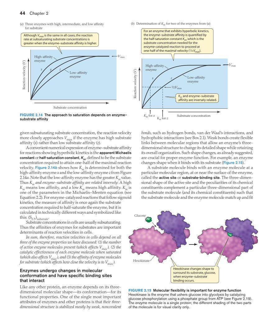
The affinity of an enzyme for its substrate affects the shape of the velocity–concentration relation at subsaturating concentra-tions of substrate (concentrations too low to saturate the reaction), as illustrated in Figure 2.14a by three enzymes with hyperbolic kinetics. Curve x in the figure represents an enzyme having a high affinity for its substrate; curve z represents one having a low affinity. All three enzymes represented in the figure have the same maximum velocity. The key difference among them is that, at any
Products
…than with catalysis.
The activation energy—the increased energy required to achieve transition state—is greater without catalysis…
Progress of the reaction
Free
ene
rgy
of m
olec
ules
Substrates
Catalyzedreaction
Uncatalyzedreaction
FIGURE 2.13 Enzymes accelerate reactions by lowering the needed activation energy Starting at their average energy level, substrate molecules must gain sufficient energy to enter a transition state before they can react to form product. The amount of energy required, the activation energy, is lowered by enzyme catalysts. Cata-lysts do not, however, alter the average free energy of either sub-strates or products; nor do they affect the relative concentrations of substrates and products at equilibrium. Vertical arrows indicate the activation energy.
02_Hill3e_Ch02.indd 43 3/8/12 2:22 PM
44 Chapter 2
given subsaturating substrate concentration, the reaction velocity more closely approaches Vmax if the enzyme has high substrate affinity (x) rather than low substrate affinity (z).
A convenient numerical expression of enzyme–substrate affinity for reactions showing hyperbolic kinetics is the apparent Michaelis constant or half-saturation constant, Km, defined to be the substrate concentration required to attain one-half of the maximal reaction velocity. Figure 2.14b shows how Km is determined for both the high-affinity enzyme x and the low-affinity enzyme z from Figure 2.14a. Note that the low-affinity enzyme has the greater Km value. Thus Km and enzyme–substrate affinity are related inversely: A high Km means low affinity, and a low Km means high affinity. Km is one of the parameters in the Michaelis–Menten equation (see Equation 2.2). For enzyme-catalyzed reactions that follow sigmoid kinetics, the measure of affinity is once again the substrate concentration required to half-saturate the enzyme, but it is calculated in technically different ways and symbolized like this: (S0.5)substrate.
Substrate concentrations in cells are usually subsaturating. Thus the affinities of enzymes for substrates are important determinants of reaction velocities in cells.
In sum, therefore, reaction velocities in cells depend on all three of the enzyme properties we have discussed: (1) the number of active enzyme molecules present (which affects Vmax ), (2) the catalytic effectiveness of each enzyme molecule when saturated (which also affects Vmax ), and (3) the affinity of enzyme molecules for substrate (which affects how close the velocity is to Vmax ).
Enzymes undergo changes in molecular conformation and have specific binding sites that interactLike any other protein, an enzyme depends on its three-dimensional molecular shape—its conformation—for its functional properties. One of the single most important attributes of enzymes and other proteins is that their three-dimensional structure is stabilized mostly by weak, noncovalent
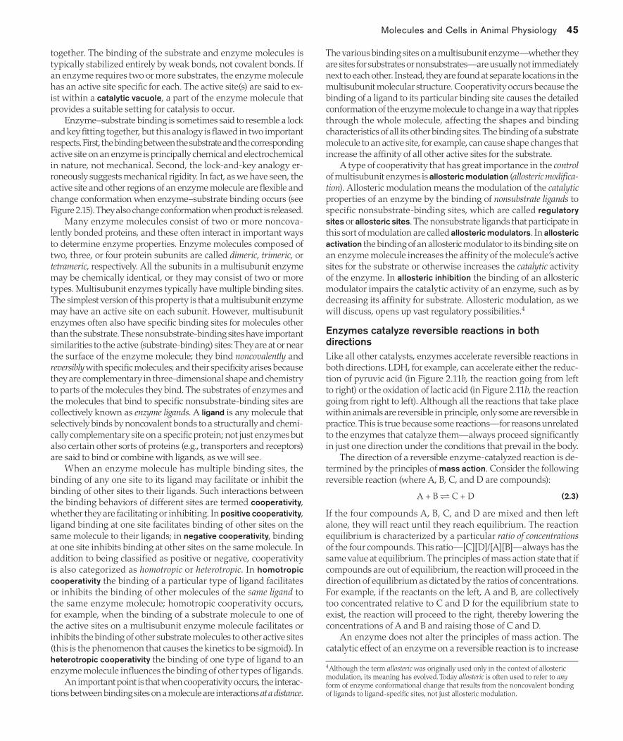
bonds, such as hydrogen bonds, van der Waal’s interactions, and hydrophobic interactions (see Box 2.1). Weak bonds create flexible links between molecular regions that allow an enzyme’s three-dimensional structure to change its detailed shape while retaining its overall organization. Such shape changes, as already suggested, are crucial for proper enzyme function. For example, an enzyme changes shape when it binds with its substrate (Figure 2.15).
A substrate molecule binds with an enzyme molecule at a particular molecular region, at or near the surface of the enzyme, called the active site or substrate-binding site. The three-dimen-sional shape of the active site and the peculiarities of its chemical constituents complement a particular three-dimensional part of the substrate molecule (and its chemical constituents) such that the substrate molecule and the enzyme molecule match up and fit
x y z
VmaxHigh-affinityenzyme
Low-affinityenzyme
Although Vmax is the same in all cases, the reaction rate at subsaturating substrate concentrations is greater when the enzyme–substrate affinity is higher.
Rea
ctio
n ve
loci
ty (V
)
Substrate concentration
(a) Three enzymes with high, intermediate, and low affinity for substrate
x z
Vmax
For an enzyme that exhibits hyperbolic kinetics, the enzyme–substrate affinity is quantified by the half-saturation constant Km, which is the substrate concentration needed for the enzyme-catalyzed reaction to proceed at one-half of the maximal velocity (1/2 Vmax).
Km and enzyme–substrateaffinity are inversely related.
Substrate concentrationKm for z
Km for x
Low-affinityenzyme
1/2Vmax
High-affinityenzyme
Rea
ctio
n ve
loci
ty (V
)
(b) Determination of Km for two of the enzymes from (a)
FIGURE 2.14 The approach to saturation depends on enzyme–substrate affinity
Hexokinase changes shape to surround its substrate, glucose, when enzyme–substrate binding occurs.
Hexokinase
Glucose
FIGURE 2.15 Molecular flexibility is important for enzyme function Hexokinase is the enzyme that ushers glucose into glycolysis by catalyzing glucose phosphorylation using a phosphate group from ATP (see Figure 2.19). The enzyme molecule is a single protein; the different shading of the two parts of the molecule is for visual clarity only.
02_Hill3e_Ch02.indd 44 3/8/12 2:22 PM
Molecules and Cells in Animal Physiology 45
together. The binding of the substrate and enzyme molecules is typically stabilized entirely by weak bonds, not covalent bonds. If an enzyme requires two or more substrates, the enzyme molecule has an active site specific for each. The active site(s) are said to ex-ist within a catalytic vacuole, a part of the enzyme molecule that provides a suitable setting for catalysis to occur.
Enzyme–substrate binding is sometimes said to resemble a lock and key fitting together, but this analogy is flawed in two important respects. First, the binding between the substrate and the corresponding active site on an enzyme is principally chemical and electrochemical in nature, not mechanical. Second, the lock-and-key analogy er-roneously suggests mechanical rigidity. In fact, as we have seen, the active site and other regions of an enzyme molecule are flexible and change conformation when enzyme–substrate binding occurs (see Figure 2.15). They also change conformation when product is released.
Many enzyme molecules consist of two or more noncova-lently bonded proteins, and these often interact in important ways to determine enzyme properties. Enzyme molecules composed of two, three, or four protein subunits are called dimeric, trimeric, or tetrameric, respectively. All the subunits in a multisubunit enzyme may be chemically identical, or they may consist of two or more types. Multisubunit enzymes typically have multiple binding sites. The simplest version of this property is that a multisubunit enzyme may have an active site on each subunit. However, multisubunit enzymes often also have specific binding sites for molecules other than the substrate. These nonsubstrate-binding sites have important similarities to the active (substrate-binding) sites: They are at or near the surface of the enzyme molecule; they bind noncovalently and reversibly with specific molecules; and their specificity arises because they are complementary in three-dimensional shape and chemistry to parts of the molecules they bind. The substrates of enzymes and the molecules that bind to specific nonsubstrate-binding sites are collectively known as enzyme ligands. A ligand is any molecule that selectively binds by noncovalent bonds to a structurally and chemi-cally complementary site on a specific protein; not just enzymes but also certain other sorts of proteins (e.g., transporters and receptors) are said to bind or combine with ligands, as we will see.
When an enzyme molecule has multiple binding sites, the binding of any one site to its ligand may facilitate or inhibit the binding of other sites to their ligands. Such interactions between the binding behaviors of different sites are termed cooperativity, whether they are facilitating or inhibiting. In positive cooperativity, ligand binding at one site facilitates binding of other sites on the same molecule to their ligands; in negative cooperativity, binding at one site inhibits binding at other sites on the same molecule. In addition to being classified as positive or negative, cooperativity is also categorized as homotropic or heterotropic. In homotropic cooperativity the binding of a particular type of ligand facilitates or inhibits the binding of other molecules of the same ligand to the same enzyme molecule; homotropic cooperativity occurs, for example, when the binding of a substrate molecule to one of the active sites on a multisubunit enzyme molecule facilitates or inhibits the binding of other substrate molecules to other active sites (this is the phenomenon that causes the kinetics to be sigmoid). In heterotropic cooperativity the binding of one type of ligand to an enzyme molecule influences the binding of other types of ligands.
An important point is that when cooperativity occurs, the interac-tions between binding sites on a molecule are interactions at a distance.
The various binding sites on a multisubunit enzyme—whether they are sites for substrates or nonsubstrates—are usually not immediately next to each other. Instead, they are found at separate locations in the multisubunit molecular structure. Cooperativity occurs because the binding of a ligand to its particular binding site causes the detailed conformation of the enzyme molecule to change in a way that ripples through the whole molecule, affecting the shapes and binding characteristics of all its other binding sites. The binding of a substrate molecule to an active site, for example, can cause shape changes that increase the affinity of all other active sites for the substrate.
A type of cooperativity that has great importance in the controlof multisubunit enzymes is allosteric modulation (allosteric modifica-tion). Allosteric modulation means the modulation of the catalyticproperties of an enzyme by the binding of nonsubstrate ligands to specific nonsubstrate-binding sites, which are called regulatory sites or allosteric sites. The nonsubstrate ligands that participate in this sort of modulation are called allosteric modulators. In allosteric activation the binding of an allosteric modulator to its binding site on an enzyme molecule increases the affinity of the molecule’s active sites for the substrate or otherwise increases the catalytic activity of the enzyme. In allosteric inhibition the binding of an allosteric modulator impairs the catalytic activity of an enzyme, such as by decreasing its affinity for substrate. Allosteric modulation, as we will discuss, opens up vast regulatory possibilities.4
Enzymes catalyze reversible reactions in both directionsLike all other catalysts, enzymes accelerate reversible reactions in both directions. LDH, for example, can accelerate either the reduc-tion of pyruvic acid (in Figure 2.11b, the reaction going from left to right) or the oxidation of lactic acid (in Figure 2.11b, the reaction going from right to left). Although all the reactions that take place within animals are reversible in principle, only some are reversible in practice. This is true because some reactions—for reasons unrelated to the enzymes that catalyze them—always proceed significantly in just one direction under the conditions that prevail in the body.
The direction of a reversible enzyme-catalyzed reaction is de-termined by the principles of mass action. Consider the following reversible reaction (where A, B, C, and D are compounds):
A + B ~ C + D (2.3)
If the four compounds A, B, C, and D are mixed and then left alone, they will react until they reach equilibrium. The reaction equilibrium is characterized by a particular ratio of concentrationsof the four compounds. This ratio—[C][D]/[A][B]—always has the same value at equilibrium. The principles of mass action state that if compounds are out of equilibrium, the reaction will proceed in the direction of equilibrium as dictated by the ratios of concentrations. For example, if the reactants on the left, A and B, are collectively too concentrated relative to C and D for the equilibrium state to exist, the reaction will proceed to the right, thereby lowering the concentrations of A and B and raising those of C and D.
An enzyme does not alter the principles of mass action. The catalytic effect of an enzyme on a reversible reaction is to increase
4 Although the term allosteric was originally used only in the context of allosteric modulation, its meaning has evolved. Today allosteric is often used to refer to any form of enzyme conformational change that results from the noncovalent bonding of ligands to ligand-specific sites, not just allosteric modulation.
02_Hill3e_Ch02.indd 45 3/8/12 2:22 PM
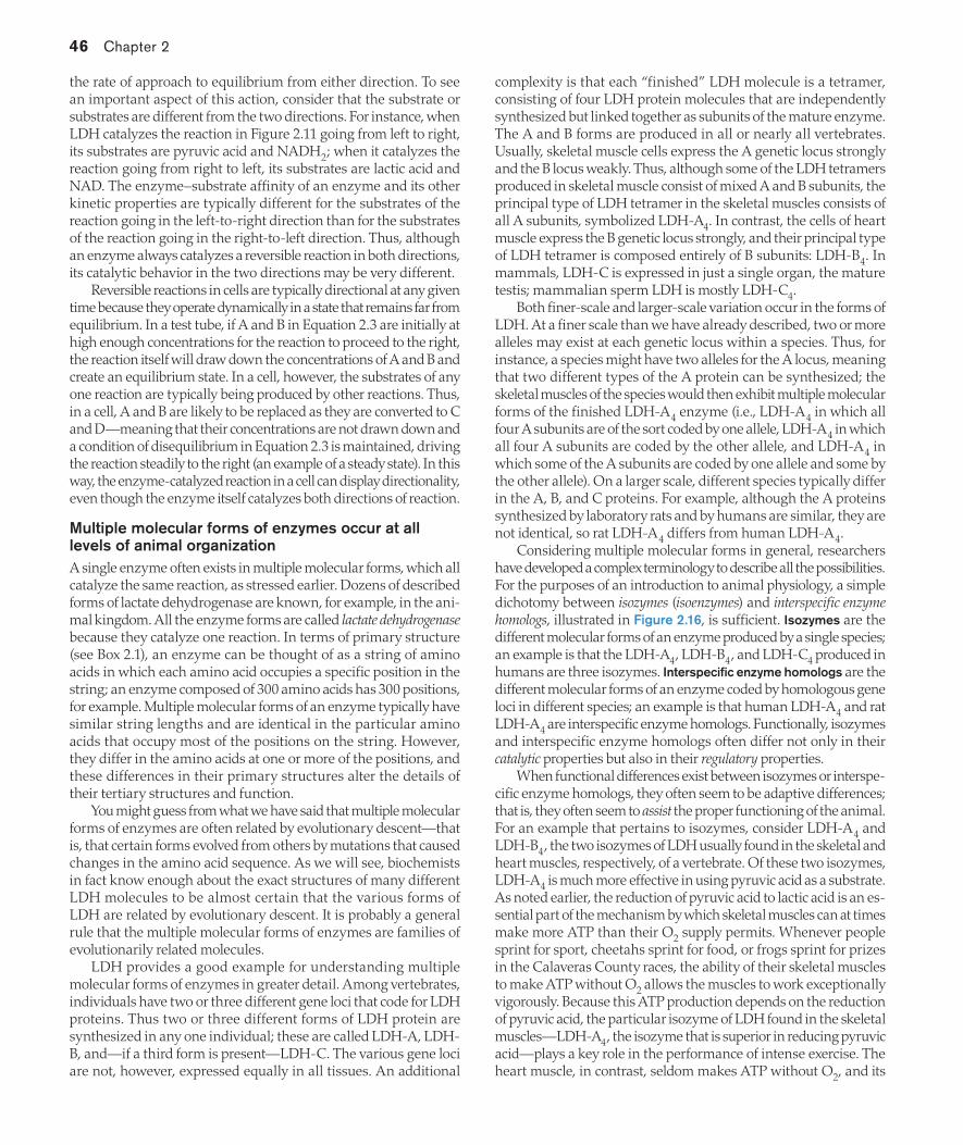
46 Chapter 2
the rate of approach to equilibrium from either direction. To see an important aspect of this action, consider that the substrate or substrates are different from the two directions. For instance, when LDH catalyzes the reaction in Figure 2.11 going from left to right, its substrates are pyruvic acid and NADH2; when it catalyzes the reaction going from right to left, its substrates are lactic acid and NAD. The enzyme–substrate affinity of an enzyme and its other kinetic properties are typically different for the substrates of the reaction going in the left-to-right direction than for the substrates of the reaction going in the right-to-left direction. Thus, although an enzyme always catalyzes a reversible reaction in both directions, its catalytic behavior in the two directions may be very different.
Reversible reactions in cells are typically directional at any given time because they operate dynamically in a state that remains far from equilibrium. In a test tube, if A and B in Equation 2.3 are initially at high enough concentrations for the reaction to proceed to the right, the reaction itself will draw down the concentrations of A and B and create an equilibrium state. In a cell, however, the substrates of any one reaction are typically being produced by other reactions. Thus, in a cell, A and B are likely to be replaced as they are converted to C and D—meaning that their concentrations are not drawn down and a condition of disequilibrium in Equation 2.3 is maintained, driving the reaction steadily to the right (an example of a steady state). In this way, the enzyme-catalyzed reaction in a cell can display directionality, even though the enzyme itself catalyzes both directions of reaction.
Multiple molecular forms of enzymes occur at all levels of animal organizationA single enzyme often exists in multiple molecular forms, which all catalyze the same reaction, as stressed earlier. Dozens of described forms of lactate dehydrogenase are known, for example, in the ani-mal kingdom. All the enzyme forms are called lactate dehydrogenasebecause they catalyze one reaction. In terms of primary structure (see Box 2.1), an enzyme can be thought of as a string of amino acids in which each amino acid occupies a specific position in the string; an enzyme composed of 300 amino acids has 300 positions, for example. Multiple molecular forms of an enzyme typically have similar string lengths and are identical in the particular amino acids that occupy most of the positions on the string. However, they differ in the amino acids at one or more of the positions, and these differences in their primary structures alter the details of their tertiary structures and function.
You might guess from what we have said that multiple molecular forms of enzymes are often related by evolutionary descent—that is, that certain forms evolved from others by mutations that caused changes in the amino acid sequence. As we will see, biochemists in fact know enough about the exact structures of many different LDH molecules to be almost certain that the various forms of LDH are related by evolutionary descent. It is probably a general rule that the multiple molecular forms of enzymes are families of evolutionarily related molecules.
LDH provides a good example for understanding multiple molecular forms of enzymes in greater detail. Among vertebrates, individuals have two or three different gene loci that code for LDH proteins. Thus two or three different forms of LDH protein are synthesized in any one individual; these are called LDH-A, LDH-B, and—if a third form is present—LDH-C. The various gene loci are not, however, expressed equally in all tissues. An additional
complexity is that each “finished” LDH molecule is a tetramer, consisting of four LDH protein molecules that are independently synthesized but linked together as subunits of the mature enzyme. The A and B forms are produced in all or nearly all vertebrates. Usually, skeletal muscle cells express the A genetic locus strongly and the B locus weakly. Thus, although some of the LDH tetramers produced in skeletal muscle consist of mixed A and B subunits, the principal type of LDH tetramer in the skeletal muscles consists of all A subunits, symbolized LDH-A4. In contrast, the cells of heart muscle express the B genetic locus strongly, and their principal type of LDH tetramer is composed entirely of B subunits: LDH-B4. In mammals, LDH-C is expressed in just a single organ, the mature testis; mammalian sperm LDH is mostly LDH-C4.
Both finer-scale and larger-scale variation occur in the forms of LDH. At a finer scale than we have already described, two or more alleles may exist at each genetic locus within a species. Thus, for instance, a species might have two alleles for the A locus, meaning that two different types of the A protein can be synthesized; the skeletal muscles of the species would then exhibit multiple molecular forms of the finished LDH-A4 enzyme (i.e., LDH-A4 in which all four A subunits are of the sort coded by one allele, LDH-A4 in which all four A subunits are coded by the other allele, and LDH-A4 in which some of the A subunits are coded by one allele and some by the other allele). On a larger scale, different species typically differ in the A, B, and C proteins. For example, although the A proteins synthesized by laboratory rats and by humans are similar, they are not identical, so rat LDH-A4 differs from human LDH-A4.
Considering multiple molecular forms in general, researchers have developed a complex terminology to describe all the possibilities. For the purposes of an introduction to animal physiology, a simple dichotomy between isozymes (isoenzymes) and interspecific enzyme homologs, illustrated in Figure 2.16, is sufficient. Isozymes are the different molecular forms of an enzyme produced by a single species; an example is that the LDH-A4, LDH-B4, and LDH-C4 produced in humans are three isozymes. Interspecific enzyme homologs are the different molecular forms of an enzyme coded by homologous gene loci in different species; an example is that human LDH-A4 and rat LDH-A4 are interspecific enzyme homologs. Functionally, isozymes and interspecific enzyme homologs often differ not only in their catalytic properties but also in their regulatory properties.
When functional differences exist between isozymes or interspe-cific enzyme homologs, they often seem to be adaptive differences; that is, they often seem to assist the proper functioning of the animal. For an example that pertains to isozymes, consider LDH-A4 and LDH-B4, the two isozymes of LDH usually found in the skeletal and heart muscles, respectively, of a vertebrate. Of these two isozymes, LDH-A4 is much more effective in using pyruvic acid as a substrate. As noted earlier, the reduction of pyruvic acid to lactic acid is an es-sential part of the mechanism by which skeletal muscles can at times make more ATP than their O2 supply permits. Whenever people sprint for sport, cheetahs sprint for food, or frogs sprint for prizes in the Calaveras County races, the ability of their skeletal muscles to make ATP without O2 allows the muscles to work exceptionally vigorously. Because this ATP production depends on the reduction of pyruvic acid, the particular isozyme of LDH found in the skeletal muscles—LDH-A4, the isozyme that is superior in reducing pyruvic acid—plays a key role in the performance of intense exercise. The heart muscle, in contrast, seldom makes ATP without O2, and its
02_Hill3e_Ch02.indd 46 3/8/12 2:22 PM

Molecules and Cells in Animal Physiology 47
isozyme, LDH-B4, is more suited to other functions than to the rapid reduction of pyruvic acid.
Interspecific enzyme homologs are often instrumental in the adaptation of species to different habitats. For example, two closely related species of thornyhead rockfish (Sebastolobus)—both found in the ocean along the West Coast of the United States—possess different genetically coded homologs of LDH-A4 in their skeletal muscles. One of the species is found in waters shallower than 500 m; the other lives at depths of 500–1500 m. A problem with the forms of LDH-A4 found in shallow-water species of marine fish, includ-ing the shallow-water rockfish, is that their affinity for substrate is highly sensitive to water pressure; these enzyme forms lose their affinity for substrate as pressure increases—so much so that at the high pressures of the deep oceans, the forms become ineffective as enzymes. The homolog of LDH-A4 synthesized by the deep-water species of rockfish is distinctive. It is relatively insensitive to water pressure and retains a suitably high affinity for substrate even when the pressure is high, helping to account for the adaptation of the species to live in the deep ocean.
SUMMARY Enzyme Fundamentals
Enzymes are protein catalysts that accelerate reactions by lowering the activation energy required for reactants to reach transition state. For most reactions to occur in cells, they must be catalyzed by enzymes. Thus a cell controls which reactions occur within it by the enzymes it synthesizes.
An enzyme must bind with its substrate to catalyze the reaction of substrate to form product. This binding, which is usually stabilized entirely by noncovalent bonds, occurs at a specific active site on the enzyme molecule, a site which is complementary in its three-dimensional chemical and electrochemical configuration to a portion of the substrate molecule. Enzyme molecules change shape when they bind to substrate or release product. These changes are permitted because the tertiary structure of a protein is stabilized by weak bonds.
Enzyme properties that determine the velocity of an enzyme-catalyzed reaction in a cell are: (1) the number of active enzyme molecules present in the cell, (2) the catalytic effectiveness of each enzyme molecule when saturated, and (3) the enzyme–substrate affinity. Enzyme-catalyzed reactions exhibit saturation kinetics because the reaction velocity is limited by the availability of enzyme molecules at high substrate concentrations. The maximal reaction velocity (Vmax) that prevails at saturation depends on properties 1 and 2: the amount and catalytic effectiveness of the enzyme. Property 3, the enzyme–substrate affinity, determines how closely the reaction velocity approaches the maximal velocity when (as is typical in cells) substrate concentrations are subsaturating. The enzyme–substrate affinity is measured by the half-saturation constant (i.e., the Michaelis constant, Km, for enzymes displaying hyperbolic kinetics).
Multisubunit enzymes often exhibit cooperativity, a phenomenon in which the binding of certain binding sites to their ligands affects (positively or negatively) the binding of other binding sites to their ligands. An important type of cooperativity is allosteric modulation, in which a nonsubstrate ligand called an allosteric modulator affects the catalytic activity of an enzyme by binding noncovalently with a specific regulatory (allosteric) binding site. Both allosteric activation and allosteric inhibition are possible.
Enzymes catalyze reversible reactions in both directions because their action is to accelerate the approach toward reaction equilibrium (determined by principles of mass action), regardless of the direction of approach.
Multiple molecular forms of enzymes occur at all levels of biological organization. Isozymes are multiple molecular forms within a single species; different isozymes may be coded by different alleles of one gene, or they may be coded by different genetic loci. Interspecific enzyme homologs are homologous forms of an enzyme in different species. Functional differences between isozymes and interspecific enzyme homologs often prove to be adaptive to different circumstances.
Regulation of Cell Function by EnzymesThe catalytic nature of enzymes often receives such exclusive at-tention that enzymes are viewed merely as molecules that speed things up. At least as important, however, is the role that cellular enzymes play as agents of regulation of cell function. The biochemical tasks in a cell are typically accomplished by sequences of enzyme-catalyzed reactions called metabolic pathways. Enzymes participate in the regulation of cell function in two principal ways. First, the types and amounts of enzymes synthesized by a cell determine which metabolic pathways are functional in the cell; any particular pathway is functional only if the cell synthesizes (through gene expression) all the enzymes the pathway requires. Second, the catalytic activities of the enzyme molecules that actually exist in a
Human LDH-B4
Human LDH-A4
Human LDH-C4
Rat LDH-B4
Rat LDH-A4
Rat LDH-C4
Interspecific enzyme hom
ologs
Isozymes
Isozymes
Each species of mammal has three major isozymes of lactate dehydrogenase (LDH), one of which is expressed just in the mature testis of the male.
FIGURE 2.16 Isozymes and interspecific enzyme homologs The forms of LDH in rats are interspecific enzyme homologs of the forms in humans.
02_Hill3e_Ch02.indd 47 3/8/12 2:22 PM

48 Chapter 2
cell at any given time can be modulated as a way of controlling the rates at which the functional metabolic pathways operate.
The types and amounts of enzymes present depend on gene expression and enzyme degradationEssentially all cells in an animal’s body have the same genome, and the genome includes the genetic code for all enzymes that the animal can produce. Cells of different tissues differ, however, in their suites of enzymes. Moreover, any one cell typically differs from time to time in the types and amounts of enzymes it contains. A gene that codes for an enzyme is said to be expressed in a cell if the cell actually synthesizes the enzyme. The reason that cells of various tissues differ in their enzymes—and that one cell can differ from time to time—is that only some genes are expressed in each cell at any given time. Gene expression is not all-or-none. Thus, for enzymes that are being synthesized by a cell, the rate of synthesis can be varied by modulation of the degree of gene expression.
The amount of a particular enzyme in a cell depends not just on the rate of enzyme synthesis but also on the rate of degradation of the enzyme. All enzymes are broken down in specific and regulated ways by various pathways, of which the ubiquitin–proteasome system discussed later in this chapter is best understood. Because of degradation, unless an enzyme is synthesized in an ongoing manner, the enzyme will disappear from a cell. The amount of an enzyme present in a cell depends, then, on an interplay of synthesis and degradation; the amount can be increased, for example, by either accelerated synthesis or decelerated degradation.
Variation in the rate of enzyme synthesis is the best-understood way that animal cells modify the amounts of their enzymes. The synthesis of an enzyme molecule requires several sequential steps: transcription of the stretch of DNA coding for the enzyme protein to form pre-messenger RNA, posttranscriptional processing to form mature mRNA, exit of the mature mRNA from the nucleus to associ-ate with ribosomes in the cytoplasm, translation of the mature RNA into the amino acid sequence of the protein, and posttranslational processing that transforms the immature polypeptide chain into a mature protein. Each of these steps is potentially modulated by a cell to control the rate at which the enzyme is synthesized.
The first step, the transcription of DNA, for instance, is typically modulated by two types of specific regulatory regions of the DNA molecule that control whether, and how fast, transcription occurs in the relevant coding region of DNA. One type of regulatory region is the promoter, a DNA sequence located just upstream (toward the 5ʹ end) from the site where transcription starts. The second type of regulatory region consists of one or more enhancers, DNA sequences that may occur at various locations, even thousands of nucleotide bases away from the promoter. Proteins called transcrip-tion factors bind with the promoter and enhancer regions of DNA by way of DNA-matching subparts of their molecular structures, and this binding controls the extent to which RNA polymerase attaches to and transcribes the DNA-coding region responsible for a given enzyme. Transcription factors are highly specific and often work in sets, permitting different genes to be independently and finely controlled.
The processes that control the rates of synthesis of enzymes act on a variety of timescales to determine which metabolic pathways are functional in a cell. A useful distinction for discussing timescales is that between constitutive and inducible enzymes. Constitutive
enzymes are present in a tissue in relatively high and steady amounts regardless of conditions. Inducible enzymes, however, are present at low levels (or not at all) in a tissue, unless their synthesis is activated by specific inducing agents.
The differentiation of tissues in an animal’s body exemplifies the control of constitutive enzymes on a long timescale. Tissues become different in their sets of functional metabolic pathways during development, and they remain different throughout life, because of long-term controls on gene expression. For example, the bone marrow cells and skin cells of mammals differ in whether they express the genes that code for the enzymes required for hemoglobin synthesis. All the genes are relatively steadily expressed—and the enzymes are, therefore, constitutive—in marrow cells but not skin cells. Accordingly, the marrow cells have a functional metabolic pathway for hemoglobin synthesis at all times throughout life, whereas skin cells do not.
Inducible enzymes that undergo up- and downregulation on rel-atively short timescales are excellently illustrated by the cytochrome P450 enzymes found in the liver, kidneys, and gastrointestinal tract of vertebrates (and also found in most or all invertebrates). The P450 enzymes are a complex family of enzymes; more than 30 kinds occur in humans, for example. Their function is to help detoxify foreign compounds by oxidizing them. The foreign compounds themselves often serve as inducing agents for the enzymes.
Low levels of P450 enzymes are found in an individual animal that has not been exposed in the immediately preceding weeks or months to suitable inducing agents, because little or no enzyme synthesis occurs in such an individual and any preexisting P450 molecules are removed by being degraded. However, even a single exposure to an inducing agent will strongly induce increased synthesis of certain P450 enzymes. A mammalian example is provided by barbiturate anesthetics. If a person or other mammal is administered an identical dose of barbiturate on two occasions that are a few days or weeks apart, the second administration will have much less effect than the first. The reason is that P450 enzymes that break down barbiturates are induced by the first administration, and the levels of the enzymes are therefore higher when the second dose is given. Another example that is particularly well understood is induction by halogenated aromatic hydrocarbons (HAHs)—a class of modern pollutants. When an animal is exposed to HAHs, the HAHs enter cells and activate an intracellular receptor, which acts as a transcription factor, causing expression of P450-coding genes. Levels of P450 enzymes then rise, poising the animal to better detoxify HAHs if a second exposure occurs. Another control system for inducible enzymes that is well understood is the system for response to hypoxia (low O2 levels) described in Box 23.1.
In Chapter 1 (see page 15), we discussed acclimation and ac-climatization—the chronic (i.e., long-term) modifications of phe-notype that individual animals commonly exhibit in response to changes in their environments. These important phenomena are often dependent at the cellular level on changes in the amounts of key enzymes.
Modulation of existing enzyme molecules permits fast regulation of cell functionCells require speedier mechanisms of regulating their functions than are provided by even the fastest inducible enzyme systems. They achieve speedier regulation by modulating the catalytic activ-
02_Hill3e_Ch02.indd 48 3/8/12 2:22 PM

Molecules and Cells in Animal Physiology 49
ity of their existing enzyme molecules. A control mechanism that depends on changing the amounts of enzymes in cells usually re-quires many hours to be even minimally effective because increasing the synthesis or the degradation of enzymes cannot alter enzyme amounts more rapidly; if such mechanisms were the only controls, a cell would be like a car that could be accelerated or decelerated only once every several hours. Changes in the catalytic proper-ties of already existing enzyme molecules, however, can often be brought about almost instantly, providing for moment-by-moment acceleration and deceleration of cell functions.
Some enzymes are better positioned than others in metabolic pathways to serve the requirements of rapid metabolic regulation. One category of well-positioned enzymes consists of those that catalyze rate-limiting reactions. In a linear metabolic pathway, it is possible in principle for the rate of one of the reactions to set the rate of the entire pathway. Suppose, for example, that of all the reactions in the pathway in Figure 2.17a, the conversion from B to C is inherently the slowest. Because the rate of the entire pathway would then be limited by the rate of that reaction, the conversion of B to C would be the rate-limiting reaction of the pathway. The catalytic effectiveness of enzyme enz2 would then be crucial.
Enzymes that catalyze branch-point reactions in metabolic pathways are another category of enzymes that are well positioned to effect rapid metabolic regulation. A branching metabolic pathway permits two or more final products to be made from a single initial reactant. For instance, in Figure 2.17b either E or H can be made from A. The relative activities of the enzymes at the branch point—enz2and enz5—determine which product is favored.
ALLOSTERIC MODULATION OF EXISTING ENZYMES Although binding sites for allosteric modulators do not occur in all enzymes, they are a common feature of enzymes that play regulatory roles. Allosteric modulation is a principal mechanism by which cell func-
tion is regulated. Recall that allosteric modulators are nonsubstrate molecules that bind noncovalently with specific sites, termed allo-steric sites or regulatory sites, on enzyme molecules and that thereby affect the catalytic activities of the enzymes. The binding of an allosteric modulator with a regulatory site is reversible and follows the principles of mass action. To illustrate, suppose that enz2, in Figure 2.17a, the rate-limiting enzyme in the reaction sequence, is allosterically modulated by a compound M. The reaction between M and the regulatory site on enz2 would then be
M + enz2 ~ M–enz2 complex (2.4)
Increasing the concentration of M shifts the reaction to the right by mass action, causing more enz2 molecules to form M–enz2 complexes (Figure 2.18a). Decreasing the concentration of M shifts the reaction to the left, causing fewer enz2 molecules to be in complexes with the allosteric modulator (Figure 2.18b). These adjustments, being driven by mass action, occur almost instantly, and they almost instantly affect the catalytic activity of the enzyme. Thus allosteric modulation can occur very rapidly.
As stressed previously, when an allosteric modulator binds with (or dissociates from) an enzyme, it alters the enzyme’s ability to catalyze the conversion of substrate to product. This outcome occurs because the binding of the modulator to its regulatory site induces changes in the conformation of the enzyme molecule that ripple through the enzyme’s molecular structure, affecting the catalytically important properties of the molecule, such as molecular flexibility or the con-formation of the active site. An enzyme that has its catalytic activity increased by a modulator is said to be upregulated; conversely, one that has its catalytic activity decreased is said to be downregulated.5 A single enzyme molecule may have two or more regulatory sites, each specific for a different allosteric modulator. In this case, the individual modulators can exert reinforcing or canceling effects on the catalytic activity of the enzyme, offering elaborate regulatory possibilities.
When an allosterically modulated enzyme is the rate-limiting en-zyme in a metabolic pathway, the entire pathway may be upregulated
5 The concepts of upregulation and downregulation are used in additional contexts as well. Another application in the study of enzymes, for example, is to the amounts of inducible enzymes in cells. When the cellular concentration of an inducible enzyme is increased, the enzyme is said to be upregulated; when the enzyme is permitted to fall to low concentration, it is said to be downregulated. Common usage also refers today to the upregulation and downregulation of processes and receptors.
A
enz1 Fast
Slow
Fast
Fast
B
enz2 enz2 enz5
C
enz3
D
enz4
E
C
enz3
D
enz4
E
F
enz6
G
enz7
H
A
enz1
B
(a) A linear metabolic pathway with a rate-limiting reaction
(b) A branching metabolic pathway
Rate-limitingreaction
Branch-pointreaction
FIGURE 2.17 Enzymes that catalyze rate-limiting reactions and branch-point reactions are well positioned to exert control over metabolism The two reaction sequences in (a) and (b) are inde-pendent. A–H are reacting compounds; enz1–enz7 are enzymes. For example, B is the substrate of enz2, and C is its product.
B
enz2
C
B
enz2
C
M
(a) High M concentration (b) Low M concentration
FIGURE 2.18 An allosteric modulator follows the principles of mass action in binding with the enzyme it modulates This figure shows how enz2 in Figure 2.17a could (a) associate with an alloste-ric modulator, M, and (b) dissociate from the modulator. B and C are reacting compounds. Combination of enz2 with M might promote or inhibit the action of the enzyme, depending on the exact nature of M, as exemplified in Figure 2.19.
02_Hill3e_Ch02.indd 49 3/8/12 2:23 PM
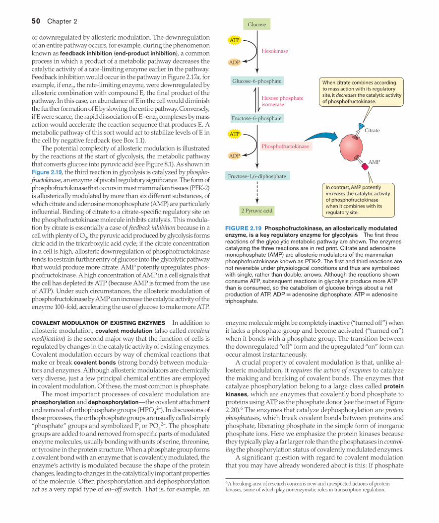
50 Chapter 2
or downregulated by allosteric modulation. The downregulation of an entire pathway occurs, for example, during the phenomenon known as feedback inhibition (end-product inhibition), a common process in which a product of a metabolic pathway decreases the catalytic activity of a rate-limiting enzyme earlier in the pathway. Feedback inhibition would occur in the pathway in Figure 2.17a, for example, if enz2, the rate-limiting enzyme, were downregulated by allosteric combination with compound E, the final product of the pathway. In this case, an abundance of E in the cell would diminish the further formation of E by slowing the entire pathway. Conversely, if E were scarce, the rapid dissociation of E–enz2 complexes by mass action would accelerate the reaction sequence that produces E. A metabolic pathway of this sort would act to stabilize levels of E in the cell by negative feedback (see Box 1.1).
The potential complexity of allosteric modulation is illustrated by the reactions at the start of glycolysis, the metabolic pathway that converts glucose into pyruvic acid (see Figure 8.1). As shown in Figure 2.19, the third reaction in glycolysis is catalyzed by phospho-fructokinase, an enzyme of pivotal regulatory significance. The form of phosphofructokinase that occurs in most mammalian tissues (PFK-2) is allosterically modulated by more than six different substances, of which citrate and adenosine monophosphate (AMP) are particularly influential. Binding of citrate to a citrate-specific regulatory site on the phosphofructokinase molecule inhibits catalysis. This modula-tion by citrate is essentially a case of feedback inhibition because in a cell with plenty of O2, the pyruvic acid produced by glycolysis forms citric acid in the tricarboxylic acid cycle; if the citrate concentration in a cell is high, allosteric downregulation of phosphofructokinase tends to restrain further entry of glucose into the glycolytic pathway that would produce more citrate. AMP potently upregulates phos-phofructokinase. A high concentration of AMP in a cell signals that the cell has depleted its ATP (because AMP is formed from the use of ATP). Under such circumstances, the allosteric modulation of phosphofructokinase by AMP can increase the catalytic activity of the enzyme 100-fold, accelerating the use of glucose to make more ATP.
COVALENT MODULATION OF EXISTING ENZYMES In addition to allosteric modulation, covalent modulation (also called covalent modification) is the second major way that the function of cells is regulated by changes in the catalytic activity of existing enzymes. Covalent modulation occurs by way of chemical reactions that make or break covalent bonds (strong bonds) between modula-tors and enzymes. Although allosteric modulators are chemically very diverse, just a few principal chemical entities are employed in covalent modulation. Of these, the most common is phosphate.
The most important processes of covalent modulation are phosphorylation and dephosphorylation—the covalent attachment and removal of orthophosphate groups (HPO4
2–). In discussions of these processes, the orthophosphate groups are usually called simply “phosphate” groups and symbolized Pi or PO4
2–. The phosphate groups are added to and removed from specific parts of modulated enzyme molecules, usually bonding with units of serine, threonine, or tyrosine in the protein structure. When a phosphate group forms a covalent bond with an enzyme that is covalently modulated, the enzyme’s activity is modulated because the shape of the protein changes, leading to changes in the catalytically important properties of the molecule. Often phosphorylation and dephosphorylation act as a very rapid type of on–off switch. That is, for example, an
enzyme molecule might be completely inactive (“turned off”) when it lacks a phosphate group and become activated (“turned on”) when it bonds with a phosphate group. The transition between the downregulated “off” form and the upregulated “on” form can occur almost instantaneously.
A crucial property of covalent modulation is that, unlike al-losteric modulation, it requires the action of enzymes to catalyze the making and breaking of covalent bonds. The enzymes that catalyze phosphorylation belong to a large class called protein kinases, which are enzymes that covalently bond phosphate to proteins using ATP as the phosphate donor (see the inset of Figure 2.20).6 The enzymes that catalyze dephosphorylation are protein phosphatases, which break covalent bonds between proteins and phosphate, liberating phosphate in the simple form of inorganic phosphate ions. Here we emphasize the protein kinases because they typically play a far larger role than the phosphatases in control-ling the phosphorylation status of covalently modulated enzymes.
A significant question with regard to covalent modulation that you may have already wondered about is this: If phosphate
6 A breaking area of research concerns new and unexpected actions of protein kinases, some of which play nonenzymatic roles in transcription regulation.
ATP
ADP
ADP
ATP
Glucose
Hexokinase
Hexose phosphate isomerase
Fructose-6-phosphate
Glucose-6-phosphate
Fructose-1,6-diphosphate
Phosphofructokinase
2 Pyruvic acid
When citrate combines accordingto mass action with its regulatory site, it decreases the catalytic activity of phosphofructokinase.
In contrast, AMP potently increases the catalytic activity of phosphofructokinase when it combines with its regulatory site.
AMP
Citrate
FIGURE 2.19 Phosphofructokinase, an allosterically modulated enzyme, is a key regulatory enzyme for glycolysis The first three reactions of the glycolytic metabolic pathway are shown. The enzymes catalyzing the three reactions are in red print. Citrate and adenosine monophosphate (AMP) are allosteric modulators of the mammalian phosphofructokinase known as PFK-2. The first and third reactions are not reversible under physiological conditions and thus are symbolized with single, rather than double, arrows. Although the reactions shown consume ATP, subsequent reactions in glycolysis produce more ATP than is consumed, so the catabolism of glucose brings about a net production of ATP. ADP = adenosine diphosphate; ATP = adenosine triphosphate.
02_Hill3e_Ch02.indd 50 3/8/12 2:23 PM
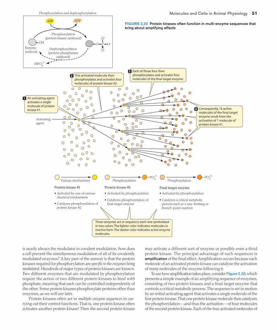
Molecules and Cells in Animal Physiology 51
is nearly always the modulator in covalent modulation, how does a cell prevent the simultaneous modulation of all of its covalently modulated enzymes? A key part of the answer is that the protein kinases required for phosphorylation are specific to the enzymes being modulated. Hundreds of major types of protein kinases are known. Two different enzymes that are modulated by phosphorylation require the action of two different protein kinases to bind with phosphate, meaning that each can be controlled independently of the other. Some protein kinases phosphorylate proteins other than enzymes, as we will see later.
Protein kinases often act in multiple-enzyme sequences in car-rying out their control functions. That is, one protein kinase often activates another protein kinase! Then the second protein kinase
may activate a different sort of enzyme or possibly even a third protein kinase. The principal advantage of such sequences is amplification of the final effect. Amplification occurs because each molecule of an activated protein kinase can catalyze the activation of many molecules of the enzyme following it.
To see how amplification takes place, consider Figure 2.20, which presents a simple example of an amplifying sequence of enzymes, consisting of two protein kinases and a final target enzyme that controls a critical metabolic process. The sequence is set in motion by an initial activating agent that activates a single molecule of the first protein kinase. That one protein kinase molecule then catalyzes the phosphorylation—and thus the activation—of four molecules of the second protein kinase. Each of the four activated molecules of
1
3
4
OH O O–
O–
O
P
ATP ADP
2
Consequently, 16 active molecules of the final target enzyme result from the activation of 1 molecule of protein kinase #1.
Each of those four then phosphorylates and activates four molecules of the final target enzyme.
This activated molecule then phosphorylates and activates four molecules of protein kinase #2.
An activating agent activates a single molecule of protein kinase #1.
Phosphorylation and dephosphorylation
Phosphorylation(protein kinase catalyzed)
Dephosphorylation(protein phosphatase
catalyzed)
Enzymemolecule
Activating agent
Protein kinase #1
chemical mechanisms
protein kinase #2
Various mechanisms Phosphorylation
Protein kinase #2 Final target enzyme
Phosphorylation
process such as a rate-limiting or
Three enzymes act in sequence, each one symbolized in two colors. The lighter color indicates molecules in inactive form. The darker color indicates active enzyme molecules.
PO4 2–PO4
2–
HPO4 2–
FIGURE 2.20 Protein kinases often function in multi-enzyme sequences that bring about amplifying effects
02_Hill3e_Ch02.indd 51 3/8/12 2:23 PM
52 Chapter 2
the second protein kinase then catalyzes the phosphorylation—and activation—of four molecules of the final target enzyme. In total, therefore, 16 target-enzyme molecules are activated. The initial activating agents of such sequences are often signaling compounds that arrive at cells in amounts that are so minute they could not by themselves exert large effects. A multi-enzyme sequence like that in Figure 2.20 allows a tiny quantity of a signaling compound to have a 16-fold greater effect than otherwise would occur.
SUMMARY Regulation of Cell Function by Enzymes
The metabolic pathways active in a cell depend on which enzymes are present in the cell, as determined by the processes of enzyme synthesis (dependent on gene expression) and enzyme degradation. The presence or absence of enzymes in a cell is regulated on long and short timescales. During individual development (an example of a long timescale), tissues acquire tissue-specific patterns of gene expression that establish tissue-specific suites of enzymes and metabolic pathways. Inducible enzymes, such as the cytochrome P450 enzymes, exemplify shorter-term regulation of the presence or absence of enzymes and metabolic pathways.
Very fast regulation of enzyme-catalyzed metabolic pathways is achieved by the modulation (upregulation or downregulation) of the catalytic activity of enzyme molecules already existing in a cell. Enzymes that catalyze rate-limiting or branch-point reactions are well positioned to mediate the rapid regulation of entire metabolic pathways in this way.
Allosteric modulation and covalent modulation are the two principal types of modulation of existing enzyme molecules. Allosteric modulation occurs by way of the noncovalent binding of allosteric modulators to regulatory sites, governed by the principles of mass action. Covalent modulation requires the enzyme-catalyzed making and breaking of covalent bonds—most commonly with phosphate. Phosphorylation is catalyzed by enzyme-specific protein kinases, which usually are the principal controlling agents in covalent modulation.
Evolution of EnzymesA great achievement of modern molecular biology is that the evolution of proteins can now be studied at the biochemical level. One major approach to this sort of study is to use data on the genes that code for proteins. We discuss that approach in Chapter 3. A second major approach, discussed here, is to use data on the proteins themselves. In studies of proteins, two principal evolutionary scales—investigated with different scientific objectives and techniques—have been of interest. We discuss these with an emphasis on enzymes.
One scale of interest in the study of enzyme protein evolution is the evolutionary relationships of the multiple enzyme forms found in sets of related species. The goal of research on these relationships is to reconstruct the family tree of the enzyme forms, so as to clarify enzyme evolution over geological scales of time. In research directed at this goal, scientists extract homologous enzymes from all the species of interest and determine the sequence of amino acids in each enzyme. They then employ the amino acid sequences to estimate the evolutionary relationships among the enzymes by drawing logical conclusions from similarities and differences in
the sequences. Figure 2.21a illustrates this approach using a set of five simplified enzymes; note, for example, that it is logical to conclude that the enzyme forms with red + green and red + blue mutations are descended from a form with only the red mutation because all the red mutations are identical.
Figure 2.21b presents a far more elaborate evolutionary tree of 24 vertebrate lactate dehydrogenases (LDHs). The evidence available indicates that there was just a single LDH gene when vertebrates first arose. All the modern, vertebrate LDH enzyme forms are coded by genes descended from that original gene and belong to a single family tree. According to amino acid sequence data, all the A forms of LDH in modern vertebrates are relatively closely related to each other (and thus all fall on one major branch—the upper branch—of the family tree in Figure 2.21b). Moreover, all the B forms are also relatively closely related to each other (and fall on a second major branch of the tree). This pattern indicates that the single original LDH gene duplicated (i.e., gave rise to two genes) early in vertebrate evolution at the point marked *. After that early duplication, each individual vertebrate animal had two LDH genes. The two diverged evolutionarily to give rise to two lineages (two evolutionary families) of genes: one coding for the A forms of LDH, and the other for the B forms. The analysis also indicates that the A gene itself duplicated prior to the appear-ance of mammals at the point marked **. Following that duplication, one copy of the gene continued to code for the A form, whereas the other diverged to produce the C form. According to the amino acid sequence data, the C forms of LDH in fish and amphibians are only distantly related to the C forms in mammals (these were all named “C” forms long ago, before their relationships were known). In sum, the study of the 24 LDHs illustrates that important features of the family tree of enzymes can be elucidated by the study of the amino acid sequences of the enzyme forms in modern animals.
The second scale of interest in the study of enzyme evolution focuses on the evolution of allele frequencies within single species. An important goal of this microevolutionary scale of research is to study evolution in action. Changes in allele frequencies within species can be highly dynamic and occur on relatively short scales of time. A case can often be made, therefore, that when differences in allele frequencies are observed—from place to place, or time to time—within a living species, the differences reflect the present-day action of natural selection or other evolutionary mechanisms.
A famous example of research on the evolution of allele frequen-cies comes from studies of the killifish Fundulus heteroclitus, a small fish (5–10 cm long) found commonly in estuaries along the Atlantic seaboard. The waters along the coast from Georgia to Maine represent one of the sharpest marine temperature gradients in the world; killifish living in Georgia experience body temperatures that, averaged over the year, are about 15°C higher than those of their relatives in Maine. In killifish, there are two major alleles of the B form of LDH (the form that occurs in the heart, red blood cells, liver, and red swimming muscles of fish). Killifish in the coastal waters of Georgia have mostly the a allele (symbolized Ba), whereas those in Maine have only the ballele (Bb). Moreover, the a allele becomes progressively less frequent from Georgia to Maine, as shown in Figure 2.22.
Several sorts of studies indicate that modern-day natural selection maintains this geographical gradient of allele frequencies. Individual killifish, for instance, have been demonstrated to travel substantial distances. Because of these long travels, interbreeding would rapidly even out the frequencies of the a and b alleles along the entire Atlantic
02_Hill3e_Ch02.indd 52 3/8/12 2:23 PM
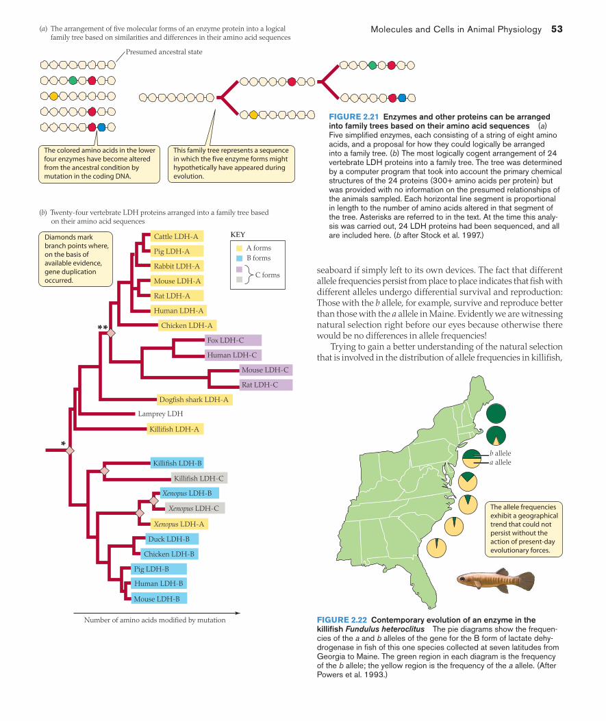
Molecules and Cells in Animal Physiology 53
seaboard if simply left to its own devices. The fact that different allele frequencies persist from place to place indicates that fish with different alleles undergo differential survival and reproduction: Those with the b allele, for example, survive and reproduce better than those with the a allele in Maine. Evidently we are witnessing natural selection right before our eyes because otherwise there would be no differences in allele frequencies!
Trying to gain a better understanding of the natural selection that is involved in the distribution of allele frequencies in killifish,
The colored amino acids in the lower four enzymes have become altered from the ancestral condition by mutation in the coding DNA.
This family tree represents a sequence in which the five enzyme forms might hypothetically have appeared during evolution.
Diamonds mark branch points where, on the basis of available evidence, gene duplication occurred.
**
*
Presumed ancestral state
(a) The arrangement of five molecular forms of an enzyme protein into a logical family tree based on similarities and differences in their amino acid sequences
(b) Twenty-four vertebrate LDH proteins arranged into a family tree based on their amino acid sequences
Number of amino acids modified by mutation
Cattle LDH-A
Pig LDH-A
Rabbit LDH-A
Mouse LDH-A
Rat LDH-A
Human LDH-A
Chicken LDH-A
Human LDH-C
Mouse LDH-C
Rat LDH-C
Dogfish shark LDH-A
Lamprey LDH
Killifish LDH-A
Killifish LDH-C
Xenopus LDH-B
Xenopus LDH-C
Xenopus LDH-A
Duck LDH-B
Chicken LDH-B
Pig LDH-B
Human LDH-B
Mouse LDH-B
Killifish LDH-B
Fox LDH-C
KEY
A formsB forms
C forms
The allele frequencies exhibit a geographical trend that could not persist without the action of present-day evolutionary forces.
b allelea allele
FIGURE 2.21 Enzymes and other proteins can be arranged into family trees based on their amino acid sequences (a) Five simplified enzymes, each consisting of a string of eight amino acids, and a proposal for how they could logically be arranged into a family tree. (b) The most logically cogent arrangement of 24 vertebrate LDH proteins into a family tree. The tree was determined by a computer program that took into account the primary chemical structures of the 24 proteins (300+ amino acids per protein) but was provided with no information on the presumed relationships of the animals sampled. Each horizontal line segment is proportional in length to the number of amino acids altered in that segment of the tree. Asterisks are referred to in the text. At the time this analy-sis was carried out, 24 LDH proteins had been sequenced, and all are included here. (b after Stock et al. 1997.)
FIGURE 2.22 Contemporary evolution of an enzyme in the killifish Fundulus heteroclitus The pie diagrams show the frequen-cies of the a and b alleles of the gene for the B form of lactate dehy-drogenase in fish of this one species collected at seven latitudes from Georgia to Maine. The green region in each diagram is the frequency of the b allele; the yellow region is the frequency of the a allele. (After Powers et al. 1993.)
02_Hill3e_Ch02.indd 53 3/8/12 2:23 PM
54 Chapter 2
physiologists have explored how the two isozymes of the B protein, coded by the a and b alleles, differ in their functional properties as enzymes. They have found that the isozyme coded by the b allele has several functional advantages at low temperatures, and that the one coded by the a allele has advantages at high temperatures. Thus mutation has given rise to two forms of this one enzyme protein, and both forms are retained because each is superior to the other in certain environments where the fish live.
Enzymes Are Instruments of Change in All Time FramesEnzymes are primary instruments of physiological change in all five of the time frames identified in Chapter 1 (see Table 1.2). Three of the time frames, you will recall, refer to changes in animal physi-ology that are responses to changes in the external environment. The first of these three is acute physiological responses by individuals, the responses that occur rapidly after the environment changes. Al-losteric modulation and covalent modulation of existing enzymes are major mechanisms of acute enzyme responses. For instance, if an animal is frightened by a predator and runs rapidly away, al-losteric upregulation of phosphofructokinase by accumulation of adenosine monophosphate (AMP) in its muscle cells will imme-diately increase the rate that glucose is processed to manufacture more ATP to sustain muscular work (see Figure 2.19).
The second major time frame of response to the environment, the chronic (long-term) physiological responses of individuals, depends on reconstructions of physiological systems requiring hours, days, or longer periods to complete. Environmentally induced changes in the expression of enzyme-coding genes constitute a major mechanism of chronic responses. For an example, consider a fish acclimated to toxin-free water. If the fish encounters toxins, it will be unable to defend itself immediately using P450 enzymes, because the enzymes must be synthesized, a process requiring many hours or days. In the long term, however, the fish will assume a new phenotype—characterized by superior toxin defenses—because of induction of its P450 enzymes.
The third time frame of response to the environment, evolutionary change, depends on shifts of gene frequencies in entire populations over multiple generations. Genes that code for enzymes are fre-quently known to evolve by mutation, natural selection, and other mechanisms on both long and short scales of evolutionary time, as we have seen in Figures 2.21b and 2.22. In this way, populations of animals take on new catalytic and regulatory phenotypes by comparison with their ancestors.
In addition to the responses of animals to their environments, there are two time frames in which the physiology of individual ani-mals is internally programmed to change, and enzymes are primary participants in these time frames as well. One time frame of internally programmed change consists of developmental (ontogenetic) changes in an animal’s physiology, the changes that occur in a programmed way as an animal matures from conception to adulthood. The expression of particular enzymes is often programmed to start at particular stages of development, as we discuss in Chapter 4 (see Figure 4.5).
Individual animals also undergo periodic physiological changes—such as changes between day and night—under control of internal biological clocks.7 Enzymes often mediate these changes, as shown
7 Biological clocks are discussed at length in Chapter 15.
by the fact that—in the tissues of animals—the catalytic activities of many enzymes rise and fall in rhythms that parallel the daily day–night cycle even when the animals have no external information on the prevailing time. Some of these enzymes affect the abilities of animals to metabolize particular foodstuffs. Others affect capabili-ties for detoxifying foreign chemicals, including medications as well as toxins. Thus food metabolism and responses to foreign agents vary between day and night because of internally programmed enzyme changes.
The Life and Death of ProteinsPhysical and chemical stresses in the lives of animals can denature enzymes and other proteins. Such stresses include high tissue tem-peratures, low cellular levels of O2, and exposure to toxic chemicals such as certain alcohols or heavy metals. When we say a protein is denatured, we mean that its three-dimensional conformation—its tertiary structure—is altered in a way that disrupts its ability to function (see Box 2.1). Usually, the primary structure—the string of amino acids—remains intact. Because the primary structure remains intact, the denatured state is potentially reversible.
One of the stunning discoveries of the last 20 years is that cells synthesize proteins termed molecular chaperones that can repair damage to other proteins by correcting reversible denaturation. The molecular chaperones use ATP-bond energy to guide the folding of other proteins. They are often active when proteins are first synthe-sized, and they are active in the repair of “old” but damaged proteins as emphasized here. Molecular chaperones assist repair by preventing protein molecules that are in unfolded states from aggregating with each other and by promoting folding patterns that restore damaged proteins to their correct three-dimensional structures. Because ATP is used, repair by molecular chaperones has a metabolic cost.
Heat-shock proteins are the most famous and best understood molecular chaperones. They are called heat-shock proteins because they were initially discovered in cells of organisms that had been exposed to stressfully high but nonlethal tissue temperatures. We realize now, however, that “heat-shock” proteins often function as molecular chaperones following many other types of cell stress, sometimes even including cold stress! An alternative name is stress proteins. The heat-shock proteins belong to several protein families of characteristic molecular weights (especially 70 and 90 kilodaltons, kDa) and display highly conserved amino acid sequences, indicating that they are evolutionarily related in most or all animals. Often they are identified by combining the prefix hsp with the molecular weight; thus hsp70 and hsp90 refer to heat-shock proteins with molecular weights of 70 and 90 kDa. Although some are constitutive proteins, heat-shock proteins are principally inducible proteins: Most are absent except during times when a stress has elicited expression of the genes that encode them.
Rocky shores along seacoasts are known from recent research to be one of the ecological settings in which heat-shock proteins routinely play critical roles. Mussels, snails, and other attached or slow-moving animals living on the rocks can experience heat stress on clear, hot days when the tide goes out and they are exposed to the sun. During or soon after such events, these animals express heat-shock proteins.
Of course, repair is not always possible, or proteins once needed may become unnecessary, or regulatory processes may require that
02_Hill3e_Ch02.indd 54 3/8/12 2:23 PM
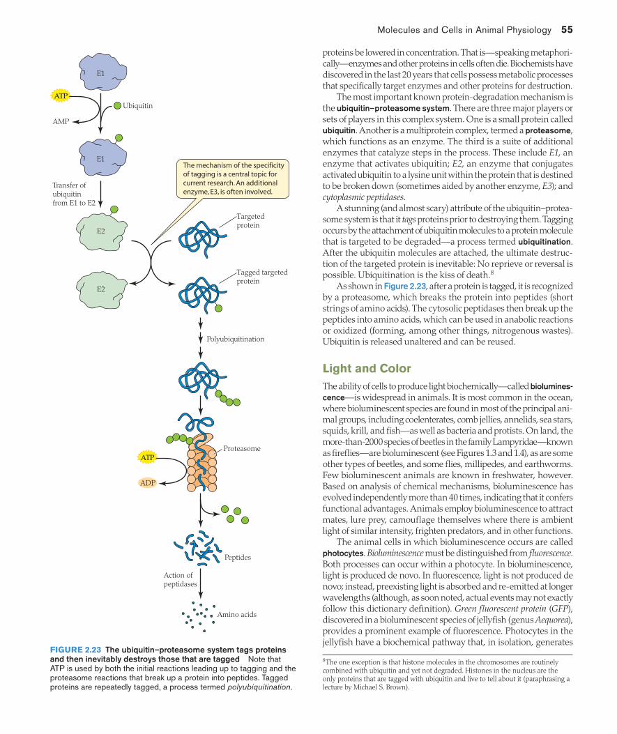
Molecules and Cells in Animal Physiology 55
proteins be lowered in concentration. That is—speaking metaphori-cally—enzymes and other proteins in cells often die. Biochemists have discovered in the last 20 years that cells possess metabolic processes that specifically target enzymes and other proteins for destruction.
The most important known protein-degradation mechanism is the ubiquitin–proteasome system. There are three major players or sets of players in this complex system. One is a small protein called ubiquitin. Another is a multiprotein complex, termed a proteasome, which functions as an enzyme. The third is a suite of additional enzymes that catalyze steps in the process. These include E1, an enzyme that activates ubiquitin; E2, an enzyme that conjugates activated ubiquitin to a lysine unit within the protein that is destined to be broken down (sometimes aided by another enzyme, E3); and cytoplasmic peptidases.
A stunning (and almost scary) attribute of the ubiquitin–protea-some system is that it tags proteins prior to destroying them. Tagging occurs by the attachment of ubiquitin molecules to a protein molecule that is targeted to be degraded—a process termed ubiquitination. After the ubiquitin molecules are attached, the ultimate destruc-tion of the targeted protein is inevitable: No reprieve or reversal is possible. Ubiquitination is the kiss of death.8
As shown in Figure 2.23, after a protein is tagged, it is recognized by a proteasome, which breaks the protein into peptides (short strings of amino acids). The cytosolic peptidases then break up the peptides into amino acids, which can be used in anabolic reactions or oxidized (forming, among other things, nitrogenous wastes). Ubiquitin is released unaltered and can be reused.
Light and ColorThe ability of cells to produce light biochemically—called biolumines-cence—is widespread in animals. It is most common in the ocean, where bioluminescent species are found in most of the principal ani-mal groups, including coelenterates, comb jellies, annelids, sea stars, squids, krill, and fish—as well as bacteria and protists. On land, the more-than-2000 species of beetles in the family Lampyridae—known as fireflies—are bioluminescent (see Figures 1.3 and 1.4), as are some other types of beetles, and some flies, millipedes, and earthworms. Few bioluminescent animals are known in freshwater, however. Based on analysis of chemical mechanisms, bioluminescence has evolved independently more than 40 times, indicating that it confers functional advantages. Animals employ bioluminescence to attract mates, lure prey, camouflage themselves where there is ambient light of similar intensity, frighten predators, and in other functions.
The animal cells in which bioluminescence occurs are called photocytes. Bioluminescence must be distinguished from fluorescence. Both processes can occur within a photocyte. In bioluminescence, light is produced de novo. In fluorescence, light is not produced de novo; instead, preexisting light is absorbed and re-emitted at longer wavelengths (although, as soon noted, actual events may not exactly follow this dictionary definition). Green fluorescent protein (GFP), discovered in a bioluminescent species of jellyfish (genus Aequorea), provides a prominent example of fluorescence. Photocytes in the jellyfish have a biochemical pathway that, in isolation, generates
8 The one exception is that histone molecules in the chromosomes are routinely combined with ubiquitin and yet not degraded. Histones in the nucleus are the only proteins that are tagged with ubiquitin and live to tell about it (paraphrasing a lecture by Michael S. Brown).
Tagged targetedprotein
Targetedprotein
E2
E2
Polyubiquitination
Proteasome
Peptides
E1
E1
Ubiquitin
Transfer of ubiquitinfrom E1 to E2
Action of peptidases
AMP
ATP
ADP
ATP
The mechanism of the specificity of tagging is a central topic for current research. An additional enzyme, E3, is often involved.
Amino acids
FIGURE 2.23 The ubiquitin–proteasome system tags proteins and then inevitably destroys those that are tagged Note that ATP is used by both the initial reactions leading up to tagging and the proteasome reactions that break up a protein into peptides. Tagged proteins are repeatedly tagged, a process termed polyubiquitination.
02_Hill3e_Ch02.indd 55 3/8/12 2:23 PM

56 Chapter 2
light at blue wavelengths. In the intact cells, however, the pathway is intimately associated with GFP, and in the final step of light pro-duction, energy from the light-producing pathway is transferred by resonance—a radiation-less process—to the GFP. The GFP emits light at green wavelengths, so the clusters of photocytes on the margin of the bell of the jellyfish glow green (Figure 2.24).
In a very general sense, light is often said to result from a lucif-erin–luciferase reaction. That is, light is said to be generated when a luciferin—a compound capable of light emission—is oxidized by the action of a luciferase—an enzyme that catalyzes luciferin oxidation. This statement is valid, but it hides the fact that there are many chemically different luciferins and luciferases, and thus an enormous diversity of light-producing reactions exists. The luciferases are par-ticularly diverse. Typically, the luciferase catalyzes combination of the luciferin with O2 to form a peroxide intermediate compound, which then spontaneously decomposes to generate a singlet electronically excited product, which in turn decays, emitting a photon of visible light. The color of the light depends on the particular luciferin–luciferase reaction and on fluorescent proteins if present. Marine animals most commonly emit at blue wavelengths—the wavelengths that travel farthest in clear seawater.
In some marine animals—notably the Aequorea jellyfish (see Figure 2.24)—luciferin, O2, and an inactive form of the catalyzing protein are assembled into a complex called a photoprotein. Light production in these cases is initiated by exposure of the photoprotein to Ca2+ or Mg2+ (or another agent), which induces a conformational change that activates catalysis.
Bioluminescent animals probably, in most cases, synthesize their own luciferin and luciferase—and produce their own light. However, many departures from this straightforward scenario are known in marine animals. Some obtain their luciferin in their diet. A more common variant is that some animals depend on symbiotic bacteria for light production, rather than having endogenous photocytes. This phenomenon is best understood in the Hawaiian bobtail squid (Euprymna scolopes), in which each generation must acquire specific light-emitting bacteria (Vibrio fischeri) from the water in its ocean environment. As discussed in Box 2.2, the squid and their bacteria provide probably the greatest insight available today on the mecha-nisms by which animal–microbial symbioses are established—as well as being a striking example of how an animal can achieve bioluminescence by the use of microbial light.
Although bioluminescence affects an animal’s color, the subject of animal color is more generally a quite different matter, depending most commonly on pigments in the skin (or other outer integument) and the wavelengths that those pigments absorb or reflect when il-luminated by solar light. If the skin is rich in a pigment that absorbs wavelengths other than green, for example, the animal looks green when viewed in solar light because only the green wavelengths are reflected into our eyes.
Speaking of animal color in this sense, a process of great physi-ological interest and ecological importance is rapid color change ( physiological color change)—the ability of individuals to change color (or color pattern) in seconds, minutes, or at most a few hours. For example, in many species of frogs, flatfish, and crayfish, individu-als darken rapidly when placed on a dark substrate, and lighten on a light substrate. Such color change depends on the function of chromatophores—flattened pigment-containing cells—in the skin or other integument. An individual may have several types of chro-matophores that differ in their pigment colors. Thus, chromatophores containing brown-black pigments, ones containing red pigments, and still others containing yellow or white pigments may be present. The pigment in a chromatophore is in the form of pigment granules(pigment-containing organelles), each about 0.3–1.0 micrometer (μm) in diameter. As a first approximation, each chromatophore cell has a fixed size in these animals. Color change is achieved by dispersing or aggregating the pigment granules within the cell. When the granules are dispersed throughout the cell, the cell as a whole takes on the color of the granules and imparts that color to the skin. When the granules within a cell are aggregated tightly together at a tiny spot in the center of the cell, the color of the granules may be essentially invisible and exert hardly any effect on the color of the skin.
The process of fully dispersing or fully aggregating pigment granules takes as little as 2–8 s in some flatfish but as long as a few hours in some frogs. Several mechanisms—still being elucidated even in vertebrates, where they are best understood—are involved. One is that the pigment granules within a chromatophore move along microtubules (part of the cytoskeleton) that radiate out to the cell periphery from the cell center in complex geometries (Figure 2.25a). Movement of the granules toward the periphery disperses
FIGURE 2.24 Bioluminescence and fluorescence in Aequorea victoria, a hydromedusan jellyfish found along the West Coast of the United States Clusters of light-producing cells are found on the margin of the bell. Although the light-producing mechanism generates blue light, the light emitted is green because of the presence of green fluorescent protein (GFP), which converts the emission wavelengths from blue to green. The light-producing photoprotein is Ca2+-activated. Both the photoprotein and the GFP, discovered in this jellyfish, have revolutionized biology because of their widespread use as molecular probes.
02_Hill3e_Ch02.indd 56 3/8/12 2:23 PM

Molecules and Cells in Animal Physiology 57
BOX 2.2 SQUID AND BIOLUMINESCENT BACTERIA, A STUDY IN CROSS-PHYLUM COORDINATION: THE EUPRYMNA SCOLOPES–VIBRIO FISCHERI SYMBIOSIS Margaret McFall-Ngai
The Hawaiian bobtail squid Euprymna scolopes forms a life-long symbiotic relationship with the Gram-negative
bioluminescent bacterium Vibrio fischeri. The animal houses populations of the bacterium in a bi-lobed light organ in the center of its mantle (body) cavity (Figure A). This squid is nocturnally active and uses the light pro-duced by the bacterial symbiont as an
antipredatory mechanism. Specifically, bacte-rial light is emitted from the ventral surface of the squid at an intensity that matches the intensity of moonlight and starlight shining down through the water (a phenomenon termed counterillumination), so that the ani-mal does not cast a shadow that can be per-ceived by a predator looking up from below. Each squid acquires its own bacteria from its environment early in life: A juvenile squid recruits V. fischeri cells from the seawater in which it develops within hours of hatching from its egg. Careful studies have revealed that this recruitment—the formation of the symbiosis—entails an intimate interaction be-tween the squid and the bacteria (Figure B). A young squid presents specialized epithelia to its seawater environment to acquire the specific bacterial symbionts, which populate deep crypts within its light organ. Once ac-quired, the symbionts initiate the life-long loss of those very epithelia, making further acquisi-tion impossible! For more on this fascinating story, see Box Extension 2.2.
FIGURE A The ecological function of the symbiosis for the squid (Photo courtesy of Margaret McFall-Ngai.)
FIGURE B Acquisition of bacterial symbionts Both images were obtained by use of immunocytochemistry and confocal microscopy. (Images courtesy of J. Foster [upper] and E. Ziegelhoffer [lower].)
Bacteria in the light organ, in the center of the mantle cavity of the squid, emit bioluminescent light downward so that at night, the squid blends in with moon- or starlight when viewed from below.
Soon after a squid hatches, Vibrio fischeri bacteria specifically attach to key epithelial surfaces that the bacteria, after they are acquired, promptly induce to be lost. Simultaneously, …
… the bacteria enter ciliated pores to populate the squid’s light organ.
(a) Microtubules (b) Aggregation and dispersal
Microtubules are seen here as fibrous white material. Note that they radiate out from the cell center.
Pigment granules are tightly aggregated at the center in this cell but can travel outward along microtubules to dispurse.
10 μm
Dispersal
Aggregation
FIGURE 2.25 Pigment aggregation and dispersal within black-pigmented chromatophores (melanophores) from the skin of a codfish (Gadus morhua) (a) Photomicrograph of a cell treated so that the microtubules are visible. Pigment granules are transported along the microtubules during dispersal and aggrega-tion. In this cell, the black pigment granules are aggregated at the cell center. (b) Diagram of a cell in aggregated and dispersed states. Note that the branched shape of the cell ensures that pigment granules will be widely spread out when in the dispersed state. (a photo courtesy of Helén Nilsson Sköld; from Nilsson and Wallin 1998.)
02_Hill3e_Ch02.indd 57 3/8/12 2:23 PM

58 Chapter 2
them, whereas movement toward the cell center aggregates them (Figure 2.25b). Movement is driven by ATP-using intracellular motor proteins such as kinesin and dynein. Chromatophores are signaled to disperse or aggregate their pigment granules by hormones, such as (i) melanocyte-stimulating hormone in amphibians and fish and (ii) several well-defined peptide hormones (e.g., red-pigment-concentrating hormone) in crustaceans. Chromatophores in some fish are also directly innervated, poising them for relatively fast neuronally-stimulated responses. In addition to exhibiting extrinsic controls, chromatophores in some crustaceans exhibit intrinsic rhythms of pigment movement controlled by biological clocks.
The most rapid color change in the animal kingdom is displayed by squids, cuttlefish, and octopuses—the cephalopod molluscs. Their color change is based on an entirely different principle than that in amphibians, fish, and crustaceans. It occurs so rapidly in some species that an individual can switch from a fully dark to a fully light coloration in less than 1 s! Color change in cephalopod molluscs is mediated by tiny color-change organs (Figure 2.26a; these organs are often called chromatophores, although this usage of “chromatophores” is entirely different from the usage we have just been discussing in the preceding paragraphs). Each of the color-change organs consists of a pigment cell of variable size that is surrounded (in three dimensions) by dozens of radially arranged muscles that are innervated directly from the brain. Relaxation of the muscle cells allows the pigment cell to contract to minimal size (e.g., 0.1 mm in diameter; Figure 2.26b). By contrast, contraction of the muscles—which can be very fast (as is typical of muscles)—ex-pands the pigment cell (to a diameter of 1.5 mm in some cases) so the pigment inside is spread out and easily visible (Figure 2.26c), imparting its color to the integument.
Reception and Use of Signals by CellsCells send signals to each other that serve to coordinate cell func-tions throughout the body. Nerve cells, for example, signal muscle cells to contract, employing neurotransmitter molecules. Moreover, endocrine cells, employing hormones, signal chromatophores to disperse or aggregate pigment granules, and they signal liver cells
to release glucose into the blood. When a signal arrives at a target cell, the cell must have mechanisms of signal reception to detect the signal. It must also have mechanisms of signal transduction—meaning mechanisms by which it modifies its intracellular activities in appropriate ways in response to the extracellular signal. Here we address signal reception and transduction.
Extracellular signals initiate their effects by binding to receptor proteinsExtracellular signaling molecules such as neurotransmitters or hormones initiate their actions on a cell by binding with certain protein molecules of the cell, called receptors. A molecule that binds specifically and noncovalently to a receptor protein is considered a ligand of the receptor. Ligand binding occurs at a specific receptor site (or sites) on the receptor protein and results in a change in the molecular conformation of the protein, a process that sets in mo-tion a further response by the cell.
Receptors may be categorized into four functional classes: (1) ligand-gated channels, (2) G protein–coupled receptors, (3) enzyme/enzyme-linked receptors, and (4) intracellular receptors (Figure 2.27). Receptors in the first three categories reside in the cell membrane. This prevalence of receptors at the cell surface reflects the fact that most signaling molecules cannot enter cells. For the most part, signaling molecules are proteins or other hydrophilic molecules that are unable to pass through the hydrophobic interior of the cell membrane. Instead of entering cells, these signaling molecules bind to receptors on the cell-membrane surface, and the receptors then mediate their intracellular effects. Only hydrophobic or very small signaling molecules can enter a cell at meaningful rates through the cell membrane; once inside, such molecules bind to intracellular receptors. Now let’s discuss the properties of the four principal classes of receptors.
LIGAND-GATED CHANNELS A ligand-gated channel is a cell-mem-brane protein that acts as both a receptor and a channel.9 This sort of channel opens to create a passageway for specific solutes, typi-cally inorganic ions, through the cell membrane when the recep-
9 Table 2.1 defines the functional categories of membrane proteins.
MusclePigment cell
0.5 mm
(a) A young squid(b) Color-change organ with muscles relaxed
(c) Color-change organ with muscles contracted
Pigment cell of a color-change organ
FIGURE 2.26 Color-change organs in squid (a) A juvenile Hawai-ian bobtail squid, Euprymna scolopes. The entire squid is only 2 mm long, accounting for the large size of the color-change organs relative to the size of the animal. The pigment cells, seen here, are expanded because the muscles of the color-change organs are contracted. (b,c) Diagrams showing a color-change organ with muscles relaxed and con-
tracted. (b) Relaxation of the muscles allows the pigment cell to contract to a small, barely visible size. (c) Contraction of the muscles expands the pigment cell so it has a prominent effect on integumentary color. Al-though the pigment cell in this case contains red pigment, neighboring pigment cells contain yellow or brown pigments. (Photo in a courtesy of Margaret McFall-Ngai; b,c after Bozler 1928.)
02_Hill3e_Ch02.indd 58 3/8/12 2:23 PM
Molecules and Cells in Animal Physiology 59
tor site or sites on the protein bind to specific signaling molecules, as diagrammed in Figure 2.27a. Ligand-gated channels function mostly in the transmission of nerve impulses across synapses, the narrow spaces between interacting nerve cells or between nerve and muscle cells (see Chapter 13). The signaling molecules that carry signals across synapses are called neurotransmitters. When a neurotransmitter is released by one cell into a synaptic gap, it dif-fuses across the gap to the receiving cell. The initial response of the receiving cell is that ligand-gated channels in its cell membrane open because of binding of the neurotransmitter to the channels. The opened channels permit increased flux of inorganic ions
ATP
GTP
Activation of the catalytic site inside the cell causes production of the second messenger cyclic GMP.
The ligand, in this case a steroid hormone, dissolves in and diffuses through the cell membrane.
In this relatively simple example, binding with the ligand…
…activates a catalytic site on the same molecule.
After binding to its ligand, a G protein–coupled receptor typically interacts with two other cell-membrane proteins—a G protein and an enzyme—to activate intracellular enzyme catalytic sites.
The catalytic activity of the enzyme produces cyclic AMP or another second messenger inside the cell.
The activated ligand–receptor complex functions as a transcription factor inside the nucleus.
KEY
Extracellular fluid Extracellular fluid
(b) G protein–coupled receptor and associated G protein system(a) Ligand-gated channel
(c) Enzyme/enzyme-linked receptor
Cytoplasm
Cytoplasm
Extracellular fluid
Cytoplasm
(d) Intracellular receptor
Extracellular fluid
Cytoplasm
Nuclear envelope (simplified)
Nucleus
DNA
Intracellularreceptor
Ligand
Cell membrane
K+
Na+
G protein–coupled receptor
G protein EnzymeCyclicAMP
(secondmessenger)
Activatedactive site
CyclicGMP
(secondmessenger)
Activatedactive site
Ligand (first messenger)
Ligand (first messenger)
Ligand
Activating interaction
In their typical mode of functioning, ligand-gated channels open to permit ions to pass through when they bind to their ligands. The flux of ions alters the electrical charge across the membrane.
FIGURE 2.27 The four types of receptor proteins involved in cell signaling (a) A ligand-gated channel. The particular example shown, a muscle cell acetylcholine receptor, must bind a ligand molecule at two sites for the channel to open. (b) A G protein–coupled receptor. Details of the molecular interactions symbolized by the yellow, double-headed arrows are discussed later in this chapter. (c) Enzyme/enzyme-linked receptors are themselves enzymes or, when activated, interact directly with other membrane proteins that are enzymes. Either way, bind-ing with the ligand activates an enzyme catalytic site inside the cell. The example shown is the atrial natriuretic peptide recep-tor, which is particularly simple because it consists of just a single protein with both a ligand-binding site and a catalytic site. (d ) Intracellular receptors are effective only for ligands that can dissolve in and diffuse through the lipid bilayer of the cell membrane. After a ligand enters the cell, it forms a complex with the receptor to initiate cellular responses. The example shown is a steroid hormone receptor, a type of receptor protein that is composed of a hormone-binding region and a region capable of binding with DNA. Binding with the hormone activates the re-ceptor, and the activated hormone–receptor complex functions as a transcription factor. ATP = adenosine triphosphate; cyclic AMP = cyclic adenosine monophosphate; cyclic GMP = cyclic guanosine monophosphate; GTP = guanosine triphosphate.
02_Hill3e_Ch02.indd 59 3/8/12 2:23 PM
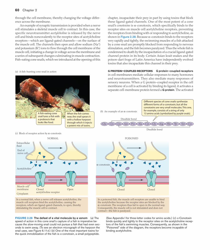
60 Chapter 2
through the cell membrane, thereby changing the voltage differ-ence across the membrane.
An example of synaptic transmission is provided when a nerve cell stimulates a skeletal muscle cell to contract. In this case, the specific neurotransmitter acetylcholine is released by the nerve cell and binds noncovalently to the receptor sites of acetylcholine receptors—which are ligand-gated channels—on the surface of the muscle cell. The channels then open and allow sodium (Na+) and potassium (K+) ions to flow through the cell membrane of the muscle cell, initiating a change in voltage across the membrane and a series of subsequent changes culminating in muscle contraction. Fish-eating cone snails, which we introduced at the opening of this
chapter, incapacitate their prey in part by using toxins that block these ligand-gated channels. One of the most potent of a cone snail’s conotoxins is α-conotoxin, which specifically binds to the receptor sites on muscle cell acetylcholine receptors, preventing the receptors from binding with or responding to acetylcholine, as shown in Figure 2.28. Because α-conotoxin binds to the receptors very rapidly and tightly, the swimming muscles of a fish attacked by a cone snail are promptly blocked from responding to nervous stimulation, and the fish becomes paralyzed. Thus the whole fish is condemned to death by the incapacitation of a crucial ligand-gated channel protein in its body. Certain Asian krait snakes and the poison-dart frogs of Latin America have independently evolved toxins that also incapacitate this channel in their prey.
G PROTEIN–COUPLED RECEPTORS G protein–coupled receptorsin cell membranes mediate cellular responses to many hormones and neurotransmitters. They also mediate many responses of sensory neurons. When a G protein–coupled receptor in the cell membrane of a cell is activated by binding its ligand, it activates a separate cell-membrane protein termed a G protein. The activated
Different species of cone snails synthesize different forms of α-conotoxin, but all the conotoxins are very small molecules. This one, for example, consists of a string of only 13 amino acids (symbolized by purple ovals).
Glu Cys Cys Asn Pro Ala Cys Gly Arg His Tyr Ser Cys
Cytoplasm
Extracellular fluid
Muscle cellmembrane Closed
acetylcholine receptorClosed Closed
α-conotoxin
End of nerve cell
(c) Block of receptor action by α-conotoxin
(b) An example of an α-conotoxin
(a) A fish-hunting cone snail in action
Disulfide bond
Disulfide bond
Acetylcholine
Open
NORMAL POISONED
In a normal fish, when a nerve cell releases acetylcholine, the muscle cell receptors bind the acetylcholine, causing the receptors, which are ligand-gated channels, to open, thereby stimulating the muscle cell to contract.
In a poisoned fish, the muscle cell receptors are unable to bind the acetylcholine because the receptor sites are blocked by the α-conotoxin. The receptors thus fail to open in the normal way. Consequently, the muscle cell is not stimulated and does not contract—the fish is paralyzed.
A fish-eating cone snail lures a fish with a proboscis that looks like food.
When the fish comes near, the snail spears it with a hollow harpoon through which it injectsa potent mix of toxins.
FIGURE 2.28 The defeat of a vital molecule by a venom (a) The speed of action in this cone snail’s capture of a fish is imperative be-cause the slow-moving snail could not pursue a fish that had even sec-onds to swim away. (To see an electron micrograph of the harpoon the snail uses, see Figure 6.11d.) (b) One of the most important toxins for the quick immobilization of the fish is α-conotoxin, a small polypeptide.
(See Appendix I for three-letter codes for amino acids.) (c) α-Conotoxin binds quickly and tightly to the receptor sites on the acetylcholine recep-tors of the fish’s swimming muscles. Consequently, as shown in the “Poisoned” side of the diagram, the receptors become incapable of binding acetylcholine.
02_Hill3e_Ch02.indd 60 3/8/12 2:23 PM

Molecules and Cells in Animal Physiology 61
G protein may then directly exert an intracellular effect, or more commonly, it interacts with still another cell-membrane protein, usually an enzyme, and activates it so that a distinctive intracellular signaling compound is synthesized in the cytoplasm of the cell by the catalytic activity of the enzyme (see Figure 2.27b).
A major difference between reception based on G protein–coupled receptors and reception based on ligand-gated channels is that in general, no sort of chemical passes through the cell membrane in the case of G protein–mediated reception. In the most common type of such reception, one chemical brings the cell-signaling message to the extracellular side of the cell membrane, and a second, dif-ferent, chemical is produced on the intracellular side to carry the signal onward to the interior of the cell. The molecules that bring signals to the cell membrane from the outside—such as hormones or neurotransmitters—are called first messengers, whereas the intracellular signaling molecules that carry the signals to the inte-rior of the cell are called second messengers. The action of the G protein–mediated mechanism in the cell membrane is analogous to a relay race in which the first messenger brings the message to a certain point but then can go no farther and must activate a second messenger for the message to go on. An example is provided by the action of epinephrine (adrenaline) on a liver cell. Epinephrine, the first messenger, binds to a G protein–coupled receptor in the cell membrane, which initiates steps resulting in intracellular synthesis of the second messenger 3′-5′-cyclic adenosine monophosphate(cyclic AMP, or cAMP). Cyclic AMP then activates the intracellular responses to the epinephrine signal. Shortly we will return to this and other second-messenger systems in greater detail.
ENZYME/ENZYME-LINKED RECEPTORS Enzyme/enzyme-linked receptors are cell-membrane proteins that either are enzymes themselves or that interact directly with enzyme proteins when activated. They are a more structurally and functionally diverse class of receptors than the two types of cell-membrane receptors we have already discussed. As in the case of G protein–mediated reception, molecules or ions do not pass through the cell membrane in this sort of reception, and enzyme/enzyme-linked receptors often activate the formation of second messengers. The simplest sort of enzyme/enzyme-linked receptor is a receptor protein that is itself an enzyme; such a protein is composed of an extracellular receptor region, a membrane-spanning region, and an intracellu-lar catalytic region (see Figure 2.27c). Binding of the extracellular signaling molecule to the receptor site activates the catalytic site at the other end of the molecule. The hormone atrial natriuretic peptide (ANP) acts on target cells in the kidney of a person to in-crease Na+ excretion by way of this sort of receptor. When ANP binds to the receptor region on the outside of a cell, the receptor molecule catalyzes the formation of a second messenger, 3′-5′-cyclic guanosine monophosphate (cyclic GMP, or cGMP), inside the cell.
INTRACELLULAR RECEPTORS Intracellular receptors are the only class of receptors not localized at the cell surface. As noted earlier, most signaling molecules cannot enter cells. Those that do are typi-cally relatively small, hydrophobic molecules that can dissolve in and diffuse through the core of the lipid bilayer of the cell membrane. These signaling molecules include steroid hormones, thyroid hor-mones, retinoic acid, vitamin D, and the gas nitric oxide (NO). The receptors for these substances are located intracellularly, in the cyto-
plasm or nucleus. The usual pattern for intracellular receptors is that, after they are activated by binding with their ligands, they interact with DNA (see Figure 2.27d) to activate specific primary-response genes, the products of which may secondarily activate other genes.
When the steroid hormone estrogen arrives at a cell, for example, it passes through the cell membrane and binds to an estrogen-specific intracellular receptor protein, forming a hormone–receptor complex. The complex is itself a transcription factor that activates specific promoter and enhancer regions of the nuclear DNA, causing the expression of specific genes. The resulting effects can alter much of a target cell’s metabolism, often promoting female cellular phenotypes.
RECEPTORS OCCUR AS MULTIPLE MOLECULAR FORMS RELATED BY
EVOLUTIONARY DESCENT In terms of molecular diversity, recep-tor proteins of any given type follow the same general principles as enzyme proteins: Each type of receptor exists in multiple molecular forms that typically are related by evolutionary descent. Most of the ligand-gated channels in modern-day animals, for example—al-though they vary in molecular details—have very similar chemical structures and are coded by a single lineage of genes that diversi-fied over evolutionary time to give rise to the channels seen today. Similarly, all the G protein–coupled receptors belong to a single family tree, as do the intracellular steroid receptors.
Cell signal transduction often entails sequences of amplifying effectsWhen signaling molecules bind to cell-membrane receptors, se-quences of amplifying effects—analogous to a chain reaction—are often involved between the moment that signal reception occurs and the moment that the final intracellular response occurs. For a classic example of this widespread pattern, let’s look at the process by which epinephrine leads to the activation of glycogen breakdown to produce glucose in vertebrate liver cells, shown in Figure 2.29.
When a human or other vertebrate experiences stress, such as the stress that occurs in anticipation of physical conflict, the adrenal glands secrete epinephrine into the blood. The circulation carries the epinephrine to the liver, where the hormone bathes liver cells, which contain abundant supplies of glycogen, a glucose-storage compound. The epinephrine itself cannot cross the cell membranes of the liver cells. “News” of its arrival reaches the inside of each cell, instead, by way of a G protein–coupled receptor system.
The receptor system itself has important amplifying properties. To set the stage for discussing these, we need to note some details of G-protein function. Recall that a G protein–coupled receptor activates a cell-membrane G protein. G proteins get their name from the fact that they are modulated by binding with guanine nucleotides. A G protein bonded with guanosine diphosphate (GDP) is inactive. A G protein is activated when it is induced to change from being bonded with GDP to being bonded with guanosine triphosphate(GTP). However, G proteins exhibit intrinsic GTP-destructive activity: When bonded with GTP, they tend to break down the GTP to GDP by hydrolysis. In this way, a G protein that has been activated by binding with GTP tends to inactivate itself by reverting to the inac-tive GDP-bonded form. The membrane G proteins, which are our focus here, are trimers in their inactive state. They dissociate into two parts when activated by GTP binding.
When epinephrine binds to its specific G protein–coupled recep-tor in the cell membrane of a liver cell, what first occurs is a series of
02_Hill3e_Ch02.indd 61 3/8/12 2:23 PM
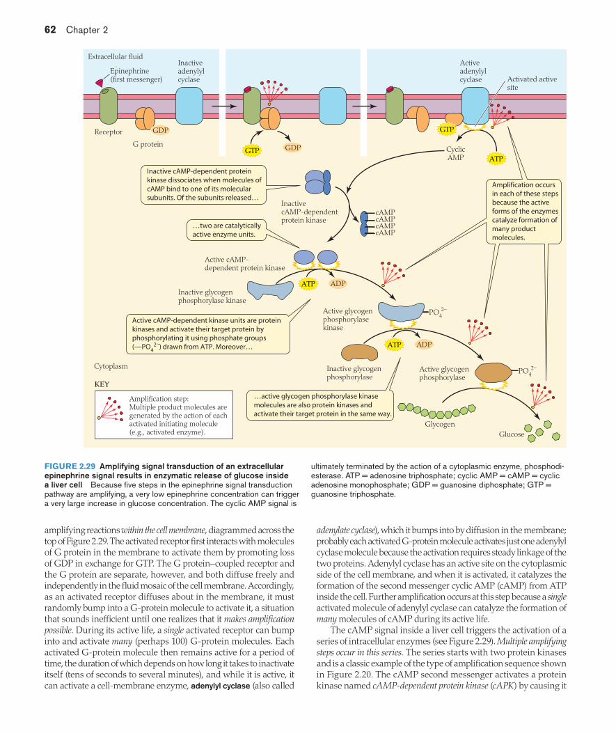
62 Chapter 2
amplifying reactions within the cell membrane, diagrammed across the top of Figure 2.29. The activated receptor first interacts with molecules of G protein in the membrane to activate them by promoting loss of GDP in exchange for GTP. The G protein–coupled receptor and the G protein are separate, however, and both diffuse freely and independently in the fluid mosaic of the cell membrane. Accordingly, as an activated receptor diffuses about in the membrane, it must randomly bump into a G-protein molecule to activate it, a situation that sounds inefficient until one realizes that it makes amplification possible. During its active life, a single activated receptor can bump into and activate many (perhaps 100) G-protein molecules. Each activated G-protein molecule then remains active for a period of time, the duration of which depends on how long it takes to inactivate itself (tens of seconds to several minutes), and while it is active, it can activate a cell-membrane enzyme, adenylyl cyclase (also called
adenylate cyclase), which it bumps into by diffusion in the membrane; probably each activated G-protein molecule activates just one adenylyl cyclase molecule because the activation requires steady linkage of the two proteins. Adenylyl cyclase has an active site on the cytoplasmic side of the cell membrane, and when it is activated, it catalyzes the formation of the second messenger cyclic AMP (cAMP) from ATP inside the cell. Further amplification occurs at this step because a singleactivated molecule of adenylyl cyclase can catalyze the formation of many molecules of cAMP during its active life.
The cAMP signal inside a liver cell triggers the activation of a series of intracellular enzymes (see Figure 2.29). Multiple amplifying steps occur in this series. The series starts with two protein kinases and is a classic example of the type of amplification sequence shown in Figure 2.20. The cAMP second messenger activates a protein kinase named cAMP-dependent protein kinase (cAPK) by causing it
GTP
Active cAMP-dependent kinase units are protein kinases and activate their target protein by phosphorylating it using phosphate groups (—PO4
2–) drawn from ATP. Moreover…
Inactive cAMP-dependent protein kinase dissociates when molecules of cAMP bind to one of its molecular subunits. Of the subunits released…
ATP
ATP
GDP
ADP
ATP ADP
PO42–
PO42–
…two are catalytically active enzyme units.
Amplification occurs in each of these steps because the active forms of the enzymes catalyze formation of many product molecules.
…active glycogen phosphorylase kinase molecules are also protein kinases and activate their target protein in the same way.
KEY
Extracellular fluid
Cytoplasm
Receptor
G protein
Inactive adenylyl cyclase
Amplification step: Multiple product molecules are generated by the action of each activated initiating molecule (e.g., activated enzyme).
Active adenylyl cyclase
CyclicAMP
Epinephrine(first messenger) Activated active
site
GDP
Inactive cAMP-dependent protein kinase
Active cAMP-dependent protein kinase
Inactive glycogen phosphorylase kinase
Active glycogen phosphorylase kinase
cAMPcAMPcAMPcAMP
Inactive glycogen phosphorylase
Active glycogen phosphorylase
GlycogenGlucose
GTP
FIGURE 2.29 Amplifying signal transduction of an extracellular epinephrine signal results in enzymatic release of glucose inside a liver cell Because five steps in the epinephrine signal transduction pathway are amplifying, a very low epinephrine concentration can trigger a very large increase in glucose concentration. The cyclic AMP signal is
ultimately terminated by the action of a cytoplasmic enzyme, phosphodi-esterase. ATP = adenosine triphosphate; cyclic AMP = cAMP = cyclic adenosine monophosphate; GDP = guanosine diphosphate; GTP = guanosine triphosphate.
02_Hill3e_Ch02.indd 62 3/8/12 2:23 PM

Molecules and Cells in Animal Physiology 63
to dissociate, forming two active enzyme units. The activated cAPK units phosphorylate, and thereby activate, a second protein kinase called glycogen phosphorylase kinase (GPK). Finally, the active GPK molecules phosphorylate and activate the ultimate target enzyme, glycogen phosphorylase (GP). Great numbers of activated GP molecules are produced. Each of them catalyzes the removal of glucose units from the glycogen polymers stored in the cell, and the glucose is then released into the blood for distribution throughout the body.
Because of the cumulative effect of all the amplifications that occur in this cell signal transduction pathway, a minute quantity of epineph-rine can cause a flood of blood glucose. A cumulative amplification of about 10,000-fold can occur between the binding of an epinephrine molecule to a G protein–coupled receptor molecule and the formation
of cAMP. Accordingly, a blood epinephrine concentration of 10–10
molar (M) can result in an intracellular concentration of cAMP of 10–6 M. Then the protein-kinase cascade within the cell can result in a further amplification of about 1000, so that the concentration of activated GP is 10 million times the blood concentration of epinephrine that initiated the signal transduction process!
Several second-messenger systems participate in cell signal transductionSeveral compounds—not just cyclic AMP—act as second mes-sengers. The common second messengers, in addition to cyclic AMP, are cyclic GMP (cGMP); inositol 1,4,5-trisphosphate (IP3); 1,2-diacylglycerol (DAG); and Ca2+ ions. Figure 2.30 provides
ATP GTP
GTP
PIP2
Ca2+
Ca2+
Ca2+Ca2+
Ca2+
Ca2+
IP3
KEY
Activating interaction
1
2
3
4 5
6
7
Activation ofmembrane protein kinases
Phosphorylation of proteins
Inositoltrisphosphate
(IP3)
Phospholipase C
G protein–coupledreceptor
Othereffects
Nitricoxide(NO)
Nitricoxide(NO)
Guanylylcyclase
Adenylylcyclase
Extracellular fluid
G protein–coupled receptor
CyclicAMP
CyclicGMP
Cytoplasm
Activation of calmodulin-dependent protein kinases or
other enzymes
IP3-gated calcium channel
Endoplasmic or sarcoplasmic reticulum
Cytoplasmic guanylyl cyclase
Phosphorylationof proteins
Phosphorylationof proteins
Activation of cAMP-
dependent protein kinases
Activation of cGMP-
dependent protein kinases
Calmodulin
Ca2+–calmodulincomplex
Diacyl-glycerol (DAG)
IP3
FIGURE 2.30 Second messengers in overview The production and the actions of five important second messengers are shown: cyclic AMP, cyclic GMP, diacylglycerol, inositol trisphosphate, and calcium ion. This figure includes only some of the major cell signal transduction pathways that employ second messengers. ➊ Some G protein–cou-pled receptor systems employ cyclic AMP as a second messenger, as seen previously in Figure 2.29. ➋ Receptor systems based on guanylyl cyclase enzymes employ cyclic GMP as a second messenger. When activated, a guanylyl cyclase produces cyclic GMP from guanosine triphosphate (GTP). In some cases, as in the atrial natriuretic peptide receptor system (see Figure 2.27c), the guanylyl cyclase is a cell-membrane enzyme. ➌ Some guanylyl cyclase enzymes are cytoplasmic. Many of the actions of nitric oxide (NO) are mediated by cyclic GMP produced by the activation of a cytoplasmic guanylyl cyclase. ➍ Some
G protein–coupled receptor systems employ diacylglycerol and inositol trisphosphate as second messengers. When such receptor systems are activated, the two second messengers are synthesized simultane-ously from a cell-membrane phospholipid, PIP2 (phosphatidylinositol 4,5-bisphosphate), by the catalytic action of activated phospholipase C, a membrane-associated enzyme. ➎ Diacylglycerol stays in the cell membrane because it is hydrophobic. ➏ Inositol trisphosphate, which is hydrophilic, enters the cytoplasm, where its major action is to open ligand-gated channels that release Ca2+ from intracellular stores such as the endoplasmic reticulum. ➐ Ca2+ ions sometimes act as second messengers, as when Ca2+ released by action of inositol trisphosphate activates the cytoplasmic protein calmodulin, which then can activate protein kinases or other enzymes.
02_Hill3e_Ch02.indd 63 3/8/12 2:23 PM
64 Chapter 2
an overview of some prominent second-messenger systems in which these compounds participate. For the most part, all of the second messengers share with cyclic AMP the property that their immediate intracellular effect is to activate a protein kinase that is already present in the cell in an inactive form, as Figure 2.30 shows. The protein kinase then activates or inactivates its target protein or proteins by phosphorylation. The target proteins are often enzymes, but they may be cell-membrane channels or receptors, channels in intracellular membranes, transcription factors that regulate gene expression, or virtually any other sort of protein. Sequences of multiple signal-amplifying reactions are a common feature of the signal transduction pathways involving second messengers.
SUMMARY Reception and Use of Signals by Cells
Extracellular signals such as hormones initiate their actions on cells by binding noncovalently with specific receptor proteins. Receptor proteins activated by binding with their signal ligands set in motion cell signal transduction mechanisms that ultimately cause cell function to be altered.
Most extracellular signaling molecules are chemically unable to enter cells because they are hydrophilic, or otherwise unable to pass through the hydrophobic, lipid interior of cell membranes. The receptors for these molecules are cell-membrane proteins that fall into three principal functional classes: ligand-gated channels, G protein–coupled receptors, and enzyme/enzyme-linked receptors. Extracellular signaling molecules that readily pass through cell membranes, such as steroid hormones, thyroid hormones, and nitric oxide (NO), have receptors that belong to a fourth functional class: intracellular receptors.
Activation of ligand-gated channels by their ligands most commonly results in changed fluxes of inorganic ions, such as Na+ and K+, across cell membranes, thereby altering voltage differences across the membranes. The altered voltage differences may then trigger other effects.
Activation of G protein–coupled receptors and enzyme/enzyme-linked receptors by their extracellular signaling ligands typically initiates the formation of second messengers, such as cyclic AMP or cyclic GMP, on the inside of the cell membrane. The second messengers, in turn, often trigger sequences of additional intracellular effects in which preexisting enzymes are modulated, most notably protein kinases. A function of these sequences is dramatic amplification of the ultimate effect.
Intracellular receptors, when activated by their ligands, usually bind with nuclear DNA and directly activate specific primary-response genes.
Study Questions1. It is becoming possible for molecular biologists to synthesize
almost any protein desired. Suppose you use a phylogenetic tree of modern-day enzymes (e.g., Figure 2.21b) to predict the amino acid sequence of a now-nonexistent ancestral enzyme form. What insights might you obtain by synthesizing the ancestral enzyme protein?
2. Using lactate dehydrogenase as an example, explain why it is true to say that “multiple molecular forms of enzymes occur at all levels of animal organization.”
3. Pollutants such as halogenated aromatic hydrocarbons (HAHs) are usually spotty in their distributions in bodies of water. Thus, even if HAHs are present, fish might be able to avoid being exposed to them. Suppose you want to determine if the fish living in an industrialized harbor are in fact more exposed to HAHs than fish in a more pristine harbor. Why might a study of liver P450 enzymes be particularly useful for your purposes?
4. What is cooperativity, and why does it not require that “cooperating” sites affect each other directly?
5. Explain why G protein–mediated receptor systems depend on membrane fluidity.
6. Describe the possible roles of allosteric modulation in the regulation of metabolic pathways.
7. Venoms nearly always consist of complex mixes of compounds. Suggest evolutionary and physiological reasons why mixes are employed rather than pure compounds. Assume that mixes imply lower amounts of individual components; for instance, assume that if a venom is composed of two compounds, each will be present in only about half the quantity than if it alone were the only component.
8. What are your views on the two sides of the debate over whether emergent properties exist? Explain and justify.
9. Outline the functional roles of conformational changes in proteins, being sure to consider the various categories of proteins such as enzymes, channels, and receptors.
10. Present additional plausible family trees for the enzyme forms in Figure 2.21a, and explain which tree you judge to be most likely.
11. Cone snails, krait snakes, and poison-dart frogs (dendrobatid frogs) have independently evolved venoms that block the muscle acetylcholine receptor. Why do you suppose this receptor has so often become a target of venoms? Explain your answer in terms of the cellular mechanisms involved.
Go to sites.sinauer.com/animalphys3e for box extensions, quizzes, flashcards,
and other resources.
ReferencesAlberts, B., A. Johnson, J. Lewis, M. Raff, K. Roberts, and P. Walter. 2007.
Molecular Biology of the Cell, 5th ed. Garland, New York. A peerless treatment of its subject, including advanced discussion of many of the topics in this chapter.
Aspengren, S., D. Hedberg, H. N. Sköld, and M. Wallin. 2008. New in-sights into melanosome transport in vertebrate pigment cells. Int. Rev. Cell. Mol. Biol. 272: 245–302. A fine, general introduction to the subject followed by details on mechanisms of pigment transport within chromatophores.
Feder, M. E., and G. E. Hofmann. 1999. Heat-shock proteins, molecular chaperones, and the stress response: Evolutionary and ecological physiology. Annu. Rev. Physiol. 61: 243–282.
Fields, P. A., and G. N. Somero. 1998. Hot spots in cold adaptation: Localized increases in conformational flexibility in lactate dehy-drogenase A4 orthologs of Antarctic notothenioid fishes. Proc. Natl. Acad. Sci. U.S.A. 95: 11476–11481.
Glickman, M. H., and A. Ciechanover. 2002. The ubiquitin-proteasome pathway: destruction for the sake of construction. Physiol. Rev. 82: 373–428.
02_Hill3e_Ch02.indd 64 3/8/12 2:23 PM
Molecules and Cells in Animal Physiology 65
Golding, G. B., and A. M. Dean. 1998. The structural basis of molecular adaptation. Mol. Biol. Evol. 15: 355–369. A daring and thought-pro-voking introduction to the new field of paleomolecular biochemistry.
Hardison, R. 1999. The evolution of hemoglobin. Am. Sci. 87: 126–137. An accessible treatment of research on the evolution of a protein, including studies of the evolution of the relevant promoter and en-hancer regions of DNA, as well as coding regions.
Hochachka, P. W., and G. N. Somero. 2002. Biochemical Adaptation: Mechanism and Process in Physiological Evolution. Oxford University Press, New York. A peerless review of the modern study of biochem-ical adaptation written by two of the foremost scientists in the field. Pedagogically exceptional.
Hofmann, G. E. 2005. Patterns of Hsp gene expression in ectothermic marine organisms on small to large biogeographic scales. Integr. Comp. Biol. 45: 247–255.
King, J., C. Haase-Pettingell, and D. Gossard. 2002. Protein folding and misfolding. Am. Sci. 90: 445–453.
Lodish, H., A. Berk, C. A. Kaiser, M. Krieger, M. P. Scott, A. Bretscher, H. Ploegh, and P. Matsudaira. 2007. Molecular Cell Biology, 6th ed. W. H. Freeman, New York.
Olivera, B. M. 1997. E. E. Just Lecture, 1996. Conus venom peptides, receptor and ion channel targets, and drug design: 50 million years of neuropharmacology. Mol. Biol. Cell 8: 2101–2109.
Powers, D. A., and P. M. Schulte. 1998. Evolutionary adaptations of gene structure and expression in natural populations in relation to a changing environment: A multidisciplinary approach to address
the million-year saga of a small fish. J. Exp. Zool. 282: 71–94. This challenging paper records one of the most successful efforts to un-derstand the molecular physiology and evolutionary biology of the ecological relationships of a species. There is probably no better ex-emplar of the future of environmental physiology.
Somero, G. N. 2002. Thermal physiology and vertical zonation of inter-tidal animals: Optima, limits, and costs of living. Integr. Comp. Biol. 42: 780–789.
Viviani, V. R. 2002. The origin, diversity, and structure function relation-ships of insect luciferases. Cell. Mol. Life Sci. 59: 1833–1850.
Widder, E. A. 2010. Bioluminescence in the ocean: origins of biologi-cal, chemical, and ecological diversity. Science 328: 704–708. An ar-ticulate, compact review—with many informative images—of light production by marine animals.
Wirgin, I., N. K. Roy, M. Loftus, R. C. Chambers, D. G. Franks, and M. E. Hahn. 2011. Mechanistic basis of resistance to PCBs in Atlantic tomcod from the Hudson River. Science 331: 1322–1325. This paper argues that rapid evolutionary increase in frequency of a mutated intracellular receptor protein provides resistance to toxic effects of polychlorinated biphenyls.
NOTE: A truly marvelous historical account of the ubiquitin–protea-some system was authored by Michael S. Brown and can be read with the year 2000 awards at the Lasker Foundation website.
See also Additional References and Figure and Table Citations
02_Hill3e_Ch02.indd 65 3/8/12 2:23 PM