characterization of msmx: a multitask atpase from bacillus ... · characterization of msmx: a...
TRANSCRIPT
Aristides Lopes de Andrade Mendes
Graduated in Cellular and Molecular Biology
Characterization of MsmX: A multitask ATPase from Bacillus subtilis
Dissertation presented to obtain the Master degree in Biotechnology
Supervisor: Isabel M. G. de Sá-Nogueira, Associate Professor w. Habilitation (Agregação), FCT/UNL
Jury Members:
President: Prof. Doutora Susana Filipe Barreiros Main Examiner: Prof. Doutor Adriano O. Henriques Supervisor: Prof. Doutora Isabel M. G. de Sá-Nogueira
September 2014

II
Characterization of MsmX: A multitask ATPase
III
Characterization of MsmX: A multitask ATPase
Copyright© reserved to Aristides Lopes Mendes, FCT/UNL, UNL
The Faculty of Science and Technology and the New University of Lisbon have the perpetual
right, and without geographical limits, to archive and publish this dissertation through press
copies in paper or digital form, or by other known form or any other that will be invented, and to
divulgate it through scientific repositories and to admit its copy and distribution with educational
or research objectives, non-commercial, as long as it is given credit to the author and editor.
A Faculdade de Ciências e Tecnologia e a Universidade Nova de Lisboa têm o direito, perpétuo
e sem limites geográficos, de arquivar e publicar esta dissertação através de exemplares
impressos reproduzidos em papel ou de forma digital, ou por qualquer outro meio conhecido ou
que venha a ser inventado, e de a divulgar através de repositórios científicos e de admitir a sua
cópia e distribuição com objectivos educacionais ou de investigação, não comerciais, desde
que seja dado crédito ao autor e editor.

IV

Characterization of MsmX: A multitask ATPase
V
Acknowledgments
“No matter what people tell you,
Words and ideas can change the world”
Robin Williams
First of all I want to thank my Professor and Supervisor, Isabel de Sá-Nogueira, for all the
support and guidance throughout this year. Thank you for all the effort in the moments I needed
help and for pushing me forward in the pursuit of a scientific career.
To Centro de Recursos Microbiológicos (CREM) da Faculdade de Ciências e Tecnologias
da Universidade Nova de Lisboa for providing good work conditions and a great environment to
develop science.
To Fundação para a Ciência e Tecnologia for partially supporting this work
through grant PEst-OE/BIA/UI0457/2013 to CREM.
To my lab mates Isabel, Mário, Lia and Viviana for the advices and teachings that helped
me become a better scientist. Thank you not only for all your patience to my constant questions
and doubts that interrupted your work and thoughts, but mainly for the support and
encouragement in the bad moments throughout the year. I could not ask for better colleagues!
My thanks to our neighbours from lab 333, especially to Raquel and Bárbara, for the good
environment and jokes at work and lunch time.
A especial thank to my biology teacher Graça Silva that was always able to motivate me
and had a determinant role in my future.
Thanks Michael for the company and friendship in this adventure of five years we have
pursued since we entered FCT; Alicia for your constant good mood and contagious laugh; Luis
and Daniel for all the support; Diana for the special person you are; Tânia for all the times you
listened to me; Rita for the good you do to me; Pedreira, Syd, Nobre, Pedro, Rute, Francisca
and many more that are always in my thoughts! To all my amazing friends that have always
supported me throughout my life, in the good and the bad times, and that make it all worth.
Thanks for all the moments that you made me laugh and smile!
Most important, I wish to thank my family! Not only because without them none of this would
be possible, but mostly because the person I am today, all my principles and beliefs… my
understanding of life… comes from them. A especial thank to my Dad for all he had to pass
through to support me and my Grandfather that always believed I would be capable of achieving
great things. Today you would be very proud of me, I will never forget you!

VI

Characterization of MsmX: A multitask ATPase
VII
Abstract
Transport across biological membranes is fundamental for cell survival and is mainly
accomplished by specialized membrane proteins known as transporters. The ABC-type
transporters constitute one of the largest and most diverse transporter superfamilies and are
found in all three domains of life (Archaea, Bacteria and Eukarya). These permeases utilize the
binding and hydrolysis of ATP to power the directional transport of a wide variety of substrates
across membranes, ranging from ions to macromolecules. The ABC transporters have a
modular architecture comprising two transmembrane domains (TMDs) that form the
translocation pore and two nucleotide-binding domains (NBDs) that hydrolyse the necessary
ATP for the translocation process.
Until the last few years, NBDs were thought to be exclusive of each transporter complex,
however it was discovered that several bacterial species ABC transporters share the same
energy-generating component. In the Gram-positive model organism Bacillus subtilis ten ABC
transporters are predicted to be involved in sugar uptake and eight of these systems do not
display a gene encoding a putative NBD protein in their vicinity. Recent studies, demonstrated
that MsmX interacts with several of these eight distinct ABC sugar importers. Thus, unlike other
NBDs, MsmX was shown to be multitask serving as energy-generating component to several
sugar importers. Other examples of multitask ATPases are also found in Streptococcus and
Streptomyces species and are involved in the uptake of carbohydrates. So a better
understanding of these multitask ATPases may have an important impact in therapy
development for pathogenic bacteria since carbohydrate utilization is essential for
microorganisms survival.
In this work we demonstrate that B. subtilis can be used as a model to study multitask
ATPases from other Gram-positive pathogenic species. A genetic system was developed to test
in vivo the functionality of MsmX homologs from pathogenic species: Bacillus thuringiensis,
Staphylococcus aureus and Streptococcus pneumoniae. The results show that the proteins are
capable to play the role of MsmX in the cell, although with a different degree of efficiency.
Functional analysis studies of MsmX indicate that modifications in the N- and C-terminal amino
acid sequence do not affect its function. Moreover, by mutagenesis we show both in vitro an in
vivo that the Lys 43 of MsmX, a conserved amino acid of NBDs, is essential for ATP hydrolysis.
Additionally, the MsmX production and purification protocol was improved for further
crystallography studies and biochemical characterization.
Keywords:
Bacillus subtilis, ABC Transporters, Multitask ATPases, MsmX, Sugars, Pathogenic bacteria.

VIII
Characterization of MsmX: A multitask ATPase
IX
Resumo
O transporte através de membranas biológicas é fundamental para a sobrevivência das
células e é realizado, principalmente, por proteínas especializadas da membrana designadas
como transportadores. Os transportadores do tipo ABC constituem uma das maiores e mais
diversificadas superfamílias de transportadores existindo em todos os três domínios dos seres
vivos (Archaea, Bacteria e Eukarya). Estas permeases utilizam a ligação e hidrólise do ATP
como força motriz para o transporte de uma ampla variedade de substratos através da
membrana, desde iões a macromoléculas. Os transportadores ABC têm uma arquitetura
modular que compreende dois domínios transmembranares (TMDs), que formam o poro de
translocação, e dois domínios de ligação a nucleótidos (NBDs), que hidrolisam o ATP
necessário para o processo de translocação.
Até recentemente, pensava-se que os NBDs eram exclusivos de cada complexo
transportador, no entanto descobriu-se que vários transportadores ABC de procariontes
partilham o mesmo componente de produção de energia. Na espécie Bacillus subtilis,
organismo modelo de bactérias Gram-positivas, existem dez transportadores ABC que se
prevêem estar envolvidos no transporte de açúcares, sendo que oito desses sistemas não
exibem um gene codificante para uma NBD putativa na sua vizinhança. Estudos recentes
demonstraram que o MsmX interage com vários desses oito importadores. Assim, ao contrário
de outros NBDs, o MsmX mostrou ser multitarefa servindo como “motor” para vários
importadores de açúcares. Outros exemplos de ATPases multitarefa são também encontrados
em espécies de Streptococcus e Streptomyces, estando envolvidos no transporte de açúcares.
Assim sendo, uma melhor compreensão destas ATPases multitarefa pode ter um impacto
importante no desenvolvimento de tratamentos para infecções bacterianas, dado que a
utilização de hidratos de carbono é essencial para a sobrevivência dos microrganismos.
Neste trabalho demonstramos que B. subtilis pode ser usado como modelo para o estudo
de ATPases multitarefa de outras espécies Gram-positivas patogénicas. De forma a testar in
vivo a funcionalidade dos homólogos do MsmX provenientes de espécies patogénicas (Bacillus
thuringiensis, Staphylococcus aureus e Streptococcus pneumoniae) foi desenvolvido um
sistema genético. Os resultados mostram que as proteínas são capazes de desempenhar o
papel do MsmX na célula, embora com diferentes graus de eficácia. Estudos funcionais do
MsmX indicam que modificações na sequência de aminoácidos do N- e C-terminal não afectam
a sua função. Além disso, mostramos através de mutagénese, tanto in vitro e in vivo, que a Lys
43 do MsmX, um aminoácido conservado de NBDs, é essencial para a hidrólise de ATP. Além
disso, o protocolo de produção e purificação do MsmX foi melhorado de modo a realizar
estudos cristalográficos e caracterizar bioquimicamente a proteína.
Palavras-chave:
Bacillus subtilis, Transportadores ABC, ATPases Multitarefa, MsmX, Açúcares, Bactérias
Patogénicas.

X

Characterization of MsmX: A multitask ATPase
XI
Contents
Acknowledgments ......................................................................................................................... V
Abstract ....................................................................................................................................... VII
Resumo ........................................................................................................................................ IX
Contents ....................................................................................................................................... XI
Figures Index .............................................................................................................................. XIII
Tables Index ............................................................................................................................... XV
Abbreviations, Symbols and Notations .................................................................................... XVII
1. General Introduction ................................................................................................................. 3
1.1 . Membrane Transport Systems .......................................................................................... 3
1.2 . ABC-type Transport Systems ............................................................................................ 3
ABC Transporters Classes ............................................................................................... 5
Transport Mechanism ....................................................................................................... 6
The Solute Binding Protein (SBP) .................................................................................... 7
The Transmembrane Domains (TMD) ............................................................................. 9
The Nucleotide Binding Domains (NBD) ........................................................................ 10
Multitask ATPases .......................................................................................................... 12
1.3 . Scope of the Thesis ......................................................................................................... 14
2. Development of an in vivo system to test the functionality of msmX Alleles and MsmX
homologs ..................................................................................................................................... 17
2.1 Introduction ........................................................................................................................ 17
2.2 Materials and Methods ...................................................................................................... 18
Substrates ....................................................................................................................... 18
Bioinformatic Analysis .................................................................................................... 18
DNA manipulation and sequencing ................................................................................ 18
Construction of plasmids and strains ............................................................................. 18

XII
Site-Directed Mutagenesis ............................................................................................. 21
Growth conditions ........................................................................................................... 21
2.3 Results and Discussion ..................................................................................................... 22
In silico Survey................................................................................................................ 22
Development of a Genetic System for Complementation Analysis ................................ 22
MsmX Functional Studies ............................................................................................... 24
Functional Analysis of MsmX Homologs ........................................................................ 27
Fine-tuning the Genetic System for Functional Analysis ................................................ 31
3. Biochemical Characterization of MsmX ................................................................................. 37
3.1 Introduction ........................................................................................................................ 37
3.2 Materials and Methods ...................................................................................................... 38
DNA manipulation and sequencing ................................................................................ 38
Site-Directed Mutagenesis ............................................................................................. 38
Construction of strains .................................................................................................... 39
Growth conditions ........................................................................................................... 39
Production and purification of recombinant proteins ...................................................... 40
Protein analysis .............................................................................................................. 41
ATPase activity assay .................................................................................................... 41
3.3 . Results and Discussion ................................................................................................... 42
Sequence Analysis ......................................................................................................... 42
Expression of msmX in E. coli ........................................................................................ 44
Purification of MsmX in E. coli ........................................................................................ 46
Analysis of the MsmX-Lys43Ala variant in vivo and in vitro ........................................... 47
4. Concluding Remarks and Future Perspectives ...................................................................... 53
5. References ............................................................................................................................. 57
6. Appendices ............................................................................................................................. 65

Characterization of MsmX: A multitask ATPase
XIII
Figures Index
Figure 1.1 - Classes of ABC transporters. .................................................................................... 6
Figure 1.2 - The transport mechanism of Type I importers.. ........................................................ 7
Figure 1.3 – TMDs and MalK interactions. ................................................................................. 10
Figure 1.4 – Conserved motifs in the NBDs domain and subdomains....................................... 12
Figure 1.5 - MsmX-dependent ABC importers in B. subtilis. ...................................................... 14
Figure 2.1 – Schematic illustration of the in vivo system developed. ......................................... 23
Figure 2.2 – Schematic representation of the amyE locus and the yxkf-msmX operon in the
chromosome of several B. subtilis strains used in this work ....................................................... 24
Figure 2.3 - Growth of B. subtilis 168T+ (wild-type) in CSK medium using glucose, arabinose
and arabinotriose as the sole carbon and energy source. .......................................................... 25
Figure 2.4 - Growth of IQB672 (ΔmsmX::cat ΔamyE::pSpank(hy)) in CSK medium using
glucose, arabinose and arabinotriose as the sole carbon and energy source. ........................... 26
Figure 2.5 - Growth of IQB673 (ΔmsmX::cat ΔamyE::pSpank(hy)-msmX) in CSK medium using
glucose, arabinose and arabinotriose as the sole carbon and energy source. ........................... 27
Figure 2.6 - Growth of IQB674 (ΔmsmX::cat ΔamyE::pSpank(hy)-msmK) in CSK medium using
glucose, arabinose and arabinotriose as the sole carbon and energy source. ........................... 29
Figure 2.7 - Growth of IQB677 (ΔmsmX::cat ΔamyE::pSpank(hy)-HD73_4301) in CSK medium
using glucose, arabinose and arabinotriose as the sole carbon and energy source. ................. 29
Figure 2.8 - Growth of IQB678 (ΔmsmX::cat ΔamyE::Pspank(hy)-ugpC) in CSK medium using
glucose, arabinose and arabinotriose as the sole carbon and energy source. ........................... 29
Figure 2.9 - Schematic representation of the modifications made in the genetic system. ......... 32
Figure 2.10 - Growth of IQB676 (ΔmsmX::cat ΔamyE::Pspank(hy)-msmX(Glu3Ser, Ile364Ser))
in CSK medium using glucose, arabinose and arabinotriose as the sole carbon and energy
source. ......................................................................................................................................... 33
Figure 3.1 - MsmX and MalK sequence alignment. ................................................................... 43
Figure 3.2 – MsmX secondary structure prevision by PSIpred. ................................................. 44

XIV
Figure 3.3 – Overproduction of rMsmX in E. coli BL21 (DE3). ................................................... 45
Figure 3.4 – Purification of rMsmX. ............................................................................................ 46
Figure 3.5 – Growth of IQB676 (ΔmsmX::cat ΔamyE::Pspank(hy)-msmX(Lys43Ala)) in CSK
medium using glucose, arabinose and arabinotriose as the sole carbon and energy source. ... 48
Figure 3.6 - Overproduction of rMsmX(Lys43Ala) in E. coli BL21 (DE3). .................................. 49
Figure 3.7 - Purification of rMsmX(Lys43Ala). ............................................................................ 49
Figure 3.8 - ATP hydrolysis by rMsmX and rMsmX(Lys43Ala). ................................................. 50
Characterization of MsmX: A multitask ATPase
XV
Tables Index
Table 2.1 - List of plasmids used or constructed during the course of this work. ....................... 19
Table 2.2 - List of oligonucleotides used in this work. ................................................................ 20
Table 2.3 - List of B. subtilis strains used or constructed during the course of this work. .......... 20
Table 2.4 – MsmX homologs selected. ....................................................................................... 22
Table 2.5 – Growth of different strains 168T+, IQB672 and IQB673 in the presence of several
saccharides as sole carbon/energy. ............................................................................................ 25
Table 2.6 - Growth of different B. subtilis strains in the presence of several saccharides as sole
carbon/energy.............................................................................................................................. 28
Table 2.7 – RBS Calculator algorithm results for each RBS. ..................................................... 31
Table 2.8 - Growth of IQB673 and IQB676 strains in the presence of several saccharides as
sole carbon/energy. ..................................................................................................................... 32
Table 3.1 - List of oligonucleotides used in this work. ................................................................ 38
Table 3.2 - List of plasmids used or constructed during the course of this work. ....................... 39
Table 3.3 - List of B. subtilis strains used or constructed during the course of this work. .......... 39
Table 3.4 – Growth of IQB673 and IQB675 in the presence of several saccharides as sole
carbon/energy.............................................................................................................................. 47

XVI

Characterization of MsmX: A multitask ATPase
XVII
Abbreviations, Symbols and Notations
aa – Amino acid
ABC – ATP-binding cassette
ADP – Adenosine diphosphate
Amp – Ampicillin
ATP – Adenosine triphosphate
A3 – 1,5-α-L-Arabinotriose
BLAST – Basic Local Alignment Search
Tool
bp – Base pairs
cat – chloramphenicol acetyltransferase gene
Cm – Chloramphenicol
CRD – C-terminal regulatory domain
CSK – C medium supplemented with
potassium succinate
CUT1 – Carbohydrate Uptake Transporter-1
DMSO – Dimethyl Sulfoxide
dNTP - Deoxynucleoside triphosphate
DNA – Deoxyribonucleic acid
ECF – Energy coupling factor
EDTA - Ethylenediaminetetraacetic acid
IPTG – Isopropyl-β-D-galactopyranoside
kDa - KiloDalton
Km - Kanamycin
LB – Luria-Bertani medium
mRNA – messenger ribonucleic acid
NBD – Nucleotide-binding domain
OD – Optical Density
ORF – Open Reading Frame
PBS – Phosphate buffer saline
PCR – Polymerase Chain Reaction
PMSF - phenylmethylsulphonyl fluoride
PTS – Phosphotransferase system
RBS – ribosome binding site
RNA - Ribonucleic acid
rpm – Revolutions per minute
SBP – Solute-binding domain
SDS-PAGE – Sodium dodecyl sulphate
polyacrilamide gel electrophoresis
Spc - Spectinomycin
Tc – tetracycline
TE – Tris-EDTA
TMD – Transmembrane domain
Tris - Tris(hydroxymethyl)aminomethane
UV – Ultraviolet light

XVIII
Amino acids – three and one letter code
Amino acid Three letter code One letter code
alanine ala A
arginine arg R
asparagine asn N
aspartic acid asp D
cysteine cys C
glutamic acid glu E
glutamine gln Q
glycine gly G
histidine his H
isoleucine ile I
leucine leu L
lysine lys K
methionine met M
phenylalanine phe F
proline pro P
serine ser S
threonine thr T
tryptophan try W
tyrosine tyr Y
valine val V
Bases - one letter code
Base Letter
Adenine A Citosyne C Guanine G Thymine T

Characterization of MsmX: A multitask ATPase
1
Chapter 1
General Introduction

Chapter 1 - General Introduction
2

Characterization of MsmX: A multitask ATPase
3
1. General Introduction
1.1. Membrane Transport Systems
Living cells are separated from the environment by lipid membranes, whereas other
subcellular membranes play essential roles in the architecture of eukaryotic cells (Seyffer
and Tampé, 2014). Transport across biological membranes is fundamental for cell survival,
which is accomplished, in the majority of the cases, by specialized membrane proteins that are
known as transporters. These proteins are responsible for the regulated and selective passage
of small molecules, ions and even some macromolecules into the cell or in the opposite
direction (Higgins, 1992; Rees et al., 2009; Eitinger et al., 2010).
The importance of membrane transport to the cell is exemplified by the fact that ~10% of
the Escherichia coli genome has been classified as participating in transport processes and,
overall, more than 550 different types of transporters have been identified (Rees et al., 2009).
Bacillus subtilis possesses a similar percentage of transporters in the genome with minor
differences. While E. coli has more sugar transporters, B. subtilis encodes more
drug/hydrophobic exporters (Saier et al., 2002).
Accordingly to the way solute transport is energized, membrane transporters are classified
in three major groups: primary active transporters, secondary transporters and group
translocators. Primary active transporters comprise very diverse protein families that use
chemical, electrical or solar energy sources to transport substrates across the membrane
against a concentration gradient. On the other hand secondary transporters often accumulate
substrates in – or deplete them from – cells by using the energy stored in ion gradients,
frequently Na+ or H+, to drive transport. This includes uniporters, antiporters, and symporters.
Group translocators chemically modify the substrate during the transport reaction, as for
example the phosphotransferase system (PTS) which transfers a phosphate group to the
substrate once it enters the cell (Davidson et al., 2008; Jaehme and Slotboom, 2014).
This dissertation will focus in one of the primary active transporters families: the ATP-
binding cassette (ABC) transporters.
1.2. ABC-type Transport Systems
The ABC transporters constitute one of the largest and most diverse transporter
superfamilies (seven distinct subfamilies) and are found in all three domains of life (Archaea,

Chapter 1 - General Introduction
4
Bacteria and Eukarya) (Yoshida et al., 1996; Rees et al., 2009; Seyffer and Tampé, 2014).
These transporters use the binding and hydrolysis of ATP (phosphate bond between the γ- and
the β-phosphate) to power the directional transport of a wide variety of substrates across
membranes, ranging from ions to macromolecules (Rees et al., 2009; Seyffer and Tampé,
2014; ter Beek et al., 2014).
The designation ABC transporters acknowledges a highly conserved ATP-binding cassette,
which is the most characteristic feature of this superfamily (Higgins, 1992). These systems are
widespread among living organisms and have been found in all species from the microbe to
man, with a high conservation of the primary sequence of the ATP-hydrolyzing domain and in
the modular architecture (Higgins, 2001; Davidson et al., 2008).
The first systems discovered and characterized in detail were the high-affinity histidine
(HisJQMP) and maltose (MalEFGK) uptake systems of Salmonella enterica serovar
typhimurium and E. coli in the 1970s (Davidson et al., 2008). Since then, a variety of systems
have been identified both in prokaryotes and eukaryotes. In E. coli and B. subtilis ~80 and 59
distinct systems were reported, respectively, while ~50 systems (five subfamilies) exist in
humans. Plants genome, like Arabidopsis thaliana, has 4120 ABC systems. However, in relation
to genome size, the highest number of ABC systems exists in bacteria (Rees et al., 2009;
Eitinger et al., 2010; Seyffer and Tampé, 2014).
In prokaryotes, these transporters are present in the plasma membrane with the ATP being
hydrolyzed in the cytoplasmic side and transport a wide variety of substrates: sugars, amino
acids, opines, peptides, phosphates, sulphates, vitamins, metallic cations, molybdenum and
organo-iron complexes. Many of these transporters are central to antibiotic and antifungal
resistance. In eukaryotes, the ABC transporters are found in organellar membranes with the
hydrolysis taking place in the cytosolic side. The transporters from mitochondria and
chloroplasts are an exception, because the domains responsible for the ATP hydrolysis are
present in the matrix or stroma side. The ABC transporters found in humans handle antibiotics,
toxins, vitamins, drugs, metals, peptides, ions, lipids, bile acids, polycarbonates and sterols.
Dysfunctions in these systems are associated with genetic diseases such as cystic fibrosis,
Tangier disease, obstetric cholestasis, and drug resistance of cancers. Here we will designate
cis-side as the side where the ATP is bound and hydrolysed and the opposite side termed as
trans-side (Quentin et al., 1999; Higgins, 2001; Seyffer and Tampé, 2014; ter Beek et al., 2014).
The ABC-type transporters are importers, which move substrates from the trans-side to the
cis-side, or exporters that transport the molecules from the cis-side to the trans-side. (ter Beek
et al., 2014). Despite this distinction between importers and exporters, they all share a common
structural organization. They have a modular architecture comprising two transmembrane
Characterization of MsmX: A multitask ATPase
5
domains (TMDs) that form the translocation pore and two nucleotide-binding domains (NBDs)
that hydrolyze ATP (Eitinger et al., 2010).
The TMDs are highly hydrophobic and each one, normally, contains six membrane-
spanning segments. These domains create the pathway for substrate transport and determine
the substrate specificity of the transporter through substrate-binding sites (Higgins, 1992, 2001;
Quentin et al., 1999). The NBDs are peripherally located at the cis-side of the membrane, where
they associate with the TMDs, and are highly conserved in structure and sequence. There are
some NBDs that do not interact with TMDs, being involved in other functions such as mRNA
translation and DNA repair. However, for these exceptions the term “ABC transporter” is not
applied and thus will not be discussed here (Higgins, 1992; Davidson et al., 2008; ter Beek et
al., 2014).
The individual domains of an ABC transporter are frequently expressed as individual
polypeptides, often found in prokaryotic species, or are fused into larger, multifunctional
polypeptides. When one of the four domains is absent, one of the remaining domains functions
as a homodimer (Higgins, 1992, 2001; Seyffer and Tampé, 2014).
In many ABC transporters auxiliary domains have been recruited for specific functions,
such as the substrate-binding protein (SBP) a domain that binds to the substrate in the trans-
side of the membrane and delivers it to the membrane-associated transport complex. The SBPs
are soluble and periplasmic in Gram-negative bacteria, while in Gram-positive bacteria they are
anchored to the membrane via an N-terminal hydrophobic lipid extension due to inexistence of
an outer membrane. Their presence gives specificity and a high degree of affinity for substrates
to the transport systems (Quentin et al., 1999; Higgins, 2001).
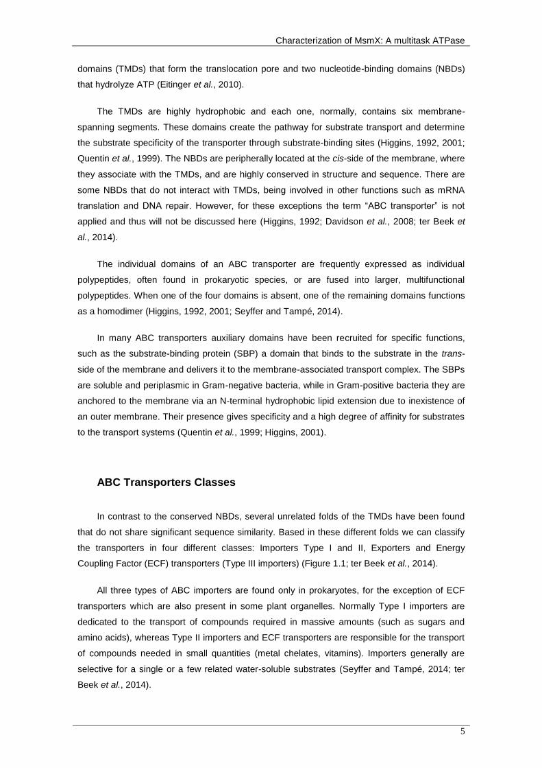
ABC Transporters Classes
In contrast to the conserved NBDs, several unrelated folds of the TMDs have been found
that do not share significant sequence similarity. Based in these different folds we can classify
the transporters in four different classes: Importers Type I and II, Exporters and Energy
Coupling Factor (ECF) transporters (Type III importers) (Figure 1.1; ter Beek et al., 2014).
All three types of ABC importers are found only in prokaryotes, for the exception of ECF
transporters which are also present in some plant organelles. Normally Type I importers are
dedicated to the transport of compounds required in massive amounts (such as sugars and
amino acids), whereas Type II importers and ECF transporters are responsible for the transport
of compounds needed in small quantities (metal chelates, vitamins). Importers generally are
selective for a single or a few related water-soluble substrates (Seyffer and Tampé, 2014; ter
Beek et al., 2014).

Chapter 1 - General Introduction
6
ABC transporters that play exporter functions can be found both in prokaryotes and
eukaryotes, where they are involved in the transport of hydrophobic compounds such as lipids,
fatty acids, cholesterol, and drugs. Some are called multidrug-resistant transporters because
they can secret a large variety of drugs out of the cell. Also, they translocate larger molecules
such as proteins (toxins, hydrolytic enzymes, S-layer proteins, lantibiotics, bacteriocins, and
competence factors) (ter Beek et al., 2014). Note that it is not excluded that the identical fold
exerts import and export functions in homologous proteins (Seyffer and Tampé, 2014).
From now on, only to the ABC Importers Type I will be refered.
Figure 1.1 - Classes of ABC transporters. The four classes share a common structural
organization: two NBDs (blue and sky blue) are attached to two TMDs (orange and yellow). Type I and II
importers can have additional domains (green), which often have a regulatory function (C-terminal
regulatory domain [CRD] fused to NBD). Type I and II importers have SBPs (magenta) located in the
periplasm (Gram-negative bacteria) or external space (Gram-positive bacteria and Archaea) which deliver
the substrates to the TMDs. ECF - energy coupling factor (ter Beek et al., 2014).
Transport Mechanism
When the transporter is not translocating any substrate, it is in a resting-state conformation
(Inward Open). In this conformation, the NBD dimer interface is open and the two TMDs line a
cavity that is accessible from the cytoplasmic side of the membrane and is sealed by a
hydrophobic gate on the outside. When substrate is present it binds to the SBP, which creates
conformational changes in the TMDs and the NBDs. The NBDs come into closer proximity (the
Pre-Translocation state) allowing ATP binding (Oldham et al., 2007; ter Beek et al., 2014).

Characterization of MsmX: A multitask ATPase
7
When ATP binds, the NBD dimer closes, with the two ATP molecules located at the
interface, and the cavity between the two TMDs is open toward the outside (Outward Open).
Then the SBP, which stabilizes in a conformation with decreased affinity to the substrate,
releases the substrate into the cavity between the TMDs where it binds to a specific site. ATP
hydrolysis does not appear to affect this state, but subsequent release of Pi and ADP is
expected to allow the TMDs to return to a conformation with the cavity exposed to the
cytoplasm (Post-hydrolysis open), which then results in the release of the substrate and
completion of the transport cycle (Oldham et al., 2007; ter Beek et al., 2014).
A key concept of this model is that the transition from the inward open to the outward open
conformation is driven by substrate binding to the SBP instead of the binding of ATP to the
NBDs (Oldham et al., 2007; ter Beek et al., 2014). The transport mechanism is summarized in
the Figure 1.2.
Figure 1.2 - The transport mechanism of Type I importers. First the substrate (orange) binds to
the SBP (green), which creates conformational changes in the TMDs (blue) and NBDs (purple), allowing
binding of ATP (yellow). When ATP binds, the NBD dimer closes and the cavity between the two TMD is
open toward the outside. Subsequent ATP hydrolysis and Pi release (blue circles) return the TMDs to the
initial position, releasing the substrate to the cytoplasm.
The Solute Binding Protein (SBP)
The SBP is a soluble constituent of ABC importers that is located on the trans-side of the
membrane. They vary in size from 25 kDa to 59 kDa and there is little sequence conservation
between binding proteins for different substrates (Higgins, 1992; ter Beek et al., 2014).
Depending on the type of cell wall the bacteria possess, the binding proteins can diffuse freely
in the periplasm between the inner and the outer membrane (Gram-negative bacteria), or be

Chapter 1 - General Introduction
8
anchored to the outer surface of the cell membrane via an N-terminal lipid moiety (Gram-
positive bacteria and archaea) or a Transmembrane helix (archaea). In some cases, SBPs are
linked in a single polypeptide with the TMD of the transporter (Eitinger et al., 2010; ter Beek et
al., 2014).
Despite their little sequence conservation, SBPs display a similar folding pattern composed
of two symmetrical lobes or domains (termed N- and C-lobe according to the protein’s termini).
Each lobe is composed of an alpha-beta fold consisting of pleated β-sheets surrounded by α-
helices and connected by loops, but the number and order of β-strands vary from lobe to lobe.
They are connected by a hinge region and the substrate binding site is located in a cleft
between the two lobes (Davidson et al., 2008; Eitinger et al., 2010).
In the absence of a ligand, the two lobes exist predominantly in an open conformation, but
upon substrate binding, the molecule occupies the cleft and induces the lobes to rotate towards
each other and close (the “Venus fly trap” model). In this new conformation, the SBP is able to
interact with the appropriate transporter, with each lobe interacting with one of the TMDs
(Higgins, 1992; Davidson et al., 2008; ter Beek et al., 2014).
These domains bind their substrates with high affinities, in the range of 0.01 to 1μM. This
high-affinity binding is responsible for the capture and accumulation of substrate in the vicinity of
the transporter at submicromolar concentrations (cells can concentrate nutrients up to 106-fold).
However, SBPs are still essential for transport even at high substrate concentrations, as
demonstrated by deletion of the gene encoding the maltose-SBP in E. coli. So they probably
also play an important functional role in the catalytic cycle of the transporter. Substrate
specificity is achieved mostly by differential H-bonding (Davidson et al., 2008; Eitinger et al.,
2010).
ABC importers are specific for a single substrate or for a family of structurally related
substrates, such as maltose and maltodextrins. However, some are more versatile, handling
structurally unrelated substrates. This is possible either because a single SBP can recognize
various substrates, as illustrated by the MalEFG1 transport system of Thermus thermophiles
HB27 (which recognizes maltose, trehalose, sucrose and palatinose), or the importer is able to
connect to multiple SBPs with different binding specificities, as illustrated by the His/Lys/Arg
transport system in Enterobacteriaceae (Higgins and Ames, 1981; Silva et al., 2005; Davidson
et al., 2008; ter Beek et al., 2014).

Characterization of MsmX: A multitask ATPase
9
The Transmembrane Domains (TMD)
The TMDs are integrated in the membrane forming a channel, located at the interface
between the two domains, which is alternately accessible from the cis-side and trans-side of the
membrane. The two domains of one transport system can be either homodimers or
heterodimers (e.g., MalF and MalG of the maltose transporter MalEFGK2 share only 13%
sequence identity but are structurally related; ter Beek et al., 2014).
The TMDs are highly hydrophobic and each one usually consists of six α-helices that span
the membrane with the N- and C-termini on the cis-side, however this number varies from
transporter to transporter ranging from five to eight. Probably all the transporters maintain a
core number of α-helices essential for functionality and the remaining exist simply to facilitate
correct folding, packing, and orientation within the membrane. The transmembrane domains
contacts the other domains (SBPs and NBDs) through intra- and extracellular protrusions that
connect the transmembrane segments (Higgins, 1992; Eitinger et al., 2010).
Indeed, in one of those intracellular protrusions there is an α-helix that plays an essential
role in the interaction between the TMDs and the NBDs. This helix is called the coupling helix,
existing in each TMD, and is a conserved element located between the transmembrane helices
3 and 4 that fits into a groove of a NBD monomer (Figure 1.3A; Higgins, 2001; Eitinger et al.,
2010; ter Beek et al., 2014).
The coupling helices are characterized by a conserved sequence of amino acids: EAA-X3-
G-X9-I-X-LP (where X is any amino acid). In the NBD, the region that interacts with the coupling
helices is the Q-loop and the groove in which the helix fits is located at the interface between
the helical subdomain and the RecA-like subdomain. However, other interactions are being
discovered. In the MalFGK2 maltose transporter of E. coli it was found that the C-terminal
segment of MalG (one of the TMDs) is partially inserted between the two MalK (NBDs), as
illustrated in Figure 1.3B (Higgins, 2001; Eitinger et al., 2010; ter Beek et al., 2014).

Chapter 1 - General Introduction
10
Figure 1.3 – TMDs and MalK interactions. A) Docking of the coupling helix into a surface cleft of
MalK. The EAA loops of MalF and MalG are compared by superposition of the two MalK subunits. The
MalK dimer is also shown as a transparent surface model. WA - Walker A motif; WB - Walker B motif. B)
Insertion of the MalG C-terminal segment into the MalK dimer interface. The two MalK subunits are
represented as a transparent surface model except for the interacting Q-loops, which are shown in stick
model. Hydrogen bonds and salt bridges are indicated by black dashed lines (adapted from Oldham et al.,
2007).
The Nucleotide Binding Domains (NBD)
The NBDs can be considered as the ‘motor domains’ of ABC transporters. These domains
are a subgroup of the diverse superfamily of P-loop NTPases and depend on magnesium ions
for catalysis. Each NBD has a core of ~200 amino acids with a highly conserved architecture
and sequence identity, varying between 30 and 50% identity depending on the transporters
being compared. These proteins consist of two subdomains: the larger RecA-like domain, which
is also found in other P-loop ATPases, and a structurally more diverse α-helical domain, which
is unique to ABC transporters. The two subdomains are interconnected by two flexible loop
regions (Higgins, 1992; Eitinger et al., 2010; ter Beek et al., 2014).
NBDs have a specific set of six highly conserved motifs:
1) The P-loop or Walker A motif (GXXGXGK(S/T)) forms a loop structure, containing a highly
conserved lysine residue that binds to the β- and γ-phosphate of ATP.
2) The Walker B motif (φφφφDE, where φ is a hydrophobic amino acid) is involved in the
coordination of Mg2+ via the conserved aspartate residue and polarizes the attacking water
molecule via the glutamic acid.

Characterization of MsmX: A multitask ATPase
11
3) The D-loop helps to form the ATP hydrolysis site.
4) The H-loop (name that derives from the highly conserved histidine residue present in this
motif) assists with the positioning of the attacking water and Mg2+.
5) The Q-loop has approximately eight residues with a conserved glutamine residue that
binds to Mg2+. Its involved in the interaction between the TMDs and NBDs.
6) The LSGGQ signature motif is located in the α-helical subdomain and is exclusive to the
ATPase from the ABC superfamily. (Figure 1.4; Oldham et al., 2007; Eitinger et al., 2010;
ter Beek et al., 2014).
In the assembled transporter, NBDs are present as a dimer but can adopt different
conformations: packed tightly against each other (closed conformation) or partially dissociated
(open conformation). ATP binding promotes the closure in a “tweezer-like” fashion with the ATP
bound between the Walker A residues of one monomer and the Signature motif of the other
monomer. In this state ATP hydrolysis occurs and the consequent release of Pi destabilizes the
dimer which reopens. When the NBD dimer is bound to ADP it resides in a semi-open state
which presumably returns to the open apo-state after the dissociation of ADP. These
conformational changes promotes the alternating access of the translocation pathway to the two
sides of the membrane, through the interactions of the Q-loops of the NBDs and the coupling
helixes of the TMDs (Rees et al., 2009; Eitinger et al., 2010; ter Beek et al., 2014). These
interactions must be specific as the ATP-binding domain from one transporter cannot normally
replace that of another (Higgins, 1992).
Additionally, the NBDs activity may be regulated through protein interactions with the C-
Terminal domain (C-Terminal Regulatory Domain) which has a common tertiary fold. The
maltose/maltodextrins transport system can be regulated at the NBD (MalK) activity level. The
C-terminal domain of MalK can interact with EIIAglc (involved in the carbon catabolite repression
in E. coli) and MalT (mal regulon activator) (Biemans-Oldehinkel et al., 2006; Cui and Davidson,
2011).
When glucose is present in the cell, MalK is inactived by EIIAglc (unphosphorylated form)
binding to the C-Terminal. This regulatory process determines the hierarchy of sugar utilization
and is known as inducer exclusion mechanism. On the other hand, MalK can inactivate mal
genes expression through binding to the maltotriose binding site of MalT. Binding of maltotriose
to MalT is essential for oligomerization and consequent DNA binding. This MalT inactivation
only occurs when there is no transport activity. Finally, the C-Terminal domain may also be
involved in a process called trans-inhibition. The accumulation of substrate in the cytoplasm can
decrease transport activity and this is called trans-inhibition. In some ATPases, the regulatory
domain forms substrate-binding pockets where the substrate can bind to the dimer locking the

Chapter 1 - General Introduction
12
transporter in the inward open conformation (Biemans-Oldehinkel et al., 2006; Cui and
Davidson, 2011).
Figure 1.4 – Conserved motifs in the NBDs domain and subdomains. The RecA-like domain
comprises the Walker A and B motifs responsible for binding ATP, the H-loop which has a highly
conserved residue that allows contact with γ-phosphate of ATP and the D loop that contacts the Walker A
of the other monomer. In the Helical subdomain is localized the signature motif, which is unique to ABC
ATPases and also contacts ATP. The Q-loop, like the H-loop, has a highly conserved residue and is
responsible for the formation and disruption of the catalytic site. This motif is present in one of the
connective loops. The C-Terminal regulatory domain is not shown (adapted from ter Beek et al., 2014).
Multitask ATPases
Initially it was thought that each transporter complex had an exclusive ATPase protein, with
each TMDs couple having a unique interaction site only recognised by one ATPase in the cell
(Schneider and Hunke, 1998). However with the increase of knowledge about these transport
systems new observations lay some doubts on this idea.
In S. mutans, the MsmEFGK system is a multiple sugar transporter responsible for the
transport of raffinose, melibiose, stachyose, isomaltose, and isomaltotriose (referred in the SBP
subsection). There is also another ABC transporter named MalXFGK which is responsible for
the uptake of maltodextrins. Both transport systems have their own ATPases (MsmK and MalK)
to energize the translocation process, which are encoded in the same operon as the other
protein domains of the respective transport system. Surprisingly it was shown that both
ATPases can interact with either their own or the alternative transporter complex (Russell et al.,
1992; Webb et al., 2008).
Streptomyces species have an ATPase named MsiK, which is involved in oligosaccharide
uptake systems. MsiK was shown to energize the transport systems responsible for the uptake
of cellobiose, xylobiose and maltose in Streptomyces lividans (Hurtubise et al., 1995; Kampers
et al., 1997), while in Streptomyces reticuli, MsiK energizes the trehalose uptake system

Characterization of MsmX: A multitask ATPase
13
(Schlösser, 2000). In Streptomyces coelicolor beside being required for the transport of N,N’-
diacetylchitobiose, some of the data suggest that MsiK plays a role in the utilization of the
disaccharides maltose and cellobiose (Saito et al., 2008).
In the Gram-positive B. subtilis an in silico analysis identified at least 78 ABC transporters
based on the identification of 86 NBDs in 78 proteins, 103 TMDs proteins, and 37 SBP proteins.
From these 78 ABC systems at least 10 are predicted to be involved in sugar uptake, however 8
of these systems do not have a NBD protein being coded in the same operon. The B. subtilis
genome encodes two potential ATPases capable of energizing the uptake of sugars: msmX and
yurJ (Quentin et al., 1999; Ferreira and de Sá-Nogueira, 2010). Recent work, demonstrated that
MsmX interacts with several of these 8 distinct ABC sugar importers orphans of an NBD, thus
energizing the transport of several sugar oligomers: maltodextrins (Schönert et al., 2006),
arabinooligosaccharides (Ferreira and de Sá-Nogueira, 2010), galactooligosaccharides,
galaturonic acid oligomers and rhamnogalacturonic acid (Ferreira and de Sá-Nogueira,
unpublished data). Thus, unlike other NBDs, MsmX was shown to be multitask serving as
energy-generating component to several sugar importers (Figure 1.5).
This type of multitask ATPases was also observed in the pathogenic species
Streptococcus pneumoniae. It was demonstrated that the MsmK ATPase, from S. pneumoniae
TIGR4, is the NBD protein partner for the raffinose, maltotetraose, fructooligosaccharides and
sialic acid ABC importer systems. Thus, MsmK has been show to energize four of the six
Carbohydrate Uptake Transporter-1 (CUT1) family transporters found in this species (Marion et
al., 2011; Linke et al., 2013).
These evidences demonstrate that bacteria evolved towards the sharing of energy-
generating components between ABC importers, from the same organism, involved in
carbohydrate utilization. Whether this evolved for genome minimization or transport regulation
remains unknown (Buckwalter and King, 2012). Therefore a better knowledge about these
multitask ATPases may have an important impact in therapy development for pathogenic
bacteria.
First of all, carbohydrate utilization is crucial for the survival of many microorganisms. If the
utilization of these substrates is impaired most probably it will be possible to prevent the
proliferation of pathogenic organisms in certain specific conditions (Buckwalter and King, 2012).
One example is MsmK which has been linked to airway colonization maintenance (Marion et al.,
2011). Secondly, biofilm formation, which confers resistance to antibiotics, has been shown to
be linked to carbohydrate metabolism. Recently it was demonstrated that B. subtilis biofilm
formation was induced by polysaccharides like pectin, arabinogalactan and xylan (Beauregard
et al., 2013). Also galactan consumption and galactose metabolism play an important role in
biofilm production (Chai et al., 2012). Furthermore, the ABC importers are a mechanism of
transport exclusive to prokaryotes, thus these ATPases may represent potential targets for
therapy since they do not exist in human cells.

Chapter 1 - General Introduction
14
Figure 1.5 - MsmX-dependent ABC importers in B. subtilis. The ABC-type importer AraNPQ is
involved in the uptake of α-1,5-arabinooligosaccharides (arabinotriose, arabinotetraose and some arabinobiose). The ABC-type importer MdxEFG is involved in the uptake of maltodextrins. The CycB/GanPQ ABC-type importer is responsible for the uptake of galactooligosaccharides. The YesOPQ and YtcQ/YesPQ transport systems are involved in the uptake of galacturonic acid oligomers and/or rhamnogalaturonic acid (adapted from Schönert et al., 2006 and Ferreira and Sá-Nogueira, 2010 and unpublished data).
1.3. Scope of the Thesis
In this work we will show that B. subtilis can be used as a model organism to study these
multitask ATPases from other Gram-positive pathogenic species, facilitating the study of these
proteins since B. subtilis is a well-known organism and easily manipulated genetically. For this
purpose, we demonstrate that these multitask ATPases from other species are able to replace
the B. subtilis MsmX function in the cell. Furthermore, we show that MsmX is in fact a protein
capable to hydrolize ATP and that amino acid K43 is essential for MsmX functionality both in
vivo and in vitro.

Characterization of MsmX: A multitask ATPase
15
Chapter 2
Development of an in vivo system to test the
functionality of msmX Alleles and MsmX homologs

Chapter 2 – Development of an in vivo system to test the functionality of msmX alleles and MsmX homologs
16
Characterization of MsmX: A multitask ATPase
17
2. Development of an in vivo system to test the functionality of
msmX Alleles and MsmX homologs
2.1 Introduction
In the Gram-positive model organism Bacillus subtilis the MsmX ATPase was shown to
energize multiple ABC carbohydrate importers. One of those importers is the AraNPQ transport
system, encoded by the ara operon, which is responsible for the uptake of arabino-
oligosaccharides and that requires the presence of MsmX for energizing the system (Ferreira
and de Sá-Nogueira, 2010).
The gene encoding MsmX seems to belong to an operon located at 340ºC where it is co-
transcribed with yxkF, a putative PucR regulatory protein (http://www.ncbi.nlm.nih.gov; Kunst et
al., 1997). However monocistronic messages were detected in both rich and minimal medium
(Yoshida et al., 2000). An in silico survey identified MsmX homologs in several pathogenic
bacteria.
The aim of the work presented in this chapter is to develop an in vivo system in order to
test if those homologs are capable of playing the role of MsmX in the cell, hence demonstrating
that the B. subtilis multitask ATPase can be used as a model to study those proteins. Genes
from Bacillus thuringiensis, Staphylococcus aureus and Streptococcus pneumoniae were
introduced in a B. subtilis msmX-null mutant genome under the control of an inducible promoter.
The capacity of each gene to complement MsmX deficiency was evaluated through their ability
to energize the AraNPQ system in B. subtilis. We show that these proteins from different
pathogenic bacteria are able to substitute MsmX and energize the B. subtilis transporter.
Moreover, we showed that modifications in the N- and C-terminal amino acids of MsmX do not
affect its functionality or its ability to interact with the AraNPQ transporter system.

Chapter 2 – Development of an in vivo system to test the functionality of msmX alleles and MsmX homologs
18
2.2 Materials and Methods
Substrates
1,5-α-L-Arabinotriose (sugar beet, purity 95%) was purchased from Megazyme
International Ireland Ltd., arabinose and glucose from Sigma-Aldrich Co.
Bioinformatic Analysis
For the identification of MsmX homologs in other Gram-positive bacteria bioinformatics
tools were used. BLASTp algorithm was used to compare MsmX amino acid sequence with the
sequence database from the National Center for Biotechnology Information at the National
Institutes of Health, Bethesda, Maryland (http://www.ncbi.nlm.nih.gov). Proteins with an identity
superior to 60% were considered as targets. An amino acid sequences alignment was made
using the multiple sequence alignment algorithm ClustalW2 (EMBL-EBI). Translation rates were
calculated using RBS Calculatorv2.0 created by Salis Lab, Pennsylvania State University (Salis
et al., 2009; Salis, 2011).
DNA manipulation and sequencing
Routine DNA manipulations were performed as described by Sambrook et al. (1989). All
restriction enzymes were purchased from Thermo Fisher Scientific Inc. and used according to
the manufacturers’ recommendations. PCR amplifications were carried out using Phusion®
High-Fidelity DNA Polymerase (Thermo Fisher Scientific Inc.). DNA from agarose gels and PCR
products were purified with the illustraTM GFXTM PCR DNA and Gel Band Purification kit (GE
Healthcare). All DNA ligations were performed using T4 DNA Ligase (Thermo Fisher Scientific
Inc.). Plasmids were purified using the QIAGEN® Plasmid Midi kit (Qiagen) or NZYMiniprep kit
(NZYTech, Lda.). DNA sequencing was performed with the ABI PRISM BigDye® Terminator
Cycle Sequencing Kit (Applied Biosystems). The sequencing reaction was purified by gel
filtration and resolved in an ABI 3730XL sequencer.
Construction of plasmids and strains
Plasmid pAM4 was obtained by amplification of the msmX gene from chromosomal DNA of
the wild-type strain B. subtilis 168T+, with the oligonucleotides ARA741 and ARA742, which

Characterization of MsmX: A multitask ATPase
19
contain unique restriction sites SalI and SphI, and the resulting fragment (1224bp) inserted
between the SalI and SphI sites of pDR111 (gift from David Rudner, Harvard University). pAM5
was obtained by amplification of the HD73_4301 gene, from chromosomal DNA of strain B.
thuringiensis serovar kurstaki str. HD73 (Bacillus Genetic Stock Center, BGSC, Ohio State
University), with the oligonucleotides ARA744 and ARA745, bearing unique restriction sites SalI
and SphI, and subsequent cloning of this fragment (1272bp) into pDR111 digested with SalI and
SphI. The amplification of the ugpC gene, from chromosomal DNA of the pathogenic strain
Staphylococcus aureus subsp. aureus ST398 (a gift from Hermínia de Lencastre, ITQB,
Universidade Nova de Lisboa), using oligonucleotides ARA746 and ARA747, which contain
unique restriction sites SalI and SphI, and cloning this fragment (1215bp) into pDR111 SalI-
SphI, yielded plasmid pAM6. The amplification of the msmK gene, from chromosomal DNA of
strain Streptococcus pneumoniae TIGR4 (a gift from Hermínia de Lencastre, ITQB,
Universidade Nova de Lisboa), with the oligonucleotides ARA748 and ARA749, harbouring
unique restriction sites SalI and SphI, and subsequent cloning of this fragment (1295bp)
between the SalI and SphI sites of pDR111, yielded plasmid pAM7. pAM9 and pAM10 were
obtained by site-directed mutagenesis and their construction is described below. Plasmid
pAM12 was obtained by amplification of the msmX sequence using pAM9 as template with the
oligonucleotides ARA741 and ARA742, which contain unique restriction sites SalI and SphI, and
then the resulting fragment (1224bp) was inserted between the SalI and SphI sites of pDR111.
Plasmids and oligonucleotides used in this work are listed in Table 2.1 and 2.2, respectively.
Table 2.1 - List of plasmids used or constructed during the course of this work.
Plasmids Relevant construction Source or Reference
pDR111 Derivative of the Pspac(hy) plasmid pJQ43; contains an additional lacO
binding site David Rudner
pAM4 PDR111 derivate, with msmX under the control of Pspank(hy) This work*
pAM5 PDR111 derivate, with HD73_4301 under the control of Pspank(hy) This work*
pAM6 PDR111 derivate, with ugpC under the control of Pspank(hy) This work*
pAM7 PDR111 derivate, with msmK under the control of Pspank(hy) This work*
pMJ1 pBluescript II KS(+)-based plasmid harboring the msmX coding region Ferreira and de Sá-
Nogueira, 2010
pAM9 pMJ1 derivate, with a new BglII restriction site in msmX coding region This work*
pAM10 pAM10 derivate, with a new NheI restriction site in msmX coding region This work*
pAM12 PDR111 derivate, with msmX from pAM10 under the control of Pspank(hy) This work*
* See Appendices from to 6.2 to 6.7 and 6.9.

Chapter 2 – Development of an in vivo system to test the functionality of msmX alleles and MsmX homologs
20
Table 2.2 - List of oligonucleotides used in this work.
Oligonucleotides Sequencea
ARA741 CTTGTGTCGACAGGGAATTGCTG
ARA742 CCCTTGCATGCGGTTTGATTCTGAG
ARA744 CGGCGTCGACTGAATCTATTCG
ARA745 TCATGGCATGCAGTAGAAGCCC
ARA746 CGGGTCGACACGAAGTGTATTGC
ARA747 ATACGCATGCCACGGCTAACGTG
ARA748 GTATCGTCGACTGGTCATCTTGCC
ARA749 GCACGCATGCACTGATATCTCTCC
ARA769 GACAGAAGTGAGATCTCGATAAGATC
ARA770 CTTATCGAGATCTCACTTCTGTCTC
ARA771 TTAGATGGCTAGCTTGCGGATGG
ARA772 CCGCAAGCTAGCCATCTAACATC
a Sequence orientation is 5’→3’. Restriction sites are underlined.
Plasmids pDR111, pAM4, pAM5, pAM6, and pAM7, were used to transform the IQB495
strain, according to the method described by Anagnostopoulos and Spizizen, 1961, resulting in
strains IQB672 (pDR111), IQB673 (pAM4), IQB674 (pAM7), IQB677 (pAM5) and IQB678
(pAM6) strains (Table 2.3). The transformants were screened for amyE- phenotype. The amyE
phenotype was tested on plates of solid LB medium containing 1% (w/v) potato starch; after
overnight incubation, plates were flooded with a solution of 0.5% (w/v) I2–5.0% (w/v) KI for
detection of starch hydrolysis.
Table 2.3 - List of B. subtilis strains used or constructed during the course of this work.
Strain Relevant genotype Sources or Referencea
168T+ Prototroph F. E. Young
IQB495 ΔmsmX::cat Ferreira and de Sá-Nogueira,
2010
IQB672 ΔmsmX::cat ΔamyE::Pspank(hy) pDR111 → IQB495
IQB673 ΔmsmX::cat ΔamyE::Pspank(hy)-msmX pAM4 → IQB495
IQB674 ΔmsmX::cat ΔamyE::Pspank(hy)-msmK pAM7 → IQB495
IQB676 ΔmsmX::cat ΔamyE::Pspank(hy)-msmX(Glu3Ser,
Ile364Ser) pAM12 → IQB495
IQB677 ΔmsmX::cat ΔamyE::Pspank(hy)-HD73_4301 pAM5 → IQB495
IQB678 ΔmsmX::cat ΔamyE::Pspank(hy)-ugpC pAM6 → IQB495
a The arrows indicate transformation and point from donor DNA to recipient strain
Characterization of MsmX: A multitask ATPase
21
Site-Directed Mutagenesis
Vector pMJ1 was used as template for site-directed mutagenesis experiments using the
mutagenic oligonucleotides ARA769 and ARA770. This pair of primers allowed the generation
of an unique BglII restriction site in the 4th nucleotide, downstream from the start codon This
originated a mutation in the residue at position 3 (Glu to Ser) in the resulting plasmid pAM9.
Afterwards, pAM9 was used as a template with mutagenic oligonucleotides ARA771 and
ARA772, which created an unique NheI restriction site in the 11th nucleotide, upstream from the
stop codon. The resulting plasmid pAM10 contains a mutation in the residue at position 364 (Ile
to Ser). For both site-directed mutagenesis experiments, a polymerase chain reaction was
carried on using 1x Phusion® GC Buffer (Thermo Fisher Scientific Inc.), 0.2 μM primers, 200
μM dNTPs, 3% DMSO, 0.8 ng/μL of Template DNA (first pMJ1 and in the second pAM9) and
0.02 U/μL of Phusion® High-Fidelity DNA Polymerase in a total volume of 50 μL. The PCR
product was digested with 10 U of DpnI, at 37 ºC, overnight. Both mutations were confirmed by
DNA sequencing.
Growth conditions
E. coli XL1Blue (Strategene) was used as host for the construction of all pDR111 derives
and E. coli DH5α (Gibco-BRL) used for pMJ1 derives. All E. coli strains were grown in liquid
Luria-Bertani (LB) medium (Miller, 1972) and on LB solidified with 1.6% (w/v) agar, ampicillin
(100 μg/mL) and/or Tetracycline (12 μg/mL) were added as required. B. subtilis was grown in
liquid LB medium, LB medium solidified with 1.6% (w/v) agar or SP medium (Martin et al., 1987)
with chloramphenicol (5 μg/mL) and spectinomycin (50 μg/mL) being added as required. Growth
kinetics parameters of the wild-type and mutant B. subtilis strains were determined in liquid
minimal medium. B. subtilis strains 168T+, IQB672, IQB673, IQB674, IQB676, IQB677 and
IQB678 were grown overnight (37 ºC, 150 rpm), from freshly streaked colonies, in C minimal
medium (Pascal et al., 1971) supplemented with L-tryptophan (100 μg/mL), potassium
glutamate (8 μg/mL) and potassium succinate (6 μg/mL) (CSK medium; Debarbouille et. al,
1990). The cell cultures were washed and resuspended, to an initial OD600nm of 0.05, in 1.5 mL
of CSK medium without potassium succinate and supplemented with different carbon and
energy sources (glucose, arabinose and arabinotriose) at a final concentration of 0.1% (w/v)
and IPTG (expression inducer) at 1 mM was added when appropriated. The cultures were
grown in sterile 50 mL Falcon tubes (Sarstedt), incubated at 37 ºC and 150 rpm in an OLS200
orbital/linear shaking bath (Grant Instruments) and the OD600nm periodically read in an Ultrospec
2100 pro UV/Visible Spectrophotometer (GE Healthcare Life Sciences).

Chapter 2 – Development of an in vivo system to test the functionality of msmX alleles and MsmX homologs
22
2.3 Results and Discussion
In silico Survey
An in silico survey was performed for the identification of MsmX homologs, in other Gram-
positive bacteria, using BLASTp tool. From this analysis Bacillus thuringiensis, Bacillus cereus,
Staphylococcus aureus and Streptococcus pneumoniae proteins were selected (Table 2.4). The
proteins from B. thuringiensis and B. cereus have the higher identity (74%) while UgpC from S.
aureus exhibits a homology of 66%. MsmK from S. pneumoniae possess the lower homology
(64%). Although we identified proteins from B. thuringiensis and B. cereus in these studies we
only used the first because they both have identical amino acid sequence. A multiple alignment
of the proteins primary sequence was performed using ClustalW2 and is shown in the Appendix
6.11. The genomic context of each gene selected for study is displayed in Appendices 6.36,
6.37 and 6.38.
Table 2.4 – MsmX homologs selected. List of MsmX homologs selected for this work, with
respective NCBI Database Reference and identity with MsmX.
Species Gene name NCBI Reference Protein Length
Identities
Bacillus thuringiensis serovar kurstaki str. HD73
HD73_4301 YP_007423400.1 366 271 (74%)
Bacillus cereus ATCC 14579 BC4016 NP_833734.1 366 271 (74%)
Staphylococcus aureus subsp. aureus ST398
ugpC (or SAPIGO223)
YP_005733033.1 365 240 (66%)
Streptococcus pneumoniae TIGR4 msmK
(or SP_1580) NP_346026.1 375 241 (64%)
Development of a Genetic System for Complementation Analysis
In order to test if other multitask ATPases (MsmX homologs), found in pathogenic species,
are able to play the roles of MsmX in the cell, we developed an in vivo system for their
expression in a B. subtilis msmX-null mutant (IQB495). For this, we placed the genes under the
control of an inducible promoter and used an integrative B. subtilis vector.
The incorporation of the genes in the chromosome guarantees no variations in the number
of copies present in the cell during the experiments and for comparison analysis. The use of an
inducible promoter allows a comparable and controlled expression of the different genes. The

Characterization of MsmX: A multitask ATPase
23
capability of the other ATPases to fully (or partially) complement the msmX absence was
evaluated using the AraNPQ transport system, which cannot transport arabinotriose in the
absence of MsmX. Determination of the growth kinetics parameters of the distinct strains,
bearing the genes encoding each one of the ATPases selected, using arabinotriose as the sole
carbon and energy source was the physiological parameter used in order to evaluate the
functionality of each protein in the cell.
The integrative vector chosen to construct this system was pDR111 (Appendix 6.1) that
enables gene expression from a modified version of the Pspac promoter, the Phyper-spank
promoter (Pspank(hy)), which is stronger and bears an additional lacO binding site to achieve a
better repression in the absence of inducer (Quisel et al., 2001; Britton et al., 2002). Gene
expression can be induced with IPTG, a compound that mimics allolactose but is not
hydrolysable, therefore maintaining sustainable levels of expression along time. The plasmid
possesses two amyE gene fragments for integration at the amyE locus of the B. subtilis
chromosome. However this plasmid does not have any ribosomal binding site (RBS) thus the
genes were cloned into this vector with their own RBS and for practical reasons also their
terminator.
A schematic illustration of the in vivo system developed and the strains constructed for this
work are represented in Figure 2.1 and 2.2, respectively.
Figure 2.1 – Schematic illustration of the in vivo system developed. The amyE locus at 28º is
represented (top left) with the genes introduced by a double-recombination event with pDR111 derivatives:
a lacI copy (red arrow), a spectinomycin resistance cassette (blue arrow), both from pDR111, a copy of the
msmX gene, or homolog (green arrow) and the amyE gene fragments (yellow arrows). At 340º, is shown
the yxkF-msmX operon with the msmX inactivated by an insertion-deletion mutation with a
chloramphenicol resistance cassette (cat).

Chapter 2 – Development of an in vivo system to test the functionality of msmX alleles and MsmX homologs
24
Figure 2.2 – Schematic representation of the amyE locus and the yxkf-msmX operon in the
chromosome of several B. subtilis strains used in this work (not drawn to scale).
MsmX Functional Studies
First the functionality of the system was tested using the msmX under the control of
Pspank(hy) in a msmX-null mutant genetic background (strain IQB673; Figure 2.2). A negative
control (strain IQB492; Figure 2.2) was constructed by transformation of strain IQB495, msmX-
null mutant genetic background, with plasmid pDR111. The wild-type strain 168T+ was used as
positive control. Growth kinetics parameters of the three strains in minimal medium using
arabinotriose (MsmX dependent uptake), glucose and arabinose (both MsmX independent
uptake) as the sole carbon and energy source, were determined and the results are
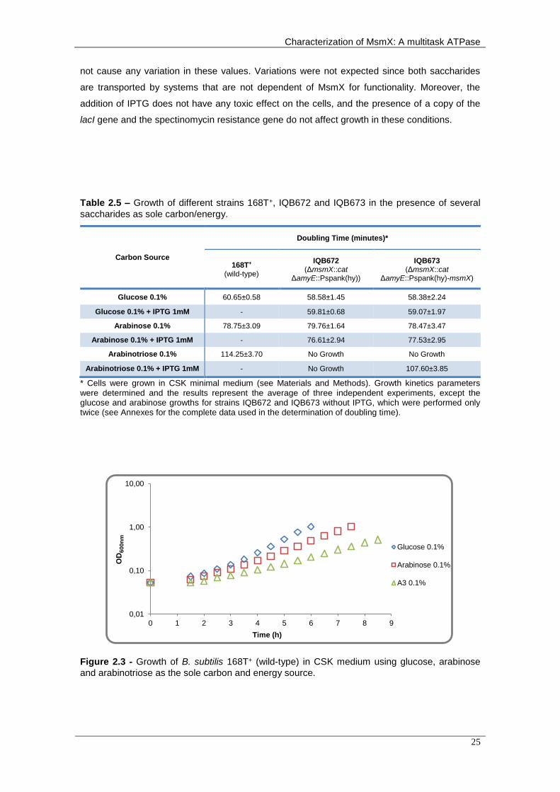
summarized in Table 2.5.
The doubling time of strains IQB672 and IQB673 in a medium supplemented with glucose
and arabinose are similar to those of the wild-type strain 168T+ and the presence of IPTG did
Characterization of MsmX: A multitask ATPase
25
not cause any variation in these values. Variations were not expected since both saccharides
are transported by systems that are not dependent of MsmX for functionality. Moreover, the
addition of IPTG does not have any toxic effect on the cells, and the presence of a copy of the
lacI gene and the spectinomycin resistance gene do not affect growth in these conditions.
Table 2.5 – Growth of different strains 168T+, IQB672 and IQB673 in the presence of several
saccharides as sole carbon/energy.
Carbon Source
Doubling Time (minutes)*
168T+
(wild-type)
IQB672 (ΔmsmX::cat
ΔamyE::Pspank(hy))
IQB673 (ΔmsmX::cat
ΔamyE::Pspank(hy)-msmX)
Glucose 0.1% 60.65±0.58 58.58±1.45 58.38±2.24
Glucose 0.1% + IPTG 1mM - 59.81±0.68 59.07±1.97
Arabinose 0.1% 78.75±3.09 79.76±1.64 78.47±3.47
Arabinose 0.1% + IPTG 1mM - 76.61±2.94 77.53±2.95
Arabinotriose 0.1% 114.25±3.70 No Growth No Growth
Arabinotriose 0.1% + IPTG 1mM - No Growth 107.60±3.85
* Cells were grown in CSK minimal medium (see Materials and Methods). Growth kinetics parameters were determined and the results represent the average of three independent experiments, except the glucose and arabinose growths for strains IQB672 and IQB673 without IPTG, which were performed only twice (see Annexes for the complete data used in the determination of doubling time).
Figure 2.3 - Growth of B. subtilis 168T+ (wild-type) in CSK medium using glucose, arabinose
and arabinotriose as the sole carbon and energy source.
0,01
0,10
1,00
10,00
0 1 2 3 4 5 6 7 8 9
OD
60
0n
m
Time (h)
Glucose 0.1%
Arabinose 0.1%
A3 0.1%

Chapter 2 – Development of an in vivo system to test the functionality of msmX alleles and MsmX homologs
26
The results obtained using only arabinotriose as a carbon/energy source display different
behaviours. As expected, the negative control, strain IQB672, was not able to use the
saccharide to support growth, even in the presence of IPTG (Table 2.5 and Figure 2.4). In this
strain, there is no MsmX present in the cells to energize the AraNPQ transport system. Strain
IQB673 was also not able to grow in the presence of arabinotriose, however when IPTG was
added to the medium, the wild-type phenotype was restored since the doubling time is similar to
that displayed by strain 168T+ (Table 2.5 and Figure 2.5).
These results show that IPTG induces expression of the msmX gene present at the amyE
locus, which allows the cell to transport arabinotriose from the medium to the cytoplasm through
the AraNPQ transporter.
Furthermore, when we compare the doubling time of strains 168T+ and IQB673 with
arabinotriose the results are very similar (considering the deviations; Table 2.5). Nonetheless, a
very slight decrease in IQB673 doubling time is observed and this variation could be due to a
small increase in the amount of MsmX present in the cell, due to the fact that the copy of msmX
present in trans is being transcribed from a different promoter. This observation however, is not
relevant for the purpose of this genetic system, which is to test the functionality of MsmX
homologs, or different alleles of msmX bearing mutations, because kinetics of growth will be
compared to strain IQB673 and not to the 168T+ strain.
Figure 2.4 - Growth of IQB672 (ΔmsmX::cat ΔamyE::pSpank(hy)) in CSK medium using
glucose, arabinose and arabinotriose as the sole carbon and energy source.
0,01
0,10
1,00
10,00
0 1 2 3 4 5 6 7 8 9
OD
60
0n
m
Time (h)
Glucose 0.1% +IPTG
Arabinose 0.1%+ IPTG
A3 0.1%
A3 0.1% + IPTG

Characterization of MsmX: A multitask ATPase
27
Figure 2.5 - Growth of IQB673 (ΔmsmX::cat ΔamyE::pSpank(hy)-msmX) in CSK medium using
glucose, arabinose and arabinotriose as the sole carbon and energy source.
In sum, the results obtained indicate that the expression of msmX in trans using the
system developed does not affect the bacteria phenotype in the presence of arabinotriose i.e.
we may conclude that using this system the intracellular concentration of MsmX is sufficient to
energize the AraNPQ transporter as observed in the wild-type strain. Cells expressing msmX in
trans, under the control of the inducible Pspank(hy) promoter, and the wild-type cells expressing
the msmX in the original locus, under the control of its own promoter, display a similar
phenotype under the tested conditions. Thus, the genetic system constructed is suitable for
studying the effect of mutations in the msmX allele and to test the functionality of MsmX
homologs in B. subtilis.
Functional Analysis of MsmX Homologs
As described above, we identified MsmX homologs in several pathogenic species and
select proteins from B. thuringiensis, S. aureus and S. pneumoniae. Using the system
developed, we tested if the homologs chosen were able to complement MsmX absence in B.
subtilis.
The different genes were placed under the control of the inducible Pspank(hy) promoter at
the amyE locus and resulted in the construction of strains IQB674 (msmK gene from S.
pneumoniae), IQB677 (HD73_4301 from B. thuringiensis) and IQB678 (ugpC from S. aureus) is
depicted in Figure 2.2. Growth kinetics parameters of the three strains in minimal medium using
arabinotriose (MsmX dependent uptake), glucose and arabinose (both MsmX independent
0,01
0,10
1,00
10,00
0 1 2 3 4 5 6 7 8 9
OD
60
0n
m
Time (h)
Glucose 0.1% +IPTG
Arabinose 0.1%+ IPTG
A3 0.1%
A3 0.1% + IPTG

Chapter 2 – Development of an in vivo system to test the functionality of msmX alleles and MsmX homologs
28
uptake) as the sole carbon and energy source, were determined and the results are
summarized in Table 2.6.
Table 2.6 - Growth of different B. subtilis strains in the presence of several saccharides as sole
carbon/energy.
Carbon Source
Doubling Time (minutes)*
IQB673 (ΔmsmX::cat
ΔamyE::Pspank(hy)-msmX)
IQB674 (ΔmsmX::cat
ΔamyE::Pspank(hy)-msmK
[S. pneumonia])
IQB677 (ΔmsmX::cat
ΔamyE::Pspank(hy)-HD73_4301
[B. thuringiensis])
IQB678 (ΔmsmX::cat
ΔamyE::Pspank(hy)-ugpC
[S. aureus])
Glucose 0.1% 58.38±2.24 59.17±1.81 59.98±1.18 59.38±0.50
Glucose 0.1% + IPTG 1mM
59.07±1.97 59.88±2.52 60.62±0.71 61.49±2.13
Arabinose 0.1% 78.47±3.47 80.97±3.81 79.01±0.59 79.98±1.65
Arabinose 0.1% + IPTG 1mM
77.53±2.95 81.40±2.20 77.22±1.98 81.40±2.20
Arabinotriose 0.1% No Growth 260.87±11.18 No Growth No Growth
Arabinotriose 0.1% + IPTG 1mM
107.60±3.85 226.05±9.55 141.18±3.76 205.47±8.28
* Cells were grown in CSK minimal medium (see Materials and Methods). Growth kinetics parameters were determined and the results represent the average of three independent experiments, except the glucose and arabinose growths without IPTG, which were performed only twice (see Annexes for the complete data used in the determination of doubling time). IQB673 results are the same presented in table 5 and are shown here to facilitate comparison.
The doubling time of strains IQB674, IQB677, and IQB678, in a medium supplemented
with glucose and arabinose are similar to those of the wild-type strain 168T+ and the presence
of IPTG did not cause any variation in these values, as previously observed with strain IQB673
(Table 2.5 and Table 2.6).
Regarding the growth with arabinotriose in inducible conditions, plus IPTG, all the three
strains were able to growth in the presence of arabinotriose as the sole carbon and energy
source. This phenotype is not observed in the absence of IPTG in strains IQB677, and IQB678
(Table 2.6 and Figures 2.7 and 2.8), unexpectedly strain IQB674 was able to growth in the
presence of arabinotriose even in the absence of IPTG (Table 2.6 and Figure 2.6). A closer look
to the sequence of msmK amplified from S. pneumonia and cloned in pDR111 revealed that in
addition to the RBS we also cloned a putative msmK promoter (PmsmK). Thus, transcription of
msmK is initiated at this promoter and is not subjected to regulation by LacI. Interestingly, the
growth of strain IQB674 in arabinotriose in the absence of IPTG is slower than that observed in
the presence of IPTG (a difference of about 34 min), suggesting that in inducing conditions
transcription of msmK is also driven by the Pspank(hy) promoter.

Characterization of MsmX: A multitask ATPase
29
Figure 2.6 - Growth of IQB674 (ΔmsmX::cat ΔamyE::pSpank(hy)-msmK) in CSK medium using
glucose, arabinose and arabinotriose as the sole carbon and energy source.
Figure 2.7 - Growth of IQB677 (ΔmsmX::cat ΔamyE::pSpank(hy)-HD73_4301) in CSK medium
using glucose, arabinose and arabinotriose as the sole carbon and energy source.
Figure 2.8 - Growth of IQB678 (ΔmsmX::cat ΔamyE::Pspank(hy)-ugpC) in CSK medium using
glucose, arabinose and arabinotriose as the sole carbon and energy source.
0,01
0,10
1,00
10,00
0 1 2 3 4 5 6 7 8 9
OD
60
0n
m
Time (h)
Glucose 0.1% +IPTG
Arabinose 0.1%+ IPTG
A3 0.1%
A3 0.1% + IPTG
0,01
0,10
1,00
10,00
0 1 2 3 4 5 6 7 8 9
OD
60
0n
m
Time (h)
Glucose 0.1% +IPTG
Arabinose 0.1%+ IPTG
A3 0.1%
A3 0.1% + IPTG
0,01
0,10
1,00
10,00
0 1 2 3 4 5 6 7 8 9
OD
60
0n
m
Time (h)
Glucose 0.1% +IPTG
Arabinose 0.1%+ IPTG
A3 0.1%
A3 0.1% + IPTG

Chapter 2 – Development of an in vivo system to test the functionality of msmX alleles and MsmX homologs
30
In conclusion these results show that msmK from S. pneumoniae, HD73_4301 from B.
thuringiensis and ugpC from S. aureus are able to play the role of msmX in energizing the B.
subtilis AraNPQ transporter, and suggest that MsmK, HD73_4301, and UgpC are able to
interact with the TMDs from the AraNPQ transporter in the absence of MsmX.
Nevertheless, the complementation of MsmX deficiency observed is only partial, the
doubling time, determined in inducing conditions in the presence of arabinotriose in strains
IQB674 (msmK from S. pneumoniae), IQB677 (HD73_4301 from B. thuringiensis) and IQB678
(ugpC from S. aureus) increased 2.1-fold, 1.3 fold, and 1.9-fold, respectively, when compared
with strain IQB673 (msmX from B. subtilis). Comparing these results it seems that the protein
HD73_4301 from B. thuringiensis is the one who reaches a value closer to full complementation
of MsmX, while between the other two proteins, UgpC from S. aureus achieves a higher
complementation degree than MsmK from S. pneumoniae. If we compare the complementation
degree with protein identity (or homology), or phylogenetic relationship between the species
used in this work, we can observe some correlation between them. The protein displaying
higher identity to MsmX is HD73_4301 (74%), which also came from the species
phylogenetically more close to B. subtilis (same genus), achieved the best complementation
degree, while the protein with less identity (64%) and also from the further species, in
phylogenetic terms, attained the worst complementation degree. So it is tempting to speculate
that a higher protein identity corresponds to a greater ability to play the MsmX role.
Interestingly, the region of the proteins predicted to interact with the TMDs of AraPQ is
highly conserved (see Appendix 6.11). However, differences in one or two amino acids near the
Q-loop, may be sufficient to alter the interaction between the ATPase and one of the TMDs.
Changes in the electrochemical properties in those positions could affect the interactions
protein-protein directly or indirectly through interaction with amino acids important for the
connection with the TMD. These changes could not prevent connection between the two
proteins, but be enough to slow down the connection and interfering with the uptake rate of
arabinotriose and, consequently, increasing the doubling time of the strains when using this
saccharide as carbon/energy source.
Another aspect that could contribute for these results is possible differences in post-
transcriptional regulation that result in variations in the total NBD protein amount produced in
each strain. Since each gene was cloned together with its own RBS, and belong to different
species, we calculated the translation initiation rates for the mRNAs transcribed from each gene
to estimate the impact of the different RBS in translation initiation. The results obtained are
summarized in Table 2.7.

Characterization of MsmX: A multitask ATPase
31
Table 2.7 – RBS Calculator algorithm results for each RBS. The translation initiation rate is a
relative value and is represented in arbitrary units (au). ∆Gtotal – Total energy necessary for translation
initiation.
Gene Translation Initiation Rate
(au) ∆Gtotal (kcal/mol)
msmX 42339.64 -6.29
HD73_4301 7519.89 -2.45
ugpC 229874.35 -10.05
msmK 18759.16 -4.48
These results clearly show us that exists differences in the post-transcriptional regulation
processes, but they do not correlate with the complementation results. In addition to differences
in the RBS strength, there are other processes that can affect post-transcriptional regulation,
such as the mRNA stability.
Since this system was created not only to study these proteins, but also other possible
multitask ATPases from other bacteria or even from B. subtilis we decided to improve our
design in order to eliminate those variables, which could influence the results obtained in the
future experiments.
Fine-tuning the Genetic System for Functional Analysis
In order to eliminate transcriptional and the post-transcriptional regulation variables, we
improved the system’s design. For this purpose we modified the N-terminal and C-terminal of
MsmX in order to allow the sub-cloning of only the Open Reading Frame (ORF) of the gene to
be tested.
The integrative vector was engineered to harbour the RBS and the terminator of B. subtilis
msmX. The msmX coding sequence was mutated in order to introduce two new unique
restriction sites at the 5’-end and 3’-end of the gene, one right after the RBS (NheI) and the
other before the stop codon (BglII). The first one was introduced between the second (Ala) and
third amino acid (Glu), and led to the transition of the Glu residue to a Ser. The second
restriction site was introduced between amino acids 363 (Arg) and 364 (Ile), substituting Ile by a
Ser residue (Figure 2.9). The resulting plasmid pAM12, a pDR111 derivative, containing the
modified version of msmX, in future studies will be digested with NheI and BglII, which enable
the removal of the msmX coding region, leaving behind the RBS and terminator, then the ORF
of the gene object of study will be cloned between those restriction sites.
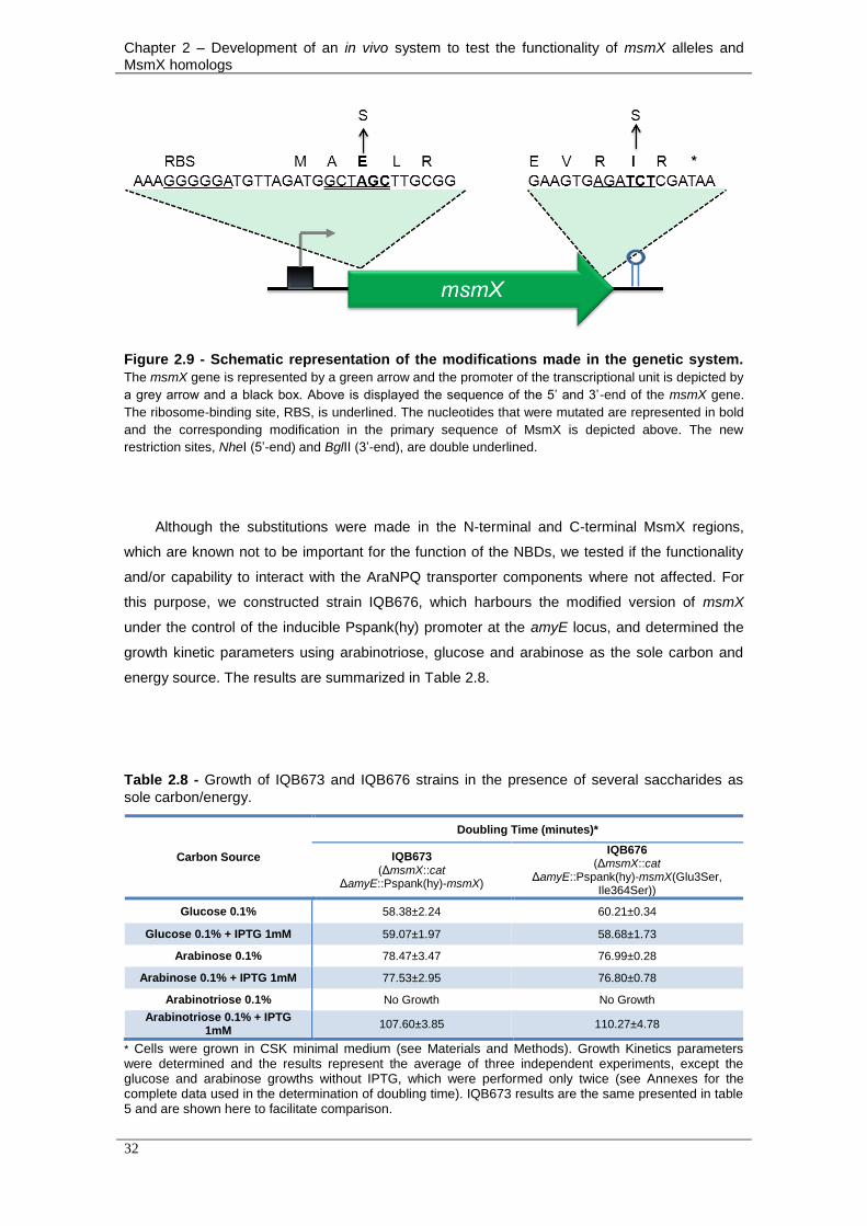
Chapter 2 – Development of an in vivo system to test the functionality of msmX alleles and MsmX homologs
32
Figure 2.9 - Schematic representation of the modifications made in the genetic system.
The msmX gene is represented by a green arrow and the promoter of the transcriptional unit is depicted by
a grey arrow and a black box. Above is displayed the sequence of the 5’ and 3’-end of the msmX gene.
The ribosome-binding site, RBS, is underlined. The nucleotides that were mutated are represented in bold
and the corresponding modification in the primary sequence of MsmX is depicted above. The new
restriction sites, NheI (5’-end) and BglII (3’-end), are double underlined.
Although the substitutions were made in the N-terminal and C-terminal MsmX regions,
which are known not to be important for the function of the NBDs, we tested if the functionality
and/or capability to interact with the AraNPQ transporter components where not affected. For
this purpose, we constructed strain IQB676, which harbours the modified version of msmX
under the control of the inducible Pspank(hy) promoter at the amyE locus, and determined the
growth kinetic parameters using arabinotriose, glucose and arabinose as the sole carbon and
energy source. The results are summarized in Table 2.8.
Table 2.8 - Growth of IQB673 and IQB676 strains in the presence of several saccharides as
sole carbon/energy.
Carbon Source
Doubling Time (minutes)*
IQB673 (ΔmsmX::cat
ΔamyE::Pspank(hy)-msmX)
IQB676 (ΔmsmX::cat
ΔamyE::Pspank(hy)-msmX(Glu3Ser, Ile364Ser))
Glucose 0.1% 58.38±2.24 60.21±0.34
Glucose 0.1% + IPTG 1mM 59.07±1.97 58.68±1.73
Arabinose 0.1% 78.47±3.47 76.99±0.28
Arabinose 0.1% + IPTG 1mM 77.53±2.95 76.80±0.78
Arabinotriose 0.1% No Growth No Growth
Arabinotriose 0.1% + IPTG 1mM
107.60±3.85 110.27±4.78
* Cells were grown in CSK minimal medium (see Materials and Methods). Growth Kinetics parameters were determined and the results represent the average of three independent experiments, except the glucose and arabinose growths without IPTG, which were performed only twice (see Annexes for the complete data used in the determination of doubling time). IQB673 results are the same presented in table 5 and are shown here to facilitate comparison.

Characterization of MsmX: A multitask ATPase
33
Figure 2.10 - Growth of IQB676 (ΔmsmX::cat ΔamyE::Pspank(hy)-msmX(Glu3Ser, Ile364Ser))
in CSK medium using glucose, arabinose and arabinotriose as the sole carbon and energy
source.
The doubling time obtained with this strain when in a medium supplemented with glucose
and arabinose is similar to IQB673 and the presence of IPTG also did not cause any variation in
these values. In the assays with arabinotriose, but without IPTG added to the medium, there
was no growth. When IPTG was added the cells were able to grow, with a doubling time similar
to IQB673. These results indicated that the amino acids substitutions introduced in MsmX do
not affect neither its functionality nor the interaction with the TMDs, and this improved system
can be used in future complementation tests.
0,01
0,10
1,00
10,00
0 1 2 3 4 5 6 7 8 9
OD
60
0n
m
Time (h)
Glucose 0.1% +IPTG
Arabinose 0.1%+ IPTG
A3 0.1%
A3 0.1% + IPTG

Chapter 2 – Development of an in vivo system to test the functionality of msmX alleles and MsmX homologs
34
Characterization of MsmX: A multitask ATPase
35
Chapter 3
Biochemical Characterization of MsmX

Chapter 3 – Biochemical Characterization of MsmX
36
Characterization of MsmX: A multitask ATPase
37
3. Biochemical Characterization of MsmX
3.1 Introduction
The first ATPase with a resolved crystal structure was HisP, from Salmonella typhimurium,
belonging to the well-characterized histidine ABC transporter HisQMP2 (Hung et al., 1998).
Since then the crystal structure of several ATPases was solved, such as the MJ0796 and
MJ1267 proteins from Methanocaldococcus jannaschii (Karpowich et al., 2001; Smith et al.,
2002). Other protein structures were determined with the NBD attached to the TMDs from the
correspondent transporter like MalK from E. coli, ModC from Archaeoglobus fulgidus and
Methanosarcina acetivorans (Chen et al., 2003; Hollenstein et al., 2007; Gerber et al., 2008).
More recently, the structure from ATPases MetNI from E. coli, involved in methionine uptake,
and LIC12079 from Leptospira spp., involved in manganese utilization, were also determined
(Rees et al., 2009; Benaroudj et al., 2013).
Beside crystallography studies, dynamic monomer/dimer equilibrium and ATPase activity
studies were performed to unravel other important structural information (Horn et al., 2003;
Verdon et al., 2003; Zoghbi et al., 2012; Benaroudj et al., 2013). Some of these studies
revealed residues important for ATP hydrolysis like the Glu residue from Walker B motif, the
second Gly residue from the Signature motif and the conserved Lys from the Walker A motif,
while the Ser residue from the Signature motif was not essential for the enzymatic activity
(Verdon et al., 2003; Benaroudj et al., 2013).
In spite of all these studies, the only well-characterized ATPase of ABC transporters
involved in the uptake of carbohydrates is MalK from E. coli. To better comprehend the energy-
generating component sharing phenomenon among distinct sugar ABC importers, structural
and biochemical characterization of multitask ATPases like MsmX must be performed. MsmX is
highly similar (>40% identity) to MalK from E. coli and MsmK from S. mutans, ATPases of
transport systems for maltose and multiple sugars, respectively (Ferreira and de Sá-Nogueira,
2010).
The aim of the work presented in this chapter was to optimize the production of MsmX in E.
coli and further purification in order to obtain the necessary amount of protein for functional and
structure characterization. Additionally functional analysis was performed by site-directed
mutagenesis. The Lys43 residue of MsmX, predicted to be essential for ATP hydrolysis, was
substituted by an Ala and the effects were observed both in vivo and in vitro.

Chapter 3 – Biochemical Characterization of MsmX
38
3.2 Materials and Methods
DNA manipulation and sequencing
Routine DNA manipulations were performed as described by Sambrook et al. (1989). All
restriction enzymes were purchased from Thermo Fisher Scientific Inc. and used according to
the manufacturers recommendations. PCR amplifications were carried out using Phusion®
High-Fidelity DNA Polymerase (Thermo Fisher Scientific Inc.). DNA from agarose gels and PCR
products were purified with the illustraTM GFXTM PCR DNA and Gel Band Purification kit (GE
Healthcare). All DNA ligations were performed using T4 DNA Ligase (Thermo Fisher Scientific
Inc.). Plasmids were purified using the QIAGEN® Plasmid Midi kit (Qiagen) or NZYMiniprep kit
(NZYTech, Lda.). DNA sequencing was performed with the ABI PRISM BigDye® Terminator
Cycle Sequencing Kit (Applied Biosystems). The sequencing reaction was purified by gel
filtration and resolved in an ABI 3730XL sequencer.
Site-Directed Mutagenesis
Vectors pMJ22 and pAM4 were used in independent experiments as template for site-
directed mutagenesis using the mutagenic oligonucleotides ARA757 and ARA758 (Table 3.1).
This pair of primers introduced the mutation of codon AAA (Lys at position 43) to GCA (Ala) in
the resulting plasmids pAM11 and pAM13, respectively. A polymerase chain reaction was
carried on using 1x Phusion® GC Buffer (Thermo Fisher Scientific Inc.), 0.2 μM primers, 200
μM dNTPs, 3% DMSO, 0.6 ng/μL of pMJ22 DNA (or 0.5 ng/μL of pAM4 DNA) and 0.02 U/μL of
Phusion® High-Fidelity DNA Polymerase in a total volume of 50 μL. The PCR product was
digested with 10 U of DpnI, at 37 ºC, overnight. The mutation was confirmed by DNA
sequencing.
Table 3.1 - List of oligonucleotides used in this work.
Oligonucleotides Sequencea
ARA757 TGCGGGGCATCAACGACGCTGCGAATGG
ARA758 CATTCGCAGCGTCGTTGATGCCCCGCAG
a Sequence orientation is 5’→3’. Mutated nucleotides are underlined

Characterization of MsmX: A multitask ATPase
39
Construction of strains
pAM13 was used to transform IQB495 strain (Table 3.2), according to the method
described by Anagnostopoulos and Spizizen, 1961, resulting in the IQB675 strain (Table 3.3).
The transformants were screened for amyE- phenotype. The amyE phenotype was tested in
plates of solid LB medium containing 1% (w/v) potato starch; after overnight incubation, plates
were flooded with a solution of 0.5% (w/v) I2–5.0% (w/v) KI for detection of starch hydrolysis.
Table 3.2 - List of plasmids used or constructed during the course of this work.
Plasmids Relevant construction Source or Reference
pT-GroE Vector for expression of GroESL chaperones under the control of T7
promoter (Yasukawa et al., 1995)
pMJ22 pET30(a)-based vector for the expression of msmX in E. coli Ferreira and de Sá-Nogueira,
unpublished data
pAM4 PDR111 derivate, with msmX under the control of Pspank(hy) This work*
pAM11 pMJ22 derivate with msmX sequence with mutated AAA
(Lys at position 43) to GCA (Ala) This work*
pAM13 pAM4 derivate with msmX sequence with mutated AAA
(Lys at position 43) to GCA (Ala) This work*
* See Appendices 6.2, 6.8 and 6.10.
Table 3.3 - List of B. subtilis strains used or constructed during the course of this work.
Strain Relevant genotype Sources or Referencea
IQB675 ΔmsmX::cat ΔamyE::pSpank(hy)-msmX(Lys43Ala) pAM13 → IQB495
a The arrows indicate transformation and point from donor DNA to recipient strain
Growth conditions
E. coli DH5α (Gibco-BRL) was used as host for routine DNA manipulations for plasmids
with pET-30(a) backbone and E. coli BL21 (DE3) pLysS (Studier et al., 1990) and E. coli BL21
(DE3) (Novagen) were used as hosts for the overproduction of recombinant MsmX and MsmX-
Lys43Ala. E. coli XL1Blue (Strategene) was used for the construction of pDR111 derivatives. All

Chapter 3 – Biochemical Characterization of MsmX
40
E. coli strains were grown in liquid Luria-Bertani (LB) medium (Miller, 1972) and on LB solidified
with 1.6% (w/v) agar, and ampicillin (100 μg/mL), Tetracycline (12 μg/mL), kanamycin (30
μg/mL) and/or chloramphenicol (25 μg/mL) were added as required.
B. subtilis was grown in liquid LB medium, LB medium solidified with 1.6% (w/v) agar or SP
medium (Martin et al., 1987) and chloramphenicol (5 μg/mL) and spectinomycin (50 μg/mL)
were added as required.
Growth kinetics parameters were determined in liquid minimal medium. B. subtilis strain
IQB675 was grown overnight (37 ºC, 150 rpm), from freshly streaked colonies, in C minimal
medium (Pascal et al., 1971) supplemented with L-tryptophan (100 μg/mL), potassium
glutamate (8 μg/mL) and potassium succinate (6 μg/mL) (CSK medium; Debarbouille et al.,
1990). The cell cultures were washed and resuspended, to an initial OD600nm of 0.05, in 1.5 mL
of CSK medium without potassium succinate and supplemented with different carbon and
energy sources (glucose, arabinose and arabinotriose) at a final concentration of 0.1% (w/v)
and with IPTG (expression inducer) at 1mM was added when appropriated. The cultures were
grown in sterile 50 mL Falcon tubes (Sarstedt), incubated at 37 ºC and 150 rpm in an OLS200
orbital/linear shaking bath (Grant Instruments) and the OD600nm periodically measured in an
Ultrospec 2100 pro UV/Visible Spectrophotometer (GE Healthcare Life Sciences)..
Production and purification of recombinant proteins
E. coli BL21 (DE3) cells harbouring both pMJ22 and pT-GroE were grown at 37 ºC and 150
rpm in 10 mL of LB medium with antibiotics (small-scale production). When the OD600nm of the
cultures reached 0.6, 5 mL of each culture were induced by the addition of IPTG to a final
concentration of 1 mM. Both the induced and non-induced cultures were grown for a further 18
hours at 25 ºC, and then the cells were harvested by centrifugation at 16000 g for 5 minutes and
resuspended in 100 μL of Lysis Buffer (20 mM sodium phosphate buffer, pH 7.4, 62.5 mM NaCl,
100 mM imidazole, glycerol 10% (v/v)). To disrupt the cells, incubation with lysozyme (1 mg/mL)
at 37 ºC for 10 minutes and three cycles of freezing in liquid nitrogen and thawing for 5 minutes
at 37 ºC were performed, followed by incubation with 0.02 U/mL of benzonase (Sigma) and
PMSF (10 mM) at 37 ºC for 10 minutes. After centrifugation at 9000 g and 4 ºC for 15 minutes,
the soluble fraction was recovered and the insoluble fraction was resuspended in 100 μL of
Lysis Buffer.
E. coli BL21 (DE3) cells harbouring pMJ22 were grown at 37 ºC and 150 rpm in 10 mL of
LB medium with antibiotics (small-scale production). When the OD600nm of the cultures reached
0.6, 5 mL of each culture was induced by the addition of IPTG to a final concentration of 0.1
mM. Both the induced and non-induced cultures were grown for a further 16 hours at 16 ºC, and
then cells were harvested and lysed as described above.

Characterization of MsmX: A multitask ATPase
41
To purify MsmX, a large-scale induction was performed. E. coli BL21 (DE3) harbouring
pMJ22 was incubated at 37 ºC and 150 rpm in 1 L of LB medium, using antibiotics. When the
OD600nm of the cultures reached 0.6, the culture was induced by the addition of IPTG to a final
concentration of 0.1 mM and grown for a further 16 hours at 16 ºC. The cells were harvested by
centrifugation at 12000 g. The pellets were resuspended in 10mL of Start Buffer (20 mM Tris-
HCl, pH 7.4, 50 mM NaCl and 10 mM imidazole) and 5 μl of benzonase at 2.5 U/mL were
added. The cells were lysed by sonication, and then centrifuged at 23500 g (30 minutes and 4
ºC). The supernatant was filtered using a 0.45 μm filter (Sarstedt) and loaded into a 1mL Hitrap
Chelating HP column (GE Healthcare). The bound proteins were eluted with a discontinuous
gradient of imidazole (40 mM, 100 mM, 200 mM, 300 mM and 500 mM).
To purify MsmX-Lys43Ala, a large-scale induction was performed with E. coli BL21 (DE3)
harbouring pAM11, using the same conditions as described to MsmX. However the purification
protocol was slightly changed instead of using the 20 mM Tris-HCl buffers, a 10 mM sodium
phosphate and 500 mM NaCl, were used.
Protein analysis
The analysis of the production, purification and molecular mass of the enzymes (monomers
and/or oligomers) was performed with SDS-PAGE (stained with Coomassie Blue), using Low
Molecular Weight Protein Marker (NZYTech). The protein quantification was determined using
Bio-Rad Protein Assay (Bio-Rad Laboratories, Inc).
ATPase activity assay
Purified MsmX and MsmX-Lys43Ala were assayed for ATP hydrolysis by monitoring the
production of inorganic phosphate at 37 °C in 20 mM Tris-HCl, pH 7.4, 50 mM NaCl and 10 mM
MgCl2. MsmX-Lys43Ala was stored in sodium phosphate buffer which was changed to the
assays buffer using PD-10 Desalting Columns (GE Healthcare Life Sciences). Activity assays
were performed with 5 μg of total protein and 0.1 mM ATP.
The assay mixture was incubated at 37ºC for 3h, with inorganic phosphate being measured
at 0 h, 1 h, 2 h and 3 h. The quantification of inorganic phosphate was determined using the
Malachite Green Phosphate Detection Kit (R&D Systems, Minneapolis, MN, USA) in
accordance with the manufacturer’s instructions. Background phosphate levels were monitored
in parallel using a control reaction without protein. OD620nm was measured in an Ultrospec 2100
pro UV/Visible Spectrophotometer (GE Healthcare Life Sciences).

Chapter 3 – Biochemical Characterization of MsmX
42
3.3. Results and Discussion
Sequence Analysis
MsmX, which can be transcribed monocistronicaly, encodes a 365 aa with a molecular
weight of ~41 kDa and an isoelectric point of 7.385 (http://subtiwiki.uni-goettingen.de; Michna et
al., 2014). MsmX is highly similar (>40% identity) to MalK from E. coli and a protein sequence
alignment of MsmX and MalK highlights the most conserved residues (Figure 2.1).
The characteristic motifs of ATP-binding proteins family are indicated in the alignment:
Walker A and B, Q-loop, Signature, D-loop and H-loop. Although these motifs are highly
conserved in both proteins, as observed in this type of ATPases, there are some minor
differences. In the Q-loop there is a semi-conservative substitution on position 84 (Asn to Ser),
while in Walker B occurs a conservative substitution in position 158 (Met to Leu), maintaining
the hydrophobic characteristic necessary for that position. Three amino acids in the H-loop were
substituted by amino acids with distinct characteristics but maintaining the conserved histidine.
The online tool PSIpred, which predicts secondary structures, was used to compare MsmX
and MalK (Jones, 1999; McGuffin et al., 2000; Bryson et al., 2005) and the results are shown in
Figure 2.2. The structures predicted with the program are highly similar from the N-terminus to
the H-loop motif. From this motif towards the C-terminal some differences are noticed in the
structure, with MsmX displaying three additional α-helices. The C-terminal domain of MalK is
known to be involved in regulatory processes through interactions with regulatory proteins
(Biemans-Oldehinkel et al., 2006).

Characterization of MsmX: A multitask ATPase
43
Figure 3.1 - MsmX and MalK sequence alignment. The amino acid sequences of MsmX and MalK
from E. coli were aligned using ClustalW2 (Thompson et al., 1994). Identical amino acids are marked
with a red asterisk, while substitutions with residues displaying the same properties are marked with ”:” and
semi-conservative substitutions are marked with a “.“. Gaps in the amino acid sequence are represented
by a ”–“. The conserved motifs are labeled in colors. The red arrow indicates the mutation performed in
MsmX (Lys43A) and described latter in this chapter.

Chapter 3 – Biochemical Characterization of MsmX
44
Figure 3.2 – MsmX secondary structure prevision by PSIpred. On the left is the predicted
secondary structure for MsmX and on the right for MalK.
Expression of msmX in E. coli
Previously, a recombinant version of MsmX (rMsmX) with a H6-tag at the C-terminal was
constructed and expressed in E. coli using the pET30 vector. The preliminary data showed that
rMsmX was functional in B. subtilis, however the soluble amount of protein obtained when
expressed in E. coli was low (Ferreira and Sá-Nogueira, unpublished results). When producing
this protein at 37 ºC with a 4 hour induction a considerable amount of protein was produced, but
almost all the protein was in the insoluble fraction.

Characterization of MsmX: A multitask ATPase
45
The low levels of soluble protein could be due to: i) the protein is not being able to achieve
its natural folding which is making the protein to be targeted to inclusion bodies, ii) although this
protein is cytoplasmic it can interact with membrane proteins from E. coli or iii) because this type
of proteins usually have a residual ATPase activity, the production of high amounts of protein
could be disrupting the energetic balances in the cell. In order to improve solubility we tested
different expression conditions. First, we co-expressed GroESL chaperones, encoded by the
pT-GroE plasmid, together with MsmX, and secondly we reduced the protein synthesis rate, by
lowering the growth temperature, and the IPTG (inducer) concentration (Figure 3.3).
The results indicate that both strategies improve solubility, however the reduction of protein
synthesis seems to have a higher effect on protein solubility. Although the total amount of
protein is lower in these conditions, the proportion of soluble protein is higher and should result
in greater protein concentration after purification.
Figure 3.3 – Overproduction of rMsmX in E. coli BL21 (DE3). On the left are shown the cell
extracts from co-expression of rMsmX and GroESL chaperones at 30 ºC for 4 h with 1 mM IPTG. On the
right are displayed the cell extracts from expression of rMsmX at 16 ºC for 16 h with 0.1 mM IPTG. P –
Insoluble Fraction; SN – Soluble Fraction; - IPTG – IPTG not added; +IPTG – IPTG added; LMW - Low
Molecular Weight Protein Marker (NZYTech). Extracts run in a 12.5% SDS-PAGE, stained with Coomassie
Blue. Soluble rMsmX is indicated with black triangle.
These results suggest that the protein insolubility may be derived from a defensive
response of the cell against the energetic balances disruption induced by over-expression of a
heterologous ATPase. Decreasing the rate of protein synthesis lead to smaller amounts of
ATPase and, consequently, decreased the toxic effect in the cell. However, we cannot discard

Chapter 3 – Biochemical Characterization of MsmX
46
possible interactions with membrane proteins because in the new conditions we still have more
insoluble protein than soluble, which could be explained by these interactions. In conclusion,
large-scale overproduction of rMsmX, for protein purification, was conducted by induction with
0.1 mM IPTG during 16 h at 16 ºC.
Purification of MsmX in E. coli
Usually, we perform protein purifications in a sodium phosphate buffer (PBS), however with
this protein we decided to use Tris-HCl buffer because inorganic phosphate will interfere with
the assay for ATPase activity determination. In the purification it was used a Ni-NTA column, as
described in the Materials and Methods section, which exclusively binds to the H6-Tag from
rMsmX.
As shown in Figure 3.4, it was possible to obtain rMsmX with a high degree of purity,
although some contaminant proteins are present. These persistent contaminants could be due
to interactions of those proteins with exposed hydrophobic regions of rMsmX. Elution of rMsmX
initiated at 200 mM of imidazole and persisted until 500 mM of imidazole. In the flow-through
and the wash fraction we observe the rMsmX band, which represents loss of protein, in addition
it is possible that some protein remained in the column since in the last elution fraction there
was still protein. These observations indicate that it is necessary to optimize the purification
protocol. Possible changes are the increase of Tris-HCl concentration and/or alter the number
of fractions eluted with each imidazole concentration.
Figure 3.4 – Purification of rMsmX. rMsmX was overexpressed, by largescale induction, in E. coli
BL21 (DE3) harbouring pMJ22 and resulting protein extracts were purified in Ni-NTA column. P and SN
represent the non-purified insoluble and soluble fractions, respectively; FT represents unbound proteins
from the soluble fraction (flow-through); W represents washed and weakly bound proteins; 40 mM (1 to 5),
100 mM (1 to 5), 200 mM (1 to 5), 300 mM (1 to 5) and 500 mM (1 to 5) represent fractions eluted with,
respectively, 40 mM, 100 mM, 200 mM, 300 mM and 500 mM imidazole. LMW represents the Low
Molecular Weight Protein Marker (NZYTech). Fractions run in a 12.5% SDS-PAGE, stained with
Coomassie Blue. rMsmX is indicated with black triangle.

Characterization of MsmX: A multitask ATPase
47
Analysis of the MsmX-Lys43Ala variant in vivo and in vitro
In the Walker A motif there is a highly conserved Lys at position 43 thought to be
responsible for ATP stabilization in the active site (see Chapter I). To evaluate its impact in the
ATPase activity of MsmX, we exchanged this residue to an Ala. The msmX allele present in
both pAM4 and pMJ22 was mutated to evaluate the effects of such mutation in vivo and in vitro.
To study the effects in vivo we applied the same methodology used to test the MsmX
homologs (Chapter II), and a strain with the msmX(Lys43Ala) mutant allele under the control of
the pSpank(hy), inserted in amyE locus, was constructed (IQB675). The kinetic parameters of
growth were calculated and are shown in Table 3.4.
The doubling time of IQB675 in the presence of glucose and arabinose are similar to the
strain IQB673, which contains the wild-type msmX in the amyE locus. However, when IPTG was
added to the medium in the presence of arabinotriose, while IQB673 is able to grow in these
conditions, no growth was observed in strain IQB675 (Figure 3.5).
Table 3.4 – Growth of IQB673 and IQB675 in the presence of several saccharides as sole
carbon/energy.
Carbon Source
Doubling Time (minutes)*
IQB673 (ΔmsmX::cat ΔamyE::pSpank(hy)-
msmX)
IQB675 (ΔmsmX::cat ΔamyE::Pspank(hy)-
msmX(Lys43Ala))
Glucose 0.1% 58.38±2.24 59.55±0.58
Glucose 0.1% + IPTG 1mM 59.07±1.97 59.81±0.68
Arabinose 0.1% 78.47±3.47 77.21±1.68
Arabinose 0.1% + IPTG 1mM 77.53±2.95 76.80±1.10
Arabinotriose 0.1% No Growth No Growth
Arabinotriose 0.1% + IPTG 1mM 107.60±3.85 No Growth
* Cells were grown in CSK minimal medium (see Materials and Methods). Growth Kinetics parameters were determined and the results represent the average of three independent experiments, except the glucose and arabinose growths without IPTG, which were performed only twice (see Annexes for the complete data used in the determination of doubling time). IQB673 results are the same presented in table 5 and are shown here to facilitate comparison.

Chapter 3 – Biochemical Characterization of MsmX
48
Figure 3.5 – Growth of IQB676 (ΔmsmX::cat ΔamyE::Pspank(hy)-msmX(Lys43Ala)) in CSK
medium using glucose, arabinose and arabinotriose as the sole carbon and energy source.
This result confirms the importance of Lys43 in the functionality of MsmX. Most probably
the ATPase is being produced by the cell and could interact with the transport system AraNPQ
in the membrane. However, due to the mutation in Lys43, the ATP hydrolysis necessary for the
transportation of arabinotriose is impaired. The MsmX Lys43 residue is the conserved Lys
present in the Walker A motif and corresponds to the amino acid Lys42 of MalK. This amino
acid is responsible for direct binding with ATP phosphate groups between which the hydrolysis
occurs (β- and γ-phosphate). Without this amino acid the phosphodiester bond may not be
placed in the right position relative to the His in the H-loop, making impossible to break it. This
way the transporter remains in the outward open position. Benaroudj et al. (2013) mutated the
same Lys of the ATPase LIC12079 from Leptospira interrogans into an Ala (Lys45Ala), which
resulted in the loss of ATPase activity.
For the characterization in vitro of the rMsmX(Lys43Ala) variant, large-scale
overexpression of rMsmX(Lys43Ala) was conducted in the same conditions used for the
production of rMsmX. Cell extracts analysis by SDS-PAGE showed that the mutation resulted in
an increase of protein solubility (Figure 3.6). This observation supports the possible existence of
energetic balances disruption caused by the overexpression of the rMsmX form discussed
above.
0,01
0,10
1,00
10,00
0 1 2 3 4 5 6 7 8
OD
60
0n
m
Time (h)
Glucose 0.1% +IPTG
Arabinose 0.1%+ IPTG
A3 0.1%
A3 0.1% + IPTG
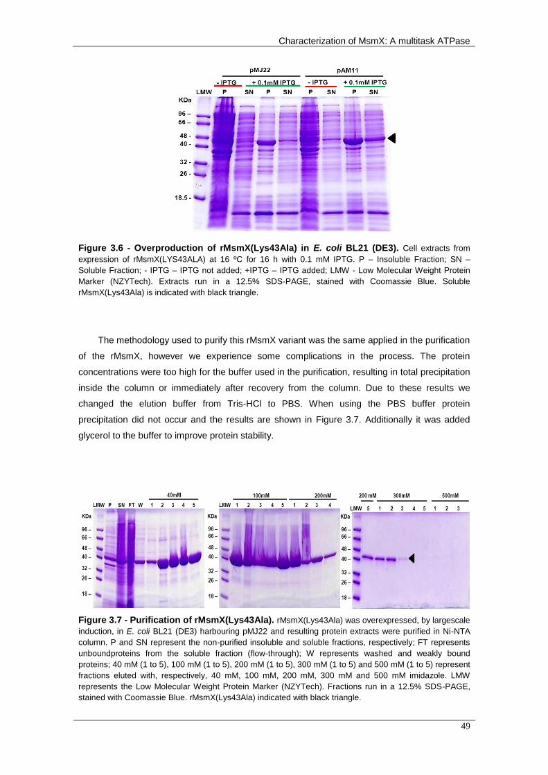
Characterization of MsmX: A multitask ATPase
49
Figure 3.6 - Overproduction of rMsmX(Lys43Ala) in E. coli BL21 (DE3). Cell extracts from
expression of rMsmX(LYS43ALA) at 16 ºC for 16 h with 0.1 mM IPTG. P – Insoluble Fraction; SN –
Soluble Fraction; - IPTG – IPTG not added; +IPTG – IPTG added; LMW - Low Molecular Weight Protein
Marker (NZYTech). Extracts run in a 12.5% SDS-PAGE, stained with Coomassie Blue. Soluble
rMsmX(Lys43Ala) is indicated with black triangle.
The methodology used to purify this rMsmX variant was the same applied in the purification
of the rMsmX, however we experience some complications in the process. The protein
concentrations were too high for the buffer used in the purification, resulting in total precipitation
inside the column or immediately after recovery from the column. Due to these results we
changed the elution buffer from Tris-HCl to PBS. When using the PBS buffer protein
precipitation did not occur and the results are shown in Figure 3.7. Additionally it was added
glycerol to the buffer to improve protein stability.
Figure 3.7 - Purification of rMsmX(Lys43Ala). rMsmX(Lys43Ala) was overexpressed, by largescale
induction, in E. coli BL21 (DE3) harbouring pMJ22 and resulting protein extracts were purified in Ni-NTA
column. P and SN represent the non-purified insoluble and soluble fractions, respectively; FT represents
unboundproteins from the soluble fraction (flow-through); W represents washed and weakly bound
proteins; 40 mM (1 to 5), 100 mM (1 to 5), 200 mM (1 to 5), 300 mM (1 to 5) and 500 mM (1 to 5) represent
fractions eluted with, respectively, 40 mM, 100 mM, 200 mM, 300 mM and 500 mM imidazole. LMW
represents the Low Molecular Weight Protein Marker (NZYTech). Fractions run in a 12.5% SDS-PAGE,
stained with Coomassie Blue. rMsmX(Lys43Ala) indicated with black triangle.

Chapter 3 – Biochemical Characterization of MsmX
50
Preliminary assays were performed to determine the ATPase activity of both rMsmX and
rMsmX(Lys43Ala). The assays were made with 5μg of protein in TrisHCl buffer, supplemented
with 10 mM MgCl2 and 0.1 mM ATP. rMsmX displayed ATPase capability (Figure 3.8), however
the ATP rate of ATP hydrolysis is 10 times lower than the activity calculated for other proteins of
the same family (Benaroudj et al., 2013) and optimization of assays conditions is required to
obtain maximum activity values. The results obtained with the rMsmX(Lys43Ala) showed a
drastic decrease in ATP hydrolysis, which confirms that the results obtained in vivo with this
protein resulted from ATPase activity decrease. Nevertheless, more independently assays in
these conditions must be conducted to confirm these results.
Figure 3.8 - ATP hydrolysis by rMsmX and rMsmX(Lys43Ala). ATP hydrolysis was determined
by measuring the production of inorganic phosphate, as described in Materials and Methods. 5 μg of protein were incubated with 0.1 mM ATP and 10 mM MgCl2. Only one assay was performed for each protein.
0
0,1
0,2
0,3
0,4
0,5
0,6
rMsmX rMsmX(K43A)
AT
P h
yd
roly
sis
(μm
ol/h.m
g)

Characterization of MsmX: A multitask ATPase
51
Chapter 4
Concluding Remarks and Future Perspectives

Chapter 4 – Concluding Remarks and Future Perspectives
52

Characterization of MsmX: A multitask ATPase
53
4. Concluding Remarks and Future Perspectives
The introduction of antibiotics in the treatment of infectious disease had a great impact in
medicine saving countless lives. However, the use of antibiotics creates a phenomenon called
antibiotic pressure which is the emergence of resistant pathogens. Although this side-effect was
predicted by Fleming, over the years there has been a negligent and abusive use of antibiotics
(French, 2010). This led to development of multidrug-resistance pathogens like Clostridium
difficile, Pseudomonas aeruginosas, S. aureus or S. pneumoniae. Also, in some cases, these
multidrug-resistance pathogens have developed more virulent traits and acquired enhanced
transmissibility (Davies and Davies, 2010).
We have reached a point where we must direct our attentions to the discovery of new
antimicrobials and therapeutic targets. The carbohydrate metabolism of bacteria plays an
important role in their survival and capacity for colonization and pathogenesis (Buckwalter and
King, 2012). One potentially attractive target to act against these pathogens is the multitask
ATPases recently discovered whose inactivation leads to inability to utilize multiple carbon
sources. This could have a great impact in the pathogen adaptability to the host niche or even
kill the bacterial by starvation. So a better understanding of these proteins functionality,
structure and regulation may have a huge impact in combat against multidrug-resistance
strains.
In the work we presented, it was shown that MsmX homologs in other pathogenic species
are able to substitute MsmX in the cell by restoring arabinooligosaccharides uptake in a B.
subtilis msmX-null mutant strain. The complementation degree varied depending on the protein
introduced in the strain genome. The B. thuringiensis protein showed to be more capable of fully
complement MsmX absence while S. aureus and S. pneumoniae proteins were only able to
partially complement it. Whether this resulted from differences in amino acid sequence that
affected the interaction with AraNPQ complex or rather from differences in the amount of protein
produced by the cell still remains unknown. Thus, here we demonstrated that B. subtilis and
MsmX could be used as a model to better comprehend this sharing of energy-generating
components between distinct ABC sugar importers.
Additionally, we proved that MsmX has ATPase activity and that the conserved Lys 43
present in the Walker A motif is essential for this activity both in vivo and in vitro. Moreover,
modifications in the N- and C-terminal primary sequence do not affect MsmX functionality or its
ability to interact with the AraNPQ transporter. In this work we also achieved important results
towards the improvement of MsmX production and purification processes, which is crucial for
crystallography studies and biochemical characterization of MsmX that will provide important
clues or even answers for the main questions arising from the discovery of multitask ATPases.

Chapter 4 – Concluding Remarks and Future Perspectives
54
The work presented here is just the tip of the iceberg regarding to the knowledge about
these multitask ATPases and the main forces that drove bacteria to develop these type of sugar
uptake mechanisms/regulation. First, we need to test the proteins used in this work with the new
system developed because it will allow us to conclude if the differences in the complementation
results observed are due to dissimilarities in the proteins or in the amount produced in the cell.
In addition, we must test the YurJ ATPase from B. subtilis, which displays 58% identity with
MsmX and is suggested to be multitask, as well as other B. subtilis ATPases thought to be
exclusive to one transporter. The conjunction of all these results may indicate which MsmX
residues are important for this multi-transporters interaction capability. Simultaneously, the
initiation of crystallography studies is of major importance to provide insights into this
mechanism. Finally, it would be interesting to ascertain if MsmX suffers any post-translational
regulation like MalK does (Biemans-Oldehinkel et al., 2006). The discovery of possible
regulatory mechanisms that control MsmX activity and their understanding may also reveal
MsmX regions for possible targeting.

Characterization of MsmX: A multitask ATPase
55
Chapter 5
References

Chapter 5 - References
56
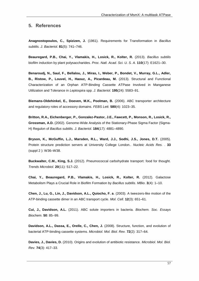
Characterization of MsmX: A multitask ATPase
57
5. References
Anagnostopoulos, C., Spizizen, J. (1961). Requirements for Transformation in Bacillus
subtilis. J. Bacteriol. 81(5): 741–746.
Beauregard, P.B., Chai, Y., Vlamakis, H., Losick, R., Kolter, R. (2013). Bacillus subtilis
biofilm induction by plant polysaccharides. Proc. Natl. Acad. Sci. U. S. A. 110(17): E1621–30.
Benaroudj, N., Saul, F., Bellalou, J., Miras, I., Weber, P., Bondet, V., Murray, G.L., Adler,
B., Ristow, P., Louvel, H., Haouz, A., Picardeau, M. (2013). Structural and Functional
Characterization of an Orphan ATP-Binding Cassette ATPase Involved in Manganese
Utilization and Tolerance in Leptospira spp. J. Bacteriol. 195(24): 5583–91.
Biemans-Oldehinkel, E., Doeven, M.K., Poolman, B. (2006). ABC transporter architecture
and regulatory roles of accessory domains. FEBS Lett. 580(4): 1023–35.
Britton, R.A., Eichenberger, P., Gonzalez-Pastor, J.E., Fawcett, P., Monson, R., Losick, R.,
Grossman, A.D. (2002). Genome-Wide Analysis of the Stationary-Phase Sigma Factor (Sigma-
H) Regulon of Bacillus subtilis. J. Bacteriol. 184(17): 4881–4890.
Bryson, K., McGuffin, L.J., Marsden, R.L., Ward, J.J., Sodhi, J.S., Jones, D.T. (2005).
Protein structure prediction servers at University College London.. Nucleic Acids Res. . 33
(suppl 2 ): W36–W38.
Buckwalter, C.M., King, S.J. (2012). Pneumococcal carbohydrate transport: food for thought.
Trends Microbiol. 20(11): 517–22.
Chai, Y., Beauregard, P.B., Vlamakis, H., Losick, R., Kolter, R. (2012). Galactose
Metabolism Plays a Crucial Role in Biofilm Formation by Bacillus subtilis. MBio. 3(4): 1–10.
Chen, J., Lu, G., Lin, J., Davidson, A.L., Quiocho, F. a. (2003). A tweezers-like motion of the
ATP-binding cassette dimer in an ABC transport cycle. Mol. Cell. 12(3): 651–61.
Cui, J., Davidson, A.L. (2011). ABC solute importers in bacteria. Biochem. Soc. Essays
Biochem. 50: 85–99.
Davidson, A.L., Dassa, E., Orelle, C., Chen, J. (2008). Structure, function, and evolution of
bacterial ATP-binding cassette systems. Microbiol. Mol. Biol. Rev. 72(2): 317–64.
Davies, J., Davies, D. (2010). Origins and evolution of antibiotic resistance. Microbiol. Mol. Biol.
Rev. 74(3): 417–33.

Chapter 5 - References
58
Debarbouille, M., Arnaud, M., Fouet, A., Klier, A., Rapoport, G. (1990). The sacT gene
regulating the sacPA operon in Bacillus subtilis shares strong homology with transcriptional
antiterminators. J. Bacteriol. 172(7): 3966–73.
Eitinger, T., Rodionov, D. a, Grote, M., Schneider, E. (2010). Canonical and ECF-type ATP-
binding cassette importers in prokaryotes: diversity in modular organization and cellular
functions. FEMS Microbiol. Rev. 35(1): 3–67.
Ferreira, M.J., de Sá-Nogueira, I. (2010). A multitask ATPase serving different ABC-type sugar
importers in Bacillus subtilis . J. Bacteriol. 192(20): 5312–8.
French, G.L. (2010). The continuing crisis in antibiotic resistance. Int. J. Antimicrob. Agents. 36
Suppl 3: S3–7.
Gerber, S., Comellas-Bigler, M., Goetz, B. a, Locher, K.P. (2008). Structural basis of trans-
inhibition in a molybdate/tungstate ABC transporter. Science. 321(5886): 246–50.
Higgins, C.F. (1992). ABC transporters: from microorganisms to man. Annu. Rev. Cell Biol. 8:
67–113.
Higgins, C.F. (2001). ABC transporters: physiology, structure and mechanism - an overview.
Res. Microbiol. 152(3-4): 205–10.
Higgins, C.F., Ames, G.F. (1981). Two periplasmic transport proteins which interact with a
common membrane receptor show extensive homology: complete nucleotide sequences. Proc.
Natl. Acad. Sci. U. S. A. 78(10): 6038–42.
Hollenstein, K., Frei, D.C., Locher, K.P. (2007). Structure of an ABC transporter in complex
with its binding protein. Nature. 446(7132): 213–6.
Horn, C., Bremer, E., Schmitt, L. (2003). Nucleotide Dependent Monomer/Dimer Equilibrium of
OpuAA, the Nucleotide-binding Protein of the Osmotically Regulated ABC Transporter OpuA
from Bacillus subtilis. J. Mol. Biol. 334(3): 403–419.
Hung, L.W., Wang, I.X., Nikaido, K., Liu, P.Q., Ames, G.F., Kim, S.H. (1998). Crystal
structure of the ATP-binding subunit of an ABC transporter. Nature. 396(6712): 703–7.
Hurtubise, Y., Shareck, F., Kluepfel, D., Morosoli, R. (1995). A cellulase/xylanase-negative
mutant of Streptomyces lividans 1326 defective in cellobiose and xylobiose uptake is mutated in
a gene encoding a protein homologous to ATP-binding proteins. Mol. Microbiol. 17(2): 367–377.
Jaehme, M., Slotboom, D.J. (2014). Diversity of membrane transport proteins for vitamins in
bacteria and archaea. Biochim. Biophys. Acta.

Characterization of MsmX: A multitask ATPase
59
Jones, D.T. (1999). Protein secondary structure prediction based on position-specific scoring
matrices. J. Mol. Biol. 292(2): 195–202.
Kampers, T., Schlösser, A., Schrempf, H. (1997). The Streptomyces ATP-binding component
MsiK assists in cellobiose and maltose transport. J. Bacteriol. 179(6): 2092–2095.
Karpowich, N., Martsinkevich, O., Millen, L., Yuan, Y.R., Dai, P.L., MacVey, K., Thomas,
P.J., Hunt, J.F. (2001). Crystal structures of the MJ1267 ATP binding cassette reveal an
induced-fit effect at the ATPase active site of an ABC transporter. Structure. 9(7): 571–86.
Kunst, F., Ogasawara, N., Moszer, I., Albertini, a M., Alloni, G., Azevedo, V., Bertero, M.G.,
Bessières, P., Bolotin, A., Borchert, S., Borriss, R., Boursier, L., Brans, A., Braun, M.,
Brignell, S.C., Bron, S., Brouillet, S., Bruschi, C. V, Caldwell, B., Capuano, V., Carter, N.M.,
Choi, S.K., Codani, J.J., Connerton, I.F., Danchin, A. (1997). The complete genome
sequence of the gram-positive bacterium Bacillus subtilis . Nature. 390(6657): 249–56.
Linke, C.M., Woodiga, S. a, Meyers, D.J., Buckwalter, C.M., Salhi, H.E., King, S.J. (2013).
The ABC transporter encoded at the pneumococcal fructooligosaccharide utilization locus
determines the ability to utilize long- and short-chain fructooligosaccharides. J. Bacteriol.
195(5): 1031–41.
Marion, C., Aten, A.E., Woodiga, S. a, King, S.J. (2011). Identification of an ATPase, MsmK,
which energizes multiple carbohydrate ABC transporters in Streptococcus pneumoniae . Infect.
Immun. 79(10): 4193–200.
Martin, I., Débarbouillé, M., Ferrari, E., Klier, A., Rapoport, G. (1987). Characterization of the
levanase gene of Bacillus subtilis which shows homology to yeast invertase. Mol. Gen. Genet.
MGG. 208: 177–184.
McGuffin, L.J., Bryson, K., Jones, D.T. (2000). The PSIPRED protein structure prediction
server. Bioinformatics. 16(4): 404–5.
Michna, R.H., Commichau, F.M., Tödter, D., Zschiedrich, C.P., Stülke, J. (2014). SubtiWiki-a
database for the model organism Bacillus subtilis that links pathway, interaction and expression
information. Nucleic Acids Res. 42(Database issue): D692–8.
Miller, J.H. (1972). Experiments in Molecular Genetics.. Cold Spring Harbor, NY: Cold Spring
Harbor Laboratory.
Oldham, M.L., Khare, D., Quiocho, F. a, Davidson, A.L., Chen, J. (2007). Crystal structure of
a catalytic intermediate of the maltose transporter. Nature. 450(7169): 515–21.

Chapter 5 - References
60
Pascal, M., Kunst, F., Lepesant, J. A. and Dedonder, R. (1971). Characterization of two
sucrase activities in Bacillus subtilis Marburg. Biochimie. 53: 1059–1066.
Quentin, Y., Fichant, G., Denizot, F. (1999). Inventory, assembly and analysis of Bacillus
subtilis ABC transport systems. J. Mol. Biol. 287(3): 467–84.
Quisel, J.D., Burkholder, W.F., Grossman, A.D. (2001). In Vivo Effects of Sporulation Kinases
on Mutant Spo0A Proteins in Bacillus subtilis . J. Bacteriol. 183(22): 6573–6578.
Rees, D.C., Johnson, E., Lewinson, O. (2009). ABC transporters: the power to change. Nat.
Rev. Mol. Cell Biol. 10(3): 218–27.
Russell, R.R., Aduse-Opoku, J., Sutcliffe, I.C., Tao, L., Ferretti, J.J. (1992). A binding
protein-dependent transport system in Streptococcus mutans responsible for multiple sugar
metabolism. J. Biol. Chem. 267(7): 4631–7.
Saier, M.H., Goldman, S.R., Maile, R.R., Moreno, M.S., Weyler, W., Yang, N., Paulsen, I.T.
(2002). Transport capabilities encoded within the Bacillus subtilis genome. J. Mol. Microbiol.
Biotechnol. 4(1): 37–67.
Saito, A., Fujii, T., Shinya, T., Shibuya, N., Ando, A., Miyashita, K. (2008). The msiK gene,
encoding the ATP-hydrolysing component of N,N’-diacetylchitobiose ABC transporters, is
essential for induction of chitinase production in Streptomyces coelicolor A3(2). Microbiology.
154(Pt 11): 3358–65.
Salis, H.M. (2011). The ribosome binding site calculator. Methods Enzymol. 498: 19–42.
Salis, H.M., Mirsky, E. a, Voigt, C. a. (2009). Automated design of synthetic ribosome binding
sites to control protein expression. Nat. Biotechnol. 27(10): 946–50.
Sambrook, J., Fritsch, E. F. and Maniatis, T. (1989). Molecular cloning: A laboratory manual.,
2nd ed., Cell. Cold Spring Harbor: Cold Spring Harbor Laboratory, NY.
Schlösser, a. (2000). MsiK-dependent trehalose uptake in Streptomyces reticuli . FEMS
Microbiol. Lett. 184(2): 187–92.
Schneider, E., Hunke, S. (1998). ATP-binding-cassette (ABC) transport systems: functional
and structural aspects of the ATP-hydrolyzing subunits/domains. FEMS Microbiol. Rev. 22(1):
1–20.
Schönert, S., Seitz, S., Krafft, H., Feuerbaum, E.-A., Andernach, I., Witz, G., Dahl, M.K.
(2006). Maltose and maltodextrin utilization by Bacillus subtilis . J. Bacteriol. 188(11): 3911–22.

Characterization of MsmX: A multitask ATPase
61
Seyffer, F., Tampé, R. (2014). ABC transporters in adaptive immunity. Biochim. Biophys. Acta.
Silva, Z., Sampaio, M.-M., Henne, A., Böhm, A., Gutzat, R., Boos, W., da Costa, M.S.,
Santos, H. (2005). The high-affinity maltose/trehalose ABC transporter in the extremely
thermophilic bacterium Thermus thermophilus HB27 also recognizes sucrose and palatinose. J.
Bacteriol. 187(4): 1210–8.
Smith, P.C., Karpowich, N., Millen, L., Moody, J.E., Rosen, J., Thomas, P.J., Hunt, J.F.
(2002). ATP binding to the motor domain from an ABC transporter drives formation of a
nucleotide sandwich dimer. Mol. Cell. 10(1): 139–49.
Studier, F. W., Rosenberg, A. H., Dunn, J. J. and Dubendorff, J.W. (1990). Use of T7 RNA
polymerase to direct expression of cloned genes. Meth. Enzym. 185: 60–89.
Ter Beek, J., Guskov, A., Slotboom, D.J. (2014). Structural diversity of ABC transporters. J.
Gen. Physiol. 143(4): 419–35.
Thompson, J.D., Higgins, D.G., Gibson, T.J. (1994). CLUSTAL W: improving the sensitivity of
progressive multiple sequence alignment through sequence weighting, position-specific gap
penalties and weight matrix choice.. Nucleic Acids Res. 22(22): 4673–4680.
Verdon, G., Albers, S.-V., van Oosterwijk, N., Dijkstra, B.W., Driessen, A.J.M., Thunnissen,
A.-M.W.H. (2003). Formation of the Productive ATP-Mg2+-bound Dimer of GlcV, an ABC-
ATPase from Sulfolobus solfataricus. J. Mol. Biol. 334(2): 255–267.
Webb, A.J., Homer, K. a, Hosie, A.H.F. (2008). Two closely related ABC transporters in
Streptococcus mutans are involved in disaccharide and/or oligosaccharide uptake. J. Bacteriol.
190(1): 168–78.
Yasukawa, T., Kanei-Ishii, C., Maekawa, T., Fujimoto, J., Yamamoto, T., Ishii, S. (1995).
Increase of Solubility of Foreign Proteins in Escherichia coli by Coproduction of the Bacterial
Thioredoxin.. J. Biol. Chem. 270(43): 25328–25331.
Yoshida, K., Ishio, I., Nagakawa, E., Yamamoto, Y., Yamamoto, M., Fujita, Y. (2000).
Systematic study of gene expression and transcription organization in the gntZ-ywaA region of
the Bacillus subtilis genome. Microbiology. 146 ( Pt 3: 573–9.
Yoshida, K., Shindo, K., Sano, H., Seki, S., Fujimura, M., Yanai, N., Miwa, Y., Fujita, Y.
(1996). Sequencing of a 65 kb region of the Bacillus subtilis genome containing the lic and cel
loci, and creation of a 177 kb contig covering the gnt-sacXY region.. Microbiology. 142(11):
3113–3123.

Chapter 5 - References
62
Zoghbi, M.E., Fuson, K.L., Sutton, R.B., Altenberg, G. a. (2012). Kinetics of the
association/dissociation cycle of an ATP-binding cassette nucleotide-binding domain. J. Biol.
Chem. 287(6): 4157–64.

Characterization of MsmX: A multitask ATPase
63
Chapter 6
Appendices

Chapter 6 - Appendices
64

Characterization of MsmX: A multitask ATPase
65
6. Appendices
Appendix 6.1 - Map of pDR111 (gift from David Rudner, Harvard University.
Appendix 6.2 - Map of pAM4. Insertion of a 1224bp fragment containing msmX gene (amplified with
primers ARA741 and ARA742 and digested with SalI and SphI) between the SalI and SphI sites of pDR111.

Chapter 6 - Appendices
66
Appendix 6.3 - Map of pAM5. Insertion of a 1272bp fragment containing HD73_4301 gene, from B.
thuringiensis serovar kurstaki str. HD73 (amplified with the primers ARA744 and ARA745 and digested with SalI and SphI) between the SalI and SphI sites of pDR111.
Appendix 6.4 - Map of pAM6. Insertion of a 1215bp fragment containing ugpC, from S. aureus subsp.
aureus ST398, (amplified with the primers ARA746 and ARA747 and digested with SalI and SphI) between
the SalI and SphI sites of pDR111.

Characterization of MsmX: A multitask ATPase
67
Appendix 6.5 - Map of pAM7. Insertion of a 1295bp fragment containing msmK, from S. pneumoniae
TIGR4, (amplified with the primers ARA748 and ARA749 and digested with SalI and SphI) between the
SalI and SphI sites of pDR111.
Appendix 6.6 - Map of pAM9. Site-directed mutagenesis of pMJ1 using the mutagenic primers set
ARA769 and ARA770. Mutagenesis generated a unique BglII restriction site in the 4th nucleotide,
downstream from the start codon.

Chapter 6 - Appendices
68
Appendix 6.7 - Map of pAM10. Site-directed mutagenesis of pAM9 using the mutagenic primers set
ARA771 and ARA772. Mutagenesis generated a unique NheI restriction site in the 11th nucleotide,
upstream from the stop codon.
Appendix 6.8 - Map of pAM11. Site-directed mutagenesis of pMJ22 using the mutagenic primers set
ARA757 and ARA758. This pair of primers allowed the mutation of codon AAA (lysine at position 45) to
GCA (alanine).

Characterization of MsmX: A multitask ATPase
69
Appendix 6.9 - Map of pAM12. Insertion of a 1224bp fragment containing msmX gene from pAM10
(amplified with primers ARA741 and ARA742 and digested with SalI and SphI) between the SalI and SphI
sites of pDR111.
Appendix 6.10 - Map of pAM13. Site-directed mutagenesis of pAM4 using the mutagenic primers set
ARA757 and ARA758. This pair of primers allowed the mutation of codon AAA (lysine at position 43) to
GCA (alanine).
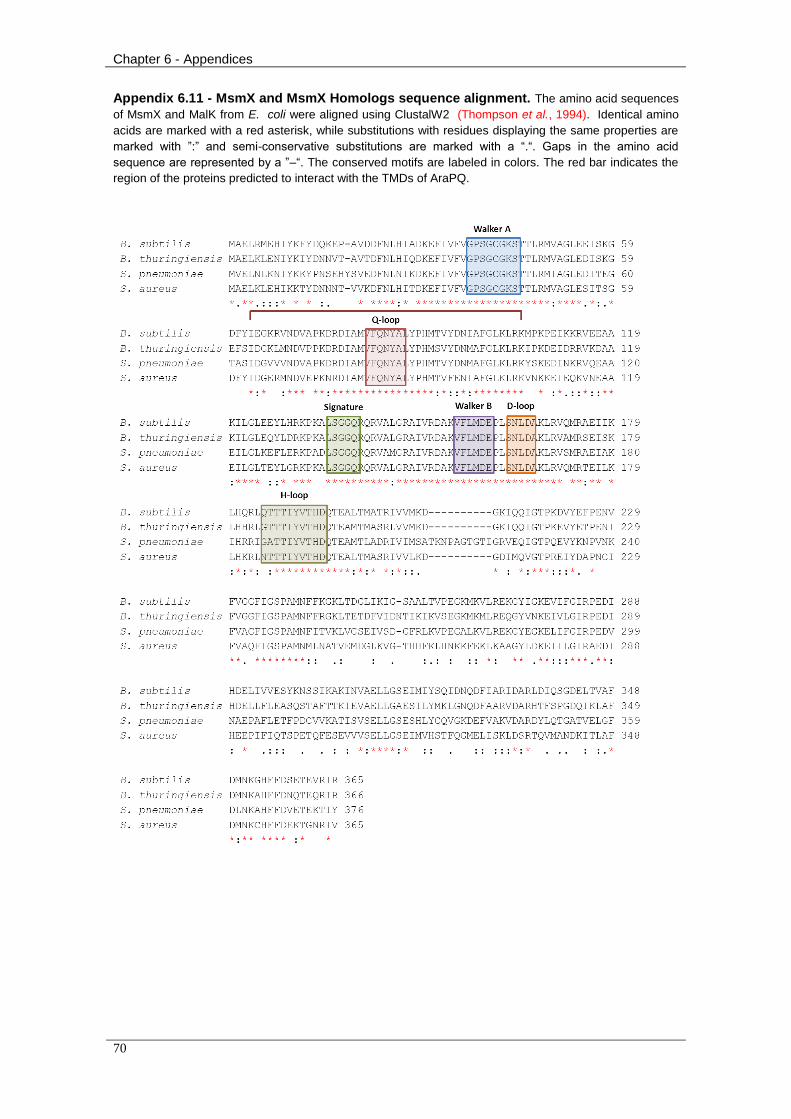
Chapter 6 - Appendices
70
Appendix 6.11 - MsmX and MsmX Homologs sequence alignment. The amino acid sequences
of MsmX and MalK from E. coli were aligned using ClustalW2 (Thompson et al., 1994). Identical amino
acids are marked with a red asterisk, while substitutions with residues displaying the same properties are
marked with ”:” and semi-conservative substitutions are marked with a “.“. Gaps in the amino acid
sequence are represented by a ”–“. The conserved motifs are labeled in colors. The red bar indicates the
region of the proteins predicted to interact with the TMDs of AraPQ.
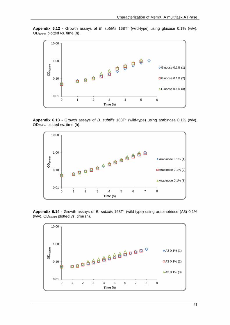
Characterization of MsmX: A multitask ATPase
71
Appendix 6.12 - Growth assays of B. subtilis 168T+ (wild-type) using glucose 0.1% (w/v).
OD600nm plotted vs. time (h).
Appendix 6.13 - Growth assays of B. subtilis 168T+ (wild-type) using arabinose 0.1% (w/v).
OD600nm plotted vs. time (h).
Appendix 6.14 - Growth assays of B. subtilis 168T+ (wild-type) using arabinotriose (A3) 0.1%
(w/v). OD600nm plotted vs. time (h).
0,01
0,10
1,00
10,00
0 1 2 3 4 5 6
OD
60
0n
m
Time (h)
Glucose 0.1% (1)
Glucose 0.1% (2)
Glucose 0.1% (3)
0,01
0,10
1,00
10,00
0 1 2 3 4 5 6 7 8
OD
60
0n
m
Time (h)
Arabinose 0.1% (1)
Arabinose 0.1% (2)
Arabinose 0.1% (3)
0,01
0,10
1,00
10,00
0 1 2 3 4 5 6 7 8 9
OD
60
0n
m
Time (h)
A3 0.1% (1)
A3 0.1% (2)
A3 0.1% (3)
Chapter 6 - Appendices
72
Appendix 6.15 - Growth assays of B. subtilis IQB672 (ΔmsmX::cat ΔamyE::Pspank(hy)) using
glucose 0.1% (w/v) and IPTG 1 mM. OD600nm plotted vs. time (h).
Appendix 6.16 - Growth assays of B. subtilis IQB672 (ΔmsmX::cat ΔamyE::Pspank(hy)) using
arabinose 0.1% (w/v) and IPTG 1 mM. OD600nm plotted vs. time (h).
Appendix 6.17 - Growth assays of B. subtilis IQB672 (ΔmsmX::cat ΔamyE::Pspank(hy)) using
arabinotriose (A3) 0.1% (w/v) and IPTG 1 mM. OD600nm plotted vs. time (h).
0,01
0,10
1,00
10,00
0 1 2 3 4 5
OD
60
0n
m
Time (h)
Glucose 0.1% (1)
Glucose 0.1% +IPTG (1)
Glucose 0.1% (2)
Glucose 0.1% +IPTG (2)
Glucose 0.1% +IPTG (3)
0,01
0,10
1,00
10,00
0 1 2 3 4 5 6 7 8
OD
60
0n
m
Time (h)
Arabinose 0.1% (1)
Arabinose 0.1% +IPTG (1)
Arabinose 0.1% (2)
Arabinose 0.1% +IPTG (2)
Arabinose 0.1% +IPTG (3)
0,01
0,10
1,00
10,00
0 1 2 3 4 5 6 7 8
OD
60
0n
m
Time (h)
A3 0.1% (1)
A3 0.1% + IPTG (1)
A3 0.1% (2)
A3 0.1% + IPTG (2)
A3 0.1% (3)
A3 0.1% + IPTG (3)
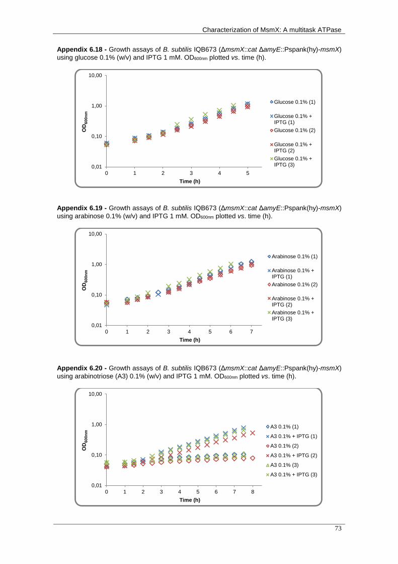
Characterization of MsmX: A multitask ATPase
73
Appendix 6.18 - Growth assays of B. subtilis IQB673 (ΔmsmX::cat ΔamyE::Pspank(hy)-msmX)
using glucose 0.1% (w/v) and IPTG 1 mM. OD600nm plotted vs. time (h).
Appendix 6.19 - Growth assays of B. subtilis IQB673 (ΔmsmX::cat ΔamyE::Pspank(hy)-msmX)
using arabinose 0.1% (w/v) and IPTG 1 mM. OD600nm plotted vs. time (h).
Appendix 6.20 - Growth assays of B. subtilis IQB673 (ΔmsmX::cat ΔamyE::Pspank(hy)-msmX)
using arabinotriose (A3) 0.1% (w/v) and IPTG 1 mM. OD600nm plotted vs. time (h).
0,01
0,10
1,00
10,00
0 1 2 3 4 5
OD
60
0n
m
Time (h)
Glucose 0.1% (1)
Glucose 0.1% +IPTG (1)
Glucose 0.1% (2)
Glucose 0.1% +IPTG (2)
Glucose 0.1% +IPTG (3)
0,01
0,10
1,00
10,00
0 1 2 3 4 5 6 7
OD
60
0n
m
Time (h)
Arabinose 0.1% (1)
Arabinose 0.1% +IPTG (1)
Arabinose 0.1% (2)
Arabinose 0.1% +IPTG (2)
Arabinose 0.1% +IPTG (3)
0,01
0,10
1,00
10,00
0 1 2 3 4 5 6 7 8
OD
60
0n
m
Time (h)
A3 0.1% (1)
A3 0.1% + IPTG (1)
A3 0.1% (2)
A3 0.1% + IPTG (2)
A3 0.1% (3)
A3 0.1% + IPTG (3)

Chapter 6 - Appendices
74
Appendix 6.21 - Growth assays of B. subtilis IQB674 (ΔmsmX::cat ΔamyE::Pspank(hy)-msmK)
using glucose 0.1% (w/v) and IPTG 1 mM. OD600nm plotted vs. time (h).
Appendix 6.22 - Growth assays of B. subtilis IQB674 (ΔmsmX::cat ΔamyE::Pspank(hy)-msmK)
using arabinose 0.1% (w/v) and IPTG 1 mM. OD600nm plotted vs. time (h).
Appendix 6.23 - Growth assays of B. subtilis IQB674 (ΔmsmX::cat ΔamyE::Pspank(hy)-msmK)
using arabinotriose (A3) 0.1% (w/v) and IPTG 1 mM. OD600nm plotted vs. time (h).
0,01
0,10
1,00
10,00
0 1 2 3 4 5 6
OD
60
0n
m
Time (h)
Glucose 0.1% (1)
Glucose 0.1% +IPTG (1)
Glucose 0.1% (2)
Glucose 0.1% +IPTG (2)
Glucose 0.1% +IPTG (3)
0,01
0,10
1,00
10,00
0 1 2 3 4 5 6 7 8
OD
60
0n
m
Time (h)
Arabinose 0.1% (1)
Arabinose 0.1% +IPTG (1)
Arabinose 0.1% (2)
Arabinose 0.1% +IPTG (2)
Arabinose 0.1% (3)
Arabinose 0.1% +IPTG (3)
0,01
0,10
1,00
10,00
0 1 2 3 4 5 6 7 8 9
OD
60
0n
m
Time (h)
A3 0.1% (1)
A3 0.1% + IPTG (1)
A3 0.1% (2)
A3 0.1% + IPTG (2)
A3 0.1% (3)
A3 0.1% + IPTG (3)

Characterization of MsmX: A multitask ATPase
75
Appendix 6.24 - Growth assays of B. subtilis IQB675 (ΔmsmX::cat ΔamyE::pSpank(hy)-
msmX(Lys45Ala)) using glucose 0.1% (w/v) and IPTG 1 mM. OD600nm plotted vs. time (h).
Appendix 6.25 - Growth assays of B. subtilis IQB675 (ΔmsmX::cat ΔamyE::pSpank(hy)-
msmX(Lys45Ala)) using arabinose 0.1% (w/v) and IPTG 1 mM. OD600nm plotted vs. time (h).
Appendix 6.26 - Growth assays of B. subtilis IQB675 (ΔmsmX::cat ΔamyE::pSpank(hy)-
msmX(Lys45Ala)) using arabinotriose (A3) 0.1% (w/v) and IPTG 1 mM. OD600nm plotted vs. time
(h).
0,01
0,10
1,00
10,00
0 1 2 3 4 5 6
OD
60
0n
m
Time (h)
Glucose 0.1% (1)
Glucose 0.1% +IPTG (1)
Glucose 0.1% (2)
Glucose 0.1% +IPTG (2)
Glucose 0.1% +IPTG (3)
0,01
0,10
1,00
10,00
0 1 2 3 4 5 6 7 8
OD
60
0n
m
Time (h)
Arabinose 0.1% (1)
Arabinose 0.1% +IPTG (1)
Arabinose 0.1% (2)
Arabinose 0.1% +IPTG (2)
Arabinose 0.1% +IPTG (3)
0,01
0,10
1,00
10,00
0 1 2 3 4 5 6 7 8 9
OD
60
0n
m
Time (h)
A3 0.1% (1)
A3 0.1% + IPTG (1)
A3 0.1% (2)
A3 0.1% + IPTG (2)
A3 0.1% (3)
A3 0.1% + IPTG (3)

Chapter 6 - Appendices
76
Appendix 6.27 - Growth assays of B. subtilis IQB676 (ΔmsmX::cat ΔamyE::Pspank(hy)-
msmX(Glu3Ser, Ile364Ser)) using glucose 0.1% (w/v) and IPTG 1 mM. OD600nm plotted vs. time
(h).
Appendix 6.28 - Growth assays of B. subtilis IQB676 (ΔmsmX::cat ΔamyE::Pspank(hy)-
msmX(Glu3Ser, Ile364Ser)) using arabinose 0.1% (w/v) and IPTG 1 mM. OD600nm plotted vs.
time (h).
Appendix 6.29 - Growth assays of B. subtilis IQB676 (ΔmsmX::cat ΔamyE::Pspank(hy)-
msmX(Glu3Ser, Ile364Ser)) using arabinotriose (A3) 0.1% (w/v) and IPTG 1 mM. OD600nm
plotted vs. time (h).
0,01
0,10
1,00
10,00
0 1 2 3 4 5
OD
60
0n
m
Time (h)
Glucose 0.1% (1)
Glucose 0.1% +IPTG (1)
Glucose 0.1% (2)
Glucose 0.1% +IPTG (2)
Glucose 0.1% +IPTG (3)
0,01
0,10
1,00
10,00
0 1 2 3 4 5 6 7 8
OD
60
0n
m
Time (h)
Arabinose 0.1% (1)
Arabinose 0.1% +IPTG (1)Arabinose 0.1% (2)
Arabinose 0.1% +IPTG (2)Arabinose 0.1% +IPTG (3)
0,01
0,10
1,00
10,00
0 1 2 3 4 5 6 7 8 9
OD
60
0n
m
Time (h)
A3 0.1% (1)
A3 0.1% + IPTG (1)
A3 0.1% (2)
A3 0.1% + IPTG (2)
A3 0.1% (3)
A3 0.1% + IPTG (3)

Characterization of MsmX: A multitask ATPase
77
Appendix 6.30 - Growth assays of B. subtilis IQB677 (ΔmsmX::cat ΔamyE::Pspank(hy)-
HD73_4301) using glucose 0.1% (w/v) and IPTG 1 mM. OD600nm plotted vs. time (h).
Appendix 6.31 - Growth assays of B. subtilis IQB677 (ΔmsmX::cat ΔamyE::Pspank(hy)-
HD73_4301) using arabinose 0.1% (w/v) and IPTG 1 mM. OD600nm plotted vs. time (h).
Appendix 6.32 - Growth assays of B. subtilis IQB677 (ΔmsmX::cat ΔamyE::Pspank(hy)-
HD73_4301) using arabinotriose (A3) 0.1% (w/v) and IPTG 1 mM. OD600nm plotted vs. time (h).
0,01
0,10
1,00
10,00
0 1 2 3 4 5 6
OD
60
0n
m
Time (h)
Glucose 0.1% (1)
Glucose 0.1% +IPTG (1)
Glucose 0.1% (2)
Glucose 0.1% +IPTG (2)
Glucose 0.1% +IPTG (3)
0,01
0,10
1,00
10,00
0 1 2 3 4 5 6 7
OD
60
0n
m
Time (h)
Arabinose 0.1% (1)
Arabinose 0.1% +IPTG (1)
Arabinose 0.1% (2)
Arabinose 0.1% +IPTG (2)
Arabinose 0.1% +IPTG (3)
0,01
0,10
1,00
10,00
0 1 2 3 4 5 6 7 8 9
OD
60
0n
m
Time (h)
A3 0.1% (1)
A3 0.1% + IPTG (1)
A3 0.1% (2)
A3 0.1% + IPTG (2)
A3 0.1% (3)
A3 0.1% + IPTG (3)

Chapter 6 - Appendices
78
Appendix 6.33 - Growth assays of B. subtilis IQB677 (ΔmsmX::cat ΔamyE::Pspank(hy)-ugpC)
using glucose 0.1% (w/v) and IPTG 1 mM. OD600nm plotted vs. time (h).
Appendix 6.34 - Growth assays of B. subtilis IQB677 (ΔmsmX::cat ΔamyE::Pspank(hy)-ugpC)
using arabinose 0.1% (w/v) and IPTG 1 mM. OD600nm plotted vs. time (h).
Appendix 6.35 - Growth assays of B. subtilis IQB677 (ΔmsmX::cat ΔamyE::Pspank(hy)-ugpC)
using arabinotriose (A3) 0.1% (w/v) and IPTG 1 mM. OD600nm plotted vs. time (h).
0,01
0,10
1,00
10,00
0 1 2 3 4 5
OD
60
0n
m
Time (h)
Glucose 0.1% (1)
Glucose 0.1% +IPTG (1)
Glucose 0.1% (2)
Glucose 0.1% +IPTG (2)
Glucose 0.1% +IPTG (3)
0,01
0,10
1,00
10,00
0 1 2 3 4 5 6 7 8
OD
60
0n
m
Time (h)
Arabinose 0.1% (1)
Arabinose 0.1% +IPTG (1)
Arabinose 0.1% (2)
Arabinose 0.1% +IPTG (2)
Arabinose 0.1% +IPTG (3)
0,01
0,10
1,00
10,00
0 1 2 3 4 5 6 7 8 9
OD
60
0n
m
Time (h)
A3 0.1% (1)
A3 0.1% + IPTG (1)
A3 0.1% (2)
A3 0.1% + IPTG (2)
A3 0.1% (3)
A3 0.1% + IPTG (3)

Characterization of MsmX: A multitask ATPase
79
Appendix 6.36 - Genomic context of HD73_4301 from Bacillus thuringiensis. The arrows
denote the genes present in the msmK gene region. Below each arrow is indicated the putative function of
encoded protein based on BLAST results.
Appendix 6.37 - Genomic context of ugpC from Staphylococcus aureus. The arrows
represent the genes present in the ugpC gene region. Below each arrow is indicated the putative function
of encoded protein based on BLAST results.
Appendix 6.38 - Genomic context of msmK from Streptococcus pneumoniae. The arrows
indicate the genes present in the HD73_4301 gene region. Below each arrow is indicated the putative
function of encoded protein based on BLAST results.