characterization of pud-1 and pud-2, two proteins up ... of pud-1 and pud-2, two proteins...
TRANSCRIPT

Characterization of PUD-1 and PUD-2, Two Proteins Up-Regulated in a Long-Lived daf-2 MutantYue-He Ding1,2☯, Yun-Guang Du1☯, Shukun Luo1☯, Yu-Xin Li1, Tie-Mei Li1, Sawako Yoshina3, Xing Wang1,Karsten Klage4, Shohei Mitani3, Keqiong Ye1*, Meng-Qiu Dong1,2*
1 National Institute of Biological Sciences, Beijing, Beijing, China, 2 Graduate Program in Chinese Academy of Medical Sciences and Peking Union MedicalCollege, Beijing, China, 3 Department of Physiology, Tokyo Women's Medical University, Tokyo, Japan, 4 Department of Cellular Biochemistry, Max PlanckInstitute of Biochemistry, Martinsried, Germany
Abstract
C. elegans PUD-1 and PUD-2, two proteins up-regulated in daf-2(loss-of-function) (PUD), are homologous 17-kDproteins with a large abundance increase in long-lived daf-2 mutant animals of reduced insulin signaling. In thisstudy, we show that both PUD-1 and PUD-2 are abundantly expressed in the intestine and hypodermis, and form aheterodimer. We have solved their crystal structure to 1.9-Å resolution and found that both proteins adopt similar β-sandwich folds in the V-shaped dimer. In contrast, their homologs PUD-3, PUD-4, PUDL-1 and PUDL-2 are allmonomeric proteins with distinct expression patterns in C. elegans. Thus, the PUD-1/PUD-2 heterodimer probablyhas a function distinct from their family members. Neither overexpression nor deletion of pud-1 and pud-2 affectedthe lifespan of WT or daf-2 mutant animals, suggesting that their induction in daf-2 worms does not contribute tolongevity. Curiously, deletion of pud-1 and pud-2 was associated with a protective effect against paralysis induced bythe amyloid β-peptide (1-42), which further enhanced the protection conferred by daf-2(RNAi) against Aβ.
Citation: Ding Y-H, Du Y-G, Luo S, Li Y-X, Li T , et al. (2013) Characterization of PUD-1 and PUD-2, Two Proteins Up-Regulated in a Long-Lived daf-2
Editor: Jiyan Ma, Ohio State University, United States of America
Received April 27, 2013; Accepted May 14, 2013; Published June 14, 2013
Copyright: © 2013 Ding et al. This is an open-access article distributed under the terms of the Creative Commons Attribution License, which permitsunrestricted use, distribution, and reproduction in any medium, provided the original author and source are credited.
Funding: The Ministry of Science and Technology of China (863 grant 2007AA02Z1A7 and 973 grant 2010CB835203 to M-QD; 863 grant 2008AA022310and 973 grant 2010CB835402 to KY) and the municipal government of Beijing funded this work. The funders had no role in study design, data collectionand analysis, decision to publish, or preparation of the manuscript.
Competing interests: The authors have declared that no competing interests exist.
* E-mail: [email protected] (KY); [email protected] (M-QD)
☯ These authors contributed equally to this work.
Introduction
The insulin/insulin-like signaling pathway (IIS) negativelyregulates lifespan in a variety of species from C. elegans,Drosophila, to mice [1,2]. Mutant C. elegans carrying a singlemissense mutation in daf-2, which encodes the only insulinreceptor in the worm, lives twice as long as the wild type (WT)[3]. The discovery that a single gene mutation can extendlifespan to such an extraordinary degree and that the lifespanregulation by IIS is conserved throughout evolution came as asurprise, because it was predicted that such genes would notexist based on a belief that the evolutionary pressure on anypost-reproductive traits should be weak, if any [4]. While agingremains one of the last frontiers in biology, exceptionally long-lived mutants such as the daf-2 worms inspire excitement—they are likely the key to understanding the molecularmechanisms of aging. Logically, one of the first steps is todetermine the changes in gene expression in the daf-2mutants.
In C. elegans, most if not all insulin signaling transmitsthrough a FOXO transcription factor called DAF-16 [1,2], thus,the downstream targets of DAF-16 are critical to the regulationof lifespan. A variety of experimental approaches have beenapplied to identifying DAF-16 targets, including microarrays,chromatin IP, quantitative mass spectrometry, and DamID[5–10].
We previously looked for DAF-16 targets by quantitativeproteomics [8]. Among the 86 proteins that are up- or down-regulated in daf-2, some are well-known DAF-16 targets suchas SOD-3, while others are more or less unexpected. Of the 86proteins, F15E11.13 (encoded by two genes F15E11.13 andY19D10B.7) and F15E11.14 (also encoded by two genesF15E11.1 and F15E11.14) are up-regulated the most (at least4-fold). We have given these genes new names, pud-1.1(F15E11.13), pud-1.2 (Y19D10B.7), pud-2.1 (F15E11.1), andpud-2.2 (F15E11.14), in which pud stands for protein up-regulated in daf-2(lf). pud-1.1 and pud-1.2 are geneduplications with exactly the same DNA sequence, encodingthe same protein PUD-1; and so are pud-2.1 and pud-2.2, both
PLOS ONE | www.plosone.org 1 June 2013 | Volume 8 | Issue 6 | e67158
Mutant. PLoS ONE 8(6): e67158. doi:10.1371/journal.pone.0067158-M

encode PUD-2. They belong to an uncharacterized genefamily, which we refer to as the PUD family. The othermembers of this gene family in C. elegans are pud-3(F15E11.15), pud-4 (F15E11.12), pudl-1 (Y54G2A.46), andpudl-2 (Y54G2A.47), where pudl stands for pud-like.
A literature search finds that although these proteins orgenes have not been characterized, they have appearedrepeatedly in previous publications, often buried in thesupplemental information as differentially expressed genesunder various conditions. For example, a temperature shift ofglp-1(e2144ts) mutant from 15 °C to 25 °C was accompaniedwith a remarkable increase of PUD-1 and PUD-2 [11]. A 7-foldincrease of the PUD-2 protein level was detected in theglp-4(bn2ts) mutant compared to WT cultured at the sametemperature of 25 °C [12]. Moreover, the PUD-1 protein wasshown to build up conspicuously in transgenic C. elegansexpressing a human 7-dehydrocholesterol reductase, anenzyme functioning in de novo biosynthesis of cholesterol [13].Similarly, when the mRNA levels were measured, elevation ofthe pud-1 and pud-2 transcripts were ranked among the topeight in the pept-1(lg601) mutant [14], and among the top 15 inrde-1(RNAi) and rde-4(RNAi) mutants [15]. Decreasedtranscript levels of these genes were also observed, inmdt-15(RNAi) worms [16], in the glp-4(bn2); sek-1(ag1) doublemutant relative to the glp-4(bn2) single mutant [17], and in WTC. elegans exposed to high concentration of CO2 [18]. It isworth noting that the glp-1 and pept-1 mutants, in whichincreased expression of these genes have been detected, havea longevity phenotype similar to that of daf-2.
To understand the function of PUD-1 and PUD-2, and thebiological significance of their up-regulation in daf-2 mutants,we set out to analyze their expression patterns, overexpressionphenotype, loss-of-function phenotype, interacting proteins,and protein structures.
Materials and Methods
Plasmids and C. elegans strainsThe plasmids and the worm strains used in this study are
listed in Tables S4 and S5 in File S1, respectively. Bristol N2was used as the wild type strain. Worms were cultured onNGM plates at 20 °C unless indicated otherwise. Transgenicstrains were constructed through microinjection of plasmids,and transgenes were chromosomally integrated by gamma-irradiation. Single copy insertion of transgenes was generatedas described [19]. All of the integrated transgenic strains andthe mutant strains were backcrossed to N2 at least four times.
RNAiRNAi by feeding was carried out at 20 °C as described [20].
For RNAi by injection, dsRNA was transcribed in vitro fromlinearized plasmid DNA using a kit (Promega), and the purifieddsRNA was injected into the C. elegans gonad. The phenotypeof the F1 progeny was analyzed.
Quantitative RT-PCRTotal RNA was extracted from synchronized young adults
using TRIZOL (Invitrogen), followed by the removal ofcontaminant DNA using DNase I. cDNAs were synthesizedfrom the total RNA templates using a reverse transcription kit(Takara). Primers used for qPCR wereoMD241[ACTTATCCGCATCCCATCCAGTGT]/oMD244[ATGTCGA CTGATCCAACTCCACCA] for pud-1,oMD246[TGACAGAACAATGGGCCAAAGTGG]/oMD247[GCCGGTATACTGATGAAGCACTTGGA] for pud-2,and actin-F[TGCCGCTCTT GTTGTAGACAATGG]/actin-R[TGACGTGGTCTTCCGACAATGGAT] for actin as theinternal standard. qPCR was carried out on an ABI 7500 Fastreal-time PCR system using a Takara realtime PCR kit (SYBRPremix Ex TaqTM II).
Mapping the boundaries of niDf209The precise boundaries of niDf209 and niDf207 in JU258
were determined by nested PCR followed by sequencing.Primers pairs were designed in 1-kb intervals to cover theannotated flanking sequences (wormbase.org). niDf209 is a15,979-bp deletion with a 1,608-bp insertion. niDf207 is a4072-bp deletion.
Generating hq5 and hq6To help introduce niDf209 (genetic position: -14.89) to the N2
background, unc-60 (genetic position: -18.88) and dpy-11(genetic position: -0.02) were used to mark recombinationevents on Chr. V. JU258 was first crossed with DR181unc-60(m35) dpy-11(e224), and DpynonUnc progeny with therecombination sites nearest to, and on the right side of niDf209were determined by PCR with oligos oMD307 and oMD308.Candidate alleles were crossed into DR35 unc-60(m35) andthe resulting UncDpy progeny containing niDf209 wereselected for further validation. hq6 was isolated using niDf208(16.7 Kb upstream of niDf209) as a marker of recombination tothe left of niDf209. hq5 was isolated using another markerupstream of niDf208. The linked unc-60(m35) anddpy-11(e224) alleles were removed through recombinationafter repeated backcrosses to N2.
Lifespan assayUnless indicated otherwise, all lifespan assays were carried
out at 20 °C as described before [8]. Kaplan-Meier survivalanalyses were carried out using the SPSS software and thelog-rank p values were reported.
Heat tolerance assayFor each strain, 30 animals were shifted from 20 °C to 35 °C
at the young adult stage and examined for survival every twohours.
PA14 fast killing assayThe assay was performed as described previously [21].
Characterization of PUD-1/PUD-2
PLOS ONE | www.plosone.org 2 June 2013 | Volume 8 | Issue 6 | e67158

Paralysis assayOn day 0, 100 L4s were transferred to fresh NGM plates
seeded with OP50 (10 worms/plate) and moved to new platesevery two days. Paralyzed worms that could only move theirheads were counted every day. Paralysis assay wasterminated after 7 days to avoid mistaking old worm as“paralyzed”.
MicroscopyAll GFP and DIC images were taken using a Zeiss
AxioImager M1 microscope.
mRNA sequencing and data analysisTotal RNAs were extracted from the WT and hq6 day-1
adults using TRIZOL (Invitrogen). The sequencing library wasconstructed by following the “mRNA sequencing samplepreparation guide” provided by Illumina. The WT and hq6mRNA sequencing libraries were bar-coded and mixed in equalamounts. The paired-end RNA sequencing data were acquiredon an Illumina GA II instrument (in two lanes). The paired-endRNA-seq 36 base pair reads were mapped to the C. elegansgenome (WS220) using the TopHat software, allowing no morethan two mismatches per sequencing read. In TopHatmapping, the expected (mean) inner distance between matepairs was set to 100 bp, the standard deviation for thedistribution on inner distances between mate pairs was set to50 bp. Only uniquely mapped paired-end reads were extractedfor subsequent analysis. Differential gene expression analysiswas performed with the bioconductor package DESeq. Geneswith reads number ≤ 10 in both samples were not included inthe DESeq analysis. In total, the two samples produced 18million uniquely mapped paired-end sequencing reads (9million per sample). In a separate experiment, single-end RNAsequencing data were acquired for three independent WT N2samples on Illumina HiSeq 2000.
Immunoprecipitation and Mass Spec analysisA single-copy insertion strain expressing FLAG-tagged
PUD-1 and the control N2 strain were cultured on High-growth(HG) plates and harvested for anti-FLAG IP. Proteins bound tothe M2 beads (Sigma) were eluted using 0.5µg/µl 3xFLAGpeptides. SDS-PAGE analysis followed by silver stainingrevealed three protein bands in the FLAG::PUD-1 IP but not inthe control IP. These bands were cut from the gel, digestedwith trypsin, and subjected to LC-MS/MS analysis on LTQ-orbitrap (Thermo, Fisher). Proteins were identified by searchingthe MS/MS spectra against a C. elegans protein databaseWS217 using Prolucid and filtering the search results withDTASelect 2.0.
Protein purificationThe PUD family proteins were expressed in the Escherichia
coli BL21(DE3) strain (Novagen) induced with 0.2 mMisopropyl β-D-1-thiogalactopyranoside normally for 8 h at 25°C. PUDL-1 and PUDL-2 were expressed at 16 °C for 16 h.PUD-1 and PUD-2 were coexpressed and copurified as adimer. The harvested cells were resuspended in buffer P500
(50 mM phosphate pH 7.6, 500 mM NaCl) and lysed using ahigh pressure cell disruptor (JNBIO) followed by sonication.The cell lysates were clarified by centrifugation and passagethrough a 0.45-μm filter and loaded onto a 5-ml HisTrapcolumn (GE healthcare). The column was washed by 50 mMimidazole in P500. The protein was eluted with 500 mMimidazole in P500 and incubated with ULP1 protease to cleavethe His-SMT3 tag. After concentrating and dilution with 20 mMHEPES-K, pH 7.6, the sample was passed through a HisTrapcolumn to remove the cleaved tag and uncleaved protein. ThePUD-1/PUD-2 complex was further purified with a Superdex200 column (GE healthcare) equilibrated in buffer E250consisting of 5 mM HEPES-K, pH 7.6 and 250 mM KCl. ThePUD-1/PUD-2 complex was labeled with seleno-methionine byinhibiting the methionine biosynthesis pathway and purifiedwith the same procedure as unlabeled complex. The proteinwas concentrated by Amicon Ultra-15 units and stored at -80°C.
Crystallization and structure determinationThe Se-labeled full-length PUD-1/PUD-2 complex was
crystallized by mixing 1 μl of protein sample (46 mg/ml in E250)with 1 μl of well solution consisting of 0.2 M NH4H2PO4, 0.1 MTris-HCl pH 8.5 and 40% 2-methyl-1,3 propanediol using thehanging drop vapor diffusion method at 20 °C. The crystal wasflash frozen in liquid nitrogen without further cryoprotection.The diffraction data were collected at the wavelength ofselenium peak at Shanghai Synchrotron Radiation Facility(SSRF) beamline BL17U and processed by HKL2000 [22]. Thestructure was solved by single-wavelength anomalousdiffraction (SAD) method using a 3.6-Å dataset. The heavyatoms were located by SHELXD [23] and the phase calculationand density modification were conducted with SHARP [24]. Thecrystal belongs to space group P3 221 and contains two copiesof PUD-1/PUD-2 heterodimer in the asymmetric unit. Thestructural model was built in COOT [25] and refined withRefmac to a free R-factor of 0.38 at 3.6 Å resolution [26].
The PUD-1 (9-151) and PUD-2 (7-152) complex (35 mg/ml inE250) was crystallized in 0.1 M HEPES-Na pH 8.5 and 4.3 MNaCl. The crystal was cryoprotected with 10% glycerol in themother solution and flash frozen in liquid nitrogen. A datasetwas collected at SSRF-BL17U to 1.9 Å resolution. Thestructure was solved by molecular replacement in PHASERusing the initial heterodimeric structure of full-length complexas a search model [27]. The crystal belongs to space groupP212121 and contains one heterodimer in the asymmetric unit.The final model was refined with Refmac to 1.9 Å resolutionand contains residues 9-151 of PUD-1, an N-terminal serineresidue from the expression vector of PUD-1, residues 7-87and 91-152 of PUD-2, one glycerol molecule, 10 chlorine ionsand 326 waters. Analysis with RAMPAGE shows that 97.9% ofthe residues are in most favored regions and 2.1% in allowedregions [28]. Structural figures were prepared in PyMOL [29].
Size exclusion chromatographyAll six C. elegans proteins of the PUD protein family were
individually purified through one-step HisTrap chromatographywith the His-SMT3 tag uncleaved. Each protein (500 μg) was
Characterization of PUD-1/PUD-2
PLOS ONE | www.plosone.org 3 June 2013 | Volume 8 | Issue 6 | e67158

loaded individually or in combination in a total of 200 μl volumeto a Superdex-200 10/300 GL column (GE healthcare) runningin buffer E250.
Results
The PUD-1 and PUD-2 protein abundance increase indaf-2 is independent of daf-16
The protein levels of PUD-1 and PUD-2 are greatly elevatedin daf-2 mutants [8]. To find out whether this is due to activatedgene expression by DAF-16, the key transcription factornormally inhibited by signaling from DAF-2 in WT C. elegans,we quantified their mRNA levels in WT, daf-2, daf-16, anddaf-16; daf-2 double mutants by quantitative PCR. The pud-1and pud-2 mRNA levels increased in the daf-2(e1370) mutantrelative to the WT, and this moderate increase required daf-16(Figure S1a in File S1). Interestingly, at the protein level boththe daf-2 mutant and the daf-16; daf-2 double mutant hadhigher amounts of PUD-1 and PUD-2 than WT (Figure S1b-c inFile S1), even though the mRNA templates of the two proteinswere reduced in the double mutant. We thus propose thatthese proteins are up-regulated in the daf-2 mutant mainlythrough a post-transcriptional mechanism independent ofDAF-16.
Overexpression of PUD-1 and PUD-2 did not extendlifespan
To find out if the increased PUD-1 and PUD-2 proteinamounts contribute to the longevity of the daf-2 mutant, weoverexpressed these proteins in WT animals to mimic theirinduction in daf-2. We generated two overexpressionconstructs, pYG2 and pWX1, in which the genomic sequencesof pud-1 and pud-2 were fused to the GFP coding sequence,respectively (Figure S2a in File S1). Because pud-1 and pud-2are adjacent genes transcribed from the same intergenicpromoter region in opposite directions, un-tagged PUD-2 and aPUD-1::GFP fusion protein were expressed from the pYG2transgene arrays hqIs24 and hqIs28, while un-tagged PUD-1and PUD-2::GFP were expressed from the pWX1 transgenearray hqIs60. PUD-1::GFP and PUD-2::GFP are bothexpressed in the intestine and hypodermis, from either themulti-copy (Figure S2 in File S1) or the single-copy transgenes(Figure 1). None of the high-copy arrays altered the WTlifespan. Perplexingly, hqIs28 and hqIs60, but not hqIs24,further extended the lifespan of daf-2(e1370) and daf-2(RNAi)animals by 27-38% (Figure S3 in File S1), and hqIs60 animalsalso showed increased resistance to thermal stress (notshown). To clarify the overexpression phenotype, we madefour additional transgene arrays (hqEx30, hqEx31, hqEx50,and hqEx52) from which untagged PUD-1 and untaggedPUD-2 were co-expressed. None of them extended the C.elegans lifespan in either the WT or daf-2(RNAi) background;hqEx30, hqEx31, and hqEx52 even shortened lifespansomewhat (Figure 2A and data not shown). hqEx52 andanother transgene hqEx47 that was made by co-injection ofHA::pud-1::GFP and HA::pud-2::Cherry under their nativepromoters failed to alter the lifespan of daf-2(RNAi) or WTworms (not shown). We also examined, hqEx30, hqEx31, and
hqEx47 animals for resistance to thermal stress at 35 °C andfound that they were not different from WT worms (Figure 2Band data not shown). We thus conclude that overexpression ofPUD-1 and PUD-2 does not extend the lifespan of WT or daf-2animals. We suspect that the longevity phenotype associatedwith hqIs60 and hqIs28 may be due to independentbackground mutations linked to the transgenes. Because thetransgenes were integrated into different chromosomes andeach was backcrossed to WT at least four times, the chancefor two mutations at different loci to cause the same phenotypeshould be extremely rare, but it appeared to be what hadoccurred in this case.
Deletion of pud-1 and pud-2 did not shorten the daf-2lifespan
We next asked whether the longevity phenotype of the daf-2mutant requires the gene activities of pud-1 and pud-2. Sinceknocking down of these genes using the RNAi-by-feedingmethod [20], either individually or in combination, failed toeffectively reduce the fluorescence intensity of PUD-1::GFP orPUD-2::GFP in transgenic strains, we set out to obtain deletionalleles. Six members of the PUD gene family in C. elegans–pud-1.2, pud-2.2, pud-3, pud-4, pud-1.1 and pud-2.1-are nextto each other on Chromosome V (Figure S4a in File S1) andthe other two–pudl-1 and pudl-2–are adjacent genes onChromosome IV (Figure S4b in File S1). Fortunately, the six-gene cluster on Chromosome V is deleted in JU258, a wild C.elegans strain isolated from the island of Madeira. We mappedthe precise boundaries of this deletion, niDf209, and found thatin addition to the PUD gene cluster, three flanking genes(Y19D10B.5, Y19D10B.6 and F15E11.11) are also deleted inniDf209 (Figure S4a in File S1). The three flanking genes eachhave at least one paralog in the C. elegans genome with 40%to 90% sequence similarity, so the deletion of them likely haslittle phenotypic consequence. Thus far, no phenotype of anykind has been associated with any of the three genes.
We transplanted niDf209 from JU258 to the standard WT N2background by homologous recombination (Figure S5 in FileS1). Briefly, JU258 was crossed to unc-60(m35) dpy-11(e224)(N2 background). The nonUnc Df Dpy worms descended fromthe first cross were mated with unc-60(m35) (N2 background),and the resulting Unc Df Dpy recombinants were selected andbackcrossed to N2 repeatedly until nonUnc Df nonDpyrecombinants were obtained, and these recombinants werefurther backcrossed to N2 for five more times. We obtained twoalleles, hq5 and hq6, that harbor niDf209 in the N2background. hq5 has two deficiencies–niDf208 and niDf209–from JU258 whereas hq6 has only niDf209 (Figure S4a in FileS1); the latter was used in most of the deletion phenotypeanalyses.
Both hq5 and hq6 are morphologically normal, theirlocomotion and egg production are also WT-like (data notshown). We found that neither hq5 nor hq6 affected thelifespan of WT or daf-2(RNAi) animals (Figure 2C). We alsomade a composite RNAi construct pYG17 in which 222-323nucleotide sequences each targeting one or two PUD genefamily members were concatenated together (Figure S6 in FileS1). Injection of the dsRNA transcribed in vitro from pYG17 into
Characterization of PUD-1/PUD-2
PLOS ONE | www.plosone.org 4 June 2013 | Volume 8 | Issue 6 | e67158
C. elegans (pud RNAi) reduced PUD-1::GFP and PUD-2::GFPlevels by at least 50% in the next generation (data not shown).Consistent with the deletion alleles, pud RNAi did not changethe lifespan of WT or daf-2(e1370) worms (Figure 2D).
hq6 conferred resistance to Aβ toxicitySince daf-2 and many other IIS mutants are not only long-
lived, but also resistant to various types of stress such as hightemperature, pathogen, and Aβ toxicity [30-32], we asked if thedeletion of pud-1 and pud-2 would affect stress tolerance. Wefound that the survival curves of hq5 and hq6 animals were
similar to that of WT at 35 °C (Figure 2E). Likewise, in a fastkilling assay using PA14, a pathogenic P. aeruginosa strain, wedid not observe consistent difference between WT and hq6, orbetween daf-2 and daf-2; hq6 (Figure 2F). However, the hq6allele protected WT animals from paralysis induced byAβ(1-42) expressed in the body-wall muscles [33], and furtherenhanced the resistance of daf-2(RNAi) animals to the Aβtoxicity (Figure 3A). It also improved the brood size of Aβtransgenic worms (Figure 3B). The fact that hq6 anddaf-2(RNAi) had a synergistic effect on Aβ resistance and thathq6 did not shorten the daf-2 lifespan both argue for the point
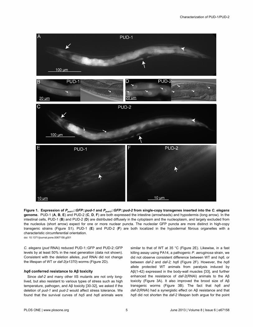
Figure 1. Expression of Ppud-1::GFP::pud-1 and Ppud-2::GFP::pud-2 from single-copy transgenes inserted into the C. elegansgenome. PUD-1 (A, B, E) and PUD-2 (C, D, F) are both expressed the intestine (arrowheads) and hypodermis (long arrow). In theintestinal cells, PUD-1 (B) and PUD-2 (D) are distributed diffusely in the cytoplasm and the nucleoplasm, and largely excluded fromthe nucleolus (short arrow) expect for one or more nuclear puncta. The nucleolar GFP puncta are more distinct in high-copytransgenic strains (Figure S1). PUD-1 (E) and PUD-2 (F) are both localized in the hypodermal fibrous organelles with acharacteristic circumferential orientation.doi: 10.1371/journal.pone.0067158.g001
Characterization of PUD-1/PUD-2
PLOS ONE | www.plosone.org 5 June 2013 | Volume 8 | Issue 6 | e67158

Figure 2. Neither overexpression nor deletion of pud-1 and pud-2 extended lifespan in WT or daf-2 animals. (A) hqEx30and hqEx31, two extra-chromosomal transgenes expressing untagged PUD-1 and PUD-2, slightly shortened lifespan of WT anddaf-2(RNAi) worms (p < 0.014 between hqEx30 or hqEx31 and WT with or without daf-2 RNAi, N>77, 20 °C). (B) hqEx30 andhqEx31 animals were as sensitive to thermal stress as the WT. Stress resistant daf-2(e1370) served as a positive control (35 °C,N>27). (C) hq5 and hq6, two deletion alleles that removed all the gene copies of pud-1 and pud-2, did not affect the lifespan of WTor daf-2(RNAi) animals (N>63). (D) Knocking down pud-1 and pud-2 by heritable RNAi (via injection of dsRNA into parent worms)had no effect on WT or daf-2(e1370) lifespan (N>79). (E-F) hq5 and hq6 did not affect the survival of C. elegans at 35 °C (E) (N>27,representative of two experiments) or the pathogenic bacteria P. aeruginosa PA14 (F) (N >39, representative of three repeats).doi: 10.1371/journal.pone.0067158.g002
Characterization of PUD-1/PUD-2
PLOS ONE | www.plosone.org 6 June 2013 | Volume 8 | Issue 6 | e67158

that PUD-1 and PUD-2 are not connected to the longevity ofdaf-2 animals.
In addition to hq5 and hq6, we also isolated tm5053 andtm6107 on chromosome IV. In tm5053, part of the codingsequence of pudl-1 and much of the intergenic region betweenpudl-1 and pudl-2 are deleted. In tm6107, both pudl-1 andpudl-2 are deleted (Figure S4b in File S1). No obvious defectswere detected in tm5053 or tm6107 worms. The tm6107;hq6and tm5053; hq6 double mutants were as resistant to Aβtoxicity as the hq6 single mutant (Figure 3C-D).
The PUD family members are likely positive regulator ofa subset of collagen genes and a subset of spermprotein genes
To find out the functions of pud-1 and pud-2 in a non-biasedway, we compared the gene expression profiles of the WT andhq6 animals using next-generation mRNA sequencing. At 5%false discovery rate and excluding the genes deleted in the hq6allele, only 21 genes were differentially expressed in hq6animals compared with the WT (Table 1), and 19 of them weredown-regulated in hq6. Most of these genes had a modestmRNA abundance change in hq6 except for haf-6 (2.5-folddecrease) and srbc-15 (no transcripts detected). Significantlyenriched in this short list of genes are the ones encodingcollagens (col-39, col-125, col-133, col-147, col-149, and
Figure 3. hq6 animals are more resistant to Aβ(1-42) toxicity. (A) In either WT or daf-2(RNAi) background, hq6 conferredresistance to paralysis induced by dvIs2, which expressed Aβ(1-42) in the body wall muscles (N ≥83, representative of threeexperiments). (B) hq6 increased the brood size of dvIs2 worms (N ≥10, representative of three experiments). (C-D) tm6107;hq6 andtm5053; hq6 double mutants showed similar resistance to Aβ toxicity as the hq6 single mutant.doi: 10.1371/journal.pone.0067158.g003
Characterization of PUD-1/PUD-2
PLOS ONE | www.plosone.org 7 June 2013 | Volume 8 | Issue 6 | e67158

col-179) or sperm proteins (a major sperm protein genemsp-77, sperm specific family class P or Q genes ssp-34,ssq-1, ssq-2, and ssq-4) (p < 0.001 for the enrichment of eithergroup of genes, hypergeometric test). The mRNA-seq datasuggest that one or more of the genes deleted in hq6 arepositive regulators of collagen or sperm protein production.
Mutations of collagen genes have been associated withmorphology or locomotion phenotypes such as Dpy (dumpy),Lon (long), Bli (blister), or Rol (roller) [34]. However, hq6animals appear to be normal in morphology and movement, sothe reduced expression of the collagen genes appears to beinconsequential. This is reminiscent of rol-6, of which the nullmutants are essentially WT-like while the missense mutationscause a strong roller phenotype [35]. Alternatively, the reducedexpression of five collagen genes might be compensated bythe up-regulation of col-179 (Table 1). No gross defects weredetected in the egg-laying behavior or the brood size of hq6animals, suggesting that hq6 sperm are functional despite areduction in the mRNA levels of five sperm proteins.
PUD-1 and PUD-2 form a heterodimer in vitro and invivo
We also tried to infer the functions of PUD-1 and PUD-2 fromtheir binding proteins. Using the MosSCI technique [19], wegenerated a single copy transgene expressing FLAG-taggedPUD-1 under its native promoter (Ppud-1::FLAG::pud-1). Usingan anti-FLAG antibody, we immunoprecipitated (IP)FLAG::PUD-1 and analyzed the protein bands in the IP bymass spectrometry. Endogenous PUD-2 and HSP-1, aconstitutively expressed Hsc70 protein that is also inducibleupon heat shock [36], were found associated withFLAG::PUD-1 (Figure 4A). The intensity of the silver-stainedprotein bands suggested that PUD-1 and PUD-2 have a 1:1stoichiometry, while HSP-1 is substoichiometric. In vitro cross-linking and gel-filtration experiments using purified recombinantPUD-1 and PUD-2 indicated that they form a heterodimer(Figure 4B–D). With this information, we revisited the proteinand mRNA quantitation results and realized that indeed, theyhad similar fold-change values in the mutants relative to theWT (Figure S1 in File S1 and [8]), and their expression patternswere the same (Figure 1 and Figure S2 in File S1). Together,our results demonstrate that PUD-1 and PUD-2 form aheterodimer in vivo and suggest that their expressions areregulated as a single unit.
The association of PUD-1 and PUD-2 with HSP-1 isintriguing. Although no sequence homology can be foundbetween the PUD family proteins and any heat shock proteins,PUD-1 and PUD-2 appear to be regulated like heat shockproteins. For example, they are markedly induced by anincrease of temperature from 20 °C to 27 °C (Figure S2d-e inFile S1) or by exposure to cadmium [37]. Moreover, from ananalysis of genes differentially regulated by the C. elegans RBprotein LIN-35, a non-E2F binding element was found in thepromoter regions of the small heat shock protein genes, and itwas also found in the promoters of pud-1 and pud-2 [38]. Wetested if the purified PUD-1/PUD-2 heterodimer displayed anychaperone activity in an in vitro assay [39], and found that it didnot (data not shown). There is a possibility that thisheterodimer may act as a co-chaperone of HSP-1, but morelikely is a substrate of HSP-1.
The crystal structure of the PUD-1 and PUD-2heterodimer
Next, we asked if these proteins have structural homologsthat could give us clues about their function. The structure ofthe PUD-1/PUD-2 complex was first determined at 3.6 Åresolution for full length proteins by Se-phasing. The initialmodel shows that a few terminal residues are disordered. Wethen prepared a fragment of PUD-1 with residues 9-151 and afragment of PUD-2 with residues 7-152 and obtained highquality crystals for the slightly truncated complex. The structureof the truncated complex was determined to 1.9 Å resolutionwith an Rwork/Rfree of 0.192/0.225 and is discussed below(Figure 5, Table S1 in File S1).
In the structure, both PUD-1 and PUD-2 adopt similar β-sandwich folds that further associate with each other into aheterodimer (Figure 5A). As expected from 54% sequencesimilarity between PUD-1 and PUD-2, the two subunit
Table 1. Differentially expressed genes inhq6 compared towild type.
Gene NameFold Change(hq6 vs N2)
Adjusted P-value Gene Description (Concise)
col-133 0.37 1.0E-07Collagens (type IV and type XIII), andrelated proteins
col-147 0.30 1.0E-07Collagens (type IV and type XIII), andrelated proteins
haf-6 0.17 2.1E-06A half-molecule ATP-binding cassette(ABC) transporter
col-39 0.37 3.8E-05Collagens (type IV and type XIII), andrelated proteins
ssp-34 0.45 2.1E-04 Sperm Specific family, class PC15C6.2 0.39 2.6E-04 Unnamed proteinssq-1 0.55 3.3E-04 Sperm-Specific family, class Q
col-149 0.49 4.5E-04Collagens (type IV and type XIII), andrelated proteins
gipc-2 0.49 2.4E-03GIPC (RGS-GAIP Interacting ProteinC) homolog
col-179 1.69 5.7E-03Collagens (type IV and type XIII), andrelated proteins
F08H9.2 0.48 5.7E-03 Unnamed protein
nspd-7 0.53 5.7E-03Nematode Specific Peptide family,group D
nspd-1 0.57 6.1E-03Nematode Specific Peptide family,group D
ssq-2 0.59 7.0E-03 Sperm-Specific family, class Qrpl-39 1.57 1.2E-02 Large ribosomal subunit L39 proteinY57G11A.2 0.46 1.4E-02 Unnamed proteinY45F10C.4 0.50 1.9E-02 Unnamed protein
srbc-15 0.00 4.3E-05Serpentine Receptor, class BC (classB-like)
col-125 0.32 2.4E-02Collagens (type IV and type XIII), andrelated proteins
msp-77 0.54 4.0E-02 Major Sperm Proteins (MSPs)ssq-4 0.61 4.7E-02 Sperm-Specific family, class Q
Characterization of PUD-1/PUD-2
PLOS ONE | www.plosone.org 8 June 2013 | Volume 8 | Issue 6 | e67158

structures can be well superimposed with a root mean squaredeviation (RMSD) of 1.277 Å over 118 Cα pairs (Figure 5B).
The β-sandwich fold of PUD-1 and PUD-2 is composed of 9major β-strands arranged into two sheets. One sheet is formedby strands β3, β2, β5, β6 and β9 in an antiparallel manner,whereas the other sheet is composed of strands β4, itsextension β4', β1, β7, β8 and β9 in a mixed manner, where alladjacent strands are aligned in antiparallel except β1 and β7that are aligned in parallel. The strand β9 pairs with bothsheets and closes one side of the sandwich. In addition,strands β5 and β6 project out from the sandwich body and forma prominent protrusion involved in dimerization.
The sandwich structures of PUD-1 and PUD-2 contact eachother in a face-to-back manner with an inclination angle of 30°,forming a V-shaped dimer. The dimerization buries 1081 Å2 ofsolvent accessible surface area per subunit and is mediated bytwo interfaces. In the first interface, one end of the PUD-1sandwich, which is composed of the β7-β8 loop, the β1-β2 loopand the exposed regions of strands β5 and β6, packs againstthe β3-β9 sheet of PUD-2 (Figure 5C). This interface isstabilized by hydrophobic and polar interactions. Thehydrophobic interactions involve residues M80, F105, F131,P132, H136 of PUD-1 and residues L31, L39, A41, W44, H76,I78, A102 and H152 of PUD-2. A number of polar and water-
Figure 4. PUD-1 and PUD-2 form a heterodimer. (A) FLAG IP followed by MS analysis identified HSP-1 and PUD-2 as bindingproteins associated with FLAG-PUD-1. Shown is the silver stained gel of the IP products before MS analysis. (B) Purifiedrecombinant PUD-1 and PUD-2 can be cross-linked together and the cross-linked species seems to be a heterodimer. (C) ThePUD-1 and PUD-2 heterodimer, each with a SMT3 tag, was also observed by gel filtration. (D) Gel filtration analysis of the C.elegans PUD gene family proteins. SMT3 tagged proteins were loaded onto a Superdex-200 10/300 column individually or inindicated combinations.doi: 10.1371/journal.pone.0067158.g004
Characterization of PUD-1/PUD-2
PLOS ONE | www.plosone.org 9 June 2013 | Volume 8 | Issue 6 | e67158

Figure 5. Structure of the PUD-1 and PUD-2 heterodimer. (A) Ribbon representation of the PUD-1 and PUD-2 heterodimer incross-eye stereoview. PUD-1 is green and PUD-2 is cyan. The β-strands are labeled with numbers and the N and C termini areindicated. (B) Structural superposition of PUD-1 and PUD-2 subunit. (C) Interactions at the dimer interface. For clarity, only residuesat the interface are shown for PUD-1. Oxygen is red, nitrogen is blue, sulfur is orange and carbon is green for PUD-1 and cyan forPUD-2. Hydrogen bonds are shown as yellow dashed lines. (D) Conserved surface. The ribbon and surface representations ofPUD-1 structure are shown side-by-side and in two opposite orientations. The residues conserved in 100% and 80% of the 31homologs of PUD-1 and PUD-2, as shown in Figure S7, are colored orange and yellow, respectively, for side chain atoms. (E)Sequence alignment of the PUD family proteins in C. elegans. Residues with 100% and 80% conservation are shaded with blackand grey, respectively. The secondary structures observed in the crystal structure are shown on the top for PUD-2 and at the bottomfor PUD-1. Residues whose surface area is buried by 10-30 Å2 and at least 30 Å2 upon dimerization are denoted by open and solidcircles, respectively.doi: 10.1371/journal.pone.0067158.g005
Characterization of PUD-1/PUD-2
PLOS ONE | www.plosone.org 10 June 2013 | Volume 8 | Issue 6 | e67158

mediated interactions are present at more peripheral regions.The second dimer interface is constituted by the extensions ofβ5 and β6 of both PUD-1 and PUD-2 (named β5' and β6' inPUD-2). They form an intermolecular antiparallel 4-strandedsheet with PUD-1 β5 aligned with PUD-2 β6' (Figure 5A.)
Despite strong similarity between PUD-1 and PUD-2structures, the dimer interface is asymmetric and engagesnonequivalent faces of each subunit. Sequence alignmentshows the residues at the dimer interface are dissimilar inPUD-1 and PUD-2, which accounts for the specificity of thedimerization mode (Figure 5E).
Functional implication of the PUD-1/PUD-2 structureA BLAST search in a non-redundant protein database of
NCBI shows that PUD-1 and PUD-2 homologs have a verylimited distribution across species and can only be identified inthree worm species, five fungi and two bacteria (Figure S7 inFile S1). Mapping the residues conserved in these sequenceson the PUD-1 structure reveals a conserved surface patch atone end of the β-sandwich, opposite to the dimerization end(Figure 5D). The patch is mainly composed of residues fromβ6-β7 loop, β4’-β5 loop and the N-terminal part of β2-β3 loop.This region is likely important to the function of the PUD familyproteins.
A DALI search shows that the β-sandwich of PUD-1/PUD-2bears some topology similarity with proteins of diverse function,such as pore-forming cytolysin, transporter transthyretin andchaperone FaeE involved in pilus assembly (Z score to 6.9).However, the functional insight provided by these structuralhomologs is vague.
To summarize, after the genetic, genomic, biochemical, andstructural analyses described above, much has been learnedabout the PUD-1 and PUD-2 proteins, but the significance oftheir biological function remains unclear.
Characterization of other C. elegans homologs ofPUD-1 and PUD-2
We determined the expression patterns of all the PUD genefamily members in C. elegans by GFP fusion proteins, and theresult is summarized in Table S2 in File S1. As describedabove, pud-1 and pud-2 are expressed strongly and uniformlyin the intestine, and less strongly in the hypodermis (Figure 1and Figure S2 in File S1). Besides, their GFP fusion proteinswere both seen in fibrous organelles, the hypodermal hemi-desmosome structures that fasten muscles to the cuticle(Figure 1E-F). At the subcellular level, both proteins were founddistributed diffusely in the cytoplasm and the nucleoplasm, andlargely excluded from the nucleolus except for one or morenucleolar puncta (Figure S2b-c in File S1).
The Ppud-3::pud-3::GFP transgene was expressed in thenuclei of hyp7, the largest hypodermal cell that wraps aroundmost of the worm body. It was also expressed in thepharyngeal muscle pm3, rectal gland cells, and less frequently,in the intestine and a few neuron-like cells in the head (Figure6A).
The expression of Ppud-4::pud-4::GFP was detected moststrongly in the hyp7 nuclei and sporadically in the intestine(Figure 6B).
Also concentrated in the hyp7 nuclei were PUDL-1::GFP andPUDL-2::GFP. Elsewhere, PUDL-1::GFP was seen in a headneuron and PUDL-2::GFP in the pharyngeal muscle pm5(Figure 6C-D).
Judging by GFP intensity, pud-1::GFP and pud-2::GFP arethe strongest expressers. From mRNA sequencing data of WTday-1 adults (Table S3 in File S1), the sequence reads ofpud-1 and pud-2 are 10-fold higher than those of pud-3 andpud-4, and about 1000-fold higher than those of pudl-1 andpudl-2. We suspect that pudl-1 and pudl-2 are minimallyexpressed in young adult worms under normal conditions. Incontrast, pud-1 and pud-2 are constitutively expressed at highlevels, and can be induced further. The nuclear localization ofthe PUD-1/2/3/4 proteins suggests that they might regulate thetranscription of the collagen genes whose mRNA levels werealtered in the hq6 mutant (Table 1).
As PUD-1 and PUD-2 form a dimer, we asked whether therest of the worm homologs are structural counterparts and canalso form a dimer. To assess their oligomeric state in solution,we preformed analytical gel filtration experiments (Figure 4C-D). Since these proteins alone have a low solubility, they wereanalyzed with a fused solubility-enhancing SMT3-tag.Separately, SMT3-PUD-1 and SMT3-PUD-2 eluted in aposition corresponding to a monomeric species. Combiningthem led to the appearance of a new peak corresponding to adimeric species, which is expected for the PUD-1/PUD-2heterodimer (Figure 4C). When either SMT3-PUD-1 or SMT3-PUD-2 was mixed with other four homologs, the elution peakstayed the same at the monomeric position (Figure 4D). Thisindicates that the four homologs cannot form a dimer witheither PUD-1 or PUD-2, or with one another. Mixing of all sixPUD family proteins led to a dimeric species that can beascribed to the PUD-1/PUD-2 dimer (Figure 4D). We thusconclude that PUD-3, PUD-4, PUDL-1, and PUDL-2 aremonomeric, in contrast with the heterodimeric structure ofPUD-1 and PUD-2. The sequence alignment shows that theresidues at the dimer interface of PUD-1 and PUD-2 are notwell conserved in other homologs (Figure 5E), corroboratingthe different oligomeric states of these proteins.
Discussion
There is no doubt that the downstream targets of DAF-16 areresponsible for the remarkable longevity of daf-2, age-1, andother IIS mutants. However, a concrete and comprehensive listof DAF-16 targets has remained elusive despite the efforts ofmultiple research groups [40]. Yet, even more difficult are thetasks of verifying the functions of candidate targets. Here wefind that PUD-1 and PUD-2, two proteins displaying the mostnotable abundance increase in a quantitative proteomicsanalysis of daf-2, appear to have no contribution to longevity(Figure 2A–D), tolerance of stress (Figure 2E-F), and dauerformation (not shown). This is similar to sod-3, which encodesan inducible mitochondrial superoxide dismutase and is a bonafide DAF-16 target once believed to be beneficial to longevity.Despite its marked increase of expression in daf-2 mutants[41], independent reports recently showed that deletion ofsod-3 had no effect on either WT or daf-2 lifespan [42-44]. In
Characterization of PUD-1/PUD-2
PLOS ONE | www.plosone.org 11 June 2013 | Volume 8 | Issue 6 | e67158

Figure 6. Expression patterns of the paralogs of PUD-1 and PUD-2 in C. elegans. (A-D) Distributions of the PUD-3, PUD-4,PUDL-1, and PUDL-2 translational GFP fusion proteins, respectively.doi: 10.1371/journal.pone.0067158.g006
Characterization of PUD-1/PUD-2
PLOS ONE | www.plosone.org 12 June 2013 | Volume 8 | Issue 6 | e67158

fact, all five C. elegans sod genes are dispensable for WTlifespan [45].
IIS controls many biological processes. Some of them arerelevant to aging and some probably not. This study and theefforts of others trying to elucidate whether and how some ofthe DAF-16 targets contribute to longevity have highlighted thecomplexity of aging regulation. Initially, many of the genes thathad been tested because their mRNA levels increased ordecreased in daf-2(-) vs. daf-2(+) backgrounds appeared tocontribute to daf-2 longevity [10]. However, as more geneswere examined, it became clear that not all changes in daf-2were in the direction of extending lifespan [8]. In fact, someprotein abundance changes in daf-2 such as the increase ofACO-2 may serve to shorten lifespan, possibly as part of acompensation mechanism acting to restore the WT status [8].daf-2 mutants are resistant to Aβ toxicity and have increasedlevels of the PUD-1/PUD-2 heterodimer. Our finding thatmutant worms lacking pud-1 and pud-2 were more resistant toparalysis caused by Aβ seems to provide another example(Figure 3).
Supporting Information
File S1. Figure S1., Increase of the PUD-1 and PUD-2 proteinabundance in the daf-2 mutant is mostly due to post-transcriptional regulation independent of daf-16. (a)Quantitative RT-PCR of pud-1 and pud-2 in WT, daf-16(mu86),daf-2(e1370), and daf-16(mu86); daf-2(e1370) worms. Dataare shown as mean ± s.e.m. of three independent experiments,each with duplicate measurements. (b) Anti-PUD-1 and anti-PUD-2 western blots showing the protein levels in whole-wormlysates of the indicated strains. The anti-tubulin signals controlfor loading. (c) Summary of the densitometry measurements ofthree independent experiments including the one shown in (b),expressed as mean ± standard deviation. * p < 0.01 vs. the wildtype N2. Figure S2. PUD-1::GFP and PUD-2::GFP expressedfrom transgene arrays under the control of their nativepromoters. (a) GFP expression constructs of PUD-1 (pYG2)and PUD-2 (pWX1). (b-c) Both are strongly expressed in theintestine and less strongly in the hypodermis. The inset showsthat PUD-1::GFP or PUD-2::GFP is expressed in thenucleoplasm of intestinal cells, largely excluded from thenucleolus except for one or more puncta. (d-e) A temperatureshift from 20 °C to 27 °C stimulated the expression ofPUD-1::GFP (d) and PUD-2::GFP (e). Figure S3. hqIs28 andhqIs60, but not hqIs24, extended daf-2 lifespan. hqIs28 andhqIs60 extended the lifespan of daf-2(e1370) (a) ordaf-2(RNAi) (b) mutants, whereas hqIs24 (c) did not. Figure
S4. The distribution of the PUD gene family members in the C.elegans genome and the location of niDf209. (a) pud-1.2,pud-2.2, pud-3, pud-4, pud-1.1 and pud-2.1 are next to eachother on Chromosome V. pud-1.2 is a perfect duplicate ofpud-1.1 and pud-2.2 is a perfect duplicate of pud-2.1. Theboundary of niDf209 as annotated in the wormbase.org is notprecise. The actual endpoints of niDf209 are indicated. Theflanking sequences are GTACTGTAGGCC [15979 bp deletion][1667 bp insertion] TGTAATTCCACG. The 1667 bp insertionconsists of a 67-bpsequence“TACTGTAGGATTACTGTAGTTTAAAAAAGGGATTTCAGCTTTCGAAAAGGTATTGAACGAAGATTAG” (indicatedby a very short blue segment right before an orange arrow)followed by an inverted duplicate of a 1600-bp fragment fromY19D10B.5 “TGTAGAATTCT C….AACGGAACGGTT” (orangearrows). (b) pudl-1 and pudl-2 are adjacent genes onChromosome IV. Figure S5. Transplanting niDf209 from JU258to the N2 background by homologous recombination. FigureS6. pYG17, an RNAi construct for knocking down multiplemembers of the PUD gene family. Figure S7. Sequencealignment of the PUD gene family proteins. Sequences werealigned by MUSCLE program and slightly adjusted. Theabbreviation of species name and Genebank ID are indicatedfor each sequence. The secondary structures of PUD-2 areindicated above the alignment. Shading of black, grey and lightgrey represent 100%, 80% and 60% conservation,respectively. Table S1. Data collection and refinementstatistics. Table S2. Endogenous promoter-driven expressionof GFP translational fusion proteins. Table S3. mRNA levels ofpud family genes. Table S4. Constructs. Table S5. C. elegansStrains.
Acknowledgements
We thank Dr. Christopher D. Link (University of Colorado) forproviding the Aβ transgenic strain CL2006 and Dr. F. UlrichHartl for help with the chaperone activity assay. Some strainswere provided by the CGC. We thank the staff in ShanghaiSynchrotron Radiation Facility beamline BL17U for assistancein data collection.
Author Contributions
Conceived and designed the experiments: M-QD Y-HD KY.Performed the experiments: Y-HD Y-GD SL T-ML XW.Analyzed the data: Y-HD Y-XL SL. Contributed reagents/materials/analysis tools: SY KK SM. Wrote the manuscript: M-QD Y-HD KY.
References
1. Kenyon C (2005) The plasticity of aging: insights from long-livedmutants. Cell 120: 449-460. doi:10.1016/j.cell.2005.02.002. PubMed:15734678.
2. Broughton S, Partridge L (2009) Insulin/IGF-like signalling, the centralnervous system and aging. Biochem J 418: 1-12. doi:10.1042/BJ20082102. PubMed: 19159343.
3. Kenyon C, Chang J, Gensch E, Rudner A, Tabtiang R (1993) A C.elegans mutant that lives twice as long as wild type. Nature 366:461-464. doi:10.1038/366461a0. PubMed: 8247153.
4. Kirkwood TB (2002) Evolution of ageing. Mech Ageing Dev 123:737-745. doi:10.1016/S0047-6374(01)00419-5. PubMed: 11869731.
5. Lee SS, Kennedy S, Tolonen AC, Ruvkun G (2003) DAF-16 targetgenes that control C. elegans life-span and metabolism. Science 300:644–647. doi:10.1126/science.1083614. PubMed: 12690206.
6. McElwee JJ, Schuster E, Blanc E, Thomas JH, Gems D (2004) Sharedtranscriptional signature in Caenorhabditis elegans Dauer larvae andlong-lived daf-2 mutants implicates detoxification system in longevity
Characterization of PUD-1/PUD-2
PLOS ONE | www.plosone.org 13 June 2013 | Volume 8 | Issue 6 | e67158

assurance. J Biol Chem 279: 44533-44543. doi:10.1074/jbc.M406207200. PubMed: 15308663.
7. Oh SW, Mukhopadhyay A, Dixit BL, Raha T, Green MR et al. (2005)Identification of direct DAF-16 targets controlling longevity, metabolismand diapause by chromatin immunoprecipitation. Nat Genet 38:251-257. PubMed: 16380712.
8. Dong MQ, Venable JD, Au N, Xu T, Park SK et al. (2007) Quantitativemass spectrometry identifies insulin signaling targets in C. elegans.Science 317: 660–663. doi:10.1126/science.1139952. PubMed:17673661.
9. Schuster E, McElwee JJ, Tullet JMA, Doonan R, Matthijssens F et al.(2010) DamID in C. elegans reveals longevity-associated targets ofDAF-16/FoxO. Mol Syst Biol 6.
10. Murphy CT, McCarroll SA, Bargmann CI, Fraser A, Kamath RS et al.(2003) Genes that act downstream of DAF-16 to influence the lifespanof Caenorhabditis elegans. Nature 424: 277-283. doi:10.1038/nature01789. PubMed: 12845331.
11. Bantscheff M, Ringel B, Madi A, Schnabel R, Glocker MO et al. (2004)Differential proteome analysis and mass spectrometric characterizationof germ line development-related proteins of Caenorhabditis elegans.Proteomics 4: 2283-2295. doi:10.1002/pmic.200400807. PubMed:15274122.
12. Krijgsveld J, Ketting RF, Mahmoudi T, Johansen J, Artal-Sanz M et al.(2003) Metabolic labeling of C. elegans and D. melanogaster forquantitative proteomics. Nat Biotechnol 21: 927-931. doi:10.1038/nbt848. PubMed: 12858183.
13. Paik YK, Jeong SK, Lee EY, Jeong PY, Shim YH (2006) C. elegans: aninvaluable model organism for the proteomics studies of thecholesterol-mediated signaling pathway. Expert Rev Proteomics 3:439-453. doi:10.1586/14789450.3.4.439. PubMed: 16901202.
14. Spanier B, Lasch K, Marsch S, Benner J, Liao W et al. (2009) How theintestinal peptide transporter PEPT-1 contributes to an obesityphenotype in Caenorhabditits elegans. PLOS ONE 4: e6279. doi:10.1371/journal.pone.0006279. PubMed: 19621081.
15. Welker NC, Habig JW, Bass BL (2007) Genes misregulated in C.elegans deficient in Dicer, RDE-4, or RDE-1 are enriched for innateimmunity genes. RNA 13: 1090-1102. doi:10.1261/rna.542107.PubMed: 17526642.
16. Taubert S, Hansen M, Van Gilst MR, Cooper SB, Yamamoto KR (2008)The Mediator subunit MDT-15 confers metabolic adaptation to ingestedmaterial. PLOS Genet 4: e1000021. PubMed: 18454197.
17. Troemel ER, Chu SW, Reinke V, Lee SS, Ausubel FM et al. (2006) p38MAPK regulates expression of immune response genes andcontributes to longevity in C. elegans. PLOS Genet 2: e183. doi:10.1371/journal.pgen.0020183. PubMed: 17096597.
18. Sharabi K, Hurwitz A, Simon AJ, Beitel GJ, Morimoto RI et al. (2009)Elevated CO2 levels affect development, motility, and fertility andextend life span in Caenorhabditis elegans. Proc Natl Acad Sci U S A106: 4024-4029. doi:10.1073/pnas.0900309106. PubMed: 19237558.
19. Frøkjær-Jensen C, Davis MW, Hopkins CE, Newman BJ, Thummel JMet al. (2008) Single-copy insertion of transgenes in Caenorhabditiselegans. Nat Genet 40: 1375-1383. doi:10.1038/ng.248. PubMed:18953339.
20. Kamath RS, Fraser AG, Dong Y, Poulin G, Durbin R et al. (2003)Systematic functional analysis of the Caenorhabditis elegans genomeusing RNAi. Nature 421: 231-237. doi:10.1038/nature01278. PubMed:12529635.
21. Tan MW, Mahajan-Miklos S, Ausubel FM (1999) Killing ofCaenorhabditis elegans by Pseudomonas aeruginosa used to modelmammalian bacterial pathogenesis. Proc Natl Acad Sci USA 96:715-720. doi:10.1073/pnas.96.2.715. PubMed: 9892699.
22. Otwinowski Z, Minor W (1997) Processing of X-ray diffraction datacollected in oscillation mode. Methods Enzymol 276: 307-326. doi:10.1016/S0076-6879(97)76066-X.
23. Sheldrick GM (2008) A short history of SHELX. Acta Crystallogr A 64:112-122. doi:10.1107/S0108767307043930. PubMed: 18156677.
24. Vonrhein C, Blanc E, Roversi P, Bricogne G (2007) Automatedstructure solution with autoSHARP. Methods Mol Biol 364: 215-230.PubMed: 17172768.
25. Emsley P, Cowtan K (2004) Coot: model-building tools for moleculargraphics. Acta Crystallogr D Biol Crystallogr 60: 2126-2132. doi:10.1107/S0907444904019158. PubMed: 15572765.
26. Murshudov GN, Vagin AA, Lebedev A, Wilson KS, Dodson EJ (1999)Efficient anisotropic refinement of macromolecular structures usingFFT. Acta Crystallogr D Biol Crystallogr 55: 247-255. doi:10.1107/S090744499801405X. PubMed: 10089417.
27. McCoy AJ, Grosse-Kunstleve RW, Adams PD, Winn MD, Storoni LC etal. (2007) Phaser crystallographic software. J Appl Crystallogr 40:658-674. doi:10.1107/S0021889807021206. PubMed: 19461840.
28. Lovell SC, Davis IW, Arendall WB 3rd, de Bakker PI, Word JM et al.(2003) Structure validation by Calpha geometry: phi,psi and Cbetadeviation. Proteins 50: 437-450. doi:10.1002/prot.10286. PubMed:12557186.
29. DeLano WL (2002) The PyMOL user’s manual. San Carlos, CA, USA:Delano Scientific.
30. Lithgow GJ, White TM, Melov S, Johnson TE (1995) Thermotoleranceand extended life-span conferred by single-gene mutations andinduced by thermal stress. Proc Natl Acad Sci USA 92: 7540-7544. doi:10.1073/pnas.92.16.7540. PubMed: 7638227.
31. Johnson , Cypser J, De Castro E, De Castro S, Henderson S et al(2000) Gerontogenes mediate health and longevity in nematodesthrough increasing resistance to environmental toxins and stressors.Exp Gerontol 35: 687-694. doi:10.1016/S0531-5565(00)00138-8.PubMed: 11053658
32. Cohen E, Bieschke J, Perciavalle RM, Kelly JW, Dillin A (2006)Opposing activities protect against age-onset proteotoxicity. Science313: 1604–1610. doi:10.1126/science.1124646. PubMed: 16902091.
33. Link CD (1995) Expression of human beta-amyloid peptide intransgenic Caenorhabditis elegans. Proc Natl Acad Sci USA 92:9368-9372. doi:10.1073/pnas.92.20.9368. PubMed: 7568134.
34. Page AP, Johnstone IL (2007) Wormbook. www.wormbook.org/chapters/www_cuticle/cuticle.html. Accessed: 2013 June 4.
35. Park EC, Horvitz HR (1986) Mutations with dominant effects on thebehavior and morphology of the nematode Caenorhabditis elegans.Genetics 113: 821-852. PubMed: 3744028.
36. Snutch TP, Heschl MF, Baillie DL (1988) The Caenorhabditis eleganshsp70 gene family: a molecular genetic characterization. Gene 64:241-255. doi:10.1016/0378-1119(88)90339-3. PubMed: 2841196.
37. Cui Y, McBride SJ, Boyd WA, Alper S, Freedman JH (2007)Toxicogenomic analysis of Caenorhabditis elegans reveals novel genesand pathways involved in the resistance to cadmium toxicity. GenomeBiol 8: R122. doi:10.1186/gb-2007-8-6-r122. PubMed: 17592649.
38. Kirienko NV, Fay DS (2007) Transcriptome profiling of the C. elegansRb ortholog reveals diverse developmental roles. Dev Biol 305:674-684. doi:10.1016/j.ydbio.2007.02.021. PubMed: 17368442.
39. Weber F, Hayer-Hartl M (2000) Refolding of bovine mitochondrialrhodanese by chaperonins GroEL and GroES. Methods in MolecularBiology – Clifton then Totowa. 140: 117-126.
40. Murphy CT (2006) The search for DAF-16/FOXO transcriptionaltargets: approaches and discoveries. Exp Gerontol 41: 910-921. doi:10.1016/j.exger.2006.06.040. PubMed: 16934425.
41. Honda Y, Honda S (1999) The daf-2 gene network for longevityregulates oxidative stress resistance and Mn-superoxide dismutasegene expression in Caenorhabditis elegans. FASEB J 13: 1385-1393.PubMed: 10428762.
42. Honda Y, Tanaka M, Honda S (2008) Modulation of longevity anddiapause by redox regulation mechanisms under the insulin-likesignaling control in Caenorhabditis elegans. Exp Gerontol 43: 520–529.doi:10.1016/j.exger.2008.02.009. PubMed: 18406553.
43. Doonan R, McElwee JJ, Matthijssens F, Walker GA, Houthoofd K et al.(2008) Against the oxidative damage theory of aging: superoxidedismutases protect against oxidative stress but have little or no effecton life span in Caenorhabditis elegans. Genes Dev 22: 3236-3241. doi:10.1101/gad.504808. PubMed: 19056880.
44. Van Raamsdonk JM, Hekimi S (2009) Deletion of the mitochondrialsuperoxide dismutase sod-2 extends lifespan in Caenorhabditiselegans. PLOS Genet 5: e1000361. PubMed: 19197346.
45. Van Raamsdonk JM, Hekimi S (2012) Superoxide dismutase isdispensable for normal animal lifespan. Proc Natl Acad Sci USA 109:5785-5790. doi:10.1073/pnas.1116158109. PubMed: 22451939.
Characterization of PUD-1/PUD-2
PLOS ONE | www.plosone.org 14 June 2013 | Volume 8 | Issue 6 | e67158