chromosome abnormalities in congenital heart disease
TRANSCRIPT

Chromosome Abnormalities in CongenitalHeart Disease
Mark C. Johnson,1* Anne Hing,2 Mary K. Wood,1 and Michael S. Watson2
1Division of Cardiology, Department of Pediatrics, Washington University School of Medicine, St. Louis, Missouri2Division of Medical Genetics, Departments of Pediatrics and Genetics, Washington University School of Medicine,St. Louis, Missouri
Refinements in cytogenetic techniques havepromoted progress in understanding therole that chromosome abnormalities play inthe cause of congenital heart disease. To de-termine if mutations at specific loci causecongenital heart disease, irrespective of thepresence of other defects, and to estimatethe prevalence of chromosome abnormali-ties in selected conotruncal cardiac defects,we reviewed retrospectively cytogeneticand clinical databases at St. Louis Chil-dren’s Hospital. Patients with known7q11.23 deletion (Williams syndrome), Ull-rich-Turner syndrome (UTS), and most au-tosomal trisomies were excluded from thisanalysis. Two groups of patients were stud-ied. Over a 6.5-year period, 57 patients withchromosomal abnormalities and congenitalheart disease were identified. Of these, 37had 22q11 deletions; 5 had abnormalities of8p; and 15 had several other chromosomeabnormalities. The prevalence of chromo-some abnormalities in selected conotruncalor aortic arch defects was estimated byanalysis of a subgroup of patients from a re-cent 22-month period. Chromosome abnor-malities were present in 12% of patientswith tetralogy of Fallot, 26% in tetralogy ofFallot/pulmonary atresia, 44% in inter-rupted aortic arch, 12% in truncus arterio-sus, 5% in double outlet right ventricle, and60% in absent pulmonary valve. We con-clude that chromosome analysis should beconsidered in patients with certain cardiacdefects. Specifically, fluorescent in situ hy-bridization (FISH) analysis of 22q11 is indi-cated in patients with conotruncal defectsor interrupted aortic arch. High resolutionanalysis should include careful evaluationof the 8p region in patients with either cono-
truncal or endocardial cushion defects. Am.J. Med. Genet. 70:292–298, 1997.© 1997 Wiley-Liss, Inc.
KEY WORDS: 22q deletion; conotruncalcongenital heart defects; 8pdeletion
INTRODUCTION
Recent clinical and basic work has shown the impor-tance of genetic factors in the cause of congenital heartdefects [Payne et al., 1995]. Chromosome abnormali-ties are associated with 8–13% of congenital heart dis-ease; however, these data were generated before fluo-rescent in situ hybridization (FISH) analysis was com-monly used [Nora et al., 1991; Ferencz et al., 1989]. Inthe past, a chromosome cause of cardiac defects wasinvestigated only if other organ system involvement orsyndromic anomalies were identified [Nora et al.,1991]. The improved resolution of cytogenetic analysisand the addition of molecular techniques have allowedadvancements in localization and detection of loci criti-cal for cardiac development. For example, delineationof the del 22(q11) (also known as CATCH-22) syndromewas initiated by the identification of chromosome22q11 monosomy in patients with DiGeorge syndrome[de la Chapelle et al., 1981; Greenberg et al., 1988;Wilson et al., 1993].
Because of the increasing use of cytogenetic tech-niques in the evaluation of patients with congenitalheart disease at St. Louis Children’s Hospital, we re-viewed our recent experience with chromosome abnor-malities and congenital heart disease to address fourquestions: (1) Is the incidence of chromosome abnor-malities in conotruncal defects higher than that esti-mated by population-based analysis [Ferencz et al.,1989]? (2) Are there specific chromosome regions thatshould be closely scrutinized as part of the cytogeneticevaluation of patients with congenital heart disease?(3) Do noncardiac manifestations predict 22q11 dele-tions in patients with congenital heart defects? (4) Do22q11 deletions occur in patients with apparently iso-lated cardiac defects?
*Correspondence to: Mark C. Johnson, MD, Pediatric Cardiol-ogy, St. Louis Children’s Hospital, One Children’s Place, St.Louis, MO 63110.
Received 28 June 1996; Accepted 14 October 1996
American Journal of Medical Genetics 70:292–298 (1997)
© 1997 Wiley-Liss, Inc.

MATERIALS AND METHODS
The St. Louis Children’s Hospital cytogenetic labora-tory database was reviewed retrospectively from Janu-ary 1989 through June 1995 to identify patients withcongenital heart disease and chromosome abnormali-ties. Samples for chromosome analysis had been sentat the discretion of the attending physicians. Abnor-malities detected by routine cytogenetic and FISHanalysis were included. Patients with the followingchromosome abnormalities were excluded from analy-sis: trisomy 13, trisomy 18, trisomy 21 patients withatrioventricular canal defects or isolated ventricularseptal defects, 45, X and related X chromosome abnor-malities, and 7q11.23 deletions (elastin). One patienthad a chromosome analysis at another institution.
The prevalence of chromosome abnormalities in se-lected cardiac defects was determined in the intervalsince FISH analysis for del 22(q11) was introduced inAugust 1993. Databases from the outpatient cardiologyservice, cardiac surgery service, echocardiography, andcatheterization laboratories were searched from thisinterval (August 1993 through June 1995) to identifythe number of patients encountered at St. Louis Chil-dren’s Hospital with the following defects: tetralogy ofFallot including pulmonary atresia, absent pulmonaryvalve, interrupted aortic arch, truncus arteriosus, anddouble outlet right ventricle. During this interval, allsamples submitted to the cytogenetic laboratory frompatients with these selected cardiac defects underwentFISH testing with the N25 probe.
Inpatient and outpatient records were reviewed toobtain clinical data. The anatomic diagnosis was veri-fied from operative findings at cardiac surgery whenapplicable. In del 22(q11) syndrome patients, minoranomalies were scored as present if they were noted inthe chart before the 22q11 deletion was identified. Pa-tients were not routinely examined by a geneticist be-fore chromosome studies were ordered. The presence ofdevelopmental delay was not assessed in patients lessthan 6 months old at the time of chart review or death.
Standard cytogenetic analysis was performed afterethidium bromide exposure of cultured skin fibroblastsor blood lymphocytes stimulated by either phytohe-magglutinin or pokeweed mitogen. FISH testing wasperformed with the N25 cosmid probe (D22S75) for thedeletion region in 22q11.2, and the control pH17 probe(D22S39) which tags the 22q13.3 band (Oncor, Inc.,Gaithersberg, MD). Scoring and validation with theseprobes in our laboratory were described previously[Johnson et al., 1995a]. Karyotypes were described inaccordance with the International System for HumanCytogenetic Nomenclature [Mitelman, 1995].
RS/1, version 4.4.1 (Bolt, Beranek & Newman, Inc.,Cambridge, MA), was used for statistical calculations.Analysis of frequencies is performed with the chi-square test or Fisher’s exact test.
RESULTS
Chromosome Abnormalities
During the entire study period (January 1989 toJune 1995), 57 patients with congenital heart defects
and chromosome abnormalities met the inclusion cri-teria. Deletion in 22q11 accounted for 37 of these pa-tients. FISH analysis identified all 37 of the 22q11 de-letions, whereas only 7 were detected with routine cy-togenetic analysis. Seven patients with the 22q11deletion, 6 with absent pulmonary valve, and one withanomalous origin of the right pulmonary artery fromthe aorta were reported previously [Johnson et al.,1995a, b].
Patients with chromosome disorders other than thedel 22(q11) syndrome are listed in Table I. The smallisodicentric marker chromosome 15 of patient 7 pre-dominately contained heterochromatin. The unaffectedmother of patient 7 was mosaic for this chromosome 15marker. Patients 11 and 12 have cardiac defects thatare uncommon for trisomy 21 but have typical noncar-diac signs of Down syndrome. The marker chromosomeof patient 13 was the size of 18p and mostly C-bandnegative. The unaffected father of patient 15 and theunaffected mother of patient 19 carried the same chro-mosome abnormalities as their children. Cytogeneticanalysis was normal in both parents of patients 3,6,8,9,and 18.
Minor anomalies and developmental abnormalitiescharacterized the 5 patients (1 to 5 in Table I) with anabnormality of the short arm of chromosome 8. Patient1 had a ridged metopic suture, bitemporal narrowing,upslanted palpebral fissures, ptosis, apparently low-setears, and micrognathia. Patient 2 had occipital plagio-cephaly, prominent eyes, posteriorly angulated earswith flattened helix, broad neck, and tapered fingers.Formal developmental testing in patient 3 showed mildmental retardation with an I.Q. of 70 at age 10 yearsand physical examination was notable for left esotropiaand micrognathia. Patient 4 had a narrow forehead,downslanted palpebral fissures, apparently low-setand posteriorly angulated ears, excess nuchal skin, andbrachydactyly. Motor and speech delay were noted atage 21 months. Patient 5 had mild microcephaly, bi-temporal narrowing, upslanted palpebral fissures,prominent nasal bridge, and micrognathia. Develop-mental evaluation at 3.5 years showed speech delayand hyperactivity.
Clinical findings of the 30 patients (10 males and 20females) with FISH confirmed deletion in 22q11 (ex-cluding the previously reported patients) are shown inTable II. Deletions in this region were also detected bystandard cytogenetic analysis in 6 of the 30 patients.Absence of the thymus in the mediastinum at the timeof surgery was documented in 7 of these patients, 5with interrupted aortic arch and 2 with ventricularseptal defects. Seven of 11 deleted patients with lym-phocyte subpopulation quantification had low CD4counts. Among the 7 patients with low CD4 counts,lymphocyte testing was performed at less than oneyear of age in all but one patient.
Seven sets of parents, 4 mothers and one father, ofpatients in Table II were not deleted by FISH analysis.The mother of patient 15 has a 22q11 deletion. She hasno heart disease but does have velopharyngeal incom-petence. The father of patient 19 has a 22q11 deletion.He has micrognathia and abnormal T-cell subpopula-tions but is otherwise phenotypically normal. An older
Congenital Heart Disease 293

sib of patient 19 died neonatally with an interruptedaortic arch, minor anomalies, absent thymus and hy-pocalcemia. FISH and cytogenetic studies are normalin the paternal grandparents of patient 19. If theseresults are combined with our previous reports[Johnson et al., 1995a,b], parental deletions were pre-sent in 4 of 11 families with completed parental evalu-ations. Parental chromosomes have not been submittedin 18 of these 37 families.
Prevalence of Chromosome Abnormalities inSelected Cardiac Defects
Prevalence data were generated for selected cono-truncal and aortic arch defects (absent pulmonaryvalve, tetralogy of Fallot, tetralogy of Fallot/pulmonaryatresia, interrupted aortic arch, truncus arteriosus,and double outlet right ventricle) during the periodsince the introduction of FISH testing (August 1993 toJune 1995). Table III shows the total number of pa-tients with these defects encountered by the cardiology(inpatient and outpatient) or cardiac surgery servicesat St. Louis Children’s Hospital during this period, thesubset of these patients with FISH testing for del22(q11) and/or routine cytogenetic testing, and the fre-quency of known chromosome abnormalities in theselesions. Patients with additional major cardiac defectsare included in this analysis. Six of the 21 patients withdouble outlet right ventricle had other major cardiacdefects including mitral atresia, hypoplastic left heart,atrioventricular canal, anomalous pulmonary venousreturn, and dextrocardia. Other combinations includedtetralogy of Fallot with atrioventricular canal (patient
3 in Table I), interrupted aortic arch with aortopulmo-nary window (patient 17 in Table I), and interruptedaortic arch with truncus arteriosus (normal cytogeneticand FISH studies). Patients in Table III with only rou-tine cytogenetic analysis were seen during the studyperiod but had samples submitted from a previous visitbefore FISH testing was available.
Frequency of Clinical Findings in del22(q11) Syndrome
To test the hypothesis that extracardiac manifesta-tions predict the 22q11 deletion in patients with car-diac defects, we reviewed clinical and cytogenetic da-tabases from the August 1993 to June 1995 study pe-riod to identify patients with FISH testing and thefollowing cardiac defects: absent pulmonary valve, te-tralogy of Fallot, tetralogy of Fallot/pulmonary atresia,interrupted aortic arch, truncus arteriosus, andanomalous origin of the pulmonary artery from theaorta. Seventy patients with these defects had cytoge-netic testing and FISH analysis for the 22q11 deletion.Patients with other chromosome abnormalities wereexcluded from this analysis. Because cytogeneticsamples from patients with isolated ventricular septaldefects were not routinely tested by FISH analysis,these patients were also excluded. Table IV comparesthe frequency of clinical findings in these 70 patientsaccording to the presence (n 4 33) or absence (n 4 37)of the 22q11 deletion. Minor anomalies, hypocalcemia,and abnormalities of the palate are predictive of 22q11deletions in patients with conotruncal and aortic archdefects. However, in 9 patients minor anomalies were
TABLE I. Chromosome Defects Other Than del(22)(q11.21q11.23)*
Chromosomeabnormality Cardiac defect Other
Developmentaldelay Syndromal
1 46,XX,del(8)(p21.1p22) AVC Lebers amaurosis Yes Yes2 46,XX,inv dup(8)(p23p12) TA,LSVC Agenesis of corpus callosum,
vertebral anomalies,duplicated right renal system
? Yes
3 46,XX,del(8)(p23.1) TOF, AVC Scoliosis Yes No4 46,XY,add(8)(p23;?) MA, DORV — Yes Yes5 46,XY,del(8)(23.1) ASD, VSD — Yes Yes6 45,XX,−15,der(22)t(15;22)(q13;qter) TAPVR, ASD Hypotonia Yes Yes7 47,XY,−13,+der(13)t(13;15)
(p10;p10),+idic(15)(q10)Dbl AA, Coart Submucous cleft palate Yes Yes
8 46,XX,add(14)t(14;?)(p13;?) PDA, PPS — No No9 46,XY,dup(4)(q31q34) TOF Seizures, cerebral atrophy Yes Yes
10 46,XX,?dup(22)(q12.1q13.1) TOF Cleft lip, hip dysplasia Yes Yes11 47,XX,+21 TOF — Yes Yes12 47,XY,+21 TOF,PDA — Yes Yes13 47,XY,+mar TOF,PDA — No No14 47,XXX ASD Strabismus, central hypotonia Yes Yes15 46,XX,t(8;12)(q22.1;q21.1) TGV,VSD — No No16 46,XX,t(2,6)(q23;q22) AVC — No No17 46,XY,r(21)(p11q12) IAA/B,APW Club feet, inguinal hernias,
dislocated hip? Yes
18 46,XX,add(4)(q31.3) TAPVR,RAA Club foot, tracheomalacia Yes Yes19 46,XX,−13,+der(13) HLH,ARSA,TAPVR — ? No20 45,X/46,X,idic(Yqter-p11::p11-qter) HLH — ? No
*APW, aortopulmonary window; ARSA, aberrant right subclavian artery; ASD, secundum atrial septal defect; AVC, atrioventricular canal; Coart,coarctation of the aorta; Dbl AA, double lumen aortic arch; DORV, double outlet right ventricle; HLH, hypoplastic left heart; IAA/B, interrupted aorticarch, type B; LSVC, left superior vena cava; MA, mitral atresia; PDA, patent ductus arteriosus; PPS, peripheral pulmonary artery stenosis; RAA, rightaortic arch; TA, truncus arteriosus; TAPVR, total anomalous pulmonary venous return; TGV, transposition great vessels; TOF, tetralogy of Fallot; VSD,ventricular septal defect.
294 Johnson et al.
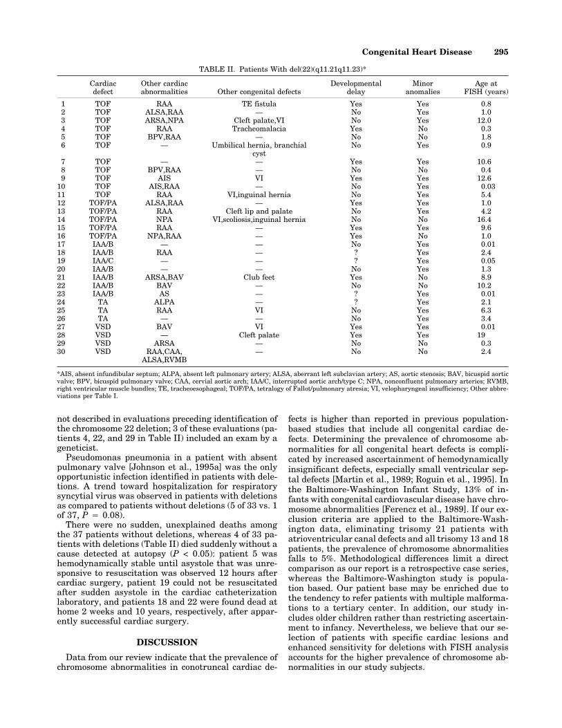
not described in evaluations preceding identification ofthe chromosome 22 deletion; 3 of these evaluations (pa-tients 4, 22, and 29 in Table II) included an exam by ageneticist.
Pseudomonas pneumonia in a patient with absentpulmonary valve [Johnson et al., 1995a] was the onlyopportunistic infection identified in patients with dele-tions. A trend toward hospitalization for respiratorysyncytial virus was observed in patients with deletionsas compared to patients without deletions (5 of 33 vs. 1of 37, P 4 0.08).
There were no sudden, unexplained deaths amongthe 37 patients without deletions, whereas 4 of 33 pa-tients with deletions (Table II) died suddenly without acause detected at autopsy (P < 0.05): patient 5 washemodynamically stable until asystole that was unre-sponsive to resuscitation was observed 12 hours aftercardiac surgery, patient 19 could not be resuscitatedafter sudden asystole in the cardiac catheterizationlaboratory, and patients 18 and 22 were found dead athome 2 weeks and 10 years, respectively, after appar-ently successful cardiac surgery.
DISCUSSION
Data from our review indicate that the prevalence ofchromosome abnormalities in conotruncal cardiac de-
fects is higher than reported in previous population-based studies that include all congenital cardiac de-fects. Determining the prevalence of chromosome ab-normalities for all congenital heart defects is compli-cated by increased ascertainment of hemodynamicallyinsignificant defects, especially small ventricular sep-tal defects [Martin et al., 1989; Roguin et al., 1995]. Inthe Baltimore-Washington Infant Study, 13% of in-fants with congenital cardiovascular disease have chro-mosome abnormalities [Ferencz et al., 1989]. If our ex-clusion criteria are applied to the Baltimore-Wash-ington data, eliminating trisomy 21 patients withatrioventricular canal defects and all trisomy 13 and 18patients, the prevalence of chromosome abnormalitiesfalls to 5%. Methodological differences limit a directcomparison as our report is a retrospective case series,whereas the Baltimore-Washington study is popula-tion based. Our patient base may be enriched due tothe tendency to refer patients with multiple malforma-tions to a tertiary center. In addition, our study in-cludes older children rather than restricting ascertain-ment to infancy. Nevertheless, we believe that our se-lection of patients with specific cardiac lesions andenhanced sensitivity for deletions with FISH analysisaccounts for the higher prevalence of chromosome ab-normalities in our study subjects.
TABLE II. Patients With del(22)(q11.21q11.23)*
Cardiacdefect
Other cardiacabnormalities Other congenital defects
Developmentaldelay
Minoranomalies
Age atFISH (years)
1 TOF RAA TE fistula Yes Yes 0.82 TOF ALSA,RAA — No Yes 1.03 TOF ARSA,NPA Cleft palate,VI No Yes 12.04 TOF RAA Tracheomalacia Yes No 0.35 TOF BPV,RAA — No No 1.86 TOF — Umbilical hernia, branchial
cystNo Yes 0.9
7 TOF — — Yes Yes 10.68 TOF BPV,RAA — No No 0.49 TOF AIS VI Yes Yes 12.6
10 TOF AIS,RAA — No Yes 0.0311 TOF RAA VI,inguinal hernia No Yes 5.412 TOF/PA ALSA,RAA — Yes Yes 1.013 TOF/PA RAA Cleft lip and palate No Yes 4.214 TOF/PA NPA VI,scoliosis,inguinal hernia No No 16.415 TOF/PA RAA — Yes Yes 9.616 TOF/PA NPA,RAA — Yes No 1.017 IAA/B — — No Yes 0.0118 IAA/B RAA — ? Yes 2.419 IAA/C — — ? Yes 0.0520 IAA/B — — No Yes 1.321 IAA/B ARSA,BAV Club feet Yes No 8.922 IAA/B BAV — No No 10.223 IAA/B AS — ? Yes 0.0124 TA ALPA — ? Yes 2.125 TA RAA VI No Yes 6.326 TA — — No Yes 3.427 VSD BAV VI Yes Yes 0.0128 VSD — Cleft palate Yes Yes 1929 VSD ARSA — No No 0.330 VSD RAA,CAA,
ALSA,RVMB— No No 2.4
*AIS, absent infundibular septum; ALPA, absent left pulmonary artery; ALSA, aberrant left subclavian artery; AS, aortic stenosis; BAV, bicuspid aorticvalve; BPV, bicuspid pulmonary valve; CAA, cervial aortic arch; IAA/C, interrupted aortic arch/type C; NPA, nonconfluent pulmonary arteries; RVMB,right ventricular muscle bundles; TE, tracheoesophageal; TOF/PA, tetralogy of Fallot/pulmonary atresia; VI, velopharyngeal insufficiency; Other abbre-viations per Table I.
Congenital Heart Disease 295

Only 36% of our patients with the cardiac defectslisted in Table III had FISH analysis. However, similarresults for the 22q11 deletion were found in small pro-spective studies from Italy and Japan. Deletions weredetected in 7% (9 of 123) of patients with tetralogy ofFallot and 15% (2 of 13) of those with tetralogy of Fal-lot/pulmonary atresia [Amati et al., 1995]. Takahashiet al. [1995] found deletions in 3 of 30 patients withtetralogy of Fallot, one of 3 patients with interruptionof the aortic arch, and one patient with origin of theright pulmonary artery from the aorta. Althoughdouble outlet right ventricle is considered a conotrun-cal cardiac defect, we and others [Takahashi et al.,1995] have not found 22q11 deletions in patients withthis anatomy.
Isolated ventricular septal defects have been re-ported in patients with DiGeorge and Shprintzen syn-dromes. A right aortic arch has been reported in 33–50% of these patients [Young et al., 1980; Van Mieropand Kutsche, 1986]. The location of these defects hasnot been well characterized. All 4 of our patients withdel 22(q11) syndrome and isolated ventricular septaldefects had surgical confirmation of paramembranous(conoventricular) defects, one also had hypoplasia ofthe infundibular septum. These data support the con-cept that the conal region of the developing heart canbe disrupted by the 22q11 deletion.
Neither cardiac nor extracardiac manifestations canreliably identify patients with the 22q11 deletion.Twelve (all infants) of 37 patients in our combined se-ries and 2 of 11 patients in a previous prospective study[Amati et al., 1995] had no extracardiac signs of del22(q11) syndrome identified before deletions were de-tected. Although data indicate that other arch andconotruncal defects such as right aortic arch, high aor-tic arch, absent infundibular septum, and aberrant
subclavian arteries are more prevalent in tetralogy pa-tients with deletions [Table IV; Momma et al., 1995],none of these are found exclusively in deleted patients.Our series had too few patients with tetralogy/pulmonary atresia to confirm observations that deletedpatients with this cardiac defect have a higher inci-dence of nonconfluent pulmonary arteries and majoraortopulmonary collaterals [Momma et al., 1996].
Reports of the spectrum of phenotype associated withdel 22(q11) syndrome to date are biased. Ascertain-ment by congenital heart anomaly has been the pri-mary indication for cytogenetic testing. Evaluation ofother relatives in heritable cases has indicated signifi-cantly milder phenotypes than in index cases. Al-though this may reflect the fact that parents of deletioncases would be expected to have milder cardiac pheno-types to have survived into child-bearing years, similarvariability has been appreciated in the affected sibs ofpropositi as well as in affected monozygotic twins[Goodship et al., 1995]. The wide range in phenotype inthis disorder points to the need for collaborative stud-ies of large numbers of families segregating the 22q11deletion to resolve better the phenotypes resultingfrom this deletion. Given the large numbers of patientsascertained over the past few years, this may be amongthe most common deletion syndromes in humans [duMontcel et al., 1996].
Ongoing investigations of the 22q11 critical regionmay allow identification of genes that are important incardiac development. A gene that encodes a putativetranscriptional regulator has been localized to the criti-cal region [Halford et al., 1993]. Cells from the neuralcrest may play a key role in the pathogenesis of cono-truncal cardiac defects [Kirby and Waldo, 1990].
The identification of 5 patients with abnormalities of8p in our series adds weight to the theory that criticalgenes for cardiac development are present in this re-gion. Atrioventricular canal and conotruncal lesionsare the most frequent cardiac anomalies associatedwith 8p deletions in our patients and in previous re-ports; however, other defects described in these pa-tients include valvular pulmonary stenosis, atrial sep-tal defect, ventricular septal defect, double outlet rightventricle, and hypoplastic left heart syndrome [Digilioet al., 1993; Gelb et al., 1991; Hutchinson et al., 1992;Wu et al., 1996]. Patient 3 (Table I) had a completeatrioventricular canal and tetralogy of Fallot. Thiswell-recognized, but infrequent, combination of cardiacdefects is associated with trisomy 21 in 35–43% of pa-
TABLE III. Prevalence of Chromosome Abnormalities in Selected Cardiac Defects
Number
Number of patients tested
22q11deletion
Otherabnormalities
Total withchromosome
abnormality (%)FISH andcytogenetic
Cytogeneticonly
Tetralogy of Fallot 140 28 7 11 6 17 (12)Tetralogy of
Fallot/pulmonary atresia19 15 2 5 0 5 (26)
Interruped aortic arch 18 11 1 7 1 8 (44)Truncus arteriosus 26 11 3 3 0 3 (12)Absent pulmonary valvea 10 9 — 6 0 6 (60)Double outlet right ventricle 21 11 4 0 1 1 (5)
aPatients with deletions previously reported.
TABLE IV. Frequency of Clinical Findings in Patients Withand Without del(22)(q11.21q11.23)
Manifestations
Withdeletion (%)
(n 4 33)
Withoutdeletion (%)
(n 4 37) P
Right aortic arch 17 (52) 11 (30) 0.06Aberrant subclavian artery 5 (15) 2 (5) 0.2Syndromal 24 (73) 10 (27) 0.0003Cleft palate/velopharyngeal
incompetence7 (21) 1 (3) 0.02
Hypocalcemia 8 (24) 0 (0) 0.001
296 Johnson et al.
tients [Nath et al., 1984; Uretzky et al., 1984]. In ourpatient with an inverted duplication 8p, the finding oftruncus arteriosus (patient 2, Table I) is unusual be-cause typically atrial and ventricular septal defects areassociated with 8p duplications [Roskes et al., 1990].However, it has been demonstrated that inverted 8pduplication patients are deleted of sequences in the8p23 region [Dill et al., 1987; Guo et al., 1995].
Developmental delay/mental retardation has been acommon finding in patients with 8p deletions, occur-ring in all of our patients and all cases reported byothers [Digilio et al., 1993; Hutchinson et al., 1992; Wuet al., 1996]. Others suggest that mental retardation isless severe in the smaller more distal deletions [Hutch-inson et al., 1992]. Agenesis of the corpus callosum wasfound in our patient with an inverted 8p duplication,and partial agenesis was reported in one patient withdel(8)(p23) and pulmonary valve stenosis [Hutchinsonet al., 1992]. In fact, a presumptive locus for develop-ment of the corpus callosum was identified on the basisof patients with trisomy 8 syndrome [Digilio et al.,1994]. Our data also confirm observations that minorfacial and genitourinary anomalies are present in pa-tients with chromosome 8p abnormalities [Digilio etal., 1993; Hutchinson et al., 1992].
We conclude that a careful physical examination andcytogenetic analysis with FISH testing for the 22q11deletion are indicated in patients with absent pulmo-nary valve, tetralogy of Fallot, tetralogy of Fallot/pulmonary atresia, interrupted aortic arch, and trun-cus arteriosus. Identification of patients with deletionswill facilitate genetic counseling and screening for as-sociated medical conditions such as immune disorders,hypocalcemia, growth delay, learning disabilities,speech disturbances, and renal anomalies [Wilson etal., 1993; Goldberg et al., 1993]. In addition, prophy-lactic treatment of hypocalcemia in the immediate pe-riod after cardiac surgery in these patients may be con-sidered. We speculate that cardiac hypocalcemia mayhave contributed to the 4 sudden deaths in our patientswith 22q11 deletions. Reduced myocardial calciumchannel activity was observed in chicks with truncusarteriosus defects secondary to cardiac neural crest ab-lation [Aiba and Creazzo, 1992]
Addition of our 5 patients with congenital heart de-fects and 8p abnormalities to those previously reported[Digilio et al., 1993; Gelb et al., 1991; Hutchinson et al.,1992] suggests that this region should be carefullyscrutinized in patients with mental retardation and ei-ther conotruncal or atrioventricular canal defects.
REFERENCES
Aiba S, Creazzo TL (1992): Calcium currents in hearts with persistenttruncus arteriosus. Am J Physiol 262:H1182–H1190.
Amati F, Mari A, Digilio MC, Mingarelli R, Marino B, Giannotti A, NovelliG, Dallapiccola B (1995): 22q11 deletions in isolated and syndromicpatients with tetralogy of Fallot. Hum Genet 95:479–482.
de la Chapelle A, Herva R, Koivisto M, Aula P (1981): A deletion in chro-mosome 22 can cause DiGeorge syndrome. Hum Genet 57:253–256.
Digilio MC, Giannotti A, Floridia G, Uccellatore F, Mingarelli R, DanesinoC, Dallapiccola B, Zuffardi O (1994): Trisomy 8 syndrome owing toisodicentric 8p chromosomes: Regional assignment of a presumptivegene involved in corpus callosum development. J Med Genet 31:238–241.
Digilio MC, Marion B, Dallapiccola B (1993): Atrioventricular canal and8p- syndrome. Am J Med Genet 47:437–438.
Dill FJ, Schertzer M, Sandercock J, Tischler B, Wood S (1987): Tandemduplication generates a duplication deficiency of chromosome 8p. ClinGenet 32:109–113.
du Montcel ST, Mendizabal H, Ayme S, Levy A, Philip N (1996): Prevalenceof 22q11 microdeletion. J Med Genet 33:719.
Ferencz C, Neill CA, Boughman JA, Rubin JD, Brenner JI, Perry LW(1989): Congenital cardiovascular malformations associated with chro-mosome abnormalities: An epidemiologic study. J Pediatr 114:79–86.
Gelb BD, Towbin JA, McCabe ERB, Sujansky E (1991): San Luis Valleyrecombinant chromosome 8 and tetralogy of Fallot: A review of chro-mosome 8 anomalies and congenital heart disease. Am J Med Genet40:471–476.
Goldberg R, Motzkin B, Marion R, Scambler PJ, Shprintzen RJ (1993):Velo-cardio-facial syndrome: A review of 120 patients. Am J Med Genet45:313–319.
Goodship J, Cross I, Scambler P, Burn J (1995): Monozygotic twins withchromosome 22q11 deletion and discordant phenotype. J Med Genet32:746–748.
Greenberg F, Elder FFB, Haffner P, Northrup H, Ledbetter DH (1988):Cytogenetic findings in a prospective series of patients with DiGeorgeanomaly. Am J Hum Genet 43:605–611.
Guo WJ, Callif-Daley F, Zapata MC, Miller ME (1995): Clinical and cyto-genetic findings in seven cases of inverted duplication of 8p with evi-dence of a telomeric deletion using fluorescence in situ hybridization.Am J Med Genet 58:230–236.
Halford S, Wadey R, Roberts C, Daw SCM, Whiting JA, O’Donnell H,Dunham I, Bentley D, Lindsay E, Baldini A, Francis F, Lehrach H,Williamson R, Wilson DI, Goodship J, Cross I, Burn J, Scambler PJ(1993): Isolation of putative transcriptional regulator from the region of22q11 deleted in DiGeorge syndrome, Shprintzen syndrome and famil-ial congenital heart disease. Hum Mol Genet 2:2099–2107.
Hutchinson R, Wilson M, Voullaire L (1992): Distal 8p deletion (8p23.1 −>8pter): A common deletion? J Med Genet 29:407–411.
Johnson MC, Strauss AW, Dowton SB, Spray TL, Huddleston CB, WoodMK, Slaugh RA, Watson MS (1995a): Deletion within chromosome 22 iscommon in patients with absent pulmonary valve syndrome. Am JCardiol 76:66–69.
Johnson MC, Watson MW, Strauss AW, Spray TL (1995b): Anomalousorigin of the right pulmonary artery from the aorta and CATCH 22syndrome. Ann Thorac Surg 60:681–683.
Kirby ML, Waldo KL (1990): Role of neural crest in congenital heart dis-ease. Circulation 82:332–340.
Martin GR, Perry LW, Ferencz C (1989): Increased prevalence of ventricu-lar septal defect: Epidemic or improved diagnosis. Pediatrics 83:200–203.
Mitelman F (1995): ‘‘An International System for Human Cytogenetic No-menclature.’’ Basel: S. Karger.
Momma K, Kondo C, Matsuoka R (1996): Tetralogy of Fallot with pulmo-nary atresia associated with chromosome 22q11 deletion. J Am CollCardiol 27:198–202.
Momma K, Kondo C, Ando M, Matsuoka R, Takao A (1995): Tetralogy ofFallot associated with chromosome 22q11 deletion. Am J Cardiol 76:618–621.
Nath PH, Soto B, Bini RM, Bargeron LM, Pacifico AD (1984): Tetralogy ofFallot with atrioventricular canal. J Thorac Cardiovasc Surg 87:421–430.
Nora JJ, Berg K, Nora AH (1991): ‘‘Cardiovascular Diseases: Genetics,Epidemiology, and Prevention.’’ New York, Oxford: Oxford UniversityPress, pp 146–168.
Payne RM, Johnson MC, Grant JW, Strauss AW (1995): Towards a mo-lecular understanding of congenital heart disease. Circulation 91:494–504.
Roguin N, Du ZD, Barak M, Nasser N, Hershkowitz S, Milgram E (1995):High prevalence of muscular ventricular septal defect in neonates. JAm Coll Cardiol 26:1545–1548.
Roskes EJ, Boughman JA, Schwartz S, Cohen MM (1990): Congenital car-diovascular malformations (CCVM) and structural chromosome abnor-malities: A report of 9 cases and literature review. Clin Genet 38:198–210.
Takahashi K, Kido S, Hoshino K, Ogawa K, Ohashi H, Fukushima Y(1995): Frequency of a 22q11 deletion in patients with conotruncal
Congenital Heart Disease 297

cardiac malformations: A prospective study. Eur J Pediatr 154:878–881.
Uretzky G, Puga F, Danielson GK, Feldt RH, Julsrud PR, Seward JB,Edwards WD, McGoon DC (1984): Complete atrioventricular canal as-sociated with tetralogy of Fallot. J Thorac Cardiovasc Surg 87:756–766.
Van Mierop LHS, Kutsche LM (1986): Cardiovascular anomalies in Di-George syndrome and importance of neural crest as a possible patho-genetic factor. Am J Cardiol 58:133–137.
Wilson DI, Burn J, Scambler P, Goodship J (1993): DiGeorge syndrome:Part of catch 22. J Med Genet 30:852–856.
Wu BL, Schneider GH, Sabatino DE, Bozovic LZ, Cao B, Korf BR (1996):Distal 8p deletion (8)(p23.1): An easily missed chromosomal abnormal-ity that may be associated with congenital heart defect and mentalretardation. Am J Med Genet 62:77–83.
Young D, Shprintzen RJ, Goldberg RB (1980): Cardiac malformations inthe velocardiofacial syndrome. Am J Cardiol 46:643–648.
298 Johnson et al.