clinical application of mn-dpdp- enhanced 3d-t1w mr
TRANSCRIPT

Clinical application of Mn-DPDP-
enhanced 3D-T1W MR
cholangiography: Additional
information to the conventional T2W
MR cholangiography
Mi-Suk Park
Department of Medicine
The Graduate School, Yonsei University

Clinical application of Mn-DPDP-
enhanced 3D-T1W MR
cholangiography: Additional
information to the conventional
T2W MR cholangiography
Directed by Professor Ki Whang Kim
The Doctoral Dieesertation submitted to the
Department of Medicine, the Graduate School of
Yonsei University in partial fulfillment of the
requirements for the degree of Doctor of Medicine
Mi-Suk Park
This certifies that the Doctoral
Dissertation of Mi-Suk Park is approved.
Ki Whang Kim: Thesis Supervisor
Sang In Lee: Thesis Committee Member
Jong Du Lee: Thesis Committee Member
Woo Jung Lee: Thesis Committee Member
Dong-Sup Yoon: Thesis Committee Member
he Graduate School Yonsei
University
December 2004
Acknowledgements
I would like to express my deepest gratitude to my thesis
supervisor and mentor Dr. Ki Whang Kim. He has inspired me to
strive for excellence not only through the course of this
thesis but also in the everyday encounters with the patients
in the hospital.
I would also like to thank Dr. Sang In Lee, Dr. Jong Du Lee,
Dr. Woo Jung Lee, and Dr. Dong-Sup Yoon whose insightful
comments were essential in completing this thesis.
I wish to thank Dr. Jeong-Sik Yu, Dr. Myeong-Jin Kim, Dr.
Kyoung Won Kim, Dr. Hyun Kwon Ha, Dr. Jae Hee Lee, and Dr. Joon
Suk Lim who generously shared his valuable ideas and subjects
of this study. I also thank Dr. Jin Young Choi who helped data analysis.
I am deeply indebted to my parents who always provided a
solid foundation for me to spread my wings.
December 2004,
Mi-Suk Park

i
Contents
Abstract-------------------------------------------------------- 1
I. Introduction ------------------------------------------------5
II. Materials and Methods -----------------------------------9
1. Study population -----------------------------------9
2. Imaging technique ----------------------------------- 10
3. Image analysis ---------------------------------- 12
4. Statistical analysis ------------------------------ 17
III. Results -----------------------------------------------19
IV. Discussion -------------------------------------------45
V. Conclusion ------------------------------------------------61
Reference --------------------------------------------------62
Abstract in Korean ---------------------------------------66
ii
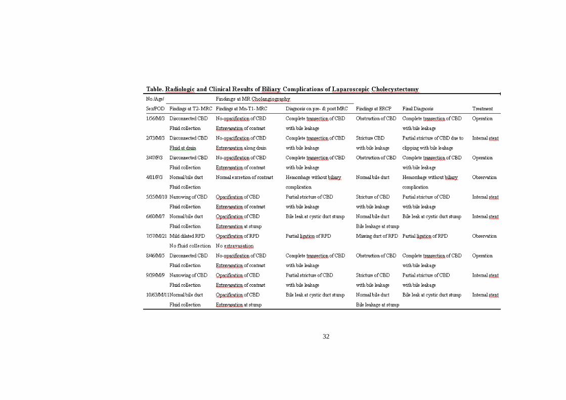
Table
Table 1.Table 1.Table 1.Table 1. Secretion time of manganese in control patients
---------------------- 19
Table 2.Table 2.Table 2.Table 2. Out-flow obstruction of gallbladder in patients with
acute cholecystitis -------- 22
Table 3.Table 3.Table 3.Table 3. Radiologic and clinical results of biliary
complications of laparoscopic cholecystectomy -– 32
Table 4Table 4Table 4Table 4.... Findings of intrahepatic choledocholithiasis at MR
cholangiography. ---------------- 40

iii
Figure
Figure 1. Figure 1. Figure 1. Figure 1. Receiver operating characteristic curves ----- 23
Figure 2.Figure 2.Figure 2.Figure 2. A 48-year-old man with typical appearance of acute
cholecystitis. ----------------------- 24
Figure 3.Figure 3.Figure 3.Figure 3. A 49-year-old woman with suspicious acute
cholecystitis. ----------------------- 26
Figure 4.Figure 4.Figure 4.Figure 4. A 56-year-old man with complete transection of CBD
with bile leakage. ------------------- 33
Figure 5. Figure 5. Figure 5. Figure 5. A 35-year-old man with partial stricture of CBD with
bile leakage. ------------------- 35
Figure 6. Figure 6. Figure 6. Figure 6. A 57-year-old man with ligation of aberrant right
posterior duct. ------------------- 37
Figure 7. Figure 7. Figure 7. Figure 7. A 47-year-old man with intrahepatic
choledocholithiasis. ------------------- 41
Figure 8. Figure 8. Figure 8. Figure 8. A 47-year-old man with intrahepatic
choledocholithiasis ------------------- 43

1
AbstractAbstractAbstractAbstract
Clinical application of MnClinical application of MnClinical application of MnClinical application of Mn----DPDPDPDPDPDPDPDP----enhanced 3Denhanced 3Denhanced 3Denhanced 3D----T1W MR T1W MR T1W MR T1W MR
cholacholacholacholangiography: Additional information to the conventional ngiography: Additional information to the conventional ngiography: Additional information to the conventional ngiography: Additional information to the conventional
T2W MR cholangiography T2W MR cholangiography T2W MR cholangiography T2W MR cholangiography
MiMiMiMi----Suk ParkSuk ParkSuk ParkSuk Park
Department of Medicine
The Graduate School, Yonsei University
(Directed by Professor Ki Whang KimKi Whang KimKi Whang KimKi Whang Kim)
PURPOSE: PURPOSE: PURPOSE: PURPOSE: To determine prospectively the additional information
and clinical application of manganese-enhanced 3D-T1W MR
cholangiography combined with conventional T2W MR
cholangiography in the various biliary diseases
MATERIALS AND METHODS:MATERIALS AND METHODS:MATERIALS AND METHODS:MATERIALS AND METHODS: Conventional heavily T2W MR
cholangiography with manganese-enhanced T1W and T2W MR
cholangiography were performed in 64 patients with high
2
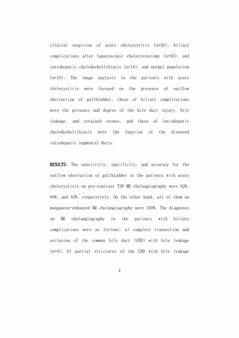
clinical suspicion of acute cholecystitis (n=30), biliary
complications after laparoscopic cholecystectomy (n=10), and
intrahepatic choledocholithiasis (n=14), and normal population
(n=10). The image analysis in the patients with acute
cholecystitis were focused on the presence of outflow
obstruction of gallbladder, those of biliary complications
were the presence and degree of the bile duct injury, bile
leakage, and retained stones, and those of intrahepatic
choledocholithiasis were the function of the diseased
intrahepatic segmental ducts.
RESULTS:RESULTS:RESULTS:RESULTS: The sensitivity, specificity, and accuracy for the
outflow obstruction of gallbladder in the patients with acute
cholecystitis on pre-contrast T2W MR cholangiography were 82%,
88%, and 83%, respectively. On the other hand, all of them on
manganese-enhanced MR cholangiography were 100%. The diagnoses
on MR cholangiography in the patients with biliary
complications were as follows: a) complete transection and
occlusion of the common bile duct (CBD) with bile leakage
(n=4); b) partial strictures of the CBD with bile leakage
3
(n=2); c) cystic duct leakage (n=2); d) partial ligation of
aberrant right hepatic duct (n=1), and hemorrhage without
biliary complication (n=1). MR cholangiography accurately
yielded the same findings as the final diagnoses on surgery or
ERCP, except one case with partial stricture of the bile duct
with bile leakage (over-diagnosed as complete occlusion on MR
cholangiography). Of 22 diseased segmental ducts in the
patients with intrahepatic choledocholithiasis on conventional
T2W MR cholangiography, eight segmental ducts were filled with
contrast at manganese-enhanced T1W MR cholangiography,
indicating that they were functioning bile ducts. The
remaining 14 segmental ducts were not filled with contrast at
manganese-enhanced T1W MR cholangiography, hence they were
non-functioning bile ducts.
CONCLUSION:CONCLUSION:CONCLUSION:CONCLUSION: T2W MR cholangiography combined with manganese-
enhanced T1W MR cholangiography provided not only the anatomic
detail, but also the functional detail. Therefore, manganese-
enhanced MR cholangiography is a useful supplement to
conventional heavily T2W MR cholangiography in the evaluation
4
of acute cholecystitis, biliary complications after
laparoscopic cholecystectomy, and intrahepatic
choledocholithiasis
Key words:Key words:Key words:Key words: bile ducts, calculi, cholangiography, magnetic
resonance, contrast
5
Clinical application of MnClinical application of MnClinical application of MnClinical application of Mn----DPDPDPDPDPDPDPDP----enhanced 3Denhanced 3Denhanced 3Denhanced 3D----T1W MR T1W MR T1W MR T1W MR
cholangiography: Additional information to the conventicholangiography: Additional information to the conventicholangiography: Additional information to the conventicholangiography: Additional information to the conventional onal onal onal
T2W MR cholangiographyT2W MR cholangiographyT2W MR cholangiographyT2W MR cholangiography
MiMiMiMi----Suk ParkSuk ParkSuk ParkSuk Park
Department of Medicine
The Graduate School, Yonsei University
(Directed by Professor Ki Whang KimKi Whang KimKi Whang KimKi Whang Kim)
ⅠⅠⅠⅠ. INTRODUCTION. INTRODUCTION. INTRODUCTION. INTRODUCTION
Since its introduction in 1991, technical advances in
heavily T2W MR cholangiopancreatography have made it possible
to generate high-quality images of the bile ducts 1-3. With a
breath-hold half-Fourier rapid acquisition with relaxation
enhancement (RARE) sequence and a phased-array body coil, we
obtained high-quality images that show both the dilated and
non-dilated biliary system without motion artifact 1-3.
Sequential acquisition of multiple 3-mm-thick sections
improved spatial resolution and allowed calculi as small as 2-
6
3 mm to be depicted. For the accuracy in the diagnosis of
various biliary diseases, heavily T2W MR
cholangiopancreatography has proven to be comparable or
superior to that of direct cholangiography 1-3. Moreover MR
cholangiography has no complications or contraindications, is
able to depict the entire biliary tree including both the
proximal and distal part of the obstruction, and shows
coexistent parenchymal lesions. MR cholangiography is now
replacing purely diagnostic direct cholangiography in the
various biliary diseases. However, heavily T2W MR
cholangiopancreatography without contrast material is not able
to reflect the dynamics of biliary system.
Mangafodipir trisodium (Mn-DPDP, manganese) is a
paramagnetic contrast agent originally designed for liver
imaging. This contrast agent consists of manganese bound to N,
N'-dipryidoxylethylenediamine-N, N'-diacetate 5, 5'-bis
(phosphate), or DPDP, is taken up by functioning hepatocytes,
and is primarily excreted in bile into the feces 4-7. Because
manganese is a paramagnetic metal ion, it primarily acts on T1
resulting in T1 shortening, although it also acts on T2

7
resulting in T2 shortening 4-7. Enhanced liver and functioning
bile ducts therefore have higher signal intensity on T1-
weighted images and lower signal intensity on T2-weighted
images 4-7. The signal intensity of the bile ducts that are
obstructed or otherwise not functioning due to stasis, however,
may not be affected because biliary stasis in the setting of
strictures or stones reduces the excretion of biliary
manganese 4-7. Theoretically, this characteristic property of
Mn-DPDP -enhanced MR cholangiography could allow noninvasive
imaging in the evaluation of biliary dynamics 4-7.
There have been a few reports about manganese-enhanced T1W
MR cholangiography in the evaluation of intrahepatic biliary
anatomy and in the detection of bile duct leaks 6,7. To our
knowledge, however, there are no studies in which conventional
heavily T2W MR cholangiography and manganese-enhanced T1W and
T2W MR cholangiography have been compared. To define the
usefulness of manganese-enhanced T1W MR cholangiography in the
evaluation of biliary ductal system, we performed manganese-
enhanced T1W and T2W MR cholangiography in addition to
conventional heavily T2W MR cholangiography. We then sought to
8
determine the influence of this technique on clinical
management decisions in a variety of disease process.

9
ⅡⅡⅡⅡ. . . . MATERIALS MATERIALS MATERIALS MATERIALS ANDANDANDAND METHODS METHODS METHODS METHODS
1. Study Population1. Study Population1. Study Population1. Study Population
Between September 2001 and August 2004, manganese-enhanced
T1W and T2W MR cholangiography in addition to conventional
heavily T2W MR cholangiography were performed in 64 patients
at two institutions. This prospective study population
consists of the patients with acute cholecystitis, biliary
complications after laparoscopic cholecystectomy, and
intrahepatic choledocholithiasis. The patients with jaundice
were excluded from our study.
Ten control patients who had no symptoms referable to the
biliary tract were examined to establish the secretion time of
the manganese into the extraheptic bile duct, gallbladder, and
duodenum.
Thirty patients with a high index of suspicion of acute
cholecystitis clinically (sudden onset of right upper quadrant
tenderness, low-grade fever, and leukocytosis without
jaundice) and at ultrasonography (visualization of impacted
stones in the gallbladder neck or cystic duct or positive US
Murphy sign with gallbladder wall thickening or distention)

10
were examined by MR cholangiography and hepatobiliary
scintigraphy.
Ten patients with high clinical suspicion of biliary
complications (abdominal pain, nausea, or vomiting accompanied
by leukocytosis, or low-grade fever) after laparoscopic
cholecystectomy were examined. All of the patients underwent
sonography or CT scans before MR cholangiography and ERCP.
Fourteen patients with a high index of suspicion of
intrahepatic choledocholithiasis clinically (recurrent attacks
of abdominal pain and fever without jaundice) and at US
(segmental intrahepatic duct dilatation with or without
stones) were examined by MR cholangiography.
2. Imaging Technique2. Imaging Technique2. Imaging Technique2. Imaging Technique
Patients were asked to fast for a minimum of six hours.
MR cholangiography was performed with a 1.5-T
superconducting unit (Magnetom Vision; Siemens Medical Systems,
Erlangen, Germany) and a phased-array torso coil. Two MR
cholangiographic techniques were applied: single-shot RARE and
multislice half-Fourier RARE sequence (HASTE; Siemens). The

11
slab of a single-shot RARE sequence was obtained at various
angles to allow optimal visualization of the bile ducts. The
multislice half-Fourier RARE images were obtained at an angle
of 20-35° to the coronal plane to simulate a right anterior
oblique projection on direct cholangiography. The imaging
parameters for a single-shot RARE sequence were TR/effective
TE, infinite/1200; echo-train length, 240; flip angle, 150°;
slab thickness, 50-70 mm; field of view, 300-350 mm; matrix,
240 x 256; and an acquisition time, 2.32 sec. The imaging
parameters for multislice half-Fourier RARE sequence were
TR/effective TE, infinite/95; echo-train length, 128; flip
angle, 150°; slab thickness, 3-5 mm with no gab; number of
slice, 13-15 (range of coverage, 52-60 mm); field of view,
300-350 mm; matrix, 240 x 256; and an acquisition time, 18 sec.
Postprocessing of the multislice half-Fourier RARE images was
performed.
After the patients were removed from the magnet, an IV
injection of mangafodipir trisodium (Mn DPDP; Teslascan,
Nycomed Amersham, Oslo, Norway) at a standard dose of 5
μmole/kg (0.1 mL/kg; maximum dose, 15 mL) was administered

12
via a slow injection over 2-3 ml/min followed by a 10 ml
saline flush. Contrast-related adverse events, only flushing,
occurred in one patient. Twenty-five to forty-five min after
injection, we obtained 3D T1-weighted fat-saturated volumetric
interpolated breath-hold images (TR/TE, 4.2/1.6; flip angle,
120; matrix 205x256; field of view, 300-350 mm; and 24
partitions interpolated to 48 slices with a thickness of 1.3
mm). Routine heavily T2-weighted MR cholangiography using the
same half-Fourier RARE sequence was repeatedly performed.
Because mangafodipir trisodium is a negative contrast agent on
T2-weighted images, we obtained post-contrast heavily T2-
weighted MR cholangiography to compare pre-contrast T2-
weighted MR cholangiography images. All MR cholangiograms were
reviewed at the console by an abdominal radiologist before the
patient was removed from the magnet, as a standard protocol.
Additional examinations were obtained with a delay of 1 and
4 hours if the radiologist deemed it necessary.
3. Image Analysis3. Image Analysis3. Image Analysis3. Image Analysis
The MR cholangiograms were interpreted prospectively by the
13
consensus of two abdominal radiologists, who were blinded to
the patients’ clinical information and the other image
findings. The source images as well as the three-dimensional
reconstruction images of conventional heavily T2-weighted MR
cholangiography and manganese-enhanced T1-, and T2-weighted MR
cholangiography were reviewed on a Picture Archiving
Communicating System (PACS) workstation monitor.
Image analysis in the patients with suspicion of acute
cholecystitis focused on the presence and location of calculi
in the gallbladder, and the associated findings, such as
gallbladder wall thickening, fluid collection around the
gallbladder, or common bile duct calculi on conventional
heavily T2W MR cholangiography. Outflow obstruction of the
gallbladder on conventional heavily T2W MR cholangiography was
definitively defined when a signal void was identified within
the cystic duct or gallbladder neck on images obtained in at
least two different projections. On manganese-enhanced T1W MR
cholangiography, the presence and time of visualization of the
gallbladder and bile duct were evaluated. Outflow obstruction
of the gallbladder was defined as no-contrast filling in
14
gallbladder until one hour. Outflow obstruction of the
gallbladder on conventional heavily T2W and manganese-enhanced
T1W MR cholangiography was compared with the hepatobiliary
scans.
Image analysis in the patients with suspicion of biliary
complications after laparoscopic cholecystectomy focused on
the presence and location of bile duct leaks, complete bile
duct transection or occlusion, partial stricture of bile duct,
or retained bile duct stones. Bile duct leakage was defined as
contrast agent extravasation adjacent to a bile duct or
contrast agent opacification of a peritoneal drain on
manganese-enhanced T1W MR cholangiography with abnormal
peritoneal fluid collection on conventional heavily T2W MR
cholangiography and signal loss of abnormal peritoneal fluid
collection on manganese-enhanced T2W MR cholangiography. A
complete transection or occlusion of the bile duct was defined
as an absence of opacification of the extrahepatic bile duct
on manganese-enhanced T1W MR cholangiography and persistence
of signal of that on manganese-enhanced T2W MR cholangiography
with disconnection of extrahepatic bile duct on conventional
15
heavily T2W MR cholangiography. Stricture of the bile duct was
defined as the opacification of the extrahepatic bile duct on
manganese-enhanced MR cholangiography and signal loss of that
on manganese-enhanced T2W MR cholangiography, despite the
presence of a narrowing or disconnected segment of the bile
duct on conventional heavily T2W MR cholangiography. The
diagnosis of complications were classified as described in
previous publications 8,9: a) complete transections and
occlusions of the bile duct with or without bile leakage; b)
partial strictures of the bile duct with or without bile
leakage; c) cystic duct leakage and accessory bile duct
leakage; d) occlusion of part of the intrahepatic duct; e)
residual stones. The same investigators reviewed the ERCP
images using the same method as for the MR cholangiographic
images. The diagnoses on the MR cholangiography were compared
with the final diagnoses on surgery or ERCP.
Image analysis in the patients with suspicion of
intrahepatic choledocholithiasis focused to identify
intrahepatic ductal dilatation and stricture, intrahepatic
duct calculi and common duct calculi on conventional heavily
16
T2W MR cholangiograms and to verify bile duct enhancement on
manganese-enhanced T1W MR cholangiograms. The final diagnosis
of diseased bile ducts was divided into two categories -
functioning or non-functioning bile duct - depending on the
combined findings of conventional heavily T2W and manganese-
enhanced T1W MR cholangiograms. Functioning bile ducts were
the bile ducts that were filled with contrast agent at
manganese-enhanced T1W MR cholangiography despite the presence
of intrahepatic ductal dilatation with stricture or calculi at
conventional T2W MR cholangiography. Non-functioning bile
ducts were the diseased segmental ducts on T2W MR
cholangiography in which contrast agent filling did not occur
at manganese-enhanced T1W MR cholangiography. The distribution
of the intrahepatic abnormalities was interpreted based on the
classification of the internal lobation and segmentation of
the liver (four segments: left lateral, left medial, right
anterior, and right posterior). Intrahepatic ductal dilatation
was diagnosed when the diameter of a duct was greater than 3mm,
and stricture was diagnosed when focal caliber change was
present at any segment. Calculi were considered present on MR
17
cholangiographic images when a signal void was identified
within the bile duct in at least two different projections.
4. Statistical Analysis4. Statistical Analysis4. Statistical Analysis4. Statistical Analysis
In the cases of acute cholecystitis, sensitivity,
specificity, accuracy, positive predictive value (PPV), and
negative predictive value (NPV) were calculated as follows:
sensitivity = true positive / (true positive + false
negative); specificity = true negative / (true negative +
false positive); accuracy = (true positive + true negative) /
(true positive + true negative + false positive + false
negative); positive predictive value (PPV) = true positive /
(true positive + false positive); negative predictive value
(NPV) = true negative / (true negative + false negative). The
differences between conventional T2W MR cholangioography and
manganese-enhanced MR cholangiography were tested for
statistical significance by using the McNemar’s test.
A five-point scale was used to assign a confidence level in
the evaluation of the cystic duct obstruction: 1, definitely
patent; 2, probably patent; 3, indeterminate; 4, probably
18
obstruct; 5, definitely obstruct. Receiver operating
characteristic (ROC) curve analysis was performed to compare
the results of readings of conventional T2W MR
cholangioography versus the results of readings of manganese-
enhanced MR cholangiography. Nonparametric receiver operating
characteristic (ROC) curves were fitted by using a computer
software package (AnalyseAnalyseAnalyseAnalyse----itititit, version 1.63; AnalyseAnalyseAnalyseAnalyse----itititit-Software,
Leeds, England). The diagnostic capability was determined by
calculating the area under the ROC curve (Az) for each reader.
For all tests, P < .05 was considered to indicate a
statistically significant difference.

19
ⅢⅢⅢⅢ. RESULTS. RESULTS. RESULTS. RESULTS
1.1.1.1. Control groupControl groupControl groupControl group
The secretion time of the manganese into the extraheptic
bile duct, gallbladder, and duodenum in ten control patients
was summarized in Table 1Table 1Table 1Table 1.
Table 1.Table 1.Table 1.Table 1. Secretion time of manganese in control group
Extrahepatic duct Gallbladder Bowel
10 min 8 3
20 min 2 6 4
30 min 1 5
60 min 1
Total 10 10 10
2. Acute cholecystitis2. Acute cholecystitis2. Acute cholecystitis2. Acute cholecystitis
Among the 30 patients with suspicion of acute
cholecystitis, outflow obstruction of gallbladder on
hepatobiliary scintigraphy was seen in 21 patients. The
sensitivity, specificity, accuracy, PPV, and NPV for the
outflow obstruction of gallbladder on pre-contrast T2-weighted
MR cholangiography were 82%, 88%, 83%, 95%, and 64%,

20
respectively. On the other hand, all of them on manganese-
enhanced MR cholangiography were 100% (Table 2)(Table 2)(Table 2)(Table 2). Statistically,
however, there is no significant difference between pre-
contrast T2-weighted MR cholangiography and manganese-enhanced
MR cholangiography (P = 0.375)
ROC curve (Fig 1)(Fig 1)(Fig 1)(Fig 1) shows that the Az for pre-contrast T2-
weighted MR cholangiography is 0.943 and that for manganese-
enhanced MR cholangiography is 1.0. There is no statistically
significant difference between them (P = 0.057).
In 18 of 30 cases, conventional heavily T2-weighted MR
cholangiography demonstrated cystic duct or gallbladder neck
calculi clearly, and the morphologic evidence of outflow
obstruction was definitive. Manganese-enhanced T1W MR
cholangiography depicted an enhanced common bile duct and
hepatic duct, but not the gallbladder, suggesting a flow
obstruction of the gallbladder. DISIDA scan failed to
visualize the gallbladder for up to four hours after IDA
injection for all of the 18 patients (Fig 2)(Fig 2)(Fig 2)(Fig 2).
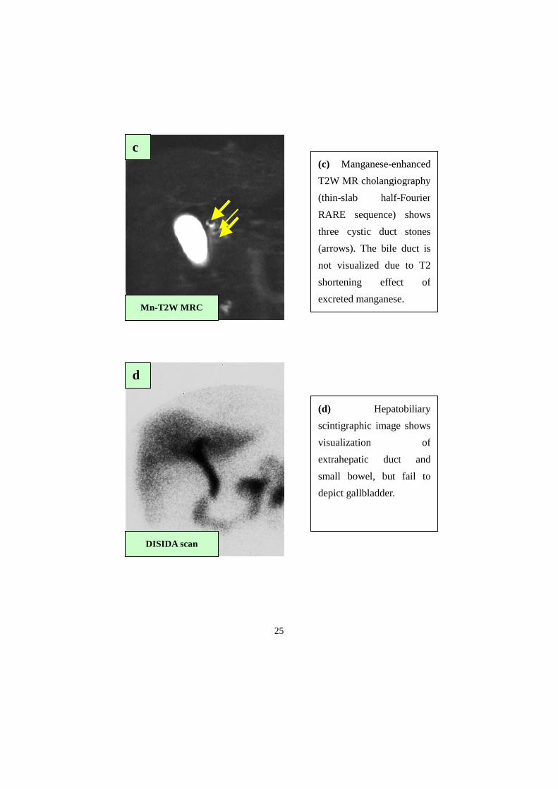
In eleven of 30 cases, heavily T2W MR cholangiography
demonstrated only floating calculi in the gallbladder lumen
21
without cystic duct or gallbladder neck calculus and there was
no evidence of outflow obstruction of the gallbladder. After
the administration of manganese, T1W MR cholangiography
depicted contrast enhancement of the gallbladder in seven
cases suggesting a patent cystic duct. In these cases, DISIDA
scan showed the gallbladder within 45 minutes after IDA
injection. In four cases, however, the gallbladder was not
visualized up to four hours on manganese enhanced T1W MR
cholangiography and DISIDA scan failed to visualize the
gallbladder for up to four hours after IDA injection. In those
cases the findings on heavily T2W MR cholangiography was
mismatched with that on manganese enhanced T1W MR
cholangiography and DISIDA scan (Fig 3)(Fig 3)(Fig 3)(Fig 3).
22
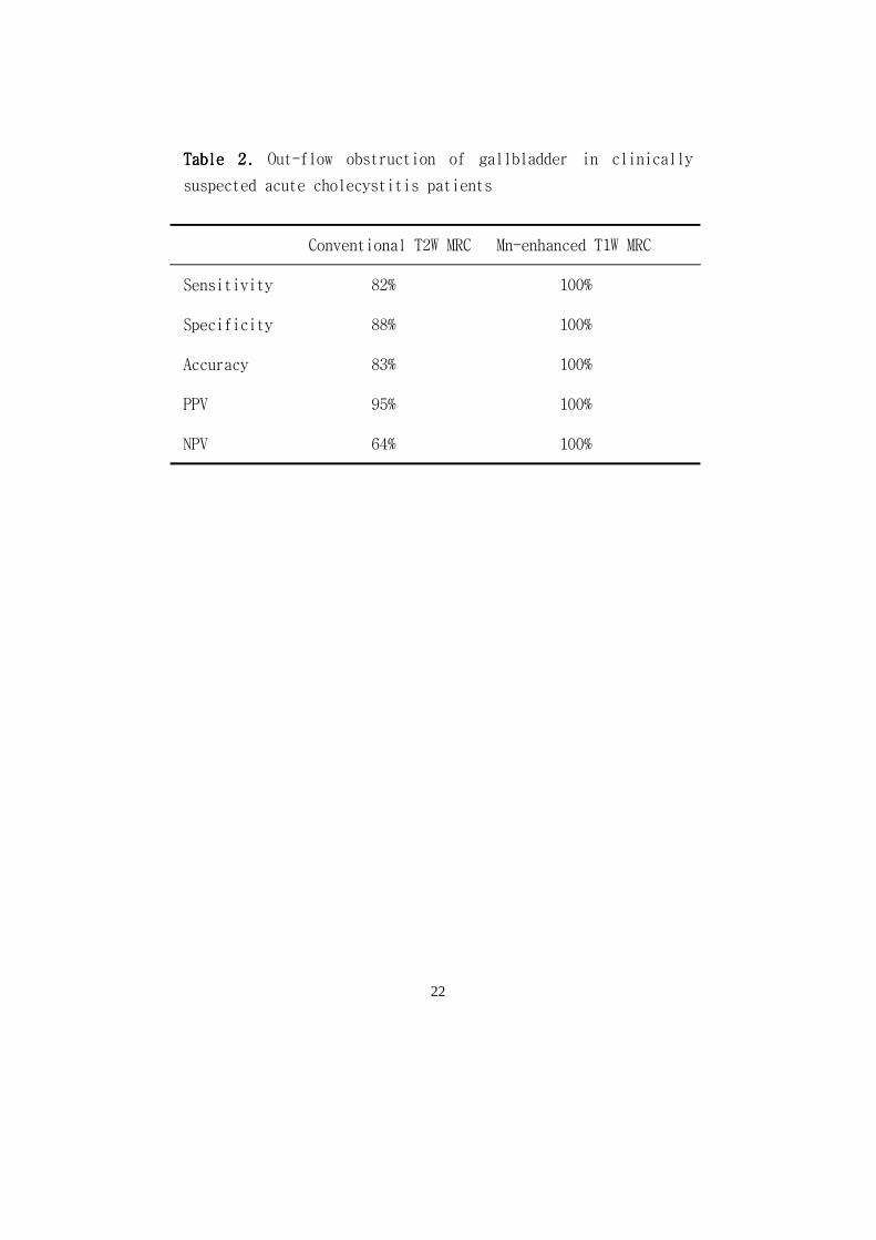
Table 2.Table 2.Table 2.Table 2. Out-flow obstruction of gallbladder in clinically
suspected acute cholecystitis patients
Conventional T2W MRC Mn-enhanced T1W MRC
Sensitivity 82% 100%
Specificity 88% 100%
Accuracy 83% 100%
PPV 95% 100%
NPV 64% 100%

23
Figure 1. Figure 1. Figure 1. Figure 1. Receiver operating characteristic curves illustrate the
increase in diagnostic confidence after manganese-enhanced T1W MR
cholangiography. Graphs are plotted to discriminate between obstruct
and patent cystic duct on conventional heavily T2W MR
cholangiography (continuous line) and on manganese-enhanced T1W
MR cholangiography (dotted line). The curves are shown against a
diagonal (right) line, which represents a review method with which
obstruct and patent ducts cannot be differentiated.
0
0.1
0.2
0.3
0.4
0.5
0.6
0.7
0.8
0.9
1
0 0.2 0.4 0.6 0.8 1
1 - Specificity (false positives)
Sen
siti
vity
(tr
ue
po
siti
ves)
No discrimination
T2
T1
24
Figure 2. Figure 2. Figure 2. Figure 2. A 48-year-old man with acute cholecystitis.
Mn-T1W MRC
(a) Conventional heavily
T2W MR cholangiography
(thin-slab half-Fourier
RARE sequence) shows
three cystic duct stones
(arrows), suggesting
outflow obstruction of
gallbladder.
a
b (b) Manganese-enhanced
3D volumetric interpolated
T1W MR cholangiography
(maximum intensity
projection image) shows
enhanced extrahepatic duct
and small bowel with
nonvisualization of
gallbladder, suggesting
outflow obstruction of
gallbladder.
Con T2W MRC
25
c
d
Mn-T2W MRC
DISIDA scan
(c) Manganese-enhanced
T2W MR cholangiography
(thin-slab half-Fourier
RARE sequence) shows
three cystic duct stones
(arrows). The bile duct is
not visualized due to T2
shortening effect of
excreted manganese.
(d) Hepatobiliary
scintigraphic image shows
visualization of
extrahepatic duct and
small bowel, but fail to
depict gallbladder.
26
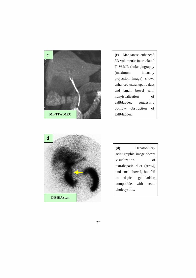
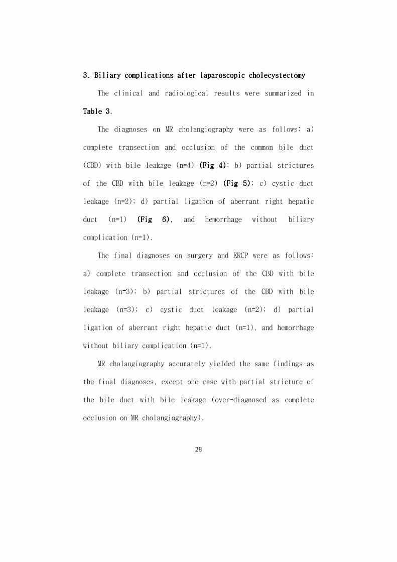
Figure 3. Figure 3. Figure 3. Figure 3. A 49-year-old woman with suspicious acute
cholecystitis.
(a and b) Serial images of
conventional heavily T2W
MR cholangiography
(thin-slab half-Fourier
RARE sequence) show
suspicious signal void
lesions (arrow in b) in the
cystic duct. However, they
cannot be differentiated
from a tortous normal
cystic duct.
Con T2W MRC
Con T2W MRC
a
b
27
(c) Manganese-enhanced
3D volumetric interpolated
T1W MR cholangiography
(maximum intensity
projection image) shows
enhanced extrahepatic duct
and small bowel with
nonvisualization of
gallbladder, suggesting
outflow obstruction of
gallbladder. Mn-T1W MRC
c
d
DISIDA scan
(d) Hepatobiliary
scintigraphic image shows
visualization of
extrahepatic duct (arrow)
and small bowel, but fail
to depict gallbladder,
compatible with acute
cholecystitis.
28
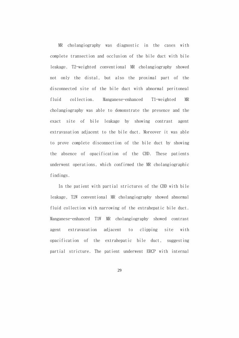
3. Biliary complications after laparoscopic cholecystectomy3. Biliary complications after laparoscopic cholecystectomy3. Biliary complications after laparoscopic cholecystectomy3. Biliary complications after laparoscopic cholecystectomy
The clinical and radiological results were summarized in
Table 3Table 3Table 3Table 3.
The diagnoses on MR cholangiography were as follows: a)
complete transection and occlusion of the common bile duct
(CBD) with bile leakage (n=4) (Fig 4)(Fig 4)(Fig 4)(Fig 4); b) partial strictures
of the CBD with bile leakage (n=2) (Fig 5)(Fig 5)(Fig 5)(Fig 5); c) cystic duct
leakage (n=2); d) partial ligation of aberrant right hepatic
duct (n=1) (Fig(Fig(Fig(Fig 6) 6) 6) 6), and hemorrhage without biliary
complication (n=1).
The final diagnoses on surgery and ERCP were as follows:
a) complete transection and occlusion of the CBD with bile
leakage (n=3); b) partial strictures of the CBD with bile
leakage (n=3); c) cystic duct leakage (n=2); d) partial
ligation of aberrant right hepatic duct (n=1), and hemorrhage
without biliary complication (n=1).
MR cholangiography accurately yielded the same findings as
the final diagnoses, except one case with partial stricture of
the bile duct with bile leakage (over-diagnosed as complete
occlusion on MR cholangiography).
29
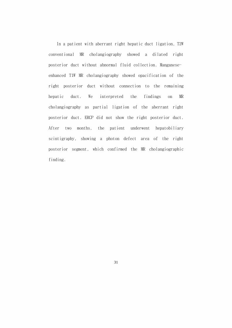
MR cholangiography was diagnostic in the cases with
complete transection and occlusion of the bile duct with bile
leakage. T2-weighted conventional MR cholangiography showed
not only the distal, but also the proximal part of the
disconnected site of the bile duct with abnormal peritoneal
fluid collection. Manganese-enhanced T1-weighted MR
cholangiography was able to demonstrate the presence and the
exact site of bile leakage by showing contrast agent
extravasation adjacent to the bile duct. Moreover it was able
to prove complete disconnection of the bile duct by showing
the absence of opacification of the CBD. These patients
underwent operations, which confirmed the MR cholangiographic
findings.
In the patient with partial strictures of the CBD with bile
leakage, T2W conventional MR cholangiography showed abnormal
fluid collection with narrowing of the extrahepatic bile duct.
Manganese-enhanced T1W MR cholangiography showed contrast
agent extravasation adjacent to clipping site with
opacification of the extrahepatic bile duct, suggesting
partial stricture. The patient underwent ERCP with internal

30
stent, which confirmed the MR cholangiographic finding. .
One patient with partial strictures of the CBD with bile
leakage was over-diagnosed on MR cholangiography. T2W
conventional MR cholangiography showed abnormal fluid
collection with narrowing of the extrahepatic bile duct.
Manganese enhanced T1W MR cholangiography showed contrast
agent opacification of the peritoneal drain without
opacification of the CBD. This case was interpreted as
complete obstruction of the bile duct with bile leakage on MR
cholangiography. The patient underwent ERCP with internal
stent, showing the communication of the extrahepatic bile duct
with bile leakage, suggesting partial stricture of the CBD.
In the patients with cystic duct leakage, T2W conventional
MR cholangiography showed abnormal fluid collection without
narrowing of the extrahepatic bile duct. Manganese-enhanced
T1W MR cholangiography showed contrast agent extravasation
adjacent to the clipping site with normal opacification of the
extrahepatic bile duct. The patients underwent ERCP with
internal stent, which confirmed the MR cholangiographic
finding.
31
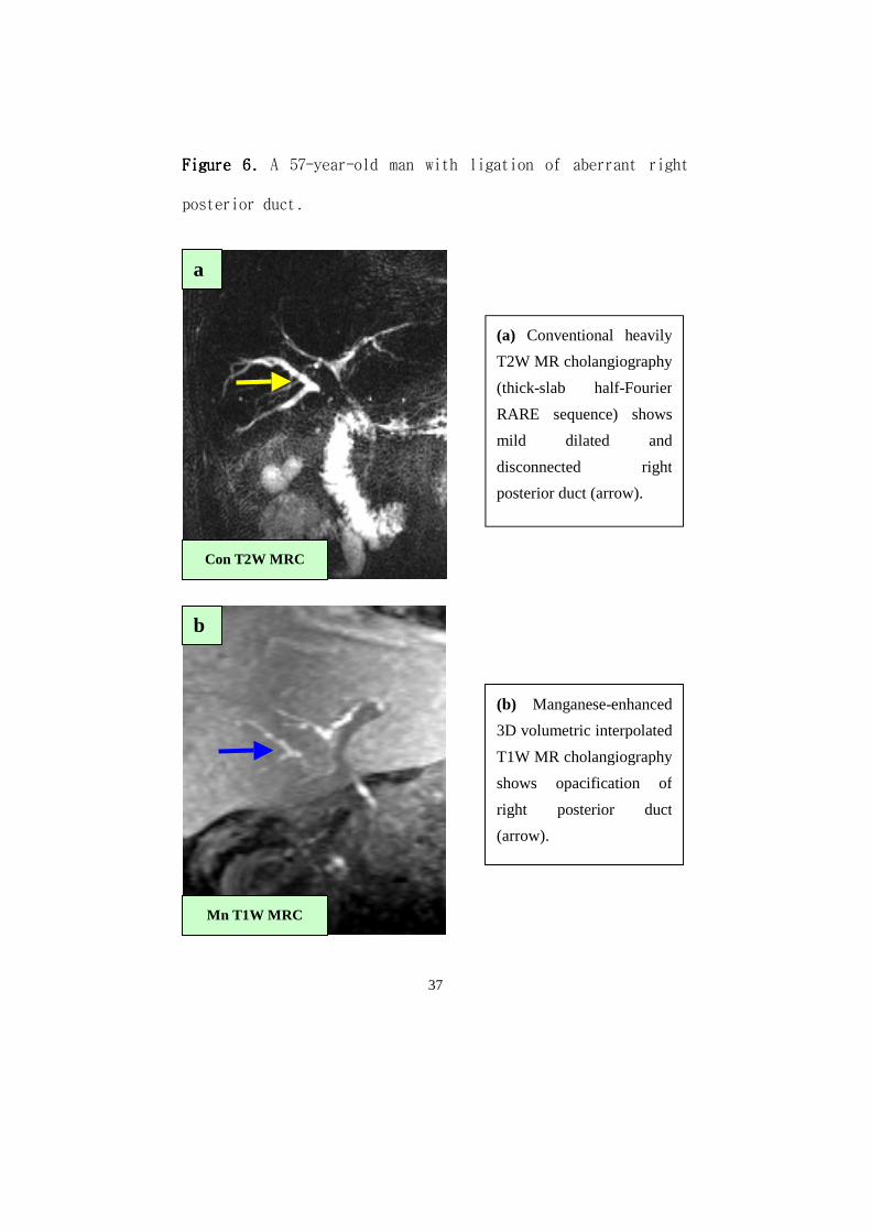
In a patient with aberrant right hepatic duct ligation, T2W
conventional MR cholangiography showed a dilated right
posterior duct without abnormal fluid collection. Manganese—
enhanced T1W MR cholangiography showed opacification of the
right posterior duct without connection to the remaining
hepatic duct. We interpreted the findings on MR
cholangiography as partial ligation of the aberrant right
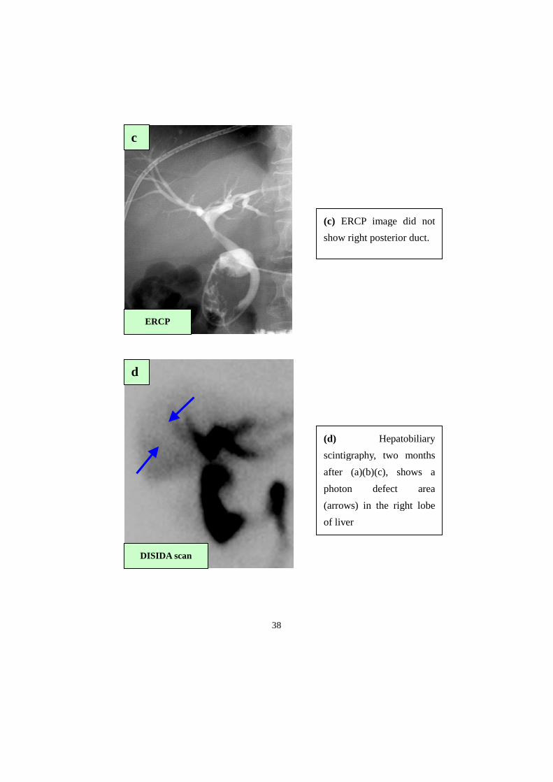
posterior duct. ERCP did not show the right posterior duct.
After two months, the patient underwent hepatobiliary
scintigraphy, showing a photon defect area of the right
posterior segment, which confirmed the MR cholangiographic
finding.
32

33
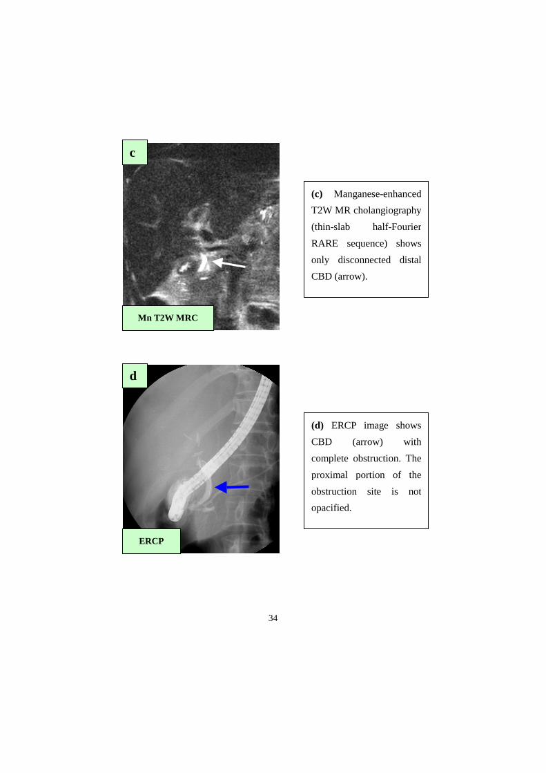
Figure 4. Figure 4. Figure 4. Figure 4. A 56-year-old man with complete transection of CBD
with bile leakage.
(a) Conventional heavily
T2W MR cholangiography
(thin-slab half-Fourier
RARE sequence) shows
disconnected CBD
(arrows), with abnormal
fluid collection (short
arrow).
(b) Manganese-enhanced
3D volumetric interpolated
T1W MR cholangiography
shows extravasation of
contrast material (short
arrow) from EHD (arrow).
Distal CBD is not
visualized.
Mn T1W MRC
a
b
Con T2W MRC
34
(c) Manganese-enhanced
T2W MR cholangiography
(thin-slab half-Fourier
RARE sequence) shows
only disconnected distal
CBD (arrow).
ERCP
c
d
(d) ERCP image shows
CBD (arrow) with
complete obstruction. The
proximal portion of the
obstruction site is not
opacified.
Mn T2W MRC
35
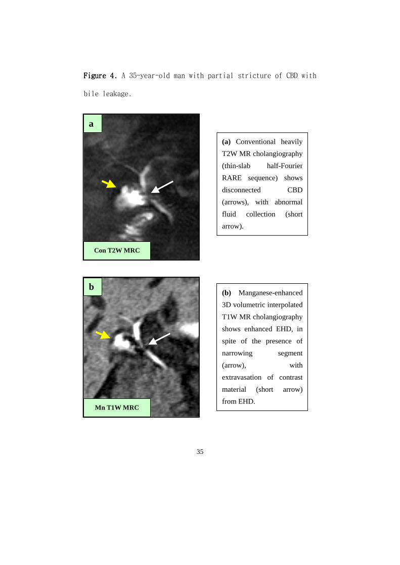
Figure 4. Figure 4. Figure 4. Figure 4. A 35-year-old man with partial stricture of CBD with
bile leakage.
(a) Conventional heavily
T2W MR cholangiography
(thin-slab half-Fourier
RARE sequence) shows
disconnected CBD
(arrows), with abnormal
fluid collection (short
arrow).
(b) Manganese-enhanced
3D volumetric interpolated
T1W MR cholangiography
shows enhanced EHD, in
spite of the presence of
narrowing segment
(arrow), with
extravasation of contrast
material (short arrow)
from EHD.
Con T2W MRC
Mn T1W MRC
a
b
36
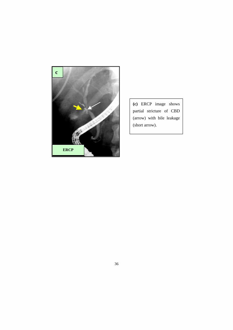
(c) ERCP image shows
partial stricture of CBD
(arrow) with bile leakage
(short arrow).
ERCP
c
37
Figure 6. Figure 6. Figure 6. Figure 6. A 57-year-old man with ligation of aberrant right
posterior duct.
(a) Conventional heavily
T2W MR cholangiography
(thick-slab half-Fourier
RARE sequence) shows
mild dilated and
disconnected right
posterior duct (arrow).
(b) Manganese-enhanced
3D volumetric interpolated
T1W MR cholangiography
shows opacification of
right posterior duct
(arrow).
Con T2W MRC
Mn T1W MRC
a
b
38
(c) ERCP image did not
show right posterior duct.
(d) Hepatobiliary
scintigraphy, two months
after (a)(b)(c), shows a
photon defect area
(arrows) in the right lobe
of liver
ERCP
DISIDA scan
c
d

39
4. Intreheptic choledocholithiasis4. Intreheptic choledocholithiasis4. Intreheptic choledocholithiasis4. Intreheptic choledocholithiasis
The findings of intrahepatic choledocholithiasis on MR
cholangiography are summarized in Table4Table4Table4Table4.
According to the findings on conventional T2W MR
cholangiography, 14 patients had 22 diseased segments.
Intrahepatic bile ductal dilatation with calculi with or
without stricture was present in eight segments. Among them,
three segmental ducts were filled with contrast at manganese-
enhanced T1W MR cholangiography. Intrahepatic bile ductal
dilatation with stricture without calculi was present in seven
segments. Among them, four segmental ducts were filled with
contrast at manganese-enhanced T1W MR cholangiography.
Impacted calculi without bile juice were present in six
segmental ducts. None of them was filled with contrast at
manganese-enhanced T1W MR cholangiography. Only one segmental
duct was dilated without calculi or stricture. That was filled
with contrast at manganese-enhanced T1W MR cholangiography.
Of 22 diseased segmental ducts on conventional T2W MR
cholangiography, 14 segmental ducts were not filled with
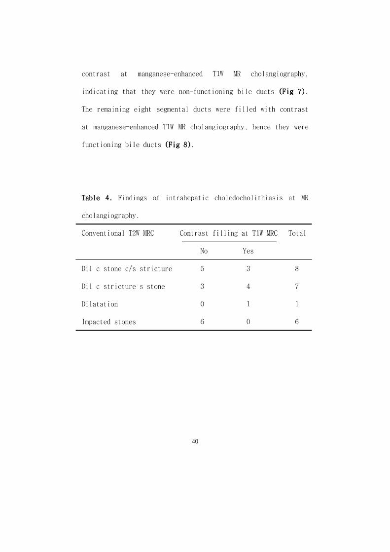
40
contrast at manganese-enhanced T1W MR cholangiography,
indicating that they were non-functioning bile ducts (Fig 7)(Fig 7)(Fig 7)(Fig 7).
The remaining eight segmental ducts were filled with contrast
at manganese-enhanced T1W MR cholangiography, hence they were
functioning bile ducts (Fig 8)(Fig 8)(Fig 8)(Fig 8).
Table 4.Table 4.Table 4.Table 4. Findings of intrahepatic choledocholithiasis at MR
cholangiography.
Conventional T2W MRC Contrast filling at T1W MRC Total
No Yes
Dil c stone c/s stricture 5 3 8
Dil c stricture s stone 3 4 7
Dilatation 0 1 1
Impacted stones 6 0 6
41
Figure 7. Figure 7. Figure 7. Figure 7. A 47-year-old man with intrahepatic
choledocholithiasis .
(a) Conventional heavily
T2W MR cholangiography
(thin-slab half-Fourier
RARE sequence) shows
markedly dilated left duct
with stone (long arrows)
and impacted multiple
stones in right posterior
duct (short arrows).
(b) Manganese-enhanced
3D volumetric interpolated
T1W MR cholangiography
shows the enhanced right
anterior duct and common
bile duct. Contrast was not
filled in left (long arrows)
and right posterior duct
(short arrows).
Con T2W MRC
Mn T1W MRC
a
b

42
(c) Manganese-enhanced
T2W MR cholangiography
(thin-slab half-Fourier
RARE sequence) still
demonstrates markedly
dilated left duct with stone
(long arrow) and impacted
multiple stones in right
posterior duct (short
arrows), suggesting no
contrast filling. The right
anterior duct and common
bile duct was not
visualized.
Mn T2W MRC
c
43
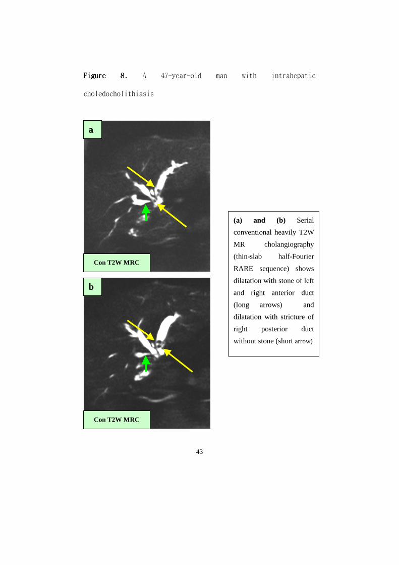
Figure 8. Figure 8. Figure 8. Figure 8. A 47-year-old man with intrahepatic
choledocholithiasis
(a) and (b) Serial
conventional heavily T2W
MR cholangiography
(thin-slab half-Fourier
RARE sequence) shows
dilatation with stone of left
and right anterior duct
(long arrows) and
dilatation with stricture of
right posterior duct
without stone (short arrow)
Con T2W MRC
Con T2W MRC
a
b
44
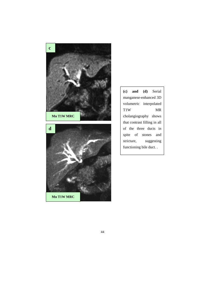
(c) and (d) Serial
manganese-enhanced 3D
volumetric interpolated
T1W MR
cholangiography shows
that contrast filling in all
of the three ducts in
spite of stones and
stricture, suggesting
functioning bile duct. .
Mn T1W MRC
Mn T1W MRC
c
d
45
ⅣⅣⅣⅣ. DISCUSSION. DISCUSSION. DISCUSSION. DISCUSSION
Mangafodipir trisodium is a paramagnetic contrast agent
originally designed for liver imaging. This contrast agent
consisting of manganese bound to DPDP is taken up by
functioning hepatocytes, and is primarily excreted via bile
into the feces 4-7. Therefore contrast-enhanced MR
cholangiography with mangafodipir trisodium provides
information about biliary dynamics similar to that obtained on
hepatobiliary scintigraphy or direct cholangiography 4-7.
Because manganese is a paramagnetic metal ion, it acts
primarily on T1, resulting in T1 shortening, although it also
acts on T2, resulting in T2 shortening 4-7. Enhanced liver and
functioning bile ducts, therefore, have higher signal
intensity on T1-weighted images and lower signal intensity on
T2W images 4-7. Therefore mangafodipir trisodium is primarily
used as a positive contrast agent of T1W MR cholangiography.
Additionally it acts as a negative contrast agent on
conventional heavily T2W MR cholangiography. On T2W MR
cholangiography after manganese administration, signal
46
intensity is lost in the functioning bile duct, but persists
in the non-functioning bile duct. This characteristic property
of combined T1W and T2W MR cholangiography before and after
administration of mangafodipir trisodium could be used as an
excellent imaging tool in the evaluation of biliary dynamics 4-
7.
We used 3D fat-saturated volumetric interpolated breath-
hold examination (VIBE) as a T1W MR cholangiographic sequence.
It is well known that properly structured 3D gradient echo
sequence provides higher signal to noise ratio, thinner
sections, no gaps, and comparable image contrast than 2D
gradient echo sequence 10. Moreover, VIBE sequence provides
isotropic or nearly isotropic spatial resolution, which allows
multiplanar reconstruction images to be generated in any
desired plane 10. Lee et al. 7 reported that manganese-enhanced
T1W MR cholangiography using 3D VIBE sequence provided the
definition of intrahepatic bile duct anatomy in non-obstructed
biliary systems with excellent image quality. In our study,
the slice thickness of manganese-enhanced 3D T1W MR
cholangiography was 1.3 mm with no gap and there was hardly
47
any partial volume artifact on maximum intensity projection
images. Therefore, the image quality of coronal 3D T1W MR
cholangiography was excellent in evaluating the various
biliary diseases.
Acute cholecystitis is an acute inflammation of the
gallbladder, consisting of leukocytic infiltration, edema,
vascular congestion, frank abscess formation, or gangrenous
necrosis 11. Most of these cases result from chemical
irritation and inflammation of the obstructed gallbladder due
to cystic duct obstruction by impacted calculi 11,12. In this
context, the key diagnostic question in acute cholecystitis is
whether or not the cystic duct is patent.
Ultrasonography has been most frequently used to assess
suspected acute cholecystits. It is, however, rarely able to
depict the impacted cystic duct calculi because of the lack of
the landmark of the cystic duct and too small amount of
surrounding bile to create an acoustic contrast 13. The
ultrasonographic diagnosis of acute cholecystitis, therefore,
rests on imaging gallstones in association with secondary
signs, such as “the sonographic Murphy sign, changes in the
48
gallbladder wall, pericholecystic fluid collection, and
intraluminal changes” rather than a direct depiction of
cystic duct obstruction 14. Cholescintigraphy has proven to be
the most sensitive method available for documenting cystic
duct patency or obstruction 15. Nonvisualization of the
gallbladder with visualization of the normal common bile duct
and bowel accurately confirms the diagnosis of acute
cholecysitis in most cases (95.2%), while visualization of the
gallbladder accurately excludes it (99.4%) 15. However,
nonvisualization of the gallbladder is the only sign of cystic
duct obstruction. The site and cause of obstruction are not
demonstrable. Furthermore, it cannot depict associated
findings, such as common bile duct calculi, pericholecystic
fluid collection or changes in the gallbladder wall and it is
not able to provide anatomic information about the organs
outside of the liver or biliary tree.
In our study, results as to the presence of cystic duct
patency on manganese-enhanced T1W MR cholangiography
completely agreed with the hepatobiliary scintigraphy. In the
cases with a patent cystic duct, this imaging demonstrated
49
both the bile duct and gallbladder as an area of high signal
intensity reflecting the normal flow of biliary system without
obstruction or stasis. In the cases of cystic duct obstruction,
however, the gallbladder was not enhanced. Therefore MR
cholangiography enhanced with manganese reflects the biliary
dynamics just like cholescintigraphy and is able to provide
functional information of the biliary system. Furthermore T1W
MR cholangiography enhanced with manganese shows the calculi
in the cystic duct or gallbladder neck, proximal to the
obstruction site, and thus it can depict the cause of
obstruction anatomically.
Conventional heavily T2W MR cholangiography is able to
detect small stones less than 3mm and to show both the
proximal and distal parts of the obstruction site in spite of
a complete obstruction. Therefore it can provide accurate
information concerning the site and cause of the obstruction.
Recently, MR cholangiography has been used to examine the
gallbladder and cystic duct in the setting of acute
cholecystitis, demonstrating a high sensitivity in detecting
an impacted cystic duct or gallbladder neck stones 16,17. In
50
this study, the impacted cystic duct or gallbladder neck
calculi were accurately demonstrated, to a resolution as small
as 3mm. In some cases, however, we were unable to
differentiate the calculus from part of the normal cystic duct.
Following manganese injection, however, MR cholangiography
depicts the cystic duct obstruction clearly, with
nonvisualization of the gallbladder on T1W images. MR
cholangiography in conjunction with conventional heavily T2W
and manganese-enhanced T1W imaging is superior to conventional
heavily T2W MR cholangiography without manganese, in the
diagnosis of acute cholecystitis, particularly when evidence
of cystic duct obstruction is equivocal.
Compared with calculus cholecystitis, acute acalculus
cholecystitis has increased the incidence of gangrene and
perforation, resulting in increased morbidity and mortality 12.
In the cases of the acalculus form, a delay in diagnosis and
definitive treatment is frequent because a demonstrable cause
of obstruction is not present. In such cases, therefore, other
imaging modalities (ultrasonography and CT) are less sensitive
than cholescintigraphy because they cannot provide functional

51
information concerning biliary dynamics 12. The case of
acalculus cholecysistitis in our study was a more complicated
one. The cholescintigraphy of this case was interpreted as a
visualization of the gallbladder. However, the visualized
gallbladder was proven to be only a portion of the neck and
not the entire gallbladder. In contrast with cholescintigraphy,
MR cholangiography, T1- and T2W imaging combined with pre- and
post-manganese injection, was able to depict the contrast-
filling portion in connection with the remaining portion of
the gallbladder. Therefore it was easier for us to accurately
approach this case with MR cholangiography than
cholescintigraphy.
It would be of great value if one imaging modality could
combine the anatomic detail of cross-sectional imaging with
the functional detail of cholescintigraphy, particulary in
cases of acalculus cholecystitis or calculus cholecystitis
with stones too small to detect in the cystic duct. We are
convinced that MR cholangiography, T1- and T2-weighted
combined with pre- and post-manganese injection, provides this
very imaging modality because it is superior to
52
ultrasonography in the depiction of the causes of cystic duct
obstruction 16 and it completely agrees with hepatobiliary
scintigraphy in the evaluation of cystic duct patency in this
study. It could replace both the cross-sectional imaging and
cholescintigraphy all at once with a higher degree of
confidence in patients with acute cholecystitis.
Leakage of the bile, bile duct injury, and retained bile
duct calculi are the main biliary complications of
laparoscopic cholecystectomy 8-9, 18-20. They may occur separately,
but complex biliary complications, such as bile duct injury
combined with bile leakage, may occur rather frequently 8-9, 18-20.
Although, there is no accepted classification of biliary
complications and no standard protocol for the management of
them, it is generally accepted that the treatment modalities
depend on the type, cause, location, and extent of
complications 9, 20. Prat et al 9. suggested that endoscopic
sphincterotomy was sufficient for the treatment of simple bile
leakage without duct injury, clip migration, and retained
stones. They 9 also recommended endoscopic stenting as a
primary option in partial CBD strictures, and surgery as a

53
definite treatment of choice in major injuries, including
complete transection and complex injury.
Most patients with complications after laparoscopic
cholecystectomy usually undergo a multitude of imaging tests -
sonography, CT, hepatobiliary scintigraphy, percutaneous
aspiration, endoscopic retrograde cholangiography, and/or
percutaneous cholangiography - because no single test is able
to provide complete and final diagnosis and some of them are
too invasive to perform for the first time 8-9, 18-20. The main
roles of CT or sonography with or without percutaneous
aspiration are to establish the presence of bile in the
peritoneal cavity and to drain it. However, they cannot
establish the combined bile duct injury and the exact location
of the bile leak. The main roles of hepatobiliary scintigraphy
are to establish the presence of a continuing bile leak.
However, it cannot provide the anatomical information. ERCP
and percutaneous cholangiography are able to establish the
presence of a continuing bile leak, provide exact anatomical
diagnosis, and treat injury by decompressing the biliary tree
or by dilating it. However they are invasive and they could
54
miss the proximal or distal part of the complete obstruction
site, and, in this case, they are not diagnostic.
Differentiating complete obstruction from partial
stricture of the bile duct is important because the former is
a definite indication of surgical treatment whereas the latter
is an indication of endoscopic stenting rather than surgery 9.
In our study, manganese-enhanced T1W MR cholangiography
differentiated complete obstruction from partial stricture.
Manganese -enhanced T1W MR cholangiography clearly
demonstrated the two cases of complete obstruction with bile
leakage, by showing extravasation of the contrast from the
bile duct without opacification of the distal part of the
extrahepatic bile duct. On the other hand, because ERCP could
not evaluate the proximal portion of the disconnected site of
the bile duct, it was not able to evaluate the presence of
bile leakage and the distance of the injured bile duct from
the confluence level, which is an important factor for
operation plan. ERCP has limitations in evaluating the
completely obstructed cases.
In our study, manganese-enhanced T1W MR cholangiography
55
clearly demonstrated the partial stricture with bile leakage,
by showing extravasation of the contrast from the bile duct
with opacification of the extrahepatic bile duct, despite the
presence of an abnormal narrowing segment of the bile duct at
conventional T2W MR cholangiography. In this case, the patient
did not have a peritoneal drain. However in a patient with a
peritoneal drain, contrast opacification of the extrahepatic
duct was absent at manganese-enhanced T1W MR cholangiography,
suggesting the complete occlusion or transection of the bile
duct. However, ERCP showed a partial stricture of the bile
duct and not complete obstruction. It may be because the main
bile flow was into the peritoneal drain rather than the
extrahepatic duct, causing the degree of obstruction to be
overestimated with manganese -enhanced T1W MR cholangiography
in the patient with a peritoneal drain.
In our study, manganese-enhanced T1W MR cholangiography
differentiated abnormal fluid collection of biliary origin
from that of non-biliary origin. Manganese-enhanced T1W MR
cholangiography showed normal contrast filling of the biliary
system without leakage or obstruction in a patient with a
56
large amount of fluid collection at sonography and
conventional T2W MR cholangiography, suggesting a non-biliary
origin of fluid collection. It was diagnosed as hemorrhage.
The traditional algorithm for the imaging of
postcholecystectomy biliary complications has been sonography,
CT and/or hepatobiliary scintigraphy, followed by diagnostic
and/or therapeutic ERCP or percutaneous cholangiography, and
some of them underwent surgery. We think that combined
conventional T2-weighted and Mn-enhanced T1-weighted MR
cholangiography can differentiate biliary complications from
non-biliary complications, simple leakage without bile duct
injury from that with bile duct injury, and complete
obstruction of the bile duct from partial stricture of the
bile duct. Moreover, they are non-invasive. Therefore, they
could be used as the first-line study for postcholecystectomy
biliary complications. After diagnosing simple leakage without
bile duct injury on combined MR cholangiography, ERCP with
sphicterotomy with or without internal stent may be performed.
After diagnosing partial stricture of the bile duct with or
without bile leakage on combined MR cholangiography, ERCP with
57
internal stent may also be performed. Furthermore, after
diagnosing complete transection or occlusion of the bile duct
with or without bile leakage on combined MR cholangiography,
prompt surgical repair should be performed. ERCP or PTC is not
necessary in those cases. Therefore, this series suggest that
combined conventional T2-weighted and Mn-enhanced 3D T1-
weighted MR cholangiography may eliminate the need for other
studies for the imaging of postcholecystectomy biliary
complications.
The management of symptomatic intrahepatic
choledochalithiasis is difficult and remains far from
satisfactory 9. In the last decade, however, the management of
this condition has been improved by a systemic approach,
advances in hepatobiliary imaging, availability of flexible
choledochoscopy, application of stone-fragmentation technology,
and innovative surgical approaches to the biliary tract. A
combination of all the various treatment modalities is
required, on a selective basis, to achieve optimal results 22.
The treatment modalities depend on identifying the exact
location and level of stones or stricture, the degree of
58
stenosis or obstruction, the degree of destruction of the
involved liver segment, and the presence of combined hepatic
abscess or cholangiocellular carcinoma. Recently, reports have
suggested MR cholangiography to be superior to direct
cholangiography in the accurate topographic evaluation of
intraheptic choledocholithiasis because of its ability to
depict all parts of the biliary tree, regardless of
obstruction or stenosis 23. However, conventional heavily T2W
MR cholangiography has some limitations in evaluating the
degree of stenosis or obstruction and the function of the
involved bile duct and liver parenchyma. In our study,
manganese-enhanced T1W MR cholangiography provided the
functional information of the bile ducts and indicated the
degree of stricture and the function of the involved duct.
Therefore, rather than conventional T2W MR cholangiography
alone, manganese-enhanced MR cholangiography combined with
conventional T2W MR cholangiography could provide further
information that is necessary for treatment decision- making
in the patients with intrahepatic choledocholithiasis.
Several limitations exist in this study. One limitation is
59
that mangafodipir-trisodium cannot be used for the patients
with jaundice because the biliary systems are not opacified
and therefore the patient population in our study was very
restricted. However, the incidence of adverse events was
comparable between the group of patients with jaundice or
cirrhosis and the group of patients without jaundice or
cirrhosis. The most commonly reported adverse events were
nausea (7%) and headache (4%), and they were mild to moderate
in intensity, not requiring treatment. A second limitation is
that there was no standard reference to the diseased segmental
ducts in the patients with intrahepatic choledochalithiasis. A
third limitation is the cost-effectiveness manganese-enhanced
MR cholangiography, especially in the patients with acute
cholecystitis. In our country, DISIDA scan and US are far more
cheaper than MR cholangiography and more their diagnostic
accuracies in the evaluation of acute cholecystitis are not so
much inferior to MR cholangiography. Therefore manganese-
enhanced MR cholangiography cannot be the first line
diagnostic modality in the evaluation of the patients with
suspected acute cholecystitis. The clinical application may be
60
restricted to the cases that the evidence of cystic duct
obstruction is equivocal.

61
ⅤⅤⅤⅤ. CONCLUSION. CONCLUSION. CONCLUSION. CONCLUSION
Conventional heavily T2W MR cholangiography combined with
manganese-enhanced MR cholangiography provided not only the
anatomic detail, but also the functional detail. Therefore,
manganese-enhanced MR cholangiography is a useful supplement
to conventional heavily T2W MR cholangiography in the
evaluation of acute cholecystitis, biliary complications after
laparoscopic cholecystectomy, and intrahepatic
choledocholithiasis

62
ReferencesReferencesReferencesReferences
1. Wallner BK, Schumacher KA, Weidenmaier W, Friedrich JM.
Dilated biliary tract: evaluation with MR
cholangiography with a T2-weighted contrast-enhanced
fast sequence. Radiology 1991; 181: 805-808.
2. Reinhold C, Bret PM. Current status of MR
Cholangiopancreatography. AJR 1996; 166: 1285-1295.
3. Fulcher AS, Turner MA, Capps GW, Zfass AM, Baker KM.
Half-Fourier RARE MR cholangiopancreatography:
experience in 300 subjects. Radiology 1998; 207: 21-32.
4. Semelka RC, Helmberger TKG. Contrast Agents for MR
Imaging of the Liver. Radiology 2001; 218: 27-38.
5. Mitchell DG, Alam F. Mangafodipir Trisodium: Effects on
T2- and T1-weighted MR cholangiography. JMRI 1999; 9:
366-368.
6. Vitellas KM, El-Dieb A, Vaswani K, et al. Detection of
Bile Duct Leaks Using MR cholangiography with
Mangafodipir trisodium (Teslascan). JCAT 2001; 25(1):
102-105.
7. Lee VS, Rofsky NM, Morgan GR, et al. Volumetric
63
Mangafodipir Trisodium-Enhanced Cholangiography to
Define Intrahepatic Biliary Anatomy. AJR 2001; 176: 906-
908.
8. Strasberg SM, Hertl M, Soper N. An analysis of the
problem of biliary injury during laparoscopic
cholecystectomy. J Am Coll Surg 1995; 180: 101-125.
9. Prat F, Pelletier G, Ponchon T, et al. What role can
endoscopy play in the management of biliary
complications after laparoscopic cholecystectomy?
Endoscopy1997; 29: 341-348.
10. Rofsky NM, Lee VS, Laub G, et al. Abdominal MR imaging
with a volumetric interpolated breath-hold examination.
Radiology 1999; 212: 876-884.
11. Crawford JM. The Liver and the Biliary Tract. In: Cotran
RS, Kumar V, Collins T, eds. Pathologic Basis of Disease.
6th ed. W.B. Saunders Company, 1999; 845-901.
12. Swayne LC. Acute Acalculus Cholecystitis: Sensitivity in
Detection Using Technetium-99m Iminodiacetic Acid
Cholescintigraphy. Radiology 1986; 160: 33-38.
13. Laing FC, Jeffrey RB Jr. Choledocholithiasis and cystic
64
duct obstruction: Difficult Ultrasonographic Diagnosis.
Radiology 1983; 146: 475-479.
14. Laing FC. Diagnostic evaluation of patients with
suspected cholecysitits. Surg Clin North Am 1984; 64: 3-
22.
15. Weissmann HS, Badia J, Sugarman LA. Spectrum of 99m-Tc-
IDA Cholescitigraphic Patterns in Acute Cholecystitis.
Radiology 1981; 138: 167-175.
16. Park MS, Yu JS, Kim YH, et al. Acute Cholecystitis:
Comparison of MR Cholangiography and US. Radiology 1998;
209: 781-785.
17. Turner MA, Fulcher AS. The Cystic Duct: Normal Anatomy
and Disease Processes. Radiographics 2001; 21: 3-22.
18. Deziel DJ. Complications of cholecystectomy: Incidence,
clinical manifestations, and diagnosis. Surg Clin North
Am 1994; 74(4): 809-823
19. Deziel DJ, Millikan KW, Economou SG, et al.
Complications of laparoscopic cholecystectomy: a
national survey of 4,292 hospitals and an analysis of
77,604 cases. Am J Surg 1993; 165: 9-14.
65
20. Slanetz PJ, Boland GW, Mueller PR. Imaging and
interventional radiology in laparoscopic injuries to the
gallbladder and biliary system. Radiology 1996; 201:
595-603.
21. Harris HW, Kunwenda ZL, Sheen-Chen SM. Recurrent
pyogenic cholangitis. Am J Surg 1998; 176: 34-37.
22. Fan ST, Choi TK, Lo CM, et al. Treatment of
hepatolithiasis: improvement of result by a systemic
approach. Surgery 1991; 109: 474-480.
23. Park MS, Yu JS, Kim YH, et al. Recurrent Pyogenic
Cholangitis: Comparison between MR Cholangiography and
Direct Cholangiography. Radiology 2001; 220: 677-687.
66
국문국문국문국문 요약요약요약요약
MnMnMnMn----DPDP DPDP DPDP DPDP 조영조영조영조영 증강증강증강증강 3D 3D 3D 3D----T1W T1W T1W T1W 자기공명자기공명자기공명자기공명 담관담관담관담관 조영술의조영술의조영술의조영술의 임상임상임상임상
적용적용적용적용: : : : 고식적고식적고식적고식적 T2W T2W T2W T2W자기공명자기공명자기공명자기공명 담관담관담관담관 조영술과의조영술과의조영술과의조영술과의 비교비교비교비교
지도교수 김김김김 기기기기 황황황황
연세대학교 대학원 의학과
박박박박 미미미미 숙숙숙숙
목목목목 적적적적: : : : 다양한 담도계 질환에서, 고식적 T2 강조 자기 공명 담관
조영술과 망간 조영 증강 삼차원 T1 강조 자기 공명 담관 조영술을
함께 시행하였을 때, 그 부가적인 정보 유무를 분석한 후, 망간
조영 증강 자기 공명 담관 조영술의 임상 활용에 대하여
연구하고자 함.
대상대상대상대상 및및및및 방법방법방법방법: : : : 임상적으로 급성 담낭염 (n=30), 복강경 유도 하
담낭 절제술 후의 담도계 합병증 (n=10), 간내 담석증 (n=14),
대조군 (n=10), 모두 64 명의 환자에서, 고식적 T2 강조 자기 공명
담관 조영술과 망간 조영 증강 삼차원 T1 과 T2 강조 자기 공명

67
담관 조영술을 함께 시행하였다. 급성 담낭염의 경우 담낭
유출로의 폐쇄 유무에 초점을 두고 영상을 분석하였으며, 복강경
유도 하 담낭 절제술 후의 담도계 합병증의 경우 담관 손상, 담즙
유출, 잔류 담석 유무, 그리고 간내 담석증의 경우 병변이 있는
간내 담도의 기능 유무에 초점을 두고 영상 분석을 하였다.
결결결결 과과과과:::: 급성 담낭염 환자에서 담낭 유출로의 폐쇄 유무에 대한,
고식적 T2 강조 자기 공명 담관 조영술의 예민도, 특이도, 정확도는
각각 82%, 88%, 83% 이었으나, 망간 조영 증강 자기 공명 담관
조영술의 그것은 모두 100% 이었다. 담낭 절제술 후의 담도계
합병증이 의심되는 환자에서 자기 공명 담관 조영술의 진단은,
총담관의 완전 협착과 담즙 유출 (n=4), 총담관의 부분 협착과
담즙 유출 (n=2), 잔류 담낭관에서의 담즙 유출 (n=2), 변이 우
담관 결찰 (n=1), 그리고 출혈 (n=1) 이었다. 자기 공명 담관
조영술의 진단은, 완전 협착으로 과 진단한 부분 협착 한 예를
제외하고는 모두 수술 혹은 역행성 담도 조영술 소견과 일치하였다.
간내 담석증의 경우 고식적 T2 강조 자기 공명 담관 조영술에서는
22 분절의 담도에 병변이 있었다. 망간 조영 증강 자기 공명 담관
조영술에서는, 8 분절의 담도는 조영 증강이 되어 아직 기능을
하고 있었으며 나머지 14 분절의 담도는 기능이 없었다.

68
결결결결 론론론론:::: 고식적 T2 강조 자기 공명 담관 조영술과 망간 조영 증강
삼차원 T1 강조 자기 공명 담관 조영술을 함께 시행하였을 때,
병변에 대한 해부학적인 정보 뿐만이 아니라 그 역동적, 기능적인
정보까지 얻을 수 있다. 따라서 망간 조영 증강 자기 공명 담관
조영술은, 급성 담낭염, 복강경 유도 하 담낭 절제술 후의 담도계
합병증, 그리고 간내 담석증의 진단에 있어서, 이제까지의 고식적
T2 강조 자기 공명 담관 조영술에 대한 추가 진단 방법으로써 매우
유용하다.
핵심 되는 말: 담관, 결석, 자기공명영상, 담도조영술,
자기공명영상 조영제