clinical policy for extended role for non-medical
TRANSCRIPT
{Insert} Name of Trust
1
Clinical Policy for extended role for non-medical
Practitioners in Cataract clinics
{Insert} Name of Trust Clinical Guideline Summary This guideline describes the processes required for non- medical staff to assess patients in cataract clinic.
Version: X.0 Status: Final : Approved: X.X.20XX Ratified: X.X.20XX
Version History
Version Date Issued Brief Summary of Change
Author
Clinical Unit or Department:
Name of author(s)
Name of responsible individual
Approved by:
Ratified by :
Date issued:
Review date
CQC relevant domains
Target audience: Nursing, orthoptists, optometrists,
ophthalmologists, ophthalmology managers

{Insert} Name of Trust
2
1. Introduction
In recent years, the involvement of non-medical healthcare professionals (HCP) in delivering
an extended scope of practice assessing and managing patients and/or performing
procedures has become widely accepted practice to cope with significantly rising demand for
eye care and to support the expansion of non-medical roles, and is supported by the Royal
College of Ophthalmologists and other HCP professional organisations as well as the NHS
England National Elective Care High Impact Intervention and GIRFT
2. Purpose
This policy sets out the process required for designated HCP to train for and to deliver
cataract assessment in extended roles to the standards required by NICE and the RCOphth.
This will contribute to the efficient delivery of the cataract service and will enhance and
develop patient-centred care, which fulfils national safety and service delivery targets.
Service provision will be more flexible and resilient, with the potential for increased capacity
for the ophthalmology service. Staff will be able to develop their roles further, increasing the
overall level of expertise in the department and promoting greater job satisfaction.
The policy provides details of:
the training and competencies
guidance for the management of patients
standard operating procedures
the process to be used for monitoring compliance with the process and outcomes
3. Scope
This policy applies to all trust sites where cataract clinics are carried out and is relevant to
ophthalmic nurses, orthoptists and optometrists who are working, or wish to work, as
advanced practitioners in cataract ophthalmology clinics, ophthalmologists including
consultants and those managing ophthalmology services.
It should be read in conjunction with other relevant trust documents:
Consent Policy
Clinical Governance/Risk Policy
Biometry/intraocular lens policy
Local Safety Standards for Invasive Procedures (SSIPs)
Preoperative assessment policy.
To be eligible for delivering this care the procedure staff must have a minimum of 1 year’s
post registration hospital ophthalmic experience and be:
Registered nurse (RN) at band 6 or above who must either hold an ophthalmic
nursing qualification or have sufficient ophthalmic experience to be judged by their
manager as competent to commence training.
Registered orthoptist at band 6 or above who has sufficient ophthalmic experience to
be judged by their manager as competent to commence training
Registered optometrist at band 6 or above who have sufficient ophthalmic experience
to be judged by their manager as competent to commence training.

{Insert} Name of Trust
3
Suitable staff members from a nursing or orthoptic background at band 5 level may
commence training for an extended role in paediatric ophthalmology and progress to band 6
on completion of their training.
4.0 Duties and responsibilities
4.1 Practitioners responsibilities
Practitioners undertaking the training are responsible for compliance with trust policies;
engaging actively with the training, keeping up to date, keeping accurate training records;
ensuring they act within their sphere of competence; completing accurately the relevant
parts of the medical records; following SOPs; reporting adverse events and safety concerns
to their supervisor, consultant or their line manager.
Once signed off as competent to practice, the HCP is required to:
keep a record of their competency sign off
undertake regular clinical update sessions or CPD on cataract ophthalmology
regularly audit their patient records and care
maintain and update their portfolio
review these as part of their annual appraisal / individual performance review.
From the point of registration, each practitioner must adhere to their professional
body/regulatory code of conduct and is accountable for his/her practice.
4.2 Consultant ophthalmologist’s responsibilities
To ensure the HCP has achieved a satisfactory knowledge base and competencies with
which to perform this enhanced role. The consultant can undertake this directly or can
delegate some or all parts to a senior colleague with appropriate experience, knowledge and
training who is a named cataract clinic trainer that is a cataract HCP with more than 2 years’
experience, or a fellow or ST 6 and above ophthalmic trainee. However the consultant
retains responsibility for the training and sign off of the HCP before they begin independent
practice.
The trainer will:
Examine the HCP to ensure she/he has the knowledge base required
Provide adequate time for the HCP to observe care and to subsequently supervise
and assess the HCP’s skills and knowledge
Only sign the competency when all aspects of the competency standards have been
demonstrated by the practitioner.
The consultant will arrange that they or another suitably qualified ophthalmologist are
available to support the HCP during clinics. The doctor should either be present on site or by
phone with a pathway in place to see a doctor urgently with the appropriate safe timescale if
required, once the HCP has undertaken any initial urgent or unplanned treatment.
The patient remains under the care of a named consultant ophthalmologist at all times.
4.3 Manager’s responsibility

{Insert} Name of Trust
4
The manager(s) [lead nurse, lead orthoptist, lead optometrist or ophthalmology department
manager] will keep a record of all competencies and a register or list of trainers and HCPs
eligible to perform advanced cataract ophthalmology practice.
Managers must only endorse practice if such development is in line with the practitioner`s
job description and existing trust policies and service requirements.
Managers must ensure that the HCP is supported in skills development in the form of:
opportunities for supervised practice
assessment of competency and sign off.
4.4 Employer’s responsibilities
The employers will ensure that the HCPs training and supervision is provided in a timely
manner, ensuring trainers and supervisors are supported to deliver the time required.
Employers will ensure HCPs are appropriately banded for the work they undertake and are
given the time to undertake the training during their current role.
The employers will ensure that, subject to following trust policy, HCPs have suitable
indemnity for this scope of practice.
5.0 Training
HCPs can only commence training after approval by their line manager.
5.1 Baseline competencies for training
Orthoptists, optometrists and nurses will have had differing training and experience in a
number of baseline skills or knowledge in terms of:
Slit lamp
Tonometry
Slit lamp fundoscopy with fundus lens
Understanding refractive errors and refractive correction
Basic knowledge of cataract and ophthalmic disease.
Consenting.
For these baseline skills and knowledge/experience, the trainer / ophthalmologist and line
manager will need to agree if there is any basic training required to bring the HCP to a level
where the extended role cataract training can commence and make a plan to train and
evidence competencies for any areas which are not covered as part of core training before
embarking on the cataract advanced practice training. Staff wishing to undertake consent for
cataract surgery must complete the trust consent training.
5.2 Cataract advanced practice training
The HCP will gain the appropriate theoretical knowledge of anatomy and physiology,
assessment and examination, disease, investigations and management from a combination
of the following:
Attending local, regional or national courses
Informal in house training or sessions with the consultant or other trainer
Additional reading around the subject area in books and journals
Reading of local and national cataract care guidelines

{Insert} Name of Trust
5
E-learning modules e.g. RCOphth cataract modules on E-Learning for Health.
The HCP will need to know:
Anatomy and physiology of the eye
Causes of cataract
Classification of cataract
Assessment of cataract and other ocular and systemic disease
Knowledge of refraction, refractive errors and refractive targets in cataract surgery
Imaging (A scan, B scan, OCT) relevant to cataract related conditions and comorbidities
Biometry, choosing an IOL, avoiding wrong IOLs
Ocular and systemic and personal risk factors for surgery and how to risk stratify cataract
surgery
Principles of cataract surgery
Latest clinical information on cataract surgery and treatment delivery
Process of cataract surgery, including the practicalities, the pathway, the on the day
journey
Anaesthetic types, risk and benefits, anaesthetic choices for cataract surgery
Any CCG thresholds for surgery
Infection control for cataract surgery
Pharmacology to include relevant drugs to assess, during and following cataract surgery,
drugs that affect cataract surgery
Recognition of intraoperative and postoperative complications and what actions to take
Is aware of any possible red flags and how to escalate concerns
Risk and legal issues around extended role development
How to audit NMP practice
The HCP will gain practical knowledge as follows:
This period will usually last at least 3 months
The HCP will initially observe practice and discuss cases with their trainer
Once the trainer agrees they are ready, the HCP will start to see patients for an initial
assessment and the trainer will then assess each patient and agree management
As the HCP progresses, they will undertake more of the assessment and
management, but continue to discuss all cases with the consultant and will sit in on
interesting cases/continue to observe the consultant’s practice
For each clinical competency area assessed (pre and post op cataracts, other
specific areas) there should be in the portfolio a disease specific logbook of at least
20 cases (Appendix) and at least 2 successfully completed work based assessments
(Appendix).
The HCP should attend at least 1 surgical session.
The HCP will maintain a portfolio of their learning, experience and performance, and will
add to this as they progress. The portfolio will contain:
Evidence of theoretical training, courses, teaching and CPD
Records of their cases and experience
A log of discussions and unfamiliar conditions seen
{Insert} Name of Trust
6
Reflective learning on a small number of cases (see Appendix: Reflective Practice
template)
Further reading e.g. books, review articles, research papers
- Written summaries of key conditions (symptoms, assessment and signs,
investigations, management, red flags, complications (see Appendix: Disease
Summary for Portfolio)
Workplace based assessments
Competency sign off documents.
At sign off, the HCP will discuss the knowledge and experience gained and the work place
based assessments in their portfolio with their consultant / trainer. The consultant / trainer
will, if satisfied, record the HCP as competent using the final competency checklist form
(Appendix).
Once signed off:
The HCP must practice in accordance with the clinic protocol (see Appendix:
Protocol for Advanced Practice Cataract Clinics).
The practitioner must be satisfied with his/her own level of competence in
accordance with the guidelines and codes of conduct from their relevant regulator
and professional body.
The HCP will undergo an informal review of practice with their trainer and/or the
consultant paediatric ophthalmologist after three to six months of independent
practice.
The HPC will undergo review of practice and the portfolio as part of their annual
appraisal / individual performance review.
5.3 Sign off for current or experienced practitioners
For Current Practitioners who have:
Completed the HCP training programme or equivalent previously and are currently
practicing in this area (eg. specialist cataract extended-role optometrists)
Completed training from another provider/trust previously and have proof of
continuing competency in the form of a completed and signed recent (within the last
two years) competency document.
You must be assessed as competent at the discretion of the supervising consultant or HCP
trainer. This should include:
Open discussion of relevant diseases to ensure theoretical competence
Successful completion of at least 1 workplace based assessment;
Creation / update and review of a portfolio
Sign off of the competency assessment form (Appendix)
For staff who have had a Gap in Service (≥6months):
Competence can be reassessed at the discretion of the supervising consultant or trainer; this
may involve some of the following:
Case discussion

{Insert} Name of Trust
7
Observed practice
The HCP observing in clinic
Work placed based assessment
The portfolio must be updated and reviewed and a competency assessment form (Appendix)
must be signed off.
6 Frequency of practice
HCP cataract clinics will be carried out according to service need. Once a practitioner has
been signed off as competent, they should be performing clinics regularly to maintain skills.
7. Outcome measures
Data to be collected is:
Record of all cases to be kept by HCPs for activity levels.
Regular audit of adherence to protocol, case management and record keeping in
conjunction with trainer
Regular documented reflective practice on cases of interest or with learning
opportunities
Regular updates of portfolio with reading/learning documents and condition
summaries
Any incidents or serious incidents or patient complaints, including the result for the
patient or of any investigation, with appropriate reflective practice and learning
recorded
Patient experience / satisfaction survey at discretion of HCP and line manager.
The HCP will undertake an audit and/or review of their practice on an annual basis as part of
their annual appraisal and individual performance review.
8.0 Stakeholder Engagements and Communication
The cataract team developed this guideline with contributions from other ophthalmic medical
staff, orthoptic, optometrist, nursing staff and the management team.
Stakeholder engagement with consultants and other relevant staff has been through insert
name of appropriate meetings and other methods e.g. emails or team meetings.
9.0 Approval and Ratification
This policy was approved by the insert name of committee and ratified by the insert
name of committee.
10.0 Dissemination and Implementation
This policy will be implemented and disseminated to all staff involved in the provision of
cataract service, and will be communicated to key stakeholders and protocol users via
email, and highlighted at team meetings and insert name of other meetings or insert
other methods of dissemination.
This policy will be published on the trust intranet site.

{Insert} Name of Trust
8
11.0 Review and Revision Arrangements
The Policy Owner/Authors will initially review this document on a 3-year basis.
Changes to the legislation or national guidelines of the administration of cataract
assessment and consultation - by non-medical personal, or any trust serious incidents
will trigger a review of this document.
12.0 Document Control and Archiving
Insert standard trust information of document storage and removal old versions/archiving
13.0 Monitoring compliance with this policy
14.0 Supporting References / Evidence Base
Nursing and Midwifery Council (2015) code of professional conduct, NMC London
http://www.nmc.org.uk/globalassets/sitedocuments/nmc-publications/revised-new-nmc-
code.pdf.
The British & Irish Orthoptic Society Code of Ethics.
https://orthoptics.org.uk/Resources/Documents/Standards/BIOS_Code_of_Ethics.pdf
The Health & Care Professions Council (HCPC) Standards of Conduct,
performance & ethics
http://www.hpcuk.
org/aboutregistration/standards/standardsofconductperformanceandethics/
BIOS – Competency document 2016..
Element to be Monitored Staff conducting Tool for
Monitoring
Frequency Responsible
Individual/Group for
results/actionst
Service delivery and unit outcomes
Lead Cataract Consultant
Audit Every 12 months
Ophthalmic or cataract
clinical lead
HCP Senior cataract
ophthalmology
clinicians and
line manager
Appraisal
and
individual
performance
review -
portfolio of
audit,
practice and
knowledge
Annually Line manager and
cataract ophthalmology
trainer
Complications or adverse
events to be recorded All staff Incident
reporting
On-going Ophthalmology CG
Complaints Complaints
team
Complaints
process
On-going Ophthalmology CG

{Insert} Name of Trust
9
General Optical Council. Standards of Practice. https://www.optical.org/en/Standards/
College of Optometrists Guidance for Professional practice. https://guidance.college-
optometrists.org/home/
Royal College of Ophthalmologists (2017) Ophthalmic Common Clinical Competency
Framework (CCCF).
Royal National Institute of Blind People. Future Sight Loss UK 1: Economic Impact of
Partial Sight and Blindness in the UK Adult Population. London: RNIB; 2009. Available
from: http://www.rnib.org.uk/aboutus/research/reports/otherresearch/pages/fsluk1.aspx. ,
2014.
RCOphth Quality Standards for cataract services. RCOphth 2015.
NICE guidance for adult cataract NG17. 2017
RCOphth/UKOA IOL quality standard 2018
Moorfields Optometrists Protocol for Cataract Clinics
Local documents Ophthalmology department guidelines Consent policy Clinical record keeping policy Clinical governance /. Risk policy Local safety standards for invasive procedures Preoperative assessment policy Mental capacity policy.
{Insert} Name of Trust
10
Appendix 1. Competencies. Cataract eye conditions: Competency checklist -
Successful completion of this competency will enable the HCP to assess specified
condition/subspecialty patients independently with the cataract service.
Aims and Objectives The Clinician is able to demonstrate supporting knowledge, understanding and has been observed as competent to adhere to the policy for extended role work in the paediatric ophthalmology clinic. The HCP is able to demonstrate supporting knowledge, understanding and has been observed as competent to effectively examine patients in the cataract subspecialty of the ophthalmology service
Training Prerequisite Prior to this assessment the practitioner has successfully completed the following: Theoretical knowledge via courses, e-learning or local training Observational work based training Background reading, learning and theory portfolio produced for cataract
Your Responsibility All staff should ensure they keep their knowledge and skills up to date through local policies, standard operating procedures and guidance. It is the responsibility of the individual to work within their own scope of competence relevant to their job role and follow their professional bodies Code of Conduct.
Employee signature/print name: ……………………………………………………………………………….. Assessor signature print name: …………………………………………………………………………………… Date: ………………………………………..
Policies, Guidelines and Protocols:
Date policy read by clinician and initials
Local policies x
Mental capacity policy
Consent policy
Trust IOL/biometry policy
NG7 NICE guidance for adult cataracts
RCOphth/UKOA IOL quality standard
Underpinning knowledge and understanding
demonstrated for:
Date and
assessor initials
Local clinical policies or guidelines
Consent policy
Mental capacity policy
Local policy etc
(key policies such as mental capacity,
{Insert} Name of Trust
11
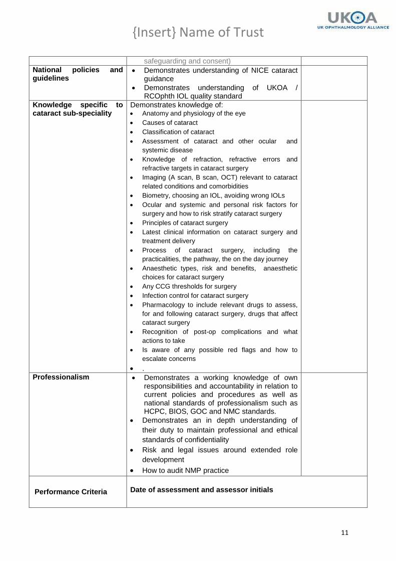
safeguarding and consent)
National policies and guidelines
Demonstrates understanding of NICE cataract guidance
Demonstrates understanding of UKOA / RCOphth IOL quality standard
Knowledge specific to cataract sub-speciality
Demonstrates knowledge of: Anatomy and physiology of the eye
Causes of cataract
Classification of cataract
Assessment of cataract and other ocular and
systemic disease
Knowledge of refraction, refractive errors and
refractive targets in cataract surgery
Imaging (A scan, B scan, OCT) relevant to cataract
related conditions and comorbidities
Biometry, choosing an IOL, avoiding wrong IOLs
Ocular and systemic and personal risk factors for
surgery and how to risk stratify cataract surgery
Principles of cataract surgery
Latest clinical information on cataract surgery and
treatment delivery
Process of cataract surgery, including the
practicalities, the pathway, the on the day journey
Anaesthetic types, risk and benefits, anaesthetic
choices for cataract surgery
Any CCG thresholds for surgery
Infection control for cataract surgery
Pharmacology to include relevant drugs to assess,
for and following cataract surgery, drugs that affect
cataract surgery
Recognition of post-op complications and what
actions to take
Is aware of any possible red flags and how to
escalate concerns
.
Professionalism Demonstrates a working knowledge of own responsibilities and accountability in relation to current policies and procedures as well as national standards of professionalism such as HCPC, BIOS, GOC and NMC standards.
Demonstrates an in depth understanding of
their duty to maintain professional and ethical
standards of confidentiality
Risk and legal issues around extended role
development
How to audit NMP practice
Performance Criteria Date of assessment and assessor initials

{Insert} Name of Trust
12
WpBA for preop cataract undertaken and passed x 2
WpBA for postop undertaken and passed x 2
Attended 1 surgical sessions
Disease specific caselog (20 patients)
{Insert} Name of Trust
13
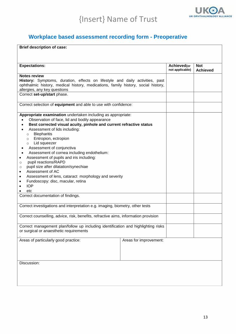
Workplace based assessment recording form - Preoperative
Brief description of case:
Expectations: Achieved(or
not applicable) Not Achieved
Notes review History: Symptoms, duration, effects on lifestyle and daily activities, past ophthalmic history, medical history, medications, family history, social history, allergies, any key questions
Correct set-up/start phase.
Correct selection of equipment and able to use with confidence:
Appropriate examination undertaken including as appropriate:
Observation of face, lid and bodily appearance
Best corrected visual acuity, pinhole and current refractive status
Assessment of lids including: o Blepharitis o Entropion, ectropion o Lid squeezer
Assessment of conjunctiva
Assessment of cornea including endothelium:
Assessment of pupils and iris including: o pupil reactions/RAPD o pupil size after dilatation/synechiae
Assessment of AC
Assessment of lens, cataract morphology and severity
Fundoscopy: disc, macular, retina
IOP
etc
Correct documentation of findings.
Correct investigations and interpretation e.g. imaging, biometry, other tests
Correct counselling, advice, risk, benefits, refractive aims, information provision
Correct management plan/follow up including identification and highlighting risks or surgical or anaesthetic requirements .
Areas of particularly good practice:
Areas for improvement:
Discussion:

{Insert} Name of Trust
14
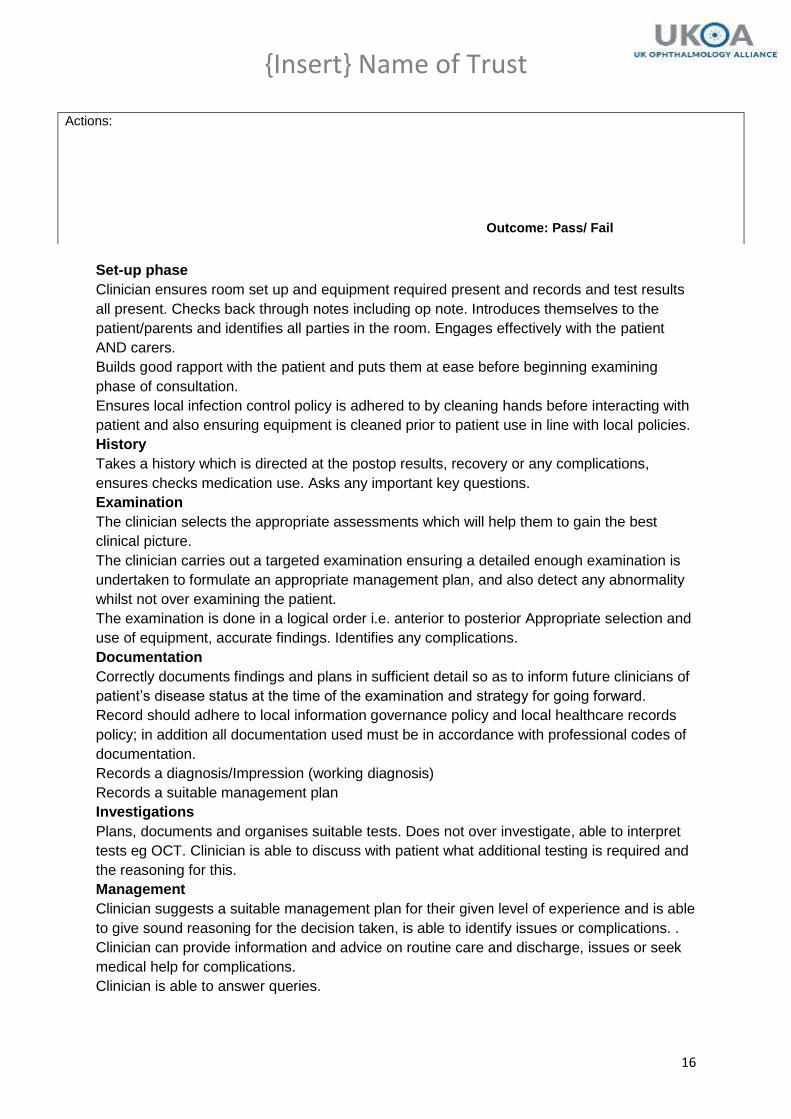
Set-up phase
Clinician ensures room set up and equipment required present and records and test results
all present. Checks back through referral and notes. Introduces themselves to the
patient/parents and identifies all parties in the room. Engages effectively with the patient
AND carers.
Builds good rapport with the patient and puts them at ease before beginning examining
phase of consultation.
Ensures local infection control policy is adhered to by cleaning hands before interacting with
patient and also ensuring equipment is cleaned prior to patient use in line with local policies.
History
Takes a history which is directed at the presenting complaint, ensures medical, social,,
medications, allergy and family history completed. Asks any important key questions.
Examination
The clinician selects the appropriate assessments which will help them to gain the best
clinical picture.
The clinician carries out a targeted examination ensuring a detailed enough examination is
undertaken to formulate an appropriate management plan, and also detect any abnormality
whilst not over examining the patient.
The examination is done in a logical order i.e. anterior to posterior Appropriate selection and
use of equipment, accurate findings..
Documentation
Correctly documents findings and plans in sufficient detail so as to inform future clinicians of
patient’s disease status at the time of the examination and strategy for going forward.
Record should adhere to local information governance policy and local healthcare records
policy; in addition all documentation used must be in accordance with professional codes of
documentation.
Records a diagnosis/Impression (working diagnosis)
Records a management plan
Investigations
Plans, documents and organises suitable tests. Does not over investigate. Able to
provisionally plan IOL and identify unusual biometry or IOl results.
Clinician is able to discuss with patient what additional testing is required and the reasoning
for this.
Management
Clinician suggests a suitable management plan for their given level of experience and is able
to give sound reasoning for the decision taken, is able to identify risk of patient and suitability
for different lists and anaesthesia. Clinician can provide information on disease, options,
risks, benefits, pathway and practicalities. Clinician is able to answer queries.
Actions: Outcome: Pass/ Fail
{Insert} Name of Trust
15
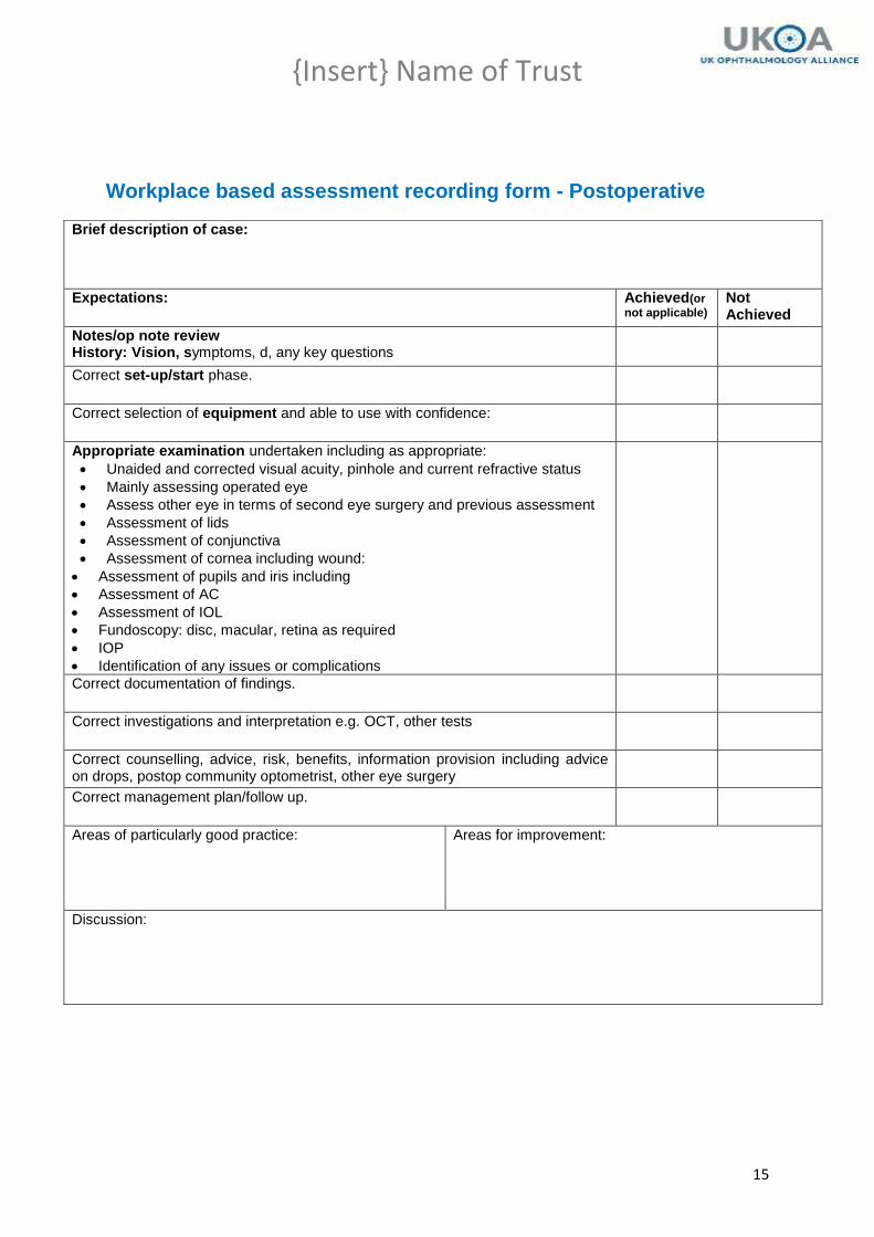
Workplace based assessment recording form - Postoperative
Brief description of case:
Expectations: Achieved(or
not applicable) Not Achieved
Notes/op note review History: Vision, symptoms, d, any key questions
Correct set-up/start phase.
Correct selection of equipment and able to use with confidence:
Appropriate examination undertaken including as appropriate:
Unaided and corrected visual acuity, pinhole and current refractive status
Mainly assessing operated eye
Assess other eye in terms of second eye surgery and previous assessment
Assessment of lids
Assessment of conjunctiva
Assessment of cornea including wound:
Assessment of pupils and iris including
Assessment of AC
Assessment of IOL
Fundoscopy: disc, macular, retina as required
IOP
Identification of any issues or complications
Correct documentation of findings.
Correct investigations and interpretation e.g. OCT, other tests
Correct counselling, advice, risk, benefits, information provision including advice on drops, postop community optometrist, other eye surgery
Correct management plan/follow up.
Areas of particularly good practice:
Areas for improvement:
Discussion:
{Insert} Name of Trust
16
Set-up phase
Clinician ensures room set up and equipment required present and records and test results
all present. Checks back through notes including op note. Introduces themselves to the
patient/parents and identifies all parties in the room. Engages effectively with the patient
AND carers.
Builds good rapport with the patient and puts them at ease before beginning examining
phase of consultation.
Ensures local infection control policy is adhered to by cleaning hands before interacting with
patient and also ensuring equipment is cleaned prior to patient use in line with local policies.
History
Takes a history which is directed at the postop results, recovery or any complications,
ensures checks medication use. Asks any important key questions.
Examination
The clinician selects the appropriate assessments which will help them to gain the best
clinical picture.
The clinician carries out a targeted examination ensuring a detailed enough examination is
undertaken to formulate an appropriate management plan, and also detect any abnormality
whilst not over examining the patient.
The examination is done in a logical order i.e. anterior to posterior Appropriate selection and
use of equipment, accurate findings. Identifies any complications.
Documentation
Correctly documents findings and plans in sufficient detail so as to inform future clinicians of
patient’s disease status at the time of the examination and strategy for going forward.
Record should adhere to local information governance policy and local healthcare records
policy; in addition all documentation used must be in accordance with professional codes of
documentation.
Records a diagnosis/Impression (working diagnosis)
Records a suitable management plan
Investigations
Plans, documents and organises suitable tests. Does not over investigate, able to interpret
tests eg OCT. Clinician is able to discuss with patient what additional testing is required and
the reasoning for this.
Management
Clinician suggests a suitable management plan for their given level of experience and is able
to give sound reasoning for the decision taken, is able to identify issues or complications. .
Clinician can provide information and advice on routine care and discharge, issues or seek
medical help for complications.
Clinician is able to answer queries.
Actions: Outcome: Pass/ Fail

{Insert} Name of Trust
17
Appendix 2. Record of 20 supervised cases
Name, designation and signature ………………………………………………..………..…………………….….…………….
Date Pt record Number Comments Signature of
practitioner
Signature of
Supervisor

{Insert} Name of Trust
18
Appendix 3. Reflective practice template
Name, designation and signature ………………………………………………..………..…………………….….…………….
Date Brief description of case and comments or
reflections by practitioner
Trainer/assessor
comments and
constructive feedback
{Insert} Name of Trust
19
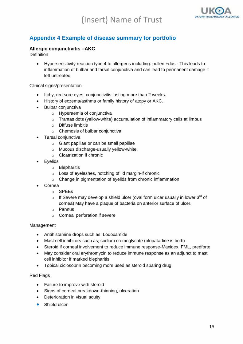
Appendix 4 Example of disease summary for portfolio
Allergic conjunctivitis –AKC
Definition
Hypersensitivity reaction type 4 to allergens including: pollen +dust- This leads to
inflammation of bulbar and tarsal conjunctiva and can lead to permanent damage if
left untreated.
Clinical signs/presentation
Itchy, red sore eyes, conjunctivitis lasting more than 2 weeks.
History of eczema/asthma or family history of atopy or AKC.
Bulbar conjunctiva
o Hyperaemia of conjunctiva
o Trantas dots (yellow-white) accumulation of inflammatory cells at limbus
o Diffuse limbitis
o Chemosis of bulbar conjunctiva
Tarsal conjunctiva
o Giant papillae or can be small papillae
o Mucous discharge-usually yellow-white.
o Cicatrization if chronic
Eyelids
o Blepharitis
o Loss of eyelashes, notching of lid margin-if chronic
o Change in pigmentation of eyelids from chronic inflammation
Cornea
o SPEEs
o If Severe may develop a shield ulcer (oval form ulcer usually in lower 3rd of
cornea) May have a plaque of bacteria on anterior surface of ulcer.
o Pannus
o Corneal perforation if severe
Management
Antihistamine drops such as: Lodoxamide
Mast cell inhibitors such as; sodium cromoglycate (olopatadine is both)
Steroid if corneal involvement to reduce immune response-Maxidex, FML, predforte
May consider oral erythromycin to reduce immune response as an adjunct to mast
cell inhibitor if marked blepharitis.
Topical ciclosoprin becoming more used as steroid sparing drug.
Red Flags
Failure to improve with steroid
Signs of corneal breakdown-thinning, ulceration
Deterioration in visual acuity
Shield ulcer
{Insert} Name of Trust
20
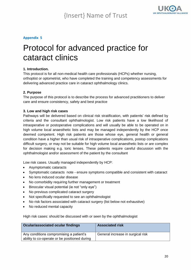
Appendix 5
Protocol for advanced practice for cataract clinics
1. Introduction.
This protocol is for all non-medical health care professionals (HCPs) whether nursing,
orthoptist or optometrist, who have completed the training and competency assessments for
delivering advanced practice care in cataract ophthalmology clinics.
2. Purpose
The purpose of this protocol is to describe the process for advanced practitioners to deliver
care and ensure consistency, safety and best practice
3. Low and high risk cases
Pathways will be delivered based on clinical risk stratification, with patients’ risk defined by
criteria and the consultant ophthalmologist. Low risk patients have a low likelihood of
intraoperative or postoperative complications and will usually be able to be operated on in
high volume local anaesthetic lists and may be managed independently by the HCP once
deemed competent. High risk patients are those whose eye, general health or general
condition have a higher than usual risk of intraoperative complications, postop complications
difficult surgery, or may not be suitable for high volume local anaesthetic lists or are complex
for decision making e.g. toric lenses. These patients require careful discussion with the
ophthalmologist and/or assessment of the patient by the consultant
Low risk cases. Usually managed independently by HCP:
Asymptomatic cataracts
Symptomatic cataracts note - ensure symptoms compatible and consistent with cataract
No lens induced ocular disease
No comorbidity requiring further management or treatment
Binocular visual potential (ie not “only eye”)
No previous complicated cataract surgery
Not specifically requested to see an ophthalmologist
No risk factors associated with cataract surgery (list below not exhaustive)
No reduced mental capacity
High risk cases: should be discussed with or seen by the ophthalmologist
Ocular/associated ocular findings
Associated risk
Any conditions compromising a patient’s
ability to co-operate or be positioned during
General increase in surgical risk

{Insert} Name of Trust
21
surgery e.g. communication and language
difficulties, hearing loss, spinal or back
problems, cough or poor breathing, tremor,
nystagmus, obesity, claustrophobia, extreme
fear/anxiety, reduced mental capacity,
dementia, psychiatric disease, lid squeezer
Age >85 years General increase in surgical risk or less good
visual outcome
Only seeing eye If serious complications, could get total loss
of vision, no “spare” eye
Complications in first eye operation Higher surgical risk
High myopia/axial length ≥ 26mm
Retinal detachment (RD), AC depth
fluctuation, IOL calculation errors
(staphyloma) and refractive surprise
High hyperopia <22mm
Shallow AC, choroidal effusion, IOL
calculation errors (refractive surprise)
Prior keratorefractive surgery
IOL calculation errors (refractive surprise),
AC depth fluctuation
Deep set eyes/high brow
Difficult surgical access
Blepharitis
Increased risk of endophthalmitis
Corneal opacification
Reduced surgical view
Corneal guttata/Fuch’s endothelial dystrophy Prolonged postoperative corneal oedema or
decompensation
Irregular corneal astigmatism (scarring,
ectasia, other causes)
IOL calculation errors (refractive surprise),
possible limited postop vision
Shallow anterior chamber Increased risk endothelial/iris damage,
technically more difficult therefore increased
surgery risk
Small dilated pupil
Poor visualisation, increased risk capsular
tear/vitreous prolapse, iris damage,
requirement for extra steps to enlarge pupil
Posterior synechiae
Intra-op miosis, prolonged post-op
inflammation, iris bleeding, inflammatory
deposits on IOL
Current or previous use of alpha adrenergic
antagonist
Tamsulosin, alfuzosin, terazosin, doxazosin
Intraoperative floppy iris syndrome (IFIS),
poor pupil dilation, progressive miosis.
Overall higher risk surgery. Greater risk with
Tamsulosin.
Active or previous uveitis Posterior synechiae, IOL deposits, cystoid
macular oedema (CMO), prolonged post-op
inflammation
Zonulopathy (laxity, dehiscence, loss)
Trauma, pseduoexfoliation, coloboma, age
>80, asymmetric anterior chamber depth
Phacodonesis (lenticular instability),
iridodonesis, lens subluxation, vitreous
prolapse, cataract loss into vitreous, late IOL
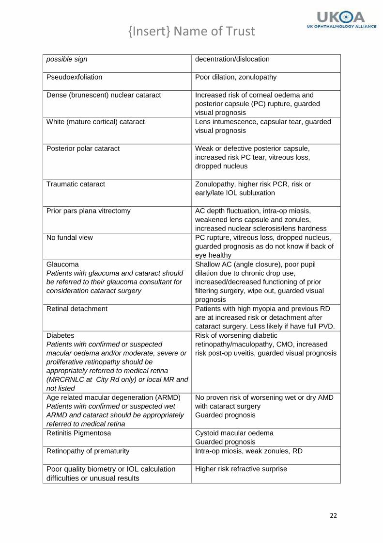
{Insert} Name of Trust
22
possible sign decentration/dislocation
Pseudoexfoliation
Poor dilation, zonulopathy
Dense (brunescent) nuclear cataract
Increased risk of corneal oedema and
posterior capsule (PC) rupture, guarded
visual prognosis
White (mature cortical) cataract
Lens intumescence, capsular tear, guarded
visual prognosis
Posterior polar cataract
Weak or defective posterior capsule,
increased risk PC tear, vitreous loss,
dropped nucleus
Traumatic cataract Zonulopathy, higher risk PCR, risk or
early/late IOL subluxation
Prior pars plana vitrectomy AC depth fluctuation, intra-op miosis,
weakened lens capsule and zonules,
increased nuclear sclerosis/lens hardness
No fundal view PC rupture, vitreous loss, dropped nucleus,
guarded prognosis as do not know if back of
eye healthy
Glaucoma
Patients with glaucoma and cataract should
be referred to their glaucoma consultant for
consideration cataract surgery
Shallow AC (angle closure), poor pupil
dilation due to chronic drop use,
increased/decreased functioning of prior
filtering surgery, wipe out, guarded visual
prognosis
Retinal detachment
Patients with high myopia and previous RD
are at increased risk or detachment after
cataract surgery. Less likely if have full PVD.
Diabetes
Patients with confirmed or suspected
macular oedema and/or moderate, severe or
proliferative retinopathy should be
appropriately referred to medical retina
(MRCRNLC at City Rd only) or local MR and
not listed
Risk of worsening diabetic
retinopathy/maculopathy, CMO, increased
risk post-op uveitis, guarded visual prognosis
Age related macular degeneration (ARMD)
Patients with confirmed or suspected wet
ARMD and cataract should be appropriately
referred to medical retina
No proven risk of worsening wet or dry AMD
with cataract surgery
Guarded prognosis
Retinitis Pigmentosa
Cystoid macular oedema
Guarded prognosis
Retinopathy of prematurity
Intra-op miosis, weak zonules, RD
Poor quality biometry or IOL calculation
difficulties or unusual results
Higher risk refractive surprise
{Insert} Name of Trust
23
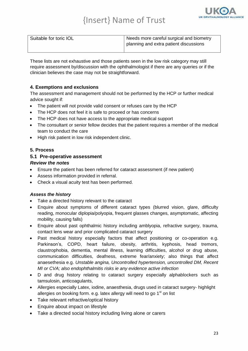
Suitable for toric IOL
Needs more careful surgical and biometry
planning and extra patient discussions
These lists are not exhaustive and those patients seen in the low risk category may still
require assessment by/discussion with the ophthalmologist if there are any queries or if the
clinician believes the case may not be straightforward.
4. Exemptions and exclusions
The assessment and management should not be performed by the HCP or further medical
advice sought if:
The patient will not provide valid consent or refuses care by the HCP
The HCP does not feel it is safe to proceed or has concerns
The HCP does not have access to the appropriate medical support
The consultant or senior fellow decides that the patient requires a member of the medical
team to conduct the care
High risk patient in low risk independent clinic.
5. Process
5.1 Pre-operative assessment
Review the notes
Ensure the patient has been referred for cataract assessment (if new patient)
Assess information provided in referral.
Check a visual acuity test has been performed.
Assess the history
Take a directed history relevant to the cataract
Enquire about symptoms of different cataract types (blurred vision, glare, difficulty
reading, monocular diplopia/polyopia, frequent glasses changes, asymptomatic, affecting
mobility, causing falls)
Enquire about past ophthalmic history including amblyopia, refractive surgery, trauma,
contact lens wear and prior complicated cataract surgery
Past medical history especially factors that affect positioning or co-operation e.g.
Parkinson’s, COPD, heart failure, obesity, arthritis, kyphosis, head tremors,
claustrophobia, dementia, mental illness, learning difficulties, alcohol or drug abuse,
communication difficulties, deafness, extreme fear/anxiety; also things that affect
anaesethesia e.g. Unstable angina, Uncontrolled hypertension, uncontrolled DM, Recent
MI or CVA; also endophthalmitis risks ie any evidence active infection
D and drug history relating to cataract surgery especially alphablockers such as
tamsulosin, anticoagulants,
Allergies especially Latex, iodine, anaesthesia, drugs used in cataract surgery- highlight
allergies on booking form. e.g. latex allergy will need to go 1st on list
Take relevant refractive/optical history
Enquire about impact on lifestyle
Take a directed social history including living alone or carers

{Insert} Name of Trust
24
Identify any specific communication needs e.g. poor hearing, English not first
language
Establish patient’s need and willingness with regard to surgical intervention
HCP must identify factors in ophthalmic and general medical history that may place
patient at higher risk of surgical or anaesthetic (LA or GA) complications or
difficulties.
Conduct the examination
Distance, corrected, pinhole vision
Observation of face and lids, posture, mobility in case of difficulties of access or
positioning
Cover test
Slit lamp assessment of eyelids, eyelid margins, conjunctiva, limbus, cornea,
anterior chamber (including angle), pupils, iris:
Pupil size and reactions including RAPD
IOP
Pupil dilatation
Examination of the lens
Examination of the vitreous gel
Dilated fundus examination including optic disc, macula and retina
Patients likely ability to comply with local anaesthesia from reaction to
examination or lid squeezing.
Investigations
Note refractive error from referral or perform or obtain focimetry or auto-refraction
for current spectacle prescription
Perform or order and interpret keratometry, biometry
Note and discuss with an ophthalmologist any unusual biometry or IOL powers
OCT for any macular pathology
B scan if no fundal view.
Treatment and management
Patients suitable for independent management the HCP should counsel and undertake
valid consent if trained to do so:
Advise patients on ability to meet driving requirements
Discuss and counsel the patient on the options including the option for doing nothing,
alternatives to surgery (eg adaptive and refractive management) , the process and
pathway for surgery, the risks and benefits, postoperative expectations and care
Establish willingness for surgery
Any guarded prognoses fully discussed with the patient and with a consultant/senior
surgeon if appropriate
Discuss the options for refractive outcomes and the limitations of refractive
predictability – most corrected for distance need readers and may need some
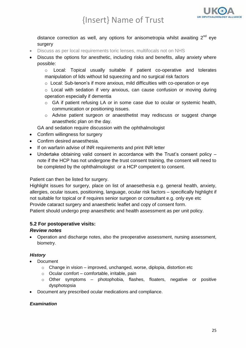
{Insert} Name of Trust
25
distance correction as well, any options for anisometropia whilst awaiting 2nd
eye
surgery
Discuss as per local requirements toric lenses, multifocals not on NHS
Discuss the options for anesthetic, including risks and benefits, allay anxiety where
possible:
o Local: Topical usually suitable if patient co-operative and tolerates
manipulation of lids without lid squeezing and no surgical risk factors
o Local: Sub-tenon’s if more anxious, mild difficulties with co-operation or eye
o Local with sedation if very anxious, can cause confusion or moving during
operation especially if dementia
o GA if patient refusing LA or in some case due to ocular or systemic health,
communication or positioning issues.
o Advise patient surgeon or anaesthetist may rediscuss or suggest change
anaesthetic plan on the day.
GA and sedation require discussion with the ophthalmologist
Confirm willingness for surgery
Confirm desired anaesthesia.
If on warfarin advise of INR requirements and print INR letter
Undertake obtaining valid consent in accordance with the Trust’s consent policy –
note if the HCP has not undergone the trust consent training, the consent will need to
be completed by the ophthalmologist or a HCP competent to consent.
Patient can then be listed for surgery.
Highlight issues for surgery, place on list of anaesethesia e.g. general health, anxiety,
allergies, ocular issues, positioning, language, ocular risk factors – specifically highlight if
not suitable for topical or if requires senior surgeon or consultant e.g. only eye etc
Provide cataract surgery and anaesthetic leaflet and copy of consent form.
Patient should undergo prep anaesthetic and health assessment as per unit policy.
5.2 For postoperative visits:
Review notes
Operation and discharge notes, also the preoperative assessment, nursing assessment,
biometry.
History
Document
o Change in vision – improved, unchanged, worse, diplopia, distortion etc
o Ocular comfort – comfortable, irritable, pain
o Other symptoms – photophobia, flashes, floaters, negative or positive
dysphotopsia
Document any prescribed ocular medications and compliance.
Examination

{Insert} Name of Trust
26
Visual acuity distance both eyes unaided, with current glasses and pinhole if 6/9 or
worse
Examine operated eyes only.
Examine the fellow eye only if there is a clinical indication to do so e..g needs
reassessment for consideration of second eye cataract surgery. If full assessment
completed at preop visit, short repeat examination of anterior segment satisfactory. If full
preop assessment second eye not completed pre-operatively, undertake full assessment
as above.
Refraction: Autorefraction of all patients at minimum, subjective refraction is ideal if
available and required if any refractive surprises (>1D sph equivalent from target)
Full external/anterior segment slit lamp examination.
IOP
Dilation of pupil if and slit lamp posterior segment examination if:
o Best corrected visual acuity worse than expected
o Any surgical complications
o Any patient complaining of flashes and floaters or other symptoms warranting
dilation
o No/poor preoperative fundus view
o All patients with diabetes
o Posterior segment co-morbidity requiring assessment postoperatively
If pupil dilation is not required postoperative fundus examination is not necessary
Any other clinical investigations if warranted
Macular OCT for all patients with diabetes, ERM, confirmed or suspected macular
pathology including patients with visual outcomes worse than expected
The second eye should be reassessed, with the level of assessment and examination at
discretion depending on the detail of the original assessment and the desire/requirement for
surgery.
If second eye surgery is desired, then the appropriate investigations should be undertaken or
checked and the treatment and management completed as above.
Treatment and management
Routine patients
HCP can independently manage all uncomplicated patients not requiring any medical
opinion as follows
Continue their postoperative drops as prescribed by the operating surgeon. Individual
surgeon prescribing habits differ but will broadly follow the post-op regime
o G chlorampenicol 0.5% qds for 1-2 weeks
o G dexamethasone 0.1% qds for 2 weeks, bd for 2 weeks
o Patients with dark irides, diabetes or other issues may have a different regime
o For patients considered at risk of pseudophakic cystoid macular oedema (PCMO)
g ketorolac 0.5% (acular) may be prescribed qds for 4 weeks
If second eye surgery is not required discharge the patient with a letter (GP and patient
copy) stating the discharge drop regime and need for refraction with local optometrist
Advise patient about obtaining community optometrist refraction at 4-6 weeks postop
If second eye surgery required, follow procedure as above for preop requirements.

{Insert} Name of Trust
27
Postoperative issues
Patients with routine postop issues can be managed by the HCP. All patients with
intraoperative complications should be seen by a doctor.
Any postoperative pathology identified and not covered below should be managed
appropriately.
Patients with ocular abnormality discovered incidentally which is unrelated to the
condition for which the patient was originally referred should be referred internally to the
appropriate service or back to the GP if the patient wishes to go elsewhere
Post-op findings Considerations Action
Lids
Postoperative
ptosis
Cosmesis
Reassure, mild ptosis may
improve over 6 months
Chronic (over 6 months) with superior
field defect or and cosmetically
unacceptable
Routine referral to adnexal
service after discussion with
medic
Conjunctiva
Conjunctival
injection
Injection around subtenons entry site
and/or sub-conjunctival haemorrhage
Reassure, expect resolution
within 6 weeks
Circumlimbal injection (ciliary flush)
usually indicative of anterior uveitis
Check anterior chamber
activity and manage
accordingly – see anterior
chamber
Diffuse injection
Drop toxicity or allergy
Manage appropriately
Blepharitis
Manage appropriately
Consider uveitis, TASS,
endophthalmitis with associated
signs and symptoms
Show to doctor
Cornea
Superficial punctuate
keratopathy/keratitis
Dry eye
Dry eye symptoms are common after cataract surgery and can take up to 3 months to resolve. Lubricate and reassure
Blepharitis Manage appropriately
Drop toxicity (usually diffuse keratopathy/keratitis)
See associated guidanceManage appropriately
Descemet’s
membrane
Mild: common after cataract surgery
If cornea clear and expected visual outcome achieved, reassure.

{Insert} Name of Trust
28
folds Postpone local refraction until resolution after six weeks if possible
Moderate to severe: significant corneal oedema and/or inflammation
Show to doctor
Descemet’s
membrane tear or
detachment
No corneal oedema If expected visual outcome achieved and cornea clear, no action required
Associated corneal oedema Show to doctor
Sutures Buried
If non-absorbable (i.e. nylon) discuss with doctor and remove if competent to do so If absorbable (i.e. vicryl) no action required
Loose Remove is competent to do so, if not call doctor/competent HCP to remove
Anterior chamber
Shallow Wound leak, Seidel positive (often associated with low IOP)
Serous choroidal effusion (often associated with low IOP)
Pupil block: Severe uveitis, capsular block syndrome (associated with high IOP)
Haemorrhagic choroidal effusion (suprachoroidal haem) (often associated with high IOP)
Show to doctor
Cells Use 1x1mm slit beam Differentiate between cells
and pigment
Grade 0 (no cells)
Grade 0.5+ (1 to 5 cells)
No action required
Grade 1+ (6 to 15 cells)
Discuss with doctor
Grade 2+ (16 to 25 cells)
Grade 3+ (26 to 50 cells)
Grade 4+ (> 50 cells)
Show to doctor
Note severe post op
inflammation is
endophthalmitis until proven
otherwise
Flare Use 1x1mm slit beam (SUN grading)
Grade 0 (none)
Grade 1+ (faint)
Grade 2+ (moderate, iris and lens
details clear)
Grade 3+ (marked, iris and lens
Flare can be difficult to
grade clinically
If grade 3+ or 4+, show to
doctor Otherwise manage
on the basis of AC cells

{Insert} Name of Trust
29
details hazy)
Grade 4+ (intense, fibrin or plastic
aqueous)
Hypopyon/Hyphaema Severe uveitis, endophthalmitis,
TASS, trauma
Show to doctor
Vitreous Vitreous strand incarcerated in wound
– peaked pupil
Show to doctor
Retained lens
fragments
Corneal oedema, anterior uveitis
Show to doctor
Anterior chamber IOL
Document haptic position, check
patency of PI, check for pigment
dispersion
Show to doctor
Iris
Trauma
Mild intraoperative iris trauma - may
result in prolonged postoperative
uveitis
Manage on the basis of AC
cells
Iris transillumination
If significant trauma and/or
patient suffering with glare,
show to doctor
Prolapse
Iris prolapse to wound Show to doctor
IOL and capsule
Anterior capsular
phimosis
Mild with no associated uveitis and visual axis clear
No action required
All other cases
Show to doctor
Posterior capsular
opacification or
plaque
Patient asymptomatic No action required
Patient symptomatic YAG laser safe to perform 4 months after surgery –list accordingly
Capsular block
syndrome (CBS)
Entrapment of fluid between the IOL
and posterior capsule
Refractive surprise (myopic shift)
Shallow AC
Show to doctor and
consider YAG capsulotomy
if indicated
Vitreous
Cells Anterior uveitis may cause spill-over of a few cells into the anterior vitreous
Manage on the basis of AC cells
Vitritis: significant infiltration of vitreous cavity with inflammatory cells/vitreous haze - suspect endophthalmitis
Show to doctor
Weiss ring
Posterior vitreous detachment
common after cataract surgery
Search for retinal breaks
(dilate) and give retinal
detachment advice
Pigment
Shafer’s sign: assume retinal break Search for retinal break
(dilate) and show to doctor
Retina
{Insert} Name of Trust
30
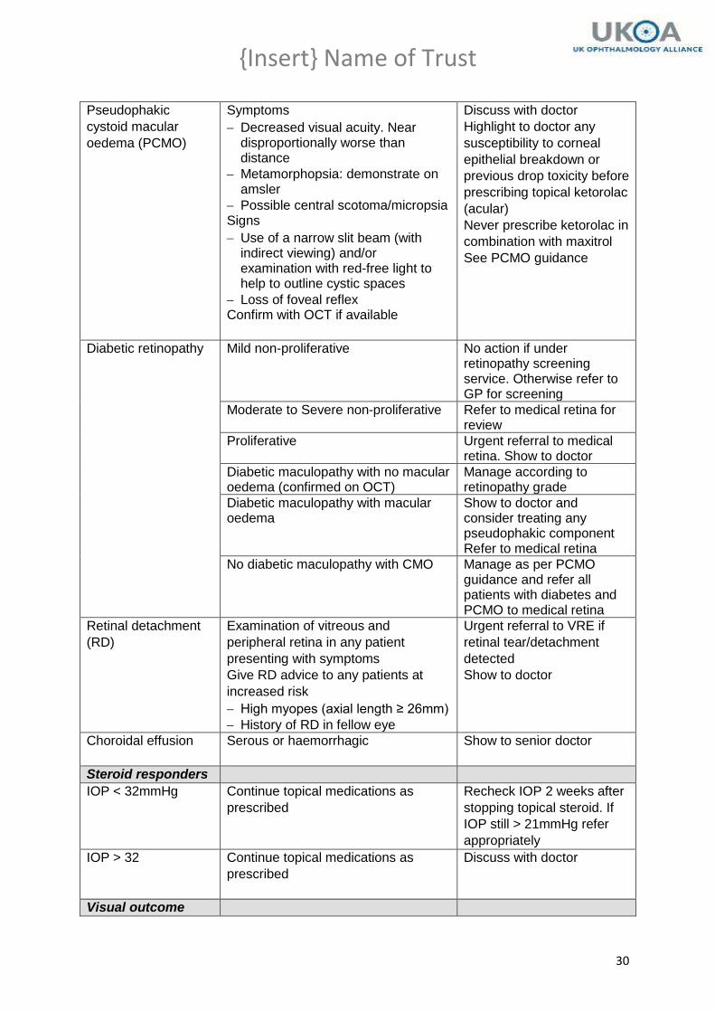
Pseudophakic
cystoid macular
oedema (PCMO)
Symptoms
Decreased visual acuity. Near disproportionally worse than distance
Metamorphopsia: demonstrate on amsler
Possible central scotoma/micropsia Signs
Use of a narrow slit beam (with indirect viewing) and/or examination with red-free light to help to outline cystic spaces
Loss of foveal reflex Confirm with OCT if available
Discuss with doctor
Highlight to doctor any
susceptibility to corneal
epithelial breakdown or
previous drop toxicity before
prescribing topical ketorolac
(acular)
Never prescribe ketorolac in
combination with maxitrol
See PCMO guidance
Diabetic retinopathy Mild non-proliferative No action if under retinopathy screening service. Otherwise refer to GP for screening
Moderate to Severe non-proliferative Refer to medical retina for review
Proliferative Urgent referral to medical retina. Show to doctor
Diabetic maculopathy with no macular oedema (confirmed on OCT)
Manage according to retinopathy grade
Diabetic maculopathy with macular oedema
Show to doctor and consider treating any pseudophakic component Refer to medical retina
No diabetic maculopathy with CMO Manage as per PCMO guidance and refer all patients with diabetes and PCMO to medical retina
Retinal detachment
(RD)
Examination of vitreous and
peripheral retina in any patient
presenting with symptoms
Give RD advice to any patients at
increased risk
High myopes (axial length ≥ 26mm)
History of RD in fellow eye
Urgent referral to VRE if
retinal tear/detachment
detected
Show to doctor
Choroidal effusion
Serous or haemorrhagic Show to senior doctor
Steroid responders
IOP < 32mmHg Continue topical medications as
prescribed
Recheck IOP 2 weeks after
stopping topical steroid. If
IOP still > 21mmHg refer
appropriately
IOP > 32
Continue topical medications as
prescribed
Discuss with doctor
Visual outcome
{Insert} Name of Trust
31
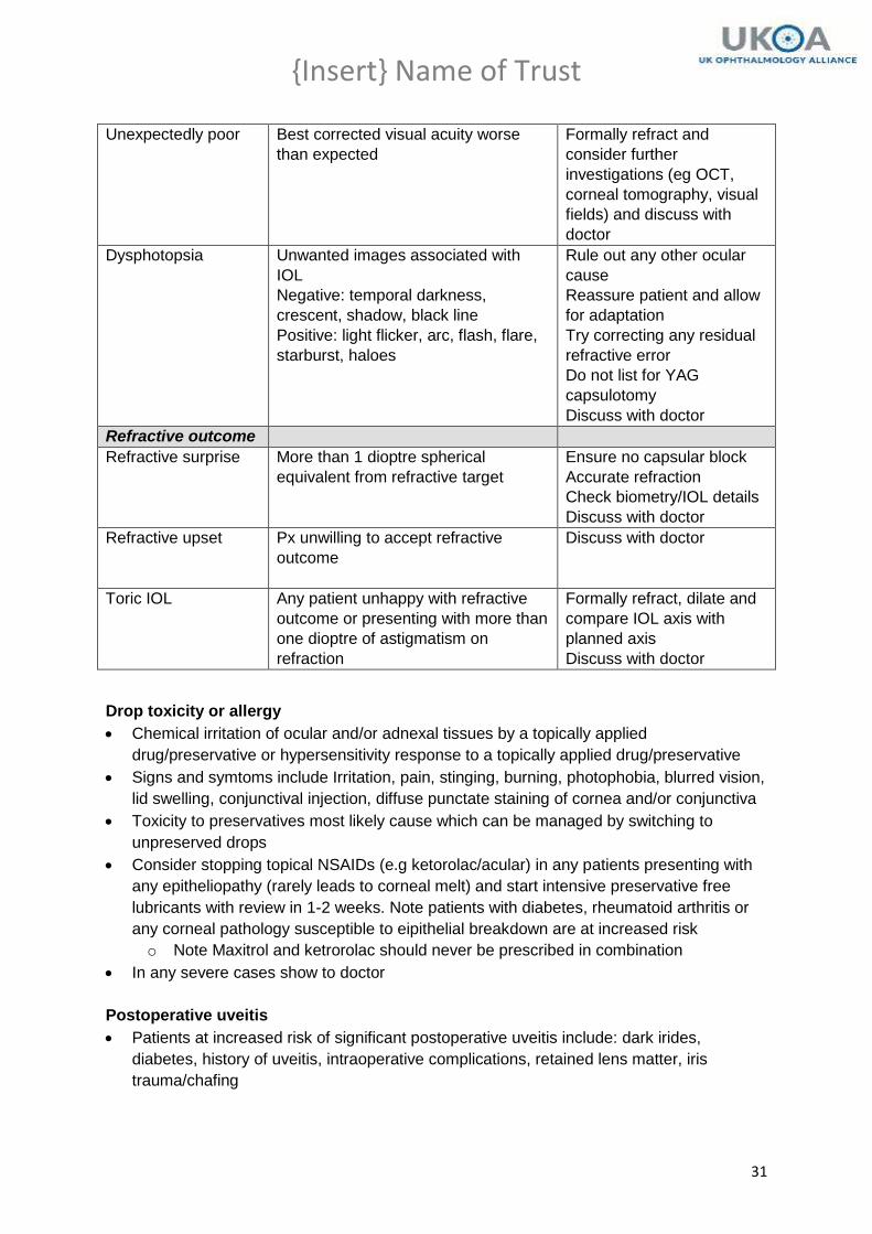
Unexpectedly poor
Best corrected visual acuity worse
than expected
Formally refract and
consider further
investigations (eg OCT,
corneal tomography, visual
fields) and discuss with
doctor
Dysphotopsia
Unwanted images associated with
IOL
Negative: temporal darkness,
crescent, shadow, black line
Positive: light flicker, arc, flash, flare,
starburst, haloes
Rule out any other ocular
cause
Reassure patient and allow
for adaptation
Try correcting any residual
refractive error
Do not list for YAG
capsulotomy
Discuss with doctor
Refractive outcome
Refractive surprise More than 1 dioptre spherical
equivalent from refractive target
Ensure no capsular block
Accurate refraction
Check biometry/IOL details
Discuss with doctor
Refractive upset Px unwilling to accept refractive
outcome
Discuss with doctor
Toric IOL
Any patient unhappy with refractive
outcome or presenting with more than
one dioptre of astigmatism on
refraction
Formally refract, dilate and
compare IOL axis with
planned axis
Discuss with doctor
Drop toxicity or allergy
Chemical irritation of ocular and/or adnexal tissues by a topically applied
drug/preservative or hypersensitivity response to a topically applied drug/preservative
Signs and symtoms include Irritation, pain, stinging, burning, photophobia, blurred vision,
lid swelling, conjunctival injection, diffuse punctate staining of cornea and/or conjunctiva
Toxicity to preservatives most likely cause which can be managed by switching to
unpreserved drops
Consider stopping topical NSAIDs (e.g ketorolac/acular) in any patients presenting with
any epitheliopathy (rarely leads to corneal melt) and start intensive preservative free
lubricants with review in 1-2 weeks. Note patients with diabetes, rheumatoid arthritis or
any corneal pathology susceptible to eipithelial breakdown are at increased risk
o Note Maxitrol and ketrorolac should never be prescribed in combination
In any severe cases show to doctor
Postoperative uveitis
Patients at increased risk of significant postoperative uveitis include: dark irides,
diabetes, history of uveitis, intraoperative complications, retained lens matter, iris
trauma/chafing

{Insert} Name of Trust
32
Rebound uveitis should be treated with an increased frequency and longer tapering
course of topical anti-inflammatories
Patients presenting with a second episode of rebound uveitis require gonioscope angle
examination to determine the presence/absence of retained lens matter – refer to doctor
if not competent
Toxic Anterior Segment Syndrome (TASS)
Sterile postoperative inflammatory reaction caused by a non-infectious substance that
enters the anterior segment and results in toxic damage to intraocular tissues
Rare, incidence unknown
Clinical picture similar to endophthalmitis but inflammatory reaction limited to anterior
chamber and presents early with onset 12-24hrs after surgery
Show to doctor
Endophthalmitis
Rare, occurring in approximately less than one in a thousand cases
Acute postoperative endophthalmitis presents upt o six weeks following surgery but
usually presents within the first two weeks
Chronic endophthalmitis can present after six weeks
Signs and symptoms include pain, visual loss, lid swelling, marked anterior chamber
inflammation with hypopyon, vitritis and often no fundal view (conjunctival injection and
corneal oedema with other associated signs)
Show to senior doctor immediately
See guidelines for management of endophthalmitis
Pseudophakic cystoid macular oedema (PCMO)
The incidence of clinical PCMO, defined as symptomatic vision loss 6/12 or worse, is
approximately 0.1% to 2.35%. PCMO as seen on OCT after modern phacoemulsification
may range from 4% to 11%
PCMO most often develops 4-6 weeks after cataract surgery. The peak incidence of
PCMO occurs at 6 weeks after surgery. Acute PCMO occurs within 6 months
postoperatively; chronic PCMO is present more than 6 months after cataract surgery
Incidence increases in patients with high-risk characteristics including diabetes mellitus,
retinitis pigmentosa, history of central retinal vein occlusion, recent history of uveitis, pre-
existing epiretinal membrane, or following complicated cataract surgery
Most patients with PCMO have spontaneous resolution of the macular oedema within 3-
4 months. One year after surgery a small minority of patients (<1%) in the absence of
treatment may still have decreased visual acuity from PCMO.
Once PCMO is confirmed by clinical findings and/or OCT, initial treatment includes the
use of topical steroidal and nonsteroidal anti-inflammatory medications (NSAIDs) e.g. g
dexamethasone 0.1% four times daily and g ketorolac four times daily for 6-8 weeks
followed by tapering
5.3 Documentation

{Insert} Name of Trust
33
Record assessment, treatment and all discussions clearly in the patient’s health records
as per trust records policy
Complete the consent form and record provision of the relevant written leaflets.
GP letter to be completed on records, filing a copy in the notes
If an unexpected event occurs, document and complete and report the incident. This is
necessary to facilitate communication within the team, meet legal requirements of
practice and enable monitoring over a time period.
Complete any documentation for listing the patient
{Insert} Name of Trust
34
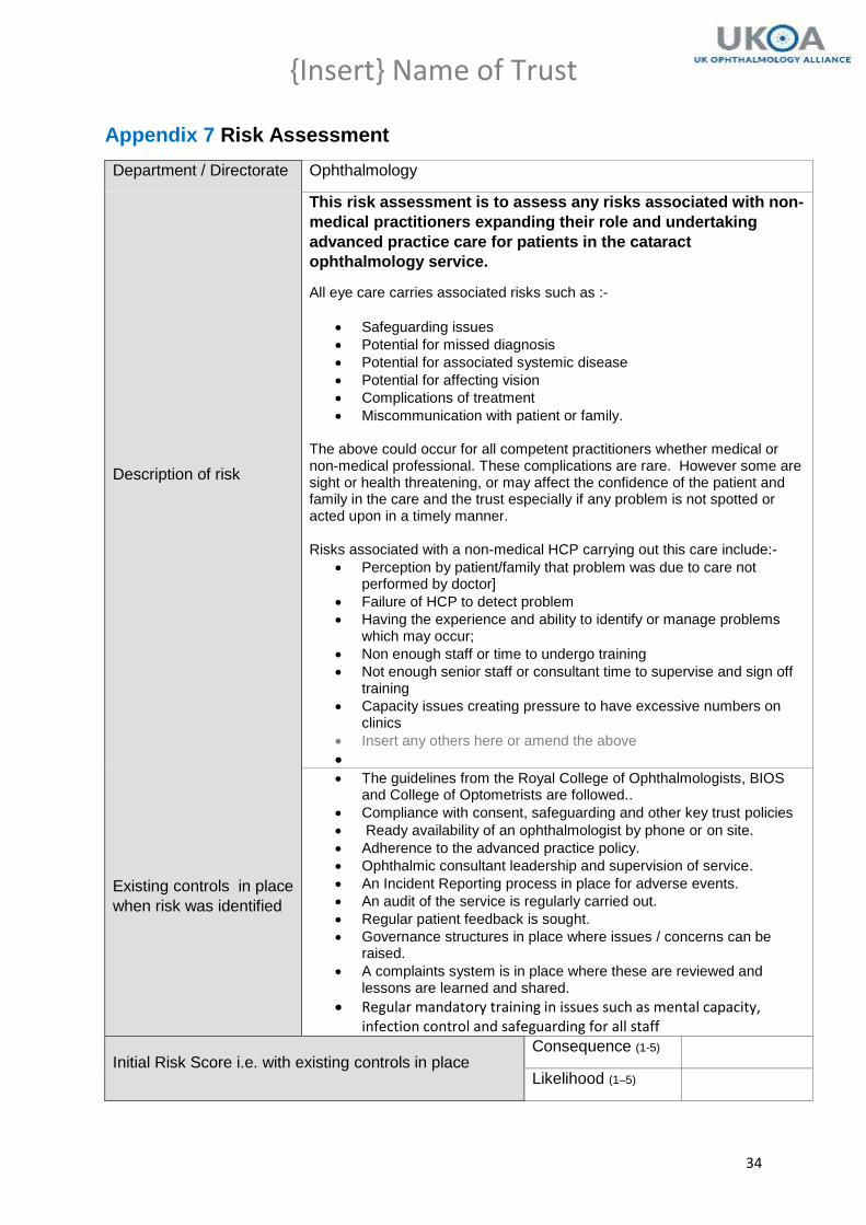
Appendix 7 Risk Assessment
Department / Directorate Ophthalmology
Description of risk
This risk assessment is to assess any risks associated with non-
medical practitioners expanding their role and undertaking
advanced practice care for patients in the cataract
ophthalmology service.
All eye care carries associated risks such as :-
Safeguarding issues
Potential for missed diagnosis
Potential for associated systemic disease
Potential for affecting vision
Complications of treatment
Miscommunication with patient or family. The above could occur for all competent practitioners whether medical or non-medical professional. These complications are rare. However some are sight or health threatening, or may affect the confidence of the patient and family in the care and the trust especially if any problem is not spotted or acted upon in a timely manner. Risks associated with a non-medical HCP carrying out this care include:-
Perception by patient/family that problem was due to care not performed by doctor]
Failure of HCP to detect problem
Having the experience and ability to identify or manage problems which may occur;
Non enough staff or time to undergo training
Not enough senior staff or consultant time to supervise and sign off training
Capacity issues creating pressure to have excessive numbers on clinics
Insert any others here or amend the above
Existing controls in place
when risk was identified
The guidelines from the Royal College of Ophthalmologists, BIOS and College of Optometrists are followed..
Compliance with consent, safeguarding and other key trust policies
Ready availability of an ophthalmologist by phone or on site.
Adherence to the advanced practice policy.
Ophthalmic consultant leadership and supervision of service.
An Incident Reporting process in place for adverse events.
An audit of the service is regularly carried out.
Regular patient feedback is sought.
Governance structures in place where issues / concerns can be raised.
A complaints system is in place where these are reviewed and lessons are learned and shared.
Regular mandatory training in issues such as mental capacity, infection control and safeguarding for all staff
Initial Risk Score i.e. with existing controls in place Consequence (1-5)
Likelihood (1–5)
{Insert} Name of Trust
35
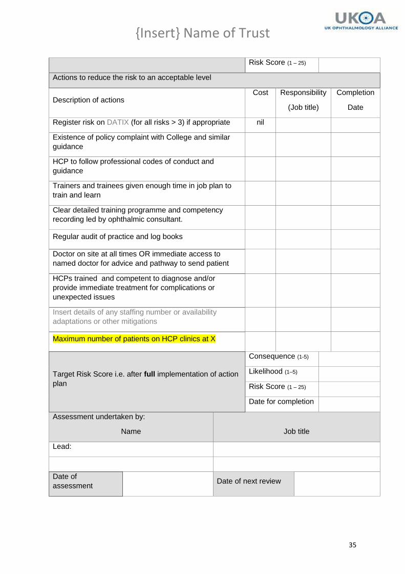
Risk Score (1 – 25)
Actions to reduce the risk to an acceptable level
Description of actions Cost Responsibility
(Job title)
Completion
Date
Register risk on DATIX (for all risks > 3) if appropriate nil
Existence of policy complaint with College and similar
guidance
HCP to follow professional codes of conduct and
guidance
Trainers and trainees given enough time in job plan to
train and learn
Clear detailed training programme and competency
recording led by ophthalmic consultant.
Regular audit of practice and log books
Doctor on site at all times OR immediate access to
named doctor for advice and pathway to send patient
HCPs trained and competent to diagnose and/or
provide immediate treatment for complications or
unexpected issues
Insert details of any staffing number or availability
adaptations or other mitigations
Maximum number of patients on HCP clinics at X
Target Risk Score i.e. after full implementation of action
plan
Consequence (1-5)
Likelihood (1–5)
Risk Score (1 – 25)
Date for completion
Assessment undertaken by:
Name
Job title
Lead:
Date of
assessment Date of next review

{Insert} Name of Trust
36
Acknowledgements: With significant thanks on authoring this document to Aneel Suri, Kat Anguige and Connor Beddow of Moorfields Hospital