closed-incision negative pressure therapy in place of
TRANSCRIPT

Received 06/09/2020 Review began 06/26/2020 Review ended 07/07/2020 Published 07/10/2020
© Copyright 2020Karlock et al. This is an open accessarticle distributed under the terms of theCreative Commons Attribution LicenseCC-BY 4.0., which permits unrestricteduse, distribution, and reproduction in anymedium, provided the original author andsource are credited.
Closed-Incision Negative Pressure Therapy inPlace of Surgical Drain Placement in PlantarFibroma Excision Surgery: A Case SeriesAbbey Karlock , Ralph J. Napolitano
1. Osteopathic Medicine, Ohio University Heritage College of Osteopathic Medicine, Athens, USA 2. Podiatry andWound Care, OrthoNeuro, Columbus, USA
Corresponding author: Abbey Karlock, [email protected]
AbstractPlantar fibromas are benign masses of fibrous tissue that develop in the arch of the foot arising from theplantar fascia. Symptomatology varies and is often related to weight bearing anatomic correlations orimpingement of neurological or musculoskeletal structures. Several treatment options are available andinclude palliative measures, non-operative interventions and surgery, all with varying degrees of successand complication risk. The aim of this case study series was to assess surgical wound healing retrospectivelyin three patients who underwent wide en bloc plantar fibroma excision surgery for symptomaticlesions. Their incision was managed with a disposable closed-incision negative pressure therapy device(Prevena™; 3M + KCI, St. Paul, MN) in lieu of surgical drains. All three patients demonstrated favorableoutcomes without complications.
Categories: Dermatology, General Surgery, OrthopedicsKeywords: closed-incision negative pressure wound therapy, plantar fibroma, foot tumors, podiatry, wound care,negative pressure wound therapy, prevena
IntroductionEpidemiology and pathophysiologyPlantar fibromatosis (PF) is a rare disease characterized by nodules in the plantar fascia caused by disorderedhyperproliferation of the fibrous tissue. It was first recorded by George Ledderhose in 1897 and is alsoreferred to as Ledderhose disease [1].
The National Institutes of Health classify PF as a “rare disease that affects fewer than 200,000 individuals inthe United States” [2]. Men are more commonly affected compared to women, and the disease tends to occurmore often in middle-aged patients but can affect patients of any age [3]. PF is also seen to occur more oftenin patients with epilepsy, diabetes mellitus, alcoholism with liver disease, repetitive trauma, and keloids [4].
PF is most often benign, but may cause pain and difficulty with ambulation and weight bearing as well aspain due to impingement of neurological or musculoskeletal structures as a result of the growing nodules[5]. The nodules are encapsulated, firm, and most commonly appear on the medial and central aspects of theplantar aponeurosis [6]. The formation of these fibromas is caused by the hyperproliferation of the fibroustissue due to increased fibroblastic activity.
Treatment optionsSymptom management is often the treatment of choice for plantar fibromas due to the characteristicallybenign nature. Some of these treatments include custom or over-the-counter orthotics to help with weightbearing, steroid injections, verapamil, radiation, extracorporeal shock wave therapy, tamoxifen hormonaltherapy, and collagenase, all with varying degrees of success and scientific data to back up the effectiveness[1,7].
If symptom management does not alleviate the pain, or if the nodules continue to grow in size, surgicalexcision is the next option. There is a high rate of re-occurrence when only removing the fibroma itself, andfor this reason excision of the entire plantar fascia is usually preferred [1,8]. Surgical management of thiscondition carries with it more significant specific complications including increased risk of hematoma,greater swelling, neurovascular damage and functional complications as well as potential painful scarformation negating the benefits of the surgery.
As with any surgical treatment, careful post-operative incision management is critical in preventing saidcomplications at the incision site. There has been much research into the most beneficial post-operativewound management, particularly the most effective way to remove fluids such as blood and lymph from thearea and keep the incision site clean. When fluids accumulate in the wound area, it can put pressure on the
1 2
Open Access CaseReport DOI: 10.7759/cureus.9110
How to cite this articleKarlock A, Napolitano R J (July 10, 2020) Closed-Incision Negative Pressure Therapy in Place of Surgical Drain Placement in Plantar FibromaExcision Surgery: A Case Series. Cureus 12(7): e9110. DOI 10.7759/cureus.9110

surrounding organs, nerves and vasculature, thus leading to pain and potential impairment of woundhealing [9].
Surgical drains are used in many post-operative patients to remove excess fluids from wound sites throughthe process of the active suction of a drain placed during the procedure [9]. Another more recent incisionmanagement technique is negative pressure wound therapy (NPWT). This is a technique developed byArgenta and Morykwas in the early 1990s [10]. The aim of this therapy is to remove excess fluid and controlthe microenvironment of the wound through negative pressure suction by drawing the edges of the woundtogether.
One area of particular interest is that of a closed-incision negative pressure therapy (ciNPT) device such asPrevena™ by 3M + KCI (St. Paul, MN). This device acts on the same principles as NPWT, but rather on aclosed incision with a constant -125 mm Hg pressure applied over the site continuously [11]. This is thoughtto help wound healing through an increase in blood flow, decreased stress on the incision, and removal oflymph from the local area. One meta-analysis of NPWT applied on closed wounds found this technique to besignificant in reducing post-operative wound infections and seromas compared to standard post-operativedressings [12]. Stannard et al. in another study found that the use of ciNPT for post-operative managementof high-risk lower extremity fractures resulted in a lower rate of wound dehiscence and infection [13]. Whilethe results of these studies were favorable to ciNPT, there is not enough evidence in the current literature torecommend this technique for all wounds and all patients.
There is a vast array of available information on this therapy for surgeries performed throughout the body.However, the foot is one that is lacking representation in the literature, particularly the plantar surface,which poses unique surgical healing challenges. There are currently no universal treatment guidelines forthe use of ciNPT devices such as Prevena™ in plantar fibroma excision surgery. The aim of this retrospectivecase series was to demonstrate and further define healing outcomes utilizing this specific ciNPWT device forthis unique surgical problem.
Case PresentationCase 1A 56-year-old female presented for the evaluation of gradual onset bilateral soft tissue masses involving theplantar surface of both feet, with the left foot being significantly more symptomatic. Her past medicalhistory included hypertension and high cholesterol. Social history included gainful employment in amoderately labor-intensive type job and several-year history of smoking. Her physical exam was essentiallynormal with the exception of somewhat firm, slightly mobile nodular masses involving both plantar footsurfaces in the region of the plantar medical arch (Figure 1). Tenderness was only noted involving the twomasses on the left foot. Plain radiographs were negative. Conservative therapies were discussed and triedthat included shoe gear and activity modification as well as analgesics. The patient declined injectiontherapy. An MRI was ordered that revealed two discrete soft tissue masses within the plantar fasciaconsistent with PF lesions without neurovascular encroachment (Figures 2, 3). Surgery was proposed and shewished to proceed with surgery on her symptomatic left foot (Figure 4). Risks and benefits were outlined forboth complete fasciectomy and wide en bloc resection. The patient chose the latter technique and wasadvised to refrain from smoking that she did for several days both pre- and post-operatively. Surgery wascompleted without complications. Wound management included placement of a battery-poweredciNPT device (Prevena™) and light compression bandaging (Figure 5). The device remained in place for oneweek and then was removed. No significant erythema or swelling was seen. Surgical pathology wasconsistent with two, benign PF lesions. Sutures remained in place for a total of three weeks and were thenremoved. At this time, the incision was found to be negligibly tender, flat and well coapted. She was non-weight bearing immediately post-operatively through suture removal at which time she was transitioned toa walking boot with instructions to gradually increase weight-bearing activities to tolerance. Rechecks wereunremarkable with the exception of a small area of dehiscence that was seen one week after suture removal.Complete incisional healing was appreciated about a week after suture removal. She returned to normal,daily activities including unrestricted work just over two months with no symptomatology or recurrence atthe one-year mark.
2020 Karlock et al. Cureus 12(7): e9110. DOI 10.7759/cureus.9110 2 of 15

FIGURE 1: Case 1 pre-operative photograph.
2020 Karlock et al. Cureus 12(7): e9110. DOI 10.7759/cureus.9110 3 of 15

FIGURE 2: Case 1 MRI showing the axial T2 weighted image of the leftfoot demonstrating a plantar fibroma involving the central and medialcords of the plantar fascia beginning at the first tarsometatarsal joint.
FIGURE 3: Case 1 MRI showing the sagittal T1 weighted image of theleft foot demonstrating a plantar fibroma starting at the firsttarsometatarsal joint with no involvement of the plantar musculature or
2020 Karlock et al. Cureus 12(7): e9110. DOI 10.7759/cureus.9110 4 of 15

flexor tendons.
FIGURE 4: Case 1 intraoperative photograph.
2020 Karlock et al. Cureus 12(7): e9110. DOI 10.7759/cureus.9110 5 of 15
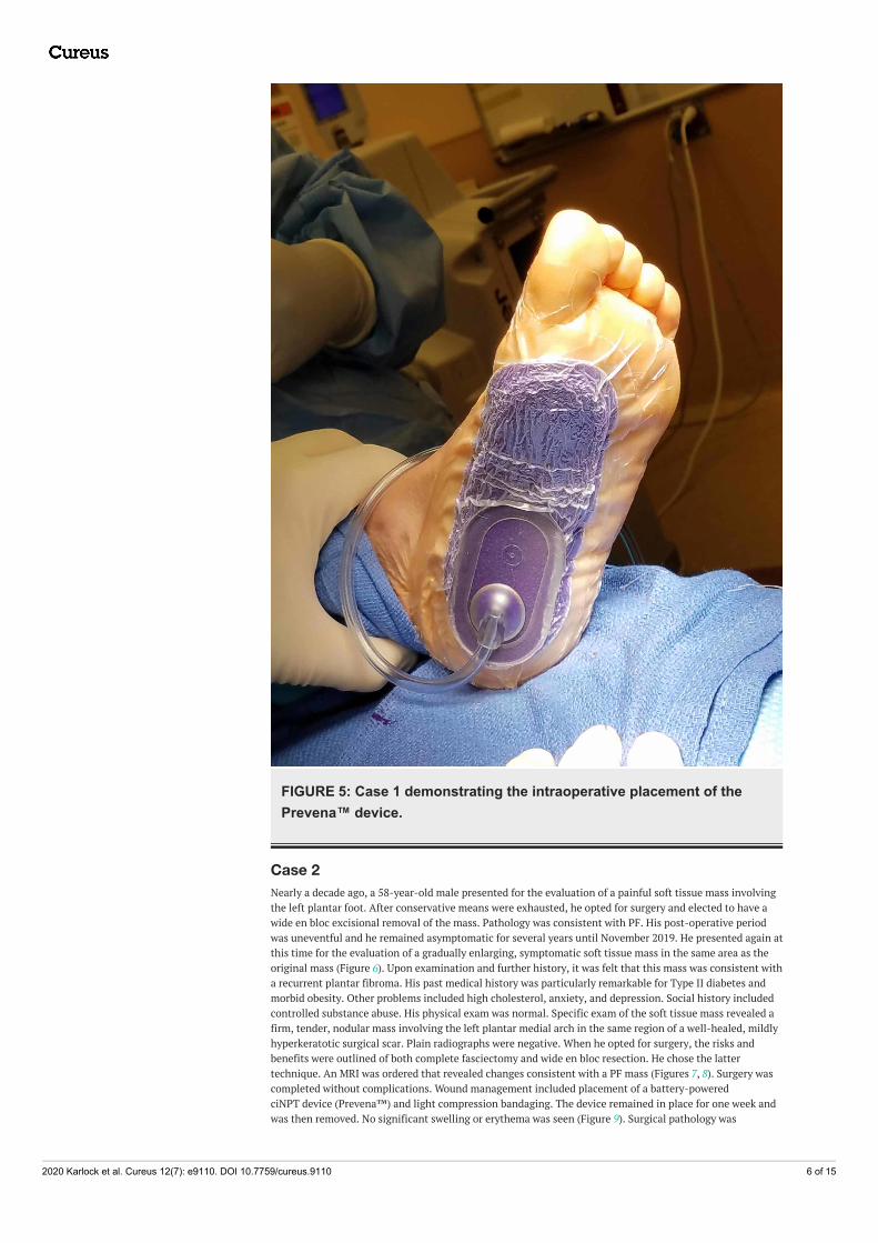
FIGURE 5: Case 1 demonstrating the intraoperative placement of thePrevena™ device.
Case 2Nearly a decade ago, a 58-year-old male presented for the evaluation of a painful soft tissue mass involvingthe left plantar foot. After conservative means were exhausted, he opted for surgery and elected to have awide en bloc excisional removal of the mass. Pathology was consistent with PF. His post-operative periodwas uneventful and he remained asymptomatic for several years until November 2019. He presented again atthis time for the evaluation of a gradually enlarging, symptomatic soft tissue mass in the same area as theoriginal mass (Figure 6). Upon examination and further history, it was felt that this mass was consistent witha recurrent plantar fibroma. His past medical history was particularly remarkable for Type II diabetes andmorbid obesity. Other problems included high cholesterol, anxiety, and depression. Social history includedcontrolled substance abuse. His physical exam was normal. Specific exam of the soft tissue mass revealed afirm, tender, nodular mass involving the left plantar medial arch in the same region of a well-healed, mildlyhyperkeratotic surgical scar. Plain radiographs were negative. When he opted for surgery, the risks andbenefits were outlined of both complete fasciectomy and wide en bloc resection. He chose the lattertechnique. An MRI was ordered that revealed changes consistent with a PF mass (Figures 7, 8). Surgery wascompleted without complications. Wound management included placement of a battery-poweredciNPT device (Prevena™) and light compression bandaging. The device remained in place for one week andwas then removed. No significant swelling or erythema was seen (Figure 9). Surgical pathology was
2020 Karlock et al. Cureus 12(7): e9110. DOI 10.7759/cureus.9110 6 of 15

consistent with a solitary benign plantar fibromatosis lesion. Sutures remained in place for a total of threeweeks. At this time, the incision was found to be negligibly tender, flat and well coapted. Partial weightbearing in a walking boot was initiated. At two weeks from the time of suture removal, he was transitioned tofull weight bearing. At just over three months, he was discharged from care symptom free without signs ofrecurrence.
FIGURE 6: Case 2 pre-operative photograph demonstrating therecurrent plantar fibroma mass (note the old mildly hyperkeratoticscar).
2020 Karlock et al. Cureus 12(7): e9110. DOI 10.7759/cureus.9110 7 of 15

FIGURE 7: Case 2 MRI showing the axial T1 weighted image of the footdemonstrating a plantar fibroma of the midfoot, demarcated with a skinmarker on the plantar aspect of the foot.
FIGURE 8: Case 2 MRI showing the sagittal T1 weighted imagedemonstrating a plantar fibroma of the first tarsometatarsal joint.
2020 Karlock et al. Cureus 12(7): e9110. DOI 10.7759/cureus.9110 8 of 15

FIGURE 9: Case 2 one week post-operatively at Prevena™ takedown(note the lack of significant edema and the well-coapted incision).
Case 3A 62-year-old female presented for the evaluation of a symptomatic soft tissue mass involving the rightplantar medial arch. Her symptomatology was relatively insidious by history and what prompted her visitwas gradual intolerance of a certain shoe gear. She reported the mass was present for over a year butsymptomology escalated about four months prior to initial evaluation. Her past medical history includedhypertension, thyroid disease, Factor V Leiden pathology with a history of deep vein thrombosis,degenerative joint disease, and morbid obesity. Social history was unremarkable. Her physical exam waslargely unremarkable except for significant pes planovalgus changes in both feet and a painful massinvolving the right foot. Specific examination of the soft tissue mass revealed a mildly firm, slightly mobile,tender soft tissue mass in the medial plantar arch. Plain films demonstrated no acute process. Both non-operative and operative treatment options were offered and the patient chose the latter, electing for wide enbloc excision instead of complete fasciectomy. Prior to surgery, an MRI was ordered that demonstratedchanges consistent with a solitary plantar fibroma lesion (Figures 10, 11). Her surgery was withoutcompromise and included perioperative anticoagulation bridge management in light of her history of deepvein thrombosis. Wound management included placement of a battery-powered ciNPT device (Prevena™)and light compression bandaging. At bandage takedown, the incison was observed to be well coapted withno complications (Figure 12). The device remained in place for one week and was then removed. No
2020 Karlock et al. Cureus 12(7): e9110. DOI 10.7759/cureus.9110 9 of 15

drainage strikethrough was seen (Figure 13). Surgical pathology was consistent with a solitary benignplantar fibromatosis lesion. Sutures remained in place for a total of three weeks. A walking boot was used fortwo weeks after suture removal. Healing progressed uneventfully with gradual increase in weight bearingand activities. Dorsal foot swelling on the operative foot persisted but eventually resolved. She ultimatelyreturned to normal shoe gear at three months and was discharged from care without symptoms or signs ofrecurrence at six months.
FIGURE 10: Case 3 MRI showing the axial T2 weighted image of theright foot demonstrating a large plantar fibroma extending from thecentral cord into the subcutaneous tissue of the plantar surface of thefoot.
2020 Karlock et al. Cureus 12(7): e9110. DOI 10.7759/cureus.9110 10 of 15

FIGURE 11: Case 3 MRI showing the T1 weighted sagittal image of theright foot demonstrating a plantar fibroma within the central portion ofthe plantar cord.
2020 Karlock et al. Cureus 12(7): e9110. DOI 10.7759/cureus.9110 11 of 15

FIGURE 12: Case 3 one week post-operatively at Prevena™ takedown(note the lack of significant edema and well-coapted incision).
2020 Karlock et al. Cureus 12(7): e9110. DOI 10.7759/cureus.9110 12 of 15

FIGURE 13: Case 3 one week post-operatively showing Prevena™dressing (note serosanguinous dressing capture without extravasationor strikethrough).
DiscussionAlthough the National Institutes of Health classify PF as a “rare” condition, this condition is certainlyencountered in private practice [2]. Developments in the non-operative intervention are ongoing withvarious degrees of success and shortcomings demonstrated in the literature. It is well accepted thatconservative, palliative, and interventional non-operative measures should be considered as first-linetreatments reserving surgery for more symptomatic cases. As stated previously, plantar fibroma surgicalcomplications, with wound healing in particular, are not uncommon and are related to the depth andbreadth of the operation. One retrospective study found that 52% of post-operative plantar fibromatosissites experienced delayed wound healing with a potential need for a skin graft [14]. Post-operative measuresto protect the incision and minimize swelling are paramount to lessen the chance of wound-healing surgicalcomplications. Techniques such as compression bandaging, non-weight bearing during incisional healing,and drain placement have been utilized successfully to decrease swelling and remove fluid at the surgicalsite. We have presented three cases in which a battery operated closed-incision negative pressure device(Prevena™) was used with favorable outcomes in lieu of surgical drain placement. This device is designed tomanage the closed surgical environment by drawing fluid away from the operative site, therefore lesseningthe chance of serosanguinous fluid collection beneath the incision. Such fluid collections can potentiate
2020 Karlock et al. Cureus 12(7): e9110. DOI 10.7759/cureus.9110 13 of 15
problems that include tension at the incision site resulting in dehiscence or pressure necrosis. Fluidcollections, if unchecked, can also serve as a nidus for infection. In this regard, Prevena™ aids in reducingthe incidence of surgical site infection by decreasing the chance of fluid collections in patients at high riskfor post-operative infections [11]. All the three patients in this case series had co-morbidities that put themat higher risk for plantar fibroma surgical complications. These included smoking, diabetes, obesity, andFactor V Leiden. All three patients demonstrated favorable outcomes without significant wound-healingcomplications. The one exception was a subtle dehiscence in the patient with the smoking history; however,the dehiscence healed uneventfully without complications.
ConclusionsSeveral treatment options are available for PF. These include palliative measures, non-operativeinterventions, and surgery, all with varying degrees of success and complication risk. We presented threecases of surgical wide en bloc resection of PF lesions in which a battery-powered ciNPT device (Prevena™)was used successfully in lieu of surgical drains. All three patients demonstrated favorable outcomes withoutsignificant complications. While additional studies are necessary, we believe this plantar fibroma surgicalcase series demonstrated that the use of a battery-powered ciNPT device (Prevena™) was beneficial inreducing wound-healing complications such as swelling, fluid collections beneath the surgical site, andsurgical site infection.
Additional InformationDisclosuresHuman subjects: Consent was obtained by all participants in this study. Conflicts of interest: Incompliance with the ICMJE uniform disclosure form, all authors declare the following: Payment/servicesinfo: All authors have declared that no financial support was received from any organization for thesubmitted work. Financial relationships: Ralph J. Napolitano, Jr. declare(s) personal fees from 3M + KCI.Ralph J. Napolitano, Jr. declare(s) personal fees from Organogenesis. Ralph J. Napolitano, Jr. declare(s)personal fees from Flower Orthopedics. Ralph J. Napolitano, Jr. declare(s) personal fees from Ortho-Dermatologics. Other relationships: All authors have declared that there are no other relationships oractivities that could appear to have influenced the submitted work.
AcknowledgementsSpecial acknowledgment to Michelle Chao, Clinical Research Coordinator for The Orthopedic Foundation,New Albany, OH, and Carrie A. Weld, Executive Assistant to Ralph J. Napolitano, Jr., DPM, CWSP, FACFAS.Special acknowledgment to the Information Technology staff at OrthoNeuro, Columbus, OH. Radiologysupport acknowledgment to Joseph E. Frondriest, MD, and Callan Bousquet, RT(R) (CT) (MRI), of LickingMemorial Hospital, Newark, OH. Radiology support acknowledgment to Joel L. Rosner, MD, and ChristinaVan Deusen of Advantage Diagnostics, Columbus, and Beachwood, Ohio.
References1. Espert M, Anderson MR, Baumhauer JF: Current concepts review: plantar fibromatosis. Foot Ankle Int. 2018,
39:751-757. 10.1177/10711007187680512. Prevalence and incidence of Ledderhose disease . (2015). Accessed: June 4, 2020:
https://www.rightdiagnosis.com/l/ledderhose_disease/prevalence.htm.3. Omor Y, Dhaene B, Grijseels S, Alard S: Ledderhose disease: clinical, radiological (ultrasound and MRI), and
anatomopathological findings. Case Rep Orthop. 2015, 2015:741461. 10.1155/2015/7414614. Goldblum JR, Folpe AL, Weiss SW: Benign fibroblastic/myofibroblastic proliferations, including superficial
fibromatoses. In Enzinger and Weiss's Soft Tissue Tumors. Elsevier, Philadelphia; 2020. 7:202-274.5. James WD, Elston DM, Treat JR, Rosenbach MA, Neuhaus IM: Dermal and subcutaneous tumors. In
Andrews' Diseases of the Skin. Elsevier, Philadelphia; 2020. 13:587-635.6. Carroll P, Henshaw RM, Garwood C, Raspovic K, Kumar D: Plantar fibromatosis: pathophysiology, surgical
and nonsurgical therapies: an evidence-based review. Foot Ankle Spec. 2018, 11:168-176.10.1177/1938640017751184
7. Young JR, Sternbach S, Willinger M, Hutchinson ID, Rosenbaum AJ: The etiology, evaluation, andmanagement of plantar fibromatosis. Orthop Res Rev. 2019, 11:1-7. 10.2147/ORR.S154289
8. Pickren JW, Smith AG, Stevenson TW Jr, AP Stout: Fibromatosis of the plantar fascia . Cancer. 1951, 4:846-856. 10.1002/1097-0142(195107)4:4<846::aid-cncr2820040422>3.0.co;2-n
9. Makama JG, Ameh EA: Surgical drains: what the resident needs to know. Niger J Med. 2008, 17:244-250.10.4314/njm.v17i3.37389
10. Orgill DP, Bayer LR: Negative pressure wound therapy: past, present and future . Int Wound J. 2013, 10:15-19. 10.1111/iwj.12170
11. PREVENA™ Incision Management System. (2020). Accessed: June 8, 2020:https://www.woundsource.com/product/prevena-incision-management-system.
12. Hyldig N, Birke-Sorensen H, Kruse M, et al.: Meta-analysis of negative-pressure wound therapy for closedsurgical incisions. Br J Surg. 2016, 103:477-486. 10.1002/bjs.10084
13. Stannard JP, Volgas DA, McGwin G III, Stewart RL, Obremskey W, Moore T, Anglen JO: Incisional negativepressure wound therapy after high-risk lower extremity fractures. J Orthop Trauma. 2012, 26:37-42.10.1097/bot.0b013e318216b1e5
2020 Karlock et al. Cureus 12(7): e9110. DOI 10.7759/cureus.9110 14 of 15

14. Sammarco GJ, Mangone PG: Classification and treatment of plantar fibromatosis. Foot Ankle Int. 2000,21:563-569. 10.1177%2F107110070002100706
2020 Karlock et al. Cureus 12(7): e9110. DOI 10.7759/cureus.9110 15 of 15