common infection strategies of pathogenic eukaryotes
TRANSCRIPT

Remodelling host-cell functions to create a suitable niche for proliferation is an extensively used virulence strategy in microbial infection of both plant and ani-mal cells1–3. It requires the delivery of microbial effector proteins into the host cell and therefore involves signals for appropriate protein transport, as well as specialized machinery dedicated to breaching the host-cell bar-rier. A wide range of bacterial pathogens use leader sequences and associated secretion systems for this pathogenic process, indicating that some mechanisms of virulence and pathogenesis are conserved in diver-gent bacterial species4. Until recently, however, little was known about how eukaryotic pathogens deliver viru-lence determinants into their host cells. This Review focuses on common infection strategies of the malaria parasite Plasmodium falciparum and the plant pathogen Phytophthora infestans, with an emphasis on the recently identified host-targeting signals that these pathogens use to deliver virulence factors and avirulence proteins into human and plant cells, respectively.
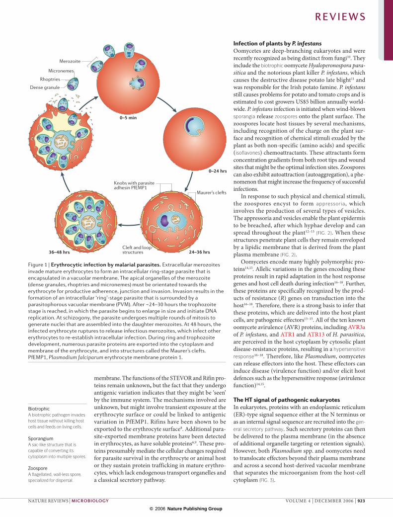
Infection of human erythrocytes by P. falciparumThe protozoan apicomplexan parasite P. falciparum causes human malaria, a major global health prob-lem5, which in Africa alone kills more than one mil-lion children annually. P. falciparum infection of erythrocytes (FIG. 1) underlies all of the symptoms and pathologies associated with malaria, many of which have been linked to parasite proteins that are secreted into red blood cells5,6. Entry into erythrocytes is medi-ated by invasion organelles located at the apical end
of the merozoite and results in the formation of an intracellular ring-stage parasite that is surrounded by a parasitophorous vacuole. Rings develop in the parasi-tophorous vacuole and after ~24–30 hours they reach the trophozoite stage, in which the parasite begins to enlarge in size and initiate DNA replication. At schizogony, the parasite undergoes multiple rounds of mitosis to generate 8–16 nuclei that are assembled into the daughter merozoites. At 48 hours the infected erythrocyte ruptures to release infectious mero-zoites, which infect other erythrocytes to re-establish intracellular infection.
Multiple structural and antigenic changes are induced in the erythrocyte on infection by malaria parasites. These include the formation of prominent cleft and loop structures in the erythrocyte cytoplasm, and the formation of prominent protrusions called knobs on the infected erythrocyte surface (FIG. 1). Resident in these structures are parasite proteins, the best characterized of which belong to a variant antigen family of adhesins called the P. falciparum erythrocyte membrane protein 1 (PfEMP1) family. Members of PfEMP1 are encoded by the var genes7, and are exposed on the erythrocyte surface concentrated at the knob protrusions. PfEMP1 mediates adhesion to endothelial cells in the blood vessels that line organs such as the brain and placenta, and is therefore linked to both cer-ebral and placental malaria5. Other secreted protein families include the STEVOR and Rifin families, which are normally situated in the cleft structures known as Maurer’s clefts that form beneath the erythrocyte
*Departments of Pathology and Microbiology-Immunology, Northwestern University, 303 East Chicago Avenue, Chicago, Illinois 60611, USA.‡Department of Plant Pathology, The Ohio State University, Ohio Agricultural Research and Development Center, Wooster, Ohio 44691, USA.Correspondence to K.H. e-mail: [email protected]:10.1038/nrmicro1549Published online6 November 2006
Avirulence proteinAn effector protein of plant pathogens that triggers disease resistance.
Parasitophorous vacuoleA vacuole within the host cell in which the parasite resides.
Maurer’s cleftsSingle-membrane-limited structures in the erythrocyte cytoplasm.
Common infection strategies of pathogenic eukaryotesKasturi Haldar*, Sophien Kamoun‡, N. Luisa Hiller*, Souvik Bhattacharje* and Christiaan van Ooij*
Pathogenic eukaryotes belong to several distinct phylogenetic lineages and have evolved the ability to colonize a range of hosts, including animals and plants. Pathogenic lifestyles have evolved repeatedly in eukaryotes, indicating that unique molecular processes are involved in host infection. However, evidence is now emerging that divergent eukaryotic pathogens might share common mechanisms of pathogenicity. The results from recent studies demonstrate that Plasmodium falciparum and Phytophthora infestans use equivalent host-targeting signals to deliver virulence adhesins and avirulence gene products into human and plant cells, respectively. Remodelling of host cells by different eukaryotic pathogens might therefore share some common features.
R E V I E W S
922 | DECEMBER 2006 | VOLUME 4 www.nature.com/reviews/micro
© 2006 Nature Publishing Group
36–48 hrs 24–36 hrs
0–24 hrs
0–5 min
Knobs with parasite adhesin PfEMP1
Cle� and loop structures
Merozoite
Rhoptries
Maurer’s cle�s
Dense granule
Micronemes
BiotrophicA biotrophic pathogen invades host tissue without killing host cells and feeds on living cells.
SporangiumA sac-like structure that is capable of converting its cytoplasm into multiple spores.
ZoosporeA flagellated, wall-less spore, specialized for dispersal.
membrane. The functions of the STEVOR and Rifin pro-teins remain unknown, but the fact that they undergo antigenic variation indicates that they might be ‘seen’ by the immune system. The mechanisms involved are unknown, but might involve transient exposure at the erythrocyte surface or could be linked to antigenic variation in PfEMP1. Rifins have been shown to be exported to the erythrocyte surface8. Additional para-site-exported membrane proteins have been detected in erythrocytes, as have soluble proteins6,9. These pro-teins presumably mediate the cellular changes required for parasite survival in the erythrocyte or animal host or they sustain protein trafficking in mature erythro-cytes, which lack endogenous transport organelles and a classical secretory pathway.
Infection of plants by P. infestansOomycetes are deep-branching eukaryotes and were recently recognized as being distinct from fungi10. They include the biotrophic oomycete Hyaloperonospora para-sitica and the notorious plant killer P. infestans, which causes the destructive disease potato late blight11 and was responsible for the Irish potato famine. P. infestans still causes problems for potato and tomato crops and is estimated to cost growers US$5 billion annually world-wide. P. infestans infection is initiated when wind-blown sporangia release zoospores onto the plant surface. The zoospores locate host tissues by several mechanisms, including recognition of the charge on the plant sur-face and recognition of chemical stimuli exuded by the plant as both non-specific (amino acids) and specific (isoflavones) chemoattractants. These attractants form concentration gradients from both root tips and wound sites that might be the optimal infection sites. Zoospores can also exhibit autoattraction (autoaggregation), a phe-nomenon that might increase the frequency of successful infections.
In response to such physical and chemical stimuli, the zoospores encyst to form appressoria, which involves the production of several types of vesicles. The appressoria and vesicles enable the plant epidermis to be breached, after which hyphae develop and can spread throughout the plant12–15 (FIG. 2). When these structures penetrate plant cells they remain enveloped by a lipidic membrane that is derived from the plant plasma membrane (FIG. 2).
Oomycetes encode many highly polymorphic pro-teins14,15. Allelic variations in the genes encoding these proteins result in rapid adaptation in the host response genes and host cell death during infection16–18. Further, these proteins are specifically recognized by the prod-ucts of resistance (R) genes on transduction into the host16–18. Therefore, there is a strong basis to infer that these proteins, which are delivered into the host plant cells, are pathogenic effectors13–15. All of the ten known oomycete avirulence (AVR) proteins, including AVR3a of P. infestans, and ATR1 and ATR13 of H. parasitica, are perceived in the host cytoplasm by cytosolic plant disease-resistance proteins, resulting in a hypersensitive response16–18. Therefore, like Plasmodium, oomycetes can release effectors into the host. These effectors can induce disease (virulence function) and/or elicit host defences such as the hypersensitive response (avirulence function)14,15.
The HT signal of pathogenic eukaryotesIn eukaryotes, proteins with an endoplasmic reticulum (ER)-type signal sequence either at the N terminus or as an internal signal sequence are recruited into the gen-eral secretory pathway. Such secretory proteins can then be delivered to the plasma membrane (in the absence of additional organelle targeting or retention signals). However, both Plasmodium spp. and oomycetes need to translocate effectors beyond their plasma membrane and across a second host-derived vacuolar membrane that separates the microorganism from the host-cell cytoplasm (FIG. 3).
Figure 1 | Erythrocytic infection by malarial parasites. Extracellular merozoites invade mature erythrocytes to form an intracellular ring-stage parasite that is encapsulated in a vacuolar membrane. The apical organelles of the merozoite (dense granules, rhoptries and micronemes) must be orientated towards the erythrocyte for productive adherence, junction and invasion. Invasion results in the formation of an intracellular ‘ring’-stage parasite that is surrounded by a parasitophorous vacuolar membrane (PVM). After ~24–30 hours the trophozoite stage is reached, in which the parasite begins to enlarge in size and initiate DNA replication. At schizogony, the parasite undergoes multiple rounds of mitosis to generate nuclei that are assembled into the daughter merozoites. At 48 hours, the infected erythrocyte ruptures to release infectious merozoites, which infect other erythrocytes to re-establish intracellular infection. During ring and trophozoite development, numerous parasite proteins are exported into the cytoplasm and membrane of the erythrocyte, and into structures called the Maurer’s clefts. PfEMP1, Plasmodium falciparum erythrocyte membrane protein 1.
R E V I E W S
NATURE REVIEWS | MICROBIOLOGY VOLUME 4 | DECEMBER 2006 | 923
© 2006 Nature Publishing Group

GcAp
Iv
Ih
Ha
Parasite
Haustorium
Host derivedhaustorialmembrane
Hostderived
PVM
a b
Parasitophorousvacuole
Parasite
IsoflavonesSpecialized polyphenolic metabolites produced by plants of the legume family.
AppressoriaSpecialized infection structures of oomycete and fungal plant pathogens that enable host penetration.
Hypersensitive responseA programmed cell-death response of plants that is associated with resistance.
General secretory pathwayPathway for protein secretion through the endoplasmic reticulum and Golgi to the plasma membrane.
Twin-arginine transport(TAT). A protein transport system that transports proteins across membranes in a fully folded state.
In the case of P. falciparum, in many of the N-terminal signal sequences, the hydrophobic core that is cru-cial for signal-sequence function is detected further downstream of the initiator methionine than it is in the signal sequences of most other eukaryotic pro-teins. However, there is general agreement that these sequences nonetheless function as adequate leader sequences for recruitment into the parasite secretory pathway. Cargo proteins are sequestered in vesicles and are transported through the secretory pathway, culminating in the fusion of vesicles with the parasite plasma membrane, which results in protein release into the parasitophorous vacuole.
The subsequent export of proteins across the sur-rounding parasitophorous vacuole membrane requires a 30–40-amino-acid vacuolar translocation sequence (VTS), which is found immediately downstream of the signal sequence19 and which is necessary and sufficient
for export. Examination of five VTSs derived from P. falciparum exported proteins led to the recognition of a shared 11-amino-acid motif (Rx1SRxLxE/D/Qx2x3x4) with a 5-amino-acid core (RxLxE/D/Q) that is conserved between diverse proteins20 (FIG. 4a). The 11-amino-acid motif was found to be required for pro-tein export into the host cell and was therefore defined as a host (cell)-targeting (HT) signal; its 5-amino-acid core was also defined as a Plasmodium export element (PEXEL)20,21. As an equivalent signal has now been found in P. infestans, the term PEXEL seems restrictive and so we will use the terms HT signal and HT motif throughout the text.
In proteins with an N-terminal signal sequence, cleavage of the signal sequence is required to gener-ate a VTS leader sequence containing an HT motif, which mediates protein export across the parasitopho-rous vacuole membrane. Such VTS leader sequences containing an HT motif have been detected in ~400 P. falciparum secretory proteins, including the large antigenic PfEMP1, STEVOR and Rifin protein families that extensively remodel the host erythrocyte during infection20,21. This novel motif, and its cargo, could therefore provide targets for new drugs and vaccines against malaria.
Although the HT motif identifies high-value candi-date proteins that are exported into the host erythrocyte, our understanding of how this motif actually functions in protein export is limited. One important feature that needs to be understood is how the HT signal can medi-ate the export of both soluble and membrane proteins to the cytoplasm and membrane of the erythrocyte, respectively. In addition, despite the importance of a conserved R residue in the 5-amino-acid core, at the time of discovery, the HT signal seemed distinct from all other known protein-transport signals, including those of the arginine-rich motif in the HIV transactivator (Tat) protein22 and the motifs that direct export through the bacterial twin-arginine transport (TAT) system23. Not all
Figure 2 | Infection of host plants by Phytophthora infestans. Schematic view of the hypersensitive cell-death response triggered in resistant plants by P. infestans. The germinated cysts (Gc) on the surface of the plant produce appressoria (Ap) to penetrate plant tissues, and then produce infection vesicles (Iv), intercellular hyphae (Ih) and haustoria (Ha) within the plant tissues. Plant cells can be infected without causing cell death. Those infected and undergoing hypersensitive cell death are shown in red. The hypersensitive response can include 1–2 cells (left) or a larger group of plant cells (right), depending on the genotypes of both the plant and the pathogen. Hypersensitivity is triggered by secreted pathogen avirulence proteins on recognition by cytoplasmic plant resistance proteins.
Figure 3 | Intracellular infection by Phytophthora infestans and Plasmodium falciparum. a | A plant cell infected by P. infestans. The P. infestans parasite colonizes the host’s intracellular spaces and forms haustoria. The host-derived haustorial membrane must be crossed by pathogenic effectors (purple squares) released into cells to mediate virulence and plant hypersensitive responses. b | A human erythrocyte infected by P. falciparum. Invasion by the extracellular merozoite stage leads to the formation of a host-derived parasitophorous vacuole, within which the parasite resides. Proteins (purple squares) secreted by the parasite must cross the parasitophorous vacuole membrane (PVM) to reach, and mediate virulence and structural changes in, the erythrocyte. Modified with permission from PloS Pathogens REF. 26.
R E V I E W S
924 | DECEMBER 2006 | VOLUME 4 www.nature.com/reviews/micro
© 2006 Nature Publishing Group

Position Position Position Position
Freq
uenc
y
Freq
uenc
y
Freq
uenc
y
Freq
uenc
y
4
3
2
1
0
4
3
2
1
0
5
4
3
2
1
0
P. falciparum
Length of upstream region
Length of upstream region
Length of upstream region
P. sojae
P. ramorum
4
3
2
1
0
002468
1012
5 10 15 20 25 30 35 40 45 50 55 60 65 70 75 80 85 90 95 100
002468
1012
5 10 15 20 25 30 35 40 45 50 55 60 65 70 75 80 85 90 95 100
002468
1012
5 10 15 20 25 30 35 40 45 50 55 60 65 70 75 80 85 90 95 100
Num
ber o
f p
rote
ins
Num
ber o
f p
rote
ins
Num
ber o
f p
rote
ins
A
D
B Ca Cb
P. falciparum proteins that are targeted to the erythrocyte contain an HT motif, indicating that there are alternative mechanisms of transport. Nonetheless, the HT signal seems to be a major new transport signal and was the first secretion signal to be defined for a eukaryotic pathogen.
The predictive algorithms that were originally used to identify the P. falciparum secretome associ-ated with the HT signal, and subsequent efforts to further characterize the set of effector proteins that carry this signal, revealed that the HT signal is used by all Plasmodium species20,21,24 but failed to detect it in any other eukaryotic or prokaryotic genomes. This indicated that either the HT signal was unique
to the genus Plasmodium or there were limitations in the algorithms that were used to look for functionally equivalent signals in divergent eukaryotes.
Analyses of four AVR proteins from plant pathogenic oomycetes revealed the presence of a conserved RxLR motif at the N terminus, followed by regions that are highly variable and under diversifying selection16–18. The RxLR motif is not required for the activation of plant defences when AVR proteins are expressed directly in plant cells25, suggesting that this motif is part of a leader sequence that is distinct from the downstream effector domains of these proteins, and which reflects a common function shared by these proteins, such as transport into the host cell.
Figure 4 | Comparative analysis of the host-targeting signal in Plasmodium falciparum and Phytophthora spp. A | Logo representation of the host targeting (HT) motif in P. falciparum secretome proteins. On the x axis, the blue bar indicates the 11-amino-acid motif recognized by the MEME program20, with a 5-amino-acid core. B | Logo representation of the HT motif in Phytopthora infestans secretome proteins. This was generated from sequences of predicted secretory proteins containing the RxLR motif in the first 100 residues after the signal sequence cleavage site (that is, ~10% of the signal sequence set). C | Comparative analyses of secretory (Ca) and cytosolic (Cb) RxLR-containing sequences in Phytophthora species showing the requirement for E and D residues downstream of the HT consensus sequence. These logo representations were generated from the sequences of predicted Phytophthora secretory proteins containing the RxLR motif in the first 100 residues after the signal sequence cleavage site (that is, ~10% of the signal sequence-containing proteins) (Ca), and the sequences of predicted Phytophthora cytosolic proteins containing the RxLR motif in the first 100 residues (that is, ~5% of the cytosolic proteins). Amino acids in A–C are represented by one-letter abbreviations and are colour coded: blue, basic residues; red, acidic residues; black, hydrophobic residues; and green, polar residues. The height of the amino acids indicates their frequency at that position. D | The positional equivalence of the HT motifs in Phytophthora ramorum, Phytophthora sojae and P. falciparum. Parts B, C and D are reproduced with permission from PloS Pathogens REF. 26.
R E V I E W S
NATURE REVIEWS | MICROBIOLOGY VOLUME 4 | DECEMBER 2006 | 925
© 2006 Nature Publishing Group

Direct testing of RxLR-containing N-terminal leader sequences for their ability to export reporter proteins from the oomycete into the plant cell remains a techni-cal challenge. However, Bhattacharjee et al.26 reported that a P. infestans leader sequence containing an RxLR motif could mediate protein export into the plant-cell cytoplasm with the same efficiency as the P. falciparum leader sequence containing the RxLxE/D/Q motif, and that this export is dependent on the RxLR motif.
A logo representation of all the predicted secretory proteins containing the P. falciparum RxLxE/D/Q HT motif and the P. infestans N-terminal RxLR HT motif is shown in FIGURE 4 (REF. 26). The residues RxL are clearly shared between these two logos. In P. infestans, the secretory RxLR-containing proteins are enriched in D or E residues ~20 amino-acids downstream of the RxLR motif, and these residues have a role in host targeting. Sequences downstream of the RxLR motif in P. infestans cytosolic proteins cannot substitute for the equivalent region in secretory RxLR-containing proteins in host targeting26. Importantly, in P. infestans proteins, the core RxL-containing motif itself is ~25–30 amino acids in length26, whereas in P. falciparum proteins this
core motif is present in the RxLxE/D/Q motif, which functions in protein export embedded in the context of a 25–30-amino-acid VTS. Therefore, the precise equivalence between these active HT signals is likely to involve a secondary and/or tertiary structure26 rather than be at the primary sequence level24. This equiva-lence in function in the P. falciparum and P. infestans HT signals, combined with the equivalence in both position and sequence requirements in the HT motifs, indicates that deep-branching eukaryotes belonging to such distinct groups as the heterokonts (P. infestans) and the alveolates (P. falciparum) share conserved secretion strategies to access host cells from both the plant and animal kingdoms16–18.
Motif conservation in pathogenic eukaryotesThe conservation of protein-transport motifs between divergent eukaryotic species can reflect convergent evolution or an ancient molecular mechanism. A short linear motif like the ER-retentive KDEL motif is preserved across phylogenetically diverse taxa and is recognized by conserved machinery27. However, short sequences can be susceptible to mutation, resulting in a loss of function. Interestingly, the positively charged arginine and lysine residues that flank the shared RxL core in both P. infestans and P. falciparum might enable some redundancy in the conserved arginine residue and preserve the function of the motif despite point mutations or changes in the host environment. By contrast, the E/D/Q residues in the 5-amino-acid core of the plasmodial HT motif are not position-ally conserved in P. infestans proteins, although the enrichment in E/D residues downstream from the P. infestans HT motif is important for host target-ing26. Analyses of P. infestans and P. falciparum leader sequences indicate that in both, the protein domain containing the information that is necessary and suf-ficient for host targeting resides in a larger peptidic structure approaching the size (25–30 amino acids) of a small globular domain27. Globular domains tend to be ordered, conserved among species and have strong binding affinities27, and therefore might be conserved across divergent eukaryotic genera, such as Plasmodium and Phytophthora. The presence of variable yet still con-strained flanking regions might allow the evolutionary plasticity needed to breach distinct host barriers, such as the parasitophorous vacuole membrane for P. fal-ciparum proteins and a plant-cell-derived membrane for P. infestans proteins.
Comparing the host-targeted secretomesAs there are multiple secretory destinations in eukaryo-tes, we define the global prediction of the Plasmodium spp. and Phytophthora spp. proteins that are delivered into host cells as the ‘host-targeted secretome’. A prominent common feature of these two host-targeted secretomes is their large size — they both comprise hundreds of proteins with many hypothetical genes, the annotation of which will provide significant insights into how eukaryotic pathogens remodel their host environment.
Table 1 | Plasmodium spp. proteins containing a SS and HT motif*
Protein or protein family Number of proteins‡
Plasmodium falciparum
Rifin 165
STEVOR 32–34
Kinases (FIKK) 18
DNAJ/heat shock (HSP40: 2; RESA and RESA-related: 8; other: 7)
17
MC-2M 13
Phosphatases 3
ABC transporter 1
KAHRP 1
Other proteins known to be exported to the erythrocyte: GBP130, GBP3, MESA1, PfHRPI, PfHRPII, PfEMP1, PfEMP2, PfEMP3
6
PfEMP1 cell-surface adhesin‡ 59
Hypothetical/unknown§ ~80–190
Plasmodium yoelii
Hypothetical family-1 39
PfATPase3 1
Plasmodium vivax PV1H14030_P putative 1
DHHC zinc finger domain, putative 1
Merozoite surface protein 4/5 1
Methanol oxidation protein 1
Aminopeptidase-like protein, related 1
Hypothetical/unknown 25*Proteins listed are those which contain a signal sequence (SS) and RxLxE/D/Q host-targeting (HT) signal. ‡Assignments based on REFS 20,21,22,28,29,30 and http://fozzie.pathology.northwestern.edu/cgi-bin/PlasmoHT/index.cgi §No leader signal sequence, internal signal sequence. ||Includes some antigenic families with ten or fewer members. ABC, ATP-binding cassette; KAHRP, knob-associated histidine-rich protein; PfEMP, Plasmodium falciparum erythrocyte membrane protein; PfHRP, Plasmodium falciparum histidine-rich protein.
R E V I E W S
926 | DECEMBER 2006 | VOLUME 4 www.nature.com/reviews/micro
© 2006 Nature Publishing Group

MAL8P1.204
PF080115
Pv088225
Pv084600
PY02866
PY01224
PFL0565w
PY07104
Pv081840
Pv118690
PFB0595w
Pv002875
PY02986
PFE0055c**
PFA0660w**
PFB0090c
PFB0925w**
PF110513
Pv080080
PY03711
PFE1170w*
PFL0055c**
PFB0085c**
PF110512**
PFA0110w**
PF110509**
PF100378**
PFB0920w**
PF110034**
PF100381**
PFL2550w**
PF140013**
PFA0675w
Syntenous orthologueAn orthologue in which the gene locus is conserved among species.
The precise composition of the P. falciparum host-targeted secretome can vary depending on the algorithm used to make the predictions20,21 (see the PlasmoHT web page, details in Further information). A more recent algorithm24 predicts additional P. falciparum and Plasmodium vivax host-targeted proteins. However, this algorithm allows for extremely low scores in the signal sequence and has not yet been extensively validated. In addition, all of the currently available algorithms are lim-ited because they are based on linear sequence alone26. Nonetheless, cumulative analyses indicate that the major antigenic STEVOR, Rifin and PfEMP1 families (alto-gether ~250 proteins) are all present in the P. falciparum
secretome. The number of other predicted proteins ranges from 120–250, and a more precise definition of the total number of proteins exported outside the major antigenic families awaits experimental validation.
Many (80–190) are hypothetical proteins (TABLE 1), have no recognizable annotation other than that they contain repeats and are enriched in one or more amino acids, and occasionally cluster into families not exceed-ing 10–15 members. Genomic analyses from multiple groups have confirmed that several are predicted by in silico annotation to be putative heat shock proteins, chaperones, kinases or phosphatases20,21,24,28–30 (TABLE 1). More than 50 are clearly expressed during the blood stages of infection. Bioinformatics analysis suggests that the HT signal is also present in insect (circumsporozoite protein (CSP)) and liver stage (liver-stage antigen 3 (LSA-3)) proteins, but experimental evidence to indicate that the signal is functional in these proteins is yet to be established.
The host-targeted secretomes of the rodent malaria parasites Plasmodium yoelii and Plasmodium berghei seem to be much smaller (~60–70 predicted effec-tors)20 than that predicted for P. falciparum. They have not been subjected to the same intense curation as the P. falciparum secretome. The P. yoelii secretome contains one large protein family of 39 members and most of the remainder are hypothetical proteins that cannot yet be annotated20. Remarkably, the P. yoelii large antigenic family Yirs31 lack the HT signal20. The rodent malaria host-targeted secretomes show little overlap (~10 proteins are syntenous orthologues, largely hypotheticals) with the P. falciparum host-targeted secretome32. Sargent et al. also predicted a distinct but small set of overlapping proteins (~10 syntenous orthologues). This shows that the size and nature of the plasmodial host-targeted secretome is strongly influenced by the host species. Strikingly, even com-pared with another human malaria parasite such as P. vivax, there is P. falciparum-specific expansion of protein families, such as those containing heat shock protein DNAJ domains (see below; FIG. 5), indicating that the functions of these proteins might be linked to virulence and disease in P. falciparum malaria. The export of a fusion protein between green fluorescent protein and a P. falciparum heat shock protein (HSP40) into the erythrocyte confirmed the prediction that this member of the heat shock protein family can indeed be delivered to the host erythrocyte20.
In Phytophthora spp., 60 P. infestans, 162 Phytophthora sojae and 131 Phytophthora ramorum proteins were pre-dicted to be secreted on the basis of signal peptides and the RxLR motif (TABLE 2). In each species ~15 proteins had putative ascribed functions that could be related to ATP-binding cassette (ABC) transporters, efflux pumps, proteases, kinases, acyltransferases and transglutami-nase elicitors (a P. sojae transglutaminase elicitor has previously been shown to induce plant defences33).
The annotation of this secretome set is still in its infancy and it will undoubtedly be refined and expanded (especially with respect to the numbers of proteins in each category) as the genomes are
Figure 5 | Families of putative host-targeted heat shock protein DNAJ-domain-containing proteins in Plasmodium species. The tree shows the protein families containing a heat shock protein DNAJ domain and a host-targeting (HT) motif in Plasmodium falciparum, Plasmodium vivax and Plasmodium yoelii. The tree was constructed using the parsimony algorithm from PHYLIP (the PHYLogeny Inference Package). Preliminary sequence data was obtained from The Institute for Genomic Research. P. falciparum genes are in bold. *Indicates the presence of an endoplasmic reticulum (ER) -type signal sequence. **Indicates the presence of an ER-type signal sequence followed by an HT motif.
R E V I E W S
NATURE REVIEWS | MICROBIOLOGY VOLUME 4 | DECEMBER 2006 | 927
© 2006 Nature Publishing Group
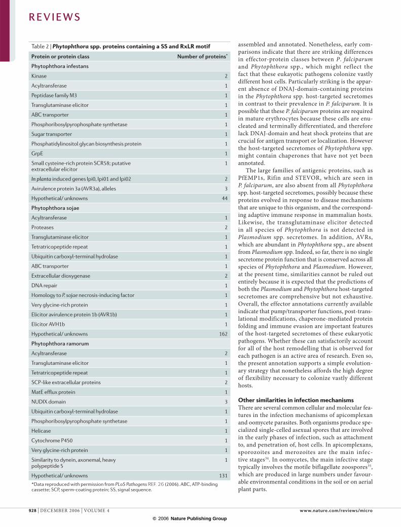
assembled and annotated. Nonetheless, early com-parisons indicate that there are striking differences in effector-protein classes between P. falciparum and Phytophthora spp., which might reflect the fact that these eukayotic pathogens colonize vastly different host cells. Particularly striking is the appar-ent absence of DNAJ-domain-containing proteins in the Phytophthora spp. host-targeted secretomes in contrast to their prevalence in P. falciparum. It is possible that these P. falciparum proteins are required in mature erythrocytes because these cells are enu-cleated and terminally differentiated, and therefore lack DNAJ-domain and heat shock proteins that are crucial for antigen transport or localization. However the host-targeted secretomes of Phytophthora spp. might contain chaperones that have not yet been annotated.
The large families of antigenic proteins, such as PfEMP1s, Rifin and STEVOR, which are seen in P. falciparum, are also absent from all Phytophthora spp. host-targeted secretomes, possibly because these proteins evolved in response to disease mechanisms that are unique to this organism, and the correspond-ing adaptive immune response in mammalian hosts. Likewise, the transglutaminase elicitor detected in all species of Phytophthora is not detected in Plasmodium spp. secretomes. In addition, AVRs, which are abundant in Phytophthora spp., are absent from Plasmodium spp. Indeed, so far, there is no single secretome protein function that is conserved across all species of Phytophthora and Plasmodium. However, at the present time, similarities cannot be ruled out entirely because it is expected that the predictions of both the Plasmodium and Phytophthora host-targeted secretomes are comprehensive but not exhaustive. Overall, the effector annotations currently available indicate that pump/transporter functions, post-trans-lational modifications, chaperone-mediated protein folding and immune evasion are important features of the host-targeted secretomes of these eukaryotic pathogens. Whether these can satisfactorily account for all of the host remodelling that is observed for each pathogen is an active area of research. Even so, the present annotation supports a simple evolution-ary strategy that nonetheless affords the high degree of flexibility necessary to colonize vastly different hosts.
Other similarities in infection mechanismsThere are several common cellular and molecular fea-tures in the infection mechanisms of apicomplexan and oomycete parasites. Both organisms produce spe-cialized single-celled asexual spores that are involved in the early phases of infection, such as attachment to, and penetration of, host cells. In apicomplexans, sporozoites and merozoites are the main infec-tive stages34. In oomycetes, the main infective stage typically involves the motile biflagellate zoospores35, which are produced in large numbers under favour-able environmental conditions in the soil or on aerial plant parts.
Table 2 | Phytophthora spp. proteins containing a SS and RxLR motif
Protein or protein class Number of proteins*
Phytophthora infestans
Kinase 2
Acyltransferase 1
Peptidase family M3 1
Transglutaminase elicitor 1
ABC transporter 1
Phosphoribosylpyrophosphate synthetase 1
Sugar transporter 1
Phosphatidylinositol glycan biosynthesis protein 1
GrpE 1
Small cysteine-rich protein SCR58; putative extracellular elicitor
1
In planta induced genes Ipi0, Ipi01 and Ipi02 2
Avirulence protein 3a (AVR3a), alleles 3
Hypothetical/ unknowns 44
Phytophthora sojae
Acyltransferase 1
Proteases 2
Transglutaminase elicitor 1
Tetratricopeptide repeat 1
Ubiquitin carboxyl-terminal hydrolase 1
ABC transporter 1
Extracellular dioxygenase 2
DNA repair 1
Homology to P. sojae necrosis-inducing factor 1
Very glycine-rich protein 1
Elicitor avirulence protein 1b (AVR1b) 1
Elicitor AVH1b 1
Hypothetical/ unknowns 162
Phytophthora ramorum
Acyltransferase 2
Transglutaminase elicitor 1
Tetratricopeptide repeat 1
SCP-like extracellular proteins 2
MatE efflux protein 1
NUDIX domain 3
Ubiquitin carboxyl-terminal hydrolase 1
Phosphoribosylpyrophosphate synthetase 1
Helicase 1
Cytochrome P450 1
Very glycine-rich protein 1
Similarity to dynein, axonemal, heavy polypeptide 5
1
Hypothetical/ unknowns 131
*Data reproduced with permission from PLoS Pathogens REF. 26 (2006). ABC, ATP-binding cassette; SCP, sperm-coating protein; SS, signal sequence.
R E V I E W S
928 | DECEMBER 2006 | VOLUME 4 www.nature.com/reviews/micro
© 2006 Nature Publishing Group

MicronemesMicronemes are located at the apical end of all three invasive stages of apicomplexan parasites and have an important role in gliding motility, host-cell adhesion and invasion.
RhoptriesRhoptries are located at the apical end of certain invasive stages of apicomplexan parasites, and have an important role in host-cell adhesion and invasion, and in the establishment of the parasitophorous vacuole.
Dense granulesSecretory granules, the contents of which are released from the parasites after discharge from the rhoptries.
Gliding motilityA form of substrate-dependent locomotion in which the parasite maintains a fixed shape.
HaustoriaSpecialized infection structures of biotrophic oomycete and fungal plant pathogens that invaginate into plant cells but remain enveloped by a modified host-cell membrane.
Invasion and penetration mechanisms. Apicomplexan sporozoites and merozoites, as well as oomycete zoospore cysts, are characterized by the accumulation of special-ized secretory vesicles (apical invasion organelles)34,35. In apicomplexans, there are three distinct apical secretory organelles: the micronemes, rhoptries and dense granules (FIG. 1). At the time of entry, the apical end of the para-site contacts the host cell, and protein and membrane release from the micronemes and rhoptries is intimately linked to entry and the formation of the nascent para-sitophorous vacuole. The release of the contents of the dense granules also contributes to the formation of the parasitophorous vacuole membrane at a subsequent step after invasion. The specialization of structurally dis-tinct organelles has not been described for oomycetes. Nonetheless, there is a high density of vesicles localized at the anterior side of oomycetes spores, which faces the host tissues or cells, and these are involved in the secre-tion of adhesins and enzymes that facilitate attachment to, and penetration of, host cells. The contents of these secretory vesicles is currently unknown but it would be interesting to find out whether it consists of secreted hydrolases and other effectors.
Recently, Robold and Hardham36 revealed that PcVsv1, a 260-kDa protein secreted by Phytophthora cinnamomi spores, shares significant sequence similarity with api-complexan adhesins. PcVsv1 contains 47 tandem copies of the thrombospondin type 1 (TSR1) repeat (InterPro IPR000884), which has been reported in numerous proteins from several apicomplexan genera, including thrombospondin-related anonymous proteins (TRAPs). PcVsv1 is an adhesin that mediates spore attachment to host cells, which is a prerequisite for invasion36. Apicomplexan TRAPs mediate host–parasite interactions and function in host-cell invasion37–39 and have also been associated with a conserved motor complex that enables gliding motility of apicomplexan cells39–42.
Apicomplexan and oomycete parasites also secrete adhesive proteins containing the PAN module/Apple domain (InterPro IPR000177), which functions in protein–protein or protein–carbohydrate interac-tions. Although this domain is found in a wide vari-ety of eukaryotic proteins, it seems to be associated with adhesins in both apicomplexans and oomycetes. Several proteins secreted through the microneme invasion organelles of apicomplexan parasites contain PAN/Apple-like domains and are thought to have a role during parasite attachment and invasion of host cells43–45. One example, MIC4, is a micronemal adhesin secreted by the apicomplexan Toxoplasma gondii that contains six PAN/Apple domains43. A second microne-mal protein, AMA1, which is found in both T. gondii and all Plasmodium species, also contains PAN/Apple domains46. AMA1 is essential for T. gondii entry into host cells47, and knocking out the gene blocks rhoptry secretion. Antibodies to AMA1 block malarial invasion and AMA1 is a leading malaria vaccine candidate48. Its function in Plasmodium spp. is likely to be similar to its function in T. gondii.
In Phytophthora parasitica, CBEL is a 34-kDa cell-wall glycoprotein that binds to cellulose and plant surfaces,
enables the agglutination of red blood cells and elicits cell death and the expression of defence genes in tobacco leaves49. CBEL contains two regions that have homology to the PAN module. Transgenic strains of P. parasitica in which the CBEL-encoding gene was silenced were impaired in adhesion to cellophane membranes and the formation of aggregating hyphae on cellulose fibres49. Recently, secreted proteins containing the PAN module were shown to be particularly diverse in several species of oomycetes, including the fish parasite Saprolegnia parasitica50.
Protease inhibition during pathogen replication. The inhibition of host proteases is a shared counterdefence strategy in animal and plant pathogenic eukaryotes. Apicomplexans and oomycetes secrete Kazal-like serine protease inhibitors (InterPro IPR002350) that are thought to contribute to virulence by protecting parasite proteins from degradation and by blocking host signalling pathways. Kazal-like proteins have been implicated in virulence in T. gondii and Neospora cani-num, apicomplexan parasites that move through the digestive tract51–54. Four putative proteins with Kazal-like domains were also identified in the genome sequence of another apicomplexan animal parasite, Cryptosporidium parvum55.
The T. gondii trypsin and chymotrypsin inhibitors TgPI-1 and TgPI-2 contain four Kazal domains and are secreted by various ‘zoites’ from secretory granules into the extracellular space or the parasitophorous vacuole51–54. Secreted protease inhibitors with Kazal domains have also been described in plant and animal pathogenic oomycetes. The function of most oomyc-ete Kazal-like proteins remains unknown but two of the P. infestans Kazal-like inhibitors, EPI1 and EPI10, have been implicated in counterdefence56. EPI1 and EPI10 specifically inhibit and interact with the patho-genesis-related P69B subtilisin-like serine protease of the host tomato plant56. The epi1, epi10 and P69B (a third inhibitor) genes are concurrently expressed and upregulated during infection of tomato by P. infestans56. Recent results indicate that EPI1 protects several secreted proteins of P. infestans from degradation by P69B (M. Tian and S. K., unpublished data). Besides serine protease inhibitors, inhibitors of cysteine pro-teases have been reported in animal parasites. One example in trypanosomatid parasites is chagasin, which is thought to target proteases of the insect vector or the mammalian host57–59. Secreted proteins with similarity to the cystatin class of cysteine protease inhibitors have also been recently reported in oomycetes60,61.
Eukaryotic parasite phylogenyParasitic lifestyles have evolved repeatedly in eukaryo-tes, indicating the use of unique molecular processes for host infection. Nonetheless, as discussed above, there is emerging evidence that divergent pathogenic eukaryotes might share common mechanisms of pathogenicity. The phylogenetic affinities between the main groups of animal and plant pathogenic parasites need to be fully appreciated by animal parasitologists
R E V I E W S
NATURE REVIEWS | MICROBIOLOGY VOLUME 4 | DECEMBER 2006 | 929
© 2006 Nature Publishing Group

Animals
Myxozoa
Choanozoa
Nucleariid amoebae
Fungi
Microsporidia
Lobose amoebae
Mycetozoa
Archamoebae
Diplomonads
RetortamonadsCarpediemonas
Parabasalids
Heteroloboseans
Trypanosomatids
Euglenids
Trimastix
Oxymonads
Malawimonas
Jakobids
Ciliates
Dinoflagellates
Apicomplexans
Brown algae
Diatoms
Oomycetes
Cryptophytes
Haptophytes
Glaucocystophytes
Rhodophytes
Chlorophytes
Radiolarians
Forminifera
Haplosporidia
Plasmodiophorids
Chlorarachniophytes
Cercomonads
Euglyphid amoebae
Opisthokonts
Amoebozoa
Excavata
Alveolates
Stramenopiles
Archaeplastids
Cercozoa
Unikonts
Bikonts
Chrom
alveolatesR
hizaria
and plant pathologists. FIGURE 6 summarizes our cur-rent understanding of eukaryotic phylogeny. Evidently, parasitic eukaryotes belong to several deep and dis-tinct phylogenetic lineages. Some major parasite groups, such as fungi, comprise both plant and animal
pathogens, whereas others, such as apicomplexans or trypanosomids, are exclusively or primarily animal parasites.
Although apicomplexans and oomycetes belong to distinct groups, accumulating studies are consistent with the suggestion that they share similarities. Phylogenetic analyses provide robust support for the chromalveo-lates, a super-group that brings together the alveolates (apicomplexans, ciliates and dinoflagellates) and the stramenopiles (oomycetes, diatoms and brown algae) as sister groups. This indicates that apicomplexans and oomycetes are more closely related to each other than they are to any other eukaryotic parasite, including major groups like trypanosomatids and fungi. Such insight into the evolution of parasitic lifestyles in eukaryotic micro-organisms further supports the view that the molecu-lar similarities discussed in this Review can be fully supported by phylogenetic analyses.
Future perspectivesWith the emerging genomics resources for eukaryo-tic parasites, it is important for both the animal and plant pathogen communities to appreciate that there are similarities between divergent organisms that can be exploited to understand fundamental concepts in the pathogenesis of eukaryotic microorganisms and to develop potential new treatments for difficult infec-tions. Comparative genomic analyses of eukaryotic microorganisms will be augmented by an understand-ing of common pathogenicity mechanisms in divergent species.
The finding that divergent eukaryotic parasites use equivalent HT sequences implies that there might be conserved machinery for the transport of effectors. The HT leader sequence might be the eukaryotic coun-terpart of the prokaryotic type III secretion system. Additional studies are needed to establish whether similar (albeit not predicted by existing algorithms) or distinct pathogenic secretion signals and the associated host-targeted secretomes function in other intracellular eukaryotic animal pathogens, such as Leishmania spp. and Trypanosoma spp., as well as Toxoplasma spp. and Cryptosporidium spp. Similarly, the secretory leader sequences used by haustoria-producing fungi remain unknown although host-delivered effectors that contain no obvious HT signature (as defined by linear sequence), such as Avr-Pita, AvrL567 and Uf-RTP1, have been described62–64. Emerging genome sequencing and func-tional genomic analysis in eukaryotic pathogens should reveal the degree to which HT signals are conserved across pathogenic eukaryotes and whether they span the same host range as the functionally equivalent HT sequences of P. falciparum and P. infestans. Further, con-servation of the molecules that underlie key processes, such as export to the host, host invasion mechanisms and counterdefence, points to novel targets for chemical intervention to manage both animal and plant diseases. One exciting possibility that this perspective brings is the opportunity to develop new targets to control infec-tions that are important to both human health and agriculture.
Figure 6 | Phylogenetic relationships among eukaryotes, showing the major groups of parasites and pathogens. The hypothetical tree summarizes current views on eukaryotic phylogeny and is based on work described elsewhere65. The main animal parasite and pathogen groups are shown. Groups that include primarily or exclusively animal parasites are shown in red, and groups that include primarily or exclusively plant pathogens are shown in green. The groups shown in blue (animals and fungi) include a large number of both animal and plant parasitic species. Note that although the trypanosomids include a small number of plant pathogenic species, and the oomycetes contain a small number of animal pathogenic species, these groups comprise primarily animal parasites and plant pathogens, respectively, and are depicted as such. The naming of the groups follows REF. 65.
R E V I E W S
930 | DECEMBER 2006 | VOLUME 4 www.nature.com/reviews/micro
© 2006 Nature Publishing Group
1. Preston, G. M., Studholme, D. J. & Caldelari, I. Profiling the secretomes of plant pathogenic Proteobacteria. FEMS Microbiol. Rev. 29, 331–360 (2005).
2. Christie, P. J. et al. Biogenesis, architecture, and function of bacterial type IV secretion systems. Annu. Rev. Microbiol. 59, 451–485 (2005).
3. Lammertyn, E. & Anne, J. Protein secretion in Legionella pneumophila and its relation to virulence. FEMS Microbiol. Lett. 238, 273–279 (2004).
4. Journet, L., Hughes, K. T. & Cornelis, G. R. Type III secretion: a secretory pathway serving both motility and virulence. Mol. Membr. Biol. 22, 41–50 (2005).
5. Miller, L. H. et al. The pathogenic basis of malaria. Nature 415, 673–679 (2002).
6. Haldar, K. et al. Protein and lipid trafficking induced in erythrocytes infected by malaria parasites. Cell. Microbiol. 4, 383–395 (2002).
7. Su, X.-Z. et al. The large diverse gene family var encodes proteins involved in cytoadherence and antigenic variation of Plasmodium falciparum-infected erythrocytes. Cell 82, 89–100 (1995).
8. Fernandez, V. et al. Small, clonally variant antigens expressed on the surface of the Plasmodium falciparum-infected erythrocyte are encoded by the rif gene family and are the target of human immune responses. J. Exp. Med. 190, 1393–1404 (1999).
9. Cooke, B. M. et al. Protein trafficking in Plasmodium falciparum-infected red blood cells. Trends Parasitol. 20, 581–589 (2004).
10. Baldauf, S. L. et al. A kingdom-level phylogeny of eukaryotes based on combined protein data. Science 290, 972–977 (2000).
11. Kamoun, S. Molecular genetics of pathogenic oomycetes. Euk. Cell 2, 191–199 (2003).
12. Huitema, E. et al. Linking sequence to phenotype in Phytophthora-plant interactions. Trends Microbiol. 12, 193–200 (2004).
13. Ellis, J., Catanzariti, A. M. & Dodds, P. The problem of how fungal and oomycete avirulence proteins enter plant cells. Trends Plant Sci. 11, 61–63 (2006).
14. Kamoun, S. A catalogue of the effector secretome of plant pathogenic oomycetes. Annu. Rev. Phytopathol. 44, 41–60 (2006).This review highlights recent findings on the structure of oomycete effectors, such as the RxLR effectors.
15. Birch, P. R. et al. Trafficking arms: oomycete effectors enter host plant cells. Trends Microbiol. 14, 8–11 (2006).
16. Allen, R. L. et al. Host-parasite coevolutionary conflict between Arabidopsis and downy mildew. Science 306, 1957–1960 (2004).The cloning of Hyaloperonospora parasitica ATR1 along with other oomycete avirulence genes resulted in the discovery of the conserved RxLR motif.
17. Rehmany, A. P. et al. Differential recognition of highly divergent downy mildew avirulence gene alleles by RPP1 resistance genes from two Arabidopsis lines. Plant Cell 17, 1839–1850 (2005).
18. Armstrong, M. R. et al. An ancestral oomycete locus contains late blight avirulence gene Avr3a, encoding a protein that is recognized in the host cytoplasm. Proc. Natl Acad. Sci. USA 102, 7766–7771 (2005).
19. Lopez-Estrano, C. et al. Cooperative domains define a unique host cell-targeting signal in Plasmodium falciparum-infected erythrocytes. Proc. Natl Acad. Sci. USA 100, 12402–12407 (2003).This study first defined minimal vacuolar translocation sequences (VTS) of about 35–40 amino acids necessary and sufficient for the export of P. falciparum proteins to the host erythrocyte.
20. Hiller, N. L. et al. A host-targeting signal in virulence proteins reveals a secretome in malarial infection. Science 306, 1934–1937 (2004).This study along with Ref. 21 simultaneously described a host-cell-targeting signal present in hundreds of parasite secretory proteins and thereby predicted a ‘secretome’ for malarial infection.
21. Marti, M. et al. Targeting malaria virulence and remodeling proteins to the host erythrocyte. Science 306, 1930–1933 (2004).
22. Schwarze, S. R. et al. In vivo protein transduction: delivery of a biologically active protein into the mouse. Science 285, 1569–1572 (1999).
23. Muller, M. & Klosgen, R. B. The Tat pathway in bacteria and chloroplasts. Mol. Membr. Biol. 22, 113–121 (2005).
24. Sargeant, T. J. et al. Lineage-specific expansion of proteins exported to erythrocytes in malaria parasites. Genome Biol. 7, R12 (2006).
25. Bos, J. I. et al. The C-terminal half of Phytophthora infestans RXLR effector AVR3a is sufficient to trigger R3a-mediated hypersensitivity and suppress INF1-induced cell death in Nicotiana benthamiana. Plant J. 48, 165–176 (2006).
26. Bhattacharjee, S. et al. The malarial host-targeting signal is conserved in the Irish potato famine pathogen. PLoS Pathog. 2, e50 (2006).This study established the molecular equivalence between host-targeting signals of Plasmodium and Phytopthora species.
27. Neduva, V. & Russell, R. B. Linear motifs: evolutionary interaction switches. FEBS Lett. 579, 3342–3345 (2005).
28. Ward, P. et al. Protein kinases of the human malaria parasite Plasmodium falciparum: the kinome of a divergent eukaryote. BMC Genomics 5, 79 (2004).
29. Sam-Yellowe, T. Y. et al. A Plasmodium gene family encoding Maurer’s cleft membrane proteins: structural properties and expression profiling. Genome Res. 14, 1052–1059 (2004).
30. Schneider, A. G. & Mercereau-Puijalon, O. A new Apicomplexa-specific protein kinase family: multiple members in Plasmodium falciparum, all with an export signature. BMC Genomics 6, 30 (2005).
31. Janssen, C. S. et al. Plasmodium interspersed repeats: the major multigene superfamily of malaria parasites. Nucleic Acids Res. 32, 5712–5720 (2004).
32. Kooij, T. W. et al. A Plasmodium whole-genome synteny map: indels and synteny breakpoints as foci for species-specific genes. PLoS Pathog. 1, e44 (2005).
33. Brunner, F. et al. Pep-13, a plant defense-inducing pathogen-associated pattern from Phytophthora transglutaminases. EMBO J. 21, 6681–6688 (2002).
34. Sibley, L. D. Intracellular parasite invasion strategies. Science 304, 248–253 (2004).This is an excellent review for apicomplexan invasion strategies.
35. Judelson, H. S. & Blanco, F. A. The spores of Phytophthora: weapons of the plant destroyer. Nature Rev. Microbiol. 3, 47–58 (2005).
36. Robold, A. V. & Hardham, A. R. During attachment Phytophthora spores secrete proteins containing thrombospondin type 1 repeats. Curr. Genet. 47, 307–315 (2005).Shows that PcVsv1, a 260-kDa protein secreted by Phytophthora spores is similar to apicomplexan adhesins.
37. Deng, M. et al. Cryptosporidium parvum genes containing thrombospondin type 1 domains. Infect. Immun. 70, 6987–6995 (2002).
38. Kappe, S. H., Buscaglia, C. A. & Nussenzweig, V. Plasmodium sporozoite molecular cell biology. Annu. Rev. Cell. Dev. Biol. 20, 29–59 (2004).
39. Kappe, S. et al. Conservation of a gliding motility and cell invasion machinery in Apicomplexan parasites. J. Cell Biol. 147, 937–944 (1999).
40. Sultan, A. A. et al. TRAP is necessary for gliding motility and infectivity of Plasmodium sporozoites. Cell 90, 511–522 (1997).
41. Menard, R. Gliding motility and cell invasion by Apicomplexa: insights from the Plasmodium sporozoite. Cell. Microbiol. 3, 63–73 (2001).
42. Baum, J. et al. A conserved molecular motor drives cell invasion and gliding motility across malaria life cycle stages and other apicomplexan parasites. J. Biol. Chem. 281, 5197–5208 (2006).
43. Brecht, S. et al. The Toxoplasma micronemal protein MIC4 is an adhesin composed of six conserved apple domains. J. Biol. Chem. 276, 4119–4127 (2001).
44. Brown, P. J. et al. A microneme protein from Eimeria tenella with homology to the Apple domains of coagulation factor XI and plasma pre-kallikrein. Mol. Biochem. Parasitol. 107, 91–102 (2000).
45. Brown, P.J. et al. Domains of invasion organelle proteins from apicomplexan parasites are homologous with the Apple domains of blood coagulation factor XI and plasma pre-kallikrein and are members of the PAN module superfamily. FEBS Lett. 497, 31–38 (2001).
46. Pizarro, J.C. et al. Crystal structure of the malaria vaccine candidate apical membrane antigen 1. Science 308, 408–411 (2005).This is an important paper for the development of a malaria vaccine.
47. Mital, J. et al. Conditional expression of Toxoplasma gondii apical membrane antigen-1 (TgAMA1) demonstrates that TgAMA1 plays a critical role in host cell invasion. Mol. Biol. Cell 16, 4341–4349 (2005).
48. Mullen, G. E. et al. Enhancement of functional antibody responses to AMA1-C1/Alhydrogel, a Plasmodium falciparum malaria vaccine, with CpG oligodeoxynucleotide. Vaccine 24, 2497–2505 (2006).
49. Gaulin, E. et al. The CBEL glycoprotein of Phytophthora parasitica var. nicotianae is involved in cell wall deposition and adhesion to cellulosic substrates. J. Cell Sci. 115, 4565–4575 (2002).
50. Torto, T. A. et al. EST mining and functional expression assays identify extracellular effector proteins from the plant pathogen Phytophthora. Genome Res. 13, 1675–1685 (2003).
51. Pszenny, V. et al. Molecular cloning, sequencing and expression of a serine proteinase inhibitor gene from Toxoplasma gondii. Mol. Biochem. Parasitol. 107, 241–249 (2000).
52. Lindh, J. G. et al. A protease inhibitor associated with the surface of Toxoplasma gondii. Mol. Biochem. Parasitol. 116, 137–145 (2001).
53. Morris, M. T. et al. Neospora caninum expresses an unusual single-domain Kazal protease inhibitor that is discharged into the parasitophorous vacuole. Int. J. Parasitol. 34, 693–701 (2004).
54. Bruno, S. et al. Identification and characterization of serine proteinase inhibitors from Neospora caninum. Mol. Biochem. Parasitol. 136, 101–107 (2004).
55. Abrahamsen, M. S. et al. Complete genome sequence of the apicomplexan, Cryptosporidium parvum. Science 304, 441–445 (2004).
56. Tian, M. et al. A Kazal-like extracellular serine protease inhibitor from Phytophthora infestans targets the tomato pathogenesis-related protease P69B. J. Biol. Chem. 279, 26370–26377 (2004).In this first report of a protease inhibitor from a plant pathogenic microorganism, the similarity with animal parasites is highlighted.
57. Santos, C. C. et al. Chagasin, the endogenous cysteine-protease inhibitor of Trypanosoma cruzi, modulates parasite differentiation and invasion of mammalian cells. J. Cell Sci. 118, 901–915 (2005).
58. Besteiro, S., Coombs, G. H. & Mottram, J. C. A potential role for ICP, a leishmanial inhibitor of cysteine peptidases, in the interaction between host and parasite. Mol. Microbiol. 54, 1224–1236 (2004).
59. Rigden, D.J., Mosolov, V. V. & Galperin, M. Y. Sequence conservation in the chagasin family suggests a common trend in cysteine proteinase binding by unrelated protein inhibitors. Protein Sci. 11, 1971–1977 (2002).
60. Torto-Alalibo, T. et al. Expressed sequence tags from the oomycete fish pathogen Saprolegnia parasitica reveal putative virulence factors. BMC Microbiol. 5, 46 (2005).
61. Win, J. et al. Computational and comparative analyses of 150 full-length cDNA sequences from the oomycete plant pathogen Phytophthora infestans. Fungal Genet. Biol. 43, 20–33 (2006).
62. Orbach, M. J. et al. A telomeric avirulence gene determines efficacy for the rice blast resistance gene Pi-ta. Plant Cell 12, 2019–2032 (2000).
63. Dodds, P. N. et al. The Melampsora lini AvrL567 avirulence genes are expressed in haustoria and their products are recognized inside plant cells. Plant Cell 16, 755–768 (2004).
64. Kemen, E. et al. Identification of a protein from rust fungi transferred from haustoria into infected plant cells. Mol. Plant-Microbe Interact. 18, 1130–1139 (2005).
65. Embley, T. M. & Martin, W. Eukaryotic evolution, changes and challenges. Nature 440, 623–630 (2006).
AcknowledgementsThis work was supported by grants from the National Institutes of Health (K.H.), American Heart Foundation (NLH) and the National Science Foundation (S.K.).
Competing interests statementThe authors declare no competing financial interests.
DATABASESThe following terms in this article are linked online to:Entrez Genome Project: http://www.ncbi.nlm.nih.gov/entrez/query.fcgi?db=genomeprjCryptosporidium parvum | Neospora caninum | Phytophthora infestans | Phytophthora ramorum | Phytophthora sojae | Plasmodium berghei | Plasmodium falciparum | Plasmodium vivax | Plasmodium yoelii | Toxoplasma gondii UniProtKB: http://ca.expasy.org/sprotATR1 | ATR13 | AVR3a | CBEL | MIC4 | PfEMP1 | Rifin | STEVOR
FURTHER INFORMATIONKasturi Haldar’s homepage: http://www.haldarlab.northwestern.edu/index.htmPlasmoHT: http://fozzie.pathology.northwestern.edu/cgi-bin/PlasmoHT/index.cgiThe Institute for Genomic Research: http://tigr.orgAccess to this links box is available online.
R E V I E W S
NATURE REVIEWS | MICROBIOLOGY VOLUME 4 | DECEMBER 2006 | 931
© 2006 Nature Publishing Group