company introduction western blot immunoprecipitation chip ... · company introduction western blot...
TRANSCRIPT

p 4. p 17. p 5-16.p 1.
Western Blot ImmunoprecipitationCompany IntroductionThe Story Behind Our Squirrel
General Protocol How Not to Fail an IP Experiment?
ChIPTroubleshooting Guide
For WB, IP, ChIP and ELISA

www.arigobio.com
Antibodies are often the main ingredient to many widely-used laboratory applications,
such as Western Blots (WB), Enzyme-Linked Immunosorbent Assays (ELISA), Chromatin
Immunoprecipitation (ChIP), Immunohistochemistry (IHC), Immunocytochemistry (ICC),
Immunoprecipitation (IP), and Flow Cytometry (FACS). In arigo, we mean quality and
make it our first priority to provide high quality products to the research community. To
this end, we screen our antibodies and validate them with scientific approaches to
determine the specificity of our products.
Arigo scientists are dedicated to giving the best service and technical support to our
customers. In this No-Fail Guide for WB, IP, ChIP and ELISA, we will discuss and share our
protocols, technical hints, failure experiences and step-by-step troubleshooting guides
to make sure that your experiments obtain a higher rate of success.
Through our products and service, we aim to provide our customers with the best
scientific tools to get quality results and papers published.
We share our failure experiences, so that you can be successful.
arigo scientists”
” About Us

02
About Us
www.arigobio.com
Have you noticed a squirrel hidden in the logo of arigo?Arigo Biolaboratories made itself synonymous with Smart Solutions using "squirrel" as a symbol.
The same as squirrels responsive to the environment, scientists in arigo thoroughly analyze the research frontier and cooperate with scientists worldwide. We bring out cutting edge research solutions in the timeliest manner, to meet the needs of unlimited possibilities in the emerging life science world.
Responsiveness :
Arigo scientists precisely screen our products and validate them with strict control. As a result we are proud to offer customers quality products that generate the most valuable results.
Quality :
To pursue a balance in laboratory life, our staff builds out many interesting ideas, to let researchers have fun while performing experiments in the labs. Our cute squirrel will always be with you to brighten your day.
Balance :
Just as squirrels save the best nuts for the future, arigo is dedicated to providing our customers with the most up to date, reliable product range to get quality results and papers published. We know that your research determines the future of mankind.
Best for the future :
Our Brand Story :
As smart as squirrels, arigo provides smart solutions allowing researchers to accelerate experiments with convincing results. Unlike many other antibody companies, our scientists are dedicated to developing the most comprehensive collection of Antibody Duos and Antibody Panels . Pathway Duos identifies prote in-prote in relationships, PTM Duos detect total and modified form of target proteins, Control Duos and ELISA Duos accelerate experimental procedures. Antibody Panels consist of several antibodies grouped together according to their scientific relevance.
Excellence :
The Story Behind Our Squirrel

About Us
WB General Protocol
IP General Protocol
ChIP General Protocol
ELISA General Protocol
Preface
The Story Behind Our Squirrel
Buffer Preparation
Procedures
How Not to Fail a Western Blot Experiment?
Internal Control Information
Protease Inhibitor Information
Western Blot Troubleshooting Guide
Buffer Preparation
Procedures
How Not to Fail an IP Experiment?
IP Troubleshooting Guide
Buffer Preparation
Procedures
How Not to Fail a ChIP Experiment?
ChIP Troubleshooting Guide
Buffer Preparation
Procedures
How Not to Fail an ELISA Experiment?
ELISA Troubleshooting Guide
www.arigobio.com
· · · · · · · · · · · · · 01
· · · · · · · · · · · · · 02
· · · · · · · · · · · · · 04
· · · · · · · · · · · · · 05
· · · · · · · · · · · · · 06
· · · · · · · · · · · · · 07
· · · · · · · · · · · · · 08
· · · · · · · · · · · · · 10
· · · · · · · · · · · · · 14
· · · · · · · · · · · · · 14
· · · · · · · · · · · · · 16
· · · · · · · · · · · · · 18
· · · · · · · · · · · · · 20
· · · · · · · · · · · · · 21
· · · · · · · · · · · · · 24
· · · · · · · · · · · · · 25
· · · · · · · · · · · · · 27
· · · · · · · · · · · · · 27
· · · · · · · · · · · · · 29
· · · · · · · · · · · · · 30

WB General Protocol
04
Buffer Preparation
SDS-PAGE
Transfer
Blocking
1o Ab incubation
Wash
2o Ab incubation
Wash
Development
Imaging
Sample preparation
Western Blotting- Work Flow -
2X SDS Loading Buffer
1M Tris pH6.8 : 2ml
50% Glycerol : 4.6ml
10% SDS : 1.6ml
0.5% Bromophenol Blue : 0.4ml
β-mercaptoethanol : 0.4ml
ddH2O : 1ml
10X SDS Running Buffer
Tris-base : 30g
Glycine : 144g
SDS : 10g
ddH2O : 1L
10X Transfer Buffer
Tris-base : 30g
Glycine : 144g
ddH2O : 1L
1X Transfer Buffer
10X Transfer Buffer : 100ml
Cold ddH2O : 800ml
Methanol : 100ml
www.arigobio.com
10X PBS
NaCl : 80g
KCl : 2g
Na2HPO4 : 14.4g
KH2PO4 : 2.4g
ddH2O : 800ml
Dissolve well.
Adjust pH to 7.4.
Bring up the volume to 1L with
ddH2O
Sterilize by autoclaving.
20X TBS
Tris-base : 48.4g
NaCl : 160g
ddH2O : 800ml
Dissolve well.
Adjust pH to 7.6 with HCl.
Bring up the volume to 1L with
ddH2O
Sterilize by autoclaving.
1.5 M Tris, pH 8.8
(stock buffer for separating gels)
Tris base : 181.65g
ddH2O : 800ml
Dissolve well.
Adjust pH to 8.8 with concentrated
HCl.
Bring up the volume to 1 L with
ddH2O
(Make sure to let the solution cool
down to room temperature before
making the final pH adjustment)
Sterilize by autoclaving.
1.5 M Tris, pH 6.8
(stock buffer for stacking gels)
Tris base : 181.65g
ddH2O : 700ml
Adjust pH to 6.8 with concentrated
HCl.
Bring up the volume to 1 L with
ddH2O
(Make sure to let the solution cool
down to room temperature before
making the final pH adjustment)
Sterilize by autoclaving.
05
WB General Protocol
Procedures
Wash cells twice with ice-cold 1X PBS gently. Harvest cells by centrifugation.Resuspend and incubate cell pellets in RIPA buffer supplemented with protease inhibitors and phosphatase inhibitors. Keep on ice for 20 mins. Sonicate for 10-15s to complete cell lysis if necessary.Centrifuge and harvest the supernatant. Perform protein quantification with Bradford assay.Add SDS loading buffer, boil the samples for 5 mins and load 30-50μg protein into each well of Polyacrylamide gel. Include a prestained protein marker.Run gel at 25mA (2 gels run at 50mA) in 1X SDS-PAGE running buffer until dye front runs towards the end of glass plates.Prepare 1X Transfer buffer. Wet PVDF membrane in methanol, or NC membrane in water. Soak membrane in transfer buffer for 10 mins.Assemble the transfer sandwich and allow protein to be transferred from SDS-PAGE gel to NC or PVDF membrane for 1-2 hours in cold transfer buffer.
Sample Preparation, SDS-PAGE and Transfer1. 2.
3. 4.
5.
6.
7.
Block blot by soaking in Blocking buffer (5% milk, 1X PBS/0.1% Tween20) for 1 hour in shaking. Incubate blot in primary antibody for 1 hour at RT or 4 ° C overnight with shaking.Wash blot 3-5 times, 5-10 minutes each in Wash buffer (PBST).Incubate blot in secondary antibody for 1 hour at RT with shaking.Wash blot 3-5 times, 5-10 minutes each in Wash buffer (PBST).Detection by freshly prepared ECL.
Blocking, Blotting and detection of proteins1.
2.
3. 4.
5. 6.
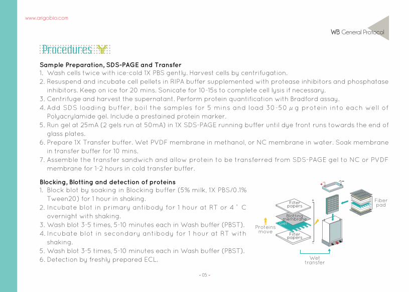
Filterpapers
Filterpapers
+
-
Blottingmembrane
Proteinsmove
Gel
Wettransfer
Fiberpad
www.arigobio.com

06
How Not to Fail a Western Blot Experiment?
How Not to Fail a Western Blot Experiment?1. Include appropriate positive and negative controls.Always include appropriate positive and negative controls in your western blot experiment. These will help narrow down possible causes just in case the experiment didn’ t work out well. Internal controls such as GAPDH, Actin or Tubulin are good candidates for positive controls. Untreated or knocked down cell lysates serve as good negative controls. Refer to our internal control guide at page 7 for more tips and advice!
2. Get a reliable antibody source and optimize it!Make sure that your antibody is WB-validated. Some antibodies do not recognized denatured epitopes, hence not suitable for use in WB experiments. Always optimize the antibody dilution factor as inappropriate dilutions will give you high background or low/weak signals. Refer to the recommended dilution factor indicated in the datasheet and try a few more dilutions to find the optimal condition.
3. Add suitable protease/phosphatase inhibitors for each sample typesProtease inhibitors or phosphatase inhibitors can be added into the freshly prepared cell lysis buffer to prevent proteolytic degradation of target proteins. Each sample type requires different combination of protease inhibitors. Understand what your sample needs and choose the right protease inhibitors to protect them. Need help? Refer to our protease inhibitor guide at page 8~9 for more details!
4. Keep things “cool” ~Proteins are sensitive to heat and the whole western blot experiment can be destroyed if high heat is applied during SDS-PAGE, transfer, blocking or antibody incubation steps. If possible, run your SDS-PAGE gel or transfer your blot at 4 ° C. Compared to RT, incubating blot with primary antibodies overnight at 4 ° C gives better antibody-target binding and a cleaner background.
www.arigobio.com

07
How Not to Fail a Western Blot Experiment?
Internal Control Information
When Western Blot or
other experiments are
p e r f o r m e d , l o a d i n g
controls are required to
ensure that (1) the same
amount of protein sample
is loaded into each lane;
(2) protein is transferred
from gel to membrane
with equal efficiency and
(3) antibody incubation
and detection is uniform.
Loading control must fulfill
certain criteria before
t h e y a r e c h o s e n a s
normalization factor in
various samples. Follow
our guide below to choose
the best internal controls
for your experiments!
COX IV(ARG54003)
kDa
17
Transferrin(ARG63114)
kDa
77Lamin B1(ARG20013)
PCMA(ARG62605)
kDa
66
29
Vinculin (ARG53723)
GAPDH (ARG10112)
β-Actin (ARG62346)
Cofilin (ARG51149)
ARG53696.ARG53698.ARG62347Tubulin
kDa116
43
3419
55
NuclearExtract
WholeCell Lysate
MitochondrialExtract Serum
What is your sample type?
Choose a control which its molecular weightis different from your protein of interest
ARG62605PCNA antibody
(PC10)
AR62347β-Tubulin antibody
(BT7R)
AR62346β-Actin antibody
(BA3R)
ARG10112GAPDH antibody
(6C5)
www.arigobio.com

08
How Not to Fail a Western Blot Experiment?
Protease Inhibitor InformationWithin a few minutes, proteases can destroy the proteins you have spent days isolating. Inhibition of proteolytic activity is therefore becoming very important to prevent unwanted degradation of proteins during their isolation and characterization.See the list below for commonly used inhibitors grouped into different classes of proteolytic enzymes.
Inhibitor Target Typical working concentrations Solvent
Serine Protease inhibitors
Leupeptin
PMSF
Aprotinin
AEBSF
Trypsin, chymotrypsin, pepsin, thrombin,calpain, cathepsinB,H, Papain etc
Broad spectrum serine protease
Broad spectrum serine protease but notthrombin or factor Xa
Broad spectrum serine protease
10-100 μM
0.1-1.0 mM
0.3 μM
0.1-1.0 mM
Water
Anhydrous ethanolor isopropanol
Water
Water
Inhibitor Target Typical working concentrations Solvent
Aspartic Protease inhibitors
Renin, Chymosin, Pepsin andotheraspartic proteases
Pepstatin A 1 μM Methanol or DMSO
www.arigobio.com

09
How Not to Fail a Western Blot Experiment?
Inhibitor Target Typical working concentrations Solvent
Cysteine Protease inhibitors
Broad spectrum cysteine proteaseand trypsin
Chymase cathepsins A,B,D,G, Papain
E-64
Chymostatin
1-10 μM
10-100 μM
Water
DMSO
Inhibitor Target Typical working concentrations Solvent
Metalloproteinase inhibitors
Broad spectrunmetalloproteinase
Aminopeptidase
EDTA
Bestatin
1-10 mM
1-10 μM
Water
Methanol
www.arigobio.com

10
Western Blot Troubleshooting Guide
Western Blot Troubleshooting Guide
Reagent contaminated Prepare all reagents freshly.
Blocking agent insufficiently dissolvedMake sure that the blocking agent such as milk or BSA is completely dissolved before
use. Alternatively, filter the blocking solution with 0.45μm filter before use.
Black Dots
Possible Causes What can you do?
Poor activi ty of ECL Prepare ECL solution freshly prior to detection.
Protein not expressed in thesample used
Make sure that the protien of interest in sufficiently induced in thesample used.
Fractionation might be necessary for some proteinsexpressed in particular
organelles.
Antibody issueMake sure that primary antibody dilution and the incubationcondition is optimal.
Compatible secondary antibody should beused.
Sodium Azide inter ferenceMake sure that there is no Sodium Azide in the antibody dilutionbuffer. Wash the
blot thoroughly before adding ECL.
Inadequate/incomplete transferBe sure that the transfer is adequate, especially for high molecularweight proteins.
Make sure that the transfer is complete bystaining the membrane with Ponceau S
solution or using a pre-stained marker as indicator.
Weak or no Signal
Possible Causes What can you do?
www.arigobio.com

11
Western Blot Troubleshooting Guide
www.arigobio.com
Improper blocking buffer used For the detection of phospho-proteins, use BSA instead of milk as blocking agent.
Antibody concentration too high Optimize the dilution factor of primary and secondary antibody.
Insufficient blockingMake sure that the blot is sufficiently blocked. Increase the percentage of skimmed
milk up to 5% if necessary.
Membrane dried out Make sure that the membrane is moist throughout the whole process of western blot.
Insufficient washingMake sure that the blot is washed in sufficent washing buffer. Increase washing time
or the percentage of Tween-20 if necessary.
High Background
Poor gel preparationMix gel completely before pouring. If SDS-PAGE should be run the day after
preparation, make sure that the gel is prepared at RT and stored in moist chamber
at 4°C.
High membrane protein concentrationin samples
If membrane fraction is used, make sure that sample is sufficiently diluted before
loading into SDS-PAGE.
Protein overloaded Make sure that total amount of protein loaded into each well is between 20-50μg.
Smeary Staining
Possible Causes What can you do?
Possible Causes What can you do?
12
Western Blot Troubleshooting Guide
Protein overloaded Make sure that total amount of protein loaded into each well is between 20-50μg.
Poor gel preparationMix gel completely before pouring. If SDS-PAGE should be run the day after
preparation, make sure that the gel is prepared at RT and store in moist chamber at
4°C.
High salt concentration in samples Make sure that the salt concentration of lysis buffer is kept between 0.15M to 0.5M.
Antibody concentration too high Optimize the dilution factor of primary and secondary antibody.
Voltage, temperature too high,field effect
Keep the voltage and temperature low while running SDS-PAGE. If necessary, run gel
in the cold room. Remove bubbles trapped at the bottom of gel to ensure even
electrophoresis.
Band Artifacts (White bands, smile effect, streaks)
Protein post-translationally modifiedor alternatively spliced
Check if the protein of interest is post-translationally modified or alternatively
spliced and produce other isoforms.
Membrane protein issueIf a membrane protein is to be detected, try low temperature (~65°C) or avoid boiling
which might cause aggregation of membrane proteins.
Incomplete protein denaturationFreshly add DTT or β-Mercaptoethanol to sample buffer, or adequaltely boil samples
to ensure complete bond breakage between peptides.
Molecular Weight Different from Predicted
www.arigobio.com
Possible Causes What can you do?
Possible Causes What can you do?

13
Western Blot Troubleshooting Guide
www.arigobio.com
Antibody concentration too high Optimize the dilution factor of primary and secondary antibody.
Protein post-translationally modifiedor alternatively spliced
Check if the protein of interest is post-translationally modified or alternatively
spliced and produce other isoforms.
Protein MultimerizationFreshly add DTT or β-Mercaptoethanol to sample buffer, or adequaltely boil samples
to ensure complete bond breakage between peptides.
Antibody issueSome antibodies use common epitopes as immunogen. Blast the immunogen region
of the antibody to make sure that the immunogen does not cross-react with epitopes
from other proteins.
Interference from secondary antibody
While performing Immunoprecippitaiotn (IP) experiment, make sure that the
secondary antibody used to detect the protein of interest is derived from a species
different from that of antibody used to pull down protein. Alternatively, use a
secondary antibody that recognize only the native form of IgG to detect IP protein.
Protein degradationAdd enough protease inhibitor to lysis buffer throughout all steps of sample
preparation. Avoid frequent freeze-thawing of samples.
Multiple bands
Possible Causes What can you do?

14
Procedures
IP General Protocol
Buffer Preparation
Cell lysis
Protein quantification
Pre-clearing
Immunoprecipitation
Wash
Elution
Protein analysis
Sample harvesting
Western Blotting- Work Flow -
Wash cells gently with ice-cold PBS.Harvest cells (107) and transfer to conical tube. Wash cells with 10ml ice-cold PBS and centrifuge at 400xg for 10 mins at 4 ° C. (The number of cells used per IP should be optimized specifically for each protein and antibody)Discard supernatant and repeat wash once.Remove supernatant completely and resuspend cell pellet in 1ml ice-cold Lysis buffer (supplemented with protease inhibitors and phosphatase inhibitors).Gently vortex and transfer to 1.5ml tube.Place tube on ice for 30 mins with occasional mixing.
Cell Lysate Preparation1. 2.
3. 4.
5. 6.
www.arigobio.com
Lysis Buffer (Non denaturing)
1M Tris-HCl, pH8: 0.5ml
10% NP-40: 1ml
5M NaCl: 0.3ml
ddH2O: 8.2ml
RIPA Buffer
1M Tris-HCl, pH8: 0.5ml
10% NP-40: 1ml
5M NaCl: 0.3ml
10% SDS: 0.1ml
10% Sodium Deoxycholate: 0.5ml
ddH2O: 7.6ml

15
IP General Protocol
Add 50ul of prepared Protein A or G slurry to 500μl cell lysates. Incubate on a rotator for 30 to 60 minutes at 4°C .Centrifuge at 2500xg for 2-3 mins at 4°C . Transfer supernatant to clean tube.
Preclearing1. 2.3.
Add 1-10μg of antibody to the pre-cleared lysates. (The concentration of antibody should be optimized)Incubate at 4°C overnight on a rotator.Add 50μl Protein A or G slurry to capture complexes. (Protein A or G should be equilibrated in the corresponding lysis buffer used).Incubate for 1-2 hour at 4°C on a rotator.Centrifuge the tube at 2500xg for 30s at 4°C.Carefully discard the supernatant. Wash beads 3-5 times with 500μl ice-cold lysis buffer. After last wash, carefully remove supernatant and add 50μl SDS Sample buffer to bead pellet.Vortex and heat at 90-100°C for 10 minutes.Centrifuge at 10,000xg for 5 mins. Collect the supernatant carefully and load onto SDS-PAGE for further analysis.
Immunoprecipitation1. 2.3.
4.5.6.7.8.9.
Centrifuge cell lysate at 10,000xg for 15-30 mins at 4°C.Carefully collect supernatant into clean tube. The protein concentration can be determined by Bradford assay. Samples can be diluted to 1μg/ul for further experiments. Cell lysates can be frozen at -80°C or used immediately for IP procedures.
7. 8.9.
10.
www.arigobio.com

16
How Not to Fail an IP Experiment?
How Not to Fail an IP Experiment?1. Lysis buffersThe denaturing ability of lysis buffer used during cell lysis can be critical. The ideal lysis buffer should protect proteins in their most native conformation while allowing adequate amounts of protein released for further analysis. Non-ionic detergents such as NP-40 or Triton X-100 are milder than ionic detergents such as SDS or Sodium Deoxycholate. Salt concentration or pH can also affect the binding capability of the antibodies to the protein of interest.
2. Elution methodThere are various ways to perform elution step in IP experiments. The harshest method would be boiling of beads in a reducing SDS sample loading buffer. This method also elute non-covalently bound antibody fragments along with the protein of interest. Other methods such as using glycine buffer or applying pH shift are also applied to avoid disruption of antibody in order to obtain lower background.
3. Choice of Protein A or Protein GProtein G is often considered a more universal IgG Binding Protein than Protein A, but different species, and subtypes of species, do vary in their binding to these proteins. Refer to the table at page 17 to find the best material that suit your antibody subclasses.
4. Secondary antibodies optimized for IP Western BlottingHeavy chains or light chains contributed by the denatured primary antibodies during IP often cause problems for the Western Blot detection of protein of interest especially if it should be found to migrate around 50kDa or 35kDa. To overcome this issue, some secondary antibodies which only recognizes native form of IgG have been developed. These antibodies significantly eliminates the detection of denatured heavy or light chains during Western Blotting.
www.arigobio.com

17
How Not to Fail an IP Experiment?
www.arigobio.com
Protein A is a cell wall component of Staphylococcus aureus which binds to the Fc region of immunoglobulins, especially IgG.
Protein G is a cell wall component isolated from Group B streptococci which binds to most mammalian immunoglobulins through their Fc region.
Protein L is a surface component of Peptostreptococcus magnus which binds immunoglobulins through their light chains. This protein is thus able to bind to antibody classes including IgA, IgD, IgE or IgM
Protein A
Protein G
Protein L
+++ Strong binding ++ Medium binding + Weak binding - No binding
Human
Total IgG
IgG1
IgG2
IgG3
IgG4
IgA,M,D
+++
+++
+++
-
+++
-
+++
+++
+++
+++
+++
-
+++
+++
+++
+++
+++
+++
Mouse
Total IgG
IgG1
IgG2
IgG3
IgG4
IgA,M,D
+++
+++
+++
-
+++
-
+++
+++
+++
+++
+++
-
+++
+++
+++
+++
+++
+++
Rat
Total IgG
IgG1
IgG2a
IgG2b
IgG2c
-
-
-
-
+
++
+
+++
+
+
+++
+++
+++
+++
+++
Chicken IgY - - -
Rabbit Total IgG +++ +++ -
Goat Total IgG - ++ -
Species Subclass Protein A Protein G Protein L

18
www.arigobio.com
IP Troubleshooting Guide
IP Troubleshooting Guide
Use secondary antibodies which only recognizes native form of IgG for
immunoblotting.
Interference from heavy or light chain
Possible Causes What can you do?
Non specific binding to agarose beads
Inadequate washing
Antibody not specific enough Use affinity purified and pre-absored antibody for IP experiments.
Concentration of antibodies too high
Use a more stringent washing buffer. Try to use a high salt washing buffer or
add 0.2% SDS or 1% Tween20 to washing buffer. Increase the number of washes.
Non specific binding to Protein A,G or LPre-block beads with BSA. Incubate beads with 2%BSA in PBS for 1 hour wash in
PBS before use.
Sample degradationAdd adequate protease inhibitors and phosphatase inhibitors throughout sample
preparation and Ip steps.
Include a pre-clear step by incubating lysate with Protein A/G/L agarose beads.
Check the recommended amount of antibody as indicated in the datasheet.
Titrate and optimize the optimal antibody amount used per IP experiment.
High Background
Possible Causes What can you do?
Secondary antibody rcognizes heavy / l i g h t c h a i n d ena t u r ed f r o m p r i ma r y antibody

IP Troubleshooting Guide
www.arigobio.com
19
No binding
Insufficient antibody
Incorrect Lysis buffer usedMake sure that the lysis buffer used is not over-denaturing and destroy the
native conformation of the target proteins.
Check the recommended amount of antibody as indicated in the datasheet.
Titrate and optimize the optimal antibody amount used per IP experiment.
Incorrect Protein A/G/L usedMake sure that the Protein A/G/L beads are capable of binding to the antibody
subclass being used.
Antibody not capable ofimmunoprecipitation
Try a different antibody. Try polyclonal antibody if monoclonal antibody does
not work well.
Target protein not present in thesample used
Make sure that the target protein is expressed at a relatively high level in the
sample used by including an Input sample in the WB.
Washes too stringent Reduce the number of washes. Reduce salt concentration in the wash buffer.
Possible Causes What can you do?

20
ChIP General Protocol
www.arigobio.com
Buffer Preparation
Quenching
Chromatin shearing
Check fragment size
Immunoprecipitation
Elution
DNA quantification
Fixation
Western Blotting- Work Flow -
TE Buffer
1M Tris-HCl (pH8): 10ml
0.5M EDTA (pH8): 1ml
ddH2O: 489ml
Total: 500ml
Elution Buffer
10% SDS: 10ml
0.5M EDTA (pH8): 10ml
1M Tris-HCl (pH8): 5ml
ddH2O: 75ml
Total: 100ml
High Salt Wash Buffer
10% SDS: 0.5ml
Triton X-100: 5ml
0.5M EDTA (pH8): 2ml
1M Tris-HCl (pH8): 10ml
5M NaCl: 50ml
ddH2O: 432.5ml
Total: 500ml
LiCl Wash Buffer
1M LiCl: 125ml
NP-40: 5ml
10% Sodium Deoxycholate: 50ml
0.5M EDTA (pH8): 1ml
1M Tris-HCl (pH8): 5ml
ddH2O: 314ml
Total: 500ml
Glycine (2.5M)
Glycine: 93.8g
ddH2O: 500ml
Gentle heating might be required
Lysis Buffer
1M HEPES-KOH (pH7.5): 10ml
5M NaCl: 5.6ml
0.5M EDTA (pH8): 0.4ml
Triton X-100: 2ml
10% SDS: 2ml
10% Sodium Deoxycholate: 2ml
ddH2O: 178ml
Total: 200ml
Protease inhibitors (add fresh)
Low Salt Wash Buffer
10% SDS: 0.5ml
Triton X-100: 5ml
0.5M EDTA (pH8): 2ml
1M Tris-HCl (pH8): 10ml
5M NaCl: 15ml
ddH2O: 467.5ml
Total: 500ml
21
ChIP General Protocol
www.arigobio.com
Procedures
Add Formaldehyde to cell culture medium at a final concentration of 1%. (Start with 50-100 million cells per experiment)Shake culture flasks for 10 minutes at RT.Add glycine to quench the reaction at a final concentration of 125mM. Shake culture flasks for 5 minutes at RT.Wash cells twice with 10-15ml of ice-cold PBS.Add 5-10ml of cold PBS to flask, scrape cells and transfer to 50ml tube.Centrifuge at 1000xg, 5 mins at 4°C.Remove supernatant and resuspend pellet in lysis buffer (750μl for 10 million cells) (For suspension cultures, pellet cells after glycine treatment, wash twice with ice-cold PBS. Resuspend pellet in lysis buffer (750μl for 10 million cells))Incubate pellet on ice for 10 minutes.Proceed with sonication or MNase digestion to shear DNA. Make sure that the sheared DNA give rise to fragment size of 200-1000bp. Optimization is needed for each cell types or tissues. After sonication, centrifuge cell lysate at 8,000xg for 10 mins at 4°C. Transfer supernatant to new tube. Remove 50μl to determine fragment size and DNA concentration, keep the rest at -80°C freezer for storage.
Cross-linking and Chromatin Preparation1.
2.3.
4. 5. 6. 7.
8.9.10.
11.12.
22
ChIP General Protocol
www.arigobio.com
Use 25μg of DNA per IP as a start. (The optimal starting material need to be optimized according to each sample type and antibody)Prepare 1 tube of chromatin for antibody IP, and another tube for control (beads only). Remove 50μl of chromatin as input sample, store temporarily at -20°C for later use.To preclear the chromatin, add 50μl Protein-A agarose/salmon sperm DNA or Protein-G agarose/salmon sperm DNA beads to chromatin and rotate for 1-2h at 4 ° C. Centrifuge chromatin samples at 2000xg for 5 mins at 4°C. Transfer supernatant to new tube.Add primary antibody to tube (except for bead-only tube). The amount of antibody used per IP can found on the datasheet or determined by user.Rotate overnight at 4°C.Add 50μl of blocked Protein A or G slurry to capture complexes. Incubate for 2 hour at 4°C on a rotator.Centrifuge the tube at 2000xg for 1min at 4°C.Carefully discard the supernatant. Wash beads once with low-salt buffer, once in high-salt buffer, once in LiCl wash buffer and once with TE buffer. Washing procedure: Resuspend beads in 1 ml wash buffer, rotate for 10 minutes at 4°C, centrifuge at 2000xg for 1 min at 4°C and remove supernatant.
Immunoprecipitation and Washing1.
2.
3.
4.
5.6.7.8.9.10.
Determination of fragment size and DNA concentration1.
2.3.
Add 70μl of elution buffer to 50μl of sheared DNA. Add 1μg RNAse A and 4μg Proteinase K, and heat at 65 ° C overnight to reverse crosslink. Purify DNA by phenol-chloroform or using a kit. Run 5μl of purified DNA sample on 1.5% agarose gel. Determine DNA concentration with spectrometer.

23
ChIP General Protocol
www.arigobio.com
Elute DNA by adding 300μl of Elution buffer (supplemented with 1μl of Proteinase K (20μg/μl) to beads. Incubate samples for 2 hours at 55°C. Gently vortex tubes occasionally. To reverse-crosslink, incubate overnight at 65 ° C. (Input should also be reverse cross-linked at the same time)Centrifuge samples at 15,000xg for 5 mins at RT.Transfer supernatant to a new tube. Purify DNA using phenol-chloroform or DNA purification kit.Resuspend or elute DNA in 50μl TE buffer and analyze by PCR or Real-Time PCR.
Elution, reverse-crosslink and DNA purification1.
2.
3. 4.5.6.

24
How Not to Fail a ChIP Experiment?
www.arigobio.com
How Not to Fail a ChIP Experiment?1. Cross-linkingCross-linking is an important step to make sure that the protein is still bound to the DNA fragment during immunoprecipitation. However, it is a time-critical process and should be optimized for each sample type or antibody type. Excessive cross-linking might mask the antibody binding sites and reduce binding ability. Therefore, it is advisable to optimize the cross-linking steps by using different concentration of formaldehyde or changing cross-linking time.
2. Fragmentation methodOptimal sonication time course or concentration of micrococcus nucleus should be determined prior to performing a ChIP experiment. Make sure that the majority of fragmented DNA falls between 200-500bp. Load 5-10μl of sample on agarose gel to analyze DNA fragment size after each sonication course.
3. Choice of Protein A or Protein GProtein G is often considered a more universal IgG Binding Protein than Protein A, but different species, and subtypes of species, do vary in their binding to these proteins. Refer to the table at page 17 to find the best material that suit your antibody subclasses.
4. Include positive and negative antibody controlsHistone H3K4me3 antibody is a popular positive control to use when studying active gene, while Histone H3K9me3 antibody is a negative control for studying inactive gene. An antibody that do not recognize chromatin epitope such as GFP antibody or IgG isotype control antibody should also be included in ChIP experiments to make sure that results came out to be valid.

25
ChIP Troubleshooting Guide
www.arigobio.com
ChIP Troubleshooting Guide
Positive signal seen in no template control
Possible Causes What can you do?
Inadequate washing
Non specific binding to Protein A,G or L
Use a more stringent washing buffer. Try to use a high salt washing buffer or
increase the number of washes.
Too much DNA template addedto the PCR reaction, or too manycycles of amplification
Add less DNA template or reduce the number of cycles of amplification.
Alternatively, real-time PCR can be used for the detection of ChIPed DNA
products.
Buffers may be contaminated Use freshly prepared lysis or wash buffers.
Include a pre-clear step by incubating lysate with Protein A/G/L agarose beads.
High Background observed in negative control
Possible Causes What can you do?
Prepare new solutions from stockPCR reagent might be contaminated

26
ChIP Troubleshooting Guide
www.arigobio.com
Not enough antibody
Not enough cells/chromatinAdd enough chromatin for each IP experiment. We suggest using at least 25 μg of
chromatin for each IP.
Cross-linking process too longOver Cross-linking with formaldehyde might mask the antibody binding sites and
reduce antibody binding ability. It is advisable to optimize the cross-linking steps by
using different concentration of formaldehyde or reducing cross-linking time.
Washes too stringent Reduce the number of washes. Reduce salt concentration in the wash buffer.
Antibody not capable ofimmunoprecipitation
Try a different antibody. Try polyclonal antibody if monoclonal antibody does not
work well.
The chromatin size might be too smallMake sure that the shearing condition is not too harsh which might results in
fragments of DNA smaller than what the primers are able to amplify.
Incomplete elution from the ProteinA/G/L beads
Incubate beads in elution buffer at 65°C with frequent mixing.
Incorrect Protein A/G/L usedMake sure that the Protein A/G/L beads are capable of binding to the antibody
subclass being used.
Titre antibody amount used for each IP to determine the optimal condition. Up to
10ug of antibody can be used for each IP experiment.
Low/No Signal
Possible Causes What can you do?
27
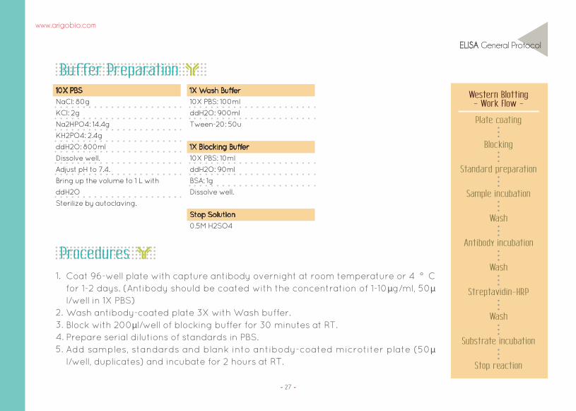
ELISA General Protocol
www.arigobio.com
Blocking
Standard preparation
Sample incubation
Wash
Antibody incubation
Wash
Streptavidin-HRP
Wash
Substrate incubation
Plate coating
Stop reaction
Western Blotting- Work Flow -
Buffer Preparation
ProceduresCoat 96-well plate with capture antibody overnight at room temperature or 4 ° C for 1-2 days. (Antibody should be coated with the concentration of 1-10μg/ml, 50μl/well in 1X PBS)Wash antibody-coated plate 3X with Wash buffer.Block with 200μl/well of blocking buffer for 30 minutes at RT.Prepare serial dilutions of standards in PBS.Add samples, standards and blank into antibody-coated microtiter plate (50μl/well, duplicates) and incubate for 2 hours at RT.
1.
2.3. 4. 5.
10X PBS
NaCl: 80g
KCl: 2g
Na2HPO4: 14.4g
KH2PO4: 2.4g
ddH2O: 800ml
Dissolve well.
Adjust pH to 7.4.
Bring up the volume to 1 L with
ddH2O
Sterilize by autoclaving.
1X Wash Buffer
10X PBS: 100ml
ddH2O: 900ml
Tween-20: 50u
1X Blocking Buffer
10X PBS: 10ml
ddH2O: 90ml
BSA: 1g
Dissolve well.
Stop Solution
0.5M H2SO4

28
ELISA General Protocol
www.arigobio.com
Wash 3X with wash buffer.Add biotinylated Capture antibody and incubate for 1 hour at RT.(Concentration of 1-10μg/ml, 50μl/well in 1X PBS)Wash 5X with Wash buffer.Add Streptavidin-HRP in Wash buffer (50μl/well) and incubate for 45 mins at RT. Wash 5X with Wash buffer. Add TMB substrate (50μl/well) and watch for color change. Stop reaction with Stop solution (50μl/well). Measure OD at 450nm in an ELISA plate reader. (Reference wavelength 620nm)
6.7.
8.9. 10. 11.12.13.

29
How Not to Fail an ELISA Experiment?
www.arigobio.com
How Not to Fail an ELISA Experiment?1. Ensure consistency between wellsUse multiwell plates, multichannel pipettes and plate washers for a more consistent result. Make sure that all pipettes are accurately calibrated on a regular basis. In the initial stage of assay development, test a range of parameters to optimize ELISA conditions.
2. Prevent sample degradationProtease inhibitors or phosphatase inhibitors can be added into the freshly prepared cell lysis buffer to prevent proteolytic degradation of target proteins. It is important to test all samples in duplicate or triplicate in conjunction with a known standard to ensure the accuracy of results.
3. Optimize coating conditionCoating of antibodies or antigens onto plastic surface is a passive absorption process which depend highly on time, temperature, pH and the concentration of coating agents. Typical coating conditions fall within the range of 1-10 μg/ml in 50-100 μl buffer, incubating overnight at 4 ° C or 1-3 hours at RT. Optimization for each assay need to be performed individually.
4. Choose the right antibodiesAntibodies of high specificity, affinity and avidity need to be used for ELISA assays. Monoclonal antibodies offer better homogeneity by targeting a single epitope while polyclonal antibodies consist of complex antibody pools that target various epitopes. For sandwich ELISA, arigo offers ELISA antibody duos for the optimized performance of ELISA assays.
“
“
Not all standard curves are straight lines. It is advisable to use program capable of generating a four parameter logistic (4-PL) for a better fitting curve.

30
ELISA Troubleshooting Guide
www.arigobio.com
ELISA Troubleshooting Guide
Wrong settings of plate reader Check the settings (wavelength, filters, gain etc) of plate reader.
Assay set up incorrectly Make sure that the instructions in the protocol is followed carefully.
Insufficient antibodies used Increase concentration of primary or secondary antibody.
Insufficient incubation Follow the incubation time as indicated in the protocol booklet.
Substrate reagents not fresh Use fresh substrate reagents
Wells dried outCover plate with adhesive film or incubate in humidified chamber
throughout experiment.
Sample concentration falls below detection limits of kit
Decrease dilution factor or concentrate samples.
Enzyme inhibitor present in buffers or reagents
Inhibitors such as Sodium Azide can affect enzyme and assay performance.
Ensure that there is no enzyme inhibitor in any buffers.
Plate washing too vigorous Check the setting of plate washer. Pipette wash buffer into wells gently.
Incorrect secondary antibody used Check if the correct secondary antibody is used.
No signal
Possible Causes What can you do?

31
ELISA Troubleshooting Guide
www.arigobio.com
High background
Wrong concentration of blocking reagent Check the recommended concentration of blocking buffer.
Too much antibodies was used Reduce the concentration of primary or secondary antibodies.
Too much substrate reagent used Use substrate with higher dilution.
Reaction not stopped Stop reactions with STOP buffer before reading.
Insufficient washing Increase washing cycles.
Incubation temperature too high Optimize incubation temperature for each experiment.
Plate left too long before reading Take measurements shortly after addition of substrate and STOP buffer.
Plate stacking during incubation lead touneven temperature throughout the plate
Avoid stacking plates together during incubation.
Pipetting error Calibrate pipettes to make sure that the correct volume is dispensed.
Reagents not mixed properlyMake sure that all reagents are mixed properly and equilibrated to room
temperature before assay
Insufficient Tween in wash buffer Use PBS+0.05% Tween as wash buffer.
Antibodies bind nonspecifically Use blocking buffer or choose another affinity-purified antibody.
Possible Causes What can you do?
Continued

32
ELISA Troubleshooting Guide
www.arigobio.com
High background
Salt concentration of incubationand wash buffer
Increase salt concentration to reduce nonspecific interaction.
Dirty plate Make sure that the bottom of plate is clean.
Substrate incubation carried out in light Perform substrate incubation in dark.
Possible Causes What can you do?
Poor standard Curve
Improper standard dilutionUse appropriate diluent as blank. Make sure that the dilution is performed as
according to protocol.
Standard improperly reconstitutedBriefly spin standard vial before opening. Make sure that there is no undissolved
material after reconstituting.
Standard degraded Store standards as according to protocol.
Curve doesn’t fit the scale Try plotting log-log or 5 parameter logistic curve fit.
Pipetting error Calibrate pipettes to make sure that the correct volume is dispensed.
Incomplete washing Increase washing cycles.
Possible Causes What can you do?

33
ELISA Troubleshooting Guide
www.arigobio.com
Weak signal
Insufficient coating Use more antigens or antibodies for coating.
Substrate reagents have expired or prepared at a wrong pH
Use fresh substrate reagents.
Possible Causes What can you do?
Variation among replicates
Improper washing Make sure that the washing is done as according to protocol.
Dirty plate Make sure that the bottom of plate is clean.
Reagents too old Make sure that the reagents are not expired. Use freshly prepared reagents.
Bubbles in wells Make sure that there is no bubble in wells before reading.
Inconsistent pipetting Calibrate pipettes to make sure that the correct volume is dispensed.
Edge effectsMake sure that the plate and reagents are equilibrated to room temperature
before starting assay.
Poor mixing of samples Mix samples gently and evenly.
Possible Causes What can you do?


Phone : +886 (3) 5621738 / E-mail : [email protected]
BIOMOL GmbH Waidmannstr. 35 22769 Hamburg Germany
[email protected] www.biomol.de
Fon: +49 (0)40-853 260 0 Fax: +49 (0)40-853 260 22