complete genome direct rna sequencing of influenza a virus · instead, the elucidation of rna...
TRANSCRIPT

Complete genome direct RNA sequencing of influenza A virus
Matthew W. Keller1, Benjamin L. Rambo-Martin2, Malania M. Wilson2, Callie A. Ridenour2, Samuel S. Shepard3, Thomas J. Stark3, Elizabeth B. Neuhaus3, Vivien G. Dugan3, David E. Wentworth3, and John R. Barnes3*
1Oak Ridge Institute of Science and Education (ORISE), Oak Ridge, Tennessee, USA 2Battelle Memorial Institute, Atlanta, Georgia, USA 3Influenza Division, National Center for Immunization and Respiratory Diseases (NCIRD), Centers for Disease Control and Prevention (CDC), Atlanta, Georgia, USA
*Address correspondence to [email protected]
ABSTRACT
For the first time, a complete genome of an RNA virus has been sequenced in its original form. Previously, RNA
was sequenced by the chemical degradation of radiolabelled RNA, a difficult method that produced only short
sequences. Instead, RNA has usually been sequenced indirectly by copying it into cDNA, which is often amplified
to dsDNA by PCR and subsequently analyzed using a variety of DNA sequencing methods. We designed an
adapter to short highly conserved termi of the influenza virus genome to target the (-) sense RNA into a protein
nanopore on the Oxford Nanopore MinION sequencing platform. Utilizing this method and total RNA extracted
from the allantoic fluid of infected chicken eggs, we demonstrate successful sequencing of the complete
influenza virus genome with 100% nucleotide coverage, 99% consensus identity, and 99% of reads mapped to
influenza. By utilizing the same methodology we can redesign the adapter in order to expand the targets to
include viral mRNA and (+) sense cRNA, which are essential to the viral life cycle. This has the potential to
identify and quantify splice variants and base modifications, which are not practically measurable with current
methods.
Introduction
Decades ago, a method was published describing the use of base-specific chemical degradation with
chromatographic and autoradiographic resolution as a way of directly sequencing short stretches of RNA1. Since
then, little progress has been made on directly sequencing RNA. Instead, the elucidation of RNA sequences is
.CC-BY-ND 4.0 International licensepeer-reviewed) is the author/funder. It is made available under aThe copyright holder for this preprint (which was not. http://dx.doi.org/10.1101/300384doi: bioRxiv preprint first posted online Apr. 12, 2018;

typically indirect and primarily requires methods that synthesize cDNA from RNA templates. While these
methods are powerful2, they suffer from limitations inherent to cDNA synthesis and amplification such as
template switching3, artifactual splicing4, loss of strandedness information5, obscuring of base modifications6,
and propagation of error7. In 2009, a method for RNA sequencing was developed on the Helicos Genetic Analysis
System where poly(A) mRNA is sequenced by the step-wise synthesis and imaging of nucleotides labeled with an
interfering but cleavable fluorescent dye8. While the input material requirements for this method are extremely
low, the long workflow and short reads are limiting. Nevertheless, these approaches expose two major
limitations of RNA sequencing: sequencing by synthesis and short read length. Overall, current technologies for
sequencing RNA templates present difficulties in the assessment of base modifications, splice variants, and
analysis of single RNA molecules.
Influenza viruses are negative-sense segmented RNA viruses9-11, and sequencing these viruses has played an
important role in their understanding for 40 years12,13 including the discovery of highly conserved viral RNA
termini14 (Figure 1A). These 3’ and 5’ termini are 12 and 13 nucleotides in length, respectively, and they are
highly conserved across the PB2, PB1, PA, HA, NP, NA, M, and NS genome segments of influenza A viruses, which
enabled the development of a universal primer set for influenza A virus genome amplification15,16. Even though
these conserved vRNA termini have been readily exploited for efficient next generation sequencing (NGS) of
influenza virus segments16-18, current methods retain some of the limitations inherent to cDNA-based
techniques3-7. A new tool for long read direct RNA sequencing could reduce these biases and greatly aid efforts
to directly sequence influenza virus and other RNA viruses.
Oxford Nanopore Technologies (ONT) recently released their direct RNA sequencing protocol. This method
involves the sequential ligations of a reverse transcriptase adapter (RTA) and a sequencing adapter19. The RTA is
a small dsDNA molecule (Figure 1B) that contains a T10 overhang designed to hybridize with poly(A) mRNA and a
5’ phosphate (Pi) that ligates to the RNA creating a DNA-RNA hybrid. The RTA also serves as a priming location
for reverse transcription of the entire length of the RNA molecule, though the cDNA generated is not sequenced.
.CC-BY-ND 4.0 International licensepeer-reviewed) is the author/funder. It is made available under aThe copyright holder for this preprint (which was not. http://dx.doi.org/10.1101/300384doi: bioRxiv preprint first posted online Apr. 12, 2018;

The DNA-RNA hybrid is then ligated to the sequencing adapter which directs the RNA strand of the assembled
library into the nanopore for sequencing19.
We describe direct RNA sequencing of an influenza A virus genome through modification of recently
released RNA methods from Oxford Nanopore Technologies19 (Figure 1C) by targeting the conserved 3’ end of
the genome with an adapter to capture it (Figure 1D), rather than a primer to amplify it. The efficacy of the
adapter is tested by sequencing the RNA genome of an influenza virus generated by reverse genetics A/Puerto
Rico/8/1934. The RNA was isolated from virus containing allantoic fluid (crude) harvested from infected
embryonated chicken eggs. The results from the nanopore sequencing are compared to the current Illumina-
based pipeline utilized by the Influenza Genomics Team at the Centers for Disease Control and Prevention.
Results
Nanopore sequencing
First, the RNA calibration strand enolase was directly sequenced on the MinION platform. Three sequencing
experiments covering 100% of the 1,314 nucleotide long RNA molecule to an average depth of 122,207 ± 8,126
(sd). Of the 169,041 ± 28,741 reads, 98.6 ± 1.7% mapped to the reference sequence (Table 1), with 100% of the
mapped reads in the sense orientation. The direction of the reads and the positive slope of the coverage
diagram (Figure S1) are indicative of directional sequencing of mRNA from the 3’ end. The distribution of read
lengths (Figure S2 and Table S1) accurately corresponds to the expected length of 1,314 nucleotides. The read
level accuracy was 90.4 ± 0.8%, and the consensus sequence was 99.7% in concordance with the known
reference.
Based on available details on the RTA system, it was possible to make further modification to target other
RNA species (Figure 1). To adapt this technique for the influenza virus genome, the target sequence of the RTA
was changed from an oligo-dT to a sequence complementary to the 12 nucleotides that are conserved at the 3’
end of the RNA segments of influenza A viruses (Table S2).
.CC-BY-ND 4.0 International licensepeer-reviewed) is the author/funder. It is made available under aThe copyright holder for this preprint (which was not. http://dx.doi.org/10.1101/300384doi: bioRxiv preprint first posted online Apr. 12, 2018;

To test the effectiveness of the modified adapter, total RNA from allantoic fluid (crude) harvested from
infected chicken eggs was sequenced via MinION. Three sequencing experiments covered 100% of the PB2, PB1,
PA, HA, NP, NA, M, and NS gene segments to an average depth of 3,269 ± 1,892 (Figure 2). Although, there is
reduced coverage at the extreme termini (Figure 3) and a heavy coverage bias towards the 3’ terminus of the
negative sense RNA, since this approach reads from the 3’ to 5’ end of the molecule. Of the 54,353 ± 15,314
reads, 98.8 ± 0.1% mapped to influenza (Table 1) in a roughly even distribution among the 8 segments (Figure
S3), with 100% of the mapped reads in the negative-sense orientation. The distribution of read lengths (Figure 4
and Table S1) corresponds well to the expected length of the respective segment. The read level accuracy was
86.3 ± 0.3%, and the consensus sequence was 98.97 ± 0.01% in concordance with consensus sequence
generated using our modified version of the multi-segment reverse transcriptase polymerase chain reaction (M-
RTPCR)15,16, Nextera, and MiSeq approach.
To provide a favorable substrate for the modified adapter and a positive control for future experiments, RNA
from two sucrose purified virus preparations (pure) was sequenced via MinION. Two sequencing experiments
covered 100% of the PB2, PB1, PA, HA, NP, NA, M, and NS viral RNA segments to an average depth 9,312 and
1,068 respectively (Figure S4) with reduced coverage at the extreme termini (Figure S5). Of the 119,860 and
13,848 reads acquired in each run, 99.6 and 99.1% mapped to influenza virus (recombinant A/Puerto
Rico/8/1934), respectively (Table S3), in a roughly even distribution among the eight vRNA segments (Figure S3)
with 100% of the mapped reads in the negative-sense orientation. The distribution of read lengths (Figure S6
and Table S1) corresponds to expected lengths of each respective segment. The read level accuracies for the
two runs were 85.2 and 83.8%, and the consensus sequences were 98.7 and 98.5% in concordance with
consensus sequence generated using our standardized M-RTPCR amplified genome and MiSeq approach.
Illumina MiSeq sequencing
The viral RNA segments from the pure and crude preparation were amplified by M-RTPCR15,16, and size
fractionation of those amplicons showed the characteristic banding pattern of the amplified influenza virus
genome (Figure S7). Sequencing of the RNA from purified virus or crude virus produced 163,264 and 143,572
.CC-BY-ND 4.0 International licensepeer-reviewed) is the author/funder. It is made available under aThe copyright holder for this preprint (which was not. http://dx.doi.org/10.1101/300384doi: bioRxiv preprint first posted online Apr. 12, 2018;
reads, respectively, of which 99.9% mapped to influenza A virus (Table 1). The reads were roughly evenly
distributed among the 8 segments (Figures S3). The mapped reads covered 100% of all 8 genome segments
(Figures 2 and S4). The read level accuracy was 99.6% and the consensus sequences, which were used as the
reference genome for the nanopore assemblies, were defined as 100% accurate and were 100% identical to
each other.
Discussion
We have demonstrated, for the first time, complete20 sequencing of an RNA virus genome by direct RNA
sequencing. Using a method originally designed to sequence mRNA, we adapted the target sequence to bind the
3’ sequence conserved among influenza A viruses. The specificity of this adapter allowed efficient sequencing of
influenza virus RNA genomic segments from RNA isolated from purified virus particles (control) or from RNA
isolated from a crude extract that contains a myriad of viral and host (chicken) RNAs. Using this adapter, 98.8%
of reads from the crude RNA preparation mapped to the influenza virus, which is practically as efficient as with
purified virus RNA sample (99.3%). This performance on crude virus stocks demonstrates that the sequence
directed library preparation is a very effective method to select specific target RNA species among a population
of RNAs, as the vast majority of reads were to A/Puerto Rico/8/1934 using 12 ribonucleotides as the target
sequence.
The data shows that other modifications to the adapter could target other RNA species such as RNAs from
specific pathogens and different RNA species within a particular pathogen. For example, one could compare (+)
sense cRNA [replication intermediate of (-) sense vRNAs], (+) sense mRNAs, or (-) sense RNAs present during
RNA virus infections (such as for influenza viruses). The data illustrates that the adapter sequence could be
modified to target specific viral families, genus, or species by extending the target sequence and or by adding
degeneracies. This is an advantage over poly(A) methods that have a reduced signal-to-noise ratio due to host
mRNA. Targeting influenza A virus vRNA and cRNA independently may prove difficult as there is
complementarity between the two conserved termini of the vRNA segments, and therefore high sequence
.CC-BY-ND 4.0 International licensepeer-reviewed) is the author/funder. It is made available under aThe copyright holder for this preprint (which was not. http://dx.doi.org/10.1101/300384doi: bioRxiv preprint first posted online Apr. 12, 2018;

identity between the 3’ termini of the (-) sense vRNA and (+) sense cRNA. Rather, cRNA and vRNA reads can be
sorted based on their (+) and (-) polarity, respectively.
In addition to avoiding any of the previously discussed limitations of cDNA synthesis and PCR amplification
strategies, the technique developed for direct RNA sequencing is highly amenable to sequencing a variety of
non-poly-adenylated RNAs from hosts and pathogens, including untranslated regions (UTRs), without biasing the
sequence to the primer. This allows the examination of the UTRs in their native form, which we have done here
with influenza A virus. However, direct RNA sequencing of UTRs is limited by read level accuracy and a loss of
coverage at the extreme 5’ end of the molecule. The extreme 3’ termini (Uni-12) of all segments were fully
sequenced and matched the expected sequence with the exception of the degeneracy at the +4 position which
was not resolved. The sequences for the extreme 5’ termini (Uni-13) that were obtained match the expected
sequences with the exception of a C to G substitution at the -9 position in the segments PB1 and PB2. The loss of
coverage at the extreme 5’ end of the molecule is most likely due to unreliable processivity as the last of the
molecule passes and resulted in the final nine nucleotides not being sequenced in some of the segments.
The data presented demonstrates the adaptability of the platform and RNA sequencing protocol. The
unmodified components were used to target enolase mRNA and could be used to target the variety of mRNA
species present in any sample. Specifically, one could dissect viral replication processes as well as host mRNAs
activated during an influenza infection at a given point in time. Genomic length and quantitative sequencing of
viral mRNA species has the potential to provide direct detection of base modifications, splice variants, and
transcriptional changes under different replication conditions, such as viruses used for vaccine production that
are transferred between mammalian and avian hosts.
The primary limitations of this technology are the high read level error rate and high input material
requirements. Reducing the error rate would enable multiplexing and more accurate consensus sequence
determination and is a requirement for understanding nucleotide polymorphisms and genome sub-populations,
particularly in viruses such as influenza that have significant intra-host diversity and or base modifications to be
identified. There are currently several bioinformatic tools for detecting DNA base modifications such as Tombo,
.CC-BY-ND 4.0 International licensepeer-reviewed) is the author/funder. It is made available under aThe copyright holder for this preprint (which was not. http://dx.doi.org/10.1101/300384doi: bioRxiv preprint first posted online Apr. 12, 2018;
Nanopolish, SignalAlign, and mCaller; however, RNA specific tools have yet to be released19. Currently, the RNA
input requirements for direct RNA sequencing are high and are not physically achievable with most original
clinical samples. Lessening the RNA input requirement of the direct RNA sequencing would take full advantage
of the unbiased nature of direct RNA sequencing and allow for the detection and description of the rich diversity
intrinsic to influenza and other viruses. Although ONT has continuously improved their basecaller Albacore,
there is still demonstrable potential for improvement. The RNA basecaller was likely developed using the very
same enolase mRNA used here, which would make it most effective at basecalling enolase mRNA. The marked
difference in accuracy between the enolase and influenza virus reads demonstrates that further development of
the RNA basecaller can, at a minimum, bring the accuracy of all RNA reads up to that of enolase reads.
Moreover, the DNA basecaller is overall more developed and more accurate than the RNA basecaller (89%
versus 85% read level accuracy for influenza samples). The continued effort to advance this technology by ONT
will undoubtedly result in higher accuracy reads and greatly improved utility.
Methods
Concentration and purification of A/Puerto Rico/8/1934 reassortant virus
A/Puerto Rico/8/1934 reassortant virus was grown in 11 day-old embryonated hen eggs at 35°C for 48
hours. Allantoic fluid was harvested from the chilled eggs and clarified at 5,400 x g, 10 minutes, 4°C, (Sorvall SLA-
1500 rotor). The virus was clarified twice more by centrifugation at 15,000 x g, 5 minutes, 4°C (Sorvall SLA-1500
rotor). Virus was pelleted by centrifugation at 39,000 x g, 3 hours at 4°C (Sorvall A621 rotor). Virus pellets were
resuspended overnight in PBS and loaded onto a 30%/55% (w/w) density sucrose gradient. The gradient was
centrifuged at 90,000 x g for 14 hours at 4°C (Sorvall AH629 rotor). The virus fractions were harvested and
sedimented at 131,000 x g (Sorvall AH629 rotor) for 2.5 hours. The resulting virus pellet was resuspended in PBS
and aliquoted for future use.
RNA isolation
Enolase II (YHR174W) mRNA is supplied in the ONT materials as the calibration RNA strand (CRS) at a
concentration of 50 ng/µL. For influenza virus samples, total RNA was isolated by InvitrogenTM TRIzol®
.CC-BY-ND 4.0 International licensepeer-reviewed) is the author/funder. It is made available under aThe copyright holder for this preprint (which was not. http://dx.doi.org/10.1101/300384doi: bioRxiv preprint first posted online Apr. 12, 2018;

extraction21 according to manufacturer’s instructions with additional considerations for biosafety. The virus was
inactivated by the addition of 10 volumes of TRIzol® in a Biosafely Level 2 biosafety cabinet. Following
inactivation, a fume hood was used for the chloroform addition and aqueous phase removal steps. RNA pellets
were resuspended in 10-40 µL nuclease free water and quantified by Quant-iTTM RiboGreen® RNA Assay Kit. Due
to the difficulty in acquiring sucrose-purified material, the pure controls were limited to one MiSeq run and two
separate MinION experiments. The availability of crude viral samples allowed it to be sequenced once on MiSeq
and three times on MinION from the same RNA preparation.
Nanopore Sequencing
The ONT direct RNA library preparation input material requirement is 500 ng of target molecule in a 9.5 µL
volume (Table S4). For mRNA sequencing of the enolase control, the protocol was used according to the
manufacturer’s instruction. For influenza viral RNA sequencing, modifications were made to the protocol
components (Table S2). We altered the supplied reverse transcriptase adapter (RTA) which has a T10 overhang
(Tm ~20°C) to target the ligation of the RTA to mRNA, with 12 nucleotides complementary to the conserved 3’
end of Influenza A virus (-) vRNA22 (Figure 1). RTA-U12 and RTA-U12.4 contained target sequences (5’ to 3’) AGC
AAA AGC AGG and AGC GAA AGC AGG (Tm ~50°C) respectively and were combined in a 2:3 molar ratio to a total
concentration of 1.4 µM. This mixture was used as a direct replacement to the RTA supplied in the protocol for
influenza samples. Though there is some disagreement regarding the segment specific degeneracies of the 12
nucleotides at the 3’ end of the genome, RTA-U12 is expected to target the segments PA, NP, M, and NS; and
RTA U-12.4 is expected to target the segments PB2, PB1, HA, and NA23,24.
Adapter ligated RNA was directly sequenced on the MinION nanopore sequencing using a FLO-MIN107
flowcell equipped with the R9.5 chemistry. The enolase sequencing experiments were operated through
MinKNOW versions 1.4.2, 1.7.7, and 1.10.11; the pure sequencing experiments were operated through
MinKNOW 1.7.7; and crude sequencing experiments were operated through MinKNOW 1.10.11. Raw data was
basecalled using Albacore 2.1.10 (released 01/26/2018), and reads were assembled using IRMA25 with the FLU-
MinION preset configuration to produce influenza consensus sequences for comparison to MiSeq-derived
.CC-BY-ND 4.0 International licensepeer-reviewed) is the author/funder. It is made available under aThe copyright holder for this preprint (which was not. http://dx.doi.org/10.1101/300384doi: bioRxiv preprint first posted online Apr. 12, 2018;

consensuses. The FLU-MinION preset differs from the default FLU module settings by the following: dropping
the median read Q-score filter from 30 to 0, raising the minimum read length from 125 to 150, raising the
frequency threshold for insertion and deletion refinement from 0.25 to 0.75 and 0.6 to 0.75 respectively, and
lowering the Smith-Waterman mismatch penalty from 5 to 3 and the gap open penalty from 10 to 6. For read-
level comparisons of MinION to MiSeq, raw fastqs from both sequencing platforms were mapped with bwa-
mem v.0.7.7 algorithm26 to MiSeq+IRMA derived consensus sequences as references. Bwa-mem settings were
left default except for the following arguments: “-A 2” and “-B 3”. Figures and tables were created in Tableau
v.10.4.3.
Error rates were calculated against the aligned plurality consensus sequence as follows:
Accuracy rate = 1 - average number of insertions, deletions, and minority alleles / sum of aligned
bases + number of deletions and insertions at left-adjacent (upstream or 5’ to the site) base per
position per segment
Insertion rate = average number of insertions, irrespective of insertion length / sum of aligned bases
+ number of insertions at left-adjacent base per position per segment
Deletion rate = average number of deletions, irrespective of deletion length / sum of aligned bases +
number of deletions at left-adjacent base per position per segment
Substitution rate = average number of minority bases / sum of aligned bases per position per
segment.
Alignment read lengths were calculated as matching + inserted bases per read (CIGAR M+I).
Illumina MiSeq Sequencing
The complete influenza genome was amplified with the RNA from both the sucrose purified virus and the
allantoic fluid. The MRT-PCR used the Uni/Inf primer set16 with SuperScript III One-Step RT-PCR with Platinum
Taq High Fidelity (Invitrogen). Following amplification, indexed paired-end libraries were generated from 2.5 µl
of 0.2 ng/µL using the Nextera XT Sample Preparation Kit (Illumina) following the manufacturer protocol using
half-volume tagmentation reactions. Libraries were purified with 0.8X AMPure XP beads (Beckman Coulter, Inc.)
.CC-BY-ND 4.0 International licensepeer-reviewed) is the author/funder. It is made available under aThe copyright holder for this preprint (which was not. http://dx.doi.org/10.1101/300384doi: bioRxiv preprint first posted online Apr. 12, 2018;

and assessed for fragment size (QIAxcel Advanced System, Qiagen) and quantitated using Quant-iT dsDNA High
Sensitivity Assay (Invitrogen). Six pmol of pooled libraries were sequenced on the Illumina MiSeq with MiSeq v2
300 cycle kit and 5% PhiX spike-in to increase the sequence diversity. Sequence analysis was performed using
IRMA25 as part of the current Illumina-based pipeline utilized by the Influenza Genomics Team at the Centers for
Disease Control and Prevention.
References
1 Peattie, D. A. Direct chemical method for sequencing RNA. Proc Natl Acad Sci USA 76, 1760-1764 (1979). 2 Wang, Z., Gerstein, M. & Snyder, M. RNA-Seq: a revolutionary tool for transcriptomics. Nat Rev Genet
10, 57-63 (2009). 3 Cocquet, J., Chong, A., Zhang, G. & Veitia, R. A. Reverse transcriptase template switching and false
alternative transcripts. Genomics 88, 127-131 (2006). 4 Roy, S. W. & Irimia, M. When good transcripts go bad: artifactual RT‐PCR ‘splicing’and genome analysis.
Bioessays 30, 601-605 (2008). 5 Haddad, F., Qin, A. X., Giger, J. M., Guo, H. & Baldwin, K. M. Potential pitfalls in the accuracy of analysis
of natural sense-antisense RNA pairs by reverse transcription-PCR. BMC Biotechnol 7, 21 (2007). 6 Ebhardt, H. A. et al. Meta-analysis of small RNA-sequencing errors reveals ubiquitous post-
transcriptional RNA modifications. Nucleic Acids Res 37, 2461-2470 (2009). 7 Nordgård, O., Kvaløy, J. T., Farmen, R. K. & Heikkilä, R. Error propagation in relative real-time reverse
transcription polymerase chain reaction quantification models: The balance between accuracy and precision. Anal Biochem 356, 182-193 (2006).
8 Ozsolak, F. et al. Direct RNA sequencing. Nature 461, 814-818 (2009). 9 Andrewes, C. H., Bang, F. B. & Burnet, F. M. A short description of the Myxovirus group (influenza and
related viruses). Virology 1, 176-184 (1955). 10 Le Clerc, J. Action of ribonuclease on the multiplication of the influenza virus. Nature 177, 578-579
(1956). 11 Pons, M. W. Studies on influenza virus ribonucleic acid. Virology 31, 523-531 (1967). 12 Air, G. M. Nucleotide sequence coding for the “signal peptide” and N terminus of the hemagglutinin
from an Asian (H2N2) strain of influenza virus. Virology 97, 468-472 (1979). 13 Air, G. M. Sequence relationships among the hemagglutinin genes of 12 subtypes of influenza A virus.
Proc Natl Acad Sci USA 78, 7639-7643 (1981). 14 Desselberger, U., Racaniello, V. R., Zazra, J. J. & Palese, P. The 3'and 5'-terminal sequences of influenza
A, B and C virus RNA segments are highly conserved and show partial inverted complementarity. Gene 8, 315-328 (1980).
15 Zhou, B. et al. Single-reaction genomic amplification accelerates sequencing and vaccine production for classical and swine origin human influenza a viruses. J Virol 83, 10309-10313 (2009).
16 Zhou, B. & Wentworth, D. E. in Influenza Virus: Methods and Protocols (eds Yoshihiro Kawaoka & Gabriele Neumann) 175-192 (Humana Press, 2012).
17 Zhao, J. et al. Nanomicroarray and multiplex next-generation sequencing for simultaneous identification and characterization of influenza viruses. Emerg Infect Dis 21, 400-408 (2015).
18 Wang, J., Moore, N., Deng, Y.-M., Eccles, D. & Hall, R. MinION nanopore sequencing of an influenza genome. Front Microbiol 6 (2015).
19 Garalde, D. R. et al. Highly parallel direct RNA sequencing on an array of nanopores. Nat Methods (2018).
.CC-BY-ND 4.0 International licensepeer-reviewed) is the author/funder. It is made available under aThe copyright holder for this preprint (which was not. http://dx.doi.org/10.1101/300384doi: bioRxiv preprint first posted online Apr. 12, 2018;

20 Ladner, J. T. et al. Standards for sequencing viral genomes in the era of high-throughput sequencing. mBio 5 (2014).
21 Chomczynski, P. A reagent for the single-step simultaneous isolation of RNA, DNA and proteins from cell and tissue samples. Biotechniques 15, 532-534, 536-537 (1993).
22 Hoffmann, E., Stech, J., Guan, Y., Webster, R. & Perez, D. Universal primer set for the full-length amplification of all influenza A viruses. Arch Virol 146, 2275-2289 (2001).
23 Ma, J. et al. Impact of the segment-specific region of the 3′-untranslated region of the influenza A virus PB1 segment on protein expression. Virus Genes 47, 429-438 (2013).
24 Widjaja, I., de Vries, E., Rottier, P. J. M. & de Haan, C. A. M. Competition between Influenza A Virus Genome Segments. PLoS ONE 7, e47529 (2012).
25 Shepard, S. S. et al. Viral deep sequencing needs an adaptive approach: IRMA, the iterative refinement meta-assembler. BMC Genomics 17, 708 (2016).
26 Li, H. & Durbin, R. Fast and accurate long-read alignment with Burrows-Wheeler transform. Bioinformatics 26, 589-595 (2010).
Acknowledgements Research reported in this publication was supported by the office of Advanced Molecular Detection (AMD CAN 939018C) at the Centers for Disease Control and Prevention. We thank Oxford Nanopore Technology’s technical support team, Bryant Catano in particular, for the recovery of QC data from early sequencing experiments.
Author contributions statement D.W. and J.B. conceived the research. M.K., M.W., and C.R. conducted the experiments. M.K., B.R-M., T.S. and S.S. analyzed the results. B.R-M. accessioned the raw data. M.K., B.R-M., M.W., C.R., S.S., T.S., E.N., V.D., D.W. and J.B. edited the manuscript.
Competing financial interest The authors declare no competing financial interests.
Data availability All raw sequence data from this work is awaiting accessioning at NCBI.
.CC-BY-ND 4.0 International licensepeer-reviewed) is the author/funder. It is made available under aThe copyright holder for this preprint (which was not. http://dx.doi.org/10.1101/300384doi: bioRxiv preprint first posted online Apr. 12, 2018;

Figure 1
A) Influenza A viruses contain highly conserved 12 and 13 nt sequences at the 3’ and 5’ termini. B) The key
component of Oxford Nanopore direct RNA sequencing is a Reverse Transcriptase Adapter (RTA) which targets
poly(A) mRNA and is ligated to the 3’ end of the mRNA. A sequencing adapter is then ligated to the RTA which
directs the RNA strand into the pore for sequencing. C) The RTA was modified to target the 3’ conserved 12 nt of
the influenza A virus genome. D) The modified RTA hybridizes and is ligated to vRNA in the first step of direct
RNA sequencing.
.CC-BY-ND 4.0 International licensepeer-reviewed) is the author/funder. It is made available under aThe copyright holder for this preprint (which was not. http://dx.doi.org/10.1101/300384doi: bioRxiv preprint first posted online Apr. 12, 2018;
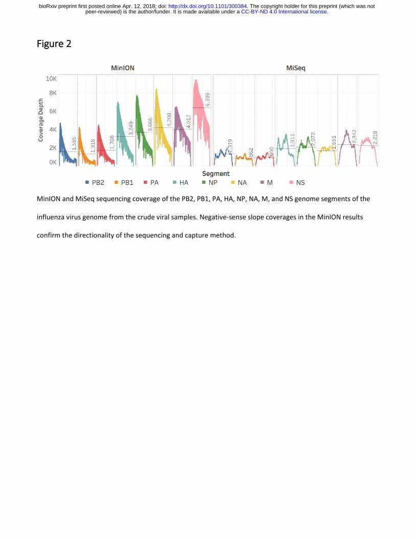
Figure 2
MinION and MiSeq sequencing coverage of the PB2, PB1, PA, HA, NP, NA, M, and NS genome segments of the
influenza virus genome from the crude viral samples. Negative-sense slope coverages in the MinION results
confirm the directionality of the sequencing and capture method.
.CC-BY-ND 4.0 International licensepeer-reviewed) is the author/funder. It is made available under aThe copyright holder for this preprint (which was not. http://dx.doi.org/10.1101/300384doi: bioRxiv preprint first posted online Apr. 12, 2018;

Figure 3
MinION coverage and consensus of the conserved 3’ (Uni-12) and 5’ (Uni-13) termini of the influenza virus RNA
segments from the crude viral samples.
.CC-BY-ND 4.0 International licensepeer-reviewed) is the author/funder. It is made available under aThe copyright holder for this preprint (which was not. http://dx.doi.org/10.1101/300384doi: bioRxiv preprint first posted online Apr. 12, 2018;

Figure 4
The aligned read length distributions correspond to the expected lengths (dashed lines) of the respective
segments (NS 890 nt; M 1,027 nt; NA 1,413 nt; NP 1,565 nt; HA 1,778 nt; PA 2,233 nt; PB1 and PB2 2,341 nt)
from the crude viral samples. As the segment length increases, the read length distribution falls further short of
the expected length, presumably due to RNA degradation. Aligned read lengths include insertion errors,
accounting for the presence of reads larger than the expected value. Due to cases of large insertion errors, 14
total reads longer than 2,500 nucleotides were observed.
.CC-BY-ND 4.0 International licensepeer-reviewed) is the author/funder. It is made available under aThe copyright holder for this preprint (which was not. http://dx.doi.org/10.1101/300384doi: bioRxiv preprint first posted online Apr. 12, 2018;

Table 1
MiSeq MinION
Crude Enolase Crude
Reads 143,572 171,135 ± 26,929 54,353 ± 15,314
Mapped Reads 143,378 169,041 ± 28,741 53,721 ± 15,145
% Mapped 99.9% 98.6 ± 1.7% 98.8 ± 0.1%
Accuracy 99.6% 90.4 ± 0.8% 86.2 ± 0.31%
Insertion 0.30% 1.49 ± 0.02% 1.66 ± 0.01%
Deletion 0.06% 5.4 ± 0.5% 8.2 ± 0.2%
Substitution 0.36% 4.7 ± 0.4% 6.4 ± 0.1%
Consensus ≡ 100% 99.7% ± 0% 98.97 ± 0.01%
Individual MiSeq experiment is compared to MinION experiments of enolase mRNA (technical triplicate) and
influenza vRNA from crude virus (triplicate). Values from triplicate experiments are presented as averages ±
standard deviation.
.CC-BY-ND 4.0 International licensepeer-reviewed) is the author/funder. It is made available under aThe copyright holder for this preprint (which was not. http://dx.doi.org/10.1101/300384doi: bioRxiv preprint first posted online Apr. 12, 2018;

Supplementary Information
Complete genome direct RNA sequencing of influenza A virus
Matthew W. Keller1, Benjamin L. Rambo-Martin2, Malania M. Wilson2, Callie A. Ridenour2, Samuel S. Shepard3, Thomas J. Stark3, Elizabeth B. Neuhaus3, Vivien G. Dugan3, David E. Wentworth3, and John R. Barnes3*
1Oak Ridge Institute of Science and Education (ORISE), Oak Ridge, Tennessee, USA 2 Battelle Memorial Institute, Atlanta, Georgia, USA 3Influenza Division, National Center for Immunization and Respiratory Diseases (NCIRD), Centers for Disease Control and Prevention (CDC), Atlanta, Georgia, USA.
*Address correspondence to [email protected]
.CC-BY-ND 4.0 International licensepeer-reviewed) is the author/funder. It is made available under aThe copyright holder for this preprint (which was not. http://dx.doi.org/10.1101/300384doi: bioRxiv preprint first posted online Apr. 12, 2018;

Figure S1
Average coverage of triplicate enolase direct RNA sequencing experiments. The MinION was able to sequence
enolase mRNA to an average coverage depth of 117,408 ± 9,617 reads. The directional nature of nanopore
sequencing results in a positive slope to the coverage for the mRNA.
.CC-BY-ND 4.0 International licensepeer-reviewed) is the author/funder. It is made available under aThe copyright holder for this preprint (which was not. http://dx.doi.org/10.1101/300384doi: bioRxiv preprint first posted online Apr. 12, 2018;

Figure S2
The aligned read length distribution is longer than the expected length of 1,314 nucleotides (dashed line) due to
insertions.
.CC-BY-ND 4.0 International licensepeer-reviewed) is the author/funder. It is made available under aThe copyright holder for this preprint (which was not. http://dx.doi.org/10.1101/300384doi: bioRxiv preprint first posted online Apr. 12, 2018;

Figure S3
Distribution of mapped reads for all experiment iterations.
.CC-BY-ND 4.0 International licensepeer-reviewed) is the author/funder. It is made available under aThe copyright holder for this preprint (which was not. http://dx.doi.org/10.1101/300384doi: bioRxiv preprint first posted online Apr. 12, 2018;

Figure S4
MinION and MiSeq sequencing coverage of the PB2, PB1, PA, HA, NP, NA, M, and NS genome segments of the
pure influenza virus genome. Negative-sense slope coverages in the MinION results confirm the directionality of
the sequencing and capture method.
.CC-BY-ND 4.0 International licensepeer-reviewed) is the author/funder. It is made available under aThe copyright holder for this preprint (which was not. http://dx.doi.org/10.1101/300384doi: bioRxiv preprint first posted online Apr. 12, 2018;

Figure S5
MiSeq coverage and consensus of the conserved termini of the influenza viral RNA genome segments from the
crude viral samples. These are the amplification sites, and the results are primer dictated sequences.
.CC-BY-ND 4.0 International licensepeer-reviewed) is the author/funder. It is made available under aThe copyright holder for this preprint (which was not. http://dx.doi.org/10.1101/300384doi: bioRxiv preprint first posted online Apr. 12, 2018;

Figure S6
The aligned read length distributions correspond to the expected lengths (dashed lines) of the respective
segments (NS 890 nt; M 1,027 nt; NA 1,413 nt; NP 1,565 nt; HA 1,778 nt; PA 2,233 nt; PB1 and PB2 2,341 nt). As
the segment length increases, the read length distribution falls further short of the expected length, presumably
due to RNA degradation. Aligned read lengths include insertion errors, accounting for the presence of reads
larger than the expected value. Due to cases of large insertion errors, 14 total reads longer than 2,500
nucleotides were observed.
.CC-BY-ND 4.0 International licensepeer-reviewed) is the author/funder. It is made available under aThe copyright holder for this preprint (which was not. http://dx.doi.org/10.1101/300384doi: bioRxiv preprint first posted online Apr. 12, 2018;

Figure S7
QIAxcel size fractionation and visualization of amplicons from (left to right): crude duplicates, negative control,
and pure duplicates. The five visible bands in lanes 1, 2, 4, and 5 are characteristic of uniform amplification of
the entire influenza virus genome. The PB2, PB1, PA, HA, NP, NA, M, and NS genome segments are represented
by only five bands because the three segments that encode the polymerases appear as a single band just larger
than 2,000 and the NP and NA segments appear as a single band just below 1500.
PB2, PB1, PA HA
NP, NA
NS M
Segments:
.CC-BY-ND 4.0 International licensepeer-reviewed) is the author/funder. It is made available under aThe copyright holder for this preprint (which was not. http://dx.doi.org/10.1101/300384doi: bioRxiv preprint first posted online Apr. 12, 2018;

Table S1
Gene & Length Average Length Mode Length
Enolase/Pure Crude Enolase/Pure Crude
Enolase 1,314 975 - 1380 -
PB2 2,341 1,177 752 440/2,240 280/2,240
PB1 2,341 1,061 718 580/2,260 260/2,260
PA 2,233 1,089 833 500/2,160 280/2,140
HA 1,778 1,111 828 1,720 1,720
NP 1,565 1,025 741 1,520 1,520
NA 1,413 980 711 1,420 1,400
M 1,027 809 649 1,020 920/1,020
NS 890 698 607 880 880
Average and mode mapped read length is shown for MinION direct RNA sequencing experiments. The presence
of short reads, particularly in the crude sample, move the average read length much lower than the mode read
length that is displayed here and in figures 4, S2, and S6. The read length distribution of the polymerases are all
bimodal with an abundance of short reads along with full length reads. The read length distribution of M from
the crude viral sample is also bimodal with a clear and well-defined peak at 920 nucleotides in addition to the
full-length peak at 1,020 nucleotides. All Illumina reads were 150 nucleotides in length.
.CC-BY-ND 4.0 International licensepeer-reviewed) is the author/funder. It is made available under aThe copyright holder for this preprint (which was not. http://dx.doi.org/10.1101/300384doi: bioRxiv preprint first posted online Apr. 12, 2018;

Table S2
Name Sequence
RTA-A /5PHOS/GGCTTCTTCTTGCTCTTAGGTAGTAGGTTC
RTA-B GAGGCGAGCGGTCAATTTTCCTAAGAGCAAGAAGAAGCCTTTTTTTTTT
RTA-B-U12 GAGGCGAGCGGTCAATTTTCCTAAGAGCAAGAAGAAGCCAGCAAAAGCAGG
RTA-B-U12.4 GAGGCGAGCGGTCAATTTTCCTAAGAGCAAGAAGAAGCCAGCGAAAGCAGG
Full sequences (5’ to 3’) of the adapters used in this study. Each RTA-B is duplexed with RTA-A. The stock RTA
was supplied with the direct RNA sequencing materials. The modified RTAs were purchased from IDT with each
of the modified RTA-B strands already duplexed to the RTA-A strand. The RTA-A has a 5’ phosphate modification
for ligation. The regions of reverse complementarity between the RTA strands are underlined, and the target
sequences are colored.
.CC-BY-ND 4.0 International licensepeer-reviewed) is the author/funder. It is made available under aThe copyright holder for this preprint (which was not. http://dx.doi.org/10.1101/300384doi: bioRxiv preprint first posted online Apr. 12, 2018;

Table S3
Individual MiSeq experiments are compared to MinION experiments of enolase mRNA (technical triplicate),
influenza vRNA from purified virus (duplicate), and influenza vRNA from crude virus (triplicate). Values from
triplicate experiments are presented as averages ± standard deviation.
MiSeq MinION
Pure Crude Enolase Pure Crude
Reads 163,264 143,572 171,135 ± 26,929 119,860 & 13,848 54,353 ± 15,314
Mapped Reads 163,130 143,378 169,041 ± 28,741 119,350 & 13,721 53,721 ± 15,145
% Mapped 99.9% 99.9% 98.6 ± 1.7% 99.6 & 99.1% 98.8 ± 0.1%
Accuracy 99.6% 99.6% 90.4 ± 0.8% 85.2 & 83.8% 86.2 ± 0.31%
Insertion 0.30% 0.30% 1.49 ± 0.02% 2.15 & 1.85% 1.66 ± 0.01%
Deletion 0.07% 0.06% 5.4 ± 0.5% 8.7 & 9.7% 8.2 ± 0.2%
Substitution 0.32% 0.36% 4.7 ± 0.4% 7.1 & 7.8% 6.4 ± 0.1%
Consensus ≡ 100% ≡ 100% 99.7% ± 0% 98.72 & 98.50% 98.97 ± 0.01%
.CC-BY-ND 4.0 International licensepeer-reviewed) is the author/funder. It is made available under aThe copyright holder for this preprint (which was not. http://dx.doi.org/10.1101/300384doi: bioRxiv preprint first posted online Apr. 12, 2018;

Table S4
Date Sample RNA (ng) Library (ng) Flowcell Pores Occupancy Time
(hours) Yield (Mb)
2017-06-19 Enolase 1 500 180 FAH04399 745 35% 48 180
2017-08-04 Enolase 2 500 133 FAH14471 666 21% 48 197
2017-11-09 Enolase 3 500 32 FAH28081 1,404 27% 48 172
2017-07-13 Pure 1 216 60 FAB49814 656 45% 24 127
2017-09-08 Pure 2 257 254 FAH15505 414 5% 14 14
2017-11-22 Crude 1 200 18 FAH36033 1,102 18% 18 59
2017-11-27 Crude 2 200 15 FAH28243 1,289 10% 16 35
2017-11-28 Crude 3 200 15 FAH36048 1,072 10% 17 34
Input material and sequencing information for the direct RNA sequencing experiments. Pore availability for mux
1-3 is displayed. These data could be recovered by ONT technical support for Enolase 1 or Pure 1. Mux 4 scan
data was not able to be recovered for these samples. The full mux (1/2/3/4) data for the other samples was:
enolase 2 (420/202/44/12), enolase 3 (509/492/403/180), pure 2 (291/110/13/1), crude 1 (483/390/229/58),
crude 2 (506/456/327/134), and crude 3 (449/393/230/53). The mux 4 pores are fewer in number and are used
lastly in long sequencing experiments, if they are used at all. Pore occupancy was estimated by ONT technical
support from QC reports.
.CC-BY-ND 4.0 International licensepeer-reviewed) is the author/funder. It is made available under aThe copyright holder for this preprint (which was not. http://dx.doi.org/10.1101/300384doi: bioRxiv preprint first posted online Apr. 12, 2018;

.CC-BY-ND 4.0 International licensepeer-reviewed) is the author/funder. It is made available under aThe copyright holder for this preprint (which was not. http://dx.doi.org/10.1101/300384doi: bioRxiv preprint first posted online Apr. 12, 2018;

.CC-BY-ND 4.0 International licensepeer-reviewed) is the author/funder. It is made available under aThe copyright holder for this preprint (which was not. http://dx.doi.org/10.1101/300384doi: bioRxiv preprint first posted online Apr. 12, 2018;

.CC-BY-ND 4.0 International licensepeer-reviewed) is the author/funder. It is made available under aThe copyright holder for this preprint (which was not. http://dx.doi.org/10.1101/300384doi: bioRxiv preprint first posted online Apr. 12, 2018;

.CC-BY-ND 4.0 International licensepeer-reviewed) is the author/funder. It is made available under aThe copyright holder for this preprint (which was not. http://dx.doi.org/10.1101/300384doi: bioRxiv preprint first posted online Apr. 12, 2018;

.CC-BY-ND 4.0 International licensepeer-reviewed) is the author/funder. It is made available under aThe copyright holder for this preprint (which was not. http://dx.doi.org/10.1101/300384doi: bioRxiv preprint first posted online Apr. 12, 2018;

.CC-BY-ND 4.0 International licensepeer-reviewed) is the author/funder. It is made available under aThe copyright holder for this preprint (which was not. http://dx.doi.org/10.1101/300384doi: bioRxiv preprint first posted online Apr. 12, 2018;

.CC-BY-ND 4.0 International licensepeer-reviewed) is the author/funder. It is made available under aThe copyright holder for this preprint (which was not. http://dx.doi.org/10.1101/300384doi: bioRxiv preprint first posted online Apr. 12, 2018;

.CC-BY-ND 4.0 International licensepeer-reviewed) is the author/funder. It is made available under aThe copyright holder for this preprint (which was not. http://dx.doi.org/10.1101/300384doi: bioRxiv preprint first posted online Apr. 12, 2018;

.CC-BY-ND 4.0 International licensepeer-reviewed) is the author/funder. It is made available under aThe copyright holder for this preprint (which was not. http://dx.doi.org/10.1101/300384doi: bioRxiv preprint first posted online Apr. 12, 2018;

.CC-BY-ND 4.0 International licensepeer-reviewed) is the author/funder. It is made available under aThe copyright holder for this preprint (which was not. http://dx.doi.org/10.1101/300384doi: bioRxiv preprint first posted online Apr. 12, 2018;

.CC-BY-ND 4.0 International licensepeer-reviewed) is the author/funder. It is made available under aThe copyright holder for this preprint (which was not. http://dx.doi.org/10.1101/300384doi: bioRxiv preprint first posted online Apr. 12, 2018;