comprehensive characterization of sphingolipid ceramide n
TRANSCRIPT
BIOTECHNOLOGICALLY RELEVANT ENZYMES AND PROTEINS
Comprehensive characterization of sphingolipid ceramideN-deacylase for the synthesis and fatty acidremodeling of glycosphingolipids
Yun-Bin Han & Lie Wu & Jamie R. Rich &
Feng-Tao Huang & Stephen G. Withers & Yan Feng &
Guang-Yu Yang
Received: 14 October 2014 /Revised: 14 January 2015 /Accepted: 19 January 2015# Springer-Verlag Berlin Heidelberg 2015
Abstract Sphingolipid ceramide N-deacylase (SCDase) cat-alyzes reversible reactions in which the amide linkage inglycosphingolipids is hydrolyzed or synthesized. WhileSCDases show great value for the enzymatic synthesis ofglycosphingolipids, they are relatively poorly characterizedenzymes. In this work, the enzymatic properties of SCDasefrom Shewanella alga G8 (SA_SCD) were systematicallycharacterized and compared with the commercially availableSCDase from Pseudomonas sp. TK4 (PS_SCD). The optimalpH values for the hydrolytic and synthetic activity ofSA_SCD were pH 6.0 and pH 7.5, respectively. Both activi-ties were strongly inhibited by Zn2+ and Cu2+, while Fe2+,Co2+, Ni2+, Mn2+, Ca2+, and Mg2+ promoted the hydrolyticactivity but inhibited the synthetic activity. SA_SCD showedvery broad substrate specificity both in hydrolysis and synthe-sis. Importantly, SA_SCD has a broader specificity for acyl
donor acceptance than does PS_SCD, especially for unsatu-rated fatty acids and fatty acids with very short or long acylchains. Further kinetic analysis revealed that the kcat/KM valuefor the hydrolytic activity of SA_SCD was 8.9-fold higherthan that of PS_SCD for GM1a, while the values for the syn-thetic activity were 38-fold higher for stearic acid and 23-foldhigher for lyso-GM1a (d18:1) than those of PS_SCD, respec-tively. The broad fatty acid specificity and high catalytic effi-ciency, together with the ease of expression of SA_SCD inEscherichia coli, make it a better biocatalyst than is PS_SCDfor the syn thes i s and s t ruc tura l remode l ing ofglycosphingolipids.
Keywords Glycosphingolipids . High-performance liquidchromatography . Sphingolipid ceramideN-deacylase .
Substrate specificity
Introduction
Glycosphingolipids (GSLs) are a class of amphipathic com-pounds on the cell surface that play extremely important rolesin cellular signaling (Wennekes et al. 2009). They are in-volved in a range of pathological processes (Butters et al.2000; Hakomori 1998; Willison and Yuki 2002) and someGSLs have even shown potential therapeutic effects on cancerand neuronal degenerative diseases (Geisler et al. 1991;Kaneko et al. 2007; Schorsch et al. 2013). However, compre-hensive study of their biological functions and their develop-ment as new drugs have been severely hampered by chal-lenges associated with their large-scale synthesis. While theisolation of GSLs from natural sources is commonly done,
Electronic supplementary material The online version of this article(doi:10.1007/s00253-015-6421-8) contains supplementary material,which is available to authorized users.
Y.<B. Han : L. Wu : F.<T. Huang :Y. Feng (*) :G.<Y. Yang (*)State Key Laboratory of Microbial Metabolism, School of LifeSciences and Biotechnology, Shanghai Jiao Tong University,Shanghai 200240, Chinae-mail: [email protected]: [email protected]
L. WuState Key Laboratory of Electroanalytical Chemistry, ChangchunInstitute of Applied Chemistry, Chinese Academy of Sciences,Changchun 130022, China
J. R. Rich : S. G. WithersDepartment of Chemistry, University of British Columbia,Vancouver, British Columbia V6T 1Z1, Canada
Appl Microbiol BiotechnolDOI 10.1007/s00253-015-6421-8

products generated in this way are subject to potential trans-mission of disease, heterogeneity, and scarcity (Rupčić andMarić 2004). The chemical synthesis of complex GSLs struc-ture requires many activation, coupling, protection, anddeprotection steps, making it extremely challenging and targetspecific (Vankar and Schmidt 2000). Enzymatic assembly ofGSLs, on the other hand, offers significant advantages due totheir much simpler syntheses, milder reaction conditions, andmore facile product purification (Rich et al. 2011; Rich andWithers 2012).
Sphingolipid ceramide N-deacylase (SCDase) is an en-zyme that can serve as a biocatalyst in the enzymatic synthesisof GSLs since it catalyzes the reversible hydrolysis/synthesisof the amide linkage between the fatty acid and the sphingo-sine base in the ceramide moiety of GSLs (Fig. 1a). The syn-thetic activity of SCDase has been used previously for theassembly of various GSLs (Mitsutake et al. 1997, 1998;
Nakagawa et al. 1999, 2005; Kita et al. 2001; Xu et al.2009; Kuchař et al. 2010), while its hydrolytic activity hasbeen used to produce N-deacylated form of GSLs (Kuritaet al. 2000). These so-called lyso-GSLs can serve as interme-diates for the production of pharmaceuticals. For example,lyso-GM1a has been used as an intermediate in the synthesisof LIGA-20, a semi-synthetic ganglioside that showed potentmultimodal neurotrophic effects (Manev et al. 1990;Mocchetti 2005). Therefore, SCDases have attracted attentionrecently for their use in the enzymatic processing of GSLs.
Four SCDase have been reported from Nocardia sp.(Hirabayashi et al. 1988), Pseudomonas sp. (Ito et al. 1995),Streptomyces sp. (Ashida et al. 1995), and Shewanella algaG8 (Furusato et al. 2002), respectively. Among them, only thegenes for the enzymes from Pseudomonas sp. TK4 (PS_SCD)and S. alga G8 (SA_SCD) have been cloned (Furusato et al.2002; Ito et al. 2004), but these two enzymes share no
Fig. 1 a SCDase catalyzes reversible reactions in which the amidelinkage in GSLs is hydrolyzed or synthesized. b The representativeHPLC chromatograms of the assay for the hydrolytic SCDase activityin the absence (dotted line) and in the presence (solid line) of SA_SCD.The reaction mixture was derivatized with OPA and detected using a
fluorescence detector (Ex=340 nm, Em=455 nm). c The representativeHPLC chromatograms of the assay for the synthetic SCDase activity inthe absence (dotted line) and in the presence (solid line) of SA_SCD. Thereaction mixture was detected using a UV detector at 195 nm
Appl Microbiol Biotechnol

sequence homology. PS_SCD is now commercially available;however, production of this enzyme is based on the fermenta-tion of Pseudomonas sp. TK4 since it has not yet been heter-ologously overexpressed. By contrast, SA_SCD can be read-ily overexpressed in Escherichia coli, which opens the possi-bility of mechanistic analysis and enzyme engineeringthrough site-directed mutagenesis (Furusato et al. 2002).However, little is known about the enzymatic properties ofSA_SCD since only its hydrolytic activity has been character-ized, and this only partially, while its synthetic activity has notbeen studied. A more extensive characterization of SA_SCDis therefore needed in order for it to be optimally used in theenzymatic synthesis of GSLs.
In this study, we first describe the development of two fast,practical, and precise HPLC-based methods for assay ofSCDase. We then use these assays to determine the specific-ities and kinetic properties of SA_SCD and compare themwith those of the commercially available PS_SCD. The resultssuggest that SA_SCD has superior properties to those ofPS_SCD in several respects, making it a valuable biocatalystfor manipulation of GSLs.
Materials and methods
Chemicals and enzymes
The sphingolipids were purchased from Avanti Polar Lipids(Alabaster, USA), except for globotetraosylceramide(Gb4Cer), which was purchased from Wako Pure ChemicalIndustries (Osaka, Japan) and GM1a, which was purchasedfrom Qilu Pharmaceutical Co., Ltd. (Jinan, China). Ortho-phthalaldehyde (OPA) was purchased from Sigma-Aldrich(St. Louis, USA). Lyso-GM1a standard and PS_SCD werepurchased from Takara Biotechnology Co., Ltd. (Dalian,China). The lyso-GM1a, lyso-GM3, lyso-sulfatide, lyso-Gb4Cer, and ω-position-modified fatty acids were synthe-sized in our lab, and methods for this will be published else-where. Sep-Pak tC18 reversed phase silica gel cartridges wereobtained from Waters (Milford, USA). HPLC solvents werepurchased from Anpel Co., Ltd. (Shanghai, China). All otherreagents were of the highest purity available.
Protein expression and purification of SA_SCD deletionmutant
The gene encoding the mature SA_SCD protein, which lacksits 38-residue N-terminal secretion signal sequence and fromwhich the 277-residue C-terminal sequence has been deleted,was codon-optimized for E. coli and synthesized (GenscriptCorporation, Nanjing, China). The gene sequence wassubcloned into pET23b vector (Novagen, Madison, USA)using the NdeI/XhoI restriction sites and was transformed into
E. coli BL21 (DE3) pLysS. Transformants were grown at37 °C in Luria-Bertani medium containing 100 μg/mL ampi-cillin until the optical density at 600 nm reached about 0.8.Then, protein expression was induced by the addition of iso-propylβ-D-1-thiogalactopyranoside (IPTG) at a final concen-tration of 0.1 mM at 16 °C for 12 h. The cells were harvestedand disrupted by sonication, and the enzyme was purified byNi2+-chelating affinity chromatography. Protein concentrationwas determined using the bicinchoninic acid protein assaywith BSA as a standard.
HPLC-based assay for SCDase-catalyzed hydrolysis
In a standard assay, the hydrolytic activity of SCDasewas measured using GM1a as substrate. The reactionmixture contained 15 nmol of GM1a and an appropriateamount of the enzyme in 30 μL of 25 mM sodiumacetate buffer (pH 6.0) with 0.1 % Triton X-100.Following incubation at 37 °C for 5 min, the reactionwas stopped by heating in a boiling water bath for5 min. An aliquot (10 μL) of reaction solution was thentaken out, mixed with 20 μL of OPA reagent (7.5 mM),and incubated at 30 °C for 5 min for the derivatization.After centrifugation at 13,000 rpm, the supernatant wastransferred to a glass vial and an aliquot (10 μL) wasinjected onto a reverse-phase HPLC column (ZorbaxSB-C18, 4.6 mm ID×150 mm, 5-μm particle size,Agilent Technologies, Santa Clara, USA) using anauto-sampler (Agilent 1260 ALS) with methanol/H2O(70:30, v/v) as the mobile phase at a flow rate of1.0 mL/min. The OPA-derivatized product was detectedusing a fluorescence detector (Agilent 1260 FLD, Ex=340 nm, Em=455 nm).
HPLC-based assay for SCDase-catalyzed synthesis
In a standard assay, the synthetic activity of SCDasewas measured using lyso-GM1a (d18:1) and stearic acidas the substrates. The reaction mixture, containing anappropriate amount of SCDase, 15 nmol of lyso-GM1a(d18:1), and 15 nmol of stearic acid, was incubated in30 μL of 25 mM Tris-HCl buffer (pH 7.5) containing0.1 % Triton X-100 and 10 % dimethyl sulfoxide(DMSO) at 37 °C for 10 min. Reaction was terminatedby heating in a boiling water bath for 5 min, and thegeneration of GM1a (d18:1/18:0) was measured byHPLC using a reverse-phase column (Zorbax EclipsePlus C18, 4.6 mm ID×100 mm, 3.5-μm particle size,Agilent Technologies, Santa Clara, USA). The mobilephase contained acetonitrile and water (80:20, v/v), plus0.03 % triethylamine, with its pH adjusted to 7.5 usingphosphoric acid. GM1a (d18:1/18:0) was eluted fromthe column at a flow rate of 1.0 mL/min and detected
Appl Microbiol Biotechnol
at a wavelength of 195 nm using a variable wavelengthdetector (Agilent 1260 VWD).
General characterization of SCDase
The effects of pH on the activities of SA_SCDwere measuredacross the pH range from 4.5 to 10.0 using a wide-range pHbuffer containing HEPES, TAPS, CAPS, MES, and aceticacid, each at 40 mM.
The effects of metal ions on the activities of SA_SCD wereassayed in the presence of 5 mM metal cations (FeCl2, CoCl2,NiCl2, CuCl2, MnCl2, ZnCl2, CaCl2, and MgCl2) or EDTA.
The effects of organic solvents on the synthetic activity ofSA_SCD were measured in the presence of organic solvents(DMSO and dimethoxyethane (DME)) at different concentra-tions (v/v).
The effects of detergents on SA_SCD synthetic activitywere measured by the standard method except that TritonX-100, sodium deoxycholate (DOC) or taurodeoxycholate(TDC) were used at different concentrations (v/v). The effectsof Triton X-100 on SA_SCD hydrolytic activity were mea-sured by the standard method except that Triton X-100 wasused at different concentrations (v/v) with the GM1a concen-tration fixed separately at 0.1, 0.5 or 1.0 mM.
In all the reaction mixtures above, 15 ng of enzyme wasused in hydrolytic activity assays, and 30 ng was used insynthetic activity assays. The activity is normalized relativeto the maximal value in each experiment, except the effect ofmetal ions in which the activity was normalized relative to thecontrol with no metal ions added.
Substrate specificities of SCDases
The hydrolytic activities of SCDases against sphingolipidswith different hydrophilic head groups (Fig. 2) were measuredwith 15 ng of SA_SCD or 35 ng of PS_SCD by the standardassay. The substrate specificity towards hydrophilic headgroups was represented by the specific hydrolytic activity.
The synthetic activities of SCDase towards lyso-GSLs con-taining different hydrophilic head groups were determinedusing 15 nmol of lyso-GSL and 30 nmol of stearic acid in30 μL of 25 mM HEPES buffer (pH 7.0) containing 0.1 %Triton X-100 and 10 % DMSO. Reactions were performed at37 °C for 12 h with 30 ng of SA_SCD. The synthetic speci-ficity towards hydrophilic head groups was represented byreaction yield (%), calculated as follows: (peak area for totallyso-GSL−peak area for remaining lyso-GSL)×100/peak ar-ea for total lyso-GSL. The HPLC detection for lyso-GSLsafter OPA derivatization was similar to that for lyso-GM1aexcept that the mobile phase ratio of methanol/H2O was ad-justed according to the polarity of lyso-GSLs.
The fatty acid specificities of SCDases in the syntheticreaction were determined using a variety of fatty acids. The
reaction mixtures containing 15 nmol lyso-GM1a (d18:1) and30 nmol of fatty acid were incubated at 37 °C for 12 h with30 ng of SA_SCD or 70 ng of PS_SCD in 30 μL of 25 mMbuffer (HEPES buffer for SA_SCD or phosphate buffer forPS_SCD), pH 7.0, containing 0.1 % Triton X-100 and 10 %DMSO.
Kinetic analysis of SCDase
For the kinetic analysis of hydrolytic activity, GM1a (0.02–2.0 mM) was incubated with 15 ng SA_SCD or 35 ngPS_SCD for 2 min in 30 μL of 25 mM sodium acetate buffer(pH 6.0) with 0.1 % Triton X-100 under standard conditions.Kinetic analysis of synthetic activity was performed usinglyso-GM1a (d18:1) and stearic acid as substrates in the pres-ence of 30 ng SA_SCD for 3 min or 70 ng PS_SCD for 20minin 30 μL of 25 mMTris-HCl buffer (pH 7.5) containing 0.1 %Triton X-100 and 10 % DMSO under standard conditions.Kinetic parameters for stearic acid were determined usingconcentrations between 0.02 and 2.0 mM at a fixed lyso-GM1a (d18:1) concentration (1 mM). Kinetic parameters forlyso-GM1a (d18:1) were determined using concentrations be-tween 0.06 and 1.0 mM at a fixed stearic acid concentration(1 mM). The parameters KM and kcat were obtained by fittingthe experimental data to the Michaelis-Menten kinetics modelusing Origin 8.0.
Nucleotide sequence accession number
Nucleotide sequence data of SA_SCD deletion mutant hasbeen deposited in the GenBank database under accessionnumber KM986461.
Results
Development of two HPLC assays of the hydrolyticand synthetic activities of SCDase
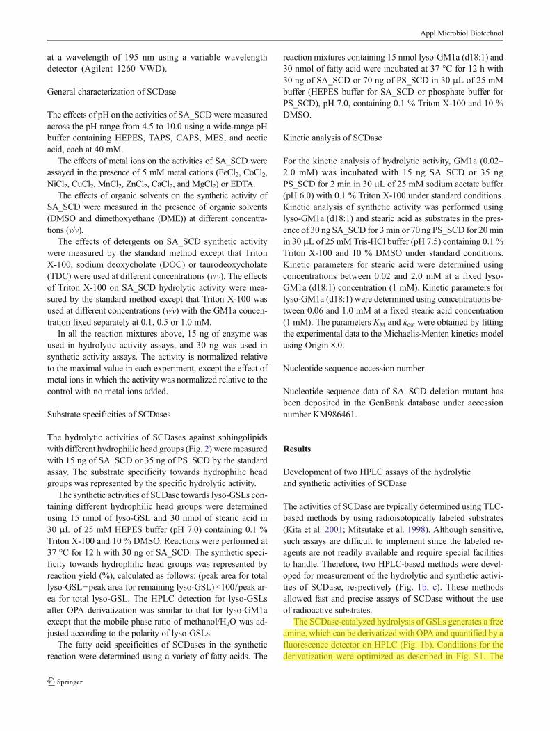
The activities of SCDase are typically determined using TLC-based methods by using radioisotopically labeled substrates(Kita et al. 2001; Mitsutake et al. 1998). Although sensitive,such assays are difficult to implement since the labeled re-agents are not readily available and require special facilitiesto handle. Therefore, two HPLC-based methods were devel-oped for measurement of the hydrolytic and synthetic activi-ties of SCDase, respectively (Fig. 1b, c). These methodsallowed fast and precise assays of SCDase without the useof radioactive substrates.
The SCDase-catalyzed hydrolysis of GSLs generates a freeamine, which can be derivatized with OPA and quantified by afluorescence detector on HPLC (Fig. 1b). Conditions for thederivatization were optimized as described in Fig. S1. The
Appl Microbiol Biotechnol

method is compatible with the analysis of various substrates,including ceramide (Cer), glucosylceramide (GlcCer),galactosylceramide (GalCer), sulfatide, lactosylceramide(LacCer), GM3, GM1a, Gb4Cer, and sphingomyelin (SM)(Fig. S2). This new method has good precision with intra-and inter-assay relative standard deviations (RSDs) all lessthan 5 % (Table S1). The HPLC-based method therefore pro-vides a reliable assay for the hydrolytic activity of SCDase.
The synthetic activity of SCDase can be directly deter-mined by monitoring the amide bond formation at 195 nmusing a UV detector on the HPLC. To achieve good separationand detection of the product, we established a modified ver-sion of the reverse-phase HPLC method reported by Gazzottiet al. (1984): instead of Gazzotti’s LiChrosorb RP-8 column,we employed a reverse-phase Zorbax Eclipse Plus C18 col-umn for HPLC analysis. When using lyso-GM1a (d18:1) andstearic acid as substrates, the product GM1a (d18:1/18:0)could be clearly separated from the substrate and buffer com-ponents using a mobile phase containing acetonitrile and wa-ter (80:20, v/v) with 0.03 % triethylamine, pH 7.5 (Fig. 1c).The RSDs of intra-assay and inter-assay for synthetic activityassay were all less than 5 % (Table S2), indicating that thisnew method also has good precision.
Basic enzymatic properties of SA_SCD
SA_SCDwas found in a marine bacterium, S. algaG8, and itsgenewas cloned by Furusato et al. (2002). It is the only knownSCDase that can be functionally expressed in E. coli.However, it is also a relatively poorly characterized enzyme.The hydrolytic activity of recombinant SA_SCD has been
partially characterized (Furusato et al. 2002), but the syntheticactivity has not been studied in detail. Herein, we thereforesystematically characterized both the hydrolytic and syntheticactivities of SA_SCD, and then compared these results withthose of the commercially available SCDase fromPseudomonas sp. TK4 (PS_SCD) to gain improved insightsinto the catalytic behavior of SCDases.
SA_SCD was expressed in E. coli and purified to homoge-neity before characterization (Fig. S3). The effect of pH on theactivities was investigated over pH range 4.5–10 using a broadrange of buffers (Fig. 3a). The enzyme maintained >80 % ofits hydrolytic activity over a broad pH range from 5.5 to 8.0,with its pH optimum at pH 6.0, which is consistent with theresults reported by Furusato et al. (2002). The synthetic reac-tion performed most efficiently between pH 7.0 and 7.5 anddecreased sharply when the pH dropped below 6.5 or in-creased above pH 7.5.
The effects of chelating agent EDTA and various divalentmetal-ions on SA_SCD were also examined. EDTA slightlyinhibited both the hydrolytic and the synthetic activity. Cu2+
and Zn2+ strongly inhibited both the hydrolytic and the syn-thetic reactions (Fig. 3b). However, Fe2+, Co2+, Ni2+, Mn2+,and Ca2+ activated hydrolysis while significantly inhibitingthe synthesis. Mg2+ activated hydrolysis and had no obviouseffect on the synthesis.
Since most of the fatty acid substrates for the syntheticactivity of SCDase are hydrophobic and do not dissolve wellin aqueous solution, organic co-solvents provide a means tofacilitate the reaction. To identify the optimal conditions forsynthesis, the effects of organic solvents on the synthetic ac-tivity were examined. While no detectable activity was
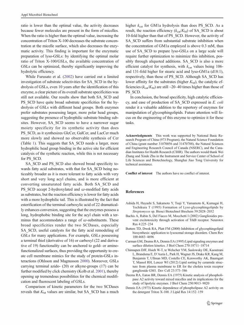
Fig. 2 Sphingolipids withdifferent head groups. Ceramide(a). GlcCer (b). GalCer (c).Sulfatide (d). LacCer (e). GM3(f). GM1a (g). Gb4Cer (h). SM (i)
Appl Microbiol Biotechnol

observable without organic co-solvent, addition of 5 % ofDME or DMSO strongly promoted the activity. Further in-creasing the DME concentrations progressively inhibited syn-thetic activity (Fig. 3c). However, low concentrations ofDMSO, up to a maximum at 10 %, enhanced synthetic activ-ity, while higher concentrations inhibited.
The effects of various detergents on the hydrolytic andsynthetic reactions of SA_SCD were also examined. Ionicdetergents, DOC, and TDC completely inhibited hydrolyticactivity (data not shown). Interestingly, the non-ionic deter-gent Triton X-100 enhanced hydrolysis, but the effectdepended on GM1a concentration (Fig. 3d). For low GM1aconcentration (0.1 mM), the optimum concentration of Triton
X-100 is low (0.1 %, w/v), but higher GM1a concentrations(1.0 mM) require a correspondingly higher concentration ofTriton X-100 (0.5 %,w/v) for the best enhancement. Syntheticactivity proceeded most efficiently in the absence of deter-gents. Addition of Triton X-100, DOC, or TDC inhibited thesynthetic reaction (Fig. 3e): no synthesis occurred when DOCor TDC concentration reached 1 %, while 60 % of the activitywas still observed with 1 % Triton X-100.
Head group specificities of SA_SCD and PS_SCD
The substrate specificity of SCDase is crucial for itspractical applications, but little information is available
Fig. 3 Characterization of therecombinant SA_SCD. a Effectsof pH. b Effects of metal cationand EDTA. c Effects of organicsolvents on the synthetic activityof SA_SCD. d Effects of TritonX-100 on the hydrolytic activityof SA_SCD at different GM1aconcentrations. e Effects ofdetergents on the syntheticactivity of SA_SCD. Valuesrepresent the mean±SD (n=3)
Appl Microbiol Biotechnol

for both SA_SCD and PS_SCD. Therefore, the substratehead group specificities of SA_SCD and PS_SCD werestudied in detail to make a systematic comparison. Forhydrolysis, SA_SCD preferred GSLs with larger sugarmoieties (GM3, GM1a, and Gb4Cer) or charged headgroups (SM and sulfatide) over those with smaller neu-tral sugar moieties (GlcCer, GalCer, and LacCer)(Table 1). PS_SCD also preferred GSLs possessing larg-er sugar moieties, but it did not show preference to-wards GSLs with charged head groups (e.g., SM).Moreover, PS_SCD hydrolyzed sulfatide faster thanGM1a and GM3, whereas SA_SCD hydrolyzed GM1aand GM3 faster. Interestingly, it was apparent thatSA_SCD hydrolyzed GSLs much faster than didPS_SCD, indicating greater efficiency. For example,specific activities of SA_SCD for GM3, GM1a, andSM were 25-, 39-, and 69-fold higher, respectively, thanthose for PS_SCD.
The substrate head group specificities of the SCDasesin the synthetic reaction were also examined (Table 1).While PS_SCD showed similar activity towards variouslyso-GSLs (Kita et al. 2001), SA_SCD preferred sub-strates with more polar head groups, such as lyso-SM(54 %), lyso-GM3 (79 %), and lyso-GM1a (81 %),while yields were low for Gal-Sph (20 %), Glc-Sph(39 %), and Lac-Sph (30 %). SA_SCD barely catalyzedthe formation of ceramide by condensation of stearicacid to Sph, whereas reaction yields of up to 64.4 %were reported previously for PS_SCD (Kita et al. 2001).The distinct substrate specificities of these two enzymesimply different enzyme-substrate binding modes.
Fatty acid specificities of SA_SCD and PS_SCD
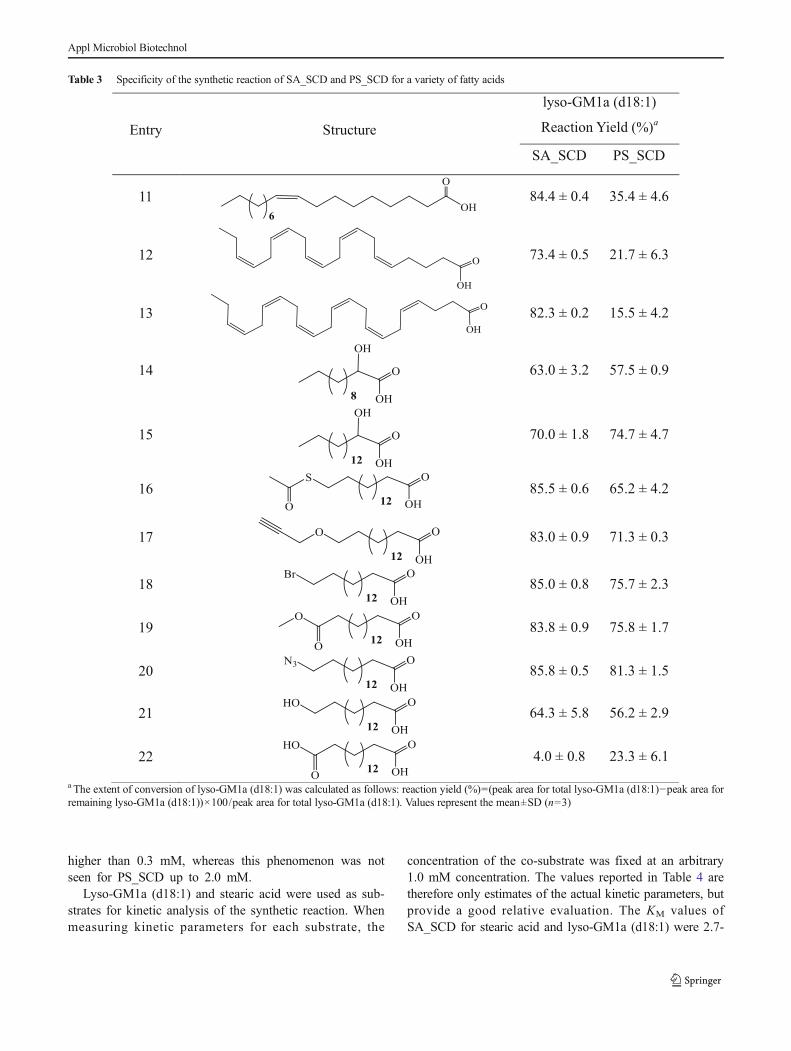
The fatty acid moieties of GSLs can significantly affecttheir physiological properties (Xu et al . 2009;Chinnapen et al. 2012; Manev et al. 1990). Therefore,remodeling of the fatty acid moieties of GSLs may helpin understanding their structure/function relationshipsand in facilitating the development of GSLs with newfunctions. Despite the obvious potential of SCDases inthe enzymatic synthesis of GSLs, their ability to utilizedifferent fatty acids hasn’t been studied in detail. Wetherefore systematically determined the substrate speci-ficities of SA_SCD and PS_SCD towards fatty acidsand their analogs, and thereby generated a library ofGM1a derivatives. In these studies, lyso-GM1a (d18:1)was used as the acceptor substrate, and generation ofthe corresponding GSLs was confirmed by mass spec-trometry (Table S3). SA_SCD accepted all ten saturatedfatty acids tested as substrates, with reaction yieldsranging from 30 to 86 % (Table 2). By contrast, onlythe fatty acids with medium acyl chain lengths (C14–C20) gave comparable yields with PS_SCD: when theacyl chain is shorter than 14 or longer than 20, theconversion for PS_SCD is much lower than that forSA_SCD. Indeed, PS_SCD barely reacted with caproicacid (1) and lignoceric acid (10), while SA_SCDachieved ~30 and 55 % conversion, respectively.SA_SCD also showed much higher activity towards un-saturated fatty acids than did PS_SCD (Table 3). Oleicacid (11), eicosapentaenoic acid (EPA, 12), anddocosahexaenoic acid (DHA, 13) can all be efficiently
Table 1 Specificities of the hydrolytic and synthetic reactions of SA_SCD and PS_SCD for hydrophilic head groups
Name Structure Hydrolysis (nmol/min/μg)a Synthesis (%)
SA_SCD PS_SCD SA_SCDb PS_SCDc
Cer Ceramide (d18:1/18:0) 0.20±0.01 0.04±0.01 0.0 64.4
GlcCer Glcβ1-1′Cer 1.46±0.1 0.31±0.01 39.0±9.4 75.5
GalCer Galβ1-1′Cer 2.01±0.03 0.26±0.02 20.1±7.3 76.8
Sulfatide HSO3-3Galβ1-1′Cer 7.36±0.4 0.88±0.01 –d 61.8
LacCer Galβ1-4Glcβ1-1′Cer 2.23±0.2 0.23±0.01 29.9±3.3 62.7
GM3 NeuAcα2-3Galβ1-4Glcβ1-1′Cer 20.9±0.8 0.84±0.02 78.8±0.9 –
GM1a Galβ1-3GalNAcβ1-4(NeuAcα2-3)Galβ1-4Glcβ1-1′Cer 28.7±0.8 0.73±0.02 81.3±0.3 65.5
Gb4Cer GalNAcβ1-3Galα1-4Galβ1-4Glcβ1-1′Cer 38.9±2.0 1.93±0.03 –d 49.1
SM Choline phosphate-Cer 8.98±0.3 0.13±0.01 54.2±2.0 60.5
a The hydrolytic specificity towards hydrophilic head groups is represented by the specific hydrolytic activities towards various GSLs. Values representthe mean±SD (n=3)b The synthetic specificity towards hydrophilic head groups is represented by the reaction yield (%), calculated as follows: (peak area for total lyso-GSL−peak area for remaining lyso-GSL)×100/peak area for total lyso-GSL. Values represent the mean±SD (n=3)c The data were taken from Kita et al. (2001)d The substrates were not tested
Appl Microbiol Biotechnol

utilized by SA_SCD with greater than 70 % conversion,whereas conversion by PS_SCD was much lower (16–35 %). Both enzymes efficiently used various 2-hydroxylated and ω-position-modified fatty acids, butthe reaction efficiency decreased for fatty acidspossessing a more polar terminal group (Table 3). Forexample, the reaction yield for 16-hydroxyhexadecanoicacid (21) is lower than that for palmitic acid (6), whilehexadecanedioic acid (22) was processed with an evenlower yield. However, efficiency increased significantly(up to 85.5 %) when the terminal carboxyl group wasprotected as its methyl ester (19).
Kinetic parameters of SA_SCD and PS_SCD
Apparent kinetic parameters for hydrolytic and syntheticactivities of SA_SCD and PS_SCD were determined(Table 4). The kinetic parameters for the hydrolytic reac-tion are termed apparent because the GM1a used in thisassay is a mixture of GM1a (d18:1/18:0) and GM1a(d20:1/18:0). The KM for the hydrolytic reaction was sim-ilar for the two enzymes, while the kcat and the kcat/KM ofSA_SCD for GM1a are 8.2- and 8.9-fold higher thanthose of PS_SCD (Table 4). Substrate inhibition was ob-served for SA_SCD when the GM1a concentration was
Table 2 Specificity of the synthetic reaction of SA_SCD and PS_SCD for saturated fatty acids
Entry Structure
Lyso-GM1a (d18:1)
Reaction yield (%)a
SA_SCD PS_SCD
1 29.7 ± 6.1 0
2 64.3 ± 2.5 17.8 ± 4.1
3 77.9 ± 1.4 50.7 ± 1.4
4 84.8 ± 0.7 67.2 ± 1.3
5 86.1 ± 0.3 81.7 ± 1.5
6 85.9 ± 0.4 85.2 ± 2.2
7 83.5 ± 1.5 82.8 ± 2.4
8 82.5 ± 1.1 76.9 ± 4.7
9 80.7 ± 0.9 36.4 ± 5.9
10 54.5 ± 0.7 9.2 ± 6.2
a The extent of conversion of lyso-GM1a (d18:1) was calculated as follows: reaction yield (%)=(peak area for total lyso-GM1a (d18:1)−peak area forremaining lyso-GM1a (d18:1))×100/peak area for total lyso-GM1a (d18:1). Values represent the mean±SD (n=3)
Appl Microbiol Biotechnol
higher than 0.3 mM, whereas this phenomenon was notseen for PS_SCD up to 2.0 mM.
Lyso-GM1a (d18:1) and stearic acid were used as sub-strates for kinetic analysis of the synthetic reaction. Whenmeasuring kinetic parameters for each substrate, the
concentration of the co-substrate was fixed at an arbitrary1.0 mM concentration. The values reported in Table 4 aretherefore only estimates of the actual kinetic parameters, butprovide a good relative evaluation. The KM values ofSA_SCD for stearic acid and lyso-GM1a (d18:1) were 2.7-
Table 3 Specificity of the synthetic reaction of SA_SCD and PS_SCD for a variety of fatty acids
Entry Structure
lyso-GM1a (d18:1)
Reaction Yield (%)a
SA_SCD PS_SCD
11 84.4 ± 0.4 35.4 ± 4.6
12 73.4 ± 0.5 21.7 ± 6.3
13 82.3 ± 0.2 15.5 ± 4.2
14 63.0 ± 3.2 57.5 ± 0.9
15 70.0 ± 1.8 74.7 ± 4.7
16 85.5 ± 0.6 65.2 ± 4.2
17 83.0 ± 0.9 71.3 ± 0.3
18 85.0 ± 0.8 75.7 ± 2.3
19 83.8 ± 0.9 75.8 ± 1.7
20 85.8 ± 0.5 81.3 ± 1.5
21 64.3 ± 5.8 56.2 ± 2.9
22 4.0 ± 0.8 23.3 ± 6.1
a The extent of conversion of lyso-GM1a (d18:1) was calculated as follows: reaction yield (%)=(peak area for total lyso-GM1a (d18:1)−peak area forremaining lyso-GM1a (d18:1))×100/peak area for total lyso-GM1a (d18:1). Values represent the mean±SD (n=3)
Appl Microbiol Biotechnol
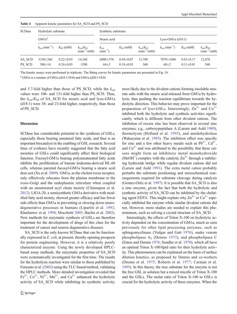
and 5.7-fold higher than those of PS_SCD, while the kcatvalues were 106- and 131-fold higher than PS_SCD. Thus,the kcat/KM of SA_SCD for stearic acid and lyso-GM1a(d18:1) were 38- and 23-fold higher, respectively, than thoseof PS_SCD.
Discussion
SCDase has considerable potential in the synthesis of GSLs,especially those bearing unnatural fatty acids, and thus is animportant biocatalyst in the enabling of GSL research. Severallines of evidence have recently suggested that the fatty acidmoieties of GSLs could significantly affect their biologicalfunction. Fucosyl-GM1a bearing polyunsaturated fatty acidsinhibits the proliferation of human leukemia-derived HL-60cells, whereas parental fucosyl-GM1a bearing a stearic aciddoes not (Xu et al. 2009). GM1a, as the cholera toxin receptor,only effectively relocates from the plasma membrane to thetrans-Golgi and the endoplasmic reticulum when coupledwith an unsaturated acyl chain moiety (Chinnapen et al.2012). LIGA-20, a semisynthetic GM1a derivative with mod-ified fatty acid moiety, showed greater efficacy and has fewerside effects than GM1a in preventing or slowing down neuro-degenerative processes in humans (Lipartiti et al. 1992;Kharlamov et al. 1994; Mocchetti 2005; Bachis et al. 2002).New methods for enzymatic synthesis of GSLs are thereforeimportant for the development of drugs of this class for thetreatment of cancer and neuron degenerative diseases.
SA_SCD is the only known SCDase that can be function-ally expressed in E. coli, at present, thereby opening prospectsfor protein engineering. However, it is a relatively poorlycharacterized enzyme. Using the newly developed HPLC-based assay methods, the enzymatic properties of SA_SCDwere systematically investigated for the first time. The resultsfor the hydrolysis reaction were similar to those published byFurusato et al. (2002) using the TLC-basedmethod, validatingthe HPLC methods. More detailed investigation revealed thatFe2+, Co2+, Ni2+, Mn2+, and Ca2+ enhanced the hydrolyticactivity of SA_SCD while inhibiting its synthetic activity,
most likely due to the divalent cations forming insoluble stea-rate salts with the stearic acid released from GM1a by hydro-lysis, thus pushing the reaction equilibrium towards the hy-drolytic direction. This behavior may prove important for thepreparation of lyso-GSLs. Interestingly, Zn2+ and Cu2+
inhibited both the hydrolytic and synthetic activities signifi-cantly, which is different from other divalent cations. Theinhibition of excess zinc has been observed in several zincenzymes, e.g., carboxypeptidase A (Larsen and Auld 1989),thermolysin (Holland et al. 1995), and amidohydrolase(Wakayama et al. 1995). The inhibition effect was specificfor zinc and a few other heavy metals such as Pb2+, Cd2+,and Cu2+ and was attributed to the possibility that these cat-ions might form an inhibitory metal monohydroxide(MeOH+) complex with the catalytic Zn2+ through a stabiliz-ing hydroxide bridge while regular divalent cations did not(Larsen and Auld 1991). The extra metal cation probablyperturbs the substrate positioning and stereochemical rear-rangements required for substrate cleavage during catalysis(Gomez-Ortiz et al. 1997). It is possible that SA_SCD is alsoa zinc enzyme, given the fact that both the hydrolytic andsynthetic activity of SA_SCD can be inhibited by the chelat-ing agent EDTA. This might explain why Zn2+ or Cu2+ espe-cially inhibited the enzyme while similar divalent cations didnot. However, more studies are needed to explain this phe-nomenon, such as solving a crystal structure of SA_SCD.
Interestingly, the effects of Triton X-100 on hydrolytic ac-tivity depended on the concentration of GM1a, much as seenpreviously for other lipid processing enzymes, such assphingomyelinase (Yedgar and Gatt 1976), snake venomphospholipase A2 (Dennis 1973), and phospholipase C(Eaton and Dennis 1976; Sundler et al. 1978), which all havean optimal Triton X-100/lipid ratio for their hydrolytic activ-ity. This phenomenon can be explained on the basis of surfacedilution kinetics, as proposed by Dennis and co-workers(Deems et al. 1975; Roberts et al. 1977; Carman et al.1995). In this theory, the true substrate for the enzyme is notthe free GSL in solution but a mixed micelle of Triton X-100and the GSLs. The molar ratio of Triton X-100 to GSLs iscrucial for the hydrolytic activity of these enzymes. When the
Table 4 Apparent kinetic parameters for SA_SCD and PS_SCD
SCDase Hydrolytic substrate Synthetic substrates
GM1aa Stearic acid Lyso-GM1a (d18:1)
kcat (min−1) KM (mM) kcat/KM
(min−1/mM)kcat(min−1)
KM (mM) kcat/KM
(min−1/mM)kcat (min
−1) KM (mM) kcat/KM
(min−1/mM)
SA_SCD 3130±260 0.22±0.03 14,160 6800±370 0.49±0.07 13,780 7870±1040 0.63±0.17 12,470
PS_SCD 380±14 0.24±0.03 1590 64±3 0.18±0.03 360 60±2 0.11±0.01 540
The kinetic assays were performed in triplicate. The fitting curves for kinetic parameters are presented in Fig. S4aGM1a is a mixture of GM1a (d18:1/18:0) and GM1a (d20:1/18:0)
Appl Microbiol Biotechnol
ratio is lower than the optimal value, the activity decreasesbecause fewer molecules are present in the form of micelles.When the ratio is higher than the optimal value, increasing theconcentration of Triton X-100 decreases the substrate concen-tration at the micelle surface, which also decreases the enzy-matic activity. This finding is important for the enzymaticpreparation of lyso-GSLs: by identifying the optimal molarratio of Triton X-100/GSLs, the available concentration ofGSLs can be optimized, thereby significantly improving thehydrolytic efficiency.
While Furusato et al. (2002) have carried out a limitedinvestigation of substrate selectivities for SA_SCD in the hy-drolysis of GSLs, even 10 years after the identification of thisenzyme, a clear picture of its overall substrate specificities wasstill not available. Our results show that both SA_SCD andPS_SCD have quite broad substrate specificities for the hy-drolysis of GSLs with different head groups. Both enzymesprefer substrates possessing larger, more polar head groups,suggesting the presence of hydrophilic substrate binding sub-sites. However, SA_SCD seems to have a narrower sugarmoiety specificity for its synthetic activity than doesPS_SCD, as it synthesizes GlcCer, GalCer, and LacCer muchmore slowly and showed no observable synthesis of Cer(Table 1). This suggests that SA_SCD needs a larger, morehydrophilic head group binding in the active site for efficientcatalysis of the synthetic reaction, while this is not necessaryfor PS_SCD.
SA_SCD and PS_SCD also showed broad specificity to-wards fatty acid substrates, with that for SA_SCD being no-ticeably broader as it is more tolerant to fatty acids with veryshort and very long acyl chains, and is more efficient atconverting unsaturated fatty acids. Both SA_SCD andPS_SCD accept 2-hydroxylated and ω-modified fatty acidsas substrates, but the reaction efficiency is lower for fatty acidswith a more hydrophilic tail. This is illustrated by the fact thatesterification of the terminal carboxylic acid of 22 dramatical-ly enhances conversion, suggesting that the enzymes possess along, hydrophobic binding site for the acyl chain with a ter-minus that accommodates a range of ω-substituents. Thesebroad specificities render the two SCDases, especiallySA_SCD, useful catalysts for the fatty acid remodeling ofGSLs for many applications. For example, GSLs possessinga terminal thiol (derivative of 16) or carboxyl (22 and deriva-tive of 19) functionality can be anchored to gold- or amino-functionalized surfaces, thus providing the opportunity to cre-ate cell membrane mimics for the study of protein-GSLs in-teractions (Ohlsson and Magnusson 2000). Moreover, GSLscarrying terminal azido- (20) or alkyne-groups (17) can befurther modified by click chemistry (Kolb et al. 2001), therebyopening up tremendous possibilities for the chemical modifi-cation and fluorescent labeling of GSLs.
Comparison of kinetic parameters for the two SCDasesreveals that KM values are similar, but SA_SCD has a much
higher kcat for GM1a hydrolysis than does PS_SCD. As aresult, the reaction efficiency (kcat/KM) of SA_SCD is about10-fold higher than that of PS_SCD. However, the activity ofSA_SCD suffers from substantial substrate inhibition whenthe concentration of GM1a employed is above 0.3 mM, thususe of SA_SCD to prepare lyso-GSLs on a large scale willrequire further optimization to minimize this inhibition, pos-sibly through aliquoted additions. SA_SCD is also a moreefficient catalyst for synthesis, with kcat values being 106-and 131-fold higher for stearic acid and lyso-GM1a (d18:1),respectively, than those of PS_SCD. Although SA_SCD haslower affinity for the substrates (higher KM), the catalytic ef-ficiencies (kcat/KM) are still ~20–40 times higher than those ofPS_SCD.
In conclusion, the broad specificity, high catalytic efficien-cy, and ease of production of SA_SCD expressed in E. colirender it a valuable addition to the repertory of enzymes formanipulation of glycosphingolipids. Future attention will fo-cus on the engineering of this enzyme to optimize it for thesetasks.
Acknowledgments This work was supported by National Basic Re-search Program of China (973 Program), the Natural Science Foundationof China (grant number 31070056 and 31470788), the Natural Sciencesand Engineering Research Council of Canada (NSERC), and the Cana-dian Institutes for Health Research (CIHR). The authors would thankWeiZhang and Xinde Zhu in the Instrument and Service Center of School ofLife Sciences and Biotechnology, Shanghai Jiao Tong University fortechnical assistance.
Conflict of interest The authors have no conflict of interest.
References
Ashida H, Hayashi S, Sakamoto Y, Tsuji Y, Yamamoto K, Kumagai H,Tochikura T (1995) Formation of Lyso-glycosphingolipids byStreptomyces sp. Biosci Biotechnol Biochem 59:2028–2032
Bachis A, Rabin S, Del Fiacco M, Mocchetti I (2002) Gangliosides pre-vent excitotoxicity through activation of TrkB receptor. NeurotoxRes 4:225–234
Butters TD, Dwek RA, Platt FM (2000) Inhibition of glycosphingolipidbiosynthesis: application to lysosomal storage disorders. Chem Rev100:4683–4696
CarmanGM, DeemsRA, Dennis EA (1995) Lipid signaling enzymes andsurface dilution kinetics. J Biol Chem 270:18711–18714
Chinnapen DJF, Hsieh W-T, te Welscher YM, Saslowsky DE, KaoutzaniL, Brandsma E, D’Auria L, Park H,Wagner JS, Drake KR, KangM,Benjamin T, Ullman MD, Costello CE, Kenworthy AK, BaumgartT, Massol RH, Lencer WI (2012) Lipid sorting by ceramide struc-ture from plasma membrane to ER for the cholera toxin receptorganglioside GM1. Dev Cell 23:573–586
Deems RA, Eaton BR, Dennis EA (1975) Kinetic analysis of phospholi-pase A2 activity toward mixed micelles and its implications for thestudy of lipolytic enzymes. J Biol Chem 250:9013–9020
Dennis EA (1973) Kinetic dependence of phospholipase A2 activity onthe detergent Triton X-100. J Lipid Res 14:152–159
Appl Microbiol Biotechnol

Eaton BR, Dennis EA (1976) Analysis of phospholipase C (Bacilluscereus) action toward mixed micelles of phospholipid and surfac-tant. Arch Biochem Biophys 176:604–609
Furusato M, Sueyoshi N, Mitsutake S, Sakaguchi K, Kita K, Okino N,Ichinose S, Omori A, Ito M (2002) Molecular cloning and charac-terization of sphingolipid ceramide N-deacylase from a marine bac-terium, Shewanella alga G8. J Biol Chem 277:17300–17307
Gazzotti G, Sonnino S, Ghidoni R, Kirschner G, Tettamanti G (1984)Analytical and preparative high-performance liquid chromatogra-phy of gangliosides. J Neurosci Res 12:179–192
Geisler FH, Dorsey FC, ColemanWP (1991) Recovery of motor functionafter spinal-cord injury—a randomized, placebo-controlled trialwith GM-1 ganglioside. N Engl J Med 324:1829–1838
Gomez-Ortiz M, Gomis-Rüth FX, Huber R, Avilés FX (1997) Inhibitionof carboxypeptidase A by excess zinc: analysis of the structuraldeterminants by X-ray crystallography. FEBS Lett 400:336–340
Hakomori S (1998) Cancer-associated glycosphingolipid antigens: theirstructure, organization, and function. Cells Tissues Organs 161:79–90
Hirabayashi Y, Kimura M, Matsumoto M, Yamamoto K, Kadowaki S,Tochikura T (1988) A novel glycosphingolipid hydrolyzing en-zyme, glycosphingolipid ceramide deacylase, which cleaves thelinkage between the fatty acid and sphingosine base inglycosphingolipids. J Biochem 103:1–4
Holland DR, Hausrath AC, Juers D, Matthews BW (1995) Structuralanalysis of zinc substitutions in the active site of thermolysin.Protein Sci 4:1955–1965
Ito M, Kurita T, Kita K (1995) A novel enzyme that cleaves the N-acyllinkage of ceramides in various glycosphingolipids as well assphingomyelin to produce their lyso forms. J Biol Chem 270:24370–24374
Ito M, Kurita T, Kita K, Sueyoshi N, Mitsutake S, Fujita M, Okino N, IzuH, Kato I (2004) Sphingolipid ceramide N-deacylase, methods forproducing sphingolipids and sphingolipid derivatives, andsphingolipid ceramide N-deacylase gene. US Patent 6821761:B2
Kaneko M, Yamada K, Miyamoto T, Inagaki M, Higuchi R (2007)Neuritogenic activity of gangliosides from echinoderms and theirstructure-activity relationship. Chem Pharm Bull 55:462–463
Kharlamov A, Zivkovic I, Polo A, Armstrong D, Costa E, Guidotti A(1994) LIGA20, a lyso derivative of ganglioside GM1, given orallyafter cortical thrombosis reduces infarct size and associated cogni-tion deficit. Proc Natl Acad Sci U S A 91:6303–6307
Kita K, Kurita T, ItoM (2001) Characterization of the reversible nature of thereaction catalyzed by sphingolipid ceramideN-deacylase—a novel formof reverse hydrolysis reaction. Eur J Biochem 268:592–602
Kolb HC, FinnMG, Sharpless KB (2001) Click chemistry: diverse chem-ical function from a few good reactions. Angew Chem Int Ed Engl40:2004–2021
Kuchař L, Rotková J, Asfaw B, Lenfeld J, Horák D, Korecká L, BílkováZ, Ledvinová J (2010) Semisynthesis of C17:0 isoforms ofsulphatide and glucosylceramide using immobilised sphingolipidceramide N-deacylase for application in analytical mass spectrome-try. Rapid Commun Mass Spectrom 24:2393–2399
Kurita T, Izu H, SanoM, ItoM, Kato I (2000) Enhancement of hydrolyticactivity of sphingolipid ceramide N-deacylase in the aqueous-organic biphasic system. J Lipid Res 41:846–851
Larsen KS, Auld DS (1989) Carboxypeptidase A: mechanism of zincinhibition. Biochemistry 28:9620–9625
Larsen KS, Auld DS (1991) Characterization of an inhibitory metal bind-ing site in carboxypeptidase A. Biochemistry 30:2613–2618
Lipartiti M, Lazzaro A, Manev H (1992) Ganglioside derivative LIGA20reduces NMDA neurotoxicity in neonatal rat brain. NeuroReport 3:919–921
Manev H, Favaron M, Vicini S, Guidotti A, Costa E (1990) Glutamate-induced neuronal death in primary cultures of cerebellar granule
cells: protection by synthetic derivatives of endogenoussphingolipids. J Pharmacol Exp Ther 252:419–427
Mitsutake S, Kita K, Okino N, Ito M (1997) [14C]Ceramide synthesis bysphingolipid ceramide N-Deacylase: new assay for ceramidase ac-tivity detection. Anal Biochem 247:52–57
Mitsutake S, Kita K, Nakagawa T, Ito M (1998) Enzymatic synthesis of14C-glycosphingolipids by reverse hydrolysis reaction ofs p h i n go l i p i d c e r am i d e N - d e a cy l a s e : d e t e c t i o n o fendoglycoceramidase activity in a seaflower. J Biochem 123:859–863
Mocchetti I (2005) Exogenous gangliosides, neuronal plasticity and re-pair, and the neurotrophins. Cell Mol Life Sci 62:2283–2294
Nakagawa T, Tani M, Kita K, Ito M (1999) Preparation of fluorescence-labeled GM1 and sphingomyelin by the reverse hydrolysis reactionof sphingolipid ceramide N-Deacylase as substrates for assay ofsphingolipid-degrading enzymes and for detection of sphingolipid-binding proteins. J Biochem 126:604–611
Nakagawa T, Morotomi A, Tani M, Sueyoshi N, Komori H, Ito M (2005)C18:3-GM1a induces apoptosis in Neuro2a cells: enzymatic remod-eling of fatty acyl chains of glycosphingolipids. J Lipid Res 46:1103–1112
Ohlsson J, Magnusson G (2000) Analogues of glycosphingolipids andglycerolipids suitable for conjugation to gold- and amino-functionalised surfaces. Tetrahedron 56:9975–9984
Rich JR, Withers SG (2012) A chemoenzymatic total synthesis ofthe neurogenic starfish ganglioside LLG-3 using anengineered and evolved synthase. Angew Chem Int Ed Engl51:8640–8643
Rich JR, Cunningham A-M, Gilbert M, Withers SG (2011)Glycosphingolipid synthesis employing a combination of recombi-nant glycosyltransferases and an endoglycoceramidaseglycosynthase. Chem Commun 47:10806–10808
Roberts MF, Deems RA, Dennis EA (1977) Dual role of interfacial phos-pholipid in phospholipase A2 catalysis. Proc Natl Acad Sci U S A74:1950–1954
Rupčić J, MarićV (2004) Cerebrosides ofCandida lipolytica yeast. ApplMicrobiol Biotechnol 64:416–420
Schorsch C, Boles E, Schaffer S (2013) Biotechnological production ofsphingoid bases and their applications. Appl Microbiol Biotechnol97:4301–4308
Sundler R, Alberts AW, Vagelos PR (1978) Enzymatic properties ofphosphatidylinositol inositolphosphohydrolase from Bacilluscereus. Substrate dilution in detergent-phospholipid micelles andbilayer vesicles. J Biol Chem 253:4175–4179
Vankar YD, Schmidt RR (2000) Chemistry of glycosphingolipids—car-bohydrate molecules of biological significance. Chem Soc Rev 29:201–216
WakayamaM, Miura Y, Oshima K, Sakai K, Moriguchi M (1995) Metal-characterization of N-Acyl-D-glutamate Amidohydrolase fromPseudomonas sp. Strain 5f-l. Biosci Biotechnol Biochem 59:1489–1492
Wennekes T, van den Berg RJBHN, Boot RG, van der Marel GA,Overkleeft HS, Aerts JMFG (2009) Glycosphingolipids-nature,function, and pharmacological modulation. Angew Chem Int EdEngl 48:8848–8869
Willison HJ, Yuki N (2002) Peripheral neuropathies and anti-glycolipidantibodies. Brain 125:2591–2625
Xu X, Goda HM, Inagaki M, Okino N, Ito M (2009) Enzymatic remod-eling of fatty acid molecules by SCDase demonstrated that FucosylGM1a possessing a polyunsaturated fatty acid induces apoptosis inHL60 human promyelocytic leukemia cells. J Fac Kyushu Univ 54:433–437
Yedgar S, Gatt S (1976) Effect of Triton X-100 on the hydrolysis ofsphingomyelin by sphingomyelinase of rat brain. Biochemistry 15:2570–2573
Appl Microbiol Biotechnol