further characterization of mammalian ceramide kinase ... · further characterization of mammalian...
TRANSCRIPT

Further characterization of mammalian ceramide kinase:
substrate delivery and (stereo)specificity, tissue
distribution, and subcellular localization studies
Helena Van Overloop, Sofie Gijsbers,1 and Paul P. Van Veldhoven2
Katholieke Universiteit Leuven, Faculteit Geneeskunde, Departement Moleculaire Celbiologie,Afdeling Farmacologie, Leuven, Belgium
Abstract Recombinant human ceramide kinase (HsCERK)was analyzed with regard to dependence on divalent cationsand to substrate delivery, spectrum, specificity, and stereo-selectivity. Depending on the chain length of the ceramide,either albumin for short-chain ceramide or a mixed micellarform (octylglucoside/cardiolipin) for long-chain ceramidewas preferred for the substrate delivery, the former re-sulting in higher activities. Bacterially expressed HsCERKwas highly dependent onMg21 ions, much less on Ca21 ions.A clear preference for the D-erythro isomer was seen. VariousN-acylated amino alcohols were no substrate, but N-hexanoyl-1-O-hexadecyl-2-desoxy-2-amino-sn-glycerol and N-tetradecanoyl-2S-amino-1-butanol were phosphorylated, sug-gesting that the secondary hydroxy group is not required forrecognition. The properties of HsCERK, expressed in CHOcells, were similar to those of the bacterially expressedprotein, including the Mg21 dependence. In mouse, thehighest activities were found in testis and cerebellum, andupon subcellular fractionation the activity was recoveredmainly in the microsomal fraction. This fits with the plasmamembrane localization in CHO cells, which was mediated bythe N-terminal putative pleckstrin domain. No evidencefor phosphorylation of ceramide by the recently describedmultiple lipid kinase was found. The latter kinase is lo-calized in the mitochondria, but no firm conclusions withregard to its substrate could be drawn. —Van Overloop, H.,S. Gijsbers, and P. P. Van Veldhoven. Further characteriza-tion of mammalian ceramide kinase: substrate delivery and(stereo)specificity, tissue distribution, and subcellular lo-calization studies. J. Lipid Res. 2006. 47: 268–283.
Supplementary key words N-acyl serinol . sphingosine . lipid kinase .
ceramide phosphate . pleckstrin . testis . anandamide
Sphingolipids, ubiquitous constituents of eukaryoticcells, are important signaling molecules with essentialroles in cell proliferation, differentiation, and apoptosis (1).Ceramide, the initial product of the sphingomyelin cycle,functions as a second messenger in a variety of biological
processes, such as cell differentiation and apoptosis (2).Ceramide can be hydrolyzed by ceramidase(s) to producesphingenine. Ceramide and sphingenine can be phosphory-lated to ceramide-1-phosphate (Cer1P) and sphingenine-1-phosphate (SeP), respectively. The phosphorylated lipidsinfluence various biological processes and are recognizedby specific phosphatases (3, 4). The responsible kinases,ceramide kinase (CERK) (5) and sphingosine kinase [type 1(6–9) and type 2 (10)], belong to a family of kinases relatedto diacylglycerol kinases.
Cer1P was initially described as the product of a Ca21-stimulated kinase that copurified with brain synapticvesicles (11). A regulatory function in the secretion ofneurotransmitters, by promoting membrane fusion, wassuggested. Phagocytosis of IgG-coated erythrocytes byhuman neutrophils was found to be associated with anincrease of Cer1P (12). A mitogenic, as well as an anti-apoptotic, effect was also attributed to Cer1P (13, 14). Byinhibition of acid sphingomyelinase, leading to an in-crease in ceramide level, Cer1P blocks both DNA frag-mentation and caspase activation (15). Cer1P was alsoshown to mediate arachidonic acid release and prosta-noid synthesis and to be a direct activator of cytosolicphospholipase A2 (16). Recently, the stimulation of Ca21
entry through voltage-operated calcium channels byCer1P was demonstrated in rat pituitary cells (17).
Compared with the effects of Cer1P, less is known aboutCERK. Reports about the subcellular distribution of thekinase activity are contradictory (5, 11), and detailed in-
Manuscript received 25 July 2005 and in revised form 2 November 2005.
Published, JLR Papers in Press, November 3, 2005.DOI 10.1194/jlr.M500321-JLR200
Abbreviations: CERK, ceramide kinase; Cer1P, ceramide-1-phos-phate; CL, cardiolipin; CMC, critical micellar concentration; DMS, N,N-dimethylsphingenine; EST, expressed sequence tag; EtOH, ethanol;GFP, green fluorescent protein; HsCERK, human ceramide kinase;LDAO, lauryldimethylammonium N-oxide; MuLK, multisubstrate lipidkinase; C2-ceramide, N-acetyl-sphingenine; C6-ceramide; N-hexanoyl-sphingenine; OG, octyl-h-D-glucopyranoside; SeP, sphingenine-1-phosphate.
1 Present address of S. Gijsbers: Katholieke Universiteit Leuven,Rega Instituut, Afdeling Virologie en Chemotherapie, Minderbroeder-straat 10, B-3000 Leuven, Belgium.
2 To whom correspondence should be addressed.e-mail: [email protected]
Copyright D 2006 by the American Society for Biochemistry and Molecular Biology, Inc.
This article is available online at http://www.jlr.org268 Journal of Lipid Research Volume 47, 2006
by guest, on June 4, 2018w
ww
.jlr.orgD
ownloaded from

formation about CERK substrate specificity and stereo-selectivity has not been reported. Given the recent reportabout another mammalian kinase, multisubstrate lipidkinase (MuLK), acting on ceramide and diacylglycerol asthe main substrate (18), additional information on CERK,and MuLK, will be beneficial for understanding ceramidesignaling. Here, we report on some properties of theseenzymes, whose cDNAs were assembled during the cloningof human sphingosine kinases (19).
MATERIALS AND METHODS
Synthesis of lipids
Sphinganine, sphingenine stereoisomers, and N-octyl-sphin-genine were obtained from Avanti Polar Lipids, Sigma, or To-ronto Research Chemicals. Other lipids, amino alcohols, andamino acids (analytical grade) were from Sigma, Aldrich, or AcrosOrganics, except for N,N-dimethylsphingenine (DMS; fromAlexis), 1-O-hexadecyl-2-desoxy-2-amino-sn-glycerol, 1-O- and 3-O-octadecyl-sn-glycerol (from Bachem), anandamide (Larodan)and CAY1044 (from Cayman). FTY720 was a gift from NovartisPharma (Basel, Switzerland).
N-[1-14C]C6-sphingenine was prepared as follows. Na-[1-14C]hexanoate (4.5 Amol; specific radioactivity, 55.4 ACi/Amol; ARC),diluted with cold Na-hexanoate (6.5 Amol), was dried overnightwith P2O5 under vacuum. After adding 240 Al of tetrahydrofurancontaining 11 Amol of 2,6-dichlorobenzoic acid, the solutionwas stirred for 2 h at room temperature followed by the addi-tion of 660 Al of 146 mM triethylamine in dichloromethanecontaining sphingenine (16.5 Amol) and 1-ethyl-3-(3-dimethyl-aminopropyl)carbodiimide (55 Amol). After a N2 flush, thereaction mixture was stirred overnight at room temperature.Phase separation was induced by adding 4.84 ml of chloroform,5.17 ml of methanol, and 4.95 ml of water, and the lower phasewas washed with 5.5 ml of methanol and 10 mM NaOH (1:1, v/v).The lower phase was dried, and the ceramide produced was fur-ther purified by preparative TLC (silica G60; Merck) with chlo-roform-methanol-acetic acid (95:5:1, v/v) as solvent. The samemethod was used for the synthesis of N-[1-14C]C2-sphingenineusing [1-14C]acetate (sodium salt; specific radioactivity, 56.5 ACi/Amol; ARC).
Other amides were made by acylation of the aminoalkanols/diols with the appropriate acylchlorides, obtained by thio-nylchloride treatment of the fatty acid, adopted from Weis andRaizman (20). Briefly, the acylchloride (2 mmol) was dissolved in1 ml of dimethylformamide, to which the aminoalkanol/diol(2 mmol), dissolved in 4 ml of dimethylformamide-pyridine(95:5, v/v), was added. After 2 h at room temperature, the mix-ture was quenched with 5 ml of 1 N HCl, and the amides wereextracted twice into 5 ml of diethylether. To remove any formedO-acylation, the dried ether extract was dissolved in 2 ml of 40%methylamine-ethanol (EtOH; 1:1) and heated to 60jC for 1 h.Water (4 ml) and EtOH (4 ml) were added, and the amideswere extracted into 2 � 5 ml of diethylether. After drying, thecrude amides were dissolved in chloroform-diisopropylether(6:4, v/v), applied to silica-SepPAK (5 g; Waters), and elutedwith chloroform-diisopropylether (6:4 or 9:1, v/v). Purity wasanalyzed by TLC in chloroform-methanol-acetic acid (90:10:1and 60:40:1, v/v) and mass spectrometry analysis (Finnigan MATTSQ 70; electrospray ionization; positive and negative mode).Structures were confirmed by daughter scan analysis.
N-Acylated amino acids were obtained by treating the aminoacid (1 mmol, dissolved in 5 ml of 0.2 M NaHCO3) withN-tetradecyl-
hydroxysuccinimide (1 mmol, dissolved in 5 ml of tetrahydrofu-ran), adopted from Lapidot, Rappoport, and Wolman (21). Afterovernight reaction, 5 ml of 1 M HCl was added and amideswere extracted into diethylether. The dried organic layer wasdissolved in chloroform-methanol (2:1, v/v) and applied on aNH2-BondElut cartridge (500 mg; Varian). After a wash withchloroform-methanol (2:1 and 1:9, v/v), amides were eluted withmethanol-acetic acid (95:5). TLC analysis was done in chloro-form-methanol-acetic acid (80:18:2, v/v).
Cloning and expression of lipid kinases
By BLAST homology searches (22), human and murine ex-pressed sequence tags (ESTs) that code for proteins related toyeast sphingosine kinases (23) were identified and clustered intofive different groups (19) (unpublished data). Related to thehuman ESTs, the inserts of IMAGE (24) clones 2392073 and6185601 (UK-HGMP Resource Center, Hinxton, England), fol-lowed later by clone hk01650 (KIAA1646; Kazusa DNA ResearchInstitute, Chiba, Japan), which all belonged to cluster 4 [lipidkinase 4 (LK4)], were sequenced (ALF DNA sequencer) with fluo-rescent plasmid primers and designed internal primers. Clonehk01650, despite its large insert (4,171 bp) (25), did not containthe putative start codon3; clone 2392073 missed both 5V and 3Vinformation and is likely produced by erratic splicing, as it con-tains some intron sequence; clone 6185601 contained boththe start and stop codons but also two deletions, 11 and 64 bp,the first resulting in a premature stop (at bases 449–451 of theopen reading frame). The insert of IMAGE clone 649534,which belonged to cluster 2 [lipid kinase 2 (LK2)], was sequencedin a similar way and contained the full open reading frame(1,269 bases). The mouse counterpart was derived from IMAGEclone 2631716.
Expression vectors were made as described in Table 1, ac-cording to standard protocols (26). For PCR, Pfx polymerase(GibcoBRL) was used, whereas Top10FV Escherichia coli cells wereused as host for recombinant work and for protein expression. Forthe expression of pETM-30-encoded proteins, E. coli BL21(DE3)was used.
Bacterially expressed HsLK4/CERK was obtained fromTop10FV E. coli cells transformed with plasmid pPVV072, grownat 37jC in growth medium (0.5% m/v yeast extract, 1.0% m/vtryptone in water, pH 7) medium supplemented with tetracycline(15 Ag/ml) and ampicillin (50 Ag/ml), and induced at an opticaldensity of 0.6 with 0.2% (w/v) arabinose. The bacteria wereharvested 4 h after induction (6,200 g for 10 min), resuspendedin PBS containing a mix of protease inhibitors (25 ml/100 mlculture), and sonicated on ice (Branson Sonifier B115, microtip).Aliquots of 1 ml were frozen in liquid nitrogen, stored at �80jC,and diluted 50-fold in PBS containing protease inhibitors beforemeasuring. The specific activity in these preparations, using N-[14C]C6-D-erythro-sphingenine bound to BSA, ranged from 17.7 to29.2 nmol/min/mg protein (three different lysate batches). Inlater experiments, the lysate was centrifuged at 10,000 g for10 min, the supernatant was discarded, and the pellet was re-suspended in PBS plus protease inhibitors (25 ml/100 ml cul-ture), resulting in 2- to 3-fold higher specific activities (68 nmol/min/mg protein; membrane batch).
For eukaryotic expression of human ceramide kinase(HsCERK), CHO cells were cultured on 100 mm plates in a-Minimal Essential Medium (GibcoBRL) supplemented with 10%
3 Because of this fact, and by using another reading frame, thisclone was predicted to code for KIAA1646 protein (AB051433).
Mammalian ceramide kinase 269
by guest, on June 4, 2018w
ww
.jlr.orgD
ownloaded from
(v/v) fetal bovine serum (GibcoBRL), L-glutamine (Invitrogen),and antibiotics/antimycotics (Invitrogen). The CHO cells weretransfected using the Lipofectamine Plus Reagent (Invitrogen)or the polyethylenimine procedure (27). The cells were har-vested 24 h after transfection, dissolved in 0.5 ml of PBS con-taining a mix of protease inhibitors, sonicated on ice (3 � 10 s),frozen in liquid nitrogen, and stored at �80jC. The kinase ac-tivity, corrected for activity in cells transfected with the ap-propriate empty vector, with N-[14C]C6-D-erythro-sphingeninebound to BSA, varied between 5.4 (Lipofectamine transfection)and 0.92 (polyethylenimine transfection) nmol/min/mg pro-tein, being severalfold higher than the endogenous activity (60 6
20 pmol/min/mg protein).The other lipid kinase, HsLK2/MuLK, was expressed in bac-
teria transformed with pSG004 and in CHO cells transfectedwith pSG005 (or empty vector). For the latter, stable transfec-tants were selected by growing the cells in the presence of400 Ag/ml G418.
Antisera
His6-GST-HsCERK269–537 fusion protein was expressed in E.coli BL21 cells, transformed with pHVO008, and induced with3 mM isopropylthio-h-galactoside for 4 h at 37jC (1 liter of
culture medium). The harvested bacteria (Kontron; 6,200 g for10 min) were resuspended in lysis buffer (6 M guanidiniumchloride, 100 mM NaH2PO4, 10 mM Tris, and10 mM imidazole,pH 8.0) and gently stirred for 1 h at 4jC. After removal ofdebris (Kontron; 7,750 g for 15 min), nickel-nitrilotriaceticacid agarose resin (Qiagen) was added to the supernatant,and the mixture was gently stirred for 30 min at 4jC. Beadswere collected and washed with 8 M urea, 100 mM NaH2PO4,10 mM Tris, and 10 mM imidazole, pH 7.0. Bound fusionproteins were eluted with elution buffer (8 M urea, 100 mMNaH2PO4, 10 mM Tris, and 250 mM imidazole, pH 7.0). Elutedproteins were mixed with Freund’s complete adjuvant (or in-complete for boosters) and injected subcutaneously into rabbits.Ten days after the fifth booster, the rabbit was bled. For affinitypurification, the serum was incubated with blot strips containing aHis6-tagged truncated HsCERK, obtained from Top10FV E. colicells transformed with pHVO006 and induced with arabinose0.2% (w/v), and purified via nickel-nitrilotriacetic acid agaroseSepharose and SDS-PAGE/blotting. Specific antibodies wereeluted from the strips with Gentle Elute Buffer (Pierce).
Anti-HsLK2/MuLK was obtained by immunizing rabbits withthe bacterially expressed and purified His6-tagged protein. Thegeneration of anti-human Pex14p (28), anti-green fluorescentprotein (GFP) (29), and anti-myc (30) has been described.
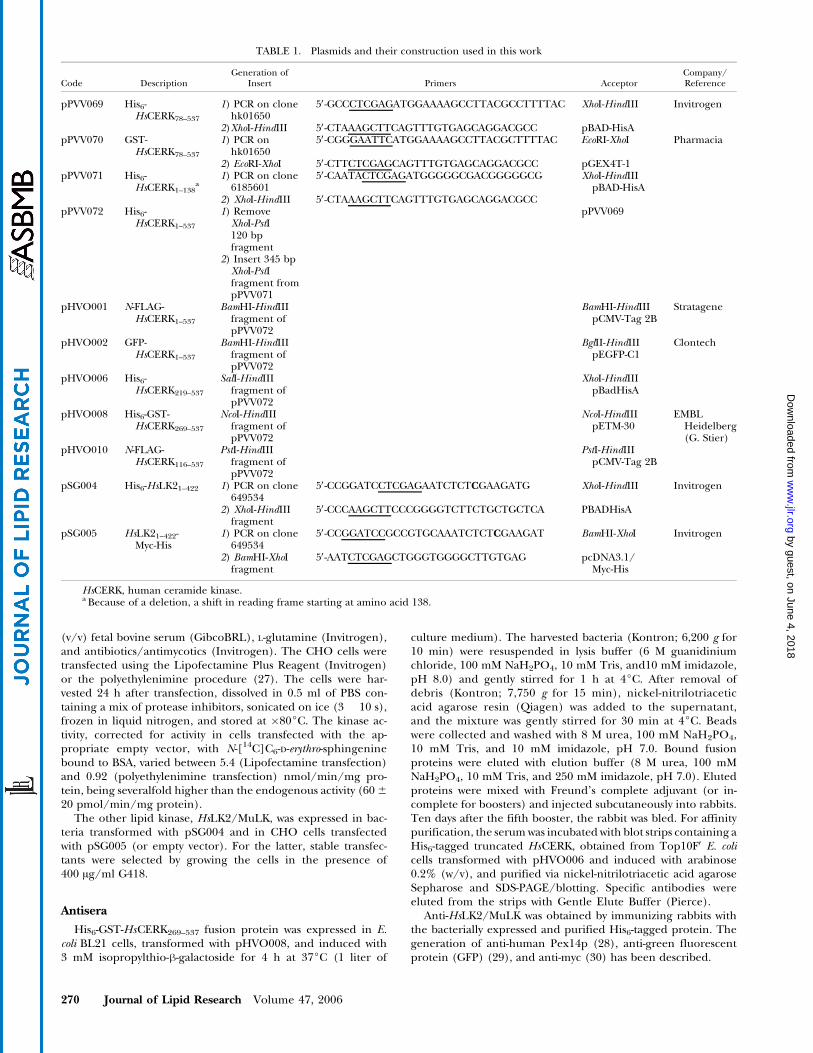
TABLE 1. Plasmids and their construction used in this work
Code DescriptionGeneration of
Insert Primers AcceptorCompany/Reference
pPVV069 His6-HsCERK78–537
1) PCR on clonehk01650
5V-GCCCTCGAGATGGAAAAGCCTTACGCCTTTTAC XhoI-HindIII Invitrogen
2)XhoI-HindIII 5V-CTAAAGCTTCAGTTTGTGAGCAGGACGCC pBAD-HisApPVV070 GST-
HsCERK78–537
1) PCR onhk01650
5V-CGGGAATTCATGGAAAAGCCTTACGCTTTTAC EcoRI-XhoI Pharmacia
2) EcoRI-XhoI 5V-CTTCTCGAGCAGTTTGTGAGCAGGACGCC pGEX4T-1pPVV071 His6-
HsCERK1–138a
1) PCR on clone6185601
5V-CAATACTCGAGATGGGGGCGACGGGGGCG XhoI-HindIIIpBAD-HisA
2) XhoI-HindIII 5V-CTAAAGCTTCAGTTTGTGAGCAGGACGCCpPVV072 His6-
HsCERK1–537
1) RemoveXhoI-PstI120 bpfragment
pPVV069
2) Insert 345 bpXhoI-PstIfragment frompPVV071
pHVO001 N-FLAG-HsCERK1–537
BamHI-HindIIIfragment ofpPVV072
BamHI-HindIIIpCMV-Tag 2B
Stratagene
pHVO002 GFP-HsCERK1–537
BamHI-HindIIIfragment ofpPVV072
BglII-HindIIIpEGFP-C1
Clontech
pHVO006 His6-HsCERK219–537
SalI-HindIIIfragment ofpPVV072
XhoI-HindIIIpBadHisA
pHVO008 His6-GST-HsCERK269–537
NcoI-HindIIIfragment ofpPVV072
NcoI-HindIIIpETM-30
EMBLHeidelberg(G. Stier)
pHVO010 N-FLAG-HsCERK116–537
PstI-HindIIIfragment ofpPVV072
PstI-HindIIIpCMV-Tag 2B
pSG004 His6-HsLK21–422 1) PCR on clone649534
5V-CCGGATCCTCGAGAATCTCTCGAAGATG XhoI-HindIII Invitrogen
2) XhoI-HindIIIfragment
5V-CCCAAGCTTCCCGGGGTCTTCTGCTGCTCA PBADHisA
pSG005 HsLK21–422-Myc-His
1) PCR on clone649534
5V-CCGGATCCGCCGTGCAAATCTCTCGAAGAT BamHI-XhoI Invitrogen
2) BamHI-XhoIfragment
5V-AATCTCGAGCTGGGTGGGGCTTGTGAG pcDNA3.1/Myc-His
HsCERK, human ceramide kinase.a Because of a deletion, a shift in reading frame starting at amino acid 138.
270 Journal of Lipid Research Volume 47, 2006
by guest, on June 4, 2018w
ww
.jlr.orgD
ownloaded from

Tissue distribution and subcellular studies
Tissues obtained from mouse (male C57/Bl6 N) or rat (maleWistar) were homogenized in 0.25 M sucrose, 5 mM Mops/NaOH, pH 7.2, and 0.1% (v/v) EtOH (HM) using Dounce (Potter-Elvehjem) homogenizers or a Polytron device (Kinematica).Subcellular fractions enriched in nuclei, mitochondria, lyso-somes and peroxisomes, microsomal vesicles, and cytosol wereprepared from testis or brain tissue homogenates, made withPotter-Elvehjem homogenizers, as described previously for ratliver (31). For the fractionation of brain tissue (cerebellum andmedulla oblongata), myelin was removed (32) by resuspendingthe lysosome and peroxisome fraction (derived from 5 g startingwet weight) in 2 ml of 0.85 M sucrose, 5 mM Mops/NaOH, pH7.2, and 0.1% (v/v) EtOH, followed by overlaying with 2 mlof homogenization medium. After centrifugation at 108,000 g(Beckman SW55) during 45 min, the supernatant, containinginterfering myelin, was discarded. Marker enzyme and proteinmeasurements were done as described previously for glutamatedehydrogenase (33), catalase (31), acid phosphatase (33), glucose-6-phosphatase (33), carboxylesterase (31), lactate dehydrogenase(33), proteins (31), and CERK.
For subcellular localization studies by immunofluorescencemicroscopy, CHO cells were grown on cover slips on 12-wellplates and transfected with selected vectors. Monolayers werefixed at 24 h after transfection with 4% (w/v) paraformaldehyde,permeabilized with 1% Triton X-100 in PBS, blocked with 5%(w/v) BSA in PBS, and processed for immunostaining as de-scribed previously (34). Antibodies used were diluted in 1% (w/v)BSA in PBS. Primary antibodies were anti-FLAG M2 (Stratagene),anti-KDEL (Affinity BioReagents), anti-biotin (Rockland), anti-GFP, anti-HsCERK, anti-myc, and anti-Pex14p; secondary anti-bodies were anti-rabbit IgG Cy3, anti-mouse IgG FITC, andanti-rabbit AP (Sigma). To visualize mitochondria, cells wereexposed to 2 AM Red Mitotracker (Red.CM-H2Xros; MolecularProbes) for 45 min before fixation. Fluorescence was observedwith a Leica DMR microscope equipped with FITC/RSGFP/Bodipy/Fluo3/DIO and Texas Red filters.
Kinase assays
To measure CERK, different assays, based on the recovery oflabeled Cer1P in the organic phase when the assay mixture wassubjected to a lipid extraction under acid conditions, wereused. The substrate was presented bound to albumin (EtOH/BSAassay), mixed with CHAPS (detergent assay), or inserted in a mixeddetergent/phospholipid micelle [octyl-h-D-glucopyranoside/car-diolipin (OG/CL) assay].
For the standard EtOH/BSA assay, a 50 Al aliquot of bacterialor CHO lysates, or homogenate or subcellular fraction (appro-priately diluted in HM), was mixed with 150 Al of reaction mix-ture. To prepare the reaction mixture, 20 nmol of substrate wasdried at the bottom of a screw-capped glass tube, dissolved inEtOH (1% of final assay volume), and mixed with 4 volumes ofBSA (resulting in a molar ceramide/BSA ratio of 2.5). Finalconcentrations were 100 AM substrate, 40 AM BSA, 5 mM ATP,50 mM Mops/NaOH, pH 7.2, 3 mM MgCl2, 40 mM NaF, 1 mMDTT, and 100 AM orthovanadate. Either labeled substrate {N-[1-14C]C6-sphingenine (z50,000 dpm/nmol), N-[1-14C]C2-sphin-genine (z50,000 dpm/nmol), N-[1-14C]C16-sphingenine(z40,000 dpm/nmol)} or [g-32P]ATP (z7,000 dpm/nmol) wasused. Purity and specific activity of the ATP were determined byultraviolet light absorbance and TLC on polyethyleneimine-cellulose plates (35). For the other assays, the dried ceramidewas solubilized in 40 Al of 2% CHAPS (detergent assay) or 40 Alof 3.75% OG/2.5 mM CL (OG/CL assay), followed by soni-cation in a water bath and addition of the other solutions. For
inhibition studies, the lipid substrate concentration was de-creased to 25 AM and the inhibitor was tested at 100 AM, andboth were dissolved together.
After an incubation of 15 min at 37jC, reactions were stoppedby the addition of 1.5 ml of chloroform-methanol (1:2, v/v) andphase-separated by adding 1 ml of chloroform and 0.7 ml of a0.5 N HCl/2 M NaCl solution. When using labeled substrate, a1 ml aliquot of the lower phase was transferred to a glass tubeand dried with nitrogen. The residue was dissolved in 60 Al ofchloroform-methanol (4:1, v/v), immediately followed by spotting20 Al on a Silica 60G glass plate with a concentration area (Merck).When using [g-32P]ATP, 250 Al of the lower phase was semiau-tomatically spotted. Plates were developed in chloroform-acetone-methanol-acetic acid-water (10:4:3:2:1, v/v). Spots of the phospho-rylated products, visualized by autoradiography (AmershamBiosciences MP film), were scraped from the plates into 0.5 mlof 1% SDS and counted by liquid scintillation chromatography.
All measurements were corrected for the presence of en-dogenous kinase substrates, possessing after phosphorylationsimilar relative mobility values as (truncated) Cer1P (assays doneplus/minus substrate) and/or endogenous kinase (transfections/transformations with empty vector), unless they proved to benegligible under the particular assay condition.
The phosphorylation of other lipids was determined in a simi-lar manner, using one of the delivery modes described above.When sphingoid bases/amino alcohols were tested, alkalinephase separations were used as described previously for sphingo-sine kinase measurements (19, 36).
The activity of the putative lipid kinase was analyzed by similarprocedures in the presence of MgCl2 or CaCl2. In addition,substrate was also delivered to the enzyme dissolved in DMSO(final concentration, 2%).
Analysis of cellular lipids
CHO cells were grown on six-well plates to 50% confluencein the medium described above and transfected with plasmidpHVO001 or the appropriate empty vector pCMV-Tag 2B usingthe Lipofectamine Plus Reagent (Invitrogen). The medium wasreplaced by phosphate-free Dulbecco’s modified Eagle’s medium(ICN), supplemented with 0.2% (v/v) Ultroser (GibcoBRL), Glu-tamax (GibcoBRL), and antibiotics/antimycotics (Invitrogen)20 h after transfection. After 6 h, cells were given 2 ml of freshphosphate-free medium containing 20 ACi of 32P and C6- or C2-ceramides, added as EtOH/4% BSA (1:4, v/v) complexes (finalconcentration, 5 AM), or N-C16-ceramide, dissolved in dodecane-methanol (37). After 15 h of incubation, medium was removedand monolayers were washed twice with PBS, scraped in 2 � 1 mlof methanol (38), and transferred to extraction tubes. Phaseseparation was introduced by the addition of 2 ml of chloro-form and 1.8 ml of a 0.5 N HCl/2 M NaCl solution to the com-bined methanol solutions. An aliquot (250 Al) of the chloro-form layer was spotted onto a silica plate and developed inchloroform-methanol-25% NH4OH (60:35:8, v/v) (first dimen-sion), allowed to dry, turned 90j and run in acetone, and subse-quently in the same direction developed in chloroform-acetone-methanol-acetic acid-water (10:4:3:2:1, v/v). Radioactive spotswere revealed by scanning (P-Imager; Molecular Dynamics) orby autoradiography.
RESULTS AND DISCUSSION
By homology with the yeast sphingosine kinases, up tofive different sequences coding for related proteins were
Mammalian ceramide kinase 271
by guest, on June 4, 2018w
ww
.jlr.orgD
ownloaded from

found in the human and mouse EST databases by differentgroups. Two cDNAs have been shown to encode sphin-gosine kinases: our cluster 1 (19) corresponded to type 1sphingosine kinase (7–9), and our cluster 3 corresponded
to type 2 (10). The proteins encoded by two other clusteredESTs were tentatively called lipid kinases (cluster 4, ac-cession number AJ457828; cluster 2, accession numbersAJ278150 and AJ401619). The first one, as shown by Sugiura
Fig. 1. Effect of substrate delivery on ceramide kinase (CERK) activity. A: Phosphorylation of ceramide(100 AM) was determined in lysates from E. coli Top10FV cells expressing human ceramide kinase (HsCERK;pPVV072) using different solubilization methods and [g-32P]ATP. The dried C6-ceramide (gray bars) wasdissolved either in ethanol (EtOH) followed by BSA (EtOH/BSA), or with mixed octyl-h-D-glucopyranoside/cardiolipin micelles (OG/CL), or with OG, CHAPS, or Triton X-100, all tested at one or five times theircritical micellar concentration (CMC). With lauryldimethylammonium N-oxide and N-laurylsarcosine, bothat 1� and 5� CMC, or with EtOH/BSA combined with detergent, phosphorylation of C6-ceramide was ,1%of the highest activity (data not shown). Activities are expressed as nanomoles of phosphorylated productper milligram of lysate protein per minute. For C16-ceramide (white bar) and C2-ceramide (black bars), onlyOG/CL and EtOH/BSA were tested. B: Phosphorylation of ceramide (100 AM) was measured in lysates fromCHO cells transfected with HsCERK (pHVO002) in the presence of [g-32P]ATP, using the EtOH/BSA (blackbars) or OG/CL (gray bars) assay as described in Materials and Methods. Activities are expressed as pico-moles of phosphorylated product per milligram of lysate protein per minute. ANA, anandamide; Cx, N-Cx-sphingenine; DAG, 1,2-sn-dioleoylglycerol; 4-OH C6, N-hexanoyl-4-OH-sphinganine.
272 Journal of Lipid Research Volume 47, 2006
by guest, on June 4, 2018w
ww
.jlr.orgD
ownloaded from
et al. (5), appeared to be CERK, a protein of z60 kDa(537 amino acids in human; 531 in mouse); the otherone was recently claimed to be a MuLK (18), a protein ofz47 kDa (422 amino acids in human; 421 in mouse). Bothcontain the conserved domains initially described forsphingosine kinases (6), and their relationship to sphingo-sine kinases has been discussed in the cited references. Inthe meantime, Tuson, Marfany, and Gonzalez-Duarte (39)found evidence for still another kinase when characterizingthe RP26 locus on chromosome 2q31.2-q32.3, one of theloci mutated in autosomal recessive retinitis pigmentosa.Based on the similarity of the predicted gene product, thegene was named ceramide kinase-like gene (CERKL).
To understand some discrepancies between our pre-liminary data, using bacterially expressed HsCERK, andthe data reported by Sugiura et al. (5), based on CERKexpressed in HEK293 cells, we further investigated indepth the enzyme characteristics. Unless indicated other-wise, the data reported below were all obtained with fusionproteins containing the full-length protein. In addition,substantial efforts were made to define the substrate ofthe other kinase (LK2), which we will refer to as MuLK tominimize confusion.
Effect of chain length and substrate delivery onCERK activity
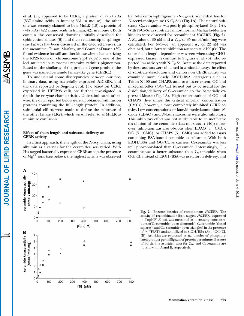
In a first approach, the length of the N -acyl chain, usingalbumin as a carrier for the ceramides, was varied. WithHis-tagged bacterially expressed CERK and in the presenceof Mg21 ions (see below), the highest activity was observed
for N-hexanoyl-sphingenine (N-C6-Se), somewhat less forN -acetyl-sphingenine (N-C2-Se) (Fig. 1A). The natural sub-strate, C16-ceramide, was poorly phosphorylated (Fig. 1A).With N-C6-Se as substrate, almost normal Michaelis-Mentenkinetics were observed for recombinant HsCERK (Fig. 2).A Km value of 30 AM and a Vmax of 35 nmol/min/mg werecalculated. For N-C2-Se, an apparent Km of 22 AM wasobtained, but substrate inhibition was seen at.100 AM. Thesame chain length dependence was seen when using CHO-expressed kinase, in contrast to Sugiura et al. (5), who re-ported low activity with N-C2-Se. Because the data reportedby these authors were obtained in a micellar assay, the effectof substrate dissolution and delivery on CERK activity wasexamined more closely. EtOH/BSA, detergents such asTriton X-100 and CHAPS, and, to a lesser extent, OG andmixed micelles (OG/CL) turned out to be useful for thedissolution/delivery of C6-ceramide to the bacterially ex-pressed kinase (Fig. 1A). High concentrations of OG andCHAPS [five times the critical micellar concentration(CMC)], however, almost completely inhibited CERK ac-tivity. Low concentrations of lauryldimethylammonium N -oxide (LDAO) and N -laurylsarcosine were also inhibitory.This inhibitory effect was not attributable to an ineffectivedissolution of the ceramide (data not shown) (40); more-over, inhibition was also obvious when LDAO (1� CMC),OG (5� CMC), or CHAPS (5� CMC) was added to assayscontaining BSA-bound ceramide as substrate. With bothEtOH/BSA and OG/CL as carriers, C2-ceramide was lesswell phosphorylated than C6-ceramide. Interestingly, C16-ceramide was a better substrate than C6-ceramide whenOG/CL instead of EtOH/BSA was used for its delivery, and
Fig. 2. Enzyme kinetics of recombinant HsCERK. Theactivity of recombinant (His)6-tagged HsCERK, expressedin Top10FV E. coli, was measured at increasing concentra-tions of C2-ceramide (open diamonds), C6-ceramide (closedsquares), and C16-ceramide (open triangles) in the presenceof [g-32P]ATP and solubilized in EtOH/BSA (A) or OG/CL(B). Activities are expressed as nanomoles of phosphory-lated product per milligram of protein per minute. Becauseof borderline activities, data for C16- and C2-ceramide arenot shown in A and B, respectively.
Mammalian ceramide kinase 273
by guest, on June 4, 2018w
ww
.jlr.orgD
ownloaded from

the activities toward C16-ceramide or ceramide containing a2-hydroxy fatty acid (ceramide IV) were comparable (datanot shown).
When these assay conditions were tested with eukary-otically expressed HsCERK, similar results were obtained(Fig. 1B). In the presence of OG/CL, C16- and C6-ceramidewere almost equally well phosphorylated (Fig. 2), and to amuch larger extent than N-C2-Se, in agreement with thefindings by Sugiura et al. (5).
Because the activities are substantially higher with trun-cated ceramides complexed to BSA than in the mixedmicellar assay, the former assay was mainly used to studythe CERK properties.
Role of divalent ions for CERK activity
In older reports concerning CERK measurements intissue/cells, a stimulation by Ca21 was reported (11, 41).
Also, Sugiura et al. (5) reported a Ca21 dependence for therecombinant CERK, reaching a maximum at z0.5 mM freeCa21. In our preliminary trials, performed in the absenceof Ca21 but in the presence of Mg21 (see above), highkinase activity was found. Therefore, the influence of Ca21
and Mg21 on HsCERK activity was determined with C6-ceramide as substrate. In the absence of Ca21, the activityof CERK, bacterially as well as eukaryotically expressed,was increased by Mg21 in a concentration-dependent man-ner (Fig. 3A). In the absence of Mg21, CERK was stimulatedby Ca21, although to a lesser extent than by Mg21, andinhibited at .1 mM Ca21 (0.04 mM free Ca21) (Fig. 3B).The highest HsCERK activity was measured in the presenceof Mg21 and without Ca21 for both forms of expressedkinase (Fig. 3C). Sugiura et al. (5) also described a stimu-lation by Mg21, which was less than by Ca21. The reason forthis discrepancy with our data is not clear. Because theMg21 dependence is seen for both the EtOH/BSA and
Fig. 3. Effect of Mg2+ and Ca2+ on CERK activity. CERKactivity was measured with 100 AM [14C]C6-ceramide assubstrate and solubilized with OG/CL under standardconditions except for the presence of 0.2 mM EDTA.Activities are expressed as picomoles of phosphorylatedC6-ceramide formed per milligram of lysate protein perminute. A, B: CERK activity in lysate from CHO cellstransfected with HsCERK (pHVO002) was measured inthe presence of increasing Mg2+ concentrations in theabsence of Ca2+ (A) or in the presence of increasing Ca2+
concentrations in the absence of Mg2+ (B). Profiles simi-lar to these, but at higher specific activities, were observedwhen using lysate from E. coli Top10FV cells expressingHsCERK (pPVV072) (data not shown). C: CERK activityin the absence or presence of Mg2+ (3 mM) and/or Ca2+
(1 mM) in a membrane fraction prepared from E. coliTop10FV cells expressing HsCERK (pPVV072) (open bars)or lysate from CHO cells transfected with HsCERK(pHVO002) (closed bars).
274 Journal of Lipid Research Volume 47, 2006
by guest, on June 4, 2018w
ww
.jlr.orgD
ownloaded from

Fig. 4. Substrate specificity of CERK. Phosphorylation of the different ceramide stereoisomers (A) and cer-amide analogs (B), each at 100 AM, was determined in lysate from E. coli Top10FV cells expressing HsCERK(pPVV072) in the presence of [g-32P]ATP using the EtOH/BSA procedure. Activities are expressed as nano-moles of phosphorylated product per milligram of protein per minute. Unless specified otherwise, aminoalcohols and amides are racemic, whereas sphingoid bases and derivatives possess the D-erythro configuration. N-tetradecanoyl-3-amino-1-propanol, N-tetradecanoyl-4-amino-1-butanol, N-tetradecanoyl-3-hydroxypyrrolidin,N-tetradecanoyl-R-serine, N-tetradecanoyl-S-serine, N-tetradecanoyl-2-benzylserine, N-tetradecanoyl-3-phenyl-serine,N-tetradecanoyl-3-amino-tyrosine,N-tetradecanoyl-1-amino-2-naphthol-4-sulfonic acid,N-tetradecanoyl-3-hydroxy-anthranilic acid, N-tetradecanoyl-4-aminophenol, N-tetradecanoyl-diethanolamine, and N-hexadeca-noyl-ethanolamine were not phosphorylated (data not shown).
Mammalian ceramide kinase 275
by guest, on June 4, 2018w
ww
.jlr.orgD
ownloaded from
the OG/CL assay, it is not attributable to the presence ofacidic phospholipids, known to bind Mg21 as well as Ca21
ions. It is also not caused by differences in the ATP con-centrations used. At all ATP concentrations tested (0.5–20 mM), Mg21 resulted in 10- to 20-fold higher activitiesthan Ca21 (data not shown). In the presence of 3 mMMg21, a Km of 400 AM was obtained for ATP, substantiallyhigher than the reported value of 32 AM using naturalceramides (5). Also, the pH profiles were analyzed with3 mM Mg21 with or without Ca21 (1 or 3 mM). A broad pHoptimum between 6.2 and 7.8 was seen, and the activityof the (bacterially expressed) kinase was always higher inthe presence of Mg21, using either the EtOH/BSA or theOG/CL assay (data not shown). As a consequence, theregulation of CERK activity by calcium levels might occurto a lesser extent than proposed (42).
Substrate stereoselectivity and specificity of CERK
To test the stereoselectivity of HsCERK, the differentstereoisomers of both C6-ceramide and C6-dihydroceramidewere initially compared. For the unsaturated compounds,the D-erythro form was clearly preferred. The absence of thedouble bond in the D-erythro isomers decreased the activityalmost 10-fold, but for the saturated substrates, both D-erythroand L-threo isomers were phosphorylated equally well(Fig. 4A). Also with the unsaturated substrates, the L-threoisomer was recognized (see below). The presence of ahydroxy group at position 4 of the base (N -C6-4-hydroxy-sphinganine) did not affect the activity (compared withN-C6-sphinganine) (Figs. 1B, 4B). In agreement with others(5), neither diacylglycerol nor sphingoid bases (or relatedbases) were phosphorylated byHsCERK. The presence of anamide bond seems essential, because N-octyl-sphingenine,also known as ceramine (43), was not a substrate (Fig. 4B).As expected, N -C6-psychosine was not a substrate.
In subsequent experiments, the influence of a selectedcollection of ceramide/sphingenine analogs was tested,first for possible inhibitory action, then as substrate. Noneof the synthetic ceramide analogs showed a great degree ofinhibition. The most potent inhibitors found were sphin-ganine, sphingenine, and DMS (data not shown), in agree-ment with the data reported by Sugiura et al. (5). Forsphingenine, an IC50 value of 40 AM was obtained in theEtOH/BSA assay (data not shown). In this respect, thesevere inhibitory effect of LDAO, bearing some structuralsimilarity to DMS, should be recalled. It also shows thatone should be careful when interpreting the cellular ef-fects of DMS, which blocks not only sphingosine kinase(44, 45), as assumed, but also CERK. Of particular inter-est were ceramide analogs containing a carboxy group(N -acylated amino acids), which could be inhibitory giventheir negative charge similar to the physiological reactionproduct. None of these, however, was inhibitory.
Only a few of the tested analogs were substrates forCERK (Fig. 4B). If the sphingoid base was shortened tothe first three C-atoms (N -C14-2-amino-1,3-propanediol),the analog was still a substrate. According to Bieberich,Kawagushi, and Yu (46), the N -C16 analog of this com-
pound (calledN-acylated serinol) acts as a ceramide mimic.When added to cultured NG108-15 or F-11 cells, it causesceramide increase and apoptosis. N -Acyl serinol is a mod-erate inhibitor of acidic ceramidase and can compete withceramide for glucosyl transfer (46), but competition withthe kinase, as shown here, could also contribute to thechanges in ceramide. When reversing the amino and sec-ondary hydroxy group (N -C14-3-amino-1,2-propanediol),the amide is poorly phosphorylated. Interestingly, the hy-droxy group at carbon 3 of the base is not necessary forrecognition by CERK, becauseN -C4-2-amino-1-hexadecanol,N -C14-2-amino-1-butanol, and N -C6-1-O-hexadecyl-2-desoxy-2-amino-sn-glycerol are good substrates. Indirectly, thesedata support the notion that the primary hydroxy group ofceramide is phosphorylated by the kinase. To documentthe importance of the amino group configuration, both R-and S -isomers of N -C14-2-amino-1-butanol were synthesizedand tested: the 2S(D) isomer is clearly a better substratethan the 2R(L) isomer (Fig. 4B). Mixing experiments sug-gest that the 2R compound, although a poor substrate,can have a stimulatory effect (data not shown). These dataagree with the selectivity seen toward the D-erythro isomerof C6-ceramide and explain the recognition of the L-threoisomers, both corresponding to the configuration of the Sisomer at carbon 2. The recognition ofN -C6-1-O-hexadecyl-2-desoxy-2-amino-sn-glycerol is interesting (Fig. 4B). Pre-viously, it was shown that the etherlipid analog, 1-O -hexa-decyl-2-desoxy-2-amino-sn-glycerol, was efficiently used byhuman sphingosine kinase type 1 (19). Hence, it is strikingthat both this base and sphingosine fit into the catalytic siteof sphingosine kinase, and after N-acylation, into that ofCERK, despite some structural differences, underlining theevolutionary relationship between both kinases. On theother hand, the N-acylated form of the immunosuppressivedrug FTY720, another sphingosine analog that can be phos-phorylated by sphingosine kinase types 1 and 2 (47–49), wasnot recognized by CERK.
Finally, we were interested to learn whether ananda-mide (N-arachidonoyl-ethanolamine) was phosphorylatedor not. Anandamide, an endocannabinoid, can be con-sidered as structurally related to ceramide, but unlike theother analogs used here, it is naturally occurring (50). Itwas a substrate, albeit a very poor one, of CERK (Fig. 1B).
The selectivity for the natural D-erythro substrates and itssubstrate spectrum, as analyzed here in detail, accentuatesthe physiological role of this kinase, classifying it as a trueCERK. It should be emphasized that short-chain ceramidessuch as N-C2-Se, so often used as a water-soluble ceramideanalog in cellular studies, and ceramide mimics such asN -acyl serinol can be efficiently phosphorylated by CERK.According to Sugiura et al. (5), N -C2-sphingenine is a poorsubstrate; however, as shown here, the affinity is very muchdependent on the way the substrate is delivered to theenzyme (see below).
Tissue distribution of HsCERK
Having established optimum assay conditions for CERKusing recombinant proteins, its presence was measured in
276 Journal of Lipid Research Volume 47, 2006
by guest, on June 4, 2018w
ww
.jlr.orgD
ownloaded from

different mouse tissues. In all tissues, Cer1P was formed,suggesting a ubiquitous expression of CERK. The level ofkinase activity varied substantially, however, being mark-edly high in testis and cerebellum, followed by pancreasand cerebrum, and negligible in skeletal muscle, usingeither EtOH/BSA (Fig. 5) or mixed micelles (data notshown) to deliver the substrate. This distribution, however,does not fully reflect the expression of CERK, which, basedon Northern analysis, is high in mouse testis, heart, andspleen (5). The lack of correlation between mRNA, pro-tein, and activity levels can be attributable to many factors,including the presence of an inhibitor or activities affect-ing the substrate or its product. In heart and lung ho-mogenates, unless diluted appropriately, residual ATPaseactivity interfered with the kinase measurements, despitethe use of NaF and orthovanadate (H. Van Overloop, L.Van den Bossche, and P. P. Van Veldhoven, unpublisheddata). When analyzing the kinase activity in testis, wenoticed the formation of an additional 32P-labeled spotthat, based on relative mobility value, represents SeP, butthis was only seen in BSA-based assays (data not shown).Presumably, ceramide bound to BSA is more accessible tohydrolysis (by ceramidases) than when inserted in a mixedmicelle. If formed, sphingenine is efficiently phosphory-lated in testis homogenates, a tissue in which we previouslyreported a high sphingosine kinase activity when usingBSA-bound substrates (36). Unfortunately, the amounts ofCERK in tissues are too low to be convincingly visualized byimmunoblotting (data not shown).
Another possibility to explain the differences betweenactivity measurements and Northern analysis could be thepresence of (an)other kinase(s) acting on ceramide, giventhe presence of different lipid kinase genes in mammaliangenomes. Recently, Waggoner et al. (18) reported on anew kinase, which they described as a MuLK, acting onceramide and diacylglycerol as main substrates, but alsoon monoacylglycerol. This kinase is highly expressed inpancreas, followed by brain and liver. Another putativelipid kinase was cloned by Tuson, Marfany, and Gonzalez-
Duarte (39), who analyzed the causative genes for retinitispigmentosa. A protein, encoded by one of these genes anddisplaying some similarity to CERK, was named CERK-likeprotein. However, this might be rather misleading. Thesimilarity to HsCERK is limited (26% amino acid identity),no information on its substrate spectrum or ATP depen-dence is available (39), and the protein does not seem toact on ceramide (51). This “kinase” has a restricted ex-pression pattern in human: based on RT-PCR, it is mod-erately expressed in pancreas, kidney, lung, and retina andalmost not expressed at all in heart and muscle (39); basedon real-time PCR, it is highly expressed in brain, followedby kidney and trachea (51).
The kinase described as MuLK was cloned some yearsago in our laboratory and named putative lipid kinase 2(accession numbers AJ278150 and AJ401619). Despitemany attempts (52), both with bacterially and eukaryoticallyexpressed proteins, no substrate, when corrected for back-ground or endogenous kinase, could be demonstrated.4
The reason for the lack of activity is not clear; no PCR-
Fig. 5. Tissue distribution of CERK activity in mouse.CERK activity was determined in mouse tissue homo-genates using 100 AM C6-ceramide, solubilized withEtOH/BSA, and using 200 Ag of protein per assay.Activities are expressed as picomoles of phosphorylatedC6-ceramide per milligram of protein per minute. Thevalues shown are means 6 SEM of separate measure-ments performed on three male mice, 60–90 days ofage. The same activity distribution pattern was seenwith C2-ceramide, complexed to BSA, or C16-ceramide,solubilized with OG/CL, as substrate (data not shown).
4 Several lipophilic phosphorylatable compounds, including all foursphinganine and sphingenine stereoisomers, 4-hydroxysphinganine,(truncated) (dihydro)ceramides, (truncated) 1,2-sn-diacylglycerol, 1-or 2-monoacylglycerols, 1-monoalkylglycerols, and phosphatidylinosi-tol, as well as less well-studied kinase substrates (tocopherol, cholesterol,farnesol) or putative substrates (galactosylceramide, anandamide),have been tested as substrates under different delivery modes (EtOH/BSA, octyl-h-D-glucopyranoside/cardiolipin, CHAPS, DMSO) in thepresence of Ca or Mg ions, with bacterially expressed kinase, lysates ofCHO cells stably transfected with HsLK2-Myc-His, mitochondria iso-lated from such cells, or in vitro-transcribed/translated kinase. Com-pared with the appropriate controls, no remarkable differences werenoticed. In addition, in lipid extracts from CHO cells cultured in thepresence of labeled glucose (precursor of glycerolipids) or serine (pre-cursor of sphingolipids) or uploaded with 32P, no additional or en-hanced radiolabeled products were revealed when analyzed by two-dimensional TLC/autoradiography and compared with cells stablytransfected with the empty vector. On the other hand, these experi-ments showed that E. coli diacylglycerol kinase can phosphorylateanandamide and that coupled transcription/translation systemsphoshorylate sphingoid bases, diacylglycerol, and ceramide (P. P. VanVeldhoven and K. De Greef, unpublished data).
Mammalian ceramide kinase 277
by guest, on June 4, 2018w
ww
.jlr.orgD
ownloaded from

Fig. 6. Subcellular localization of HsCERK. CHO cells were analyzed 24 h after Lipofectamine-mediatedtransfection with plasmids encoding a full-length FLAG-HsCERK fusion (pHVO001) (A, B, D1), greenfluorescent protein (GFP)-HsCERK (pHVO002) (C), N-truncated FLAG-HsCERK116-537 (pHVO010) (D2),or appropriate control vectors (data not shown) by direct (C1) or indirect (A1, B1, B2, C2, D1, D2) immuno-fluorescence microscopy. FLAG fusions were stained with monoclonal mouse anti-FLAG M2 (Stratagene)/anti-mouse FITC (A1, B1, D1, D2); plasma membrane was visualized with the Image-iTTM LIVE plasma membranelabeling kit (Molecular Probes) (A2); endoplasmic reticulum was visualized with rabbit anti-KDEL (AffinityBioReagents)/anti-rabbit Cy3 (B2), and CERK was visualized with affinity-purified rabbit anti-CERK/anti-rabbitCy3 (C2). Overlays are shown in A3, B3, and C3. In E, CHO cells transfected with pEGFP-C1 (lane 1) orpHVO002, coding for a GFP-HsCERK fusion (lane 2), were analyzed 24 h after transfection by immunoblottingusing anti-CERK antibody or anti-GFP. Both antibodies immunodecorated a band of the expected size for thefusion protein (87.7 kDa). Presumably, anti-CERK also recognizes the endogenous CERK in CHO cells (leftpanel, arrowhead); the asterisk in the right panel indicates a protein cross-reacting with anti-GFP. Migration ofprotein standards, expressed in kDa, is indicated.
278 Journal of Lipid Research Volume 47, 2006
by guest, on June 4, 2018w
ww
.jlr.orgD
ownloaded from

introduced errors appear to be present in the plasmidsused, and the cDNAs cloned by Waggoner et al. (18) werebased on our database entries. It is possible that the tag,although at different ends in the bacterial and eukaryoticconstructs, interferes with the kinase activity. Because ofthe lack of experimental detail on the way MuLK was con-structed (18), we cannot explore this possibility. Anotherconsideration is that the majority of our data were obtainedwith the human kinase, whereas those reported byWaggoner et al. (18) relate to the murine kinase, charac-terized by one additional amino acid (Q271).
The expression of MuLK in human tissues, estimated byRT-PCR techniques by Waggoner et al. (18), is highest inpancreas, followed by brain, liver, and kidney. AlthoughMuLK, expressed in CHO cells, was recognized by ourantiserum, we failed to see signals of the correct size(z50 kDa) upon immunoblotting of various rat or mousetissues, indicating low expression. Given the tissue distri-bution of MuLK, if active, it is rather unlikely that itcontributes to ceramide phosphorylation under our assayconditions. In addition, the fact that the same tissuedistribution was seen when using EtOH/BSA or mixedmicelles to deliver the substrate, either short- or long-chain ceramides (Fig. 5; data not shown), also suggeststhat the measured activity resides in a single enzyme.Moreover, the subcellular localization of MuLK is differentfrom that of CERK (see below).
Subcellular localization of HsCERK and HsMuLK
The enzyme(s) catalyzing the phosphorylation of cera-mide was (were) previously reported to be membrane-as-sociated (11, 41). Upon overexpression of CERK in HEK293cells, again a membrane-bound activity was found (5). Mit-sutake et al. (42) recently showed that HsCERK was predom-inantly localized in the cytosol of RBL-2H3 cells but was
present at the plasma membrane in a few cells, and Carreet al. (53) reported on a Golgi localization in Cos-1 andHUVEC cells and translocation of HsCERK to the plasmamembrane upon osmotic swelling. In our hands, taggedforms of HsCERK (FLAG-HsCERK and EGFP-HsCERKfusions), upon expression in CHO cells, were mainly lo-calized to the plasma membrane, based on colocalizationwith appropriate markers (Fig. 6A), whereas no evidence foran ER association was found (Fig. 6B).
Immunostaining of such cells with purified anti-CERKantibody confirmed this localization pattern (Fig. 6C).An N-truncated form of CERK, lacking the first 115amino acids, failed to associate with the plasma membrane(Fig. 6D) and was not active. These findings are in agree-ment with data published by Carre et al. (53) showing thatremoval of the first 123 amino acids, which mediate bind-ing to liposomes (pleckstrin domain), abolished activityand that the truncated protein became cytosolic. In fact,bacterially expressed CERK lacking the first 77 aminoacids, either as a His6 or GST fusion, was also inactive (H.Van Overloop and P. P. Van Veldhoven, unpublisheddata), although it contained the conserved domains situ-ated more C terminally. Therefore, the N-terminal regionseems to be required for enzyme activity/folding. In CHOcells overexpressing CERK, formation of Cer1P fromendogenous ceramides could be seen upon 32P upload-ing, and exogenously added C6-ceramide, as well asC2-ceramide, were efficiently converted to their phos-phate esters (Fig. 7). Exogenously added long-chain cer-amide, however, was not phosphorylated, likely because ofpoor uptake.
The localization of the other cloned kinase was analyzedby expressing it with a C-terminal Myc-His6 tag. Waggoneret al. (18) reported murine MuLK-EGFP to be associatedwith undefined endomembranes. Our colocalization stud-
Fig. 7. C6-ceramide is phosphorylated by CHO cells overexpressing HsCERK. C6-ceramide was added toCHO cells without (A) or with (B) overexpression of HsCERK, uploaded with 32P, followed by lipidextraction after overnight incubation. Lipids were analyzed by two-dimensional TLC [first dimension,chloroform-methanol-NH4OH, 60:35:8 (v/v); second dimension, acetone wash followed by chloroform-acetone-methanol-acetic acid-water, 10:4:3:2:1 (v/v)], followed by detection by autoradiography. Phosphor-ylated C6-ceramide is indicated by a solid arrow. Phosphorylation of endogenous ceramides (asterisks) wasalso seen in CHO cells overexpressing HsCERK without the addition of ceramide (data not shown). Thearrowheads indicate the position of sphingenine-1-phosphate, resulting from C6-ceramide breakdown byceramidase(s) and subsequent action of endogenous sphingosine kinase. Similar findings were obtainedwhen fortifying the cells with C2-ceramide (data not shown).
Mammalian ceramide kinase 279
by guest, on June 4, 2018w
ww
.jlr.orgD
ownloaded from

ies clearly reveal an exclusive mitochondrial pattern forthe HsMuLK-Myc-His6 fusion (Fig. 8). This is consistentwith the presence of an N-terminal targeting sequence andtransit peptide, as predicted by different algorithms [Sig-nalP 3.0 (54), http://www.cbs.dtu.dk/services/SignalP/#submission; ProtComp 6.0, http://sun1.softberry.com/berry.phtml?topic=protcompan&group=programs&subgroup=proloc), likely cleaved between positions 31 and32 (...LYG31-K32HC..) for both human and murine kinase.The submitochondrial localization of HsMuLK was fur-ther investigated by immunocytochemistry and selectivepermeabilization of the plasma membrane. When thefixed cells were exposed to high detergent concentra-tions [0.03–1% (w/v) Triton X-100], HsMuLK was recog-nized by anti-myc antibodies (Fig. 8A, C). Likewise, under
these conditions, anti-biotin decorated the mitochondrialmatrix (Fig. 8D), known to contain various biotinylatedproteins (55). However, at 0.015% Triton X-100, nostaining of HsMuLK or biotinylated proteins was observed(Fig. 8G). Under these conditions, Pex14p, a peroxisomalmembrane protein facing the cytosol (56), was still im-munodecorated (Fig. 8H), indicating that the plasma mem-brane was permeabilized but that MuLK was not accessible.After sonication of transfected CHO cells in hypotonicalkaline medium [10 mM Na-pyrophosphate buffer, pH 9(57)], the fusion protein was recovered in the high-speedpellet (data not shown), suggesting a tight membraneassociation, in agreement with Waggoner et al. (18).
To document further the cellular localization of CERKunder physiological conditions, ceramide phosphoryla-
Fig. 8. Subcellular localization of human multisubstrate lipid kinase. CHO cells, stably transfected withpSG005 coding for HsLK2-Myc-His, were analyzed by indirect immunofluorescence microscopy, afterpermeabilization with 1% (A, B), 0.03% (w/v) (C–E), or 0.015% (w/v) (F–H) Triton X-100, usingmonoclonal mouse anti-myc as primary antibody and anti-mouse FITC as secondary antibody (A, C, F).Colocalization studies were performed using Mitotracker Red CM-H2�Ros (Molecular Probes) (B) orpolyclonal rabbit anti-biotin and anti-rabbit-Cy3 (D, G). In E and H, cells were stained with polyclonal rabbitanti-Pex14p and anti-rabbit Cy3. Control incubations with cells stably transfected with pcDNA/Myc-Hisshowed that the staining with anti-myc was attributable to the expressed fusion protein.
280 Journal of Lipid Research Volume 47, 2006
by guest, on June 4, 2018w
ww
.jlr.orgD
ownloaded from
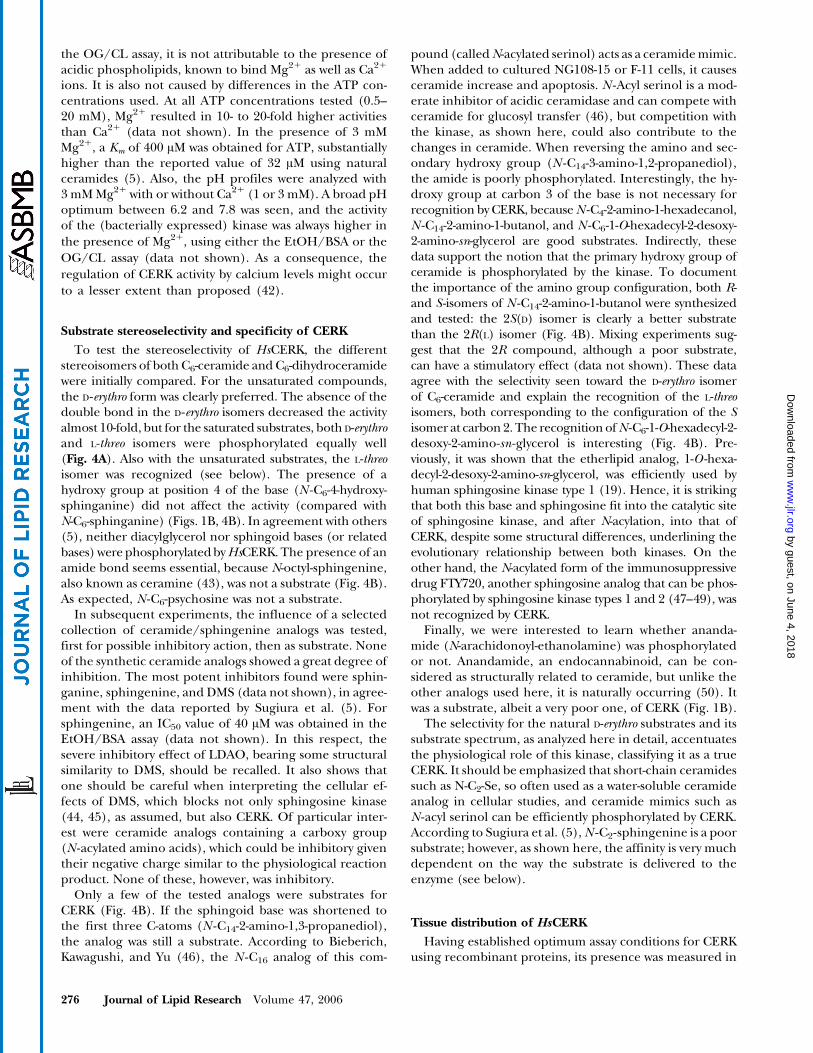
tion was analyzed in subcellular fractions prepared fromtestis, a tissue with a high CERK activity, and in brain, inwhich the expression of MuLK is high. The majority ofCERK activity was recovered in the so-called microsomalfraction (Fig. 9). This fraction is known to contain vesiclesderived from plasma membrane and ER, as can be de-duced from the enrichment in specific markers (alkalinephosphodiesterase, 2.76-fold for testis and 2.15-fold forbrain; carboxylesterase, 3.75-fold for testis and 3.4-fold forbrain). Hence, these data are in agreement with the dataobtained in cells overexpressing CERK. In the mitochon-drial fraction enriched in mitochondria, CERK activity isvery low, an additional argument that MuLK, enriched in
this fraction, does not contribute to the ceramide phos-phorylation described in this study.
To summarize, CERK is a Mg21-dependent lipid kinasehighly selective for D-erythro ceramides and can be effi-ciently measured using BSA-complexed C6-ceramide. Com-pounds containing an unsaturated sphingoid base arepreferred, but the presence of a secondary hydroxy groupis not required. Considerable variation is allowed withregard to the chain length of the base (up to ethanol-amine) and of the fatty acid (up to acetyl) of the amide, butthe configuration at carbon 2, carrying the amide bond,should be 2S. In this respect, one should be aware that N-C2-Se, a very popular ceramide analog in cellular studies,
Fig. 9. Subcellular distribution of ceramide phosphorylating activities in rat brain and testis. Fresh rat braintissue (cerebellum + medulla oblongata) (left panels) and testis (right panels) homogenates were frac-tionated into a nuclear (N), a heavy mitochondrial (M), a light mitochondrial (L), a microsomal (P), and acytosolic (S) fraction. In each fraction, protein, marker enzymes, and ceramide phosphorylation (100 AM C6-ceramide, solubilized with BSA/EtOH) were measured. Results are expressed as relative specific activities(RSA) versus cumulative percentage of total protein according to de Duve et al. (64). RSA is defined as thepercentage of total activity present in a particular fraction divided by the corresponding percentage of totalprotein. Recoveries varied between 82% and 102%. CERK activity in rat brain and testis amounted to 27 and26 pmol/min/mg protein, respectively. Other markers (not shown) included catalase, acid phosphatase,glucose-6-phosphatase, 5V-nucleotidase, and lactate dehydrogenase, with the highest relative specific activityin the L, L, P, P, and S fractions, respectively. Although glutamate dehydrogenase is generally considered amarker for mitochondria, a soluble activity has been reported in nonhepatic tissues (65), likely explainingthe rather high amount in the S fraction of testis.
Mammalian ceramide kinase 281
by guest, on June 4, 2018w
ww
.jlr.orgD
ownloaded from

is phosphorylated by CERK and that the amount ofC2-Cer1P formed will depend on the expression level ofCERK in the cells studied. The enzyme is mostly active intestis and cerebellum, but its function in these organs isnot clear. In brain, it was postulated to be linked to synapseformation. With regard to testis, it is remarkable that manyother sphingosine/ceramide-metabolizing enzymes arepreferentially expressed in this tissue [sphingosine kinase(36), dihydroceramide desaturase (40)] and that levels ofSeP are also high (58). Perhaps this is linked to the con-tinuous process of cell maturation and apoptosis to formgametes in the gonadal tissues (59). The roles of MuLK(18) and certainly of CERKL (39) are unclear. The re-semblance of MuLK to E. coli 1,2-sn-dioleoylglycerol kinasewith regard to substrate spectrum, both being unspecifickinases acting on ceramide, diacylglycerol, and monoacyl-glycerol, and its association with mitochondria, couldsuggest some ancestral function. The mitochondrial lo-calization of MuLK certainly adds another dimension tosphingolipid signaling. Changes in the mitochondrialintegrity, leading to cytochromec release and finally totype 2 apoptosis, have been linked to increases in ceramide(60), and the presence of a mitochondrion-specific neutralceramidase (61) suggests a specific pool of ceramide inthese organelles. Through the action of MuLK, a mito-chondrial pool of Cer1P is feasible as well, which could bea mediator in the known ceramide-dependent processes,but this remains to be proven.
Note. Very recently, Bektas et al. (62) published theirwork on MuLK and claimed that this kinase is not at allactive on ceramide, in agreement with our negative data,but is able to phosphorylate monoacylglycerols and dia-cylglycerols. During the reviewing process, a paper byWijesinghe et al. (63) on the substrate specificity of CERKappeared in this journal. Using Triton X-100 micelles todeliver the substrate, similar conclusions about D-erythrospecificity were reached. However, the reported require-ments for a sphingosine backbone, at least 12 carbonatoms long, and the secondary hydroxy group for substraterecognition are not supported by our study.
This work was supported by grants from the Flemish Fonds voorWetenschappelijk Onderzoek (G.0405.02) and from the BelgianMinistry of Federaal Wetenschapsbeleid Interuniversitaire At-tractiepolen (IAP-P5/05). H.V.O. is an aspirant from the FlemishFonds voor Wetenschappelijk Onderzoek. Technical support byS. Asselberghs, V. Brys, K. De Greef, and G. Van der Hoeven tovarious parts of this work is highly appreciated. The authorsthank Dr. T. Nagase (Kazusa DNA Research Institute, Chiba,Japan) for access to clone hk01650, Dr. G. Stier (EMBL, Heidel-berg, Germany) for the pETM-30 expression vector, and Dr. V.Brinkmann (Novartis Pharma, Basel, Switzerland) for the sampleof FTY720.
REFERENCES
1. Futerman, A. H., and Y. A. Hannun. 2004. The complex life ofsimple sphingolipids. EMBO Rep. 5: 777–782.
2. Pettus, B. J., C. E. Chalfant, and Y. A. Hannun. 2002. Ceramide in
apoptosis: an overview and current perspectives. Biochim. Biophys.Acta. 1585: 114–125.
3. Spiegel, S., and S. Milstien. 2002. Sphingosine 1-phosphate, a keycell signaling molecule. J. Biol. Chem. 277: 25851–25854.
4. Gomez-Munoz, A. 2004. Ceramide-1-phosphate: a novel regulatorof cell activation. FEBS Lett. 562: 5–10.
5. Sugiura, M., K. Kono, H. Liu, T. Shimizugawa, H. Minekura, S.Spiegel, and T. Kohama. 2002. Ceramide kinase, a novel lipidkinase. J. Biol. Chem. 277: 23294–23300.
6. Kohama, T., A. Olivera, L. Edsall, M. M. Nagiec, R. Dickson, and S.Spiegel. 1998. Molecular cloning and functional characterizationof murine sphingosine kinase. J. Biol. Chem. 273: 23722–23728.
7. Nava, V. E., E. Lacana, S. Poulton, H. Liu, M. Sugiura, K. Kono, S.Milstien, T. Kohama, and S. Spiegel. 2000. Functional character-ization of human sphingosine kinase-1. FEBS Lett. 473: 81–84.
8. Pitson, S. M., P. A. B. Moretti, J. R. Zebol, P. Xia, J. R. Gamble, M. A.Vadas, R. J. D’Andrea, and B. W. Wattenberg. 2000. Expression of acatalytically inactive sphingosine kinase mutant blocks agonist-induced sphingosine kinase activation. A dominant-negativesphingosine kinase. J. Biol. Chem. 275: 33945–33950.
9. Melendez, A. J., E. Carlos-Dias, M. Gosink, J. M. Allen, and L.Takacs. 2000. Human sphingosine kinase: molecular cloning, func-tional characterization and tissue distribution. Gene. 251: 19–26.
10. Liu, H., M. Sugiura, V. E. Nava, L. C. Edsall, K. Kono, S. Poulton, S.Milstien, T. Kohama, and S. Spiegel. 2000. Molecular cloning andfunctional characterization of a novel mammalian sphingosinekinase type 2 isoform. J. Biol. Chem. 275: 19513–19520.
11. Bajjalieh, S. M., T. F. Martin, and E. Floor. 1989. Synaptic vesiclekinase. A calcium-stimulated lipid kinase that co-purifies with brainsynaptic vesicles. J. Biol. Chem. 264: 14354–14360.
12. Hinkovska-Galcheva, V. T., L. A. Boxer, P. J. Mansfield, D. Harsh, A.Blackwood, and J. A. Shayman. 1998. The formation of ceramide-1-phosphate during neutrophil phagocytosis and its role in liposomefusion. J. Biol. Chem. 273: 33203–33209.
13. Gomez-Munoz, A., P. A. Duffy, A. Martin, L. O’Brien, H. S. Byun, R.Bittman, and D. N. Brindley. 1995. Short-chain ceramide-1-phos-phates are novel stimulators of DNA synthesis and cell division: anta-gonism by cell-permeable ceramides. Mol. Pharmacol. 47: 833–839.
14. Gomez-Munoz, A., L. M. Frago, L. Alvarez, and I. Varela-Nieto.1997. Stimulation of DNA synthesis by natural ceramide-1-phosphate. Biochem. J. 325: 435–440.
15. Gomez-Munoz, A., J. Y. Kong, B. Salh, and U. P. Steinbrecher. 2004.Ceramide-1-phosphate blocks apoptosis through inhibition of acidsphingomyelinase in macrophages. J. Lipid Res. 45: 99–105.
16. Pettus, B. J., A. Bielawska, S. Spiegel, P. Roddy, Y. A. Hannun, andC. E. Chalfant. 2003. Ceramide kinase mediates cytokine- and cal-cium ionophore-induced arachidonic acid release. J. Biol. Chem.278: 38206–38213.
17. Tornquist, K., T. Blom, R. Shariatmadari, and M. Pasternack. 2004.Ceramide-1-phosphate enhances calcium entry through voltage-operated calcium channels by a protein kinase C-dependentmechanism in GH4C1 rat pituitary cells. Biochem. J. 380: 661–668.
18. Waggoner, D. W., L. B. Johnson, P. C. Mann, V. Morris, J. Guastella,and S. M. Bajjalieh. 2004. MuLK, a eukaryotic multi-substrate lipidkinase. J. Biol. Chem. 279: 38228–38235.
19. Gijsbers, S., S. Asselberghs, P. Herdewijn, and P. P. Van Veldhoven.2002. 1-O-Hexadecyl-2-desoxy-2-amino-sn-glycerol, a substrate forhuman sphingosine kinase. Biochim. Biophys. Acta. 158: 1–8.
20. Weis, B., and P. Raizman. 1958. II. Synthesis of long chain fatty acidamines of sphingosine and dihydrosphingosine. J. Am. Chem. Soc.80: 4657–4658.
21. Lapidot, Y., S. Rappoport, and Y. Wolman. 1967. Use of esters of N-hydroxysuccinimide in the synthesis of N-acylamino acids. J. LipidRes. 8: 142–145.
22. Altschul, S. F., T. L. Madden, A. A. Schaffer, J. Zhang, Z. Zhang, W.Miller, and D. J. Lipman. 1997. Gapped BLAST and PSI-BLAST: anew generation of protein database search programs. Nucleic AcidsRes. 25: 3389–3402.
23. Nagiec, M. M., M. Skrzypek, E. E. Nagiec, R. L. Lester, and R. C.Dickson. 1998. The LCB4 (YOR171c) and LCB5 (YLR260w) genesof Saccharomyces encode sphingoid long chain base kinases. J. Biol.Chem. 273: 19437–19442.
24. Lennon, G. G., C. Auffray, M. Polymeropoulos, and M. B. Soares.1996. The I.M.A.G.E. Consortium: an integrated molecular analysisof genomes and their expression. Genomics. 33: 151–152.
25. Hirosawa, M., T. Nagase, Y. Murahashi, R. Kikuno, and O. Ohara.2001. Identification of novel transcribed sequences on human
282 Journal of Lipid Research Volume 47, 2006
by guest, on June 4, 2018w
ww
.jlr.orgD
ownloaded from

chromosome 22 by expressed sequence tag mapping. DNA Res. 28:1–9.
26. Sambrook, J., E. F. Fritsch, and T. Maniatis. 1989. MolecularCloning: A Laboratory Manual. Cold Spring Harbor LaboratoryPress, Cold Spring Harbor, NY.
27. Boussif, O., F. Lezoualc’h, M. A. Zanta, M. D. Mergny, D. Scherman,B. Demeneix, and J. P. Behr. 1995. A versatile vector for gene andoligonucleotide transfer into cells in culture and in vivo: poly-ethylenimine. Proc. Natl. Acad. Sci. USA. 92: 7297–7301.
28. Amery, L., M. Fransen, K. De Nys, G. P. Mannaerts, and P. P. VanVeldhoven. 2000. Mitochondrial and peroxisomal targeting of 2-methylacyl-CoA racemase in humans. J. Lipid Res. 41: 1752–1759.
29. Fransen, M., T. Wylin, C. Brees, G. P. Mannaerts, and P. P. VanVeldhoven. 2001. Human Pex19p binds peroxisomal integral mem-brane proteins at regions distinct from their sorting sequences.Mol. Cell. Biol. 21: 4413–4424.
30. Ghys, K., M. Fransen, G. P. Mannaerts, and P. P. Van Veldhoven.2002. Functional studies on human Pex7p: subcellular localizationand interaction with proteins containing a peroxisome-targetingsignal type 2 and other peroxins. Biochem. J. 365: 41–50.
31. Van Veldhoven, P. P., and G. P. Mannaerts. 1991. Subcellularlocalization and membrane topology of sphingosine-1-phosphatelyase in rat liver. J. Biol. Chem. 266: 12502–12507.
32. Kovacs, W. J., P. L. Faust, G. A. Keller, and S. K. Krisans. 2001. Purifi-cation of brain peroxisomes and localization of 3-hydroxy-3-meth-ylglutaryl coenzyme A reductase. Eur. J. Biochem. 268: 4850–4859.
33. Van Veldhoven, P. P., E. Baumgart, and G. P. Mannaerts. 1996.Iodixanol (Optiprep), an improved density gradient medium forthe iso-osmotic isolation of rat liver peroxisomes. Anal. Biochem.237: 17–23.
34. Amery, L., M. Fransen, K. De Nys, G. P. Mannaerts, and P. P. VanVeldhoven. 2000. Mitochondrial and peroxisomal targeting of 2-methylacyl-CoA racemase in humans. J. Lipid Res. 41: 1752–1759.
35. Preiss, J. E., C. R. Loomis, R. M. Bell, and J. E. Niedel. 1987. Quan-titative measurement of sn-1,2-diacylglycerols. Methods Enzymol. 141:294–300.
36. Gijsbers, S., G. Van der Hoeven, and P. P. Van Veldhoven. 2001.Subcellular study of sphingoid base phosphorylation in rat tissues:evidence for multiple sphingosine kinases. Biochim. Biophys. Acta.1532: 37–50.
37. Ji, L., G. Zhang, S. Uematsu, Y. Akahori, and Y. Hirabayashi. 1995.Induction of apoptotic DNA fragmentation and cell death bynatural ceramide. FEBS Lett. 358: 211–214.
38. Van Veldhoven, P. P., and R. M. Bell. 1988. Effect of harvestingmethods, growth conditions and growth phase on diacylglycerollevels in cultured human adherent cells. Biochim. Biophys. Acta. 959:185–196.
39. Tuson, M., G. Marfany, and R. Gonzalez-Duarte. 2004. Mutation ofCERKL, a novel human ceramide kinase gene, causes autosomalrecessive retinitis pigmentosa (RP26).Am. J. Hum. Genet. 74: 128–138.
40. Causeret, C., L. Geeraert, G. Van der Hoeven, G. P. Mannaerts, andP. P. Van Veldhoven. 2000. Further characterization of rat dihydro-ceramide desaturase: tissue distribution, subcellular localization,and substrate specificity. Lipids. 35: 1117–1125.
41. Kolesnick, R. N., and M. R. Hemer. 1990. Characterization of aceramide kinase activity from human leukemia (HL-60) cells.Separation from diacylglycerol kinase activity. J. Biol. Chem. 265:18803–18808.
42. Mitsutake, S., T. J. Kim, Y. Inagaki, M. Kato, T. Yamashita, and Y.Igarashi. 2004. Ceramide kinase is a mediator of calcium-dependentdegranulation in mast cells. J. Biol. Chem. 279: 17570–17577.
43. Karasavvas, N., R. K. Erukulla, R. Bittman, R. Lockshin, and Z.Zakeri. 1996. Stereospecific induction of apoptosis in U937 cells byN-octanoyl-sphingosine stereoisomers and N-octyl-sphingosine.The ceramide amide group is not required for apoptosis. Eur. J.Biochem. 236: 729–737.
44. Yatomi, Y., F. Ruan, T. Megidish, T. Toyokuni, S. Hakomori, and Y.Igarashi. 1996. N,N-dimethylsphingosine inhibition of sphingosinekinase and sphingosine 1-phosphate activity in human platelets.Biochemistry. 35: 626–633.
45. Edsall, L. C., J. R. Van Brocklyn, O. Cuvillier, B. Kleuser, and S.Spiegel. 1998. N,N-Dimethylsphingosine is a potent competitiveinhibitor of sphingosine kinase but not of protein kinase C: mod-
ulation of cellular levels of sphingosine 1-phosphate and cera-mide. Biochemistry. 37: 12892–12898.
46. Bieberich, E., T. Kawagushi, and R. K. Yu. 2000. N-Acylated serinolis a novel ceramide mimic inducing apoptosis in neuroblastomacells. J. Biol. Chem. 275: 177–181.
47. Mandala, S., R. Hajdu, J. Bergstrom, E. Quackenbush, J. Xie, J.Milligan, R. Thornton, G. J. Shei, D. Card, C. Keohane, et al. 2002.Alteration of lymphocyte trafficking by sphingosine-1-phosphatereceptor agonists. Science. 296: 346–349.
48. Paugh, S. W., S. G. Payne, S. E. Barbour, S. Milstien, and S. Spiegel.2003. The immunosuppressant FTY720 is phosphorylated bysphingosine kinase type 2. FEBS Lett. 554: 189–193.
49. Billich, A., F. Bornancin, P. Devay, D. Mechtcheriakova, U. Urtz,and T. Baumruker. 2003. Phosphorylation of the immunomodu-latory drug FTY720 by sphingosine kinases. J. Biol. Chem. 278:47408–47415.
50. Schmid, H. H., and E. V. Berdyshev. 2002. Cannabinoid receptor-inactive N-acylethanolamines and other fatty acid amides: metab-olism and function. Prostaglandins Leukot. Essent. Fatty Acids. 66:363–376.
51. Bornancin, F., D. Mechtcheriakova, S. Stora, C. Graf, A. Wlachos, P.Devay, N. Urtz, T. Baumruker, and A. Billich. 2004. Characteriza-tion of a ceramide kinase-like protein. Biochim. Biophys. Acta. 1687:31–43.
52. Gijsbers, S. 2002. Metabolism and Biological Effects of Sphingo-sine-1-phosphate. PhD Dissertation. Acta Biomedica Lovaniensia,Leuven, The Netherlands.
53. Carre, A., C. Graf, S. Stora, D. Mechtcheriakova, R. Csonga, N. Urtz,A. Billich, T. Baumruker, and F. Bornancin. 2004. Ceramide kinasetargeting and activity determined by its N-terminal pleckstrinhomology domain. Biochem. Biophys. Res. Commun. 324: 1215–1219.
54. Nielsen, H., J. Engelbrecht, S. Brunak, and G. von Heijne. 1997.Identification of prokaryotic and eukaryotic signal peptides andprediction of their cleavage sites. Protein Eng. 10: 1–6.
55. Hollinshead, M., J. Sanderson, and D. J. Vaux. 1997. Anti-biotinantibodies offer superior organelle-specific labeling of mitochondriaover avidin or streptavidin. J. Histochem. Cytochem. 45: 1053–1057.
56. Fransen, M., S. R. Terlecky, and S. Subramani. 1998. Identificationof a human PTS1 receptor docking protein directly requiredfor peroxisomal protein import. Proc. Natl. Acad. Sci. USA. 95:8087–8092.
57. Van Veldhoven, P. P., W. W. Just, and G. P. Mannaerts. 1987.Permeability of the peroxisomal membrane to cofactors of beta-oxidation. Evidence for the presence of a pore-forming protein.J. Biol. Chem. 262: 4310–4318.
58. Yatomi, Y., R. J. Welch, and Y. Igarashi. 1997. Distribution ofsphingosine 1-phosphate, a bioactive sphingolipid, in rat tissues.FEBS Lett. 404: 173–174.
59. Tilly, J. L., and R. N. Kolesnick. 1999. Sphingolipid signalingin gonadal development and function. Chem. Phys. Lipids. 102:149–155.
60. Birbes, H., S. E. Bawab, L. M. Obeid, and Y. A. Hannun. 2002.Mitochondria and ceramide: intertwined roles in regulation ofapoptosis. Adv. Enzyme Regul. 42: 113–129.
61. El Bawab, S., P. Roddy, T. Qian, A. Bielawska, J. J. Lemasters, andY. A. Hannun. 2000. Molecular cloning and characterization of ahuman mitochondrial ceramidase. J. Biol. Chem. 275: 21508–21513.
62. Bektas, M., S. G. Payne, H. Liu, S. Goparaju, S. Milstien, and S.Spiegel. 2005. A novel acylglycerol kinase that produces lysophos-phatidic acid modulates cross talk with EGFR in prostate cancercells. J. Cell Biol. 169: 801–811.
63. Wijesinghe, D. S., A. Massiello, P. Subramanian, Z. Szulc, A.Bielawska, and C. E. Chalfant. Substrate specificity of humanceramide kinase. J. Lipid Res. Epub ahead pf print. September 18,2005; doi:10.1194/jlr.M500313-JLR200.
64. de Duve, C., B. C. Pressman, R. Gianetto, R. Wattiaux, and F.Appelmans. 1955. Tissue fractionation studies. VI. Intracellulardistribution patterns of enzymes in rat-liver tissue. Biochem. J. 60:604–617.
65. Colon, A. D., A. Plaitakis, A. Perakis, S. Berl, and D. D. Clarke.1986. Purification and characterization of a soluble and a particu-late glutamate dehydrogenase from rat brain. J. Neurochem. 46:1811–1819.
Mammalian ceramide kinase 283
by guest, on June 4, 2018w
ww
.jlr.orgD
ownloaded from