contemplating the akin - podiatrym.com · october 2016 | podiatry management 87 interphalangeus...
TRANSCRIPT

www.podiatrym.com OCTOBER 2016 | PODIATRY MANAGEMENT
87
Interphalangeus deformity is de-scribed as articular deviation at the head of the proximal phalanx. Wedge resection to correct an angular defor-mity is best determined by finding the center of axis of rotation (CORA). CORA is typically used to describe angular deformities of long bones. However, it can be applied to the first ray when discussing hallux ab-ducto valgus surgery as well.
Figure 1 shows how CORA is evaluated by determining the point at which the longitudinal axis intersects on each side of a deformity. Lines drawn perpendicular to each longitu-dinal axis form an angle that would correct the deformity to a straight line. Of course a perfectly straight line may not always be the goal of a surgical procedure but in many cases it is. When re-alignment of a joint is the surgical goal, the CORA often will be determined to be too close or even crossing into the joint itself. When arthrodesis of the joint is planned, this is appropriate. However, when a wedge osteotomy is planned, the proximity to the joint has practi-cal implications regarding fixation
The proximal phalangeal os-teotomy for the correction of hallux valgus was first described by Akin in 1925.1 The procedure described
medial eminence resection of both the proximal phalangeal base and the first metatarsal head. The osteotomy was a transversely oriented wedge that was fixated only by splinting of the toe. This procedure remains relevant today
primarily as an adjunct procedure and has been modified by various sur-geons to allow other forms of fixation to be utilized. Although its indication in some cases is obvious, often this procedure is opted once the first meta-tarsal procedure has been completed and the abductus or valgus position of the hallux is deemed unsatisfactory. Osteotomy design has been mod-ified to allow the use of new forms
of fixation that have evolved since the procedure was first described. The Akin procedure remains relevant today but is considered by many foot surgeons a necessary evil at best and at worst a “cheater” procedure when the primary procedure fails to ade-quately reduce the deformity. The indications and anatomic consider-ations will be presented. A focused discussion on the oblique closing wedge osteotomy of the first proxi-mal phalanx in hallux valgus repair will be presented.
Level of Deformity Determining the level at which the deformity occurs is important in procedure planning. An increased distal articular set angle indicates a proximal deformity of the phalanx.
Here’s a focused discussion on the oblique closing wedge osteotomy of the first proximal phalanx.
Contemplatingthe Akin
By Bradley d. Castellano, dPM
Continued on page 88
Figure 1: The center of rotation axis or CORA is used to describe the apex of an angular defor-mity. CORA is defined as the intersection point between longitudinal axis proximal and distal to a deformity. The wedge that would completely correct the deformity is represented by the red perpendicular lines to each axis that intersect within the confines of the bone itself.
Determining the level at which the deformity occurs is important
in procedure planning.
SURGICAL PodIatry

www.podiatrym.comOCTOBER 2016 | PODIATRY MANAGEMENT
88
SURGICAL PodIatry
Surgical Technique Surgical exposure of the proximal phalanx is achieved with a dorsal linear incision just medial to the ex-tensor halluces longus tendon. The dorsal and medial surfaces of the phalanx are exposed with sharp dis-section and the use of a periosteal elevator leaving the capsular, tendon, and ligamentous attachments intact (Figure 4). Depending on whether
a distal or proximally based wedge is planned, additional dissection to expose the planned apex of the oste-otomy is performed. Once adequate exposure for visualization and fixa-tion placement is achieved, an axis guide pin can be used to help plan the osteotomy alignment. The first osteotomy of the prox-imal phalanx is placed at a point halfway between a line paralleling the articular surface of the phalanx
and stabilization of the osteotomy. Fixation through a joint is general-ly avoided to prevent trauma to the articular surfaces and the need to remove fixation that impedes range of motion or irritates soft tissues in-volved in joint movement. For that reason, compromises in the placement of osteotomies are often made despite their lack of proximity to the CORA. In the case of the Akin-type phalangeal osteotomy or the treatment of increased distal articular set angle,
this is typically not a significant issue. However, interphalangeus deformity typically will have CORA at or very near the interphalangeal joint, making proximal osteotomy on the phalanx inappropriate (Figure 2).
Oblique Wedge Osteotomy of the Proximal Phalanx While somewhat more technically difficult, there are two major advan-
tages of the obliquely-oriented oste-otomy of the proximal phalanx. The first is more obvious in that it allows more rigid internal fixation in the form of screws. The second advantage is that the hinge of the osteotomy can be located at the attachment of strong soft tissue at the proximal lateral and distal lateral aspects of the phalanx. In the event that the boney hinge is compromised while executing the bone wedge resection, these attach-ments can act as a “soft hinge” for the osteotomy. Osteoto-mies made transversely lose this advantage as they are usually located in the diaphysis of the phalanx and often have less than rigid fixation. Experience has shown that transverse osteoto-mies can be quite suc-cessful but non-weight-bearing or at least more rigorous immobilization techniques are probably best to protect against disruption of the oper-ative site. Even a distal Akin can take advantage of this premise. Boberg described an oblique os-teotomy of the proximal phalanx that allowed screw fixation and the added benefit of soft tis-sue hinge at the distal lateral collateral liga-ment origin (Figure 3).2
Akin (from page 87)
Continued on page 89Figure 2: Proximal osteotomy for a distal defor-mity may create prominence of the phalangeal head medially, and in this case prominence of the fixation.
Figure 3: Tendon and ligament attachments at the head and base of the phalanx including the flexor halluces brevis, abductor halluces, and lateral collateral ligament. The attachments can act as a soft tissue hinge in a well-placed hinge osteotomy.
Figure 4: In this example, a Lapi-dus bunionecto-my has already corrected the intermetatarsal angle. Dissection of the dorsal, medial, and proximal lateral aspect of the hallux allows both visualization of the hinge axis lo-cation and room for placement of fixation. In the oblique wedge procedure, the first osteotomy is oriented halfway
(45 degrees) from the lines parallel to the longitudinal axis of the phalanx and a line parallel to the joint proximal joint surface. Plan-ning the osteotomy in this way should ensure a large enough medial bone segment to allow two screw fixation even if mild deviation occurs during the execution of the osteotomy. The second bone cut is typically made angulating distally to the first osteotomy. A K-wire is inserted dorsal to plantar at the apex can be helpful in maintaining the correct plane for each cut.
Surgical exposure of the proximal phalanx is achieved with a dorsal linear incision just medial
to the extensor halluces longus tendon.

www.podiatrym.com OCTOBER 2016 | PODIATRY MANAGEMENT
89
SURGICAL PodIatry
wedge with the first pass of the blade (Figure 5). Once the osteotomy is complete, a bone hook or single skin hook can be used to dislodge the wedge and remove it. Feathering the hinge to allow easy closure of the wedge os-teotomy is carefully performed until the osteotomy is easily clamped in a reduced position. It is important to place the clamp in the correct ori-entation to prevent hinge fracture. The points of the clamp should be perpendicular to the osteotomy. In many cases, clamping lateral to the long extensor tendon can be help-ful in allowing visualization of the proper clamp orientation (Figure 6). Intra-operative radiographs are best performed at this stage in the proce-dure to determine if the correction of the deformity is adequate prior to placement of internal fixation. Self-drilling/self-tapping screws are the most effective in this rather small bone that requires bi-cortical purchase to achieve rigid fixation. The screws are placed medially to laterally. The long oblique osteotomy allows standard
and a line parallel to the long axis of the bone. This 45-degree first cut ensures that the osteotomy will be long enough to allow a two-screw fixation; a standard AO concept in oblique osteotomy/fracture fixation. The second cut is usually made an-gulating distal to the first, making sure to stay in the same plane as the first osteotomy by following the axis guide pin. It is important to note that a relatively large wedge will be resected compared to a transverse wedge simply because the base of the resected wedge is much further away from the apex. Understanding this is beneficial since the surgeon will be more likely to resect a large enough
Once the osteotomy is complete, a bone hook or single skin hook can be used to dislodge the wedge and
remove it. Feathering the hinge to allow easy closure of the wedge osteotomy is carefully performed until the
osteotomy is easily clamped in a reduced position.
Akin (from page 88)
Continued on page 90
Figure 6A: Note that the retractor has been moved to retract the skin and superficial fascia to allow the bone clamp to be moved lateral to the long extensor tendon. Clamping perpendicular to the osteotomy ensures compression of the oste-otomy following the direction of the hinge axis, making fracture of the hinge less likely.
Figure 5: To create the same degree of correc-tion, an oblique wedge osteotomy will appear much larger with more medial cortical wall re-moved. The tendency when first attempting the oblique wedge is to under-correct the deformity.
Figure 6B: Once the osteotomy has been reduced, the retractor can again be used to retract the tendon lateral to allow visualization of the bone to be fixated.
Figure 7: Intra-operative radiographs are used to confirm that appropriate screw length is achieved. Here, a sponge clamp is used to twist the hallux under fluoroscopy for visualization to ensure the guide pin for the cannulated screw exits the far cortex only slightly. Excessively long screws are frequently painful once the patient begins normal ambulation. However, if the screw does not purchase the far cortex, the fixation is likely to fail to maintain stability of the osteotomy.
www.podiatrym.comOCTOBER 2016 | PODIATRY MANAGEMENT
90
SURGICAL PodIatry
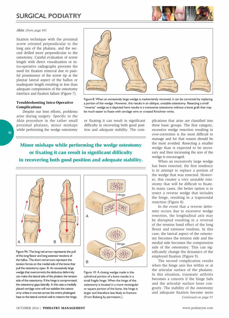
plications that arise are classified into three basic groups. The first category, excessive wedge resection resulting in over-correction is the most difficult to manage and for that reason should be the most avoided. Resecting a smaller wedge than is expected to be neces-sary and then increasing the size of the wedge is encouraged. When an excessively large wedge has been resected, the first tendency is to attempt to replace a portion of the wedge that was resected. Howev-er, this creates a very unstable oste-otomy that will be difficult to fixate. In many cases, the better option is to resect a reverse wedge that includes the hinge, resulting in a trapezoidal resection (Figure 8). In the event that a reverse defor-mity occurs due to excessive wedge resection, the longitudinal axis may be disrupted resulting in a reversal of the tension band effect of the long flexor and extensor tendons. In this case, the lateral aspect of the osteoto-my becomes the tension side and the medial side becomes the compression side of the osteotomy. This can sig-nificantly change the dynamics of the employed fixation (Figure 9). The second complication results when the hinge axis lies within or at the articular surface of the phalanx. In this situation, traumatic arthritis becomes a concern if the hinge fails and the articular surface loses con-gruity. The stability of the osteotomy and adequate fixation become more
fixation technique with the proximal screw oriented perpendicular to the long axis of the phalanx, and the sec-ond drilled more perpendicular to the osteotomy. Careful evaluation of screw length with direct visualization or in-tra-operative radiographs prevents the need for fixation removal due to pain-ful prominence of the screw tip at the plantar lateral aspect of the hallux or inadequate length resulting in less than adequate compression of the osteotomy interface and fixation failure (Figure 7).
Troubleshooting Intra-Operative Complications Despite our best efforts, problems arise during surgery. Specific to the Akin procedure in the rather small proximal phalanx, minor mishaps while performing the wedge osteotomy
or fixating it can result in significant difficulty in recovering both good posi-tion and adequate stability. The com-
Akin (from page 89)
Figure 8: When an excessively large wedge is inadvertently removed, it can be corrected by replacing a portion of the wedge. However, this results in an oblique, unstable osteotomy. Resecting a small “reverse” wedge as is depicted here results in a transverse osteotomy without a bone graft that may be much easier to fixate with cerclage wire or crossed Kirschner wires.
A B
Figure 9A: The long red arrow represents the pull of the long flexor and long extensor tendons of the hallux. The short red arrows represent the tension forces on the medial side of the bone that pull the osteotomy open. B. An excessively large wedge that overcorrects the abductus deformity can make the lateral side of the phalanx the tension side of the osteotomy. If the hinge is compromised, the osteotomy gaps laterally. In this case a medially placed cerclage wire will not stabilize the osteot-omy unless it courses across the entire phalangeal base to the lateral cortical wall to restore the hinge.
A
B
Hinge
Hinge
Figure 10: A closing wedge made in the cylindrical portion of a bone results in a small fragile hinge. When the hinge of the osteotomy is located in a more rectangular or square portion of the bone, the hinge is larger and therefore less likely to fracture (From Boberg by permission.). Continued on page 91
Minor mishaps while performing the wedge osteotomy or fixating it can result in significant difficulty
in recovering both good position and adequate stability.

www.podiatrym.com OCTOBER 2016 | PODIATRY MANAGEMENT
91
SURGICAL PodIatry
Discussion While the indication for digital re-alignment may be radiographical-ly or even clinically obvious in select cases, there are likely few other surgical procedures that are performed as an afterthought than the proximal phalan-geal osteotomy during the correction of hallux valgus. There are two circum-stances that frequently contribute to this impromptu procedure. At times, a significant increase in the distal articu-lar set angle is revealed after the inter-metatarsal angle has been addressed. But perhaps more frequently, the cor-rection of the hallux abducto valgus de-formity is determined to be insufficient. In either case, the surgeon is forced to decide whether the residual deformity is significant enough to warrant an ad-ditional osteotomy. The literature is rife with studies proving that patients are “satisfied” with their surgical result following hallux valgus repair and that cosmetic appearance plays a very small role in this contentment. The proximal pha-langeal osteotomy seems to lie some-where between the idioms “better is the enemy of good” and “a job worth doing is worth doing right.” Hopefully the decision is not based on the sur-geon’s discomposure with the surgical procedure. Taking advantage of the soft tissue hinge while performing the wedge osteotomy simplifies the pro-cedure, making any chosen fixation technique more effective. The oblique wedge allows this advantage and plac-es a significant portion of the osteot-omy in the metaphyseal bone that is known to have a better proclivity to rapid healing and creates a large hinge that is less likely to fracture. PM
paramount to protect the joint from significant deformity. Experience has shown that minor incongruities do not result in significant symptoms. The final category is loss of an intact hinge. This probably occurs more frequently in osteotomies per-formed in the shaft of the phalanx because the hinge in the oval shaft portion of bone is much smaller than a hinge in a more rectangular area at the metaphyseal (Figure 10). Hinge fractures also result when the hinge becomes “boxed.” In this situation, the theoretical hinge of the osteotomy lies outside the phalanx
itself. To correct this problem, fracture of the hinge is frequently necessary to allow reduction of the wedge osteoto-my. Two-point fixation to give the os-teotomy satisfactory stability may be required. In the transverse osteotomy, this may be achieved with two staples or stainless steel wire loops oriented perpendicular to one another. Crossed Kirshner wires buried or percutaneous may also be con-sidered. An obliquely-oriented oste-otomy may still lend itself to screw fixation, though again two points of fixation are preferred once the boney hinge has failed. However, if a sturdy soft tissue hinge remains intact, a single screw may be satisfactory.
Akin (from page 90)
dr. Castellano graduated from the Pennsylvania College of Podiatric Medicine in 1989. He completed the three-year residen-cy at Northlake Med-ical Center in Tucker, Georgia. After practic-ing in Fort Myers, Flor-
ida for many years, he returned to Georgia and practices in Hoschton, GA. Brad is a Fellow, American College of Foot and Ankle Surgeons, a Diplomate, American Board of Foot and Ankle Surgery and an active Faculty Member of the Podiatry Institute.
the Podiatry Institute Mission is to be a supportive, global net-work of members, colleagues and friends, which enhances the
quality of life for patients with foot, ankle, and leg disorders through innovative education, research, and service. The Podiatry Institute is a non-profit foundation founded by E. Dalton McGlamry, D.P.M. in 1972, for the purpose of advancing po-diatric medical education through seminars, workshops, publications and audiovisual media. The faculty of the Podiatry Insti-tute is an entirely voluntary group who give their time to lecture, write and participate in medical mission trips to advance the above stated mission. All the Podiatry Institute faculty lecturing at a Podiatry Institute conference are doing so without receiving an honorarium. The faculty of the Podiatry Institute participate truly as a labor of love. They participate to share their knowledge and expe-rience. The faculty is made up primarily of graduates of DeKalb Med-ical Center in Decatur, Georgia; formerly Northlake Medical Center (Doctors Hospital) of Tucker, Georgia. Additional faculty include in-dividuals that have been recognized for their outstanding dedication and contribution to the field. Simply put, The Podiatry Institute’s primary product is education. This takes many forms including regional meetings, cadaveric surgical skills workshops, and production of educational materials for both professional and lay audiences. The Podiatry Institute hosts over a dozen meetings per year, many in conjunction with state or local asso-ciations, and have consistently drawn over 2,500 attendees each year. Please check out their website and all that the Podiatry Institute has to offer. www.podiatryinstitute.com •
about the Podiatry Institute