continuing education program: focus … · 618 o. seror centrifugal the radial ablation compared to...
TRANSCRIPT
Diagnostic and Interventional Imaging (2015) 96, 617—624
CONTINUING EDUCATION PROGRAM: FOCUS. . .
Ablative therapies: Advantages anddisadvantages of radiofrequency,cryotherapy, microwave andelectroporation methods, or how to choosethe right method for an individual patient?
O. Serora,b,c,∗
a Department of Radiology, Hôpital Jean-Verdier, Paris — Seine-Saint-Denis UniversityHospitals, Assistance publique—Hôpitaux de Paris, avenue du 14-Juillet, 93143 Bondy, Franceb UFR SMBH, Paris 13 Sorbonne Paris Cité University, 93000 Bobigny, Francec Inserm, UMR-1162, Solid Tumor Functional Genomics, IUH, 75010 Paris, France
KEYWORDSPercutaneousablation;Radiofrequency;Microwave;Irreversibleelectroporation
Abstract Several ablation techniques are currently available. Except for electroporation, allof these methods cause fatal damage at a cellular level and irreversible architectural decon-struction at a tissue level by thermal effects. Ablation of a tumor using one of these techniques,whether thermal or otherwise, requires applicators to be positioned from which the energy isdelivered in situ. Some techniques, however, require several applicators to be inserted (multi-bipolar radiofrequency, cryotherapy and electroporation) whereas a single applicator is oftensufficient with other technologies (monopolar radiofrequency and microwave). These methodsare conceptually very similar but are distinguished from each other in practice through thetechnologies they use. It is essential to understand these differences as they influence theadvantages and limitations of each of the techniques. There is no such thing as the perfectmultifunctional ablation device and choice is dictated on an individual patient basis dependingon the aim of treatment, which itself depends on each patient’s clinical situation.
© 2015 Éditions francaises de radiologie. Published by Elsevier Masson SAS. All rights reserved.
∗ Corresponding author at: Service de radiologie, hôpital Jean-Verdier,E-mail address: [email protected]
http://dx.doi.org/10.1016/j.diii.2015.04.0072211-5684/© 2015 Éditions francaises de radiologie. Published by Elsevie
93140 Bondy, France.
r Masson SAS. All rights reserved.
6
Ccdp
Iatocamgcanaattai[gptbcdgtircot(uimraaaegsmtatarr
fiowqc
ottdunssubatibatugiitbtsozocbt
aftaeatrwaftepadotz(ct(fo3c
18
entrifugal radial ablation compared toonvergent centripetal ablation: theifficult balance between simplicity andredictability
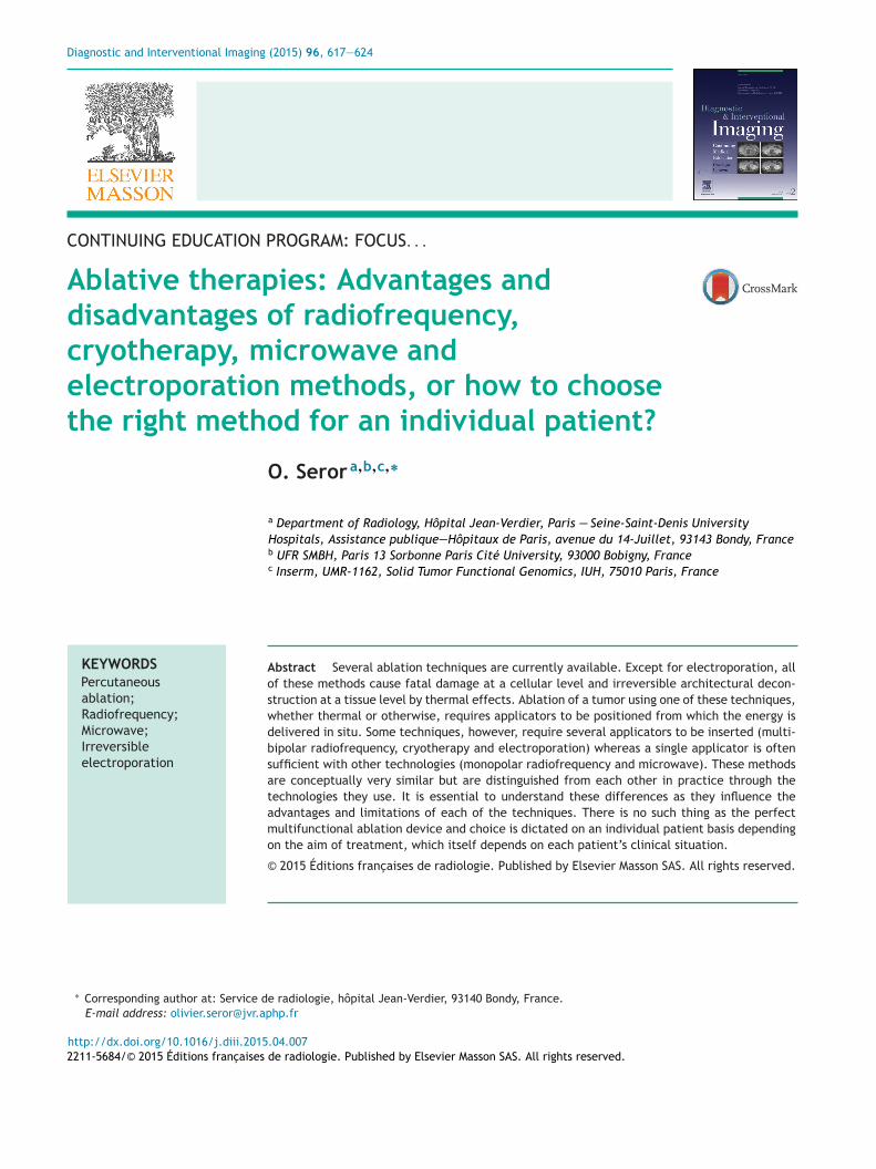
n situ destruction of a tumor has long been obtained using centrifugal coverage method: energy dissipates towardshe periphery from an applicator inserted into the centerf the tumor target [1]. Ideally energy has to isotropi-ally radiate with a minimum of loss in order to produce
fatal effect on the tumor, and beyond that over a targetedargin and a minimal thickness (Fig. 1). This centrifu-
al dispersive ablation strategy which is derived from thehemical ablation techniques is still the most widely useds it is the most simple to apply. It is the basis of tech-iques such as monopolar radiofrequency, microwave, lasernd cryotherapy. Using these techniques, an overlappingblation strategy is required for tumors over 3 cm in diame-er (1.5 cm for laser and cryotherapy) in order to destroyhe whole lesion with a sufficient margin. This may bechieved simultaneously if several applicators are used dur-ng the procedure (generally with laser and cryotherapy)2]. Fundamentally, using several applicators with centrifu-al dispersion methods does not change the operation of therocedure and final ablation is planned by simple summa-ion of several overlapping centrifugal destructions causedy each of the applicators. If the applicators are sufficientlylose together (which is essential to achieve continuousestruction between them) a more or less extensive syner-istic effect occurs. It is important, however, to understandhat each of the ablated regions created remains overallndependent. As such, they represent areas of centrifugaladial destruction, the contribution of which to the suc-ess of the whole procedure is mostly influenced by theperator’s ability to implant the applicator in the cen-er of each of the desired individual destruction zonesFig. 1). The conceptual simplicity of ablations carried outsing dispersion techniques is their main advantage. Thiss particularly apparent for monopolar radiofrequency andicrowave ablation which can destroy a relatively wide
ange of tumors up to approximately 3 cm in diameter using single applicator and therefore with a single puncture. Thisssumes, however, that the shape of the targets treated iss spherical as possible and that the assumption of isotropicnergy propagation is observed. These two conditions areenerally met for small tumors (<2.5 cm) although over thisize, large deviations from the ideal centrifugal ablationodel are seen because of the heterogeneous nature of
issue properties (electrical and thermal conduction, lightbsorption and micro- and macrocirculatory thermal convec-ion). Because of the overall spherical expansion of theblation zone it is desirable for the target treated to beemote from important structures in order to reduce theisks of complications due to collateral thermal damage.
Another ablation strategy involves convergence of energyrom the periphery towards the center of the tumor. A min-mum of two applicators are inserted into the periphery
f the tumor in order to deliver the energy concentricallyithin the target. In practice at present, only radiofre-uency and electroporation which deliver a bipolar RFurrent can use this strategy satisfactorily. The continuitycrtm
O. Seror
f the treatment zone is governed by the distance betweenhe electrodes: 3 cm for radiofrequency and 2.5 cm for elec-roporation are the distances beyond which the risk ofiscontinuity of the treated areas becomes high. For vol-metric dosimetry reasons, targets over 2 cm in diametereed to be treated along several axes of energy delivered,imilarly to the crossed beams in external conformationaltereotactic radiotherapy methods. When these systems aresed, it is recommended that at least three electrodese implanted for ‘‘one shot’’ treatment of lesions with
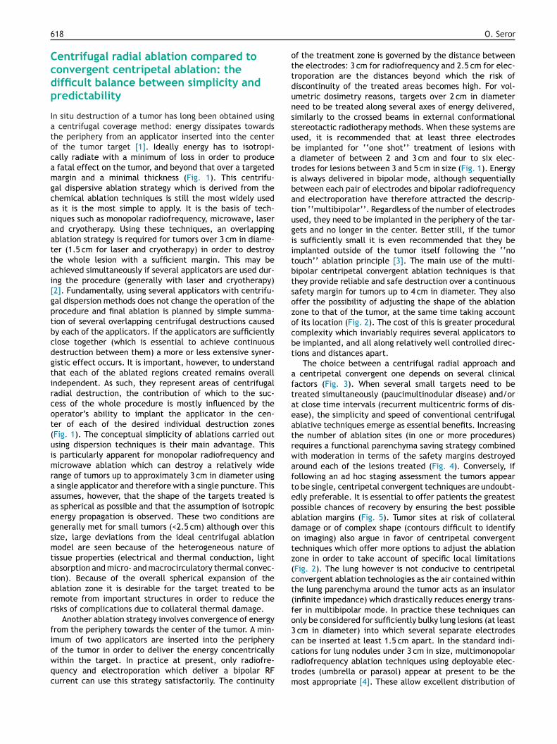
diameter of between 2 and 3 cm and four to six elec-rodes for lesions between 3 and 5 cm in size (Fig. 1). Energys always delivered in bipolar mode, although sequentiallyetween each pair of electrodes and bipolar radiofrequencynd electroporation have therefore attracted the descrip-ion ‘‘multibipolar’’. Regardless of the number of electrodessed, they need to be implanted in the periphery of the tar-ets and no longer in the center. Better still, if the tumors sufficiently small it is even recommended that they bemplanted outside of the tumor itself following the ‘‘noouch’’ ablation principle [3]. The main use of the multi-ipolar centripetal convergent ablation techniques is thathey provide reliable and safe destruction over a continuousafety margin for tumors up to 4 cm in diameter. They alsoffer the possibility of adjusting the shape of the ablationone to that of the tumor, at the same time taking accountf its location (Fig. 2). The cost of this is greater proceduralomplexity which invariably requires several applicators toe implanted, and all along relatively well controlled direc-ions and distances apart.
The choice between a centrifugal radial approach and centripetal convergent one depends on several clinicalactors (Fig. 3). When several small targets need to bereated simultaneously (paucimultinodular disease) and/ort close time intervals (recurrent multicentric forms of dis-ase), the simplicity and speed of conventional centrifugalblative techniques emerge as essential benefits. Increasinghe number of ablation sites (in one or more procedures)equires a functional parenchyma saving strategy combinedith moderation in terms of the safety margins destroyedround each of the lesions treated (Fig. 4). Conversely, ifollowing an ad hoc staging assessment the tumors appearo be single, centripetal convergent techniques are undoubt-dly preferable. It is essential to offer patients the greatestossible chances of recovery by ensuring the best possibleblation margins (Fig. 5). Tumor sites at risk of collateralamage or of complex shape (contours difficult to identifyn imaging) also argue in favor of centripetal convergentechniques which offer more options to adjust the ablationone in order to take account of specific local limitationsFig. 2). The lung however is not conducive to centripetalonvergent ablation technologies as the air contained withinhe lung parenchyma around the tumor acts as an insulatorinfinite impedance) which drastically reduces energy trans-er in multibipolar mode. In practice these techniques cannly be considered for sufficiently bulky lung lesions (at least
cm in diameter) into which several separate electrodesan be inserted at least 1.5 cm apart. In the standard indi-
ations for lung nodules under 3 cm in size, multimonopolaradiofrequency ablation techniques using deployable elec-rodes (umbrella or parasol) appear at present to be theost appropriate [4]. These allow excellent distribution of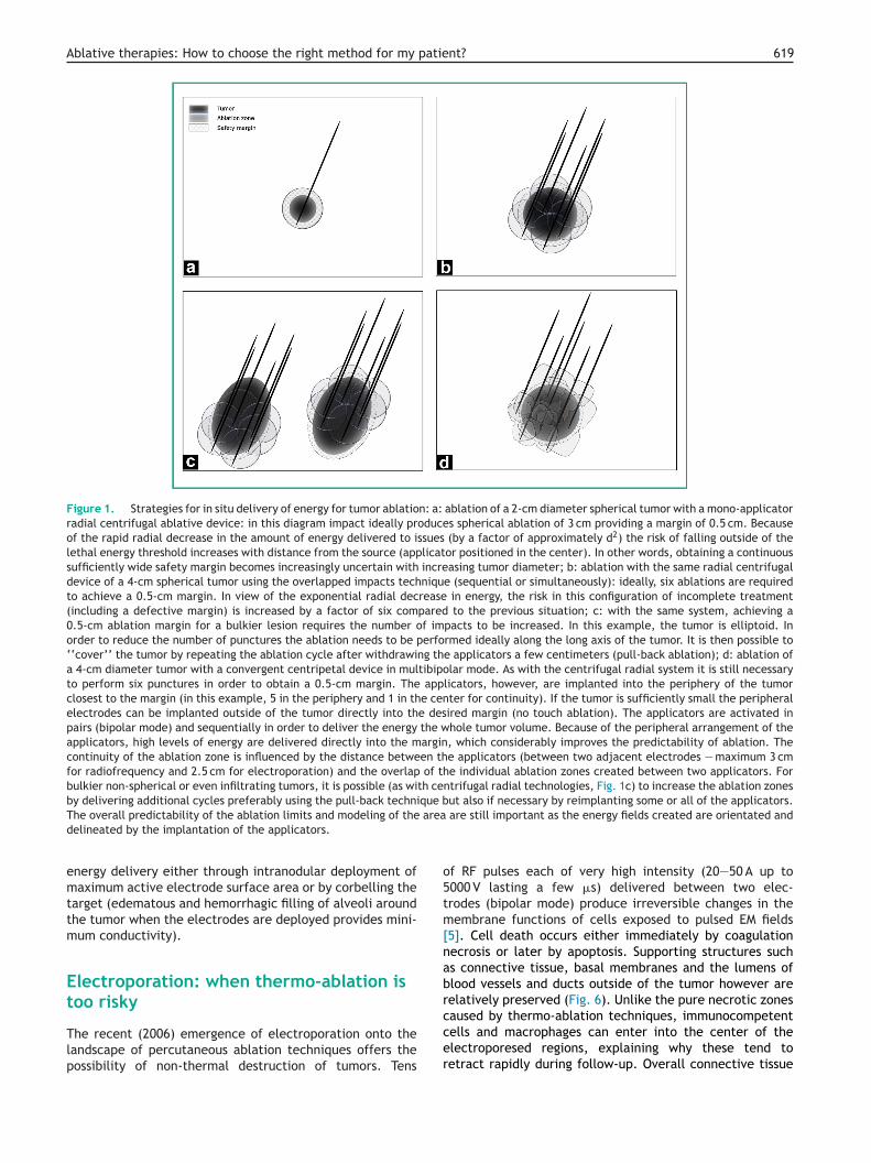
Ablative therapies: How to choose the right method for my patient? 619
Figure 1. Strategies for in situ delivery of energy for tumor ablation: a: ablation of a 2-cm diameter spherical tumor with a mono-applicatorradial centrifugal ablative device: in this diagram impact ideally produces spherical ablation of 3 cm providing a margin of 0.5 cm. Becauseof the rapid radial decrease in the amount of energy delivered to issues (by a factor of approximately d2) the risk of falling outside of thelethal energy threshold increases with distance from the source (applicator positioned in the center). In other words, obtaining a continuoussufficiently wide safety margin becomes increasingly uncertain with increasing tumor diameter; b: ablation with the same radial centrifugaldevice of a 4-cm spherical tumor using the overlapped impacts technique (sequential or simultaneously): ideally, six ablations are requiredto achieve a 0.5-cm margin. In view of the exponential radial decrease in energy, the risk in this configuration of incomplete treatment(including a defective margin) is increased by a factor of six compared to the previous situation; c: with the same system, achieving a0.5-cm ablation margin for a bulkier lesion requires the number of impacts to be increased. In this example, the tumor is elliptoid. Inorder to reduce the number of punctures the ablation needs to be performed ideally along the long axis of the tumor. It is then possible to‘‘cover’’ the tumor by repeating the ablation cycle after withdrawing the applicators a few centimeters (pull-back ablation); d: ablation ofa 4-cm diameter tumor with a convergent centripetal device in multibipolar mode. As with the centrifugal radial system it is still necessaryto perform six punctures in order to obtain a 0.5-cm margin. The applicators, however, are implanted into the periphery of the tumorclosest to the margin (in this example, 5 in the periphery and 1 in the center for continuity). If the tumor is sufficiently small the peripheralelectrodes can be implanted outside of the tumor directly into the desired margin (no touch ablation). The applicators are activated inpairs (bipolar mode) and sequentially in order to deliver the energy the whole tumor volume. Because of the peripheral arrangement of theapplicators, high levels of energy are delivered directly into the margin, which considerably improves the predictability of ablation. Thecontinuity of the ablation zone is influenced by the distance between the applicators (between two adjacent electrodes — maximum 3 cmfor radiofrequency and 2.5 cm for electroporation) and the overlap of the individual ablation zones created between two applicators. Forbulkier non-spherical or even infiltrating tumors, it is possible (as with centrifugal radial technologies, Fig. 1c) to increase the ablation zonesby delivering additional cycles preferably using the pull-back technique but also if necessary by reimplanting some or all of the applicators.
area
o5tm[nabr
The overall predictability of the ablation limits and modeling of thedelineated by the implantation of the applicators.
energy delivery either through intranodular deployment ofmaximum active electrode surface area or by corbelling thetarget (edematous and hemorrhagic filling of alveoli aroundthe tumor when the electrodes are deployed provides mini-mum conductivity).
Electroporation: when thermo-ablation istoo risky
The recent (2006) emergence of electroporation onto thelandscape of percutaneous ablation techniques offers thepossibility of non-thermal destruction of tumors. Tens
ccer
are still important as the energy fields created are orientated and
f RF pulses each of very high intensity (20—50 A up to000 V lasting a few �s) delivered between two elec-rodes (bipolar mode) produce irreversible changes in theembrane functions of cells exposed to pulsed EM fields
5]. Cell death occurs either immediately by coagulationecrosis or later by apoptosis. Supporting structures suchs connective tissue, basal membranes and the lumens oflood vessels and ducts outside of the tumor however areelatively preserved (Fig. 6). Unlike the pure necrotic zones
aused by thermo-ablation techniques, immunocompetentells and macrophages can enter into the center of thelectroporesed regions, explaining why these tend toetract rapidly during follow-up. Overall connective tissue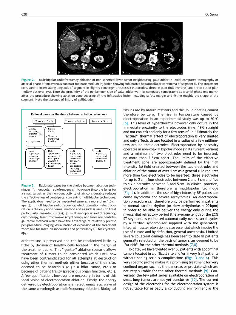
620 O. Seror
Figure 2. Multibipolar radiofrequency ablation of non-spherical liver tumor neighbouring gallbladder: a: axial computed tomography atarterial phase of intravenous contrast iodinate medium injection showing infiltrative hepatocellular carcinoma of segment 5. The treatmentconsisted to insert along long axis of segment in slightly convergent routes six electrodes, three in plan (full overlays) and three out of plan(hollow out overlays). Note the proximity of the peritoneum side of gallbladder wall; b: computed tomography at arterial phase one monthafter the procedure showing ablation zone covering all the infiltrative lsegment. Note the absence of injury of gallbladder.
Figure 3. Rationale bases for the choice between ablation tech-niques: *: monopolar radiofrequency, microwave (into the lungs fora small target as the non-conductivity of air considerably reducesthe effectiveness of centripetal concentric multibipolar techniques.The applicators need to be implanted generally more than 1.5 cmapart); †: multibipolar radiofrequency, electroporation (electropo-ration is the only non-thermal method and as such is useful to treatparticularly hazardous sites); ‡: multimonopolar radiofrequency,cryotherapy, laser, microwave (cryotherapy and laser are centrifu-gal radial methods which have the advantage of relatively precisepza
altthudbAidt
tte[ia‘atoantiamfte(ctiimSbIusg‘
twvcn
er-procedure imaging visualization of expansion of the treatmentone: MRI for laser, all modalities and particularly CT for cryother-py).
rchitecture is preserved and can be recolonized little byittle by division of healthy cells located in the margin ofhe treatment zone. This ‘‘gentle’’ ablation scenario allowsreatment of tumors to be considered which until nowave been contraindicated for all attempts at destructionsing other thermal methods either because of their site,eemed to be hazardous (e.g.: a hilar tumor, etc.) orecause of patient frailty (precarious organ function, etc.).
few qualifications however are necessary in terms of thisdeal vision of electroporation ablation. Firstly, the energyelivered by electroporation is an electromagnetic wave ofhe same wavelength as radiofrequency ablation. Biological
vsdn
esion including safety margin and fitting roughly the shape of the
issues are by nature resistors and the Joule heating cannotherefore be zero. The rise in temperature caused bylectroporation in an experimental study was up to 60 ◦C6]. This level of hyperthermia however only occurs in themmediate proximity to the electrodes (fine, 19 G straightnd not cooled) and only for a few tens of �s. Ultimately the‘actual’’ thermal effect of electroporation is very limitednd only affects tissues located in a radius of a few millime-ers around the electrodes. Electroporation by necessityperates in non-coaxial bipolar mode (in its current version)nd a minimum of two electrodes need to be inserted,o more than 2.5 cm apart. The limits of the effectivereatment zone are approximately defined by the highntensity EM field created between the two electrodes andblation of the tumor of over 1 cm as a general rule requiresore than two electrodes to be inserted: three electrodes
or up to 2 cm, four electrodes between 2 and 3 cm and fiveo six electrodes between 3 and 5 cm. In clinical practice,lectroporation is therefore a multibipolar techniqueFig. 1). In addition, the use of high intensity RF pulses canause myoclonia and severe arrhythmias. An electropora-ion procedure can therefore only be performed in patientsn normal cardiac rhythm (or slow arrhythmias <100 bpm)n order to be able to deliver the energy only during theyocardial refractory period (the average length of the ECG
T segments is estimated automatically over several cyclesy a cardiac synchronizer supplied with the generator).ntegral muscle relaxation is also essential which implies these of curare and by definition, general anesthesia. Limitedevere collateral damage has been seen to date in patientsenerally selected on the basis of tumor sites deemed to be‘at risk’’ for the other thermal methods [7,8].
To date, we have treated over 50 patients with abdominalumors located in a difficult site and/or in very frail patientsithout seeing serious complications (Figs. 3 and 6). Thisery specific profile makes it a promising treatment for veryonfined organs such as the pancreas or prostate which areot very suitable for the other thermal methods [9]. Con-
ersely, the few pilot series available on electroporation ofmall lung tumors are not yet conclusive [10]. The currentesign of the electrodes for the electroporation system isot suitable for as badly a conducting environment as the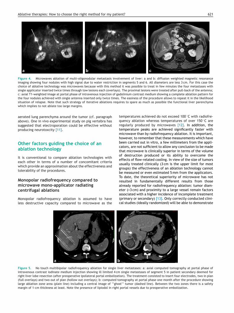
Ablative therapies: How to choose the right method for my patient? 621
Figure 4. Microwaves ablation of multi-oligonodular metastasis involvement of liver: a and b: diffusion weighted magnetic resonanceimaging showing four nodules with high signal due to water restriction in segments 5 and 6. All diameters are less 3 cm. For this case thechoice of ablative technology was microwaves because with this method it was possible to treat in few minutes the four metastases withsingle applicator inserted twice times through tow lesions each (overlays). The proximal lesions were treated after pull-back of the antenna;c: axial T1-weighted image at portal phase of intravenous injection of gadolinium contrast medium showing a complete ablation pattern forthe four nodules achieved with single antenna inserted only twice times. The easiness of the procedure allows to repeat it in the likelihood
requ
tqrtmhbctoeugbTraeter (>3 cm) and proximity to a large vessel remain factors
situation of relapse. Note that such strategy of iterative ablations
which implies to not ablate too large margins.
aerated lung parenchyma around the tumor (cf. paragraphabove). One in vivo experimental study on pig vertebra hassuggested that electroporation could be effective withoutproducing neurotoxcity [11].
Other factors guiding the choice of anablation technology
It is conventional to compare ablation technologies witheach other in terms of a number of concomitant criteriawhich provide an approximation about the effectiveness andtolerability of the procedures.
Monopolar radiofrequency compared tomicrowave mono-applicator radiating
centrifugal ablationsMonopolar radiofrequency ablation is assumed to haveless destructive capacity compared to microwave as the
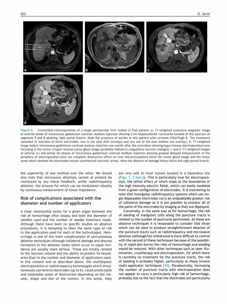
a(c
Figure 5. No touch multibipolar radiofrequency ablation for single livintravenous contrast iodinate medium injection showing ill limited 4 cmright liver lobe resection (after preoperative ipsilateral portal embolizati(full overlays) and two out of plan (hollow out overlays); b: computed tolarge ablation zone area (plain line) including a central image of ‘‘ghosmargin of 1-cm thickness at least. Note the presence of lipiodol in right
ires to spare as much as possible the functional liver parenchyma
emperatures achieved do not exceed 100 ◦C with radiofre-uency ablation whereas temperatures of over 150 ◦C areegularly produced by microwaves [12]. In addition, theemperature peaks are achieved significantly faster withicrowave than by radiofrequency ablation. It is important,
owever, to remember that these measurements which haveeen carried out in vitro, a few millimeters from the appli-ators, are not sufficient to allow any conclusion to be madehat microwave is clinically superior in terms of the volumef destruction produced or its ability to overcome theffects of flow-related cooling. In view of the size of tumorssually treated clinically (3 cm is the upper limit for mostroups) the effectiveness of an ablation technology cannote measured or even estimated 5 mm from the applicators.o date, the theoretical superiority of microwave has notesulted in fundamentally different results from thoselready reported for radiofrequency ablation: tumor diam-
ssociated with a higher incidence of incomplete treatmentprimary or secondary) [13]. Only correctly conducted clini-al studies (ideally randomized) will be able to demonstrate
er metastases: a: axial computed tomography at portal phase of single metastases of segment 5 in patient secondary deemed foron). The treatment consisted to insert four electrodes, two in planmography at portal phase one month after the procedure showingt’’ tumor (dashed line). Between the two zones there is a safetyportal vessels due to preoperative embolization.
622 O. Seror
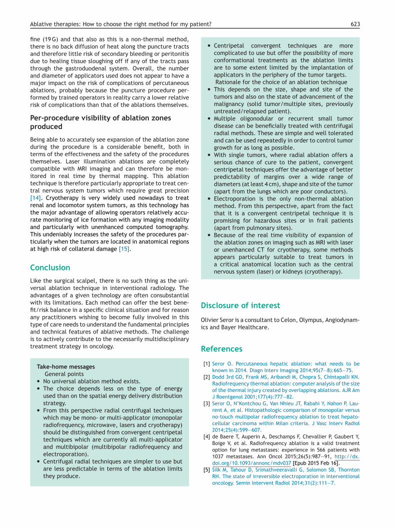
Figure 6. Irreversible electroporation of a single perivascular liver nodule in frail patient: a: T1-weighted resonance magnetic imageat arterial phase of intravenous gadolinium contrast medium injection showing 2 cm hepatocellular carcinoma located at the junction ofsegments 5 and 8 abutting right portal branch. Note the presence of ascites in this patient with cirrhosis Child-Pugh B. The treatmentconsisted in insertion of three electrodes, two in the plan (full overlays) and one out of the plan (hollow out overlay); b: T1-weightedimage before intravenous gadolinium contrast medium injection one month after the procedure showing hypo-intense electroporated zoneincluding in the center a hyper-intense tumor ghost image (probably related to coagulative necrotic changes); c and d: T1-weighted imagesat arterial (c) and portal (d) phases of intravenous gadolinium contrast medium injection showing gradual delayed enhancement of thep on lia eas).
tamab
Rd
ArnAptoafdtaIetau
a(ttfngot
orawtawiwmio
eriphery of electroporated zone (no complete destructive effectreas which abutted the electrodes remain unenhanced (necrotic ar
he superiority of one method over the other. We shouldlso note that microwave ablations cannot at present beonitored by any tissue feedback, unlike radiofrequency
blation, the process for which can be modulated robustlyy continuous measurement of tissue impedance.
isk of complications associated with theiameter and number of applicators
clear relationship exists for a given organ between theisk of hemorrhage after biopsy and both the diameter ofeedles used and the number of needle insertions made.lthough there have been no specific studies on ablationrocedures, it is tempting to liken the same type of risko the applicators used for each of the technologies. Hem-rrhage is one of the main complications of percutaneousblation techniques although collateral damage and abscessormation in the ablation zones which occur in equal inci-ence are usually more serious. These are mostly relatedo the necrosis volume and the ability to predict its bound-ries than to the number and diameter of applicators used.n this context and as described above, the multibipolar
lectroporation or radiofrequency technologies which simul-aneously use several electrodes (up to 6), cause predictablend modulable zones of destruction depending on the vol-me, shape and site of the tumors. In this sense, theymtnp
ver microcirculation) while the tumor ghost image and the linear Note the absence of damage biliary ducts and right portal branch.
re very safe to treat tumors located in a hazardous siteFigs. 1, 2 and 6). This is particularly true for electropora-ion, the lethal effect of which stops at the boundaries ofhe high intensity electric fields, which can easily modeledrom a given configuration of electrodes. It is interesting toote that monopolar radiofrequency systems which use sin-le deployable electrodes carry an undoubtedly greater riskf collateral damage as it is not possible to monitor all ofhe paths of the electrodes by imaging as they are deployed.
Conversely, in the same way as for hemorrhage, the riskf seeding of malignant cells along the puncture tracts iselated to the number of punctures performed. As these areblative techniques it is reasonable to consider that thosehich can be used to produce straightforward ablation of
he puncture tracts such as radiofrequency and microwaveblation (although hot withdrawal is more difficult to controlith the second of these techniques because of the possibil-
ty of rapid skin burns) the risks of hemorrhage and seedingould be reduced. With other techniques such as laser illu-ination, cryotherapy and electroporation, for which there
s currently no treatment for the puncture tracts, the riskf seeding is probably higher, particularly as these involve
ulti-applicator techniques [10]. Paradoxically, increasinghe number of puncture tracts with electroporation doesot appear to carry a particularly high risk of hemorrhage,robably due to the fact that the electrodes are particularly
patie
D
Oi
R
Ablative therapies: How to choose the right method for my
fine (19 G) and that also as this is a non-thermal method,there is no back diffusion of heat along the puncture tractsand therefore little risk of secondary bleeding or peritonitisdue to healing tissue sloughing off if any of the tracts passthrough the gastroduodenal system. Overall, the numberand diameter of applicators used does not appear to have amajor impact on the risk of complications of percutaneousablations, probably because the puncture procedure per-formed by trained operators in reality carry a lower relativerisk of complications than that of the ablations themselves.
Per-procedure visibility of ablation zonesproduced
Being able to accurately see expansion of the ablation zoneduring the procedure is a considerable benefit, both interms of the effectiveness and the safety of the proceduresthemselves. Laser illumination ablations are completelycompatible with MRI imaging and can therefore be mon-itored in real time by thermal mapping. This ablationtechnique is therefore particularly appropriate to treat cen-tral nervous system tumors which require great precision[14]. Cryotherapy is very widely used nowadays to treatrenal and locomotor system tumors, as this technology hasthe major advantage of allowing operators relatively accu-rate monitoring of ice formation with any imaging modalityand particularly with unenhanced computed tomography.This undeniably increases the safety of the procedures par-ticularly when the tumors are located in anatomical regionsat high risk of collateral damage [15].
Conclusion
Like the surgical scalpel, there is no such thing as the uni-versal ablation technique in interventional radiology. Theadvantages of a given technology are often consubstantialwith its limitations. Each method can offer the best bene-fit/risk balance in a specific clinical situation and for reasonany practitioners wishing to become fully involved in thistype of care needs to understand the fundamental principlesand technical features of ablative methods. The challengeis to actively contribute to the necessarily multidisciplinarytreatment strategy in oncology.
Take-home messagesGeneral points
• No universal ablation method exists.• The choice depends less on the type of energy
used than on the spatial energy delivery distributionstrategy.
• From this perspective radial centrifugal techniqueswhich may be mono- or multi-applicator (monopolarradiofrequency, microwave, lasers and cryotherapy)should be distinguished from convergent centripetaltechniques which are currently all multi-applicatorand multibipolar (multibipolar radiofrequency andelectroporation).
• Centrifugal radial techniques are simpler to use butare less predictable in terms of the ablation limitsthey produce.
nt? 623
• Centripetal convergent techniques are morecomplicated to use but offer the possibility of moreconformational treatments as the ablation limitsare to some extent limited by the implantation ofapplicators in the periphery of the tumor targets.Rationale for the choice of an ablation technique
• This depends on the size, shape and site of thetumors and also on the state of advancement of themalignancy (solid tumor/multiple sites, previouslyuntreated/relapsed patient).
• Multiple oligonodular or recurrent small tumordisease can be beneficially treated with centrifugalradial methods. These are simple and well toleratedand can be used repeatedly in order to control tumorgrowth for as long as possible.
• With single tumors, where radial ablation offers aserious chance of cure to the patient, convergentcentripetal techniques offer the advantage of betterpredictability of margins over a wide range ofdiameters (at least 4 cm), shape and site of the tumor(apart from the lungs which are poor conductors).
• Electroporation is the only non-thermal ablationmethod. From this perspective, apart from the factthat it is a convergent centripetal technique it ispromising for hazardous sites or in frail patients(apart from pulmonary sites).
• Because of the real time visibility of expansion ofthe ablation zones on imaging such as MRI with laseror unenhanced CT for cryotherapy, some methodsappears particularly suitable to treat tumors ina critical anatomical location such as the centralnervous system (laser) or kidneys (cryotherapy).
isclosure of interest
livier Seror is a consultant to Celon, Olympus, Angiodynam-cs and Bayer Healthcare.
eferences
[1] Seror O. Percutaneous hepatic ablation: what needs to beknown in 2014. Diagn Interv Imaging 2014;95(7—8):665—75.
[2] Dodd 3rd GD, Frank MS, Aribandi M, Chopra S, Chintapalli KN.Radiofrequency thermal ablation: computer analysis of the sizeof the thermal injury created by overlapping ablations. AJR AmJ Roentgenol 2001;177(4):777—82.
[3] Seror O, N’Kontchou G, Van Nhieu JT, Rabahi Y, Nahon P, Lau-rent A, et al. Histopathologic comparison of monopolar versusno-touch multipolar radiofrequency ablation to treat hepato-cellular carcinoma within Milan criteria. J Vasc Interv Radiol2014;25(4):599—607.
[4] de Baere T, Auperin A, Deschamps F, Chevallier P, Gaubert Y,Boige V, et al. Radiofrequency ablation is a valid treatmentoption for lung metastases: experience in 566 patients with1037 metastases. Ann Oncol 2015;26(5):987—91, http://dx.
doi.org/10.1093/annonc/mdv037 [Epub 2015 Feb 16].[5] Silk M, Tahour D, Srimathveeravalli G, Solomon SB, ThorntonRH. The state of irreversible electroporation in interventionaloncology. Semin Intervent Radiol 2014;31(2):111—7.
6
[
[
[
[
[
24
[6] Dunki-Jacobs EM, Philips P, Martin 2nd RC. Evaluation of ther-mal injury to liver, pancreas and kidney during irreversibleelectroporation in an in vivo experimental model. Br J Surg2014;101(9):1113—21.
[7] Narayanan G, Bhatia S, Echenique A, Suthar R, Barbery K,Yrizarry J. Vessel patency post-irreversible electroporation.Cardiovasc Intervent Radiol 2014;37(6):1523—9.
[8] Scheffer HJ, Melenhorst MC, van Tilborg AA, Nielsen K, vanNieuwkerk KM, de Vries RA, et al. Percutaneous irreversibleelectroporation of a large centrally located hepatocellular ade-noma in a woman with a pregnancy wish. Cardiovasc InterventRadiol 2014 [Epub ahead of print].
[9] Scheffer HJ, Nielsen K, de Jong MC, van Tilborg AA, Vieveen JM,Bouwman AR, et al. Irreversible electroporation for nonthermaltumor ablation in the clinical setting: a systematic review ofsafety and efficacy. J Vasc Interv Radiol 2014;25(7):997—1011.
10] Ricke J, Jurgens JH, Deschamps F, Tselikas L, Uhde K, Kosiek
O, et al. Irreversible electroporation (IRE) fails to demon-strate efficacy in a prospective multicenter phase ii trial onlung malignancies: the ALICE trial. Cardiovasc Intervent Radiol2015;38(2):401—8.[
O. Seror
11] Tam AL, Abdelsalam ME, Gagea M, Ensor JE, Moussa M, AhmedM, et al. Irreversible electroporation of the lumbar vertebrae ina porcine model: is there clinical-pathologic evidence of neuraltoxicity? Radiology 2014;272(3):709—19.
12] Simon CJ, Dupuy DE, Mayo-Smith WW. Microwave abla-tion: principles and applications. Radiographics 2005;25(Suppl.1):S69—83.
13] Vogl TJ, Farshid P, Naguib NN, Darvishi A, BazrafshanB, Mbalisike E, et al. Thermal ablation of liver metas-tases from colorectal cancer: radiofrequency, microwaveand laser ablation therapies. Radiol Med 2014;119(7):451—61.
14] Rahmathulla G, Recinos PF, Kamian K, Mohammadi AM,Ahluwalia MS, Barnett GH. MRI-guided laser interstitialthermal therapy in neuro-oncology: a review of its cur-rent clinical applications. Oncology 2014;87(2):67—82,http://dx.doi.org/10.1159/000362817 [Epub 2014 Jul 3].
15] Zargar H, Atwell TD, Cadeddu JA, de la Rosette JJ, JanetschekG, Kaouk JH, et al. Cryoablation for small renal masses:selection criteria, complications, and functional and oncologicresults. Eur Urol 2015.