continuing medical education activity in american … in adults - update on...continuing medical...
TRANSCRIPT

Continuing medical education activityin American Journal of Hematology
CME Editor: Ayalew Tefferi, MDAuthor: Animesh Pardanani, MBBS, PhD
Article Title: Systemic mastocytosis in adults: 2012update on Diagnosis, Risk-stratification and ManagementIf you wish to receive credit for this activity, please refer
to the website: www.wileyhealthlearning.com
Accreditation and designation statement:Blackwell Futura Media Services is accredited by the
Accreditation Council for Continuing Medical Education toprovide continuing medical education for physicians.Blackwell Futura Media Services designates this journal-
based CME for amaximum of 1 AMAPRACategory 1 CreditTM.Physicians should only claim credit commensurate with theextent of their participation in the activity
Educational objectivesUpon completion of this educational activity, participants
will be better able to:
! To review current pathogenetic concepts, classifica-tion, diagnosis and risk stratification of systemic mas-tocytosis.
! To review current management of systemic mastocyto-sis, including an update of investigational therapies
Activity disclosuresNo commercial support has been accepted related to the
development or publication of this activity.CME Editor—Ayalew Tefferi, MD has no conflicts of inter-
est to discloseAuthor—Animesh Pardanani, MBBS, PhD has no con-
flicts of interest to discloseThis activity underwent peer review in line with the stand-
ards of editorial integrity and publication ethics maintainedby American Journal of Hematology. The peer reviewershave no conflicts of interest to disclose. The peer reviewprocess for American Journal of Hematology is single
blinded. As such, the identities of the reviewers are not dis-closed in line with the standard accepted practices of medi-cal journal peer review.Conflicts of interest have been identified and
resolved in accordance with Blackwell Futura MediaServices’s Policy on Activity Disclosure and Conflict ofInterest. The primary resolution method used was peerreview and review by a non-conflicted expert.
Instructions on receiving creditThis activity is intended for physicians. For information
on applicability and acceptance of continuing medical edu-cation credit for this activity, please consult your professio-nal licensing board.This activity is designed to be completed within one hour;
physicians should claim only those credits that reflect the timeactually spent in the activity. To successfully earn credit,participants must complete the activity during the valid creditperiod, which is up to two years from initial publication.Follow these steps to earn credit:
! Log on to www.wileyhealthlearning.com
! Read the target audience, educational objectives, andactivity disclosures.
! Read the activity contents in print or online format.
! Reflect on the activity contents.
! Access the CME Exam, and choose the best answerto each question.
! Complete the required evaluation component of theactivity.
! Claim your Certificate.
This activity will be available for CME credit for twelvemonths following its launch date. At that time, it will bereviewed and potentially updated and extended for an addi-tional twelve months.
VVC 2012 Wiley Periodicals, Inc.
American Journal of Hematology 401 http://wileyonlinelibrary.com/cgi-bin/jhome/35105

ANNUAL CLINICAL UPDATES IN HEMATOLOGICAL MALIGNANCIES: A CONTINUING
MEDICAL EDUCATION SERIES
Systemic mastocytosis in adults: 2012 Update on diagnosis, riskstratification, and management
Animesh Pardanani*
Disease Overview: Systemic mastocytosis (SM) results from a clonal proliferation of abnormal mast cells(MC) in one or more extra-cutaneous organs.Diagnosis: The major criterion is presence of multifocal clusters of morphologically abnormal MC in thebone marrow. Minor diagnostic criteria include elevated serum tryptase level, abnormal MC expression ofCD25 and/or CD2, and presence of KITD816V.Risk Stratification: The prognostic relevance of the 2008 World Health Organization (WHO) classification ofSM has recently been confirmed. Classification of SM patients into indolent (SM), aggressive SM (ASM), SMassociated with a clonal non-MC lineage disease (SM-AHNMD) and mast cell leukemia (MCL) subgroups isa useful first step in establishing prognosis.Management: SM treatment is generally palliative. ISM patients have a normal life expectancy and receivesymptom-directed therapy; infrequently, cytoreductive therapy may be indicated for refractory symptoms.ASM patients have disease-related organ dysfunction; interferon-a (±corticosteroids) can control dermato-logical, hematological, gastrointestinal, skeletal, and mediator-release symptoms, but is hampered by poortolerability. Similarly, cladribine has broad therapeutic activity, with particular utility when rapid MC debulk-ing is indicated; the main toxicity is myelosuppression. Imatinib has a therapeutic role in the presence ofan imatinib-sensitive KIT mutation or in KITD816-unmutated patients. Treatment of SM-AHNMD is governedprimarily by the non-MC neoplasm; hydroxyurea has modest utility in this setting.Investigational Drugs: Dasatinib’s in vitro anti-KITD816V activity has not translated into significant therapeuticactivity in most SM patients. In contrast, preliminary data suggest that Midostaurin may produce significantdecreases in MC burden in some patients. Am. J. Hematol. 87:402–411, 2012. VVC 2012 Wiley Periodicals, Inc.
Disease Overview and PathogenesisMastocytosis is one of eight subcategories of myeloproli-
ferative neoplasms (MPN) per the 2008 World Health Orga-nization (WHO) classification of tumors of hematopoieticand lymphoid tissues [1]. It results from a clonal, neoplasticproliferation of morphologically and immunophenotypicallyabnormal mast cells (MC) that accumulate in one or moreorgan systems. The sine qua non of mastocytosis is thepresence of multifocal clusters of abnormal MC, which incontrast to normal MC are variable in appearance, rangingfrom round to fusiform variants with long, polar cytoplasmicprocesses, and may display cytoplasmic hypogranularitywith uneven distribution of fine granules, as well as atypicalnuclei with monocytoid appearance [2–4].The clinical presentation of mastocytosis is heterogene-
ous, ranging from skin-limited disease (cutaneous mastocy-tosis, CM), particularly in pediatric cases where the majorityhave disease-onset within the first 2 years of life and com-monly experience spontaneous regression of skin lesions[5–8], to a more aggressive variant with extra-cutaneousinvolvement (systemic mastocytosis, SM) that may beassociated with multiorgan dysfunction/failure and short-ened survival, that is generally seen in adult patients [9].The WHO document distinguishes the usually KIT-
mutated SM from a Philadelphia chromosome-negativeMPN with hematological features of chronic eosinophilicleukemia associated with splenomegaly, marked elevationof serum vitamin B12, elevation of serum tryptase, andincreased bone marrow (BM) MC commonly in scattered ornoncohesive clusters [10]. The latter entity is commonlyassociated with rearrangement of PDGFRA (i.e. FIP1L1-PDGFR) and less commonly, PDGFRB (e.g. PRKG2-PDGFRB), and is sensitive to treatment with imatinib [11–18]. WHO-defined SM is sometimes associated with a
clonally-related second myeloid neoplasm [19–22], which isnot surprising considering its origin as a stem cell diseasewith multilineage clonal involvement [23–25]. Conversely,an otherwise well-defined myeloid malignancy, such asmyelodysplastic syndrome (MDS) or a non-mast cell dis-ease MPN, might also harbor neoplastic mast cells [26].Mastocytosis is frequently associated with somatic gain-
of-function point mutations within KIT. KIT (CD117) is aType III receptor tyrosine kinase that is characterized by anextracellular domain comprised of five immunoglobulin (Ig)-like subdomains, a hydrophobic transmembrane region, anegative regulatory juxta-membrane intracellular domain,and a cytoplasmic tyrosine kinase domain that is split by a77-amino acid hydrophilic insert sequence into adenosinetriphosphate binding and phosphotransferase domains [27].KIT is notably expressed by MC, hematopoietic progenitorcells, germ cells, melanocytes, and interstitial cells of Cajalin the gastrointestinal tract and is therefore functionally rel-evant for normal mast cell development, hematopoiesis,gametogenesis, melanogenesis, and regulation of slowgastric waves [28]. KIT expression is down regulated upondifferentiation of hematopoietic progenitors into mature cells
Division of Hematology, Department of Medicine, Mayo Clinic, Rochester,Minnesota
Conflict of interest: Nothing to report
*Correspondence to: Animesh Pardanani, Department of Medicine, Divisionof Hematology, Mayo Clinic, 200 First St. SW, Rochester, MN 55905. E-mail:[email protected]
Received for publication 17 January 2012; Accepted 18 January 2012
Am. J. Hematol. 87:402–411, 2012.
Published online in Wiley Online Library (wileyonlinelibrary.com).DOI: 10.1002/ajh.23134
AJH Educational Material
VVC 2012 Wiley Periodicals, Inc.
American Journal of Hematology 402 http://wileyonlinelibrary.com/cgi-bin/jhome/35105

of all lineages, except mast cells, which retain high levelsof cell surface KIT expression. The interaction between KITand its ligand, stem cell factor (SCF), plays a key role inregulating mast cell proliferation, maturation, adhesion, che-motaxis, and survival [29].Gain-of-function somatic mutations in the KIT tyrosine ki-
nase domain, particularly the D816V mutation, have beenfound to occur in a majority of cases of adult SM, irrespec-tive of WHO SM subtype [9,30]. Other less common (<5%)somatic KIT mutations identified in adult SM include V560G[31,32], D815K [33], D816Y [30,33–35], insVI815-816 [30],D816F [33,35], D816H [36], and D820G [37]. Recent stud-ies have confirmed that childhood-onset mastocytosis isalso clearly clonal in nature, and is associated with germ-line or acquired activating KIT mutations [38–40]. In onestudy of pediatric CM that screened the entire KIT codingsequence for mutations using skin lesional DNA, only 42%of cases harbored missense mutations targeting KITD816;in 44% of cases, genetic alterations (insertions (insFF419),deletions (D419), deletion-insertions (D417-419insY), inter-nal tandem duplications (ITD SA501-502, ITD AY502-503,ITD NFAF505-508), and missense mutations (D816V,D816Y, D816I, C443Y, S476I, K509I, D572A, M541L)) werefound to mainly involve exons 8 and 9, which encode thefifth Ig (D5) domain and the extracellular region near thetransmembrane domain, regions that have previously beenshown to be affected in core-binding factor-acute myeloidleukemia (CBF-AML) and in gastrointestinal stromal tumors(GISTs), respectively [41,42]. The aforementioned muta-tions in the exons 8 and 9 have rarely been described inpediatric or adult mastocytosis, although individual muta-tions have been reported in CBF-AML (D417-419insY)[41,43], kindreds with familial GISTs and mastocytosis (D419) [44], familial mastocytosis (K509I) [45], and GISTs(ITD AY502-503) [42]. As with KITD816V, every one of themutations in exons 8 and 9 that was tested was found toconstitutively activate KIT kinase activity. Other rare germ-line KIT mutations that target the transmembrane domainand that are associated with familial mastocytosis includeF522C and A533D [46,47].While activating KIT mutations are frequently associated
with human mastocytosis, they do not occur universally,and the question as to whether individual mutations arenecessary and sufficient to cause mast cell transformationand whether such mutations alone explain the diverse clini-cal presentations of mastocytosis remains currently unset-tled. Furthermore, while childhood- and adult-onset masto-cytosis are both associated with activating KIT mutations,the natural history of the two conditions is quite different,with the former often exhibiting skin-limited disease thatspontaneously regresses with age; in contrast, the latter ischaracterized by persistent multiorgan involvement, oftenwith second non-MC hematologic neoplasm.Experimental data with regard to this issue have not
been conclusive; in transgenic mice expressing humanKITD816V in mature MC (under the control of the chymasepromoter), only a subset (30%) of mice developed a limitedform of mastocytosis (some with cutaneous-limited disease)at an old age (12–18 months) [48]. Although BM-derivedMC from the transgenic animals eventually became growthfactor independent and could be maintained in long-termcultures, the incomplete disease penetrance in this modelsuggested that additional somatic mutations are necessaryfor full MC transformation. In another transgenic mousemodel that allowed conditional expression of murineKITD814V (the homolog of human KITD816V) driven bythe KIT promoter, expression of mutant KIT in adult miceincluding in hematopoietic precursors caused severe mas-tocytosis with 100% penetrance at a young age [49].
Approximately half of the mice developed a non-MC lineagehematologic neoplasm, most frequently a leukemic diseasederived from an immature B-cell precursor. The mice alsodeveloped a severe focal inflammatory colitis associated witha massive increase in mucosal mast cell numbers. In con-trast, when mutant KIT expression in this model was limitedto more mature MC, disease expression was significantlyattenuated; while half of the mice developed MC tumors anderosive skin lesions and all developed severe colitis, thedisease occurred significantly later and progressed muchslower. While both the aforementioned transgenic murinemodels are imperfect (abnormal intracellular processing/traf-ficking of human KITD816V resulting in low oncogenicity inthe former, and low transgene expression in the latter), theycumulatively suggest that the effects of constitutive KITsignaling depend on the developmental stage of the celltargeted by the gain-of-function mutation. As has been notedin mastocytosis patients [30], mutations targeting undifferen-tiated progenitors result in multilineage involvement andexpression of a severe systemic disease phenotype; incontrast, mutations that target committed MC progenitors ormature MC result in milder forms of the disease.Other oncogenic mutations recently identified in mastocy-
tosis patients include those in TET2 (TET oncogene familymember 2) and N-RAS [50,51]. These mutations are notspecific to mastocytosis and their pathogenetic role and/orprognostic impact is currently uncertain. TET2 is a putativetumor suppressor gene; in one study, mutational frequencyin SM was 29% and the presence of TET2 mutations wasassociated with monocytosis. Further, TET2 mutations co-segregated with KITD816V but did not appear to affect sur-vival in SM. Interestingly, expression of an activated M-RASmutant (Q71L) in primary murine BM cells reproduciblygenerated a lethal mastocytosis and mast cell leukemia(MCL); in contrast, expression of constitutively activated H-RAS (G21V) produced a lethal histiocytic/monocytic leuke-mia, presumably reflecting significant differences in down-stream signaling pathways in these disease models [52].
DiagnosisThe diagnosis of mastocytosis is based on identification
of neoplastic MC by morphological, immunophenotypic,and/or genetic (molecular) criteria using well-establishedcriteria as outlined by the 2008 WHO document (Fig. 1) [1].Biopsy of organs other than BM, such as liver or spleen, isinfrequently pursued, either for diagnostic purposes or todemonstrate MC infiltration as the cause of impaired organdysfunction.
Bone marrow histologyIn practice, the current diagnostic approach for SM starts
with a BM examination since this site is almost universallyinvolved in adult mastocytosis, and histological diagnosticcriteria for non-BM, extracutaneous organ involvement inSM have not been firmly established or widely accepted asof yet. Further, BM examination also allows detection of asecond hematologic neoplasm, if present [19,21].In general, the pathognomonic multifocal dense MC
aggregates, frequently in perivascular and/or paratrabecularBM locations (major diagnostic criterion; Fig. 1), may not bereadily recognized by standard dyes such as Giemsa, partic-ularly when MC exhibit significant hypogranulation or abnor-mal nuclear morphology, or in cases with extensive BMinvolvement by a second hematological neoplasm (e.g.,acute myeloid leukemia), or when significant reticulin fibrosisis present. Among the immunohistochemical markers,tryptase is the most sensitive, given that virtually all MC,irrespective of their stage of maturation, activation status, ortissue of localization express this marker, and consequentlyallows for detection of even small and/or immature MC
American Journal of Hematology 403
annual clinical updates in hematological malignancies: a continuing medical education series

infiltrates [53–55]. It must be emphasized, however, thatneither tryptase nor KIT/CD117 immunostaining is able todistinguish between normal and neoplastic MC [56]. Also,abnormal basophils seen in some cases of acute and chronicbasophilic leukemia, as well as in chronic myeloid leukemia(CML), and blasts in some AML cases may be tryptasepositive, and may prove difficult to distinguish from MC [19].In contrast, immunohistochemical detection of aberrant
CD25 expression on bone marrow MC appears to be a reli-able diagnostic tool in SM, given its ability to detect abnor-mal MC in all SM subtypes, including the rare cases with aloosely scattered, interstitial pattern of MC involvement(see below) [55].CD30 (Ki-1 antigen) has been reported to be preferentially
expressed (proportion of cells as well as intensity of stain-ing) in neoplastic MC from patients with ASM or MCL (11 of13; 85%) as compared to ISM (12 of 45; 27%) [57]. In thelatter group, CD30 expression was significantly correlatedwith serum tryptase level "50 ng/mL. The clinical implica-tions of this finding are currently unclear given lack of inde-pendent confirmation of this relatively subjective assess-ment, small number of cases studied (particularly ASM/MCL) and overlap of CD30 expression between ISM andASM/MCL (e.g. SSM cases were uniformly CD30-positive).
Mast cell immunophenotypingNeoplastic MC generally express CD25 and/or CD2, and
the abnormal expression of at least one of these two anti-gens counts as a minor criterion toward the diagnosis ofSM per the WHO system (Fig. 1) [1]. Expression of CD2on MC, as assessed by either flow cytometry or immuno-staining, has been noted to be variable in SM, and conse-quently, CD25 expression may be more reliable marker forneoplastic MC [58,59]. The aforementioned immunostainingand immunophenotyping studies enhance the morphologi-cal and immunophenotypic distinction between normal(round and CD25-negative) and abnormal (spindle-shapedand CD25-positive) mast cells, respectively [54,59].
Serum tryptase levelNormal MC display a spectrum of ‘activation levels’ in
vivo, and the mechanisms governing the secretory pheno-type and mediator release patterns are not completelyunderstood [60]. In SM, an elevated serum tryptase level(>20 ng/mL) counts as a minor diagnostic criterion per theWHO framework (Fig. 1) [1], while the levels vary widely,serum tryptase is elevated in the majority of SM patientsacross all WHO subgroups; a significantly greater propor-tion of ASM and SM-AHNMD patients exhibit a markedlyelevated serum tryptase level (>200 ng/mL) compared withthose with ISM [9]. Serum tryptase levels are also elevatedin a significant proportion of cases with AML, CML, andMDS [61]; consequently, this test has limited diagnostic util-ity in the presence of a second SM-associated myeloidneoplasm. The correlation between MC mediator levels andpresence of MC mediator-release symptoms (MCMRS) orsystemic MC burden remains incompletely understood; inone study of indolent mastocytosis patients, MC mediatorlevels were significantly correlated with BM MC burden, butnot MCMRS [62].
Molecular studiesIdentification of KITD816V counts as a minor diagnostic
criterion per the WHO system (Fig. 1) [1]. Of note, there isa high correlation between KIT mutation detection and theproportion of lesional cells in the sample, as well as thesensitivity of the screening method employed [63]. Sensitiv-ity of detection may be enhanced by enriching lesional MCby laser capture microdissection, or magnetic bead- orFACS-based cell sorting, respectively [20,30,64], or throughthe use of highly sensitive PCR techniques [33]. Outside ofa research setting, it is currently not standard practice toscreen for KIT mutations other than those involving D816.The frequency of involvement of non-MC lineages (gener-ally myeloid, but occasionally lymphoid lineages) byKITD816V appears to be greater in cases of ASM or MCL,as compared with ISM [30]. In contrast, KITD816V is
Figure 1. Diganostic algorithm for systemic mastocytosis (SM).
404 American Journal of Hematology
annual clinical updates in hematological malignancies: a continuing medical education series

variably present in cells representing the second hemato-logical neoplasm in SM-AHNMD cases, depending uponthe particular AHNMD subtype (CMML > MPN, AML >lymphoid neoplasms) [65].It is important to recognize that rare SM cases may ex-
hibit a well-differentiated phenotype (i.e. relatively normalBM MC morphology; absence of aberrant MC CD25/CD2expression); these cases are associated with non-D816VKIT mutations (e.g. germline F522C or somatic I817V) and,in the case of KITF522C-associated SM, has been shownto be sensitive to imatinib therapy [30,46].In the presence of blood eosinophilia, screening for
FIP1L1-PDGFRA, using either FISH or RT-PCR, is war-ranted [16]. In contrast, conventional cytogenetics analysisgenerally permits identification of cases of BM mastocytosisassociated with a PDGFRB rearrangement (i.e. chromo-somal translocations involving 5q31-32) [18]. These caseswith PDGFRA/PDGFRB-rearranged MPN with BM MChyperplasia are appropriately classified as ‘‘Myeloid orlymphoid neoplasms with eosinophilia and abnormalities ofPDGFRA, PDGFRB or FGFR1’’ per the WHO classification[10].
Risk StratificationThis section focuses on adult SM patients; their life ex-
pectancy, when considered as a group, appears to beshorter as compared with age- and gender-matched con-
trols, with the excess deaths in this group occurring withinthe first 3 to 5 years after diagnosis (Fig. 2A) [9,66].The categorization of adult SM patients per the 2008
WHO classification system remains the most practical firststep in risk stratifying newly diagnosed patients [1]. TheWHO classification recognizes several categories of masto-cytosis including (i) cutaneous mastocytosis (limited to theskin; variants include urticaria pigmentosa, diffuse cutane-ous mastocytosis and solitary mastocytoma of the skin), (ii)extracutaneous mastocytoma (unifocal nondestructive mastcell tumor with low-grade cellular atypia), (iii) mast cell sar-coma (destructive unifocal mast cell tumor with poorly dif-ferentiated mast cells, and tendency to metastasize and/orevolve into MCL), and (iv) SM. The latter is subclassifiedinto four subcategories: indolent SM (ISM; no evidence ofextracutaneous organ dysfunction), aggressive SM (ASM;presence of extracutaneous organ dysfunction), SM associ-ated with another clonal hematological non-MC lineage dis-ease (SM-AHNMD), and mast cell leukemia (MCL).A recent study of 342 adult patients validated, for the first
time, the prognostic value of the WHO classification forSM [9].
Indolent SMIn the aforementioned series, ISM comprised the largest
subgroup (n 5 159; 46%) [9]. Compared with patients withASM and SM-AHNMD, ISM patients were significantlyyounger at presentation (median age 49 years) and had ahigher prevalence (66%–75%) of UP-like skin lesions,MCMRS and gastrointestinal symptoms; ISM patients weresignificantly less likely, however, to exhibit constitutionalsymptoms or hepatosplenomegaly (<20%).The WHO system recognizes two provisional ISM sub-
variants: smoldering SM (SSM) and isolated bone marrow(BM) mastocytosis (BMM) [1]. SSM is characterized by ahigher burden of MC defined by the presence of "2 ‘‘B-findings’’ (Fig. 1). Of the 159 ISM patients in the aforemen-tioned series, 22 (14%) had SSM, 36 (23%) BMM, and theremaining 101 (63%) did not fit in with either category(ISM-other) [62]. SSM patients were significantly older (me-dian age 64 years) than patients with BMM or ISM-otherand more frequently presented with constitutional symp-toms (45%), anemia (55%), and elevated MC mediator lev-els. In contrast, BMM patients more frequently presentedwith MCMRS (86%), including anaphylaxis (78%). Overallmedian survival in ISM was 198 months, which was not sig-nificantly different than that of the age- and sex-matchedU.S. control population (Fig. 2B, red curve). SSM patientshad a significantly inferior survival (median 120 months) ascompared with those with ISM-other (median 301 months)or BMM (not reached).In a multivariable analysis, advanced age was the pri-
mary determinant of inferior survival and accounted for themarked difference in survival between SSM and the othertwo groups. The overall risk of transformation to acute leu-kemia or ASM was low (<1% and 3%, respectively) butwas significantly higher in SSM (18%). Another recentstudy confirmed the low-rate of disease progression in ISM;after a median follow-up of 147 months (range, 61–329),the progression rate was 3%; predictors of disease pro-gression were serum b2-microglobulin level and multiline-age presence of KITD816V [67].
Systemic mastocytosis with associated clonalhematological nonmast cell lineage diseaseSM-AHNMD was the second most common SM sub-
group (n 5 138; 40%) in the aforementioned series [9,22].Of these, 123 (89%) had an associated myeloid neoplasm,while the remainder had lymphoma (n 5 7), myeloma(n 5 5), chronic lymphocytic leukemia (n 5 2), or primary
Figure 2. Survival of systemic mastocytosis patients. (A) The observed Kaplan-Meier survival for systemic mastocytosis patients (red) compared with theexpected age- and sex-matched US population’s survival (blue). (B) The observedKaplan-Meier survival for systemic mastocytosis patients classified by diseasetype ISM (red), ASM (green), AHNMD (gold), and MCL (violet) compared with theexpected age- and sex-matched US population’s survival (blue) for the entirecohort. [Color figure can be viewed in the online issue, which is available atwileyonlinelibrary.com.]
American Journal of Hematology 405
annual clinical updates in hematological malignancies: a continuing medical education series
amyloidosis (n 5 1). Of the patients with an associatedmyeloid malignancy, 55 (45%) had SM-MPN, 36 (29%)SM-chronic myelomonocytic leukemia (SM-CMML), and 28(23%) SM-MDS. A significant proportion (n 5 42; 34%)exhibited prominent eosinophilia ("1.5 3 109/L), especiallythose with SM-MPN (n 5 31; 56%); of the latter, 12 (39%)harbored the FIP1L1-PDGFRA fusion.Overall median survival in SM-AHNMD was 24 months
(Fig. 2B, gold curve). SM-MPN patients had a significantlylonger median survival (31 months) as compared withpatients with SM-CMML (15 months), SM-MDS (13months), or SM-AL (11 months). Leukemic transformation(13% overall) was seen significantly more frequently in SM-MDS (29%), as compared with SM-MPN (11%) or SM-CMML (6%). Clinical outcome was similar between SM-MPN patients with or without eosinophilia.
Aggressive systemic mastocytosisASM was the third most common subgroup (n 5 41;
12%) in the aforementioned series [9]. ASM patients fre-quently displayed constitutional symptoms (60%), hepatos-plenomegaly (50%), lymphadenopathy (30%), severe ane-mia (Hgb < 10 g/dL; 24%) or thrombocytopenia (platelets<100 3 109/L; 27%), leukocytosis (41%), and markedlyelevated serum tryptase levels (>200 ng/mL; 40%). Overallmedian survival in ASM was 41 months (Fig. 2B, greencurve) and leukemic transformation occurred in two patients(5%).
Mast cell leukemiaMCL was relatively rare (n 5 4; 1%) in the aforemen-
tioned series [9]; the prognosis in these cases was dismalwith median survival of only 2 months (Fig. 2B, violet curve).In addition to WHO SM subtype, multivariable analysis
shown a significant and independent association betweeninferior survival and advanced age (P < 0.0001), history ofweight loss (P 5 0.01), anemia (P 5 0.007), thrombocyto-penia (P 5 0.0008), hypoalbuminemia (P 5 0.0008), andexcess BM blasts (>5%; P 5 0.004) [9].
Peripheral blood immunophenotyping has revealed thepresence of circulating CD34-/KIT1 precursor mast cells inSM patients; circulating levels correlated with advanced SM(SM-AHNMD > ASM > ISM), albeit with overlap betweensubgroups [68]. Further, in a small number of cases, thelevel of circulating precursor mast cells correlated witheffectiveness of MC cytoreductive therapy and with clinicaldisease relapse, thereby pointing to the potential future util-ity of such testing.KITD816V, which is the hallmark of adult SM, has been
shown to occur in BM hematopoietic cell compartmentsother than MC, particularly in cases of SM-AHNMD, ASM,and MCL, but less frequently in ISM, thereby indicatinginvolvement of a pluripotent stem cell in such cases [30].Further, comprehensive immunophenotyping has shownthat an immature BM MC phenotype (CD251/FceRIlo/FSClo/SSClo/CD45lo), in the absence of coexisting normalMC in the BM, correlated with multilineage hematopoieticinvolvement by KITD816V, regardless of the WHO SM sub-type [69]. In contrast, BM MC from patients with ISM sub-types displayed a mature activated MC phenotype (e.g.increased expression of MC activation markers CD63,CD69, and CD203c in patients with BMM) [70]. While suchassays require considerable technical expertise, and conse-quently are not routinely available, these data indicate theprognostic value of the aforementioned observations; inone study, multilineage KITD816V involvement was themost important prognostic criterion for progression of ISMto more aggressive SM subtypes [67].
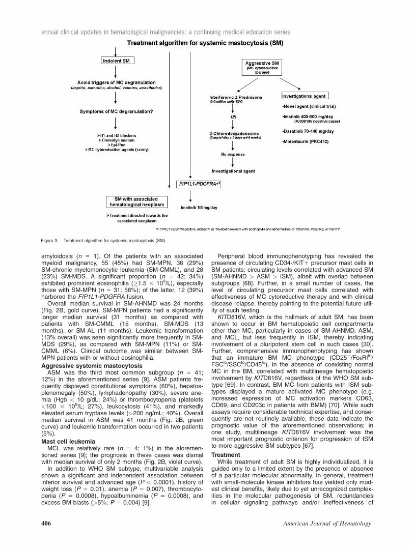
TreatmentWhile treatment of adult SM is highly individualized, it is
guided only to a limited extent by the presence or absenceof a particular molecular abnormality. In general, treatmentwith small-molecule kinase inhibitors has yielded only mod-est clinical benefits, likely due to yet unrecognized complex-ities in the molecular pathogenesis of SM, redundanciesin cellular signaling pathways and/or ineffectiveness of
Figure 3. Treatment algorithm for systemic mastocytosis (SM).
406 American Journal of Hematology
annual clinical updates in hematological malignancies: a continuing medical education series

currently available in vivo KITD816V-inhibitors. For the rareSM patient with a transmembrane KIT mutation (e.g.,F522C or K509I), dramatic clinical responses to imatinibtherapy can be observed [45,46]. Overall, however,although progress has been achieved with some of thenewer investigational agents (e.g., midostaurin/PKC412),the promise of truly ‘‘targeted therapy’’ in the vast majorityof SM patients (akin to imatinib therapy in CML) has yet tobe realized. Drug therapy has not been shown to favorablyaffect survival in SM and the experience with allogeneicstem cell transplantation is too limited to comment on [71].Therefore, current therapy in WHO-defined SM is largelypalliative and directed at MC degranulation symptoms (e.g.pruritus, urticaria, angioedema, flushing, nausea, vomiting,abdominal pain, diarrhea, episodic anaphylactoid attacks),symptomatic skin disease (e.g. urticaria pigmentosa), and/or organ dysfunction from MC tissue infiltration (e.g. hyper-splenism or pathologic fracture). Treatment options in SMrange from observation alone (supplemented by preventa-tive measures to avoid precipitating MCMRS), to symptommanagement (e.g. managing pruritus or diarrhea), to sup-portive measures (e.g. red blood cell transfusion or osteo-porosis treatment), to cytoreductive therapy for MC debulk-ing in the setting of aggressive, advanced, or treatment-re-fractory disease. An additional challenge in SM treatment isthe cumbersome nature of the current treatment responsecriteria [72], which makes it difficult to compare efficacy of agiven treatment across different centers. In this regard, alter-native criteria that are more objective, reproducible, and SM-subtype specific as compared with the current criteria haverecently been proposed, albeit not yet validated [73].Currently used agents for SM therapy are presented
below. Our current algorithm for SM treatment is illustratedin Fig. 3.
1. Interferon (IFN)-a: IFN-a is often considered the first-line cytoreductive therapy in symptomatic SM; sincethe initial report in 1992 [74], several case reports orsmall series have shown IFN-a (IFN-a2b in mostinstances) to improve symptoms of MC degranulation,decrease bone marrow MC infiltration, and amelioratemastocytosis-related ascites/hepatosplenomegaly, cyto-penias, skin findings, and osteoporosis [75–87]. IFN-atreatment is not uniformly effective [88], and the fre-quency of major response (i.e. complete resolution ofone or more baseline ‘‘C’’ findings) is approximately 20to 30%; the optimal dose and duration of IFN-a therapyfor SM remain unclear, however, concurrent administra-tion of corticosteroids (prednisone) may improve its effi-cacy (up to 40% major response rate) and tolerability[82,89]. The time to best response may be a year orlonger [82] and delayed responses to therapy havebeen described [90]. IFN-a treatment is frequently (upto 50%) complicated by toxicities, including flu-likesymptoms, bone pain, fever, cytopenias, depression,and hypothyroidism; consequently, the adverse dropoutrate with IFN-a treatment is not trivial [82,91,92].Finally, a significant proportion of patients will relapsewithin a short period of IFN-a treatment being discon-tinued, illustrating the cytostatic rather than cytolyticeffects of the drug [92].In a French study, 20 SM patients (16 ASM and 4ISM) were treated with IFN-a starting at 1 MU/day withprogressive increase to 5 MU/m2/day; 13 patients weretreated for at least 6 months (median dose 3.2 MU/day) [92]. All 13 patients exhibited responses (nonwere complete) in systemic and cutaneous diseasemanifestations that were associated with decrease incirculating MC mediator levels, but not in BM MC bur-
den. Adverse effects were frequent (cytopenias anddepression in nine and seven patients, respectively);there were two deaths during the treatment phase.Four responding patients experienced prompt relapseof symptoms after treatment cessation.In the Mayo Clinic study, 47 patients received IFN-awith or without prednisone [91]; the median weeklydose was 15 MU per week (range 3.5–30 MU perweek) and the initial dose of prednisone ranged from20 mg to 60 mg per day with a slow tapering overweeks or months in some patients. In 40 evaluablepatients, the overall response rate (ORR) was 53%(ISM and ASM 60%; SM-AHNMD 45%). Overall me-dian duration of response was 12 months (range, 1–67months). Responses were not significantly differentwhen comparing patients who did and did not receiveprednisone. Absence of systemic mediator-relatedsymptoms was significantly associated with inferiorresponse to IFN-a; 41% versus 77%, respectively.Major toxicities included fatigue, depression and throm-bocytopenia.Summary: IFN-a has activity in all SM subcategoriesand has been shown to improve dermatological, hema-tological, gastrointestinal, and systemic symptoms asso-ciated with histamine release. IFN-a also has a role intreating skeletal symptoms because of its ability toincrease bone density. Use of higher doses of IFN-ahas the potential to decrease the BM MC burden insome patients. We commonly start treatment at thedose of 1 to 3 million units (MU) subcutaneously threetimes per week, followed by gradual escalation to 3 to 5MU three to five times per week, if tolerated. Predni-sone (30–60 mg/day) is commonly added at the start oftreatment to improve tolerability and response, and istapered over a 2- to 3-month period. IFN-a treatment isgenerally continued as long as a response is observedand there are no intolerable adverse effects.
2. Cladribine or 2-chlorodeoxyadenosine (2-CdA) hasdemonstrated in vitro and in vivo activity against neo-plastic MC; the published experience suggests that 2-CdA has therapeutic activity in all SM subtypes includ-ing in MCL [93–98].In the Mayo Clinic study, 2-CdA was administered to26 patients (eight as first-line); the dose was 5 mg/m2
per day or 0.13 to 0.17 mg/kg per day for 5 days as a2-hr intravenous (IV) infusion, and median number oftreatment cycles was 3 (range 1–9) [91]. Treatmentresponse was evaluable in 22 patients and the ORRwas 55% (ORR in ISM, ASM, and SM-AHNMD was56%, 50%, and 55%, respectively). Median durationof response was 11 months (range, 3–74 months).Presence of circulating immature myeloid cells wassignificantly associated with inferior response to 2-CdA (0% vs. 75%). Major toxicities were myelosup-pression and infection.In a recent French study, 44 patients with mastocyto-sis were treated with 2-CdA (CM 5 3, ISM 5 19,SSM 5 3, ASM 5 12, SM-AHNMD 5 6 and MCL 51) [99]. All patients had failed previous symptomatictherapy and/or IFN-a (n 5 10) or kinase inhibitors(n 5 7); 2-CdA was given at 0.15 mg/kg/day in a 2-hrinfusion or subcutaneously for 5 days, repeated every1 to 2 months, for a median of four cycles. After a me-dian follow-up of 35 months, no opportunistic infec-tions were seen with the exception of zoster infectionin two patients. Responses occurred in 24 of 31patients with urticaria pigmentosa, 17 of 35 withfatigue, 14 of 24 with flushing, 9 of 24 with pruritus, 9of 21 with abdominal pain, 1 of 9 with ascites, 11 of
American Journal of Hematology 407
annual clinical updates in hematological malignancies: a continuing medical education series

23 with diarrhea, 8 of16 with weight loss, 4 of 14 withheadache, 5 of 10 with cough, 7 of 20 with spleno-megaly, 2 of 6 with lymphadenopathy, 0 of 2 with pleu-ral effusions, and 5 of 19 with neuropsychologicalsymptoms. In addition, eosinophil count normalized in7 of 10 cases and a substantial decrease in tryptaselevels was also noted. Overall, major (MR) and partial(PR) responses were observed in 7 of 12 patientswith ASM, 3 of 3 SSM, 17 of 19 ISM, 2 of 3 CM butin none of the patients with SM-AHNMD. Median dura-tion of response was approximately 20 months.Summary: The 2-CdA has activity in all SM subtypes.We use 2-CdA as first-line treatment in cases whererapid MC debulking is indicated, or in symptomaticpatients who are refractory or intolerant to IFN-a.Potential toxicities of 2-CdA include myelosuppressionand lymphopenia with increased risk of opportunisticinfections. The limited activity of 2-CdA in SM-AHNMDpatients noted in the French study is discrepant withpublished data; the discrepancy may relate to alterna-tive interpretation of the treatment response data andneeds to be confirmed.
3. Imatinib mesylate (IM) demonstrates in vitro efficacyagainst wild-type KIT and certain transmembrane(F522C) and juxta-membrane (V560G) KIT mutants,but not the common kinase (D816V) domain mutants[46,100–102]. Similarly, not all juxta-membrane muta-tions may be sensitive to IM (e.g. V559I) [103].In the Mayo Clinic study that excluded FIP1L1-PDGFRA-positive cases, IM was administered to 27SM patients; the median starting dose was 400 mgper day (range 100–400 mg/day), and the mainte-nance dose in responding patients ranged from 200mg/day to 400 mg/day [91]. In 22 evaluable patients,the ORR was 18% (ORR in ISM, ASM, and SM-AHNMD was 14%, 50%, and 9%, respectively), andmedian duration of response was 19.6 months (range,9–69 months). Responses included improvement inUP and decrease in the BM MC burden. The majority(86%) of IM-treated patients were KITD816V posi-tive—ORR in mutation-positive and -negative patientswas 17% and 33%, respectively. None of the sixpatients with SM and associated eosinophilia (allKITD816V-positive) responded to IM treatment. Majortoxicities included diarrhea and peripheral edema; twopatients developed interstitial pneumonitis.Data from another study, however, suggested an ORRof 36% in KITD816V-positive SM patients [104]. In yetanother study of 20 SM patients treated with IM, onlyone KITD816V-negative patient responded while sixother patients reported symptomatic improvement[105]. Finally, in another study wherein 17 SM patientsreceived IM treatment, the response rate was 29%(one complete and four partial remissions), all inKITD816V-negative patients [106].Summary: While IM is the only SM treatment currentlyapproved by the Food and Drug Administration (FDA)(specific indication is treatment of adult patients withASM without the KITD816V mutation or with unknownKIT mutational status), it has a limited role in the treat-ment of unselected SM patients, the majority of whomlikely harbor KITD816V. The rare SM cases that harboran IM-sensitive KIT mutation, or those that areKITD816-unmutated may be appropriate candidates forIM treatment.
4. Hydroxyurea (HU): In the Mayo Clinic study, HU wasgiven to 30 SM patients (28 with SM-AHNMD) [91].The drug was used as first-line therapy in 24 patients.The dose ranged from 500 mg every other day to
2000 mg per day. Treatment response was evalu-able in 26 patients; control of thrombocytosis, leu-kocytosis, and/or hepatosplenomegaly wasobserved in five SM-AHNMD patients (ORR 519%). Median duration of response was 31.5months (range, 5–50 months) and the major toxic-ity was myelosuppression.Summary: The utility of HU in treating SM-AHNMDstems from its myelosuppressive activity. HU doesnot, however, exhibit any substantial anti-MC effect.
1. Investigational agents:(i) Dasatinib has shown efficacy in vitro against various
KIT mutants including D816V [107,108]. Further-more, dasatinib may synergize with PKC412 andchemotherapy in this regard [109–111]. In the largeststudy of dasatinib therapy in SM [112], the drug wasgiven at a starting dose of 70 mg PO bid to 33 SMpatients: 18 ISM, 9 ASM, and 6 with SM-AHNMD.Two (6%) patients, both of whom were D816V-nega-tive, achieved complete remission. Nine (27%)patients experienced symptomatic improvement.Grade 3 toxicities were observed in 19 (58%)patients. In another report [113], four SM patients (allKITD816V positive; two with ASM, one SM-AHNMD,and one ISM) were treated with dasatinib at a doseranging from 50 mg to 100 mg twice daily. Twopatients (eight each with ASM and SM-AHNMD) hada major response, which in the case of the SM-AHNMD patient was accompanied by decrease inthe BM MC burden. Both responders experienced aninitial exacerbation of MCMRS and rash lasting sev-eral days before the benefits of dasatinib therapybecame evident.Summary: Dasatinib appears to have modest activityin KITD816V-positive SM. The cumulative publishedexperience to date does not clarify as to which groupof SM patients is likely to obtain the most benefitfrom dasatinib therapy.
(ii) Midostaurin (PKC412) has in vitro activity against ki-nase domain KIT mutants (D816Y and D816V)[94,114], and treatment of a patient with MCL whoharbored KITD816V resulted in transient clinicalbenefit [115]. In a recent phase-2 study PKC412was orally administered to 26 SM patients at 100mg BID [116]. SM variants included ASM (n 5 4),SM-CMML (n 5 14), SM-MDS (n 5 3), SM-MDS/MPN-U (n 5 1), and MCL (n 5 4). Major responserate per conventional criteria was 38%. Majorresponses included normalization of hypoalbumine-mia, improvement of hemoglobin and plateletcounts, resolution of liver function abnormalities,improvement of pleural effusion and ascites, and re-version of weight loss. Some of these responseswere accompanied by improvement in hepatosple-nomegaly, a >50% decrease in serum tryptase leveland/or BM MC burden, and improvement inMCMRS. One patient with MCL had achieved anear complete remission with a decrease of serumtryptase from 763 to <20 ng/mL, and decrease ofmarrow MC burden from 60–70% to 5%. The mostcommon drug side effects were nausea, vomiting,diarrhea, and fatigue. Asymptomatic hyperlipasemiaoccurred in five patients. Treatment had to be dis-continued in 18 (69%) patients.Summary: PKC412 has activity in SM and mightproduce substantial reduction in MC burden in somepatients. However, it is currently not clear whichpatients with SM stand to benefit from such treat-ment and more studies are needed to clarify the
408 American Journal of Hematology
annual clinical updates in hematological malignancies: a continuing medical education series

advantage of PKC412 over treatment with IFN-a orcladribine, which are currently considered first linetreatment in ASM or symptomatic indolent SM.
(iii) Masatinib mesilate (AB1010) has been shown toinhibit human and murine KIT with juxtamembraneactivating KIT mutations, as well as PDGFRA-a/b,and Lyn kinases at nanomolar concentrations incell-based assays [117]. In contrast, masatinibonly weakly inhibited KITD816V-driven cell prolif-eration (micromolar concentrations). Masatinibwas administered to 25 patients with symptomaticcutaneous or ISM that was refractory to conven-tional therapy, and where KITD816V was absentin at least one MC infiltrated organ (skin or BM)[118]. Patients were randomized to receive 3 or 6mg/kg/day for 12 weeks; the primary endpointwas change in symptoms at week 12 relative tobaseline. There was a significant improvement infrequency of flushing, pruritus score, and Hamiltonrating for depression by 64%, 36%, and 43%,respectively. The overall clinical response ("50%improvement in baseline symptom without deterio-ration or emergence of another symptom) was56%. Twenty-two patients (88%) completed thestudy; two discontinued due to adverse events(AE). In the initial 12-week phase, 21 patients(84%) experienced at least one masatinib-relatedAE (mild [n 5 11], moderate [n 5 19], and severe[n 5 9]). The most common AE were nausea/vomiting (52%), edema (44%), muscle spasms(28%), and rash (28%). One patient developedmasatinib-related agranulocytosis that was revers-ible. Seventeen patients (68%) entered the exten-sion phase and at the time of publication, eightpatients (32%) were still receiving treatment. Inanother report of 35 ISM patients (22 and 2patients with mild-moderate and severe depres-sion, respectively), depression scores were signifi-cantly improved (20% improvement in initial score)in 67% of cases after masatinib therapy for 12weeks [119].Summary: Given its lack of activity againstKITD816V, masatinib appears to be at a disadvant-age as compared with other ‘‘targeted’’ therapiesfor the treatment of adult SM. In the absence of ahead-to-head trial, it is unclear if masatinib hasany clear advantage over imatinib for the treatmentof symptomatic ISM. The frequency of AE in theformer study was relatively high, which likelyexplains why the QLQ-C30 symptom score showedlittle improvement as compared with baseline.
References1. Horny HP, Metcalfe DD, Bennett JM, et al. Mastocytosis. In: Swerdlow SH,
Campo E, Harris NL, et al., Editors. WHO Classification of Tumors of He-matopoietic and Lymphoid Tissues. Lyon: International Agency for Researchand Cancer (IARC); 2008. pp 54–63.
2. Brunning RD, McKenna RW, Rosai J, et al. Systemic mastocytosis. Extracu-taneous manifestations. Am J Surg Pathol 1983;7:425–438.
3. Horny HP, Parwaresch MR, Lennert K. Bone marrow findings in systemicmastocytosis. Hum Pathol 1985;16:808–814.
4. Stevens EC, Rosenthal NS. Bone marrow mast cell morphologic featuresand hematopoietic dyspoiesis in systemic mast cell disease. Am J ClinPathol 2001;116:177–182.
5. Caplan RM. The natural course of urticaria pigmentosa. Analysis and follow-up of 112 cases. Arch Dermatol 1963;87:146–157.
6. Uzzaman A, Maric I, Noel P, et al. Pediatric-onset mastocytosis: a long termClinical follow-up and correlation with bone marrow histopathology. PediatrBlood Cancer 2009;53:629–634.
7. Azana JM, Torrelo A, Mediero IG, Zambrano A. Urticaria pigmentosa: Areview of 67 pediatric cases. Pediatr Dermatol 1994;11:102–106.
8. Kettelhut BV, Metcalfe DD. Pediatric mastocytosis. Ann Allergy 1994;73:197–202; quiz 202–207.
9. Lim KH, Tefferi A, Lasho TL, et al. Systemic mastocytosis in 342 consecu-tive adults: Survival studies and prognostic factors. Blood 2009;113:5727–5736.
10. Bain BJ, Gilliland DG, Horny HP, Vardiman JW.Myeloid and lymphoid neo-plasms with eosinophilia and abnormalities of PDGFRA, PDGFRB orFGFR1. In:Swerdlow SH, et al., Editors. WHO Classification of Tumors ofHematopoietic and Lymphoid Tissues. Lyon: International Agency forResearch and Cancer (IARC); 2008. pp 68–73.
11. Cools J, DeAngelo DJ, Gotlib J, et al. A tyrosine kinase created by fusion ofthe PDGFRA and FIP1L1 genes as a therapeutic target of imatinib in idio-pathic hypereosinophilic syndrome. N Engl J Med 2003;348:1201–1214.
12. Klion AD, Noel P, Akin C, et al. Elevated serum tryptase levels identify asubset of patients with a myeloproliferative variant of idiopathic hypereosino-philic syndrome associated with tissue fibrosis, poor prognosis, and imatinibresponsiveness. Blood 2003;101:4660–4666.
13. Klion AD, Robyn J, Akin C, et al. Molecular remission and reversal of myelo-fibrosis in response to imatinib mesylate treatment in patients with the mye-loproliferative variant of hypereosinophilic syndrome. Blood 2004;103:473–478.
14. Pardanani A, Brockman SR, Paternoster SF, et al. FIP1L1-PDGFRA fusion:Prevalence and clinicopathologic correlates in 89 consecutive patients withmoderate to severe eosinophilia. Blood 2004;104:3038–3045.
15. Pardanani A, Elliott M, Reeder T, et al. Imatinib for systemic mast-cell dis-ease. Lancet 2003;362:535–536.
16. Pardanani A, Ketterling RP, Brockman SR, et al. CHIC2 deletion, a surro-gate for FIP1L1-PDGFRA fusion, occurs in systemic mastocytosis associ-ated with eosinophilia and predicts response to imatinib mesylate therapy.Blood 2003;102:3093–3096.
17. Dalal BI, Horsman DE, Bruyere H, Forrest DL. Imatinib mesylate respon-siveness in aggressive systemic mastocytosis: Novel association with aplatelet derived growth factor receptor beta mutation. Am J Hematol2007;82:77–79.
18. Walz C, Metzgeroth G, Haferlach C, et al. Characterization of three newimatinib-responsive fusion genes in chronic myeloproliferative disorders gen-erated by disruption of the platelet-derived growth factor receptor beta gene.Haematologica 2007;92:163–169.
19. Horny HP, Sotlar K, Sperr WR, Valent P. Systemic mastocytosis with associ-ated clonal haematological non-mast cell lineage diseases: A histopathologi-cal challenge. J Clin Pathol 2004;57:604–608.
20. Sotlar K, Fridrich C, Mall A, et al. Detection of c-kit point mutation Asp-816–> Val in microdissected pooled single mast cells and leukemic cells in apatient with systemic mastocytosis and concomitant chronic myelomonocyticleukemia. Leuk Res 2002;26:979–984.
21. Travis WD. Li CY, Yam LT, et al. Significance of systemic mast cell diseasewith associated hematologic disorders. Cancer 1988;62:965–972.
22. Pardanani A, Lim KH, Lasho TL, et al. Prognostically relevant breakdown of123 patients with systemic mastocytosis associated with other myeloidmalignancies. Blood 2009;114:3769–3772.
23. Pardanani A, Reeder T, Li CY, Tefferi A. Eosinophils are derived from theneoplastic clone in patients with systemic mastocytosis and eosinophilia.Leuk Res 2003;27:883–885.
24. Taylor ML, Sehgal D, Raffeld M, et al. Demonstration that mast cells, Tcells, and B cells bearing the activating kit mutation D816V occur in clusterswithin the marrow of patients with mastocytosis. J Mol Diagn 2004;6:335–342.
25. Tefferi A, Lasho TL, Brockman SR, et al. FIP1L1-PDGFRA and c-kit D816Vmutation-based clonality studies in systemic mast cell disease associatedwith eosinophilia. Haematologica 2004;89:871–873.
26. Dunphy CH. Evaluation of mast cells in myeloproliferative disorders andmyelodysplastic syndromes. Arch Pathol Lab Med 2005;129:219–222.
27. Yarden Y, Kuang WJ, Yang-Feng T, et al. Human proto-oncogene c-kit: Anew cell surface receptor tyrosine kinase for an unidentified ligand. EMBO J1987;6:3341–3351.
28. Miettinen M, Lasota J. KIT (CD117): A review on expression in normal andneoplastic tissues, and mutations and their clinicopathologic correlation. ApplImmunohistochem Mol Morphol 2005;13:205–220.
29. Valent P, Spanblochl E, Sperr WR, et al. Induction of differentiation ofhuman mast cells from bone marrow and peripheral blood mononuclear cellsby recombinant human stem cell factor/kit-ligand in long-term culture. Blood1992;80:2237–2245.
30. Garcia-Montero AC, Jara-Acevedo M, Teodosio C, et al. KIT mutation inmast cells and other bone marrow hematopoietic cell lineages in systemicmast cell disorders: A prospective study of the Spanish Network on Mastocy-tosis (REMA) in a series of 113 patients. Blood 2006;108:2366–2372.
31. Buttner C, Henz BM, Welker P, et al. Identification of activating c-kit mutationsin adult-, but not in childhood-onset indolent mastocytosis: A possible explana-tion for divergent clinical behavior. J Invest Dermatol 1998;111:1227–1231.
32. Furitsu T, Tsujimura T, Tono T, et al. Identification of mutations in the codingsequence of the proto-oncogene c-kit in a human mast cell leukemia cell linecausing ligand-independent activation of c-kit product. J Clin Invest1993;92:1736–1744.
33. Sotlar K, Escribano L, Landt O, et al. One-step detection of c-kit point muta-tions using peptide nucleic acid-mediated polymerase chain reaction clamp-ing and hybridization probes. Am J Pathol 2003;162:737–746.
American Journal of Hematology 409
annual clinical updates in hematological malignancies: a continuing medical education series

34. Beghini A, Cairoli R, Morra E, Larizza L. In vivo differentiation of mast cellsfrom acute myeloid leukemia blasts carrying a novel activating ligand-inde-pendent C-kit mutation. Blood Cells Mol Dis 1998;24:262–270.
35. Longley BJ Jr, Metcalfe DD, Tharp M, et al. Activating and dominant inacti-vating c-KIT catalytic domain mutations in distinct clinical forms of humanmastocytosis. Proc Natl Acad Sci USA 1999;96:1609–1614.
36. Pullarkat VA, Pullarkat ST, Calverley DC, Brynes RK. Mast cell diseaseassociated with acute myeloid leukemia: Detection of a new c-kit mutationAsp816His. Am J Hematol 2000;65:307–309.
37. Pignon JM, Giraudier S, Duquesnoy P, et al. A new c-kit mutation in a caseof aggressive mast cell disease. Br J Haematol 1997;96:374–376.
38. Bodemer C, Hermine O, Palmerini F, et al. Pediatric mastocytosis is a clonaldisease associated with D816V and other activating c-KIT mutations. JInvest Dermatol 2010;130:804–815.
39. Yanagihori H, Oyama N, Nakamura K, Kaneko F. c-kit Mutations in patientswith childhood-onset mastocytosis and genotype-phenotype correlation. JMol Diagn 2005;7:252–257.
40. Verzijl A, Heide R, Oranje AP, van Schaik RH. C-kit Asp-816-Val mutationanalysis in patients with mastocytosis. Dermatology 2007;214:15–20.
41. Gari M, Goodeve A, Wilson G, et al. c-kit proto-oncogene exon 8 in-framedeletion plus insertion mutations in acute myeloid leukaemia. Br J Haematol1999;105:894–900.
42. Lux ML, Rubin BP, Biase TL, et al. KIT extracellular and kinase domainmutations in gastrointestinal stromal tumors. Am J Pathol 2000;156:791–795.
43. Goemans BF, Zwaan CM, Miller M, et al. Mutations in KIT and RAS are fre-quent events in pediatric core-binding factor acute myeloid leukemia. Leuke-mia 2005;19:1536–1542.
44. Hartmann K, Wardelmann E, Ma Y, et al. Novel germline mutation of KITassociated with familial gastrointestinal stromal tumors and mastocytosis.Gastroenterology 2005;129:1042–1046.
45. Zhang LY, Smith ML, Schultheis B, et al. A novel K509I mutation of KITidentified in familial mastocytosis-in vitro and in vivo responsiveness to imati-nib therapy. Leuk Res 2006;30:373–378.
46. Akin C, Fumo G, Yavuz AS, et al. A novel form of mastocytosis associatedwith a transmembrane c-kit mutation and response to imatinib. Blood2004;103:3222–3225.
47. Tang X, Boxer M, Drummond A, et al. A germline mutation in KIT in familialdiffuse cutaneous mastocytosis. J Med Genet 2004;41:e88.
48. Zappulla JP, Dubreuil P, Desbois S, et al. Mastocytosis in mice expressinghuman Kit receptor with the activating Asp816Val mutation. J Exp Med2005;202:1635–1641.
49. Gerbaulet A, Wickenhauser C, Scholten J, et al. Mast cell hyperplasia, Bcell malignancy, and intestinal inflammation in mice with conditional expres-sion of a constitutively active kit. Blood 2011;117:2012–2021.
50. Tefferi A, Levine RL, Lim KH, et al. Frequent TET2 mutations in systemicmastocytosis: Clinical, KITD816V and FIP1L1-PDGFRA correlates. Leukemia2009;23:900–904.
51. Wilson TM, Maric I, Simakova O, et al. Clonal analysis of NRAS activatingmutations in KIT-D816V systemic mastocytosis. Haematologica2011;96:459–463.
52. Guo X, Schrader KA, Xu Y, Schrader JW. Expression of a constitutivelyactive mutant of M-Ras in normal bone marrow is sufficient for induction of amalignant mastocytosis/mast cell leukemia, distinct from the histiocytosis/monocytic leukemia induced by expression of activated H-Ras. Oncogene2005;24:2330–2342.
53. Horny HP, Sillaber C, Menke D, et al. Diagnostic value of immunostaining fortryptase in patients with mastocytosis. Am J Surg Pathol 1998;22:1132–1140.
54. Horny HP, Valent P. Histopathological and immunohistochemical aspects ofmastocytosis. Int Arch Allergy Immunol 2002;127:115–117.
55. Sotlar K, Horny HP, Simonitsch I, et al. CD25 indicates the neoplastic phe-notype of mast cells: A novel immunohistochemical marker for the diagnosisof systemic mastocytosis (SM) in routinely processed bone marrow biopsyspecimens. Am J Surg Pathol 2004;28:1319–1325.
56. Jordan JH, Walchshofer S, Jurecka W, et al. Immunohistochemical proper-ties of bone marrow mast cells in systemic mastocytosis: Evidence forexpression of CD2, CD117/Kit, and bcl-x(L). Hum Pathol 2001;32:545–552.
57. Sotlar K, Cerny-Reiterer S, Petat-Dutter K,Hessel et al. Aberrant expressionof CD30 in neoplastic mast cells in high-grade mastocytosis. Mod Pathol2011;24:585–595.
58. Escribano L, Garcia Montero AC, Nunez R, Orfao A. Flow cytometricanalysis of normal and neoplastic mast cells: Role in diagnosis and fol-low-up of mast cell disease. Immunol Allergy Clin North Am2006;26:535–547.
59. Pardanani A, Kimlinger T, Reeder T, et al. Bone marrow mast cell immuno-phenotyping in adults with mast cell disease: A prospective study of 33patients. Leuk Res 2004;28:777–783.
60. Galli SJ, Kalesnikoff J, Grimbaldeston MA, et al. Mast cells as "tunable"effector and immunoregulatory cells: Recent advances. Annu Rev Immunol2005;23:749–786.
61. Sperr WR, El-Samahi A, Kundi M, et al. Elevated tryptase levels selectivelycluster in myeloid neoplasms: A novel diagnostic approach and screenmarker in clinical haematology. Eur J Clin Invest 2009;39:914–923.
62. Pardanani A, Lim KH, Lasho TL, et al. WHO subvariants of indolent masto-cytosis: Clinical details and prognostic evaluation in 159 consecutive adults.Blood 2010;115:150–151.
63. Akin C. Molecular diagnosis of mast cell disorders: A paper from the 2005William Beaumont Hospital Symposium on Molecular Pathology. J Mol Diagn2006;8:412–419.
64. Yavuz AS, Lipsky PE, Yavuz S, et al. Evidence for the involvement of a he-matopoietic progenitor cell in systemic mastocytosis from single-cell analysisof mutations in the c-kit gene. Blood 2002;100:661–665.
65. Sotlar K, Colak S, Bache A, et al. Variable presence of KITD816V in clonalhaematological non-mast cell lineage diseases associated with systemicmastocytosis (SM-AHNMD). J Pathol 2010;220:586–595.
66. Travis WD, Li CY, Bergstralh EJ, et al. Systemic mast cell disease. Anal-ysis of 58 cases and literature review. Medicine (Baltimore) 1988;67:345–368.
67. Escribano L, Alvarez-Twose I, Sanchez-Munoz L, et al. Prognosis in adultindolent systemic mastocytosis: A long-term study of the Spanish Networkon Mastocytosis in a series of 145 patients. J Allergy Clin Immunol2009;124:514–521.
68. Georgin-Lavialle S, Lhermitte L, Baude C, et al. Blood CD34-c-Kit1 cell ratecorrelates with aggressive forms of systemic mastocytosis and behaves likea mast cell precursor. Blood 2011;118:5246–5249.
69. Teodosio C, Garcia-Montero AC, Jara-Acevedo M, et al. An immature immu-nophenotype of bone marrow mast cells predicts for multilineage D816V KITmutation in systemic mastocytosis. Leukemia 2011. doi: 10.1038/leu.2011.293. [Epub ahead of print].
70. Teodosio C, Garcia-Montero AC, Jara-Acevedo M, et al. Mast cells from dif-ferent molecular and prognostic subtypes of systemic mastocytosis displaydistinct immunophenotypes. J Allergy Clin Immunol 2010;125:719–726, 726e1–726e4.
71. Nakamura R, Chakrabarti S, Akin C, et al. A pilot study of nonmyeloablativeallogeneic hematopoietic stem cell transplant for advanced systemic masto-cytosis. Bone Marrow Transplant 2006;37:353–358.
72. Valent P, Akin C, Escribano L, et al. Standards and standardization in mas-tocytosis: Consensus statements on diagnostics, treatment recommenda-tions and response criteria. Eur J Clin Invest 2007;37:435–453.
73. Pardanani A, Tefferi A. Proposal for a revised classification of systemic mas-tocytosis. Blood 2010;115:2720–2721.
74. Kluin-Nelemans HC, Jansen JH, Breukelman H, et al. Response to inter-feron alfa-2b in a patient with systemic mastocytosis. N Engl J Med1992;326:619–623.
75. Butterfield JH. Response of severe systemic mastocytosis to interferonalpha. Br J Dermatol 1998;138:489–495.
76. Butterfield JH, Tefferi A, Kozuh GF. Successful treatment of systemic masto-cytosis with high-dose interferon-alfa: long-term follow-up of a case. LeukRes 2005;29:131–134.
77. Kolde G, Sunderkotter C, Luger TA. Treatment of urticaria pigmentosa usinginterferon alpha. Br J Dermatol 1995;133:91–94.
78. Lehmann T, Beyeler C, Lammle B, et al. Severe osteoporosis due to sys-temic mast cell disease: Successful treatment with interferon alpha-2B. Br JRheumatol 1996;35:898–900.
79. Lehmann T, Lammle B. IFNalpha treatment in systemic mastocytosis. AnnHematol 1999;78:483–484.
80. Takasaki Y, Tsukasaki K, Jubashi T, et al. Systemic mastocytosis withextensive polypoid lesions in the intestines; successful treatment with inter-feron-alpha. Intern Med 1998;37:484–488.
81. Weide R, Ehlenz K, Lorenz W, et al. Successful treatment of osteoporosisin systemic mastocytosis with interferon alpha-2b. Ann Hematol 1996;72:41–43.
82. Hauswirth AW, Simonitsch-Klupp I, Uffmann M, et al. Response to therapywith interferon alpha-2b and prednisolone in aggressive systemic mastocyto-sis: Report of five cases and review of the literature. Leuk Res 2004;28:249–257.
83. Lippert U, Henz BM. Long-term effect of interferon alpha treatment in masto-cytosis. Br J Dermatol 1996;134:1164–1165.
84. Petit A, Pulik M, Gaulier A, et al. Systemic mastocytosis associated withchronic myelomonocytic leukemia: Clinical features and response to inter-feron alfa therapy. J Am Acad Dermatol 1995;32:850–853.
85. Worobec AS, Kirshenbaum AS, Schwartz LB, Metcalfe DD. Treatment ofthree patients with systemic mastocytosis with interferon alpha-2b. LeukLymphoma 1996;22:501–508.
86. Brunel V, Tadrist Z, Cailleres S, et al. [Interferon alpha and pamidronate: Auseful combination in the treatment of osteoporosis and systemic mastocyto-sis]. Presse Med 1998;27:64.
87. Czarnetzki BM, Algermissen B, Jeep S, et al. Interferon treatment ofpatients with chronic urticaria and mastocytosis. J Am Acad Dermatol1994;30:500–501.
88. Hennessy B, Giles F, Cortes J, et al. Management of patients with systemicmastocytosis: Review of M. D. Anderson Cancer Center experience. Am JHematol 2004;77:209–214.
89. Delaporte E, Pierard E, Wolthers BG, et al. Interferon-alpha in combinationwith corticosteroids improves systemic mast cell disease. Br J Dermatol1995;132:479–482.
90. Lippert U, Henz BM. Long-term effect of interferon alpha treatment in masto-cytosis [comment]. Br J Dermatol 1996;134:1164–1165.
91. Lim KH, Pardanani A, Butterfield JH, et al. Cytoreductive therapy in 108adults with systemic mastocytosis: Outcome analysis and response predic-tion during treatment with interferon-alpha, hydroxyurea, imatinib mesylate or2-chlorodeoxyadenosine. Am J Hematol 2009;84:790–794.
410 American Journal of Hematology
annual clinical updates in hematological malignancies: a continuing medical education series

92. Simon J, Lortholary O, Caillat-Vigneron N, et al. Interest of interferon alphain systemic mastocytosis. The French experience and review of the litera-ture. Pathol Biol (Paris) 2004;52:294–299.
93. Bohm A, Sonneck K, Gleixner KV, et al. In vitro and in vivo growth-inhibitoryeffects of cladribine on neoplastic mast cells exhibiting the imatinib-resistantKIT mutation D816V. Exp Hematol 2010;38:744–755.
94. Gleixner KV, Mayerhofer M, Aichberger KJ, et al. PKC412 inhibits in vitrogrowth of neoplastic human mast cells expressing the D816V-mutated vari-ant of KIT: comparison with AMN107, imatinib, and cladribine (2CdA) andevaluation of cooperative drug effects. Blood 2006;107:752–759.
95. Penack O, Sotlar K, Noack F, et al. Cladribine therapy in a patient withan aleukemic subvariant of mast cell leukemia. Ann Hematol2005;84:692–693.
96. Tefferi A, Li CY, Butterfield JH, Hoagland HC. Treatment of systemic mast-cell disease with cladribine. N Engl J Med 2001;344:307–309.
97. Kluin-Nelemans HC, Oldhoff JM, Van Doormaal JJ, et al. Cladribine therapyfor systemic mastocytosis. Blood 2003;102:4270–4276.
98. Pardanani A, Hoffbrand AV, Butterfield JH, Tefferi A. Treatment of systemicmast cell disease with 2-chlorodeoxyadenosine. Leuk Res 2004;28:127–131.
99. Hermine O, Hirsh I, Damaj G, et al. Long term efficacy and safety of cladri-bine in adult systemic mastocytosis: A French multicenter study of 44patients. ASH Annual Meeting Abstracts. Blood 2010;116:1982.
100. Zermati Y, De Sepulveda P, Feger F, et al. Effect of tyrosine kinase inhibitorSTI571 on the kinase activity of wild-type and various mutated c-kit recep-tors found in mast cell neoplasms. Oncogene 2003;22:660–664.
101. Akin C, Brockow K, D’Ambrosio C, et al. Effects of tyrosine kinase inhibitorSTI571 on human mast cells bearing wild-type or mutated c-kit. Exp Hema-tol 2003;31:686–692.
102. Ma Y, Zeng S, Metcalfe DD, et al. The c-KIT mutation causing human mas-tocytosis is resistant to STI571 and other KIT kinase inhibitors; kinases withenzymatic site mutations show different inhibitor sensitivity profiles than wild-type kinases and those with regulatory-type mutations. Blood 2002;99:1741–1744.
103. Nakagomi N, Hirota S. Juxtamembrane-type c-kit gene mutation found inaggressive systemic mastocytosis induces imatinib-resistant constitutive KITactivation. Lab Invest 2007;87:365–371.
104. Droogendijk HJ, Kluin-Nelemans HJ, van Doormaal JJ, et al. Imatinib mesy-late in the treatment of systemic mastocytosis: A phase II trial. Cancer2006;107:345–351.
105. Vega-Ruiz A, Cortes JE, Sever M, et al. Phase II study of imatinib mesylateas therapy for patients with systemic mastocytosis. Leuk Res 2009;33:1481–1484.
106. Pagano L, Valentini CG, Caira M, et al. Advanced mast cell disease: An Ital-ian Hematological Multicenter experience. Int J Hematol 2008;88:483–488.
107. Schittenhelm MM, Shiraga S, Schroeder A, et al. Dasatinib (BMS-354825),a dual SRC/ABL kinase inhibitor, inhibits the kinase activity of wild-type,juxtamembrane, and activation loop mutant KIT isoforms associated withhuman malignancies. Cancer Res 2006;66:473–481.
108. Shah NP, Lee FY, Luo R, et al. Dasatinib (BMS-354825) inhibits KITD816V,an imatinib-resistant activating mutation that triggers neoplastic growth inmost patients with systemic mastocytosis. Blood 2006;108:286–291.
109. Gleixner KV, Mayerhofer M, Sonneck K, et al. Synergistic growth-inhibitoryeffects of two tyrosine kinase inhibitors, dasatinib and PKC412, on neoplasticmast cells expressing the D816V-mutated oncogenic variant of KIT. Haema-tologica 2007;92:1451–1459.
110. Aichberger KJ, Sperr WR, Gleixner KV, et al. Treatment responses to cladri-bine and dasatinib in rapidly progressing aggressive mastocytosis. Eur J ClinInvest 2008;38:869–873.
111. Ustun C, Corless CL, Savage N, et al. Chemotherapy and dasatinib inducelong-term hematologic and molecular remission in systemic mastocytosiswith acute myeloid leukemia with KIT D816V. Leuk Res 2009;33:735–741.
112. Verstovsek S, Tefferi A, Cortes J, et al. Phase II study of dasatinib in Phila-delphia chromosome-negative acute and chronic myeloid diseases, includingsystemic mastocytosis. Clin Cancer Res 2008;14: 3906–3915.
113. Purtill D, Cooney J, Sinniah R, et al. Dasatinib therapy for systemic masto-cytosis: four cases. Eur J Haematol 2008;80:456–458.
114. Growney JD, Clark JJ, Adelsperger J, et al. Activation mutations of humanc-KIT resistant to imatinib mesylate are sensitive to the tyrosine kinase inhib-itor PKC412. Blood 2005;106:721–724.
115. Gotlib J, Berube C, Growney JD, et al. Activity of the tyrosine kinase inhibi-tor PKC412 in a patient with mast cell leukemia with the D816V KIT muta-tion. Blood 2005;106:2865–2870.
116. Gotlib J, DeAngelo DJ, George TI, et al. KIT Inhibitor midostaurin exhibits ahigh rate of Clinically meaningful and durable responses in advanced sys-temic mastocytosis: Report of a fully accrued phase II trial. ASH AnnualMeeting Abstracts. Blood 2010;116:316.
117. Dubreuil P, Letard S, Ciufolini M, et al. Masitinib (AB1010), a potentand selective tyrosine kinase inhibitor targeting KIT. PLoS One2009;4:e7258.
118. Paul C, Sans B, Suarez F, et al. Masitinib for the treatment of systemic andcutaneous mastocytosis with handicap: A phase 2a study. Am J Hematol2010;85:921–925.
119. Moura DS, Sultan S, Georgin-Lavialle S, et al. Depression in patients withmastocytosis: Prevalence, features and effects of masitinib therapy. PLoSOne 2011;6:e26375.
American Journal of Hematology 411
annual clinical updates in hematological malignancies: a continuing medical education series