cronicon · the leaf sap, or a rhizome decoction, is taken to et al treat malaria. externally, the...
TRANSCRIPT

CroniconO P E N A C C E S S EC PHARMACOLOGY AND TOXICOLOGY
Research Article
Evaluation of Protective Effect of Aqueous Leave Extract of Costus afer on Female Albino Wistar Rats Exposed to Lead Acetate
Ezejiofor Anthonet Ndidiamaka* and Orisakwe Orish Ebere
Department of Experimental Pharmacology and Toxicology, Faculty of Pharmaceutical Sciences, University of Port Harcourt, Rivers State, Nigeria
*Corresponding Author: Ezejiofor Anthonet Ndidiamaka, Department of Experimental Pharmacology and Toxicology, Faculty of Phar-maceutical Sciences, University of Port Harcourt, Rivers State, Nigeria.
Citation: Ezejiofor Anthonet Ndidiamaka and Orisakwe Orish Ebere. “Evaluation of Protective Effect of Aqueous Leave Extract of Costus afer on Female Albino Wistar Rats Exposed to Lead Acetate”. EC Pharmacology and Toxicology 4.2 (2017): 75-92.
Received: July 25, 2017; Published: August 30, 2017
Abstract
Lead (Pb2+) is a toxic metal that induces a broad range of physiological, biochemical and neurological dysfunctions in humans as well as in animal model. Lead (Pb) is a well-known multi-organ toxicant which damages the liver, kidney, reproductive system and other physiological organs. It is a divalent cation that settles in the proximal tubule of the nephron, leading to nephrotoxicity, it induces oxidative damage and production of reactive oxygen species. The present research aimed at investigating the protective effect of Costus afer on some pathological damages induced by lead. The study involves 5 groups of five rats each, shared accord-ing to their weight. First and second groups serve as normal and toxic control that received deionized water and leaded water (CH3COO)2Pb·3H2O) respectively. Groups 3, 4 and 5 were administered by oral gavage 750, 1500 and 2250 mg/kg of the Costus afer respectively while receiving leaded water (Pb2+) ad libitium. BLL significantly increased in lead only exposed group compared to control. Lead only exposed rats showed lower body weight gain, decreased in haematological parameters, hormonal level, sero-bio-chemical parameters and lower activities of antioxidant enzymes with increased malondialdehyde (MDA) activities found in liver and kidney compared with control. The present result indicate that chronic Pb2+ intoxication induces oxidative stress seen as pathological changes in rats whose effect can be ameliorated using aqueous leave extract of Costus afer.
Keywords: Costus afer; Lead Acetate; Rats; MDA
IntroductionCostus afer a perennial, herbaceous and unbranched rhizomatous herb that grows up to 4m tall have been proved to have a wide range
of therapeutic effects. Costus afer has been reported to reduce significantly blood sugar level [1,2]. Costus afer exhibits hepatoprotective, nephroprotective and antioxidant role as reported by Ezejiofor., et al [1-3]. Costus afer have proven to possess antimicrobial, anti-inflam-matory [4,5], Physical properties of Costus afer have been investigated by Okwu and Okwu [6]; Akpan., et al. [4]; Ezejiofor., et al [7]. Costus afer possesses hypolipidemic effect (Emeh., et al. 2014), antinociceptive property [8]. The leaf sap, or a rhizome decoction, is taken to treat malaria. Externally, the leaf sap is used as eye drops to treat eye troubles and as nose drops to treat headache with vertigo. It is used in frictions to treat oedema and fever.
Increasing ecological and global public health due to environmental contamination by heavy metals has posed a very big concern. Exposure of humans to these metals has also risen and this is as a result of an exponential increase of their use in several industrial, ag-ricultural, domestic and technological applications [9]. The essential heavy metals exert biochemical and physiological functions in both plants and animals. They are important constituents of several key enzymes and play important roles in various oxidation-reduction reactions [10], example is copper, zinc. Lead is ubiquitous in nature and numerous pathological conditions have been associated with it. Lead is one of the five elements that rank among the priority metals that are of great public health significance, this is because of its

76
Evaluation of Protective Effect of Aqueous Leave Extract of Costus afer on Female Albino Wistar Rats Exposed to Lead Acetate
Citation: Ezejiofor Anthonet Ndidiamaka and Orisakwe Orish Ebere. “Evaluation of Protective Effect of Aqueous Leave Extract of Costus afer on Female Albino Wistar Rats Exposed to Lead Acetate”. EC Pharmacology and Toxicology 4.2 (2017): 75-92.
high degree of toxicity [11]. Acute exposure to lead induces brain damage, kidney damage, and gastrointestinal diseases while chronic exposure may cause adverse effects on the blood, central nervous system, blood pressure, kidneys and vitamin D metabolism [12]. Lead poisoning occurs when small amounts of lead builds up in the body during a period of prolong exposure. The mechanism of lead induced stress includes the effects of lead on membranes, DNA, and antioxidant defense systems of cells. Hypo or hyperthyroidism can also result from lead exposure (Klages., et al. 1987).
However, the toxic consequences of most of this heavy metal has been shown to avert treatment with modern medicine and modern medicine coupled with its consequences of increase toxicity, high cost and unaffordability has made it almost impossible to the people of middle and low economic class who faces the health challenge emanating from heavy metal exposure to sort for medical care. Hence, there is a resurgence of interest in natural antidotes as chemo-preventive agents considering their availability, safety and affordability. This study was therefore designed to evaluate the protective effect of the aqueous leaf extract of Costus afer ker gawl on female albino rats exposed to lead acetate.
Materials and MethodsChemicals: All the chemicals were of analytical grade and obtained from commercial sources.Most of the analysis were carried out using kit (IEMA/ELISA; EIA)- Accubind Elisa Microwells, Monobind Inc Lake Forest; CA 92630.USA.
Collection of plant material
Costus afer leaves were collected from a farm land in Abuja campus, University of Port Harcourt in Ikwerre Local Government Area of Rivers State, Nigeria in October, 2016.
Identification of plant sample
The plant was identified and authenticated by A.O. Ozioko, International Centre for Ethnomedicine and Drug Development (INTER-CEDD), University of Nigeria, Nsukka, Enugu State and confirmed by Professor M.E. Bassey of the Department of Botany, University of Uyo, Akwa Ibom State, Nigeria. The voucher number is INTERCEED/033.
Preparation of plant extract
The fresh leaf samples were washed with clean water to remove any dirt or sand present. The leaves were placed in a well-ventilated area, away from direct sunlight so as to drain off the water. The leaf was then pulverized manually with the aid of a grinding machine and stored properly. Then 250g of the pulverized leaf was weighed out and macerated in 500 ml of deionized water in a stoppered container and was allowed to stand for 48 hours with constant agitations at intervals. After which the mixture was strained, the marc was pressed and the liquid was filtered. This was then stored in a refrigerator at 4˚C. The solution was discarded every third day and fresh preparation made and the process was repeated till the end of the study [7].
Preparation of 2500ppm Leaded Water
A 50g of lead acetate (CH3COO)2Pb·3H2O) was dissolved in 12 ml of 1N hydrochloric acid (HCl) and made up to 20L with deionized water. After which 10g of glucose was added to improve the taste [13].
Animals care handling
Twenty-five (25) adult female Wistar rat aged 60 days weighing between 80 - 173g obtained from animal house of Experimental Pharmacology and Toxicology Department, University of Port Harcourt, Rivers State, Nigeria were used for the study. The animals kept in polypropylene cages were maintained under standard conditions prescribed by the committee for the purpose of control and supervi-sion on experiments on animals (CPCSEA). The experimental protocol was approved by the Institutional Animal Ethics Committee and the ethical number UPH/PHARM/2017/0333 was assigned. The animals were fed with standard diet (Boiler finisher) and lead water ad libitum with the exception of normal control which receive deionized water ad libitium.

Citation: Ezejiofor Anthonet Ndidiamaka and Orisakwe Orish Ebere. “Evaluation of Protective Effect of Aqueous Leave Extract of Costus afer on Female Albino Wistar Rats Exposed to Lead Acetate”. EC Pharmacology and Toxicology 4.2 (2017): 75-92.
Evaluation of Protective Effect of Aqueous Leave Extract of Costus afer on Female Albino Wistar Rats Exposed to Lead Acetate77
Experimental design
A total of 25 rats were randomly divided into five groups with five rats each according to their weight. Group 1 was maintained as normal control and was given deionized water, while group 2 rats acted as Pb toxicity control that received Pb as (CH3COO)2Pb·3H2O) dissolved in 12 ml of IN HCl with little glucose added to improve the taste and made up to 20L with deionized water. Groups 3, 4 and 5 constitute the treatment group which received leaded water and 750 mg/kg, 1500 mg/kg and 2250 mg/kg b.w. of aqueous leave extract of Costus afer respectively by oral gavage.
Group 1: water only (normal control)Group 2: leaded water (toxic control)Group 3: leaded water + 750 mg/kg Costus afer Group 4: leaded water + 1150 mg/kg Costus afer Group 5: leaded water + 2250 mg/kg Costus afer
The research lasted for 28 days. The dose of the plant extract administered was based on the previous work done by Ezejiofor., et al. [7] while the dose of the lead (Pb) was according to the previous work of Sadeghi., et al. 2013. Body weights were recorded at the beginning and at the end of experiment while the fluid and feed intake of the rats in all the groups were monitored daily for 28 days.
Sample collection
At the end of the experiment, the animals were fasted overnight; the blood samples were collected from retro-orbital plexus of experi-mental rats for studying serum biochemical profile (ALT, BUN, creatinine and total protein). Then all the rats were euthanized. Uterus along with ovaries was collected immediately and ovaries were kept in ice cold phosphate buffer. A portion of the ovaries, uterus, liver and kidney was homogenized with tissue homogenizer individually to make 10% homogenate to assay antioxidants, peroxidation and functional markers. Pieces of tissues from ovary, uterus liver, kidney and heart were immediately kept in 10% of formalin fixative to study histological alterations if any.
Biochemical analysis
Hormonal Assay
Quantitative Determination of Follicle Stimulating Hormone (FSH), Luteinizing Hormone (LH) using (IEMA/ELISA)
The assays for the plasma concentrations of testosterone, LH and FSH were performed using a commercial microplate enzyme immuno-assay kits as per manufacturer’s instructions (Monobind Inc., USA). The Testosterone AccuBind™ Microplate EIA Test System has sensitiv-ity of 0.0576 ng/ml and with a negligible cross reactivity with other androgen derivatives like androstenedione, 5α-dihydrotestosterone, and methyltestosterone.
Quantitative Determination of Triiodothyronine (T3 and T4) and thyroxine: The plasma levels of Total triiodothyronine and thyrox-ine were determined using the commercial enzyme immune assay kits (Biocheck Inc, Foster city, CA) as per manufacturer’s instruction. Range of the assays in normal individual falls within 4.8 to 12.0 µg/dl for thyroxine and 0.6 to 1.85 ng/ml for triiodothyronine. The levels of plasma total triiodothyronine and thyroxine were expressed as ng/ml and ng/dl respectively. All the samples were assayed on the same day to avoid the inter assay variation.
Lipid Profile: Total Cholesterol, Triglycerides, HDL Cholesterol were assayed.
Antioxidant markers
Superoxide Dismutase (SOD) was estimated by the method that involved inhibition of superoxide-dependent reduction of tetrazolium dye methyl thiazolyl tetrazolium (MTT) to its formazan [14]. The activity of superoxide dismutase was determined by the method of Misra and Fridovich [15].

Citation: Ezejiofor Anthonet Ndidiamaka and Orisakwe Orish Ebere. “Evaluation of Protective Effect of Aqueous Leave Extract of Costus afer on Female Albino Wistar Rats Exposed to Lead Acetate”. EC Pharmacology and Toxicology 4.2 (2017): 75-92.
Evaluation of Protective Effect of Aqueous Leave Extract of Costus afer on Female Albino Wistar Rats Exposed to Lead Acetate78
Reduced Glutathione (GSH) Level: The method of Sedlak and Lindsay (1968) was followed in estimating the level of reduced glutathi-one. GSH was estimated based on a reaction of reduced glutathione with 5-5ditiobis-2-nitrobenzoic acid (DTNB).
Peroxidation markers: Malondialdehyde, the product of lipid peroxidation, was estimated by reaction with thiobarbituric acid (TBA) as per the method prescribed by Balasubramanian., et al [16].
Catalase Activity Assay: Catalase activity was determined according to Clairborne [17] with slight modifications.
Glutathione-S-Trasferase Activity Assay: Glutathione-S-transferase (GST) activity was determined according to Habig., et al [18].
Assay for Glutathione Peroxidase Activity: The activity of GSH-Px was assessed according to established methods of Rotruck., et al (1973).
Sero-biochemical markers: ALT, ALP, AST, BUN and creatinine were estimated in serum using the standard diagnostic kits.
Total Protein: Total protein in the ovarian tissue was quantified as per Lowry., et al.’s method.
Haematological Analysis: the following heamatological parameter such as Hb, PCV, WBC, platelet and RBC were assayed using the gen-eral laboratory technique.
Blood Lead Level (BLL): BLL was measured in the whole blood samples using Solaar Thermo Elemental Atomic Absorption Spectrometer (BUCK Scientific; model: 210, USA).
Histology
For light microscopy examination, the formalin fixed tissues were dehydrated through ascending grades of alcohol, cleared in three changes of xylene, and were embedded in paraffin. Serial sections, each of 4-micron thickness, were cut and stained with H and E as per standard protocols [19]. For transmission electron microscopy (TEM), the glutaraldehyde-fixed tissues were used. Stained sections were microscopically evaluated and the pictures of the slides were taken for comparison.
Statistical analysis
The data were subjected to statistical analysis by applying one way ANOVA using statistical package for social sciences (SPSS) version 12.0. Differences between means were tested using Duncan’s multiple comparison tests and significance was set at P < 0.05 and the result expressed as Mean ± Standard deviation (SD).
Results
Treatments Absolute Weight (G) Relative Weight (%)Ovary+Uterus Heart Liver Kidney Ovary+ Uterus Heart Liver Kidney
F1 0.50 ± 0.10 0.50 ± 0.00 4.50 ± 0.60 1.46 ± 0.03 0.41 0.42 3.59 1.22F2 0.47 ± 0.12 0.50 ± 0.00 4.90 ± 0.62 0.67 ± 0.02* 0.40 0.24 2.90 0.53F3 0.73 ± 0.32** 0.60 ± 0.00 4.60 ± 0.20 0.80 ± 0.01 0.53 0.44 3.20 0.54F4 0.90 ± 0.35** 0.63 ± 0.06** 4.40 ± 0.60 0.81 ± 0.01 0.49 0.33 3.80 0.47F5 0.43 ± 0.12 0.51 ± 0.09 5.80 ± 0.21 0.84 ± 0.01 0.21 0.42 3.38 0.42
All values represent Mean ± SD (n = 5); * Values differ significantly from control (p < 0.05). ** Values differ significantly from Pb alone (p < 0.05). F1=H2O only; F2 = PbH2O only; F3 = PbH2O + 750 mg/kg CA; F4 = PbH2O + 1500 mg/kg CA; F5 = PbH2O + 2250 mg/kg CA
Table 1: Effect of Aqueous leave extract of Costus afer on absolute and relative weight of organs.

Citation: Ezejiofor Anthonet Ndidiamaka and Orisakwe Orish Ebere. “Evaluation of Protective Effect of Aqueous Leave Extract of Costus afer on Female Albino Wistar Rats Exposed to Lead Acetate”. EC Pharmacology and Toxicology 4.2 (2017): 75-92.
Evaluation of Protective Effect of Aqueous Leave Extract of Costus afer on Female Albino Wistar Rats Exposed to Lead Acetate
79
Treatments BLL (mg/ml) TET (ng/ml) LH (m/u/ml) FSH (m/u/ml) T3 (ng/ml) T4 (ng/ml)F1 0.006 ± 0.01 0.70 ± 0.06 14.97 ± 4.80 6.93 ± 4.05 0.43 ± 0.01 5.40 ± 0.30F2 0.140 ± 0.04 0.51 ± 0.09* 2.43 ± 0.25 * 2.90 ± 0.40* 0.31 ± 0.02* 4.80 ± 0.20*
F3 0.00 ± 0.00 0.62 ± 0.05** 2.27 ± 0.25 3.80 ± 0.40** 0.40 ± 0.0** 4.77 ± 0.15F4 0.002 ± 0.00 0.63 ± 0.12** 2.93 ± 0.15 3.67 ± 1.15** 0.41 ± 0.0** 4.63 ± 0.45F5 0.00 ± 0.00 0.61 ± 0.12** 3.13 ± 4.04** 3.50 ± 1.57** 0.42 ± 0.0* 4.77 ± 0.45
All values represent Mean ± SD (n = 5); * Values differ significantly from control (p < 0.05). ** Values differ significantly from Pb alone (p < 0.05). F1 = H2O only; F2 = PbH2O only; F3 = PbH2O + 750 mg/kg CA; F4 = PbH2O + 1500 mg/kg CA; F5 = PbH2O + 2250 mg/kg CA
Table 2: Effect of aqueous extract of Costus afer on Hormones.
Treatments T.C (mmol/L) T.G (mmolL) HDL (mmolL) LDL (mmolL)F1 3.70 ± 1.21 0.89 ± 0.25 1.53 ± 0.55 1.80 ± 0.36F2 3.33 ± 0.47 0.60 ± 0.17* 1.03 ± 0.15* 2.37 ± 0.31*
F3 2.93 ± 0.21** 0.79 ± 0.22 1.20 ± 0.20 1.63 ± 0.25**
F4 4.07 ± 0.12** 1.89 ± 2.23** 1.10 ± 0.10 1.17 ± 0.06**
F5 2.47 ± 0.15** 1.07 ± 0.09** 0.93 ± 0.06** 0.93 ± 0.06**
All values represent Mean ± SD (n = 5); * Values differ significantly from control (p < 0.05). ** Values differ significantly from Pb alone (p < 0.05). F1 = H2O only; F2 = PbH2O only; F3 = PbH2O + 750 mg/kg CA; F4 = PbH2O + 1500 mg/kg CA; F5 = PbH2O + 2250 mg/kg CA
Table 3: Effect of aqueous extract of Costus afer on the lipid profile.
Treatments Hb (g/dl) PCV (%) RBC (×1012/L)
WBC (×109/L)
Platelet (×109/L)
N B
F1 13.67 ± 0.35 41.00 ± 1.00 6.23 ± 0.25 3.77 ± 1.00 200.00 ± 30.00 24.00 ± 5.29 76.00 ± 5.39F2 10.90 ± 0.35* 32.67 ± 1.15* 4.17 ± 0.21 * 4.93 ± 0.25* 210.00 ± 52.92 68.33 ± 10.41 * 68.33 ± 10.41*F3 11.67 ± 0.85** 35.00 ± 2.65 4.50 ± 0.30 6.83 ± 0.65** 246.67 ± 25.17** 74.33 ± 4.04** 74.33 ± 4.04**
F4 12.77 ± 0.68** 38.33 ± 2.08** 5.60 ± 0.87 ** 5.90 ± 0.66** 266.67 ± 75.72** 67.67 ± 3.52 67.67 ± 2.52F5 13.47 ± 1.08** 40.33 ± 3.21** 5.77 ± 0.95 ** 5.57 ± 0.60** 273.33 ± 25.17** 36.67 ± 4.73 ** 63.33 ± 4.73**
All values represent Mean ± SD (n = 5); * Values differ significantly from control (p < 0.05). ** Values differ significantly from Pb alone (p < 0.05). F1 = H2O only; F2 = PbH2O only; F3 = PbH2O + 750 mg/kg CA; F4 = PbH2O + 1500 mg/kg CA; F5 = PbH2O + 2250 mg/kg CA.
Table 4: Effect of aqueous leaf extract of Costus afer on the hematological parameters.
Samples GSH CAT MDA GPH GST SODFU1Mean ± SD 2.1 ± 0.2 1.3 ± 0.4 0.1 ± 0.06 0.16 ± 0.31 0.64 ± 0.13 0.14 ± 0.03Max 2.3 1.6 0.11 0.14 0.79 0.13Min 1.9 0.9 0.11 0.11 0.55 0.05FU2Mean ± SD 1.87 ± 0.2* 0.89 ± 0.7* 0.12 ± 0.15 0.13 ± 0.15* 0.57 ± 012* 0.08 ± 0.05*
80
Evaluation of Protective Effect of Aqueous Leave Extract of Costus afer on Female Albino Wistar Rats Exposed to Lead Acetate
Citation: Ezejiofor Anthonet Ndidiamaka and Orisakwe Orish Ebere. “Evaluation of Protective Effect of Aqueous Leave Extract of Costus afer on Female Albino Wistar Rats Exposed to Lead Acetate”. EC Pharmacology and Toxicology 4.2 (2017): 75-92.
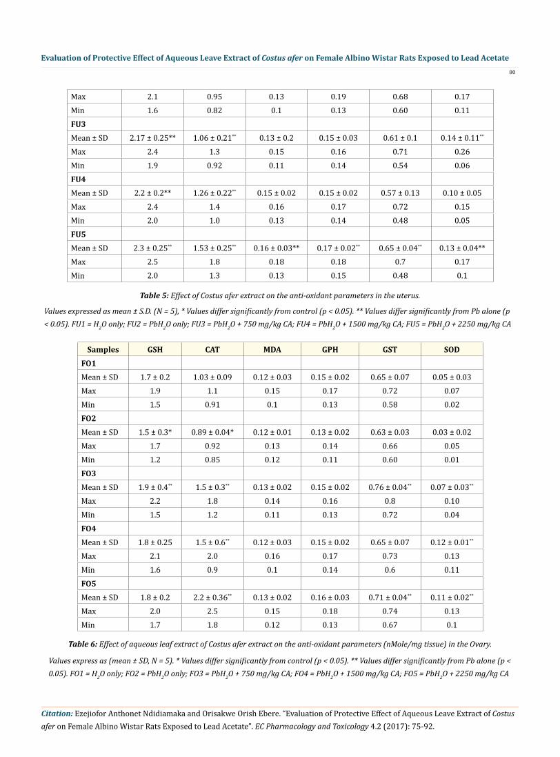
Max 2.1 0.95 0.13 0.19 0.68 0.17Min 1.6 0.82 0.1 0.13 0.60 0.11FU3Mean ± SD 2.17 ± 0.25** 1.06 ± 0.21** 0.13 ± 0.2 0.15 ± 0.03 0.61 ± 0.1 0.14 ± 0.11**
Max 2.4 1.3 0.15 0.16 0.71 0.26Min 1.9 0.92 0.11 0.14 0.54 0.06FU4Mean ± SD 2.2 ± 0.2** 1.26 ± 0.22** 0.15 ± 0.02 0.15 ± 0.02 0.57 ± 0.13 0.10 ± 0.05Max 2.4 1.4 0.16 0.17 0.72 0.15Min 2.0 1.0 0.13 0.14 0.48 0.05FU5Mean ± SD 2.3 ± 0.25** 1.53 ± 0.25** 0.16 ± 0.03** 0.17 ± 0.02** 0.65 ± 0.04** 0.13 ± 0.04**Max 2.5 1.8 0.18 0.18 0.7 0.17Min 2.0 1.3 0.13 0.15 0.48 0.1
Table 5: Effect of Costus afer extract on the anti-oxidant parameters in the uterus.
Values expressed as mean ± S.D. (N = 5), * Values differ significantly from control (p < 0.05). ** Values differ significantly from Pb alone (p < 0.05). FU1 = H2O only; FU2 = PbH2O only; FU3 = PbH2O + 750 mg/kg CA; FU4 = PbH2O + 1500 mg/kg CA; FU5 = PbH2O + 2250 mg/kg CA
Samples GSH CAT MDA GPH GST SODFO1Mean ± SD 1.7 ± 0.2 1.03 ± 0.09 0.12 ± 0.03 0.15 ± 0.02 0.65 ± 0.07 0.05 ± 0.03Max 1.9 1.1 0.15 0.17 0.72 0.07Min 1.5 0.91 0.1 0.13 0.58 0.02FO2Mean ± SD 1.5 ± 0.3* 0.89 ± 0.04* 0.12 ± 0.01 0.13 ± 0.02 0.63 ± 0.03 0.03 ± 0.02Max 1.7 0.92 0.13 0.14 0.66 0.05Min 1.2 0.85 0.12 0.11 0.60 0.01FO3Mean ± SD 1.9 ± 0.4** 1.5 ± 0.3** 0.13 ± 0.02 0.15 ± 0.02 0.76 ± 0.04** 0.07 ± 0.03**
Max 2.2 1.8 0.14 0.16 0.8 0.10Min 1.5 1.2 0.11 0.13 0.72 0.04FO4Mean ± SD 1.8 ± 0.25 1.5 ± 0.6** 0.12 ± 0.03 0.15 ± 0.02 0.65 ± 0.07 0.12 ± 0.01**
Max 2.1 2.0 0.16 0.17 0.73 0.13Min 1.6 0.9 0.1 0.14 0.6 0.11FO5Mean ± SD 1.8 ± 0.2 2.2 ± 0.36** 0.13 ± 0.02 0.16 ± 0.03 0.71 ± 0.04** 0.11 ± 0.02**
Max 2.0 2.5 0.15 0.18 0.74 0.13Min 1.7 1.8 0.12 0.13 0.67 0.1
Values express as (mean ± SD, N = 5). * Values differ significantly from control (p < 0.05). ** Values differ significantly from Pb alone (p < 0.05). FO1 = H2O only; FO2 = PbH2O only; FO3 = PbH2O + 750 mg/kg CA; FO4 = PbH2O + 1500 mg/kg CA; FO5 = PbH2O + 2250 mg/kg CA
Table 6: Effect of aqueous leaf extract of Costus afer extract on the anti-oxidant parameters (nMole/mg tissue) in the Ovary.

81
Evaluation of Protective Effect of Aqueous Leave Extract of Costus afer on Female Albino Wistar Rats Exposed to Lead Acetate
Citation: Ezejiofor Anthonet Ndidiamaka and Orisakwe Orish Ebere. “Evaluation of Protective Effect of Aqueous Leave Extract of Costus afer on Female Albino Wistar Rats Exposed to Lead Acetate”. EC Pharmacology and Toxicology 4.2 (2017): 75-92.
Samples GSH CAT MDA GPH GST SODFK1
Mean ± SD 1.97 ± 0.15 1.57 ± 0.5 0.11 ± 0.02 0.23 ± 0.02 0.72 ± 0.03 0.05 ± 0.01Max 2.1 2.0 0.13 0.25 0.74 0.06Min 1.8 1.1 0.1 0.21 0.69 0.04FK2
Mean ± SD 1.3 ± 0.25* 1.23 ± 0.21 0.94 ± 0.61* 0.21 ± 0.02 0.69 ± 0.03 0.04 ± 0.02Max 1.6 1.4 0.99 0.2 0.72 0.06Min 1-1 1.0 0.87 0.19 0.67 0.03FK3
Mean ± SD 1.6 ± 0.1** 2.2 ± 0.45 0.79 ± 0.15** 0.2 ± 0.02 0.93 ± 0.05** 0.09 ± 0.03**
Max 1.7 2.7 0.9 0.20 0.98 0.13Min 1.5 1.8 0.62 0.17 0.88 0.07FK4
Mean ± SD 1.6 ± 0.1** 2.2 ± 0.7 0.67 ± 0.2** 0.22 ± 0.01 0.79 ± 0.06** 0.05 ± 0.004Max 1.7 2.7 0.78 0.22 0.84 0.05Min 1.5 1.4 0.47 0.17 0.73 0.04FK5
Mean ± SD 2.0 ± 0.1** 3.2 ± 1.2** 0.44 ± 0.04** 0.22 ± 0.01 0.71 ± 0.05 0.06 ± 0.004**
Max 2.1 4.5 0.47 0.23 0.75 0.06Min 1.9 2.3 0.04 0.21 0.65 0.05
The data are expressed as mean ± S.D. (N = 5), * Values differ significantly from control (p < 0.05). ** Values differ significantly from Pb alone (p<0.05). FK1 = H2O only; FK2 = PbH2O only; FK3 = PbH2O + 750 mg/kg CA; FK4 = PbH2O + 1500 mg/kg CA; FK5 = PbH2O + 2250 mg/kg CA
Table 7: Effect of Costus afer extract on the anti-oxidant parameters in the Kidney.
Samples GSH CAT MDA GPH GST SODFL1Mean ± SD 1.7 ± 0.1 5.2 ± 0.97 0.7 ± 0.1 0.17 ± 0.03 0.86 ± 0.05 0.34 ± 0.07Max 1.8 6.1 0.85 0.2 0.92 0.47Min 1.6 4.2 0.65 0.15 0.82 0.26FL2Mean ± SD 1.5 ± 0.1* 4.2. ± 1.1* 0.9 ± 0.08 0.10 ± 0.01* 0.71 ± 0.02 0.20 ± 0.1*Max 1.6 4.9 0.97 0.18 0.73 0.27Min 1.4 3.9 0.82 0.16 0.70 0.14FL3Mean ± SD 1.6 ± 0.05 5.9 ± 0.9** 0.34 ± 0.09** 0.19 ± 0.02 0.9 ± 0.09** 0.35 ± 0.12**
Max 1.6 6.9 0.42 0.20 0.98 0.48Min 1.5 5.2 0.24 0.17 0.80 0.25FL4

Citation: Ezejiofor Anthonet Ndidiamaka and Orisakwe Orish Ebere. “Evaluation of Protective Effect of Aqueous Leave Extract of Costus afer on Female Albino Wistar Rats Exposed to Lead Acetate”. EC Pharmacology and Toxicology 4.2 (2017): 75-92.
Evaluation of Protective Effect of Aqueous Leave Extract of Costus afer on Female Albino Wistar Rats Exposed to Lead Acetate
82
Mean ± SD 1.8 ± 0.15** 5.5 ± 1.5** 0.37 ± 0.12** 0.19 ± 0.01 0.91 ± 0.09** 0.31 ± 0.08**
Max 2.0 7.0 0.47 0.20 0.97 0.40Min 1.7 4.0 0.24 0.18 0.81 0.25FL5Mean ± SD 1.9 ± 0.25*8 6.9 ± 0.5** 0.41 ± 0.17** 0.21 ± 0.03** 0.91 ± 0.07 0.38 ± 0.1**
Max 2.2 7.5 0.56 0.23 0.99 0.45Min 1.7 6.5 0.23 0.18 0.85 0.26
The data are expressed as mean ± S.D. (N = 5), * Values differ significantly from control (p < 0.05). ** Values differ significantly from Pb alone (p < 0.05). FL1 = H2O only; FL2 = PbH2O only; FL3 = PbH2O + 750 mg/kg CA; FL4 = PbH2O + 1500 mg/kg CA; FL5 = PbH2O + 2250 mg/kg CA
Table 8: Effect of Costus afer extract on the anti-oxidant parameters in the Liver.
Samples AST ALT ALP UR CR TP ALB Na K HCO3 TB CB
F1
Mean ± SD 21 ± 5.0 9.3 ± 1.8 24 ± 11.8 1.23 ± 0.3 175.3 ± 1.5 65.7 ± 8.3 37.7 ± 4.5 135 ± 1.5 4.9 ± 0.4 31 ± 1.2 4.9 ± 1.1 2.8 ± 0.8
Max 26 11 31 1.5 177 75 42 137 32 32 5.6 3.7
Min 16 7.5 10 1.0 174 59 33 134 30 30 3.7 2.4
F2
Mean ± SD 14 ± 3.5* 5.6 ± 1.9* 18.7 ± 11* 1.27 ± 0.1 175.7 ± 2.1 62.3 ± 11 36.3 ± 2.1 142 ± 14* 4.8 ± 0.1 26 ± 3.5* 5.6 ± 1.9 3.3 ± 0.8
Max 16 7.5 29 1.4 178 70 38 155 4.9 28 7.4 3.7
Min 10 3.7 7.0 1.2 174 50 34 127 4.7 22 3.6 2.4
F3
Mean ± SD 46 ± 11** 16.3 ± 1.2** 32.3 ± 0.6** 4.7 ± 2.2** 174.3 ± 1.5 68.3 ± 3.5** 43 ± 6.1** 138 ± 5.1** 5.0 ± 1.3 29 ± 1.2** 8.7 ± 2.8a 5.0 ± 2.3a
Max 57 17 33 7.1 176 72 50 144 5.9 30 11.1 6.9
Min 36 15 32 2.8 173 65 39 134 3.5 28 5.6 2.4
F4
Mean ± SD 48 ± 13** 15.7 ± 3.1** 27.0 ± 2.0** 4.3 ± 1.0** 173.7 ± 1.2 72.3 ± 4.5** 39.7 ± 5.7** 132 ± 2** 5.0 ± 0.2 29 ± 3.1** 9.7 ± 2.5a 7.1 ± 1.5b
Max 62 19 29 5.4 175 77 46 134 5.3 32 12.3 5.5
Min 36 13 25 3.4 173 68 35 130 4.9 26 7.4 5.6
F5
Mean ± SD 78 ± 5.0** 52. ± 4.6** 30.3 ± 0.6** 4.6 ± 1.29** 165 ± 3.0** 71.7 ± 1.5** 37.7 ± 2.1 135 ± 5** 5.2 ± 0.8** 29 ± 3.1** 17 ± 1.0b 12.6 0.7b
Max 83 58 31 6 168 73 40 140 5.7 32 18.5 13.4
Min 73 50 30 3.5 162 70 36 130 4.2 26 16.7 12.2
Table 9: Effect of aqueous leaf extract of Costus afer on Sero-biochemical parameters.
The data are expressed as mean ± S.D. (N = 5), * Values differ significantly from control (p < 0.05). ** Values differ significantly from Pb alone (p < 0.05). F1 = H2O only; F2 = PbH2O only; F3 = PbH2O + 750 mg/kg CA; F4 = PbH2O + 1500 mg/kg CA; F5 = PbH2O + 2250 mg/kg CA

Citation: Ezejiofor Anthonet Ndidiamaka and Orisakwe Orish Ebere. “Evaluation of Protective Effect of Aqueous Leave Extract of Costus afer on Female Albino Wistar Rats Exposed to Lead Acetate”. EC Pharmacology and Toxicology 4.2 (2017): 75-92.
Evaluation of Protective Effect of Aqueous Leave Extract of Costus afer on Female Albino Wistar Rats Exposed to Lead Acetate83
Figure 1: histogram of the weight gain, feed and fluid intake. F1 (H20), F2 (Pb), F3 (Pb + 750 mg/kg CA), F4 (Pb + 1500 mg/kg CA) and F5 (Pb + 2250 mg/kg CA).
Figure 2: Photomicrograph of the Heart: F1 (H20), F2 (Pb), F3 (Pb + 750 mg/kg CA), F4 (Pb + 1500 mg/kg CA) and F5 (Pb + 2250 mg/kg CA). All panels were stained with haematoxylin and eosin. magnification x100. N (Central Nuclei); BM (Branching Muscle fibre).
Citation: Ezejiofor Anthonet Ndidiamaka and Orisakwe Orish Ebere. “Evaluation of Protective Effect of Aqueous Leave Extract of Costus afer on Female Albino Wistar Rats Exposed to Lead Acetate”. EC Pharmacology and Toxicology 4.2 (2017): 75-92.
Evaluation of Protective Effect of Aqueous Leave Extract of Costus afer on Female Albino Wistar Rats Exposed to Lead Acetate
84
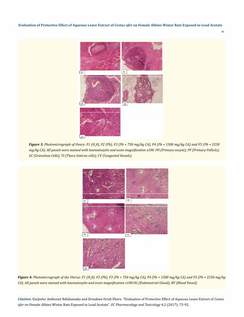
Figure 3: Photomicrograph of Ovary: F1 (H20), F2 (Pb), F3 (Pb + 750 mg/kg CA), F4 (Pb + 1500 mg/kg CA) and F5 (Pb + 2250 mg/kg CA). All panels were stained with haematoxylin and eosin magnification x100. PO (Primary oocyte); PF (Primary Follicle); GC (Granulosa Cells); TI (Theca Interna cells); CV (Congested Vessels).
Figure 4: Photomicrograph of the Uterus: F1 (H20), F2 (Pb), F3 (Pb + 750 mg/kg CA), F4 (Pb + 1500 mg/kg CA) and F5 (Pb + 2250 mg/kg CA). All panels were stained with haematoxylin and eosin magnification x100.UG (Endometrial Gland); BV (Blood Vessel).

Citation: Ezejiofor Anthonet Ndidiamaka and Orisakwe Orish Ebere. “Evaluation of Protective Effect of Aqueous Leave Extract of Costus afer on Female Albino Wistar Rats Exposed to Lead Acetate”. EC Pharmacology and Toxicology 4.2 (2017): 75-92.
Evaluation of Protective Effect of Aqueous Leave Extract of Costus afer on Female Albino Wistar Rats Exposed to Lead Acetate85
Figure 5: Photomicrograph of the Kidney: F1 (H20), F2 (Pb), F3 (Pb + 750 mg/kg CA), F4 (Pb + 1500 mg/kg CA) and F5 (Pb + 2250 mg/kg CA). All panels were stained with haematoxylin and eosin magnification x100. The slides above shows no histologic change in the kidney, all slides shows the glomeruli and renal tubules.
Figure 6: Photomicrograph of the Liver: F1 (H20), F2 (Pb), F3 (Pb + 750 mg/kg CA), F4 (Pb + 1500 mg/kg CA) and F5 (Pb + 2250 mg/kg CA). All panels were stained with haematoxylin and eosin magnification x100. The slide shows no histologic change in all the groups. All the slides showed the central and portal veins with corresponding hepatocytes.

Citation: Ezejiofor Anthonet Ndidiamaka and Orisakwe Orish Ebere. “Evaluation of Protective Effect of Aqueous Leave Extract of Costus afer on Female Albino Wistar Rats Exposed to Lead Acetate”. EC Pharmacology and Toxicology 4.2 (2017): 75-92.
Evaluation of Protective Effect of Aqueous Leave Extract of Costus afer on Female Albino Wistar Rats Exposed to Lead Acetate86
Discussion
The main health hazards of contamination by heavy metals have been associated with exposure to cadmium, lead and mercury – cur-rently the most widely distributed pollutants in the environment which, at the same time, demonstrate a high level of toxicity against living organisms is lead. Over the years there have been extensive toxicological studies reporting their adverse effect on humans such as neurotoxicity, immunodeficiency, osteoporosis, kidneys and other organ failures, as well as potential implications in impaired fertil-ity [20,21]. There is enough epidemiological data concerning acute metal poisonings in which the real health concern mainly relates to chronic exposures to low concentrations of Pb which can potentially affect a large part of the human population [22]. Infertility has been already recognized by the World Health Organization as a considerable public health issue worldwide and has become a serious medical challenge [23]. It is believed that approximately 15% - 30% of couples are diagnosed with unexplained infertility [24]. It is beyond any doubt that lifestyle and quality of the ambient environment can play a fundamental role in human reproductive success [25]. It has been reported that women with severe lead intoxication are more prone to prolonged and abnormal menstruation, miscarriage, still birth, premature delivery and infertility [26]. Lead can affect a woman’s reproductive hormone levels and this might lead to infertility. The exact mechanism of lead induced reproductive stress remains obscure but some studies suggests that lead causes oxidative stress. This presents an imbalance between the production of free radicals and the biological system’s ability to readily detoxify the reactive interme-diates or to repair the resulting damage [26]. This study was carried out in order to investigate the protective effects of Costus afer on lead induced pathological damage in female rats.
Most of the actions of Costus afer can be attributed to its antioxidant properties which emanate from its numerous bioactive com-pounds which are of immense medicinal properties. The phytoconstituents of aqueous leaf extract of Costus afer include alkaloids, sapo-nins, flavonoids, tannins, phenols, cardiac glycosides and terpenoids, these phytoconstituents are believed to be responsible for the bio-logical activity of the plant extract. Flavonoids are a group of plant metabolites thought to provide health benefits through cell signaling pathways and antioxidant effects. They are polyphenolic compounds containing fifteen atoms and are water soluble [27]. Phenols are a class of chemical compounds consisting of a hydroxyl group bonded to an aromatic hydrocarbon group. Natural phenolic compounds play an important role in cancer prevention and treatment. They have antioxidant, anticarcinogenic and anti-inflammatory effects. Tannins are water soluble polyphenolic substances found in plant product of secondary metabolism. They have a great structural diversity but in gen-eral, two classes are distinguished, the hydrolysable type and the condensed type [28]. Tannin solutions are acidic and have an astringent taste. Tannins have well documented antimicrobial activities. Tannins have also been reported to have other physiological effects such as to accelerate blood clotting, reduce blood pressure, decrease serum lipid level and modulate immune responses [29]. Terpenoids also called isoprenoids are secondary metabolites occurring in most organisms, particularly plants. A large number of terpenoids exhibit cyto-toxicity against a variety of tumour cells and cancer preventive as well as anticancer efficacy in pre-clinical animal models [30]. Terpenes constitute a vast number off products based on the different possible arrangements of bonded isoprene units [31]. Cardiac glycosides are a class of medications used to treat heart failure and certain irregular heartbeats [32]. Cardiac glycosides are potent inhibitors of cellular Na+/K+-ATPase. This ion transport function is necessary for cell survival. The ion transport move sodium ions out of the cell and brings potassium ions into the cell [33]. Several plants have been reported to have central nervous system depressant and anxiolytic activity due to the presence of triterpenoids, flavonoids [34] and saponins [35]. Triterpenoids, saponins are reported to have agonistic/facilitatory activities at GABAA receptor complex, which led to the hypothesis that they act as benzodiazepine-like molecules [36].
Effect on the Absolute and Relative Weight of Organs
There was a slight reduction in the relative weight of the uterus and fallopian tube in the group receiving lead only (Table 1) and the reduction was not statistically significant when compared with the control. This observation is not in agreement with the observations of Wiebe., et al. [37] and Udayraj., et al. [38] who failed to observe any significant changes in the weights of uterus, ovary and fallopian tube in rats exposed to lead. Another study by Dumitrescu., et al. [39] observed a progressive decline in the weights of the uterus and fallopian tube after six weeks of exposure to lead which was significant. The plant extract did not exert any significant effect on the weight of organs (both absolute and relative weight), this observation may be due to the dose and duration of the study.
Citation: Ezejiofor Anthonet Ndidiamaka and Orisakwe Orish Ebere. “Evaluation of Protective Effect of Aqueous Leave Extract of Costus afer on Female Albino Wistar Rats Exposed to Lead Acetate”. EC Pharmacology and Toxicology 4.2 (2017): 75-92.
Evaluation of Protective Effect of Aqueous Leave Extract of Costus afer on Female Albino Wistar Rats Exposed to Lead Acetate
87
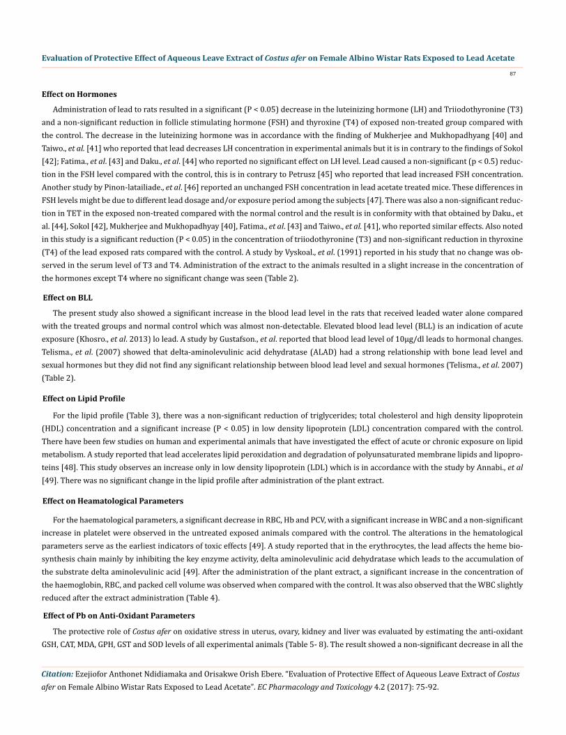
Effect on Hormones
Administration of lead to rats resulted in a significant (P < 0.05) decrease in the luteinizing hormone (LH) and Triiodothyronine (T3) and a non-significant reduction in follicle stimulating hormone (FSH) and thyroxine (T4) of exposed non-treated group compared with the control. The decrease in the luteinizing hormone was in accordance with the finding of Mukherjee and Mukhopadhyang [40] and Taiwo., et al. [41] who reported that lead decreases LH concentration in experimental animals but it is in contrary to the findings of Sokol [42]; Fatima., et al. [43] and Daku., et al. [44] who reported no significant effect on LH level. Lead caused a non-significant (p < 0.5) reduc-tion in the FSH level compared with the control, this is in contrary to Petrusz [45] who reported that lead increased FSH concentration. Another study by Pinon-latailiade., et al. [46] reported an unchanged FSH concentration in lead acetate treated mice. These differences in FSH levels might be due to different lead dosage and/or exposure period among the subjects [47]. There was also a non-significant reduc-tion in TET in the exposed non-treated compared with the normal control and the result is in conformity with that obtained by Daku., et al. [44], Sokol [42], Mukherjee and Mukhopadhyay [40], Fatima., et al. [43] and Taiwo., et al. [41], who reported similar effects. Also noted in this study is a significant reduction (P < 0.05) in the concentration of triiodothyronine (T3) and non-significant reduction in thyroxine (T4) of the lead exposed rats compared with the control. A study by Vyskoal., et al. (1991) reported in his study that no change was ob-served in the serum level of T3 and T4. Administration of the extract to the animals resulted in a slight increase in the concentration of the hormones except T4 where no significant change was seen (Table 2).
Effect on BLL
The present study also showed a significant increase in the blood lead level in the rats that received leaded water alone compared with the treated groups and normal control which was almost non-detectable. Elevated blood lead level (BLL) is an indication of acute exposure (Khosro., et al. 2013) lo lead. A study by Gustafson., et al. reported that blood lead level of 10µg/dl leads to hormonal changes. Telisma., et al. (2007) showed that delta-aminolevulinic acid dehydratase (ALAD) had a strong relationship with bone lead level and sexual hormones but they did not find any significant relationship between blood lead level and sexual hormones (Telisma., et al. 2007) (Table 2).
Effect on Lipid Profile
For the lipid profile (Table 3), there was a non-significant reduction of triglycerides; total cholesterol and high density lipoprotein (HDL) concentration and a significant increase (P < 0.05) in low density lipoprotein (LDL) concentration compared with the control. There have been few studies on human and experimental animals that have investigated the effect of acute or chronic exposure on lipid metabolism. A study reported that lead accelerates lipid peroxidation and degradation of polyunsaturated membrane lipids and lipopro-teins [48]. This study observes an increase only in low density lipoprotein (LDL) which is in accordance with the study by Annabi., et al [49]. There was no significant change in the lipid profile after administration of the plant extract.
Effect on Heamatological Parameters
For the haematological parameters, a significant decrease in RBC, Hb and PCV, with a significant increase in WBC and a non-significant increase in platelet were observed in the untreated exposed animals compared with the control. The alterations in the hematological parameters serve as the earliest indicators of toxic effects [49]. A study reported that in the erythrocytes, the lead affects the heme bio-synthesis chain mainly by inhibiting the key enzyme activity, delta aminolevulinic acid dehydratase which leads to the accumulation of the substrate delta aminolevulinic acid [49]. After the administration of the plant extract, a significant increase in the concentration of the haemoglobin, RBC, and packed cell volume was observed when compared with the control. It was also observed that the WBC slightly reduced after the extract administration (Table 4).
Effect of Pb on Anti-Oxidant Parameters
The protective role of Costus afer on oxidative stress in uterus, ovary, kidney and liver was evaluated by estimating the anti-oxidant GSH, CAT, MDA, GPH, GST and SOD levels of all experimental animals (Table 5- 8). The result showed a non-significant decrease in all the

Citation: Ezejiofor Anthonet Ndidiamaka and Orisakwe Orish Ebere. “Evaluation of Protective Effect of Aqueous Leave Extract of Costus afer on Female Albino Wistar Rats Exposed to Lead Acetate”. EC Pharmacology and Toxicology 4.2 (2017): 75-92.
Evaluation of Protective Effect of Aqueous Leave Extract of Costus afer on Female Albino Wistar Rats Exposed to Lead Acetate
88
parameters measured with the exception of MDA which showed no change on the ovary and uterus and a significant increase in liver and kidney in the exposed non-treated group compared with the control. The use of the extract showed a promising effect by a non-significant increase in the reduced antioxidant level measured and a decrease in the level of MDA in both liver and kidney. This indicates that the protective effect of Costus afer leave to lead toxicity is mediated through its antioxidant potential. This finding is in agreement with previous work of Dorostghoal [50]; Sharma and Bhattacharya [51]; Ansar., et al. [52] and Elgawisha and Abdelrazekb [53]. Pb is a ubiquitous environmental and industrial pollutant that induces a broad range of toxic manifestations within biological systems. Although the exact mechanism of lead toxicity still remains unclear but cumulative data showed that Pb exposure induces over-production of ROS and depletes the cellular antioxidant capacity. An imbalance of pro-oxidant/antioxidant ratio in tissue and cellular components is known to cause damage to membranes, DNA, or proteins, and finally destroy the tissues or systems (Hsu and Guo, 2002). Therefore, exogenous supplementation of antioxidant molecules would have an advantageous role on the cell’s antioxidant defenses to counteract Pb intoxica-tion. The edible leaves of Costus afer contains phenolics and flavonoids which are known plant antioxidant metabolites [54]. In this pres-ent study, Costus afer has been found to possess an inhibitory activity over Pb-acetate induced toxicity in experimental rats, as can be seen in its potential to increase the levels of anti-oxidants in biological system. Tissue antioxidant markers, liver and kidney function markers were chosen as quantitative markers for this study.
Effect on Liver and Kidney Parameters
In the present investigation, Pb exposure must have caused significant increase in Pb burden in the tissue of experimental rats. Many studies however have revealed that plant phenolics could decrease accumulation of heavy metal in tissue [55-57]. Which might be the case with Costus afer as it causes a significant increase (P < 0.05) in some of the hepatic and (Kidney) nephro function biomarkers evalu-ated. Generally, these results may indicate degenerative and necrotic changes in liver and kidney and prolonged exposure to this toxic metal may result in hepatic and kidney injuries [58]. Also the increased levels of blood urea with decreased levels of total protein (TP) may be indicative to protein catabolism and kidney dysfunction [59]. These results clearly showed that Pb -acetate has a harmful and stressful influence on the hepatic and renal tissues. Pb is known to alter the activity of lipid metabolizing enzymes in liver [60], which can limit the biosynthesis of bile acids. Bile acid plays important role in elimination of cholesterol from the body [61,62]. The present study revealed that administration of Costus afer induced significant (P < 0.05) decrease in some of the hepatic and Kidney function parameters such as AST, ALT, ALP, Na+, and HCO3- whereas the other parameters showed a non-significant decrease (UR, CR, TP, and K+) when com-pared with the control (Table 9).
Effect on Weight Gain, Feed and Fluid Intake
In the present investigation, the reduction of the weight gain for the animals receiving the lead only was not significant when com-pared with the control (Figure 1). There was a significant reduction (P < 0.05) in the feed and fluid intake of the animals which is in ac-cordance with the report by Udayraj., et al [38]. The reduction in the weight gain after the lead exposure may be due to its influence on the feeding behavior via central nervous system or secretion of growth hormone [63].
Effect on Histopathology
From the histopathological result shown in figures 1-5, the administration of lead did not exhibit any sign of toxicity to all the organs evaluated. Though some changes was noted for the absolute and relative weight as seen in table 2 but such changes did not reflect on the histology of the organs examined, this may be due to some factors like dose of the lead acetate administered coupled with exposure period to the toxicant.
Conclusion
In conclusion, this study has shown that though lead (Pb) having been implicated in most of the pathological conditions in animal model and the modern medicine having lost its potential in the management and treatment of most of these health challenges imposed by heavy metal contamination due to its increase toxicity and unaffordability there could still be hope resulting from the use of natural antidote which is less toxic, affordable and accessible, hence the use of Costus afer seems to be an answer to it.

Citation: Ezejiofor Anthonet Ndidiamaka and Orisakwe Orish Ebere. “Evaluation of Protective Effect of Aqueous Leave Extract of Costus afer on Female Albino Wistar Rats Exposed to Lead Acetate”. EC Pharmacology and Toxicology 4.2 (2017): 75-92.
Evaluation of Protective Effect of Aqueous Leave Extract of Costus afer on Female Albino Wistar Rats Exposed to Lead Acetate
89
Bibliography
1. Ezejiofor AN., et al. “Cytological and biochemical Studies during Progression of Alloxan Induced Diabetes and Possible Protection of aqueous leaf extract of Costus afer”. Chinese Journal of Natural Medicines 12.10 (2014): 745-752.
2. Anthonet Ndidiamaka Ezejiofor Chinna Nneka Orish and Orish E Orisakwe. “Morphological changes in Pancreases and Glucose re-duction of aqueous extract of Costus afer ker gawl leaf on alloxan-induced diabetic rats”. Journal of Basic and Clinical Physiology and Pharmacology 26.6 (2014): 595-601.
3. Anthonet Ndidiamaka Ezejiofor and Orish Ebere Orisakwe. “Assessment of the Hepatoprotective and Antioxidant Effect of Aqueous Leaf Extract of Costus afer “Ker Gawl” on Cyclosporine A Induced Hepatotoxicity”. Toxicology International 22.3 (2015): 83-91.
4. Akpan MM., et al. “Antimicrobial assessment of ethanolic extract of Costus afer leaves”. Asian Journal of Plant Science and Research 2.3 (2012): 335-341.
5. Anthonet Ndidi Ezejiofor., et al. “Nephroprotective and antioxidant effect of aqueous leaf extract of Costus afer Ker gawl on cyclospo-rine-a (Csa) induced nephrotoxicity”. Clinical Phytoscience 2 (2016): 11.
6. Okwu DE and Okwu ME. “Chemical composition of Spondias mombin Linn Plants parts”. Journal of Sustainable Agriculture and Envi-ronmental 6 (2004): 140-147.
7. Ezejiofor AN., et al. “Effect of aqueous extract of Costus afer ker Gawl (zingiberaceae) on the liver and kidney of male albino Wister rat”. Ancient Science of Life 33.1 (2013): 4-9.
8. Ijioma SN., et al. “Antinociceptive Property of Costus afer Ker Stem Juice and Ethanol Leaf Extract in Albino Rats”. Comprehensive Journal of Medical Sciences 2.2 (2014): 14-19.
9. Brads H. “Heavy Metals in the environment: Origin interaction and Remediation volume 6”. London : Academic Press (2002).
10. World Health Organization, Switzerland: Geneva. “Trace elements in human nutrition and health” (1996).
11. Yedjou GC and Tchounwou PB. “N- acetyl cysteine affords protection against lead induced cytotoxicity and oxidative stress in human liver carcinoma(Hep G2 ) cells”. Metal Ions in Biology and Medicine 10 (2008): 419-424.
12. Agency for Toxic Substances and Disease Registry (ATSDR). Public Health Services. Atlanta. Public Health Service, US. Department of Health and Human Services: Toxicological profile for lead (1999).
13. Sadeghi A., et al. “The Effect of Ascorbic Acid and Garlic Administration on LeadInduced Neural Damage in Rat Offspring’s Hippocam-pus”. Iranian Journal of Basic Medical Sciences 16.2 (2013): 157-164.
14. Okhawa H., et al. “Assay for lipid peroxidesin animal tissues by Thiobarbituric acid reaction”. Analytical Biochemistry 95.2 (1982): 351-358.
15. Misra HP and Fridovich I. “The role of superoxide anion in the auto-oxidation of epinephrine and a simple assay for superoxide dis-mutase”. Journal of Biological Chemistry 247.10 (1972): 3170-3175.
16. Balasubramanian KA., et al. “An unidentified inhibitor of lipid peroxidation in intestinal mucosa”. Biochimica et Biophysica Acta 962.1 (1988): 51-58.

Citation: Ezejiofor Anthonet Ndidiamaka and Orisakwe Orish Ebere. “Evaluation of Protective Effect of Aqueous Leave Extract of Costus afer on Female Albino Wistar Rats Exposed to Lead Acetate”. EC Pharmacology and Toxicology 4.2 (2017): 75-92.
Evaluation of Protective Effect of Aqueous Leave Extract of Costus afer on Female Albino Wistar Rats Exposed to Lead Acetate
90
17. Clairborne A. “Catalase activity”. In: Greewald AR (ed.). Handbook of Methods for oxygen radical research. CRC Press, Boca Raton, FL (1995): 237-242.
18. Habig WH., et al. “Glutathione S- transferase. The first enzymatic step in mercapturic acid formation”. Journal of Biological Chemistry 249.22 (1974): 7130-7139.
19. Bancroft JD and Gamble M. “Theory and practice of histological techniques”. 5th edition. New York: Churchill Livingstone (2003): 593-620.
20. Jang DH and Hoffman RS. “Heavy metal chelation in neurotoxic exposures”. Neurologic Clinics 29.3 (2011): 607-622.
21. Youness ER., et al. “Cadmium impact and osteoporosis: mechanism of action”. Toxicology Mechanism and Methods 22.7 (2012): 560-567.
22. Hu H. “Human health and heavy metals exposure”. In: McCally M. (ed.). Life support: the environment and human health. MIT press, Massachusetts (2002).
23. Vayena E., et al. “Medical, ethical & social aspects of assisted reproduction. Current practices & controversies in assisted reproduc-tion: Report of a WHO meeting”. Geneva, Switzerland (2001).
24. Quaas A and Dokras A. “Diagnosis and Treatment of Unexplained Infertility”. Reviews in Obstetrics and Gynecology 1.2 (2008): 69-76.
25. Sharpe RM and Franks S. “Environment, lifestyle and infertility - an intergenerational issue”. Nature Cell Biology 4.1 (2002): S33-S40.
26. Flora S., et al. “Arsenic, cadmium and lead”. Reproductive and Developmental Toxicology (2011): 415-438.
27. Robertson Sally. “What are flavonoids?” (2014).
28. Haslam E. “Practical polyphenolics: From structure to molecular recognition and physiological action”. Cambridge UK: Cambridge University Press (1998): 9810-9883.
29. Chung KT., et al. “Tannins and human health: a review”. Critical Review Food Science Nutrition 38.6 (1998): 421-464.
30. Roslin J Thopil and Anupam Bishayee. “Terpenoids as potential chemoprotective and therapeutic agents in liver cancer”. World Jour-nal of Hepatology 3.9 (2011): 228-249.
31. Zwenger R Sam and Basu Chandak. “Plant terpenoids: Applications and future potentials”. Biotechnology and Molecular Biology Re-views 3.1 (2008): 1-7.
32. Heller L Jacob. “Cardiac glycoside overdose” (2014).
33. Klabunde E Richard. “Cardiac glycosides Cardiovascular pharmacology concepts” (2015).
34. Datta BK., et al. “Analgesic, anti-inflammatory and central nervous system depressant activities of sesquiterpenes and a flavonoid glycoside from Polygonum viscosum”. Pharmazie 59.3 (2004): 222-225.
35. Dubois M., et al. “Cussonoides A and B, two triterpene saponins from Cussoria barteri”. Plant Medicine (1986): 5680-5683.
36. Uma A Bhosale., et al. “Study of the central nervous system depressant and behavioural activity of an ethanol extract of Achyranthes aspera (Agadha) in different animal models”. International Journal of Applied and Basic Medical Research 1.2 (2011): 104-108.

Citation: Ezejiofor Anthonet Ndidiamaka and Orisakwe Orish Ebere. “Evaluation of Protective Effect of Aqueous Leave Extract of Costus afer on Female Albino Wistar Rats Exposed to Lead Acetate”. EC Pharmacology and Toxicology 4.2 (2017): 75-92.
Evaluation of Protective Effect of Aqueous Leave Extract of Costus afer on Female Albino Wistar Rats Exposed to Lead Acetate
91
37. Wiebe JP., et al. “Effect of prenatal and neonatal exposure of lead on gonadotropin receptors and steroidogenesis in rats ovaries”. Journal of Toxicological Environmental Health 24.4 (1988): 461-476.
38. Udayraj PN., et al. “Lead induced adverse effects on the reproductive system of rats with particular reference to histopathological changes in uterus”. Indian Journal of Pharmacology 47.1 (2015): 22-26.
39. Dumitrescu E., et al. “The consequences of chronic lead acetate intake on exposure and morphological integrity biomarkers (lead level and weight of sexual organs) in female”. Lucrări $Tiinłifice Medicină Veterinară 41 (2008): 619-622.
40. Mukherjee S and Mukhopadhyay PK. “Studies on lead and arsenic toxicity in male rat gonads and its protection by high dietary pro-tein supplementation”. Al Ameen Journal of Medical Sciences 2.1 (2009): 73-77.
41. Taiwo AM., et al. “Assessments of Possible Gonadotoxic Effect of Lead on Experimental Male Rabbits”. Global Veterinaria 5.5 (2010): 282-286.
42. Sokol R and Berman N. “Effects of Lead Exposure on GnRH and LH Secretion in Male Rats: Response to castration and -methyl-p-thyrosine AMPT challenge”. Reproductive Toxicology 12.3 (1998): 347-355.
43. Fatima R., et al. “Protective role of ginger on lead induced derangement in plasma testosterone and luteinizing hormone levels on male Sprague Dawley Rats”. Journal Ayub Medical College Abbottabad 23.4 (2011): 24-27.
44. Daku AB., et al. “Age-related effects of lead poisoning on sex hormones in adult male Wistar rats”. Journal of Physiology and Patho-physiology 7.4 (2016): 23-27.
45. Petrusz P., et al. “Lead poisoning and reproduction: effects on pituitary and serum gonadotropins in neonatal rats”. Environmental Research 19.2 (1979): 383-391.
46. Pinon-Lataillade G., et al. “Reproductive toxicity of chronic lead exposure in male and female mice”. Human and Experimental Toxicol-ogy 14.11 (1995): 872-878.
47. Mohsen V., et al. “Review article: How does lead induce male infertility”. Iranian Journal of Reproductive Medicine 9.1 (2011): 1-8.
48. Ribarov SR., et al. “The effect of lead on hemoglobin catalyzed lipid peroxidation”. Biochimica et Biophysica Acta 664.3 (1981): 453-459.
49. Annabi A., et al. “Oxidative stress, biochemical alterations and hyperlipidemia in female rats induced by lead chronic toxicity during puberty and post puberty periods”. Iranian Journal of Basic Medical Sciences 18.10 (2015): 1034-1043.
50. Dorostghoal M., et al. “Protective effects of Fumaria parviflora L. on lead-induced testicular toxicity in male rats”. International Jour-nal of Andrologia 46.4 (2013): 437-446.
51. Sharma DN and Bhattacharya L. “Enzymatic toxicity induced by lead acetate and its possible reversal by α tocopherol in testicular tissue of Swiss albino mice”. International Journal of Biomedical Research 6.11 (2015): 874-879.
52. Ansar S., et al. “The protective effect of rutin against renal toxicity induced by lead acetate”. Toxin Reviews 35.1-2 (2016): 58-62.
53. Elgawisha RA and Abdelrazek HMA. “Effects of lead acetate on testicular function and caspase- 3expression with respect to the pro-tective effect of cinnamon in albino rats”. Toxicology Reports 1 (2014): 795-801.

Citation: Ezejiofor Anthonet Ndidiamaka and Orisakwe Orish Ebere. “Evaluation of Protective Effect of Aqueous Leave Extract of Costus afer on Female Albino Wistar Rats Exposed to Lead Acetate”. EC Pharmacology and Toxicology 4.2 (2017): 75-92.
Evaluation of Protective Effect of Aqueous Leave Extract of Costus afer on Female Albino Wistar Rats Exposed to Lead Acetate
92
54. Ezejiofor AN., et al. “Evaluations of Some Biological Properties of Ethanolic Leave Extract of Costus Afer (Ker Gawl)”. IOSR Journal of Pharmacy and Biological Sciences (IOSR-JPBS) 12.1 (2017): 62-68.
55. Xia D., et al. “Protective effect of Smilax glabra extract against lead-induced oxidative stress in rats”. Journal of Ethnopharmacology 130.2 (2010): 414-420.
56. Liu C., et al. “Protective role of puerarin on lead-induced alterations of the hepatic glutathione antioxidant system and hyperlipid-emia in rats”. Food and Chemical Toxicology 49.12 (2011): 3119-3127.
57. Amudha K and Pari L. “Beneficial role of naringin, a flavanoid on nickel induced nephrotoxicity in rats”. Chemico-Biological Interac-tions 193.1 (2011): 57-64.
58. El-Nekeety AA., et al. “Protective effect of Aquilegia vulgaris (L.) against lead acetate-induced oxidative stress in rats”. Food and Chemical Toxicology 47.9 (2009): 2209-2215.
59. Abdel-Wahhab MA., et al. “Inulacrithmoides extract protect against ochratoxin A-induced oxidative stress, clastogenic and muta-genic alterations in male rats”. Toxicon 52.4 (2008): 566-573.
60. Kojima M., et al. “Tumor necrosis factor-a independent down regulation of hepatic cholesterol 7a-hydroxylase gene in mice treated with lead nitrate”. Toxicological Sciences 87.2 (2005): 537-542.
61. Mudipalli A. “Lead hepatotoxicity and potential health effects”. Indian Journal of Medical Research 126.6 (2007): 518-527.
62. Newairy ASA and Abdou HM. “Protective role of flax lignans against lead acetate induced oxidative damage and hyperlipidemia in rats”. Food and Chemical Toxicology 47.4 (2009): 813-818.
63. Huseman CA., et al. “Neuroendocrine effects of toxic and low blood lead levels in children”. Pediatrics 90 (1992): 186-189.
Volume 4 Issue 2 August 2017© All rights reserved by Ezejiofor Anthonet Ndidiamaka and Orisakwe Orish Ebere.