curcuma mangga-mediated synthesis of gold nanoparticles ... · in this study, we demonstrated the...
TRANSCRIPT

nanomaterials
Article
Curcuma mangga-Mediated Synthesis of GoldNanoparticles: Characterization, Stability,Cytotoxicity, and Blood Compatibility
Yiing Yee Foo 1,*, Vengadesh Periasamy 2,*, Lik Voon Kiew 3, G. Gnana Kumar 4 andSri Nurestri Abd Malek 1
1 Institute of Biological Sciences, Faculty of Science, University of Malaya, Kuala Lumpur 50603, Malaysia;[email protected]
2 Low Dimensional Materials Research Centre (LDMRC), Department of Physics, Faculty of Science,University of Malaya, Kuala Lumpur 50603, Malaysia
3 Department of Pharmacology, Faculty of Medicine, University of Malaya, Kuala Lumpur 50603, Malaysia;[email protected]
4 Department of Physical Chemistry, School of Chemistry, Madurai Kamaraj University, Madurai 625021,Tamil Nadu, India; [email protected]
* Correspondence: [email protected] (Y.Y.F.); [email protected] (V.P.); Tel.: +60-37-967-4038 (V.P.)
Academic Editor: Eva PellicerReceived: 4 March 2017; Accepted: 27 April 2017; Published: 27 May 2017
Abstract: The utilization of toxic chemicals as reducing and stabilizing agents in the preparationof gold nanoparticles (AuNPs) has increased in vivo toxicity and thus limited its application inclinical settings. Herein, we propose an alternative method of preparing highly stable AuNPs,where non-toxic Curcuma mangga (CM) extract was used as a single reducing and stabilizingagent to overcome the aforementioned constraints. The morphological images enunciated thatthe homogeneously dispersed AuNPs exhibited spherical morphology with an average particlediameter of 15.6 nm. Fourier Transform infrared (FTIR) and cyclic voltammetry analysis demonstratedthat carbonyl groups of terpenoids in CM extract played an important role in the formation andstabilization of AuNPs. Green-synthesized AuNPs were found to have good stability in physiologicalmedia after 24 h of dispersion. The AuNPs were also cytocompatible with human colon fibroblastcell (CCD-18Co) and human lung fibroblast cell (MRC-5). Hemocompatibility tests revealed thatthe AuNPs were blood-compatible, with less than 10% of hemolysis without any aggregation oferythrocytes. The current study suggests potential in employing a CM-extract-based method in thepreparation of AuNPs for anticancer diagnosis and therapy.
Keywords: gold nanoparticles; green synthesis; in vitro stability; biocompatible; phytochemicals;hemocompatibility
1. Introduction
In recent years, the application of gold nanoparticles (AuNPs) as a nano-diagnostic, in drugdelivery, and as a therapeutic agent has received extensive attention due to the remarkable physicaland chemical properties of AuNPs [1]. These includes the capability to harness energy from thetissue penetrating near infra-red radiations, excellent photothermal properties, high drug loadingand the ability to functionalize with targeting moieties [2]. To date, AuNP products on clinical trials,recombinant human tumor necrosis factor alpha bound PEGylated gold nanoparticles (CYT-6091) andAuroShell particles have shown positive outcomes for cancer therapy and photothermal therapy forcoronary atherosclerosis, respectively [3,4].
Nanomaterials 2017, 7, 123; doi:10.3390/nano7060123 www.mdpi.com/journal/nanomaterials

Nanomaterials 2017, 7, 123 2 of 14
Only two out of thousands of AuNP formulations from the literature have progressed to humanclinical trials. Poor stability in physiological environments remains one of the major limitations thatcontribute to poor translation of nanoparticle formulations from bench to clinic [5]. Physiologicalenvironments abundant with proteins and salts can influence the stability and interaction of AuNPswith biological systems. The high ionic strength of physiological media can cause AuNPs aggregation,thus reducing cellular internalization of AuNPs [6]. For instance, AuNPs capped with commonstabilizing agents such as citrate and cetyltrimethylammonium bromide (CTAB) have been found tobe unstable in buffers of pH 4, 7, 8, or 10 [7]. Thus, the stabilization of AuNPs with capping agents areoften required to enable any biological applications of AuNPs [8].
Apart from the stability, biocompatibility, and biosafety of AuNPs, there are two other importantfactors to be considered. The synthesis of AuNPs often involves the use of toxic reducing agents orsurfactants. Wan et al. reported that CTAB-coated gold nanorods (CTAB-AuNPs) are toxic to bothtumors and non-malignant transformed cells at nanomolar concentrations [9]. CTAB-AuNPs haveshown to aggregate on the cell membrane and cause damage to the membrane. CTAB-AuNPs alsoincreased lysosomal membrane permeation and decreased mitochondrial membrane potential, whichsubsequently induced cell death [10]. Freese et al. demonstrated that the higher amount of citrate onthe surface of AuNPs increases the cytotoxicity and decreases the proliferation rate of human dermalmicrovascular endothelial cells (HDMEC) and the human cerebral microvascular endothelial cell line(hCMEC/D3) [11]. Vijayakumar and Ganesan investigated the cytotoxic effect of the stabilizing agentsof AuNPs. The citrate-stabilized AuNPs (citrate-AuNPs) were found to have increased cytotoxicitycompared to starch- and gum arabic-stabilized AuNPs [12]. These studies suggested that the surfacechemistry of AuNPs accounts for the biosafety of AuNPs in biological applications. Therefore, the useof non-toxic stabilizing agents in the preparation of AuNPs is necessary.
Green-synthesized AuNPs have emerged in recent times as a potential solution to overcomethe stability and biocompatibility problems of chemically synthesized AuNPs. Generally, AuNPsare synthesized through physical and chemical routes, which are costly, consume high amounts ofenergy, and produce toxic wastes [13]. In recent years, the green synthesis of AuNPs has emerged asan alternative method due to its simplicity, cost, and energy effectiveness and considerable reductionin the generation of hazardous waste [13]. Green synthesis utilizes natural biomolecules such aspolysaccharides [14], microbial enzymes [15], and phytochemicals [16] as the reducing and stabilizingagents in the formation of AuNPs. In contrast to physical and chemical synthesis, this process normallydoes not require heat or pressure, which ultimately reduces the energy usage and therefore lowers theproduction cost [17].
In this study, we demonstrated the synthesis of AuNPs using Curcuma mangga aqueous ethanol(CM) extract through a simple, one-step method. C. mangga is a type of ginger, traditionally usedin the treatment for fever, stomachaches, and cancer [18]. It has also been reported extensively forits anticancer [18], antioxidant [19], and antimicrobial properties [20]. Numerous phytochemicalswith antioxidant activity have been reported to be present in C. mangga [19], which could act as thereducing agent for reducing precursor Au3+ ions. Upon AuNP synthesis, we further investigatephysico-chemical characteristics, the in vitro stability in physiological conditions, cytocompatibility,and blood compatibility.
2. Results
2.1. Characterization of AuNPs by Ultraviolet-Visible (UV-Vis) Spectroscopy and TransmissionElectron Microscopy
The formation of AuNPs can be observed as the change in color from yellow to red-violet. Thered-violet color observed was a result of the surface plasmon resonance (SPR) of AuNPs, which isattributed to the oscillation of free conducting electrons induced by the electromagnetic field [21].UV-Vis spectroscopy was carried out to ascertain the formation, and to monitor the stability, of AuNPs.Figure 1a shows the UV-Vis spectra of AuNPs at different incubation times. An absorption peak

Nanomaterials 2017, 7, 123 3 of 14
at a visible wavelength of 535 nm was observed after 15 min of incubation, which corresponds tothe SPR band of AuNPs. The intensity of the absorption peak increased significantly for the firsthour of incubation. There was no significant increase in the intensity of the SPR band after a further24 h of incubation, indicating the completion of a reaction at one hour of incubation. The yield ofAuNPs synthesized by CM extract was found to be 52.5% using microwave plasma-atomic emissionspectroscopy (MP-AES).
Figure 1. (a) Ultraviolet-visible (UV-Vis) spectra of gold nanoparticles (AuNPs) at incubation times of15, 30, 45, 60, and 90 min and 24 h; (b) Transmission electron micrographs of AuNPs synthesized fromCurcuma mangga (CM) extract at magnification 100,000×. (Inlet) Size distribution of AuNPs analyzedfor about 400 particles using Image J.
The size and shape of AuNPs were determined using transmission electron microscopy (TEM).TEM image (Figure 1b) illustrates that the AuNPs synthesized from CM extract were monodispersedand predominantly spherical in shape. The core sizes of AuNPs were in the range of 2–35 nm, withan average size of 15.6 nm. It was also observed that the sizes of AuNPs were distributed normallyas shown in the histogram in Figure 1b (inlet). Studies have shown that nanoparticles less than 5 nmeasily undergo renal clearance [22], while particles more than 200 nm will be retained in the spleen [23].Thus, AuNPs with sizes in the range of 10–200 nm will be highly desirable for systemic administration.
2.2. Fourier Transform Infrared Spectroscopy
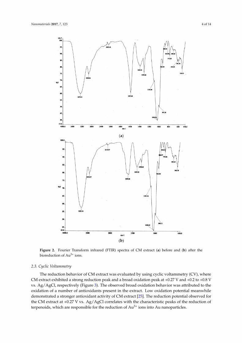
Fourier Transform infrared (FTIR) spectroscopy was conducted to investigate the possiblefunctional groups involved in the formation of AuNPs. Two intense bands were observed at 1036and 3273 cm−1 in the FTIR spectra of CM before and after bioreduction of Au3+ (Figure 2a,b), whichcorresponds to C–OH and O–H bonds respectively [24]. The results support the chemical compositionsof CM extract, where the C–OH and O–H bonds can be found in polyphenolics, curcuminoids, andterpenoids [18]. Other bands that are visible in both spectra (Figure 2a,b) are at 2929, 1394, and1309 cm−1. Bands observed at ~500–868 cm−1 signify the C–H groups that are abundant in CM extract.Intense bands at 1588 cm−1 in the FTIR spectrum of CM extract before bioreduction was related tothe unsaturated carbonyl group in terpenoids. The intensity of the band was reduced and shifted to1610 cm−1 after the bioreduction of Au3+, suggesting that the carbonyl group of terpenoids in the CMextract reduces Au3+ in the synthesis of AuNPs.
Nanomaterials 2017, 7, 123 4 of 14
Figure 2. Fourier Transform infrared (FTIR) spectra of CM extract (a) before and (b) after thebioreduction of Au3+ ions.
2.3. Cyclic Voltammetry
The reduction behavior of CM extract was evaluated by using cyclic voltammetry (CV), whereCM extract exhibited a strong reduction peak and a broad oxidation peak at +0.27 V and +0.2 to +0.8 Vvs. Ag/AgCl, respectively (Figure 3). The observed broad oxidation behavior was attributed to theoxidation of a number of antioxidants present in the extract. Low oxidation potential meanwhiledemonstrated a stronger antioxidant activity of CM extract [25]. The reduction potential observed forthe CM extract at +0.27 V vs. Ag/AgCl correlates with the characteristic peaks of the reduction ofterpenoids, which are responsible for the reduction of Au3+ ions into Au nanoparticles.

Nanomaterials 2017, 7, 123 5 of 14
Figure 3. Cyclic voltammetry (CV) of CM extract in pH 7.4 at a scan rate of 30 mV s−1.
2.4. In Vitro Stability Study
The stability of AuNPs in buffer and biological media is an essential criteria before using AuNPsfor cell-based studies or systemic use. The presence of electrolytes and proteins, for instance, humanserum albumin (HSA) in blood stream or fetal bovine serum (FBS) in cell culture media may destabilizethe AuNPs [26]. Thus, the stability of AuNPs was investigated by dispersing AuNPs in phosphatebuffered saline (PBS) of pH 5.0, 7.4, and 9.0, 0.5% HSA, Dulbecco’s Modified Eagle Medium (DMEM),and DMEM with 10% FBS. Ultrapure water was used as a negative control. The spectra change wasmonitored by UV-Vis spectroscopy. After 24 h of dispersion, compared with the negative control, nospectral change was observed in the UV-Vis spectra of AuNPs dispersed in the physiological buffertested (Figure 4).
Figure 4. UV-Vis absorption spectra of AuNPs after 24 h of incubation in phosphate buffered saline(PBS) of pH 5.0, 7.4, 9.0, 0.5% human serum albumin (HSA), Dulbecco’s Modified Eagle Medium(DMEM), DMEM with 10% fetal bovine serum (FBS), and ultrapure water.
Zeta potential values of AuNPs in ultrapure water were found to be −38.2 ± 0.6 mV (Table 1).A slight decrease in zeta potential values (−26.4, −29.2, −33.3, and −34.4 mV) were observed forAuNPs dispersed in PBS of pH 5, 7, 9, and 0.5% HSA. Meanwhile, AuNPs in DMEM media andDMEM with 10% FBS demonstrated significant decreases in zeta potential values to −20.9 and−13.5 mV, respectively.

Nanomaterials 2017, 7, 123 6 of 14
Table 1. Zeta potential values and hydrodynamic sizes of gold nanoparticles after 24 h of incubation inPBS of pH 5.0, 7.4, and 9.0, 0.5% human serum albumin (HAS), Dulbecco’s Modified Eagle Medium(DMEM) media, and DMEM media with 10% fetal bovine serum (FBS).
Physiological Media Zeta Potential (mV) Size (nm)
PBS pH 5.0 −26.4 ± 1.0 * 82.9 ± 1.0PBS pH 7.4 −29.2 ± 0.8 * 55.4 ± 1.5 *PBS pH 9.0 −33.3 ± 0.6 * 68.8 ± 2.0 *0.5% HSA −34.4 ± 1.0 * 77.3 ± 1.3
DMEM −20.9 ± 0.4 * 75.8 ± 1.8DMEM with 10% FBS −13.5 ± 0.1 * 114.7 ± 1.4 *
Control −38.2 ± 0.6 78.7 ± 0.5
Results presented as mean ± S.E, with n = 3. * significant difference compared to control at p < 0.05, obtained byone-way ANOVA.
The hydrodynamic radius of AuNPs in ultrapure water was 78.7 ± 0.5 nm after 24 h. There wasdiscrepancy in size determination using dynamic light scattering (DLS) and TEM analysis, due tothe difference in the analytical techniques. DLS measures the hydrodynamic radius of an NP, whichincludes the coating on the surface of AuNPs, while TEM measures the core size of AuNPs. Therefore,the hydrodynamic radius of a nanoparticle is usually bigger than the core size determined by TEM [27].The incubation of AuNPs in PBS of pH 5.0, 0.5% HSA, and DMEM media did not cause any significantchange in the hydrodynamic size of the AuNPs (Table 1). The incubation of the AuNPs in PBS ofpH 7.4 and pH 9.0 decreased the hydrodynamic size of the AuNPs to 55.4 and 68.8 nm, respectively.Meanwhile, the increase in the hydrodynamic size to 114.7 nm was observed for AuNPs incubated inDMEM with 10% FBS [28].
2.5. Cytocompatibility Study
Cell viability assay was carried out in order to investigate the in vitro toxicity profile of AuNPs.In this experiment, we used a sulforhodamine B (SRB) assay to determine cell viability. SRB is anegatively charged pink aminoxanthine dye. The SRB assay depends on the ability of SRB to bind tobasic amino acid in mild acidic condition and release the dye in a basic condition. SRB is simpler, moresensitive and reproducible, compared to other cytotoxicity assays [29].
Two normal human fibroblast cell lines were used in this study, which are the human lungfibroblast cell (MRC-5) and human colon fibroblast cell (CCD-18Co). The percentage of viability ofCCD-18Co and MRC-5 (Figure 5a,b) at 72 h of exposure to AuNPs at concentrations of 25 µg/mL andlower were maintained above 90% and 80%, respectively. No significant toxic effect to CCD-18Cowas observed upon treatment with AuNPs at 50 µg/mL for 24 h, with a percentage of viability of94.0%. AuNPs only result in a slight inhibition to CCD-18Co after 72 h of treatment with a percentageof viability of 65.3%. AuNPs at high concentration show a more pronounced effect to MRC-5, where65.4% and 13.3% of cell viability was observed for treatment after 24 and 72 h, respectively. CM extractat concentrations up to 50 µg/mL have not shown any toxic effect to both CCD-18Co and MRC-5 after24 and 72 h of treatment (Figure 5c,d).

Nanomaterials 2017, 7, 123 7 of 14
Figure 5. The percentage of viability by the SRB assay for (a) human colon fibroblast cell (CCD-18Co)and (b) human lung fibroblast cell (MRC-5) after exposure to AuNPs for 24 and 72 h in a dose-dependentmanner. The percentage of viability of (c) CCD-18Co and (d) MRC-5 upon treatment with CM extractfor 24 and 72 h. Concentrations of AuNPs and CM extract used in the experiments were 3.13, 6.25,12.50, 25.00, and 50.00 µg/mL. Results are shown as mean ± S.E. with n = 3.
2.6. In Vitro Blood Compatibility
Blood compatibility test is an important factor to evaluate toxicity profile of nanoparticles forintravenous use. AuNPs interact with blood cells immediately after administration into biologicalsystems. Thus, it is essential to evaluate hemocompatibility of AuNPs. In this study, hemolysis andthe red blood cell (RBC) aggregation test were used to measure the destruction of RBC upon exposureto green-synthesized AuNPs. The hemolysis assay is a colorimetric assay used to estimate the amountof released red-colored hemoglobin, which indicate the extent of the damage of RBC [30]. FromFigure 6, the percentage of hemolysis of RBC were found to be 2.7%, 1.8%, 2.6%, 6.2%, and 9.9% uponexposure to AuNPs with concentrations of 3.13, 6.25, 12.50, 25.00, and 50.00 µg/mL, respectively. Nohemolysis of RBC was observed upon exposure to CM extract up to concentrations of 50 µg/mL.Citrate-AuNPs results in 2.9% of hemolysis of RBC at a concentration of 50 µg/mL. The exposure ofRBC to CTAB-AuNPs of concentrations 25.00 and 50.00 µg/mL results in 44.7% and 96.0% of hemolysis.
Microscopy images in Figure 7 show the aggregation of RBCs upon incubation with the positivecontrol. RBCs treated by AuNPs, CM extract, and citrate-AuNPs maintained healthy smooth biconcavestructures, which have a morphology similar to the negative control. No aggregation was observed inthe RBCs treated by all concentrations of the AuNPs, CM extract, and citrate-AuNPs tested. Meanwhile,RBC treated with low concentrations (3.13–12.5 µg/mL) of CTAB-AuNPs showed RBC deformation.Lysed RBC was observed upon treatment with CTAB-AuNPs with a concentration of 25 µg/mL. Only

Nanomaterials 2017, 7, 123 8 of 14
cell debris can be seen for RBC treated with 50 µg/mL of CTAB-AuNPs since almost all of the RBCunderwent hemolysis.
Figure 6. Percentage of hemolysis of red blood cells after exposure to AuNPs at concentrations of 3.13,6.25, 12.50, 25.00, and 50.00 µg/mL. Each point represents mean ± S.E., with n = 3 (if not indicated, theerror bar was within the size of the symbol).
Figure 7. Micrographs of erythrocytes after 2 h of incubation with the positive control, the negativecontrol, the AuNPs, the CM extract, the citrate-AuNPs, and cetyltrimethylammonium bromide(CTAB)-AuNPs at concentrations of 3.13, 6.25, 12.50, 25.00, and 50.00 µg/mL at a magnificationof 100×.

Nanomaterials 2017, 7, 123 9 of 14
3. Discussion
The applications of AuNPs have shown promising advancements in the field of medicine.However, very few AuNPs have gone through clinical trials and have proven to be beneficial for humanapplications, due to the lack of knowledge in the physical characteristics of AuNPs in the physiologicalsystem. This study therefore involves in vitro stability, cytocompatibility, and hemocompatibilityinvestigations as a prerequisite evaluation on AuNPs for in vivo applications.
A simple, one-step method was demonstrated for the green synthesis of AuNPs using CMextract. This method is fast, cheap, and energy-efficient, where no heating and stabilizingagent was required for the formation of AuNPs. A bottom-up approach, where Au3+ ionsfrom chloroauric acid (HAuCl4·3H2O) act as the building blocks for the formation of AuNPs,was adopted [13]. The antioxidants in CM extract serve as a reducing agent in the synthesisof AuNPs [31]. From FTIR analysis, carbonyl groups in the CM extract were shown to beinvolved in the reduction of Au3+ ions. CM extract is composed of terpenoids, for instance,labda-8(17), 12-diene-15,16-dial, 15,16-bisnor-labda-8(17),11-diene-13-on, calcatarin, and ZeruminA [18,19]. Labda-8(17), 12-diene-15,16-dial, calcatarin, and Zerumin A have aldehyde groups, while15,16-bisnor-labda-8(17),11-diene-13-on has a keto group. These compounds are capable of reducingthe Au3+ to Au0. This result is consistent with a previous study that showed predominant involvementof carbonyl group in the reduction of Au3+ ions [32].
The reduction capability of CMEW extract in reducing the Au3+ ions into AuNPs was furthersupported by cyclic voltammetry analysis. As clearly depicted in Figure 3, the reduction potentialwas observed for CM extract at +0.27 V vs. Ag/AgCl, which is well correlated with the characteristicreduction potential of terpenoids. The obtained cyclic voltammogram has demonstrated the reductioncapability of terpenoids in CM extract for the reduction of Au3+ ions into AuNPs. We propose amechanism for the formation of AuNPs as shown in Scheme 1. The reduction of Au3+ to Au leads tothe formation of nuclei. The nuclei coalesce and contribute to the growth of AuNPs [33]. Terpenoidswith carboxylic groups, such as Longpene A, Coronadiene, and Zerumin A can bind to AuNP surfacesand act as a stabilizing agent [34].
Scheme 1. The proposed mechanism for the formation of AuNPs synthesized using CM extract.
The evaluation on the stability of AuNPs in biological media is important to predict the fate ofAuNPs after systemic administration. Bloodstream or physiological media contain serum proteins,electrolytes, nutrients, and metabolites that contribute to a high ionic strength. The aggregation ofchemically synthesized AuNPs may occur when they were subject to the blood stream, limiting thein vivo human applications of AuNPs [6]. Chemically synthesized AuNPs, such as citrate-cappedAuNPs and CTAB-coated AuNPs, are unstable and form aggregates in physiological media [7,35].Wang et al. reported a rapid drop in absorption intensities of citrate-capped and CTAB-coated AuNPsin the first 15 min of incubation in PBS of pH 4, 7, 8, and 10, which indicate the instability of theAuNPs in PBS. Another study has shown that diluting citrate-capped AuNPs in PBS results in theformation of agglomerates 2–3 times larger in size [36]. In our experiment, the dispersion of AuNPsin PBS, 0.5% HSA, DMEM, and DMEM with 10% FBS did not alter the UV-Vis spectra, suggesting

Nanomaterials 2017, 7, 123 10 of 14
that the green-synthesized AuNPs were stable in all conditions tested. There were also no significantchanges in the size of AuNPs upon their dispersion in PBS, the HSA solution, and DMEM, suggestingthat AuNPs synthesized from CM extract are stable. The 46% increase in size of AuNPs after 24 hof dispersion in DMEM with 10% of FBS due to the formation of protein corona on the surface ofAuNPs contributed by FBS. This is a common phenomenon where instantaneous absorption of proteinoccurred once the nanoparticles were exposed to media containing protein [5].
The zeta potential values decreased by 12.8 to 30.9% upon incubation with PBS, which might bedue to the high ionic strength of PBS disrupting the surface charge of AuNPs. The zeta potential valuesbecome less negative as the pH of PBS decreased, which is due to the higher concentration of H+ ionsin acidic PBS, which reduced the negative surface charge of AuNPs [37]. The significant reductionin zeta potential values of AuNPs in DMEM media is caused by a high ionic environment in themedia. DMEM media contain a high percentage of inorganic salts, amino acids, and vitamins [5]. Thealteration in zeta potential, however, did not affect the stability of AuNPs in all physiological media,as demonstrated by UV-Vis spectroscopy and hydrodynamic size results. All these data support thefact that AuNPs synthesized by CM extract were stable in all physiological conditions tested.
AuNPs synthesized by CM extract were biocompatible. Cytocompatibility results suggestedthat AuNPs synthesized from CM extract did not have toxic effects on the two normal cells testedat the concentration of 25 µg/mL. Therefore, AuNPs are non-toxic for biomedical applications. Thecytocompatibility of AuNPs was attributed to the biocompatibility of CM extract used as a stabilizingagent. In the preparation of AuNPs via chemical synthesis, the residual contamination from thesynthesis or the desorption of the stabilizing agent from AuNPs may contribute to the toxicity ofAuNPs. A common capping agent for gold nanorods, CTAB, has a high cytotoxicity against cells atnanomolar concentrations. This resulted in a significant cytotoxicity of CTAB-capped gold nanorods.As such, overcoating the CTAB-capped gold nanorods with polymers was required to reduce thetoxicity of CTAB-capped gold nanorods [38]. Herein, we demonstrated that the use of phytochemicalsas capping agents might be a better option for the synthesis of biocompatible AuNPs.
The investigation of toxicity towards RBC is an important parameter to evaluate the safety andcirculation characteristics of AuNPs. This study has shown that AuNPs synthesized from CM extractare hemocompatible. No more than 10% of hemolysis was observed for all tested concentrations up to50 µg/mL, which is below the safe hemolytic ratio for biomaterials according to ISO/TR7406 [39]. Theaggregation of RBC was also not observed for all concentrations of AuNPs tested. The hemocompatiblecharacteristic of AuNPs is attributed to its negative zeta potential and hemocompatibility of the CMextract. Meanwhile, we have shown that CTAB-AuNPs are not blood compatible, where 44.7% and96.0% of hemolysis was observed upon treatment with 25 and 50 µg/mL CTAB-AuNPs. This may bedue to desorption of CTAB from CTAB-AuNPs. Free CTAB molecules can cause lysis to RBC at lowconcentrations [40]. Thus, CTAB-AuNPs is not recommended for biomedical applications.
4. Materials and Methods
4.1. Materials
Gold (III) chloride trihydrate (HAuCl4·3H2O), citrate-stabilized gold nanoparticles (20 nm),Eagle’s Minimum Essential Medium (EMEM), fetal bovine serum (FBS), sodium pyruvate,amphotericin B, penicillin/streptomycin, human serum albumin (HSA), and sulforhodamine B (SRB)dye were purchased from Sigma-Aldrich (Saint Louis, MO, USA). The human normal colon fibroblastcell line (CCD-18Co) and the human lung fibroblast cell line (MRC-5) were purchased from AmericanType Culture Collection (ATCC, Manassas, VA, USA). Dulbecco’s Modified Eagle Medium (DMEM)without phenol red was purchased from Nacalai (Kyoto, Japan). Fresh human blood was collected inEDTA tubes. The blood was centrifuged at 4 ◦C, 1000× g for 10 min to obtain red blood cells (RBC).C. mangga rhizomes were obtained from Yogjakarta, Indonesia. CM extract was obtained through theextraction of C. mangga rhizomes by 50% aqueous ethanol and dried under vacuum evaporator.

Nanomaterials 2017, 7, 123 11 of 14
4.2. Synthesis and Purification of AuNPs
A stock solution of 10.0 mg/mL of the CM extract in 50% aqueous ethanol was prepared forthe synthesis of AuNPs. The AuNPs were prepared as previously described [41]. Briefly, 2 mLof CM extract was added to 5 mL of 1 mM aqueous chloroauric acid (HAuCl4·3H2O) and themixture was allowed to incubate at room temperature. The formation of AuNPs was indicatedby the change of color of the suspension from yellow to red-violet. The AuNPs produced werethen purified by repeated centrifugation at 14,000 rpm for 30 min at 4 ◦C, after which AuNPpellets were suspended in ultrapure water. The microwave plasma-atomic emission spectroscopy(MP-AES) (Agilent Technologies, Santa Cara, CA, USA) technique was utilized to determine the finalconcentration of AuNPs in the suspension.
4.3. Characterization of AuNPs
UV-Vis absorption spectra of AuNPs were measured by Synergy H1 hybrid reader (BioTek, VT,USA) at a resolution of 1.0 nm between 300 and 800 nm. Transmission electron microscopy (TEM)measurements were carried out using JEM-7600F high resolution-TEM (JEOL, Tokyo, Japan) withan accelerating voltage of 120 kV. TEM samples were prepared by dropping as-synthesized AuNPson carbon-coated copper grid and allowed to dry in the oven overnight. The size distribution wasdetermined by measuring the diameter of about 400 particles using Image J software (National Instituteof Health, Bethesda, MD, USA). Fourier transform infrared (FTIR) spectroscopy was performed todetermine the possible biomolecules involved in the synthesis of AuNPs. FTIR spectra of CM extractand CM after the reduction of Au3+ were measured using a Spectrum 400 (PerkinElmer, Boston, MA,USA) in the spectral range of 4000–450 cm−1 with a resolution of 1 cm−1. Cyclic voltammetry (CV)measurement was carried out with the aid of a three-electrode configuration cell comprising an indiumtin oxide (ITO) working electrode of (1 × 2) cm dimension, an Ag/AgCl reference electrode, and a Ptwire counter electrode. The three-electrode configuration cell was placed in 0.2 wt% CM extract with a0.2 M phosphate buffer solution (pH = 6.6). CV measurements were performed using a multi-channelAutolab (PGSTAT30) potentiostat workstation (Metrohm Autolab, Utrecht, Netherlands) under apotential range of −0.1 to 1.0 V vs. Ag/AgCl and at a scan rate of 30 mV/s.
4.4. In Vitro Stability Study
In vitro stability study was performed as described by Basha et al. with modification [16]. Briefly,0.5 mL of AuNP suspension were added to 0.5 mL of 0.5% HSA, phosphate buffer saline at pH 5.0,7.4, and 9.0, DMEM media, and DMEM with 10% FBS, respectively. A total of 0.5 mL of ultrapurewater was used as control. The change in the absorbance spectra of AuNPs after 24 h was monitoredusing UV-Vis spectroscopy. Hydrodynamic size and zeta potential of AuNPs was measured using aZetasizer Nano ZS (Malvern Instruments, Malvern, UK) operated at 25 ◦C.
4.5. Cytocompatibility Study
CCD-18Co and MRC-5 were maintained in the EMEM medium supplemented with 10% FBS, 1%sodium pyruvate, amphotericin B, and penicillin/streptomycin, respectively, incubated at 37 ◦C in a 5%CO2 incubator. When the cells reached 70–80% confluency, the cells were plated in 96-well plate with8000 cells/well for 24 h. The cells were then incubated with AuNPs and CM extract at concentrationsof 3.13, 6.25, 12.50, 25.00, and 50.00 µg/mL for 24 and 72 h. Cell viability was determined by the SRBassay as described by Skehan et al. [42] The absorbance values of SRB were obtained using the SynergyH1 hybrid reader (BioTek, Winooski, VT, USA) at a wavelength of 492 nm.
4.6. Hemocompatibility Test
Hemolysis assay was conducted as described by Evans et al. [43] with modification. Onehundred microliters of 2% RBC were mixed with 100 µL of AuNPs, CM extract, citrate-AuNPs,

Nanomaterials 2017, 7, 123 12 of 14
and CTAB-AuNPs of different concentrations in PBS. PBS and 1% v/v Triton-X were used as negativeand positive controls, respectively. Absorbance of hemoglobin released was measured at 550 nmafter 5 h of incubation at 37 ◦C. The percentage of hemolysis was calculated using absorbance ofpositive control as 100% hemolysis. The aggregation test was conducted according to Singhal and Raywith modification [44]. One hundred microliters of 2% RBC suspension were mixed with 100 µL ofAuNPs, CM extract, citrate-AuNPs and CTAB-AuNPs of different concentrations in PBS. RBC at 2%concentration, suspended in anti-coagulant (sodium citrate), was used as the negative control, whilethe positive control was prepared as a solution with no sodium citrate. Photomicrographs of RBC wereobtained at 100× magnification after incubation at 37 ◦C for 2 h.
5. Conclusions
We demonstrated a successful green synthesis of AuNPs from precursor Au3+ ions mediated byCM extract. This one-step synthesis facilitates easier and practically economic routes for industrialscale-up production with reduced environmental risks. In vitro stability studies show that the AuNPswere stable in PBS of pH 5, 7, and 9, HSA solution, and DMEM media. Slight alterations in size andzeta potential value were observed in AuNPs dispersed in DMEM with 10% FBS. AuNPs synthesizedby CM extract also demonstrated good cytocompatibility and blood compatibility. Generally, thepositive outcomes of the work warrant the need for further investigations using animal models for aclinical translation of AuNPs.
Acknowledgments: This research is supported by High Impact Research (HIR) MoE GrantUM.C/625/1/HIR/MoE/SC/02 from the Ministry of Education Malaysia and University Malaya Research Grant(UMRG) RG321-15AFR and PPP grant, PG199-2014B. The authors would also like to acknowledge the Center forFundamental and Frontier Sciences in Nanostructure Self-Assembly (FSSA), Chemistry Department, University ofMalaya for the Zeta potential measurement. HIR central facility is acknowledged for MP-AES measurements.Lastly, we would also like to acknowledge Prof. Dr. Ramesh Subramaniam and his student from Department ofPhysics for the CV measurement.
Author Contributions: Vengadesh Periasamy, Sri Nurestri Abd Malek, and Yiing Yee Foo conceived and designedthe experiments; Yiing Yee Foo performed the experiments; Sri Nurestri Abd Malek, Vengadesh Periasamy,Yiing Yee Foo, and G. Gnana Kumar analyzed the data; Sri Nurestri Abd Malek, Vengadesh Periasamy andLik Voon Kiew contributed reagents/materials/analysis tools; Yiing Yee Foo wrote the paper.
Conflicts of Interest: The authors declare no conflict of interest.
References
1. Jiao, P.F.; Zhou, H.Y.; Chen, L.X.; Yan, B. Cancer-targeting multifunctionalized gold nanoparticles in imagingand therapy. Curr. Med. Chem. 2011, 18, 2086–2102. [CrossRef] [PubMed]
2. Mieszawska, A.J.; Mulder, W.J.; Fayad, Z.A.; Cormode, D.P. Multifunctional gold nanoparticles for diagnosisand therapy of disease. Mol. Pharm. 2013, 10, 831. [CrossRef] [PubMed]
3. Kharlamov, A.N.; Tyurnina, A.E.; Veselova, V.S.; Kovtun, O.P.; Shur, V.Y.; Gabinsky, J.L. Silica–goldnanoparticles for atheroprotective management of plaques: Results of the nanom-fim trial. Nanoscale2015, 7, 8003–8015. [CrossRef] [PubMed]
4. Libutti, S.K.; Paciotti, G.F.; Byrnes, A.A.; Alexander, H.R.; Gannon, W.E.; Walker, M.; Seidel, G.D.;Yuldasheva, N.; Tamarkin, L. Phase I and pharmacokinetic studies of CYT-6091, a novel pegylated colloidalgold-rhtnf nanomedicine. Clin. Cancer Res. 2010, 16, 6139–6149. [CrossRef] [PubMed]
5. Yallapu, M.M.; Chauhan, N.; Othman, S.F.; Khalilzad-Sharghi, V.; Ebeling, M.C.; Khan, S.; Jaggi, M.;Chauhan, S.C. Implications of protein corona on physico-chemical and biological properties of magneticnanoparticles. Biomaterials 2015, 46, 1–12. [CrossRef] [PubMed]
6. Alkilany, A.M.; Murphy, C.J. Toxicity and cellular uptake of gold nanoparticles: What we have learned sofar? J. Nanopart. Res. 2010, 12, 2313–2333. [CrossRef] [PubMed]
7. Wang, A.; Ng, H.P.; Xu, Y.; Li, Y.; Zheng, Y.; Yu, J.; Han, F.; Peng, F.; Fu, L. Gold nanoparticles: Synthesis,stability test, and application for the rice growth. J. Nanomater. 2014, 2014, 6. [CrossRef]

Nanomaterials 2017, 7, 123 13 of 14
8. Moore, T.L.; Rodriguez-Lorenzo, L.; Hirsch, V.; Balog, S.; Urban, D.; Jud, C.; Rothen-Rutishauser, B.;Lattuada, M.; Petri-Fink, A. Nanoparticle colloidal stability in cell culture media and impact on cellularinteractions. Chem. Soc. Rev. 2015, 44, 6287–6305. [CrossRef] [PubMed]
9. Wan, J.; Wang, J.-H.; Liu, T.; Xie, Z.; Yu, X.-F.; Li, W. Surface chemistry but not aspect ratio mediates thebiological toxicity of gold nanorods in vitro and in vivo. Sci. Rep. 2015, 5, 11398. [CrossRef] [PubMed]
10. Wang, L.; Jiang, X.; Ji, Y.; Bai, R.; Zhao, Y.; Wu, X.; Chen, C. Surface chemistry of gold nanorods: Origin ofcell membrane damage and cytotoxicity. Nanoscale 2013, 5, 8384–8391. [CrossRef] [PubMed]
11. Freese, C.; Uboldi, C.; Gibson, M.I.; Unger, R.E.; Weksler, B.B.; Romero, I.A.; Couraud, P.-O.; Kirkpatrick, C.J.Uptake and cytotoxicity of citrate-coated gold nanospheres: Comparative studies on human endothelial andepithelial cells. Part. Fibre Toxicol. 2012, 9, 23. [CrossRef] [PubMed]
12. Vijayakumar, S.; Ganesan, S. In vitro cytotoxicity assay on gold nanoparticles with different stabilizingagents. J. Nanomater. 2012, 2012, 9. [CrossRef]
13. Nath, D.; Banerjee, P. Green nanotechnology–a new hope for medical biology. Environ. Toxicol. Pharmacol.2013, 36, 997–1014. [CrossRef] [PubMed]
14. Maity, S.; Sen, I.K.; Islam, S.S. Green synthesis of gold nanoparticles using gum polysaccharide ofcochlospermum religiosum (katira gum) and study of catalytic activity. Physica E 2012, 45, 130–134.[CrossRef]
15. Husseiny, M.I.; El-Aziz, M.A.; Badr, Y.; Mahmoud, M.A. Biosynthesis of gold nanoparticles usingpseudomonas aeruginosa. Spectrochim. Acta Part A 2007, 67, 1003–1006. [CrossRef] [PubMed]
16. Basha, S.K.; Govindaraju, K.; Manikandan, R.; Ahn, J.S.; Bae, E.Y.; Singaravelu, G. Phytochemical mediatedgold nanoparticles and their PTP 1B inhibitory activity. Colloids Surf. B 2010, 75, 405–409. [CrossRef][PubMed]
17. Duan, H.; Wang, D.; Li, Y. Green chemistry for nanoparticle synthesis. Chem. Soc. Rev. 2015, 44, 5778–5792.[CrossRef] [PubMed]
18. Malek, S.N.A.; Lee, G.S.; Hong, S.L.; Yaacob, H.; Wahab, N.A.; Weber, J.-F.F.; Shah, S.A.A. Phytochemical andcytotoxic investigations of curcuma mangga rhizomes. Molecules 2011, 16, 4539–4548. [CrossRef] [PubMed]
19. Liu, Y.; Nair, M.G. Labdane diterpenes in curcuma mangga rhizomes inhibit lipid peroxidation,cyclooxygenase enzymes and human tumour cell proliferation. Food Chem. 2011, 124, 527–532. [CrossRef]
20. Philip, K.; Malek, S.N.; Sani, W.; Shin, S.K.; Kumar, S.; Lai, H.S.; Serm, L.G.; Rahman, S.N. Antimicrobialactivity of some medicinal plants from malaysia. Am. J. Appl. Sci. 2009, 6, 1613.
21. Petryayeva, E.; Krull, U.J. Localized surface plasmon resonance: Nanostructures, bioassays andbiosensing—A review. Anal. Chim. Acta 2011, 706, 8–24. [CrossRef] [PubMed]
22. Choi, H.S.; Liu, W.; Misra, P.; Tanaka, E.; Zimmer, J.P.; Ipe, B.I.; Bawendi, M.G.; Frangioni, J.V. Renal clearanceof quantum dots. Nat. Biotechnol. 2007, 25, 1165–1170. [CrossRef] [PubMed]
23. Blanco, E.; Shen, H.; Ferrari, M. Principles of nanoparticle design for overcoming biological barriers to drugdelivery. Nat. Biotechnol. 2015, 33, 941–951. [CrossRef] [PubMed]
24. Smith, B. Infrared Spectral Interpretation: A Systematic Approach; CRC Press: Boca Raton, FL, USA, 1999.25. Sochor, J.; Dobes, J.; Krystofova, O.; Ruttkay-Nedecky, B.; Babula, P.; Pohanka, M.; Jurikova, T.; Zitka, O.;
Adam, V.; Klejdus, B. Electrochemistry as a tool for studying antioxidant properties. Int. J. Electrochem. Sci.2013, 8, 8464–8489.
26. Sperling, R.A.; Parak, W. Surface modification, functionalization and bioconjugation of colloidal inorganicnanoparticles. Philos. Trans. R. Soc. Lond. A 2010, 368, 1333–1383. [CrossRef] [PubMed]
27. Regmi, R.; Gumber, V.; Rao, V.S.; Kohli, I.; Black, C.; Sudakar, C.; Vaishnava, P.; Naik, V.;Naik, R.; Mukhopadhyay, A. Discrepancy between different estimates of the hydrodynamic diameterof polymer-coated iron oxide nanoparticles in solution. J. Nanopart. Res. 2011, 13, 6869–6875. [CrossRef]
28. Caracciolo, G.; Callipo, L.; De Sanctis, S.C.; Cavaliere, C.; Pozzi, D.; Laganà, A. Surface adsorption ofprotein corona controls the cell internalization mechanism of DC-Chol–DHOL/DNA lipoplexes in serum.Biochim. Biophys. Acta (BBA) Biomembr. 2010, 1798, 536–543. [CrossRef] [PubMed]
29. Vichai, V.; Kirtikara, K. Sulforhodamine b colorimetric assay for cytotoxicity screening. Nat. Protoc. 2006, 1,1112–1116. [CrossRef] [PubMed]
30. Evani, S.J.; Ramasubramanian, A.K. Hemocompatibility of nanoparticles. In Nanobiomaterials Handbook;Sitharaman, B., Ed.; CRC Press: Boca Raton, FL, USA, 2011; pp. 1–17.

Nanomaterials 2017, 7, 123 14 of 14
31. Elia, P.; Zach, R.; Hazan, S.; Kolusheva, S.; Porat, Z.; Zeiri, Y. Green synthesis of gold nanoparticles usingplant extracts as reducing agents. Int. J. Nanomed. 2014, 9, 4007.
32. Rajan, A.; MeenaKumari, M.; Philip, D. Shape tailored green synthesis and catalytic properties of goldnanocrystals. Spectrochim. Acta Part A 2014, 118, 793–799. [CrossRef] [PubMed]
33. Polte, J.; Kraehnert, R.; Radtke, M.; Reinholz, U.; Riesemeier, H.; Thünemann, A.F.; Emmerling, F. NewInsights of the Nucleation and Growth Process of Gold Nanoparticles via in siTu Coupling of Saxs and Xanes.J. Phys. Conf. Ser. 2010, 247, 012051. [CrossRef]
34. Aziz, M.A.; Kim, J.-P.; Oyama, M. Preparation of monodispersed carboxylate-functionalized goldnanoparticles using pamoic acid as a reducing and capping reagent. Gold Bull. 2014, 47, 127–132. [CrossRef]
35. Du, S.; Kendall, K.; Toloueinia, P.; Mehrabadi, Y.; Gupta, G.; Newton, J. Aggregation and adhesion of goldnanoparticles in phosphate buffered saline. J. Nanopart. Res. 2012, 14, 758. [CrossRef]
36. Gosens, I.; Post, J.A.; de la Fonteyne, L.J.; Jansen, E.H.; Geus, J.W.; Cassee, F.R.; de Jong, W.H. Impact ofagglomeration state of nano- and submicron sized gold particles on pulmonary inflammation. Part. FibreToxicol. 2010, 7, 37. [CrossRef] [PubMed]
37. Pfeiffer, C.; Rehbock, C.; Hühn, D.; Carrillo-Carrion, C.; de Aberasturi, D.J.; Merk, V.; Barcikowski, S.;Parak, W.J. Interaction of colloidal nanoparticles with their local environment: The (ionic) nanoenvironmentaround nanoparticles is different from bulk and determines the physico-chemical properties of thenanoparticles. J. R. Soc. Interface 2014, 11, 20130931. [CrossRef] [PubMed]
38. Alkilany, A.M.; Nagaria, P.K.; Hexel, C.R.; Shaw, T.J.; Murphy, C.J.; Wyatt, M.D. Cellular uptake andcytotoxicity of gold nanorods: Molecular origin of cytotoxicity and surface effects. Small 2009, 5, 701–708.[CrossRef] [PubMed]
39. Jain, T.; Kumar, S.; Dutta, P.K. Dibutyrylchitin nanoparticles as novel drug carrier. Int. J. Biol. Macromol. 2016,82, 1011–1017. [CrossRef] [PubMed]
40. Lin, Y.-S.; Haynes, C.L. Impacts of mesoporous silica nanoparticle size, pore ordering, and pore integrity onhemolytic activity. J. Am. Chem. Soc. 2010, 132, 4834–4842. [CrossRef] [PubMed]
41. Foo, Y.Y.; Periasamy, V.; Malek, S.N.A. Green synthesis of gold nanoparticles using aqueous ethanol extractof curcuma mangga rhizomes as reducing agent. AIP Conf. Proc. 2015, 1657, 060008.
42. Skehan, P.; Storeng, R.; Scudiero, D.; Monks, A.; McMahon, J.; Vistica, D.; Warren, J.T.; Bokesch, H.; Kenney, S.;Boyd, M.R. New colorimetric cytotoxicity assay for anticancer-drug screening. J. Natl. Cancer Inst. 1990, 82,1107–1112. [CrossRef] [PubMed]
43. Evans, B.C.; Nelson, C.E.; Shann, S.Y.; Beavers, K.R.; Kim, A.J.; Li, H.; Nelson, H.M.; Giorgio, T.D.; Duvall, C.L.Ex vivo red blood cell hemolysis assay for the evaluation of ph-responsive endosomolytic agents for cytosolicdelivery of biomacromolecular drugs. JoVE (J. Vis. Exp.) 2013, 73, e5016. [CrossRef] [PubMed]
44. Singhal, J.P.; Ray, A.R. Synthesis of blood compatible polyamide block copolymers. Biomaterials 2002, 23,1139–1145. [CrossRef]
© 2017 by the authors. Licensee MDPI, Basel, Switzerland. This article is an open accessarticle distributed under the terms and conditions of the Creative Commons Attribution(CC BY) license (http://creativecommons.org/licenses/by/4.0/).