daftar pustaka -...
TRANSCRIPT

63
DAFTAR PUSTAKA
1. Komisi Penanggulangan AIDS Indonesia. Info HIV dan AIDS[Internet].
Jakarta:2012 - 2013 [cited 2013 9 November]; Available from:
http://www.aidsindonesia.or.id/contents/37/78/Info-HIV-dan-
AIDS#sthash.BudXf6LH.dpds
2. Djoerban Z. HIV/AIDS di Indonesia. In: Sudoyo AW, Setiyohadi B, Alwi I,
Simadibrata M,, Setiadi S, editor. Buku Ajar Ilmu Penyakit Dalam. Jakarta:
Pusat Penerbitan Ilmu Penyakit Dalam; 2009. p. 2861.
3. Sufiawati I, Febrina RP. Manifestasi oral yang berhubungan dengan tingkat
imunosupresi pada anak-anak yang terinfeksi HIV/AIDS dan
penatalaksanaannya (Studi Pustaka). Jurnal Kedokteran Gigi Universitas
Padjadjaran. 2005;17:3 - 4.
4. Naidoo S, Chikte U. Oro-facial Manifestations in Paediatric HIV: A
Comparative Study of Institutionalized and Hospital Outpatients. Oral
Disease. 2004;10(1):13 - 8.(abstract)
5. Trapero JC, Sanchez JC, Guerrero JR, Lopez LAM. Dental Management of
Patient with Human Immunodeficiency Virus. Quintessence International.
2003;34:515-25.(abstract)
6. Nielsen K, McSherry G, Petru A. A descriptive survey of pediatric
immunedeficiency virus-infected long-time survivors. Pediatrics. 1997;99:4.
7. UNAIDS. Regional HIV and AIDS statistics and features. Geneva
(Switzerland): UNAIDS, 2012.
8. Direktorat Jenderal Pemberantasan Penyakit Menular dan Penyehatan
Lingkungan Kementrian Kesehatan RI. Data HIV dan AIDS di Jawa Tengah
tahun 2005 – 30 September 2013. Jakarta: Direktorat Jenderal Pengendalian

64
Penyakit Menular dan Penyehatan Lingkungan Kementrian Kesehatan RI,
2013.
9. Yani FF, Arwin APA, Bambang S, Darmawan BS, Nia K, Nastiti K. Penyakit
Respiratorik pada Anak dengan HIV. Sari Pediatri. 2006;8:188-94.
10. Gona P, Russell BVD, Paige LW, Wayne MD, Miriam CC, Sharon AN, et al.
Incidence of Opportunistic and Other Infections in HIV-Infected Children in
the HAART Era. Journal American Medical Assosiation (JAMA).
2006;296:292-300.
11. Álvaro-Meca A, Julia J, Dariela M, Asunción D, Dolores G, Salvador R. Rate
of candidiasis among HIV-infected children in Spain in the era of highly
active antiretroviral therapy (1997–2008). BMC Infectious Diseases.
2013;13:115.
12. Brahmbhatt H, Godfrey K, Fred WM, David S, Tom L, Fred N, et al.
Mortality in HIV-Infected and Uninfected Children of HIV-Infected and
Uninfected Mothers in Rural Uganda. Journal Acquired Immune Deficiency
Syndrome. 2006;41:504-8.
13. Barasch A, Monika MS, Frank AC, Daniel HF, Ralph VK. Oral soft tissue
manifestations in HIV-positive vs. HIVnegative children from an inner city
population: A two-year observational study. Pediatrics Dentistry.
2000;22:215-20.
14. Djoerban Z. Membidik AIDS Ikhtiar Memahami HIV dan ODHA. In:
Rustamaji NA, editor. Yogyakarta: Galang Press Yogyakarta dan Yayasan
Memajukan Ilmu Penyakit Dalam; 2000. p. 3.
15. UNICEF. Child Info Monitoring The Situation of Children and
Women[Internet]. Jakarta.c2013 [updated November 2013; cited 2013 1
December ]; Available from: http://www.childinfo.org/hivaids.html.
16. Prof. H. Herry Garna dr.SpA(K), PhD. Buku Ajar Divisi Infeksi dan Penyakit
Tropis Departemen Kesehatan Anak Fakultas Kedokteran Universitas
Padjadjaran/RSUP Dr. Hasan Sadikin Bandung. Jakarta: Sagung Seto; 2012.
286 - 7, 305 - 6, 23, 31, 33 - 34 p.
17. UNAIDS. Global Report AIDS Epidemic 2013. Geneva (Switzerland): 2013.

65
18. Direktorat Jenderal Pengendalian Penyakit dan Penyehatan Lingkungan
Departemen Kesehatan RI. Laporan Situasi Perkembangan HIV dan AIDS di
Indonesia Tahun 2013. Jakarta: Direktorat Jenderal Pengendalian Penyakit
dan Penyehatan Lingkungan Departemen Kesehatan RI, 2013.
19. Komisi Penanggulangan AIDS Provinsi Jawa Tengah. Kondisi HIV&AIDS di
Jawa Tengah 1993 - Desember 2012. 2012; Available from:
http://www.aidsjateng.or.id/data/Data%20HIV%20dan%20AIDS%20Prov.%2
0Jateng%20per%20Desember%202012.ppt.
20. Fowler MG, Simonds RJ, Roongpisuthipong A. Update on Perinatal HIV
transmission. Pediatc Clin North Am. 2002;1(47):21 - 38.(abstract)
21. Behrman R, Kliegman RM, Jenson HB, editor. Nelson textbook of pediatrics
17th Ed. Philadelphia: WB Saunders; 2004. 1109 - 21 p.
22. Preble EA, Piwoz EG. Prevention of mother-to-child transmission of HIV in
Asia: practical guidance for programs. Washington: Linkages Projects; 2002.
23. Roitt IM, Peter JD. Essential Immunology. 10th
ed. London: Blackwell
Publishing Company; 2001. 314 - 9 p.
24. National Institute of Allergy and Infectious Diseases. Mechanism and
Pathogenesis of Pediatric HIV-1 Infection. 1998.
25. Direktorat Jenderal Pengendalian Penyakit dan Penyehatan Lingkungan
Departemen Kesehatan RI. Pedoman Tatalaksana Infeksi HIV dan Terapi
Antiretroviral Pada Anak Di Indonesia. Jakarta: Direktorat Jenderal
Pengendalian Penyakit dan Penyehatan Lingkungan Departemen Kesehatan
RI; 2010. p. 15-6, 62-3.
26. Colette JS, Caroline AS, Mike SY, Sabine KL, Fiona CL, Sara M, et.al.
Factors Influencing Increases in CD4 Cell Counts of HIV-Positive Persons
Receiving Long-Term Highly Active Antiretroviral Therapy. The Journal of
Infectious Disease 2004;190:1860 - 8.
27. Nasronudin. Profilaksis dan Penatalaksanaan HIV/AIDS Penderita Dewasa.
In: Barakbah J, Soewandojo E, Suharto, Hadi U, Astuti WD, editor. HIV dan
AIDS Pendekatan Biologi Molekuler, Klinis, dan Sosial. Surabaya: Airlangga
University Press; 2007. p. 250; 3 - 4.

66
28. WHO. HIV/AIDS programme. Consolidated Guidelines on the use of
Antiretroviral Drugs for Treating and Preventing HIV Infection June 2013.
29. Lallemant M, Shing C, Rachel C, Bernard P. Pediatric HIV-A Neglected
Disease? The New England Journal of Medicine. 2011;365;7:581 - 3.
30. Collins IJ, Gonzague J, Rawiwan H, Suparat Kanjanavanit, Suchat H, Chaiwat
N, et al. Long-Term Survival of HIV-Infected Children Receiving
Antiretroviral Therapy in Thailand: A 5-Year Observational Cohort Study.
Clinical Infectious Diseases. 2010;51(12):1449 - 57.
31. Kristin LC. Psychosocial Aspects of HIV/AIDS: Children and Adolescents.
32. Nasronudin. Pengembangan pengetahuan penyakit infeksi HIV dan AIDS. In:
Barakbah J, Soewandojo E, Suharto, Hadi U, Astuti WD, editor. HIV dan
AIDS Pendekatan Biologi Molekuler, Klinis, dan Sosial. Surabaya: Airlangga
University Press; 2007. p. 279 - 303.
33. Simon H. Pneumonia. United States of America: University of Maryland
Medial Center; 2013.
34. Cars O, Per N. Antibiotic resistance-The faceless threat. International Journal
of Risk & Safety in Medicine. 2005;17:103 - 10.
35. Mark G, J de Boer. Linking Pneumocystis Epidemiology, Transmission, and
Virulence. Clinical Infectious Diseases. 2012.
36. Schaller M, Borelli C, Korting HC, Hube B. Hydrolytic enzymes as virulence
factors of Candida albicans. US National Library of Medicine National
Institutes of Health. 2005;48(6):365 - 77.(abstract)
37. Singh G, Gurpreet S, Dispensrasinh J, Jagdeep K. Lipid hydrolizing enzymes
in virulence : Mycobacterium tuberculose as a model system. Critical Reviews
in Microbiology. 2010;36(3):259 - 69.
38. Departement of Health and Human Service USA. Guidelines for the
Prevention and Treatment of Opportunistic Infections in HIV-Infected Adults
and Adolescents 2013.
39. Pneumonia Pada Anak : UNICEF dan WHO Menyebutkan Pneumonia
Sebagai Penyebab Kematian Tertinggi Anak Balita[Internet]. Jakarta [updated

67
18 July 2012; cited 2013 5 December]; Available from:
http://www.pdpersi.co.id/content/news.php?mid=5&nid=866&catid=9.
40. Saukkonen JJ. Lymphocytic Interstitial Pneumonia[Internet]. USA [updated
2013 20 September; cited 2013 5 December]; Available from:
http://www.emedicine.medscape.com/article/299643-overview.
41. Fagundes SMS, Linda MCM, Mariana D, Consuelo MCF, Angelica EM.
Acute Cor Pulmonale due to Lymphocytic Interstitial Pneumonia in a Child
With AIDS. 2012; 16. Available from:
http://www.scielo.br/scielo.php?pid=S141386702012000300013&script=sciar
ttext.
42. Akpan A, R Morgan. Oral Candidiasis. Postgrad Med J. 2002;78:455-9.
43. Sofro MAU. Mengenal Infeksi Cytomegalovirus[Internet]. Suara Merdeka
Cetak; 2012 [updated 2012 12 December; cited 2013 7 December]; Available
from: http://www.suaramerdeka.com/v1/index.php/read/cetak/2012/12/12/
208330/Mengenal-Infeksi-Sitomegalovirus-.
44. Kartasasmita CB. Epidemiologi Tuberkulosis. Bagian Ilmu Kesehatan Anak
Fakultas Kedokteran Universitas Padjdjaran/RS Hasan Sadikin Bandung. Sari
Pediatri. 2009;11(2):124 - 9.
45. Manifestasi Klinis HIV pada Anak[Internet]. 2009 [updated 2009 14 January;
cited 2013 7 December]; Available from:
http://childrenhivaids.wordpress.com/2009/01/14/tanda-dan-gejala-hiv-dan-
aids-pada-anak/
46. Iroezindu MO, Eugenia OO, Harry H, Brian VW. Prevalence and Risk Factors
for Opportunistic Infections in HIV Patients Receiving Antiretroviral Therapy
in a Resource-Limited Setting in Nigeria. J AIDS Clinic Res. 2013;83.
47. Nasronudin. Stres Oksidatif, Antioksidan, dan Pengaruhnya terhadap
Progresivitas Infeksi HIV. In: Barakbah J, Soewandojo E, Suharto, Hadi U,
Astuti WD, editor. HIV dan AIDS Pendekatan Biologi Molekuler, Klinis, dan
Sosial. Surabaya: Airlangga University Press; 2007. p. 96 -7.
68
48. Mahlungulu S, Grobler LA, Visser ME, Volmink J. Nutritional interventions
for reducing morbidity and mortality in people with HIV. NCBI.
2013;2:CD004536.(abstract)
49. Astari L, Sawitri, Safitri YE, Hinda D. Viral Load pada infeksi HIV. Berkala
Ilmu Kesehatan Kulit dan Kelamin. 2009;21(1):31 - 8.
50. Tsigrelis C, Berbari E, Temesgen Z. Viral opportunistic infections in HIV-
infected adults. 2006;54(2):91 - 6.(abstract)
51. Horn T. HIV drug resistance and resistance testing. 2001.
52. Morrow BM, Catherine MS, Marco Z, Andrew W, Heathler JZ. Pneumocystis
pneumonia in South African Children diagnosed by molecular methods. BMC
Research Notes. 2014;7(26):1 - 6.
53. Ashir GM, Mustapha MG, Adamu IR, Farouk B, Ibrahim UH. HIV-related
oral candidiasis in Nigerian children: a marker of HIV disease progression. SA
Journal of Child Health. 2008;2(4):152 - 4.
54. Al-Attar I, John EO, Exil V, Sarah AV, Steven EL. Predictors of Cardiac
Morbidity and Related Mortality in Children With Acquired
Immunodeficiency Syndrome. JACC. 2003;41(9):1598 - 1605.
55. Wamalwa DC, Elizabeth MO, Carey F, Barbra AR, Dorothy AM, Irene I, et
al. Predictors of mortality in HIV-1 infected children on antiretroviral therapy
in kenya: a prospective cohort. BMC pediatrics. 2010;10(33):1 - 8.
56. Hesseling AC, Westra AE, Werschkull H, Donald PR, Beyers, Hussey GD, et
al. Outcome of HIV infected children with culture confirmed tuberculosis.
Arch Dis Child. 2005;90:1171 - 4.

69
LAMPIRAN
Lampiran 1. Ethical clearance

70
Lampiran 2. Surat ijin penelitian

71
Lampiran 3. Informed consent
JUDUL PENELITIAN : HUBUNGAN JENIS INFEKSI
OPORTUNISTIK TERHADAP MORTALITAS ANAK HUMAN
IMMUNODEFICIENCY VIRUS/ACQUIRED IMMUNE DEFICIENCY
SYNDROME STUDI DI RSUP Dr. KARIADI SEMARANG
INSTANSI PELAKSANA : FAKULTAS KEDOKTERAN
UNIVERSITAS DIPONEGORO SEMARANG
PersetujuanSetelahPenjelasan
(INFORMED CONSENT)
Bapak/Ibu/SdrYth :
SayaOlfien Noer Primanti Kusumo Negoro mahasiswi Fakultas Kedokteran
Universitas Diponegoro, Progam Studi Kedokteran Umum. Saya bermaksud
melakukan penelitian mengenai ”Hubungan jenis infeksi oportunistik
terhadap mortalitas anak Human Immunodeficiency Virus/Acquired Immune
Deficiency Syndrome studi di RSUP Dr. Kariadi Semarang”. Penelitian ini
dilakukan sebagai tahap akhir dalam penyelesaian studi di Fakultas
Kedokteran Universitas Diponegoro, Program Studi Kedokteran Umum. Pada
penelitian ini akan dilakukan pengambilan data pada catatan medis untuk
mengetahui apakah jenis infeksi oportunistik (Pneumocystis Jiroveci
Pneumonia, Limfoid Interstitial Pneumonitis, kandidiasis, infeksi
Cytomegalovirus, dan tuberkulosis) berhubungan dengan mortalitas anak
HIV/AIDS di RSUP Dr. Kariadi Semarang.
Peneliti akan menjaga kerahasiaan identitas dan informasi yang diberikan dan
hanya digunakan untuk kepentingan penelitian.
Demikian informasi ini saya sampaikan, atas partisipasi Bapak/Ibu/Sdr, saya
ucapkan terima kasih.
SetelahmendengardanmemahamipenjelasanPenelitian,
denganinisayamenyatakan
SETUJU / TIDAK SETUJU
Untukikutsebagairesponden / sampelpenelitian.
Semarang,………………
Saksi : NamaTerang :
NamaTerang : Alamat :
Alamat :

72
Lampiran 4. Hasil analsis
Hasil Analisis Deskriptif Frequencies
Jenis Kelamin
Frequency Percent Valid Percent Cumulative Percent
Valid Perempuan 18 51,4 51,4 51,4
Laki laki 17 48,6 48,6 100,0
Total 35 100,0 100,0
Stadium Klinis
Frequency Percent Valid Percent Cumulative Percent
Valid Stadium III 24 68,6 68,6 68,6
Stadium IV 11 31,4 31,4 100,0
Total 35 100,0 100,0
Limfoid Interstitial Pneumonitis
Frequency Percent Valid Percent Cumulative Percent
Valid Tidak 35 100,0 100,0 100,0
Kandidiasis
Frequency Percent Valid Percent Cumulative Percent
Valid Ya 27 77,1 77,1 77,1
Tidak 8 22,9 22,9 100,0
Total 35 100,0 100,0
Infeksi Cytomegalovirus
Frequency Percent Valid Percent Cumulative Percent
Valid Ya 5 14,3 14,3 14,3
Tidak 30 85,7 85,7 100,0
Total 35 100,0 100,0
Pneumocystis Jiroveci Pneumonia
Frequency Percent Valid Percent Cumulative Percent
Valid Ya 11 31,4 31,4 31,4
Tidak 24 68,6 68,6 100,0
Total 35 100,0 100,0

73
Tuberkulosis
Frequency Percent Valid Percent Cumulative Percent
Valid Ya 27 77,1 77,1 77,1
Tidak 8 22,9 22,9 100,0
Total 35 100,0 100,0
Mortalitas
Frequency Percent Valid Percent Cumulative Percent
Valid Ya 7 20,0 20,0 20,0
Tidak 28 80,0 80,0 100,0
Total 35 100,0 100,0

74

75

76
Umur (bulan)
Case Processing Summary
Cases
Valid Missing Total
N Percent N Percent N Percent
Umur (bulan) 32 91,4% 3 8,6% 35 100,0%
Descriptives
Statistic Std. Error
Umur (bulan) Mean 53,9688 7,62510
95% Confidence Interval for Mean Lower Bound 38,4173
Upper Bound 69,5202
5% Trimmed Mean 51,2222
Median 44,5000
Variance 1860,547
Std. Deviation 43,13406
Minimum 5,00
Maximum 158,00
Range 153,00
Interquartile Range 51,75
Skewness 1,070 ,414
Kurtosis ,185 ,809

77
Tests of Normality
Kolmogorov-Smirnova Shapiro-Wilk
Statistic df Sig. Statistic df Sig.
Umur (bulan) ,156 32 ,046 ,862 32 ,001
a. Lilliefors Significance Correction

78
Jumlah CD4 (sel/mm3)
Case Processing Summary
Cases
Valid Missing Total
N Percent N Percent N Percent
Jumlah CD4 (sel/mm3) 35 100,0% 0 0,0% 35 100,0%

79
Descriptives
Statistic Std. Error
Jumlah CD4
(sel/mm3)
Mean 128,7429 21,74042
95% Confidence Interval for
Mean
Lower
Bound 84,5610
Upper
Bound 172,9247
5% Trimmed Mean 118,4048
Median 117,0000
Variance 16542,608
Std. Deviation 128,61807
Minimum 8,00
Maximum 478,00
Range 470,00
Interquartile Range 183,00
Skewness 1,117 ,398
Kurtosis ,363 ,778
Tests of Normality
Kolmogorov-Smirnova Shapiro-Wilk
Statistic df Sig. Statistic df Sig.
Jumlah CD4 (sel/mm3) ,180 35 ,006 ,844 35 ,000
a. Lilliefors Significance Correction

80

81
Hasil Analisa Chisquare
Case Processing Summary
Cases
Valid Missing Total
N Percent N Percent N Percent
PCP * Mortalitas 35 100,0% 0 0,0% 35 100,0%
LIP * Mortalitas 35 100,0% 0 0,0% 35 100,0%
Kandidiasis * Mortalitas 35 100,0% 0 0,0% 35 100,0%
Infeksi CMV * Mortalitas 35 100,0% 0 0,0% 35 100,0%
TB paru * Mortalitas 35 100,0% 0 0,0% 35 100,0%
Pneumocystis Jiroveci Pneumonia * Mortalitas Crosstabulation
Mortalitas
Total Ya Tidak
Pneumocystis Jiroveci
Pneumonia
Ya Count 0 11 11
% within Pneumocystis Jiroveci
Pneumonia 0,0% 100,0% 100,0%
Tidak Count 7 17 24
% within Pneumocystis Jiroveci
Pneumonia 29,2% 70,8% 100,0%
Total Count 7 28 35
% within Pneumocystis Jiroveci
Pneumonia 20,0% 80,0% 100,0%
Chi-Square Tests
Value df
Asymp. Sig. (2-
sided)
Exact Sig. (2-
sided)
Exact Sig. (1-
sided)
Pearson Chi-Square 4,010a 1 ,045
Continuity Correctionb 2,395 1 ,122
Likelihood Ratio 6,054 1 ,014
Fisher's Exact Test ,072 ,051
Linear-by-Linear
Association 3,896 1 ,048
N of Valid Cases 35
a. 2 cells (50,0%) have expected count less than 5. The minimum expected count is 2,20.
b. Computed only for a 2x2 table

82
Risk Estimate
Value
95% Confidence Interval
Lower Upper
For cohort Mortalitas = Tidak 1,412 1,092 1,825
N of Valid Cases 35
Limfoid Interstitial Pneumonitis * Mortalitas Crosstabulation
Mortalitas
Total Ya Tidak
Limfoid Interstitial
Pneumonitis
Tidak Count 7 28 35
% within Limfoid Interstitial
Pneumonitis 20,0% 80,0% 100,0%
Total Count 7 28 35
% within Limfoid Interstitial
Pneumonitis 20,0% 80,0% 100,0%
Chi-Square Tests
Value
Pearson Chi-Square .a
N of Valid Cases 35
a. No statistics are computed because Limfoid Interstitial Pneumonitis is a constant.

83
Risk Estimate
Value
Odds Ratio for Limfoid Interstitial Pneumonitis (Tidak / .) .a
a. No statistics are computed because Limfoid Interstitial Pneumonitis is a constant.
Kandidiasis * Mortalitas Crosstabulation
Mortalitas
Total Ya Tidak
Kandidiasis Ya Count 7 20 27
% within Kandidiasis 25,9% 74,1% 100,0%
Tidak Count 0 8 8
% within Kandidiasis 0,0% 100,0% 100,0%
Total Count 7 28 35
% within Kandidiasis 20,0% 80,0% 100,0%

84
Chi-Square Tests
Value df
Asymp. Sig. (2-
sided)
Exact Sig. (2-
sided)
Exact Sig. (1-
sided)
Pearson Chi-Square 2,593a 1 ,107
Continuity Correctionb 1,225 1 ,268
Likelihood Ratio 4,125 1 ,042
Fisher's Exact Test ,166 ,132
Linear-by-Linear
Association 2,519 1 ,113
N of Valid Cases 35
a. 1 cells (25,0%) have expected count less than 5. The minimum expected count is 1,60.
b. Computed only for a 2x2 table
Risk Estimate
Value
95% Confidence Interval
Lower Upper
For cohort Mortalitas = Tidak ,741 ,593 ,926
N of Valid Cases 35

85
Infeksi Cytomegalovirus * Mortalitas Crosstabulation
Mortalitas
Total Ya Tidak
Infeksi Cytomegalovirus Ya Count 1 4 5
% within Infeksi Cytomegalovirus 20,0% 80,0% 100,0%
Tidak Count 6 24 30
% within Infeksi Cytomegalovirus 20,0% 80,0% 100,0%
Total Count 7 28 35
% within Infeksi Cytomegalovirus 20,0% 80,0% 100,0%
Chi-Square Tests
Value df
Asymp. Sig. (2-
sided)
Exact Sig. (2-
sided)
Exact Sig. (1-
sided)
Pearson Chi-Square ,000a 1 1,000
Continuity Correctionb ,000 1 1,000
Likelihood Ratio ,000 1 1,000
Fisher's Exact Test 1,000 ,744
Linear-by-Linear
Association ,000 1 1,000
N of Valid Cases 35
a. 2 cells (50,0%) have expected count less than 5. The minimum expected count is 1,00.
b. Computed only for a 2x2 table
Risk Estimate
Value
95% Confidence Interval
Lower Upper
Odds Ratio for Infeksi Cytomegalovirus (Ya / Tidak) 1,000 ,094 10,664
For cohort Mortalitas = Ya 1,000 ,151 6,643
For cohort Mortalitas = Tidak 1,000 ,623 1,605
N of Valid Cases 35

86
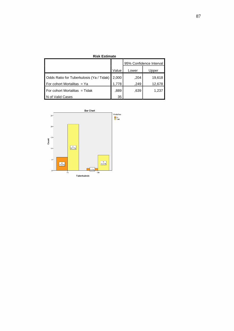
Tuberkulosis * Mortalitas Crosstabulation
Mortalitas
Total Ya Tidak
Tuberkulosis Ya Count 6 21 27
% within Tuberkulosis 22,2% 77,8% 100,0%
Tidak Count 1 7 8
% within Tuberkulosis 12,5% 87,5% 100,0%
Total Count 7 28 35
% within Tuberkulosis 20,0% 80,0% 100,0%
Chi-Square Tests
Value df
Asymp. Sig. (2-
sided)
Exact Sig. (2-
sided)
Exact Sig. (1-
sided)
Pearson Chi-Square ,365a 1 ,546
Continuity Correctionb ,010 1 ,920
Likelihood Ratio ,396 1 ,529
Fisher's Exact Test 1,000 ,484
Linear-by-Linear
Association ,354 1 ,552
N of Valid Cases 35
a. 1 cells (25,0%) have expected count less than 5. The minimum expected count is 1,60.
b. Computed only for a 2x2 table
87
Risk Estimate
Value
95% Confidence Interval
Lower Upper
Odds Ratio for Tuberkulosis (Ya / Tidak) 2,000 ,204 19,618
For cohort Mortalitas = Ya 1,778 ,249 12,678
For cohort Mortalitas = Tidak ,889 ,639 1,237
N of Valid Cases 35

88
Lampiran 5. Biodata mahasiswa
Identitas
Nama : Olfien Noer Primanti Kusumo Negoro
NIM : 22010110120056
Tempat/tanggal lahir : Yogyakarta/21 Februari 1992
Jenis Kelamin : Perempuan
Alamat : Pondok Ungu Permai B25/1 Bekasi Utara
Nomor HP : 085693402967
e-mail : [email protected]
Riwayat Pendidikan Formal
1. SD : SDN HARAPAN JAYA 1 BEKASI Lulus tahun : 2003
2. SMP : SMPN 5 BEKASI Lulus tahun : 2006
3. SMA : SMAN 1 SRAGEN Lulus tahun : 2009
4. FK UNDIP : Masuk tahun : 2010