dcb potential beyond the selected sfa lesions types ... · dcb potential beyond the selected sfa...
TRANSCRIPT
DCB Potential Beyond the Selected SFA
Lesions Types Studied in Trials to Date
What We Know About the ‘Real World’
Krishna Rocha-Singh, MDSaint John’s Hospital
Springfield, IL
Revisiting “Nothing Left Behind”
Defining DCB Efficacy in Complex Disease
• DCB technologies enter into a new era of ‘data
evolution’ to define their role in the management of
complex FPA disease
• Beyond RCTs, robust adjudicated ‘real world’
multicenter registries are essential to that ‘data
evolution’
• Importantly, a uniform definition of terms of what
constitutes a “complex FPA lesions”, how ‘vessel
preparation’ is used and assessed requires
collaboration between physicians, industry and
regulators
3
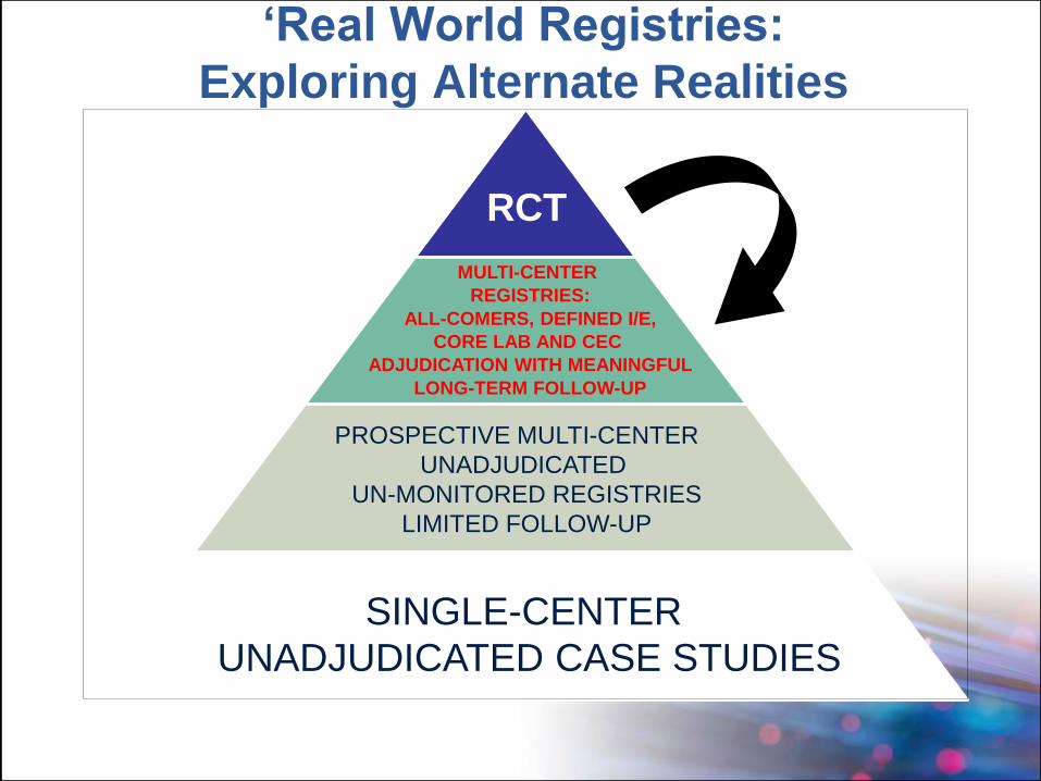
‘Real World Registries:
Exploring Alternate Realities
RCT
SINGLE-CENTER
UNADJUDICATED CASE STUDIES
PROSPECTIVE MULTI-CENTER
UNADJUDICATED
UN-MONITORED REGISTRIES
LIMITED FOLLOW-UP
MULTI-CENTER
REGISTRIES:
ALL-COMERS, DEFINED I/E,
CORE LAB AND CEC
ADJUDICATION WITH MEANINGFUL
LONG-TERM FOLLOW-UP
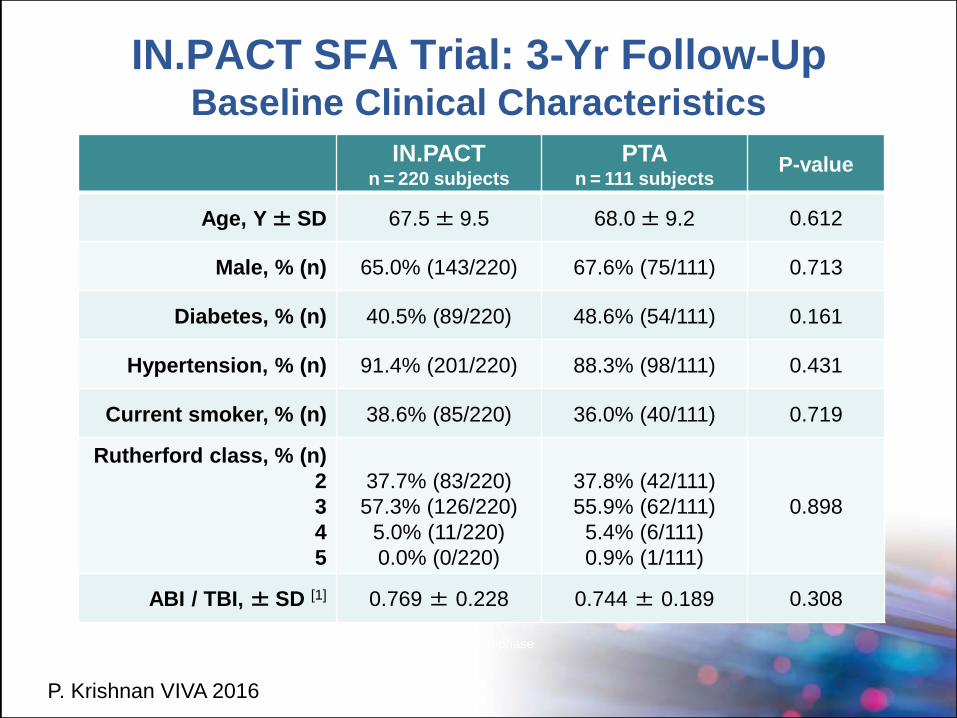
IN.PACT SFA Trial: 3-Yr Follow-Up Baseline Clinical Characteristics
IN.PACTn = 220 subjects
PTAn = 111 subjects
P-value
Age, Y± SD 67.5± 9.5 68.0± 9.2 0.612
Male, % (n) 65.0% (143/220) 67.6% (75/111) 0.713
Diabetes, % (n) 40.5% (89/220) 48.6% (54/111) 0.161
Hypertension, % (n) 91.4% (201/220) 88.3% (98/111) 0.431
Current smoker, % (n) 38.6% (85/220) 36.0% (40/111) 0.719
Rutherford class, % (n)
2
3
4
5
37.7% (83/220)
57.3% (126/220)
5.0% (11/220)
0.0% (0/220)
37.8% (42/111)
55.9% (62/111)
5.4% (6/111)
0.9% (1/111)
0.898
ABI / TBI, ± SD [1] 0.769 ± 0.228 0.744 ± 0.189 0.308
1. TBI allowed in cases of incompressible vessels in IN.PACT SFA II phase
P. Krishnan VIVA 2016
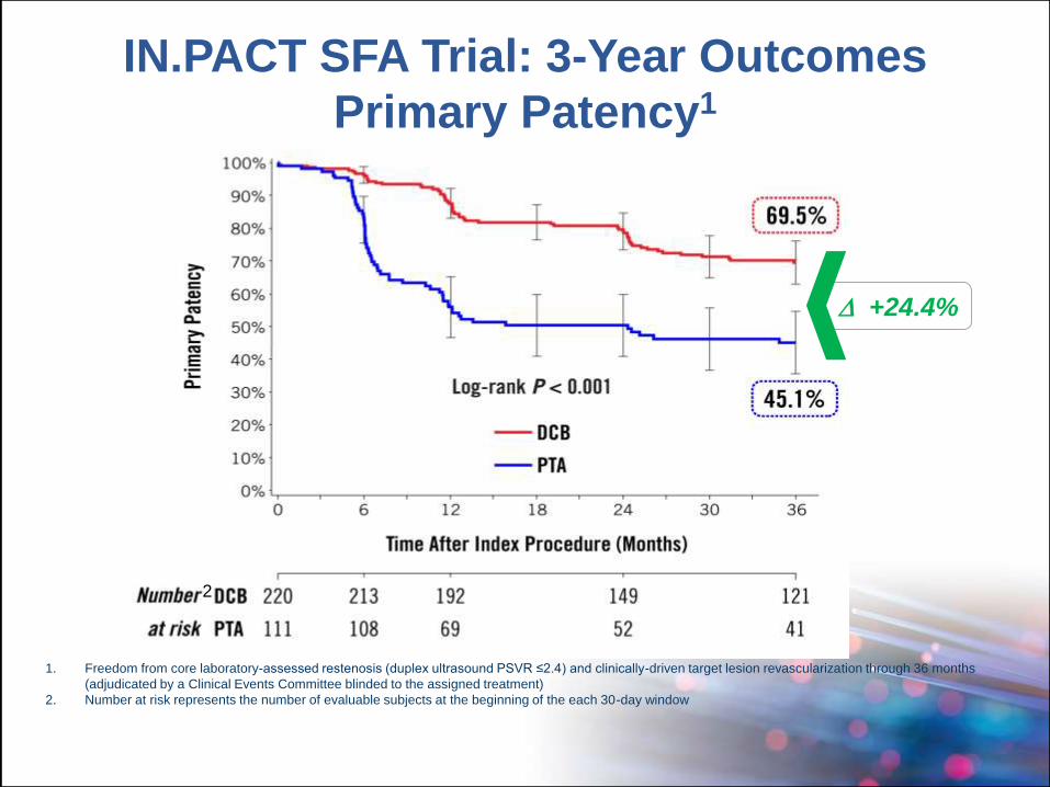
1. Freedom from core laboratory-assessed restenosis (duplex ultrasound PSVR ≤2.4) and clinically-driven target lesion revascularization through 36 months
(adjudicated by a Clinical Events Committee blinded to the assigned treatment)
2. Number at risk represents the number of evaluable subjects at the beginning of the each 30-day window
2
+24.4%
IN.PACT SFA Trial: 3-Year Outcomes
Primary Patency1
P. Krishnan VIVA 2016
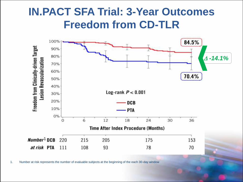
IN.PACT SFA Trial: 3-Year Outcomes
Freedom from CD-TLR
1. Number at risk represents the number of evaluable subjects at the beginning of the each 30-day window
1
-14.1%
P. Krishnan VIVA 2016
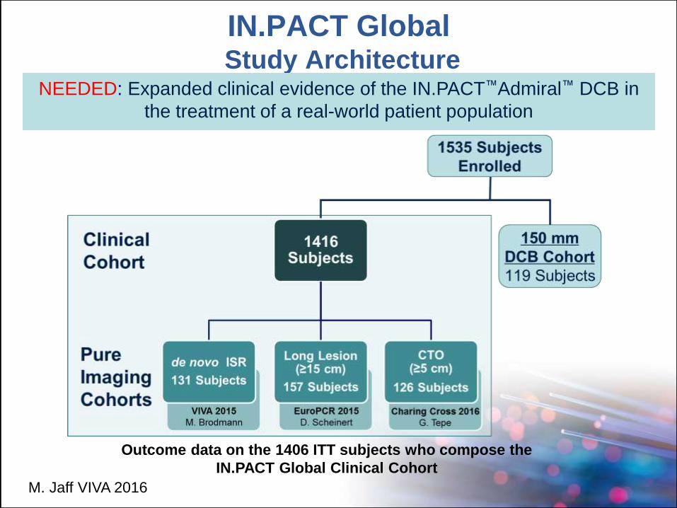
IN.PACT Global Study Architecture
NEEDED: Expanded clinical evidence of the IN.PACT™Admiral™ DCB in
the treatment of a real-world patient population
M. Jaff VIVA 2016
Outcome data on the 1406 ITT subjects who compose the
IN.PACT Global Clinical Cohort
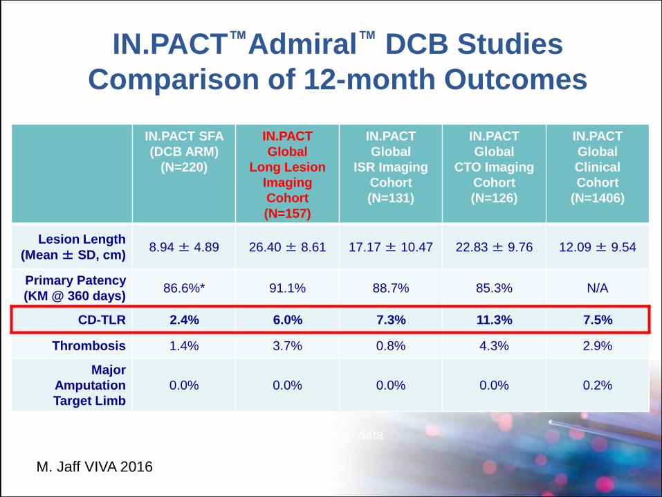
IN.PACT™Admiral™ DCB Studies
Comparison of 12-month Outcomes
IN.PACT SFA
(DCB ARM)
(N=220)
IN.PACT
Global
Long Lesion
Imaging
Cohort
(N=157)
IN.PACT
Global
ISR Imaging
Cohort
(N=131)
IN.PACT
Global
CTO Imaging
Cohort
(N=126)
IN.PACT
Global
Clinical
Cohort
(N=1406)
Lesion Length
(Mean± SD, cm)8.94 ± 4.89 26.40 ± 8.61 17.17 ± 10.47 22.83 ± 9.76 12.09 ± 9.54
Primary Patency
(KM @ 360 days) 86.6%* 91.1% 88.7% 85.3% N/A
CD-TLR 2.4% 6.0% 7.3% 11.3% 7.5%
Thrombosis 1.4% 3.7% 0.8% 4.3% 2.9%
Major
Amputation
Target Limb
0.0% 0.0% 0.0% 0.0% 0.2%
*KM Day 360 rate of 87.5% using 2-year data
M. Jaff VIVA 2016
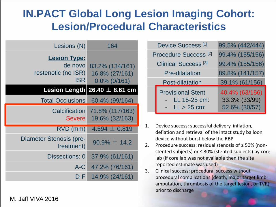
IN.PACT Global Long Lesion Imaging Cohort:
Lesion/Procedural Characteristics
Device Success [1] 99.5% (442/444)
Procedure Success [2] 99.4% (155/156)
Clinical Success [3] 99.4% (155/156)
Pre-dilatation 89.8% (141/157)
Post-dilatation 39.1% (61/156)
Provisional Stent
- LL 15-25 cm:
- LL > 25 cm:
40.4% (63/156)
33.3% (33/99)
52.6% (30/57)
Lesions (N) 164
Lesion Type:
de novo
restenotic (no ISR)
ISR
83.2% (134/161)
16.8% (27/161)
0.0% (0/161)
Lesion Length 26.40 ± 8.61 cm
Total Occlusions 60.4% (99/164)
Calcification
Severe
71.8% (117/163)
19.6% (32/163)
RVD (mm) 4.594 ± 0.819
Diameter Stenosis (pre-
treatment)90.9% ± 14.2
Dissections: 0 37.9% (61/161)
A-C 47.2% (76/161)
D-F 14.9% (24/161)
1. Device success: successful delivery, inflation, deflation and retrieval of the intact study balloon device without burst below the RBP
2. Procedure success: residual stenosis of ≤ 50% (non-stented subjects) or ≤ 30% (stented subjects) by core lab (if core lab was not available then the site reported estimate was used)
3. Clinical success: procedural success without procedural complications (death, major target limb amputation, thrombosis of the target lesion, or TVR) prior to discharge
M. Jaff VIVA 2016
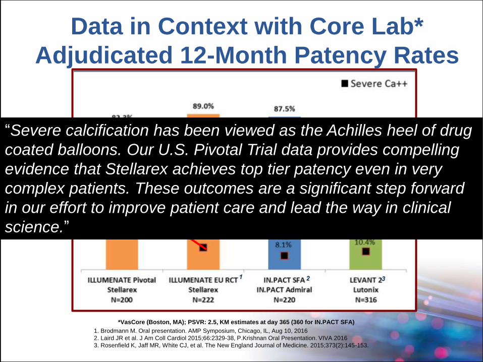
Data in Context with Core Lab*
Adjudicated 12-Month Patency Rates
1. Brodmann M. Oral presentation. AMP Symposium, Chicago, IL, Aug 10, 2016
2. Laird JR et al. J Am Coll Cardiol 2015;66:2329-38, P.Krishnan Oral Presentation. VIVA 2016
3. Rosenfield K, Jaff MR, White CJ, et al. The New England Journal of Medicine. 2015;373(2):145-153.
*VasCore (Boston, MA); PSVR: 2.5, KM estimates at day 365 (360 for IN.PACT SFA)
“Severe calcification has been viewed as the Achilles heel of drug
coated balloons. Our U.S. Pivotal Trial data provides compelling
evidence that Stellarex achieves top tier patency even in very
complex patients. These outcomes are a significant step forward
in our effort to improve patient care and lead the way in clinical
science.”
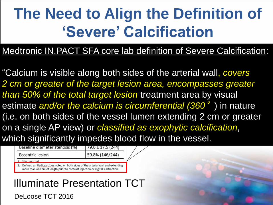
Radiopacities noted on both sides of the
arterial wall and extending more than
ONE cm of length prior to contrast
injection or digital substraction
The Need to Align the Definition of
‘Severe’ Calcification
DeLoose TCT 2016
Medtronic IN.PACT SFA core lab definition of Severe Calcification:
“Calcium is visible along both sides of the arterial wall, covers
2 cm or greater of the target lesion area, encompasses greater
than 50% of the total target lesion treatment area by visual
estimate and/or the calcium is circumferential (360°) in nature
(i.e. on both sides of the vessel lumen extending 2 cm or greater
on a single AP view) or classified as exophytic calcification,
which significantly impedes blood flow in the vessel.
Illuminate Presentation TCT
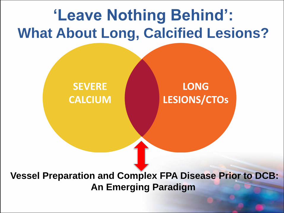
SEVERECALCIUM
LONGLESIONS/CTOs
Vessel Preparation and Complex FPA Disease Prior to DCB:
An Emerging Paradigm
‘Leave Nothing Behind’:What About Long, Calcified Lesions?
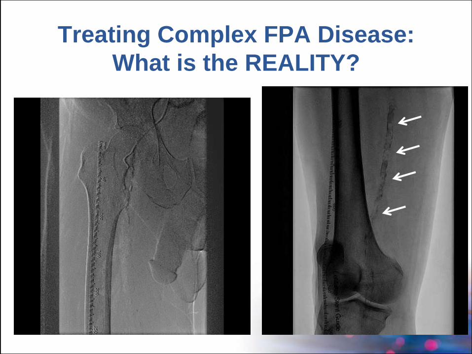
Treating Complex FPA Disease:
What is the REALITY?
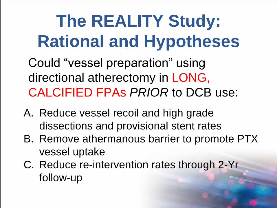
The REALITY Study:
Rational and Hypotheses
Could “vessel preparation” using
directional atherectomy in LONG,
CALCIFIED FPAs PRIOR to DCB use:
A. Reduce vessel recoil and high grade
dissections and provisional stent rates
B. Remove athermanous barrier to promote PTX
vessel uptake
C. Reduce re-intervention rates through 2-Yr
follow-up
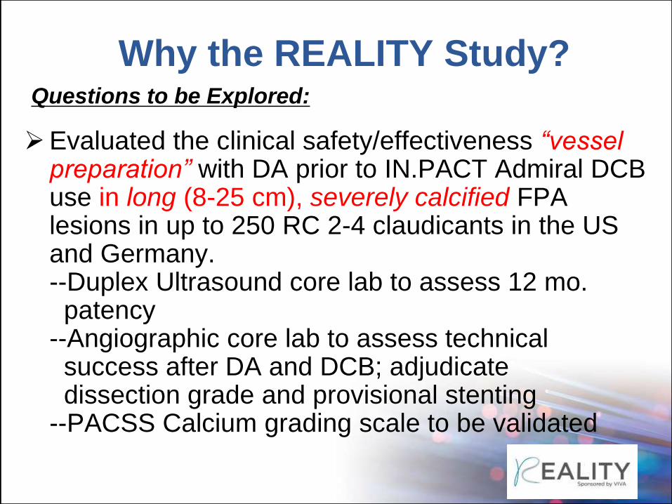
Evaluated the clinical safety/effectiveness “vessel preparation” with DA prior to IN.PACT Admiral DCBuse in long (8-25 cm), severely calcified FPAlesions in up to 250 RC 2-4 claudicants in the US and Germany.--Duplex Ultrasound core lab to assess 12 mo. patency
--Angiographic core lab to assess technical success after DA and DCB; adjudicate dissection grade and provisional stenting
--PACSS Calcium grading scale to be validated
Why the REALITY Study?Questions to be Explored:
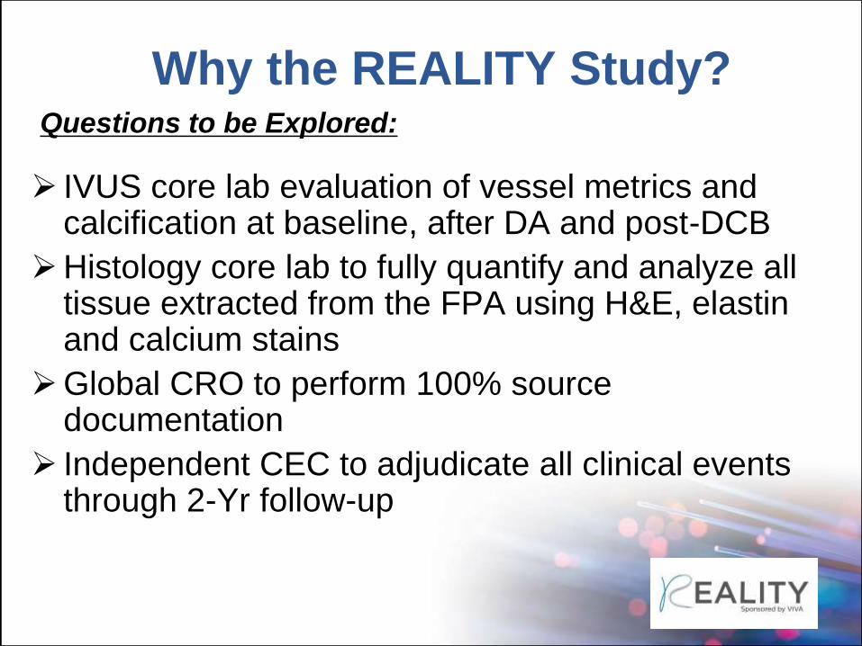
IVUS core lab evaluation of vessel metrics and calcification at baseline, after DA and post-DCB
Histology core lab to fully quantify and analyze all tissue extracted from the FPA using H&E, elastin and calcium stains
Global CRO to perform 100% source documentation
Independent CEC to adjudicate all clinical events through 2-Yr follow-up
Why the REALITY Study?Questions to be Explored:
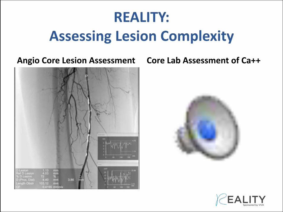
REALITY: Assessing Lesion Complexity
Angio Core Lesion Assessment Core Lab Assessment of Ca++
IVUS Core Lab Assessment
• IVUS metrics: lumen
diameter, CSA, plaque
burden, dissection,
Ca++ are assessed at
baseline, post-DA,
post-DCB
• All investigator are
blinded to IVUS images
REALITY:
Assessing Lesion Complexity
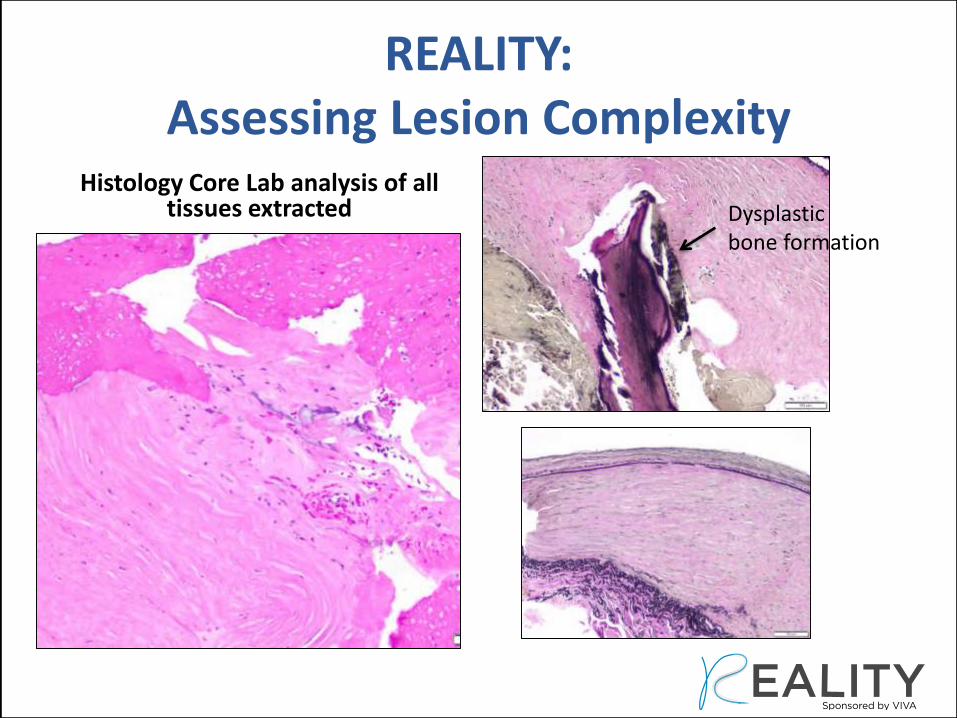
Histology Core Lab analysis of all tissues extracted
REALITY: Assessing Lesion Complexity
Dysplasticbone formation
Finally, We Must Avoid Our Natural
Tendency of ‘Irrational Exuberance’
• Regardless of the device combinations
evaluated, all stakeholders should openly
collaborate, align definitions, and disclose
outcomes
• While a RCT to evaluate the “best” device
combinations to treat complex disease is
impractical, robust/adjudicated registries
provide important insights

DCB Potential Beyond the Selected SFA
Lesions Types Studied in Trials to Date
What We Know About the ‘Real World’
Krishna Rocha-Singh, MDSaint John’s Hospital
Springfield, IL