deficits in dopaminergic transmission precede neuron loss
TRANSCRIPT
Deficits in dopaminergic transmission precede neuronloss and dysfunction in a new Parkinson modelStephanie Janezica,b,1, Sarah Threlfella,b,1, Paul D. Dodsona,c,1, Megan J. Dowiea,c, Tonya N. Taylora,b,Dawid Potgietera,b, Laura Parkkinena,d, Steven L. Seniora,b, Sabina Anwara,b, Brent Ryana,b, Thierry Deltheila,b,Polina Kosilloa,b, Milena Ciorocha,b, Katharina Wagnera,b, Olaf Ansorgea,d, David M. Bannermana,e,J. Paul Bolama,c, Peter J. Magilla,c, Stephanie J. Cragga,b, and Richard Wade-Martinsa,b,2
aOxford Parkinson’s Disease Centre bDepartment of Physiology, Anatomy and Genetics, cMedical Research Council Anatomical Neuropharmacology Unit,Department of Pharmacology, dNuffield Department of Clinical Neuroscience, John Radcliffe Hospital, and eDepartment of Experimental Psychology,University of Oxford, Oxford OX1 3QX, United Kingdom
Edited by Thomas C. Südhof, Stanford University School of Medicine, Stanford, CA, and approved September 5, 2013 (received for review May 15, 2013)
The pathological end-state of Parkinson disease is well describedfrom postmortem tissue, but there remains a pressing need to defineearly functional changes to susceptible neurons and circuits. Inparticular, mechanisms underlying the vulnerability of the dopamineneurons of the substantia nigra pars compacta (SNc) and the im-portance of protein aggregation in driving the disease process re-main to be determined. To better understand the sequence of eventsoccurring in familial and sporadic Parkinson disease, we generatedbacterial artificial chromosome transgenic mice (SNCA-OVX) that ex-press wild-type α-synuclein from the complete human SNCA locus atdisease-relevant levels and display a transgene expression profile thatrecapitulates that of endogenous α-synuclein. SNCA-OVXmice displayage-dependent loss of nigrostriatal dopamine neurons and motorimpairments characteristic of Parkinson disease. This phenotype ispreceded by early deficits in dopamine release from terminals inthe dorsal, but not ventral, striatum. Such neurotransmission deficitsare not seen at either noradrenergic or serotoninergic terminals. Do-pamine release deficits are associated with an altered distribution ofvesicles in dopaminergic axons in the dorsal striatum. Aged SNCA-OVX mice exhibit reduced firing of SNc dopamine neurons in vivomeasured by juxtacellular recording of neurochemically identifiedneurons. These progressive changes in vulnerable SNc neurons wereobserved independently of overt protein aggregation, suggestingneurophysiological changes precede, and are not driven by, aggre-gate formation. This longitudinal phenotyping strategy in SNCA-OVXmice thus provides insights into the region-specific neuronal distur-bances preceding and accompanying Parkinson disease.
dopamine transmission | in vivo electrophysiology | voltammetry |neurodegeneration | behavioral phenotyping
The development of new disease-modifying therapies for Par-kinson disease (PD) is critically dependent on animal models
that accurately recapitulate pathophysiological sequelae in anage-dependent manner. The generation of such models usinggenetically altered animals has proved challenging. The tradi-tional use of heterologous gene promoters in the generation oftransgenic mouse models precluded an endogenous transgeneexpression profile and produced additional phenotypes that arenot characteristic of PD (1). In contrast, bacterial artificialchromosome (BAC) technology can enable expression of a de-sired transgene under the control of its native promoter andregulatory elements to achieve a correct spatiotemporal expres-sion profile, thereby providing a more physiological model forinvestigating molecular mechanisms of the disease.The α-synuclein gene (SNCA) has been implicated in PD
through three dominant point mutations (2–4) and locus multi-plication (5, 6). SNCA duplications and triplications cause auto-somal-dominant PD in which the age of onset and diseaseseverity are related in a gene dosage-dependent manner (5, 6).More recently, genome-wide association studies (GWASs) havedemonstrated a link between common SNCA variants and sporadic
forms of PD, with disease-associated polymorphisms proposed toincrease α-synuclein (α-syn) expression either by increasing tran-scription or stabilizing mRNA (7–9). Although α-syn aggregationinto cytoplasmic Lewy bodies is seen in human PD patient post-mortem tissue, it is not known whether neurophysiological dys-function and cell death precede the formation of aggregates.To better understand the mechanisms by which elevated α-syn
leads to the progressive neuronal dysfunction seen in PD, we havegenerated BAC transgenic mice (SNCA-OVX) that express humanwild-type α-syn at twice the endogenous level of mouse α-syn. Usinga longitudinal “deep phenotyping” strategy in these SNCA-OVXmice, in vitro and in vivo, we find disturbances to midbrain dopa-mine neuron function at discrete phases that precede and/or ac-company PD-like loss of dopamine neurons and motor deficits.Specifically, dopamine transmission and vesicular organization arecompromised selectively in dorsal striatum before a subsequent lossof dopamine neurons, abnormal firing in remaining dopamineneurons in vivo, and movement deficits. These findings thus providekey insights into the progression of events in PD.
ResultsMolecular Characterization of SNCA-OVX Mice.We generated SNCAtransgenic mice using a BAC construct carrying the complete
Significance
Elevated expression of the presynaptic protein α-synucleinunderlies familial and sporadic Parkinson disease (PD). How-ever, our understanding of how increases in α-synuclein levelsdrive the sequence of events leading to PD is incomplete. Here,we apply a multidisciplinary longitudinal analysis to a newα-synuclein transgenic mouse model. We show that early-stagedecreases in dopamine release and vesicle reclustering precedelate-stage changes in neuronal firing properties, measured byin vivo recordings from vulnerable neurons. Accumulated defi-cits in dopamine neurotransmission and altered neuronal firingare associated with cell death and motor abnormalities, in theabsence of protein aggregation in the substantia nigra. Thesefindings have important implications for developing therapies.
Author contributions: O.A., D.M.B., J.P.B., P.J.M., S.J.C., and R.W.-M. designed research;S.J., S.T., P.D.D., M.J.D., T.N.T., D.P., L.P., S.L.S., S.A., B.R., T.D., P.K., M.C., and K.W. per-formed research; S.J., S.T., P.D.D., M.J.D., T.N.T., D.P., L.P., B.R., T.D., D.M.B., J.P.B., P.J.M.,S.J.C., and R.W.-M. analyzed data; and S.J., S.T., P.D.D., J.P.B., P.J.M., S.J.C., and R.W.-M.wrote the paper.
The authors declare no conflict of interest.
This article is a PNAS Direct Submission.
Freely available online through the PNAS open access option.1S.J., S.T., and P.D.D. contributed equally to this work.2To whom correspondence should be addressed. E-mail: [email protected].
This article contains supporting information online at www.pnas.org/lookup/suppl/doi:10.1073/pnas.1309143110/-/DCSupplemental.
E4016–E4025 | PNAS | Published online September 30, 2013 www.pnas.org/cgi/doi/10.1073/pnas.1309143110
Dow
nloa
ded
by g
uest
on
Nov
embe
r 24
, 202
1
111-kb wild-type human SNCA locus (Fig. S1A). Two mouse lineswere generated: SNCA-OVX, which overexpresses human α-synto levels that model those associated with SNCA multiplications(10), and hα-syn, which expresses human α-syn at a low/moderatelevel as a control for expression of human α-syn. Both lines were
bred to a mouse α-syn-null (Snca−/−) pure C57/Bl6 background(11) to preclude confounding interactions with endogenous α-syn.Complete transgene integration was confirmed by exon PCRamplification of all six human SNCA exons (Fig. S1B). ForSNCA-OVX mice, a single site of integration was identified by
B
Dα-syn
hα-synSNCA-OVXSnca-/- C57/Bl6
A
C
E
3 m
on
ths
h -synSNCA-Snca-/-
18 m
on
ths
h -synSNCA-OVXOVX Snca-/-F
G H
α-synuclein TH α-synuclein/ TH
h α-s
ynSnca
-/-
SNCA
-OVX
α-synuclein TH α-synuclein/ TH
hα-s
ynSnca
-/-
SNCA
-OVX
SNCA-OVX Snca-/-
+AC
+ PK
LB
509
Syn-1+FA
20 m20 m
20 m
20 m 20 m
20 m
20 m
20 m
20 m
20 m20 m
Thioflavine S
PD control
20 m
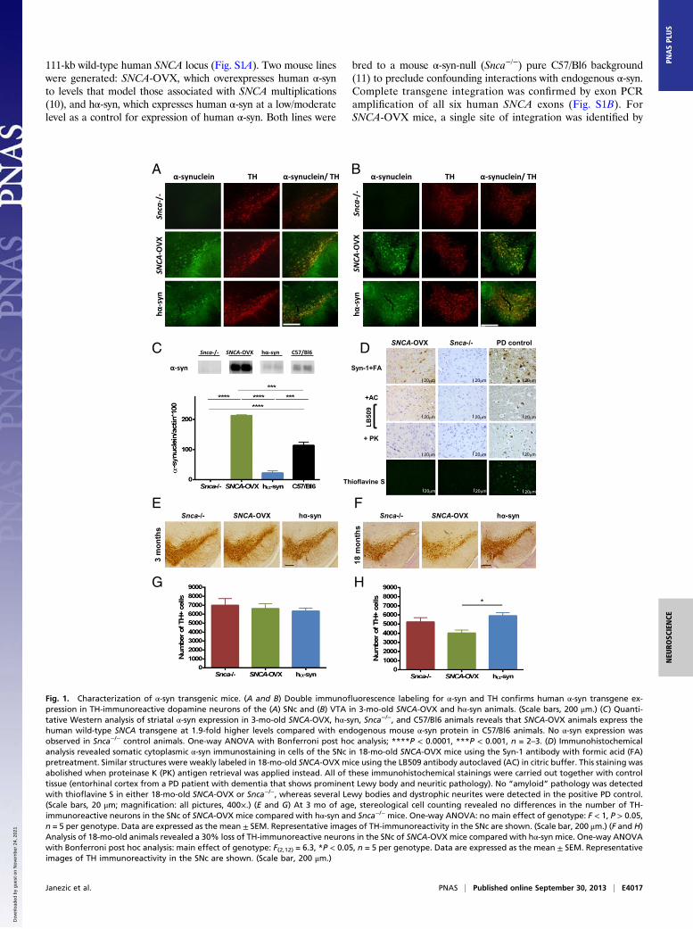
Fig. 1. Characterization of α-syn transgenic mice. (A and B) Double immunofluorescence labeling for α-syn and TH confirms human α-syn transgene ex-pression in TH-immunoreactive dopamine neurons of the (A) SNc and (B) VTA in 3-mo-old SNCA-OVX and hα-syn animals. (Scale bars, 200 μm.) (C) Quanti-tative Western analysis of striatal α-syn expression in 3-mo-old SNCA-OVX, hα-syn, Snca−/−, and C57/Bl6 animals reveals that SNCA-OVX animals express thehuman wild-type SNCA transgene at 1.9-fold higher levels compared with endogenous mouse α-syn protein in C57/Bl6 animals. No α-syn expression wasobserved in Snca−/− control animals. One-way ANOVA with Bonferroni post hoc analysis; ****P < 0.0001, ***P < 0.001, n = 2–3. (D) Immunohistochemicalanalysis revealed somatic cytoplasmic α-syn immunostaining in cells of the SNc in 18-mo-old SNCA-OVX mice using the Syn-1 antibody with formic acid (FA)pretreatment. Similar structures were weakly labeled in 18-mo-old SNCA-OVX mice using the LB509 antibody autoclaved (AC) in citric buffer. This staining wasabolished when proteinase K (PK) antigen retrieval was applied instead. All of these immunohistochemical stainings were carried out together with controltissue (entorhinal cortex from a PD patient with dementia that shows prominent Lewy body and neuritic pathology). No “amyloid” pathology was detectedwith thioflavine S in either 18-mo-old SNCA-OVX or Snca−/−, whereas several Lewy bodies and dystrophic neurites were detected in the positive PD control.(Scale bars, 20 μm; magnification: all pictures, 400×.) (E and G) At 3 mo of age, stereological cell counting revealed no differences in the number of TH-immunoreactive neurons in the SNc of SNCA-OVX mice compared with hα-syn and Snca−/− mice. One-way ANOVA: no main effect of genotype: F < 1, P > 0.05,n = 5 per genotype. Data are expressed as the mean ± SEM. Representative images of TH-immunoreactivity in the SNc are shown. (Scale bar, 200 μm.) (F and H)Analysis of 18-mo-old animals revealed a 30% loss of TH-immunoreactive neurons in the SNc of SNCA-OVX mice compared with hα-syn mice. One-way ANOVAwith Bonferroni post hoc analysis: main effect of genotype: F(2,12) = 6.3, *P < 0.05, n = 5 per genotype. Data are expressed as the mean ± SEM. Representativeimages of TH immunoreactivity in the SNc are shown. (Scale bar, 200 μm.)
Janezic et al. PNAS | Published online September 30, 2013 | E4017
NEU
ROSC
IENCE
PNASPL
US
Dow
nloa
ded
by g
uest
on
Nov
embe
r 24
, 202
1
fluorescence in situ hybridization analysis near the centromere ofchromosome 4 and, for hα-syn mice, a single site near the telo-mere of chromosome 2 (Fig. S1C).Double immunofluorescence labeling for tyrosine hydroxylase
(TH) and α-syn revealed transgene expression in TH-immuno-reactive dopamine neurons in the substantia nigra pars compacta(SNc) (Fig. 1A) and ventral tegmental area (VTA) (Fig. 1B) ofSNCA-OVX and hα-syn (but not Snca−/−) mice. Transgene ex-pression was also found to recapitulate the regional expressionpattern of endogenous mouse α-syn in other brain regions in-cluding cortex, hippocampus, and striatum (Fig. S1E). Humanα-syn levels in the striatum of SNCA-OVX mice were 1.9-foldhigher relative to the levels of endogenous mouse α-syn in age-matched C57/Bl6 animals (Fig. 1C). We observed no change inβ-synuclein or γ-synuclein levels in SNCA-OVX mice (Fig. S1D).Soluble high-molecular-weight α-syn species were observed in
the midbrain of SNCA-OVX mice at both 3 and 18 mo of age,but not in hα-syn mice, as assessed by native PAGE (Fig. S2A).The absence of soluble high-molecular-weight species in hα-synmice was confirmed by loading increasing amounts of protein,such that the levels of α-syn in the samples were comparable,without the appearance of the smear of higher-molecular-weightspecies being seen. In addition, it was confirmed that these sol-uble species were similar to those observed in PD, by comparisonwith SNc tissue from a PD patient. A C-terminal truncated formof α-syn was observed at low levels in the midbrain of 18-mo-oldSNCA-OVX mice, as demonstrated by Western blotting with anantibody raised against the central portion (aa 18–123) of α-synfollowed by reprobing with an antibody raised against theC terminus (aa 118–123) of α-syn (Fig. S2B). This truncationgenerated a fragment comparable in size to that observed in theSNc of a PD patient but was not observed in hα-syn mice or inany of the samples probed with the C-terminal antibody. Noevidence of α-syn phosphorylation at S129 was observed in any ofthe mice strains using a number of commercial S129P antibodies(Fig. S2C).We looked for the presence of pathological α-syn aggregates in
the SNCA-OVX line at 18 mo of age. Proteinase K treatment,which enhances the immunoreactivity of “abnormal” α-syn (i.e.,that in intracytoplasmic aggregates) and also diminishes that ofdiffuse physiological expression (12), abolished the immunore-activity of α-syn in the SNc (Fig. 1D) and striatum (Fig. S3A) ofaged SNCA-OVX mice. Similarly, thioflavine S, which detectsthe β-sheet–rich amyloid and p62, a common constituent in se-veral types of disease-associated inclusions, revealed no patho-logical protein aggregations in the SNc of SNCA-OVX mice (Fig.S3B). No qualitative differences in astrogliosis or microglial ac-tivation were detected between SNCA-OVX mice and Snca−/−
controls, as analyzed by immunohistochemistry using antibodiesagainst GFAP and Iba-1, respectively (Fig. S3B). Hyperphos-phorylated tau (AT8) and β-amyloid (4G8) staining was negativein all animals (Fig. S3B).
Aged SNCA-OVX Animals Lose Nigrostriatal Dopamine Neurons. Toassess whether progressive degeneration of SNc dopamine neu-rons, a cardinal pathological feature of PD, occurs as a conse-quence of overexpressing α-syn, an unbiased stereological countof TH-immunoreactive neurons was performed in the SNc of3- and 18-mo-old mice. It is already known that Snca−/− micedisplay a small decrease in dopamine neurons (13, 14); therefore,to evaluate the effects of increased levels of α-syn we comparedthe number of dopamine neurons in SNCA-OVX to those in hα-syn mice. In young adults (3 mo), no difference was found in thenumber of TH-immunoreactive neurons between genotypes (Fig.1 E and G). However, in aged mice (18 mo), SNCA-OVX ani-mals displayed a 30% reduction in TH-immunoreactive neuronsin the SNc compared with the hα-syn mice (Fig. 1 F and H).At 18 mo of age, SNCA-OVX animals also displayed a 22%
reduction in hematoxylin-counterstained neurons in theSNc compared with the hα-syn mice (Fig. S4B), whereas nodifference in hematoxylin-stained cells was observed at 3 moof age (Fig. S4A), confirming that the loss of TH-immunore-active neurons in aged SNCA-OVX mice was not due toa down-regulation of TH expression. The loss of SNc dopa-mine neurons in aged SNCA-OVX mice was observed in theabsence of overt aggregation pathology in the SNc and stria-tum, suggesting α-syn aggregation is not required for neuronaldeath in the SNc.
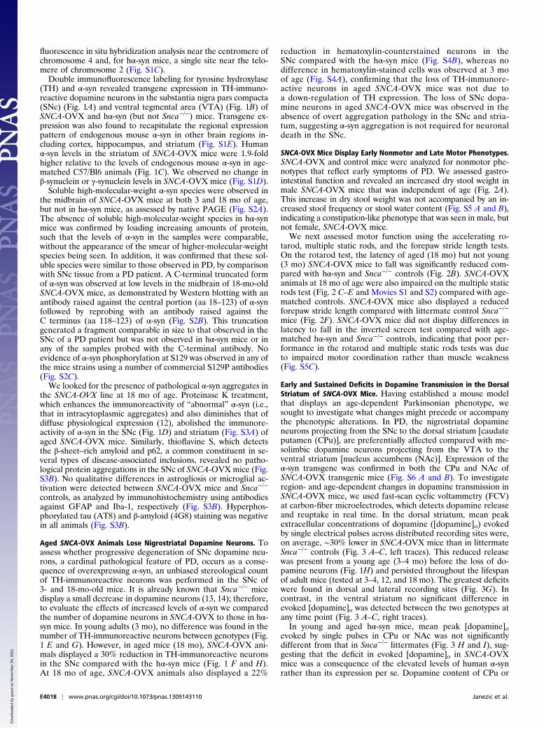
SNCA-OVX Mice Display Early Nonmotor and Late Motor Phenotypes.SNCA-OVX and control mice were analyzed for nonmotor phe-notypes that reflect early symptoms of PD. We assessed gastro-intestinal function and revealed an increased dry stool weight inmale SNCA-OVX mice that was independent of age (Fig. 2A).This increase in dry stool weight was not accompanied by an in-creased stool frequency or stool water content (Fig. S5 A and B),indicating a constipation-like phenotype that was seen in male, butnot female, SNCA-OVX mice.We next assessed motor function using the accelerating ro-
tarod, multiple static rods, and the forepaw stride length tests.On the rotarod test, the latency of aged (18 mo) but not young(3 mo) SNCA-OVX mice to fall was significantly reduced com-pared with hα-syn and Snca−/− controls (Fig. 2B). SNCA-OVXanimals at 18 mo of age were also impaired on the multiple staticrods test (Fig. 2 C–E and Movies S1 and S2) compared with age-matched controls. SNCA-OVX mice also displayed a reducedforepaw stride length compared with littermate control Snca−/−
mice (Fig. 2F). SNCA-OVX mice did not display differences inlatency to fall in the inverted screen test compared with age-matched hα-syn and Snca−/− controls, indicating that poor per-formance in the rotarod and multiple static rods tests was dueto impaired motor coordination rather than muscle weakness(Fig. S5C).
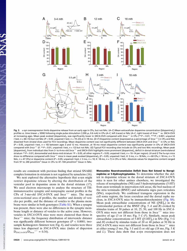
Early and Sustained Deficits in Dopamine Transmission in the DorsalStriatum of SNCA-OVX Mice. Having established a mouse modelthat displays an age-dependent Parkinsonian phenotype, wesought to investigate what changes might precede or accompanythe phenotypic alterations. In PD, the nigrostriatal dopamineneurons projecting from the SNc to the dorsal striatum [caudateputamen (CPu)], are preferentially affected compared with me-solimbic dopamine neurons projecting from the VTA to theventral striatum [nucleus accumbens (NAc)]. Expression of theα-syn transgene was confirmed in both the CPu and NAc ofSNCA-OVX transgenic mice (Fig. S6 A and B). To investigateregion- and age-dependent changes in dopamine transmission inSNCA-OVX mice, we used fast-scan cyclic voltammetry (FCV)at carbon-fiber microelectrodes, which detects dopamine releaseand reuptake in real time. In the dorsal striatum, mean peakextracellular concentrations of dopamine ([dopamine]o) evokedby single electrical pulses across distributed recording sites were,on average, ∼30% lower in SNCA-OVX mice than in littermateSnca−/− controls (Fig. 3 A–C, left traces). This reduced releasewas present from a young age (3–4 mo) before the loss of do-pamine neurons (Fig. 1H) and persisted throughout the lifespanof adult mice (tested at 3–4, 12, and 18 mo). The greatest deficitswere found in dorsal and lateral recording sites (Fig. 3G). Incontrast, in the ventral striatum no significant difference inevoked [dopamine]o was detected between the two genotypes atany time point (Fig. 3 A–C, right traces).In young and aged hα-syn mice, mean peak [dopamine]o
evoked by single pulses in CPu or NAc was not significantlydifferent from that in Snca−/− littermates (Fig. 3 H and I), sug-gesting that the deficit in evoked [dopamine]o in SNCA-OVXmice was a consequence of the elevated levels of human α-synrather than its expression per se. Dopamine content of CPu or
E4018 | www.pnas.org/cgi/doi/10.1073/pnas.1309143110 Janezic et al.
Dow
nloa
ded
by g
uest
on
Nov
embe
r 24
, 202
1
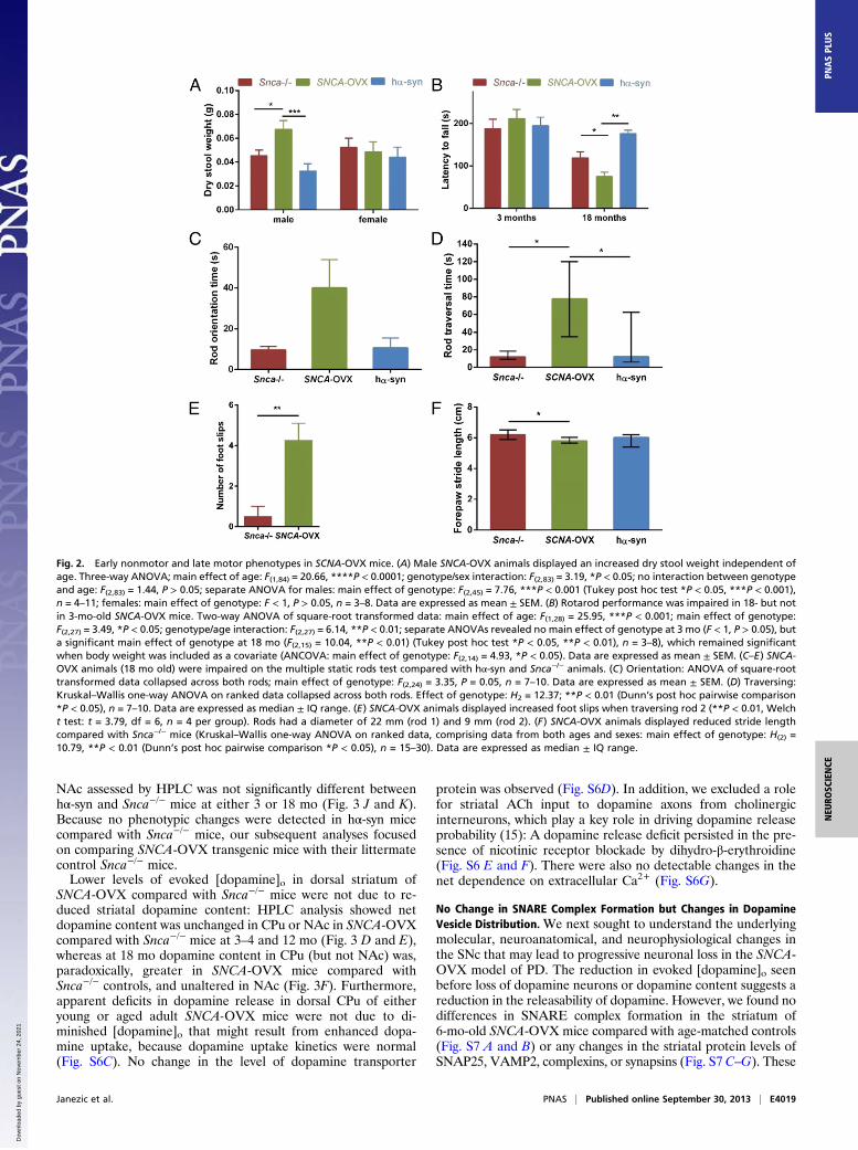
NAc assessed by HPLC was not significantly different betweenhα-syn and Snca−/− mice at either 3 or 18 mo (Fig. 3 J and K).Because no phenotypic changes were detected in hα-syn micecompared with Snca−/− mice, our subsequent analyses focusedon comparing SNCA-OVX transgenic mice with their littermatecontrol Snca−/− mice.Lower levels of evoked [dopamine]o in dorsal striatum of
SNCA-OVX compared with Snca−/− mice were not due to re-duced striatal dopamine content: HPLC analysis showed netdopamine content was unchanged in CPu or NAc in SNCA-OVXcompared with Snca−/− mice at 3–4 and 12 mo (Fig. 3 D and E),whereas at 18 mo dopamine content in CPu (but not NAc) was,paradoxically, greater in SNCA-OVX mice compared withSnca−/− controls, and unaltered in NAc (Fig. 3F). Furthermore,apparent deficits in dopamine release in dorsal CPu of eitheryoung or aged adult SNCA-OVX mice were not due to di-minished [dopamine]o that might result from enhanced dopa-mine uptake, because dopamine uptake kinetics were normal(Fig. S6C). No change in the level of dopamine transporter
protein was observed (Fig. S6D). In addition, we excluded a rolefor striatal ACh input to dopamine axons from cholinergicinterneurons, which play a key role in driving dopamine releaseprobability (15): A dopamine release deficit persisted in the pre-sence of nicotinic receptor blockade by dihydro-β-erythroidine(Fig. S6 E and F). There were also no detectable changes in thenet dependence on extracellular Ca2+ (Fig. S6G).
No Change in SNARE Complex Formation but Changes in DopamineVesicle Distribution. We next sought to understand the underlyingmolecular, neuroanatomical, and neurophysiological changes inthe SNc that may lead to progressive neuronal loss in the SNCA-OVX model of PD. The reduction in evoked [dopamine]o seenbefore loss of dopamine neurons or dopamine content suggests areduction in the releasability of dopamine. However, we found nodifferences in SNARE complex formation in the striatum of6-mo-old SNCA-OVX mice compared with age-matched controls(Fig. S7 A and B) or any changes in the striatal protein levels ofSNAP25, VAMP2, complexins, or synapsins (Fig. S7 C–G). These
Fig. 2. Early nonmotor and late motor phenotypes in SCNA-OVX mice. (A) Male SNCA-OVX animals displayed an increased dry stool weight independent ofage. Three-way ANOVA; main effect of age: F(1,84) = 20.66, ****P < 0.0001; genotype/sex interaction: F(2,83) = 3.19, *P < 0.05; no interaction between genotypeand age: F(2,83) = 1.44, P > 0.05; separate ANOVA for males: main effect of genotype: F(2,45) = 7.76, ***P < 0.001 (Tukey post hoc test *P < 0.05, ***P < 0.001),n = 4–11; females: main effect of genotype: F < 1, P > 0.05, n = 3–8. Data are expressed as mean ± SEM. (B) Rotarod performance was impaired in 18- but notin 3-mo-old SNCA-OVX mice. Two-way ANOVA of square-root transformed data: main effect of age: F(1,28) = 25.95, ***P < 0.001; main effect of genotype:F(2,27) = 3.49, *P < 0.05; genotype/age interaction: F(2,27) = 6.14, **P < 0.01; separate ANOVAs revealed no main effect of genotype at 3 mo (F < 1, P > 0.05), buta significant main effect of genotype at 18 mo (F(2,15) = 10.04, **P < 0.01) (Tukey post hoc test *P < 0.05, **P < 0.01), n = 3–8), which remained significantwhen body weight was included as a covariate (ANCOVA: main effect of genotype: F(2,14) = 4.93, *P < 0.05). Data are expressed as mean ± SEM. (C–E) SNCA-OVX animals (18 mo old) were impaired on the multiple static rods test compared with hα-syn and Snca−/− animals. (C) Orientation: ANOVA of square-roottransformed data collapsed across both rods; main effect of genotype: F(2,24) = 3.35, P = 0.05, n = 7–10. Data are expressed as mean ± SEM. (D) Traversing:Kruskal–Wallis one-way ANOVA on ranked data collapsed across both rods. Effect of genotype: H2 = 12.37; **P < 0.01 (Dunn’s post hoc pairwise comparison*P < 0.05), n = 7–10. Data are expressed as median ± IQ range. (E) SNCA-OVX animals displayed increased foot slips when traversing rod 2 (**P < 0.01, Welcht test: t = 3.79, df = 6, n = 4 per group). Rods had a diameter of 22 mm (rod 1) and 9 mm (rod 2). (F) SNCA-OVX animals displayed reduced stride lengthcompared with Snca−/− mice (Kruskal–Wallis one-way ANOVA on ranked data, comprising data from both ages and sexes: main effect of genotype: H(2) =10.79, **P < 0.01 (Dunn’s post hoc pairwise comparison *P < 0.05), n = 15–30). Data are expressed as median ± IQ range.
Janezic et al. PNAS | Published online September 30, 2013 | E4019
NEU
ROSC
IENCE
PNASPL
US
Dow
nloa
ded
by g
uest
on
Nov
embe
r 24
, 202
1
results are consistent with previous finding that striatal SNAREcomplex formation in striatum is not regulated by synucleins (16).We next explored the alternative hypothesis that α-syn might
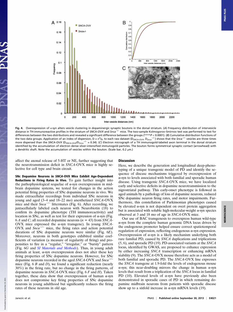
restrict dopamine release by altering the distribution of thevesicular pool in dopamine axons in the dorsal striatum (17).We used electron microscopy to analyze the structure of TH-immunoreactive synaptic and nonsynaptic axonal profiles in theCPu of 3-mo-old SNCA-OVX and Snca−/− mice. The meancross-sectional area of profiles, the number and density of vesi-cles per profile, and the distance of vesicles to the plasma mem-brane were similar in both genotypes (Table S1). When a synapsewas present, there were also no differences in the synaptic mem-brane length or distance of vesicles to the active zone. However,vesicles in SNCA-OVX mice were more clustered than those inSnca−/− mice; the frequency distribution of intervesicle distancewas significantly different between genotypes (P < 0.0001; two-sample Kolmogorov–Smirnov test; Fig. 4), and vesicles were threetimes less dispersed in SNCA-OVX mice (index of dispersionDSNCA-OVX/DSnca
−/− = 0.34).
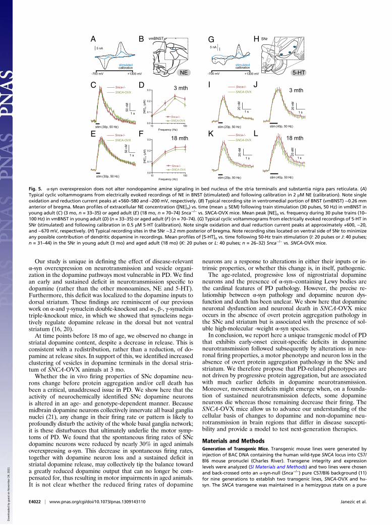
Monoamine Neurotransmission Deficit Does Not Extend to Norepi-nephrine or 5-Hydroxytryptamine. To determine whether the def-icit in dopamine release in the dorsal striatum of SNCA-OVXmice is seen for other amines elsewhere, we investigated therelease of norepinephrine (NE) and 5-hydroxytryptamine (5-HT)from axon terminals in innervation-rich areas, the bed nucleus ofthe stria terminalis (BNST) and substantia nigra pars reticulata(SNr), respectively. We confirmed transgene expression in thecell soma regions, the locus coeruleus and the dorsal raphe nu-cleus, in SNCA-OVX mice by immunohistochemistry (Fig. S8).Mean peak extracellular concentrations of NE ([NE]o) in theventromedial portion of the BNST (Fig. 5 A and B) evoked bystimulus trains of 30 pulses at 10, 25, 50, and 100 Hz in SNCA-OVX mice were not different from those in Snca−/− mice irre-spective of age (3 or 18 mo; Fig. 5 C–F). Similarly, mean peakextracellular concentrations of 5-HT ([5-HT]o) in SNr (Fig. 5 Gand H) evoked by stimulus trains of 20 or 40 pulses at 50 Hz inSNCA-OVX mice were not different from those in Snca−/− miceat either young (3 mo, Fig. 5 I and J) or old age (18 mo, Fig. 5 Kand L). These data show that α-syn overexpression does not
3-4 mth 12 mth 18 mthSnca (-/-)SNCA OVX
0.5 μM
0.5 s
Snca (-/-)SNCA OVX
0.5 μM
0.5 s
0.5 μM
0.5 s
*** ** ***
A B C
Snca (-/-)hα-syn
0.5 s
0.5 μM
3 mth
Snca (-/-)hα-syn
E F
18 mth
20 nA
-700 mV +1300 mV
CPu NAc
CPu NAc
D
CPu
NAc
0.5 s
0.5 μM
**
*
*Snca (-/-)SNCA OVX
0.5 μM
0.5 s
CPu NAc0
20
40
60
80
100
120
140
Region
DA
cont
ent
CPu NAc0
20
40
60
80
100
120
140
RegionCPu NAc
0
20
40
60
80
100
120
140
Region
CPu NAc0
20
40
60
80
100
120
140
RegionCPu NAc
0
20
40
60
80
100
120
140
Region
**
DA
cont
ent
DA
cont
ent
DA
cont
ent
DA
cont
ent
G H I
J K
CPu NAc CPu NAc
CPu NAc
Stim
Stim
Fig. 3. α-syn overexpression limits dopamine release from an early age in CPu, but not NAc. (A–C) Mean extracellular dopamine concentration ([dopamine]o)profiles vs. time (mean ± SEM) following single pulse stimulation (↑200 μs, 0.6 mA) in CPu (A–C, left traces) or NAc (A–C, right traces) of Snca−/− vs. SNCA-OVXat increasing ages. Mean peak evoked [dopamine]o was significantly lower in SNCA-OVX compared with Snca−/− in CPu (**P < 0.01, ***P < 0.001; unpairedt test; n = 60–72) but not NAc (P > 0.05; unpaired t test; n = 19–23) at 3–18 mo. (D–F) Dopamine content (expressed as a percentage of Snca−/−) in CPu and NAcdissected from striatal slices used for FCV recordings. Mean dopamine content was not significantly different between SNCA-OVX and Snca−/− in CPu or NAc(P > 0.05, unpaired t test, n = 10) between ages 3 and 12 mo. However, at 18 mo mean dopamine content was significantly greater in CPu of SNCA-OVXcompared with Snca−/− (F; **P < 0.01, unpaired t test, n = 12) but not NAc. (G) Typical FCV recording sites include six CPu and two NAc recordings. Mean peak[dopamine]o from individual sites from 3- to 4-mo-old Snca−/− and SNCA-OVX highlights more prominent [dopamine]o deficit in dorsal striatum (ventrolateralstriatum **P < 0.01; dorsomedial and dorso-mid striatum *P < 0.05; all other regions P > 0.05; unpaired t test; n = 10–14 per region). (H and I) The hα-syn miceshow no difference compared with Snca−/− mice in mean peak evoked [dopamine]o (P > 0.05; unpaired t test; H: 3 mo, n = 18 NAc, n = 60 CPu; I: 18 mo, n = 15NAc, n = 47 CPu) or dopamine content (P > 0.05; unpaired t test; J: 3 mo, n = 10; K: 18 mo, n = 7) in CPu or NAc. Absolute values for dopamine content rangedfrom 97 to 240 pmol/mm3 tissue in CPu to 47–104 pmol/mm3 tissue in NAc.
E4020 | www.pnas.org/cgi/doi/10.1073/pnas.1309143110 Janezic et al.
Dow
nloa
ded
by g
uest
on
Nov
embe
r 24
, 202
1
affect the axonal release of 5-HT or NE, further suggesting thatthe neurotransmission deficit in SNCA-OVX mice is highly se-lective for cell type and brain circuit.
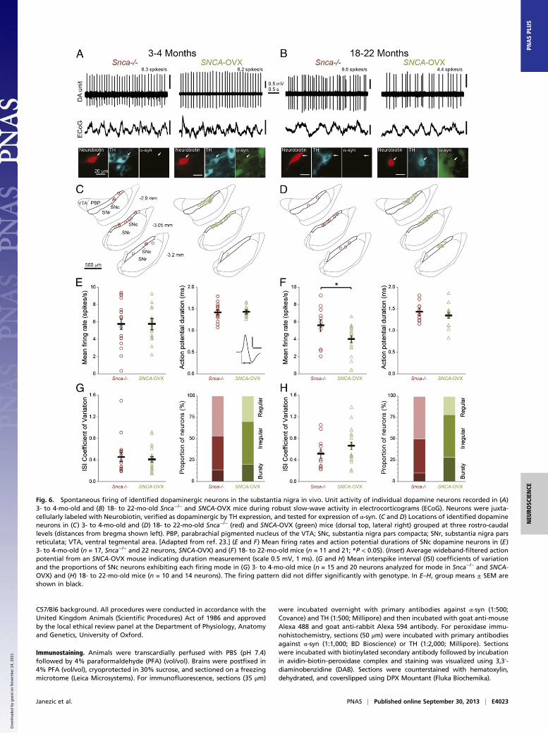
SNc Dopamine Neurons in SNCA-OVX Mice Exhibit Age-DependentReductions in Firing Rates in Vivo. To gain further insight intothe pathophysiological sequelae of α-syn overexpression in mid-brain dopamine systems, we tested for changes in the actionpotential firing properties of SNc dopamine neurons in vivo. Wemade extracellular recordings from individual SNc neurons inyoung and aged (3–4 and 18–22 mo) anesthetized SNCA-OVXmice and their Snca−/− littermates (Fig. 6). After recording, wejuxtacellularly labeled each neuron with Neurobiotin (18) toconfirm its dopamine phenotype (TH immunoreactivity) andlocation in SNc, as well as test for their expression of α-syn (Fig.6 A and C; all recorded dopamine neurons (n = 43) from SNCA-OVX mice expressed the α-syn transgene). In young SNCA-OVX and Snca−/− mice, the firing rates and action potentialdurations of SNc dopamine neurons were similar (Fig. 6E).Moreover, neurons in both genotypes exhibited similar coef-ficients of variation (a measure of regularity of firing) and pro-pensities to fire in a “regular,” “irregular,” or “bursty” pattern(Fig. 6G and SI Materials and Methods). Thus, in young adultanimals at least, α-syn overexpression does not alter these keyfiring properties of SNc dopamine neurons. However, for SNcdopamine neurons recorded in the aged SNCA-OVX and Snca−/−
mice (Fig. 6 B and D), we found a significant reduction (nearly30%) in the firing rate, but no change in the firing patterns, ofdopamine neurons in SNCA-OVX mice (Fig. 6 F and H). Takentogether, these data show that overexpression of human α-syndoes not compromise key firing properties of SNc dopamineneurons in young adulthood but significantly reduces the firingrates of these neurons in old age.
DiscussionHere, we describe the generation and longitudinal deep-pheno-typing of a unique transgenic model of PD and identify the se-quence of disease mechanisms triggered by overexpression ofα-syn to levels associated with both familial and sporadic humandisease. Using transgenic SNCA-OVX mice, we have localizedearly and selective deficits in dopamine neurotransmission to thenigrostriatal pathway. This early-onset phenotype is followed inaged animals by a pathology of loss of dopamine neurons, reducedSNc dopamine neuron firing rates, and motor impairments. Fur-thermore, this constellation of Parkinsonian phenotypes causedby elevated α-syn is not dependent on overt protein aggregationbut is associated with soluble high-molecular-weight α-syn speciesobserved at 3 and 18 mo of age in SNCA-OVX mice.Our use of BAC transgenesis to overexpress human wild-type
α-syn from the complete human genomic DNA locus driven bythe endogenous promoter helped ensure correct spatiotemporalregulation of expression, reflecting endogenous α-syn expression.Overexpression of α-syn is a likely mechanism underlying bothrare familial PD, caused by SNCA duplications and triplications(5, 6), and sporadic PD (19). PD-associated variants at the SNCAlocus, identified by GWAS, are proposed to enhance expressionby either increasing SNCA transcription or enhancing mRNAstability (9). The SNCA-OVX mouse therefore acts as a model ofboth familial and sporadic PD. The SNCA-OVX line expressesthe SNCA transgene at 1.9-fold the levels of endogenous mouseα-syn; this near-doubling mirrors the change in human α-synlevels that result from a triplication of the SNCA locus in familialPD (10). Elevated levels of α-syn have previously also beendemonstrated in sporadic cases of PD in which remaining do-pamine midbrain neurons from patients with sporadic diseaseshow up to a sixfold increase in α-syn mRNA levels (19).
Fig. 4. Overexpression of α-syn alters vesicle clustering in dopaminergic synaptic boutons in the dorsal striatum. (A) Frequency distribution of intervesicledistance in TH-immunoreactive profiles in the striatum of SNCA-OVX and Snca−/− mice. The two-sample Kolmogorov–Smirnov test was performed to test fordifferences between the two distributions and revealed a significant difference between the groups (****P < 0.0001). (B) Cumulative distribution functions ofthe two data groups. Application of an index of dispersion, D = σ2/μ, to each raw dataset (DSNCA-OVX, DSnca
−/−) shows that the Snca−/− vesicles are three timesmore dispersed than the SNCA-OVX (DSNCA-OVX/DSnca
−/− = 0.34). (C) Electron micrograph of a TH immunogold-labeled axon terminal in the dorsal striatumidentified by the accumulation of electron dense silver-intensified immunogold particles. The bouton forms symmetrical synaptic contact (arrowhead) witha dendritic shaft. Note the accumulation of vesicles within the bouton. (Scale bar, 0.2 μm.)
Janezic et al. PNAS | Published online September 30, 2013 | E4021
NEU
ROSC
IENCE
PNASPL
US
Dow
nloa
ded
by g
uest
on
Nov
embe
r 24
, 202
1
Our study is unique in defining the effect of disease-relevantα-syn overexpression on neurotransmission and vesicle organi-zation in the dopamine pathways most vulnerable in PD. We findan early and sustained deficit in neurotransmission specific todopamine (rather than the other monoamines, NE and 5-HT).Furthermore, this deficit was localized to the dopamine inputs todorsal striatum. These findings are reminiscent of our previouswork on α-and γ-synuclein double-knockout and α-, β-, γ-synucleintriple-knockout mice, in which we showed that synucleins nega-tively regulate dopamine release in the dorsal but not ventralstriatum (16, 20).At time points before 18 mo of age, we observed no change in
striatal dopamine content, despite a decrease in release. This isconsistent with a redistribution, rather than a reduction, of do-pamine at release sites. In support of this, we identified increasedclustering of vesicles in dopamine terminals in the dorsal stria-tum of SNCA-OVX animals at 3 mo.Whether the in vivo firing properties of SNc dopamine neu-
rons change before protein aggregation and/or cell death hasbeen a critical, unaddressed issue in PD. We show here that theactivity of neurochemically identified SNc dopamine neuronsis altered in an age- and genotype-dependent manner. Becausemidbrain dopamine neurons collectively innervate all basal ganglianuclei (21), any change in their firing rate or pattern is likely toprofoundly disturb the activity of the whole basal ganglia network;it is these disturbances that ultimately underlie the motor symp-toms of PD. We found that the spontaneous firing rates of SNcdopamine neurons were reduced by nearly 30% in aged animalsoverexpressing α-syn. This decrease in spontaneous firing rates,together with dopamine neuron loss and a sustained deficit instriatal dopamine release, may collectively tip the balance towarda greatly reduced dopamine output that can no longer be com-pensated for, thus resulting in motor impairments in aged animals.It is not clear whether the reduced firing rates of dopamine
neurons are a response to alterations in either their inputs or in-trinsic properties, or whether this change is, in itself, pathogenic.The age-related, progressive loss of nigrostriatal dopamine
neurons and the presence of α-syn–containing Lewy bodies arethe cardinal features of PD pathology. However, the precise re-lationship between α-syn pathology and dopamine neuron dys-function and death has been unclear. We show here that dopamineneuronal dysfunction and neuronal death in SNCA-OVX miceoccurs in the absence of overt protein aggregation pathology inthe SNc and striatum but is associated with the presence of sol-uble high-molecular -weight α-syn species.In conclusion, we report here a unique transgenic model of PD
that exhibits early-onset circuit-specific deficits in dopamineneurotransmission followed subsequently by alterations in neu-ronal firing properties, a motor phenotype and neuron loss in theabsence of overt protein aggregation pathology in the SNc andstriatum. We therefore propose that PD-related phenotypes arenot driven by progressive protein aggregation, but are associatedwith much earlier deficits in dopamine neurotransmission.Moreover, movement deficits might emerge when, on a founda-tion of sustained neurotransmission defects, some dopamineneurons die whereas those remaining decrease their firing. TheSNCA-OVX mice allow us to advance our understanding of thecellular basis of changes to dopamine and non-dopamine neu-rotransmission in brain regions that differ in disease suscepti-bility and provide a model to test next-generation therapies.
Materials and MethodsGeneration of Transgenic Mice. Transgenic mouse lines were generated byinjection of BAC DNA containing the human wild-type SNCA locus into C57/Bl6 mouse pronuclei (Charles River). Transgene integrity and expressionlevels were analyzed (SI Materials and Methods) and two lines were chosenand back-crossed onto an α-syn-null (Snca−/−) pure C57/Bl6 background (11)for nine generations to establish two transgenic lines, SNCA-OVX and hα-syn. The SNCA transgene was maintained in a hemizygous state on a pure
stim (30p, 50 Hz)
1 s
50 n
M
3 mth
stim (30p, 50 Hz)
18 mth
1 s
50 n
M
NE 5-HT
Snca-/-SNCA-OVX
20nM
1 s
Snca-/-SNCA-OVX
stim (20p, 50 Hz)
3 mth
18 mth
20nM
1 s
stim (40p, 50 Hz)
Snca-/-SNCA-OVX
20nM
1 s
stim (20p, 50 Hz)
20nM
1 s
stim (40p, 50 Hz)
vmBNST SNrA B
C D
E F
5 nA
stimulatedcalibration
-700 mV +1300 mV
G H
Snca-/-SNCA-OVX
I
calibrationstimulated
5 nA
-700 mV +1300 mV
J
K L10 100
0.0
0.1
0.2
0.3
Snca-/-SNCA-OVX
Frequency (Hz)
[NE]
o(μ
M)
10 1000.0
0.1
0.2
0.3
Frequency (Hz)
[NE]
o(μ
M)
Snca-/-SNCA-OVX
Fig. 5. α-syn overexpression does not alter nondopamine amine signaling in bed nucleus of the stria terminalis and substantia nigra pars reticulata. (A)Typical cyclic voltammograms from electrically evoked recordings of NE in BNST (stimulated) and following calibration in 2 μM NE (calibration). Note singleoxidation and reduction current peaks at +560–580 and –200 mV, respectively. (B) Typical recording site in ventromedial portion of BNST (vmBNST) ∼0.26 mmanterior of bregma. Mean profiles of extracellular NE concentration ([NE]o) vs. time (mean ± SEM) following train stimulation (30 pulses, 50 Hz) in vmBNST inyoung adult (C) (3 mo, n = 33–35) or aged adult (E) (18 mo, n = 70–74) Snca−/− vs. SNCA-OVX mice. Mean peak [NE]o vs. frequency during 30 pulse trains (10–100 Hz) in vmBNST in young adult (D) (n = 33–35) or aged adult (F) (n = 70–74). (G) Typical cyclic voltammograms from electrically evoked recordings of 5-HT inSNr (stimulated) and following calibration in 0.5 μM 5-HT (calibration). Note single oxidation and dual reduction current peaks at approximately +600, −20,and −670 mV, respectively. (H) Typical recording sites in the SNr ∼3.2 mm posterior of bregma. Note recording sites located on ventral side of SNr to minimizeany possible contribution of dendritic dopamine in recordings. Mean profiles of [5-HT]o vs. time following 50-Hz train stimulation (I: 20 pulses or J: 40 pulses;n = 31–44) in the SNr in young adult (3 mo) and aged adult (18 mo) (K: 20 pulses or L: 40 pulses; n = 26–32) Snca−/− vs. SNCA-OVX mice.
E4022 | www.pnas.org/cgi/doi/10.1073/pnas.1309143110 Janezic et al.
Dow
nloa
ded
by g
uest
on
Nov
embe
r 24
, 202
1
C57/Bl6 background. All procedures were conducted in accordance with theUnited Kingdom Animals (Scientific Procedures) Act of 1986 and approvedby the local ethical review panel at the Department of Physiology, Anatomyand Genetics, University of Oxford.
Immunostaining. Animals were transcardially perfused with PBS (pH 7.4)followed by 4% paraformaldehyde (PFA) (vol/vol). Brains were postfixed in4% PFA (vol/vol), cryoprotected in 30% sucrose, and sectioned on a freezingmicrotome (Leica Microsystems). For immunofluorescence, sections (35 μm)
were incubated overnight with primary antibodies against α-syn (1:500;Covance) and TH (1:500; Millipore) and then incubated with goat anti-mouseAlexa 488 and goat anti-rabbit Alexa 594 antibody. For peroxidase immu-nohistochemistry, sections (50 μm) were incubated with primary antibodiesagainst α-syn (1:1,000; BD Bioscience) or TH (1:2,000; Millipore). Sectionswere incubated with biotinylated secondary antibody followed by incubationin avidin–biotin–peroxidase complex and staining was visualized using 3,3′-diaminobenzidine (DAB). Sections were counterstained with hematoxylin,dehydrated, and coverslipped using DPX Mountant (Fluka Biochemika).
Fig. 6. Spontaneous firing of identified dopaminergic neurons in the substantia nigra in vivo. Unit activity of individual dopamine neurons recorded in (A)3- to 4-mo-old and (B) 18- to 22-mo-old Snca−/− and SNCA-OVX mice during robust slow-wave activity in electrocorticograms (ECoG). Neurons were juxta-cellularly labeled with Neurobiotin, verified as dopaminergic by TH expression, and tested for expression of α-syn. (C and D) Locations of identified dopamineneurons in (C) 3- to 4-mo-old and (D) 18- to 22-mo-old Snca−/− (red) and SNCA-OVX (green) mice (dorsal top, lateral right) grouped at three rostro-caudallevels (distances from bregma shown left). PBP, parabrachial pigmented nucleus of the VTA; SNc, substantia nigra pars compacta; SNr, substantia nigra parsreticulata; VTA, ventral tegmental area. [Adapted from ref. 23.] (E and F) Mean firing rates and action potential durations of SNc dopamine neurons in (E)3- to 4-mo-old (n = 17, Snca−/− and 22 neurons, SNCA-OVX) and (F) 18- to 22-mo-old mice (n = 11 and 21; *P < 0.05). (Inset) Average wideband-filtered actionpotential from an SNCA-OVX mouse indicating duration measurement (scale 0.5 mV, 1 ms). (G and H) Mean interspike interval (ISI) coefficients of variationand the proportions of SNc neurons exhibiting each firing mode in (G) 3- to 4-mo-old mice (n = 15 and 20 neurons analyzed for mode in Snca−/− and SNCA-OVX) and (H) 18- to 22-mo-old mice (n = 10 and 14 neurons). The firing pattern did not differ significantly with genotype. In E–H, group means ± SEM areshown in black.
Janezic et al. PNAS | Published online September 30, 2013 | E4023
NEU
ROSC
IENCE
PNASPL
US
Dow
nloa
ded
by g
uest
on
Nov
embe
r 24
, 202
1
Western analysis. Striatal tissue was homogenized in phosphate bufferedsaline (pH 7.4) containing 1% (vol/vol) Igepal CA-630, 0.1% (vol/vol) SDS, 0.5%(mass/vol) sodium deoxycholate and protease inhibitor cocktail, using a TissueTearor (Biospec Products, Inc). Protein content was quantified using a BACassay kit (Sigma) and proteins were analyzed by Western blotting. See TableS2 for primary antibodies. Bands were visualized using horseradish peroxi-dase-conjugated goat anti-mouse IgG (Bio-Rad) and the chemiluminescentECL+ kit (GE Healthcare). Bands were quantified using ImageJ software.Data was analyzed using one-way ANOVA and Bonferroni post hoc analysisusing SPSS Statistics 20 (IBM).
Neuropathology. To detect potential α-syn–related pathology, the mAbs Syn-1 (1:1,000; BD Transduction Labs) against aa 91–99 and LB509 (1:1,000;Invitrogen) against aa115–122 were used. Brains were fixed in 10% formalin(vol/vol), embedded in paraffin, and cut into 6-μm-thick sections, depar-affinized, and rehydrated. Peroxidase activity was eliminated by treatmentwith 3% H2O2 (mass/vol). Antigen retrieval for Syn-1 involved treatmentwith formic acid for 15 min, and for LB509 involved either autoclaving (121 °C)for 10 min in citrate buffer or treatment with 20 μg/mL proteinase K (Roche)for 10 min at room temperature. The mouse-on-mouse kit (M.O.M.; VectorLabs) was used to mAb to minimize the background and staining was visu-alized using DAB (Vector Labs). Thioflavine S staining was carried out asdescribed by Sun et al. (22).
Stereological Cell Count. The total number of TH-immunoreactive and he-matoxylin-counterstained nigral dopamine neurons was estimated with theoptical fractionator method of unbiased stereology using the Stereo-Investigator software (MicroBrightField). Sections (50 μm) were processed forTH immunoreactivity as described above. Cells were counted under a 40×objective of a Zeiss Imager M2 microscope using a randomly placed countingframe of 50 × 50 μm on a sample grid of 120 × 160 μm. A 22-μm opticaldissector with 2-μm upper and lower guard zones was used. The SNc wasdelineated using a mouse brain atlas (23). Every second section from 2.7 to3.88 mm posterior to bregma was counted. Data are expressed as meannumber of TH-immunoreactive neurons ± SEM and were analyzed usingone-way ANOVA and Bonferroni post hoc analysis using SPSS Statistics20 (IBM).
Behavior. One-hour stool collection.Animals (male and female) were placed intoseparate clean cages and fecal pellets were collected over a 1-h period (1600–1700 hours). Pellets were weighed to obtain wet stool weight, dried over-night at 65 °C, and reweighed to obtain dry stool weight and stool watercontent (24).Rotarod. In the rotarod test, female mice were placed on a rod that accel-erated from 4 to 40 rpm over a 5-min period. The length of time which theanimals were able to stay on the rotating rod was recorded as latency to fall.Animals were tested three times a day for two consecutive days and per-formance was averaged for each day (20).Multiple static rods test. In the multiple static rods test, two wooden dowels of22-mm (rod 1) or 9-mm (rod 2) diameter were clamped to a supportingplatform 50 cm above a cushioned surface. Female mice were placed at theend of the rod facing away from the platform. The time taken to turn aroundby 180° to face the platform, time taken to traverse the rod, and the numberof foot slips made when traversing the rod were recorded (20).Forepaw stride length. In the forepaw stride length test, animals’ forepawswere placed in ink and animals walked across a sheet of paper toward theirhome cage. The distance between steps on the same side of the body, fromthe middle toe of the first step to the heel of the second step, was measuredand averaged to obtain stride length. Only steps during normal walking(straight line, no stopping) were included in the analysis (24). Male and fe-male animals were tested.Statistics. Data analyzed using a parametric one-, two- or three-way ANOVAfollowed by a Tukey post hoc test are expressed as means ± SEM. Data an-alyzed using a nonparametric Kruskal–Wallis ANOVA followed by Dunn’spairwise multiple comparison are expressed as median ± interquartile (IQ)range. Analyses were completed using the SPSS Statistics 21 software (IBM).
FCV. Mice were killed by cervical dislocation and decapitated. Coronal slices(300 μm) containing the region of interest (striatum, BNST, or SNr) wereprepared as described previously (15, 25) in ice-cold Hepes-buffered artificialcerebrospinal fluid (aCSF) saturated with 95% O2/5% CO2. Slices were thenmaintained in a bicarbonate-buffered aCSF at room temperature beforerecording. Extracellular dopamine, NE, or 5-HT concentration ([dopamine]o,
[NE]o, or [5-HT]o) were monitored using FCV with 7-μm-diameter carbonfiber microelectrodes (tip length 50–100 μm) and a Millar Voltammeter(Julian Millar, Barts and the London School of Medicine and Dentistry,London, UK) as described previously (15, 26, 27). In brief, the scanningvoltage was a triangular waveform (−0.7 V to +1.3 V range vs. Ag/AgCl) ata scan rate of 800 V/s and frequency of 8 Hz. Electrodes were calibrated in0.5–2 μM 5-HT, dopamine, or NE in experimental media. All data areexpressed as means ± SEM, and the sample size, n, is the number ofobservations. The number of animals in each FCV dataset was ≥3. Compar-isons for differences in means were assessed by two-way ANOVA and posthoc Bonferroni t test or unpaired t test using GraphPad Prism 4.0(GraphPad Software).
HPLC with Electrochemical Detection. Dopamine content was measured byHPLC with electrochemical detection as described previously (16, 20). Fol-lowing FCV recordings, tissue punches from the dorsal (2 mm in diameter)and ventral striatum (1.2 mm in diameter) from two brain slices per animalwere taken and analyzed for dopamine content by HPLC.
Electron Microscopy. Ultrastructural analyses were performed on 3-mo-oldmice. Three SNCA-OVX and three littermate Snca−/− animals were anes-thetized and perfuse-fixed with 0.1% glutaraldehyde and 4% PFA (vol/vol)in 0.1 M phosphate buffer. Sagittal vibratome sections (60 μm) were in-cubated with primary antibody against TH to identify dopamine neurons,followed by a gold-conjugated secondary antibody and silver-intensificationof the gold particles (see ref. 28). Sections were subsequently postfixed in1% osmium tetroxide and mounted on slides in an electron microscopicresin. Samples of dorsolateral striatum were identified, cut from the sec-tions, and reembedded onto resin blocks and serial ultrathin sections (50–70nm) were collected and stained with lead citrate. For each animal, 50 imagesof TH-immunoreactive structures were acquired in a systematically ran-domized manner (150 images per genotype). In addition, TH-immunoreac-tive structures forming synaptic specializations were randomly acquired(SNCA-OVX = 33; Snca −/− = 29). Digital images of TH-immunoreactivestructures and synapses were analyzed using ImageJ and ImageJ pluginsPointDensity and PointDensitySyn (16, 29) to measure terminal size, lengthof synaptic active zone, vesicle number, and distribution. At all stages oftissue processing and ultrastructural analysis the investigators were blindedto animal genotype. Statistical tests were performed using SPSS/PASW Sta-tistics (SPSS) and Matlab (Mathworks).
In Vivo Electrophysiological Recording and Juxtacellular Labeling of SingleNeurons. Experiments were performed in urethane-anesthetized (1.5 g/kg,i.p.) 3- to 4-mo-old mice (five Snca−/− and six SNCA-OVX) and 18- to 22-mo-old mice (five Snca−/− and six SNCA-OVX). Extracellular recordings of single-unit activity in the SNc were made using glass electrodes (10–25 MΩ in situ;tip diameter ∼1.5 μm) containing 0.5 M NaCl solution and Neurobiotin (1.5%wt/vol; Vector Laboratories). An electrocorticogram was also recorded abovethe frontal cortex (18). Following electrophysiological characterization, sin-gle neurons were juxtacellularly labeled with Neurobiotin for post hoc ver-ification of location and neurochemical identity (18). We focused ouranalysis on single-unit activity recorded during robust slow-wave activity, asquantitatively defined from the simultaneous electrocorticogram record-ings. Firing pattern was defined according to one of three modes (regular,bursty, or irregular), as detailed previously (30, 31) and in SI Materials andMethods. Before statistical comparisons, a Shapiro–Wilk test was used tojudge whether datasets were normally distributed (P < 0.05 to reject). Sta-tistical comparisons were made using Student t tests or Welch t tests. Theprevalence of firing patterns was compared with a Fisher’s exact test.
ACKNOWLEDGMENTS.We thank J. Alegre-Abarrategui, R. Deacon, K. Jennings,C. Johnson, and E. Pissadaki for their expertise in experimental techniques. Wethank E. Volpi (Cytogenetics Core, Wellcome Trust Centre for Human Genetics,Oxford; Wellcome Trust Core Award, Grant 090532/Z/09/Z) for FISH analysis. Wethank the animal care and veterinary support staff at Oxford UniversityBiomedical Services. This work was supported by the Monument Trust DiscoveryAward from Parkinson’s UK, Wellcome Trust Research Career Development Fel-lowship GR073141MA (to R.W.-M.), Medical Research Council (MRC) UK AwardsU138164490 and U138197109 (to J.P.B. and P.J.M., respectively), and WellcomeTrust Neuroscience Studentship in Neuroscience GR072324MA (to S.L.S.). M.J.D.holds a Girdlers’ New Zealand Health Research Council Fellowship; S.J., D.P., andS.A. hold MRC studentships; P.K. is supported by the Clarendon Fund; and D.M.B.holds a Wellcome Trust Senior Research Fellowship.
E4024 | www.pnas.org/cgi/doi/10.1073/pnas.1309143110 Janezic et al.
Dow
nloa
ded
by g
uest
on
Nov
embe
r 24
, 202
1
1. Chesselet M-F, Richter F (2011) Modelling of Parkinson’s disease in mice. LancetNeurol 10(12):1108–1118.
2. Polymeropoulos MH, et al. (1997) Mutation in the alpha-synuclein gene identified infamilies with Parkinson’s disease. Science 276(5321):2045–2047.
3. Krüger R, et al. (1998) Ala30Pro mutation in the gene encoding alpha-synuclein inParkinson’s disease. Nat Genet 18(2):106–108.
4. Zarranz JJ, et al. (2004) The new mutation, E46K, of alpha-synuclein causes Parkinsonand Lewy body dementia. Ann Neurol 55(2):164–173.
5. Chartier-Harlin MC, et al. (2004) Alpha-synuclein locus duplication as a cause of fa-milial Parkinson’s disease. Lancet 364(9440):1167–1169.
6. Singleton AB, et al. (2003) alpha-Synuclein locus triplication causes Parkinson’s dis-ease. Science 302(5646):841.
7. Satake W, et al. (2009) Genome-wide association study identifies common variants atfour loci as genetic risk factors for Parkinson’s disease. Nat Genet 41(12):1303–1307.
8. Simón-Sánchez J, et al. (2009) Genome-wide association study reveals genetic riskunderlying Parkinson’s disease. Nat Genet 41(12):1308–1312.
9. Venda LL, Cragg SJ, Buchman VL, Wade-Martins R (2010) α-Synuclein and dopamine atthe crossroads of Parkinson’s disease. Trends Neurosci 33(12):559–568.
10. Miller DW, et al. (2004) Alpha-synuclein in blood and brain from familial Parkinsondisease with SNCA locus triplication. Neurology 62(10):1835–1838.
11. Abeliovich A, et al. (2000) Mice lacking alpha-synuclein display functional deficits inthe nigrostriatal dopamine system. Neuron 25(1):239–252.
12. Neumann M, et al. (2002) Misfolded proteinase K-resistant hyperphosphorylated al-pha-synuclein in aged transgenic mice with locomotor deterioration and in humanalpha-synucleinopathies. J Clin Invest 110(10):1429–1439.
13. Robertson DC, et al. (2004) Developmental loss and resistance to MPTP toxicityof dopaminergic neurones in substantia nigra pars compacta of gamma-synuclein,alpha-synuclein and double alpha/gamma-synuclein null mutant mice. J Neurochem89(5):1126–1136.
14. Al-Wandi A, et al. (2010) Absence of alpha-synuclein affects dopamine metabolismand synaptic markers in the striatum of aging mice. Neurobiol Aging 31(5):796–804.
15. Threlfell S, et al. (2012) Striatal dopamine release is triggered by synchronized activityin cholinergic interneurons. Neuron 75(1):58–64.
16. Anwar S, et al. (2011) Functional alterations to the nigrostriatal system in mice lackingall three members of the synuclein family. J Neurosci 31(20):7264–7274.
17. Nemani VM, et al. (2010) Increased expression of alpha-synuclein reduces neuro-transmitter release by inhibiting synaptic vesicle reclustering after endocytosis.Neuron 65(1):66–79.
18. Brown MT, Henny P, Bolam JP, Magill PJ (2009) Activity of neurochemically hetero-geneous dopaminergic neurons in the substantia nigra during spontaneous anddriven changes in brain state. J Neurosci 29(9):2915–2925.
19. Gründemann J, Schlaudraff F, Haeckel O, Liss B (2008) Elevated alpha-synuclein mRNAlevels in individual UV-laser-microdissected dopaminergic substantia nigra neurons inidiopathic Parkinson’s disease. Nucleic Acids Res 36(7):e38.
20. Senior SL, et al. (2008) Increased striatal dopamine release and hyperdopaminergic-like behaviour in mice lacking both alpha-synuclein and gamma-synuclein. Eur JNeurosci 27(4):947–957.
21. Smith Y, Kieval JZ (2000) Anatomy of the dopamine system in the basal ganglia.Trends Neurosci 23(10, Suppl):S28–S33.
22. Sun A, Nguyen XV, Bing G (2002) Comparative analysis of an improved thioflavin-sstain, Gallyas silver stain, and immunohistochemistry for neurofibrillary tangle dem-onstration on the same sections. J Histochem Cytochem 50(4):463–472.
23. Franklin KBJ, Paxinos G (2007) The Mouse Brain in Stereotaxic Coordinates (Academic,New York), 3rd Ed.
24. Taylor TN, et al. (2009) Nonmotor symptoms of Parkinson’s disease revealed in an an-imal model with reduced monoamine storage capacity. J Neurosci 29(25):8103–8113.
25. Threlfell S, et al. (2010) Striatal muscarinic receptors promote activity dependence ofdopamine transmission via distinct receptor subtypes on cholinergic interneurons inventral versus dorsal striatum. J Neurosci 30(9):3398–3408.
26. Jennings KA, Lesch KP, Sharp T, Cragg SJ (2010) Non-linear relationship between 5-HTtransporter gene expression and frequency sensitivity of 5-HT signals. J Neurochem115(4):965–973.
27. Threlfell S, et al. (2004) Histamine H3 receptors inhibit serotonin release in substantianigra pars reticulata. J Neurosci 24(40):8704–8710.
28. Moss J, Bolam JP (2008) A dopaminergic axon lattice in the striatum and its re-lationship with cortical and thalamic terminals. J Neurosci 28(44):11221–11230.
29. Larsson M, Broman J (2005) Different basal levels of CaMKII phosphorylated atThr286/287 at nociceptive and low-threshold primary afferent synapses. Eur J Neu-rosci 21(9):2445–2458.
30. Tepper JM, Martin LP, Anderson DR (1995) GABAA receptor-mediated inhibition ofrat substantia nigra dopaminergic neurons by pars reticulata projection neurons.J Neurosci 15(4):3092–3103.
31. Paladini CA, Tepper JM (1999) GABA(A) and GABA(B) antagonists differentially affectthe firing pattern of substantia nigra dopaminergic neurons in vivo. Synapse 32(3):165–176.
Janezic et al. PNAS | Published online September 30, 2013 | E4025
NEU
ROSC
IENCE
PNASPL
US
Dow
nloa
ded
by g
uest
on
Nov
embe
r 24
, 202
1