department of biochemistry, microbiology & immunology ...€¦ · fumaric acid esters 21 dmf as...
TRANSCRIPT

PHARMACOLOGICAL IMPROVEMENT OF ONCOLYTIC VIROTHERAPY
Mohammed Selman
Thesis submitted to the
Faculty of Graduate and Postdoctoral Studies
in partial fulfillment of the requirements
for the Doctorate of Philosophy in Biochemistry
Department of Biochemistry, Microbiology & Immunology
Faculty of Medicine
University of Ottawa
© Mohammed Selman, Ottawa, Canada, 2018

ii
Abstract
Oncolytic viruses (OV) are an emerging class of anticancer bio-therapeutics that induce
antitumor immunity through selective replication in cancer cells. However, the efficacy
of OVs as single agents remains limited. We postulate that resistance to oncolytic
virotherapy results in part from the failure of tumor cells to be sufficiently infected. In
this study, we provide evidence that in the context of sarcoma, a highly heterogeneous
malignancy, the infection of tumors by different oncolytic viruses varies greatly.
Similarly, for a given oncolytic virus, productive infection of tumors across patient
samples varies by many orders of magnitude. To overcome this issue, we hypothesize
that the infection of resistant tumors can be achieved through the use of selected small
molecules. Here, we have identified two novel drug classes with the ability to improve
the efficacy of OV therapy: fumaric and maleic acid esters (FMAEs) and vanadium
compounds. FMAEs are enhancing infection of cancer cells by several oncolytic viruses
in cancer cell lines and human tumor biopsies. The ability of FMAEs to enhance viral
spread is due to their ability to inhibit type I IFN production and response, which is
associated with their ability to block nuclear translocation of transcription factor NF-κB.
Vanadium-based phosphatase inhibitors enhance OV infection of RNA viruses in vitro
and ex vivo, in resistant cancer cell lines. Mechanistically, this involves subverting the
antiviral type I IFN response towards a death-inducing and proinflammatory type II IFN
response, leading to improved OV spread, increased bystander killing of cancer cells, and
enhanced anti-tumor immune-stimulation. Both FMAEs and vanadium compounds
improve therapeutic outcomes of OV treatment in syngeneic tumor models, leading to
durable responses, even in models otherwise refractory to OV and drug alone. Overall,
we showcased novel avenues for the development of improved immunotherapy
strategies.

iii
Acknowledgements
This thesis would not have been possible without the aid and support of several people.
First, I would like to thank my supervisor Dr. Jean-Simon Diallo for his guidance,
motivation and continuous support during the duration of my PhD. Furthermore, I would
like to thank to my thesis committee members, Dr. Mary-Ellen Harper, Dr. Tommy Alain
and Dr. Douglas Gray for their helpful advice throughout the course of the program. I
would also like to thank the many collaborators for their work and valuable input: Dr.
Debbie C. Crans, Dr. Brian A. Keller, Dr. Larissa Pikor, Dr. Fabrice Le Boeuf, Dr. John
C. Bell and Dr. Carolina Ilkow. A special thanks to Dr. Fanny Tzelepis for teaching me
new skills and all her effort and help with the many immunological experiments, and
Andrew Chen for all his help managing the numerous in vivo experiments. Sincere thanks
for all the hard work from honors students that I had the privilege to supervise and work
with: Chris Rousso, Paula Ou and Anabel Bergeron. Finally, thanks to family, friends and
all current and former lab members of the Diallo lab for their ongoing support.

iv
Table of Contents
Abstract ii
Acknowledgements iii
List of Abbreviations vi
List of Figures ix
Chapter 1 - General Introduction 1
Cancer 1
Oncolytic virotherapy 1
Mechanism of action of oncolytic viruses 2
Oncolytic virus platforms 4
Oncolytic herpesvirus 5
Oncolytic poxviruses 6
Oncolytic reovirus 7
Oncolytic rhabdoviruses 9
Type I and type II interferon pathways 11
The NF-κB pathway 13
Defects in type I IFN signaling in cancer 14
Resistance to OV 16
Breaking resistance to OV: Viral sensitizers 17
Fumaric acid esters 21
DMF as cancer treatment 23
Vanadium 25
Vanadium compounds are promising anticancer agents 27
Sarcoma: a rare cancer in need of new therapies 28
Rationale and aim I 29
Rationale and hypothesis II 30
Chapter 2 - Oncolytic Maraba virus MG1 as a treatment for Sarcoma 32
Abstract 33
Introduction 34
Materials and Methods 36
Results 40

v
Discussion 52
Chapter 3 - Dimethyl Fumarate Potentiates Oncolytic Virotherapy through NF-kB
inhibition 54
Abstract 55
Introduction 56
Results 58
Discussion 79
Chapter 4 - Multi-Modal Potentiation of Oncolytic Virotherapy by Vanadium
Compounds 91
Abstract 92
Introduction 93
Results 95
Discussion 119
Materials and Methods 123
Chapter 5 - General discussion 132
Crosstalk between type I and II interferon pathways 132
The antiapoptotic and proapoptotic effects of vanadate 135
Expanding OV tropism 137
Improving Oncolytic rhabdovirus therapy in the treatment of Sarcoma 137
Cancer therapy targeting the NF-κB pathway 138
Treatment delivery 139
Beyond oncolytic immunotherapy 140
Concluding thoughts 141
Appendices I – Chapter 2 Supplemental information 143
Appendices II – Chapter 3 Supplemental information 147
Appendices III – Chapter 3 Supplemental information 157
References 178
vi
List of Abbreviations
7-AAD 7-Aminoactinomycin D
ANOVA Analysis of variance
Bax BCL2 Associated X
BMOV Bismaltolato oxovanadium
BSO Buthionine sulfoximine
CDK Cyclin-dependent kinase
CTCL Cutaneous T-cell lymphoma
CTLA-4 Cytotoxic T-lymphocyte-associated protein-4
DEF Diethyl fumarate
DEM Diethyl maleate
DMEM Dulbecco’s Modified Eagle’s Medium
DMF Dimethyl fumarate
DMM Dimethyl maleate
DNA Deoxyribonucleic acid
ECM Extracellular matrix
eIF2a eukaryotic initiation factor 2 alpha
FA Fumaric acid
FAE Fumaric acid esters
FDA U S Food and Drug Administration
FMAE Fumaric and maleic acid esters
G Glycoprotein
GAF IFN-γ-regulated DNA-binding factor
GAS IFN-γ activation sequence
GCL Glutamate Cysteine Ligase
GM-CSF Granulocyte-macrophage colony-stimulating factor
GO Gene Ontology
GSH Glutathione
HDAC Histone deacetylase inhibitor
HEPS 4-(2-hydroxyethyl)-1-piperazineethanesulfonic acid
HR Higher responders
HSV-1 Herpes simplex virus 1
IAP Inhibitors of apoptosis proteins
IFIT1 Interferon-induced protein with tetratricopeptide repeats 1
IFITM1 Interferon-induced transmembrane protein 1
IFN Interferon
IFNGR1 IFN-γ receptor 1
IKK IκB kinase
IRF IFN-regulatory factor

vii
IRSE Interferon-stimulated response element
ISGF3 Interferon-stimulated gene factor 3
IT Intratumoral
IV Intravenous
IVIS in vivo imaging system
JAK Janus kinase
JC John Cunningham
JX-594 Pexastimogene devacirepvec
KEAP1 Kelch-like ECH-associated protein 1
LPS Lipopolysaccharides
LR Lower responders
M Matrix
MG1 Marabex
MMF Monomethyl fumarate
MOI Multiplicity of infection
MS Multiple sclerosis
mTOR mammalian target of rapamycin
MV Measles
MxA Myxoma resistance protein A
N Nucleoprotein
NF-κB Nuclear factor kappa-light-chain-enhancer of activated B cells
NK Natural killer
NRF2 Nuclear factor (erythroid-derived 2)-like 2
OHSN-REB Ottawa Health Science Network Research Ethics Board
OV Oncolytic Virus
P Phosphoprotein
PBS Phosphate-buffered saline
PD-L1 Programmed death-ligand 1
PD1 Programmed cell death protein 1
PDA Pancreatic ductal adenocarcinomas
PFU Plaque forming units
PI3K Phosphoinositide 3-kinase
PKR Protein kinase R
PML Progressive multifocal leukoencephalopathy
PRR Pattern recognition receptors
PTP Protein tyrosine phosphatases
PTP1B Protein-tyrosine phosphatase 1B
qPCR Quantitative polymerase chain reaction
RECIST Response Evaluation Criteria In Solid Tumors
RIG-1 Retinoic Acid Inducible Gene 1
viii
RIPK3 Receptor Interacting Protein Kinase 3
RNA Ribonucleic acid
ROS Reactive oxygen species
RPMI Roswell Park Memorial Institute
SC Sub-cutaneous
Smac Second mitochondrial activator of caspase
SMC Smac mimetic compounds
STAT1 Signal transducer and activator of transcription 1
STAT2 Signal transducer and activator of transcription 2
T-Vec Talimogene laherparepvec
TBK1 TANK-binding kinase 1
TFNα Tumor necrosis factor alpha
TGF-β Transforming growth factor-β
TK Thymidine kinase
TLR Toll-like receptors
VOx Vvanadium(V) oxytriethoxyde
VS Vanadium(IV) oxide sulphate
VSV Vesicular stomatitis virus
VVdd double-deleted vaccinia virus
wt wild-type

ix
List of Figures
Figure 2.1. Cytopathic effect of oncolytic viruses in sarcoma cells lines in vitro.
Figure 2.2. Productive infection of human sarcoma explants by MG1 and VV.
Figure 2.3. Maraba virus MG1 infects and replicates in murine sarcoma S180 tumors,
leading to persistent anti-tumor effects
Figure 3.1. Dimethyl fumarate promotes viral infection
Figure 3.2. DMF enhances infection ex vivo and in human clinical samples.
Figure 3.3. Fumaric and maleic acid esters promote infection by VSVΔ51.
Figure 3.4. Dimethyl fumarate enhances VSV∆51 therapeutic efficacy in syngeneic and
xenograft tumor models.
Figure 3.5. FMAEs inhibit antiviral cytokine production and response to type I interferon.
Figure 3.6. DMF inhibits NF-κB translocation upon infection.
Figure 4.1. Vanadate enhances VSV∆51 infection in cancer cells but not normal cells.
Figure 4.2. Viral enhancement is dependent on Vanadium.
Figure 4.3. Vanadate facilitates virus-induced type I interferon and ROS mediated cell
death
Figure 4.4. Vanadate increases VSV∆51 efficacy in resistant syngeneic tumor models.
Figure 4.5. Vanadate /VSV∆51 co-treatment triggers T-cell infiltration and anti-tumor
immunity.
Figure 4.6. Vanadate re-wires antiviral type I IFN signaling towards pro-inflammatory
type II IFN response

1
Chapter 1 - General Introduction
Cancer
Cancer is not simply defined as the abnormal growth of cells, but is in fact an
intricate and dynamic disease driven by mutation, epigenetic reprogramming, and
crosstalk with its microenvironment. The development of cancer is shepherded by well
described hallmarks of cancer biology such as sustained proliferative signaling, evasion
of growth suppressors, resistance to cell death and reprogramming of energy metabolism
(1). These factors result in, for instance, replicative immortality, induction of
angiogenesis, and the ability to evade immune detection (1). Today, multiple strategies
are used for the treatment of cancer, from standard surgery to cytotoxic treatment such as
radiation and chemotherapy. In addition, there has been a rise in the field of
immunotherapy which promotes the body's immune system to specifically target the
cancer such as checkpoint inhibitors targeting immunosuppressive signalling pathways
on T-cells or cancer cells (2). Despite tremendous advancement in cancer treatment
technologies, cancer is still the leading cause of death in Canada; with an average of 565
Canadians diagnosed with cancer every day of which 221 Canadians succumb to it.
According to estimates, 1 out of 4 Canadians is expected to die from cancer (3, 4) .
Oncolytic virotherapy
Oncolytic virotherapy has emerged as a viable alternative to conventional cancer
therapies, such as radiation and chemotherapy that often lack in efficacy and cause severe
2
side effects. Oncolytic virotherapy refers to treatments using replicating viruses that
preferentially infect and kill cancer cells without harming normal cells.
Some early clinical observations of cancer patients with tumor regression or
remission were associated with patients’ viral infections, for instance, influenza infection
was linked to the remission of leukemia as documented in 1904 (5, 6). This was followed
by a report in 1922 where vaccinia virus was shown to inhibit the growth of several
mouse and rat tumors (7). This led to the first patient trials using natural viruses to treat
cancer in the 1950s, which showed some anti-tumor effect but unacceptable toxicities (5).
Since the advent of molecular engineering, a broad spectrum of DNA and RNA viruses
have been selected or engineered to improve viral oncolysis and have been tested
clinically for their anticancer properties, including adenovirus, measles virus, parvovirus,
reovirus, vesicular stomatitis virus, seneca valley virus, and coxsackievirus (8). Despite
the diversity of the viruses tested, only two OVs have been approved for clinical use.
First, in 2005, Oncorine, a type-5 adenovirus with a partial deletion in the E1B gene was
approved in China for the treatment of head and neck cancer (9). In 2015, the U.S. Food
and Drug Administration (FDA) approved the first OV in North America for the
treatment of melanoma, talimogene laherparepvec (T-Vec) an engineered herpes simplex
virus (10).
Mechanism of action of oncolytic viruses
3
The mechanisms of action of OV therapy are multi-modal and targets several
pathways. These can be mediated by the virus’ natural ability or through transgene
expression. Altogether, this results in a combination of direct lysis of tumor cells, anti-
angiogenic effects, and the induction of an antitumor immune response (11). Lysis of
cancer cells occurs following selective viral replication in the tumor. The spread of viral
infection within malignant tissue results in the continuous amplification of the parental
virus, until it is stopped by an antiviral immune response or a lack of susceptible cancer
cells. Oncolytic viruses can be classified into two groups - viruses with a natural tropism
for cancer cells or viruses that are genetically modified to selectively replicate in cancer
cells (8). For example, the removal of the viral thymidine kinase (TK) from the herpes or
vaccinia viruses attenuates those viruses in normal tissues due to a paucity of endogenous
nucleotides available for efficient viral replication. In contrast, tumors functionally
complement this gene deletion in support of their own replicative needs and thus support
viral replication (12, 13). Viral proteins that support the attachment of the virus to host
cells can also be modified to target the virus to specific receptors overexpressed in
malignant tumors (14, 15). Furthermore, replacing endogenous viral promoters with
tissue-specific promoters for viral genes that are crucial for successful replication can
improve selectivity of OVs (16, 17).
Owing to multiple factors, including low expression of major histocompatibility
complex (MHC) antigens as well as cytokine secretion profiles that antagonize local
immune responses, tumors and their microenvironments are inherently
immunosuppressive (18). Infection of tumor cells by OVs induces release of several
antiviral and pro-inflammatory cytokines, leading to a localized inflammatory response

4
(8, 11). This is followed by an infiltration of various innate and adaptive immune cells
into the tumor in addition to the activation of resident lymphocytes. MHC I proteins,
recognized by the cytotoxic T lymphocytes (CTLs) infiltrated within the tumor, display
the viral- and cancer-associated antigens on the cell surface, resulting in a systemic
antitumor (and antiviral) immune response (11, 19). The generation of systemic adaptive
antitumor immunity is thought to be critical and result in long-term protection against
tumor recurrence (20, 21).
Oncolytic virus platforms
Most of the oncolytic viruses currently in development exhibit a relatively broad
spectrum of antitumor activity in vitro, typically against both epithelial and hematological
malignancies(8). Naturally occurring viruses have different entry routes, life cycles,
structures, and tropisms manifesting in distinctive clinical symptoms. Therefore, OVs
derived from these viruses can also be expected to be more efficient against specific
malignancies. OVs can also be engineered to target specific cell-surface receptors or
nuclear transcription factors to target specific malignancies (8). Evaluating and
comparing the potency of various OV platforms is therefore a reasonable first step toward
identifying the best candidate for clinical translation of OVs for the treatment of specific
malignancies. For example, in human pancreatic ductal adenocarcinomas (PDA),
oncolytic activity of VSV, adenoviruses, sendai virus, and respiratory syncytial virus
were compared. In vitro, oncolytic VSV showed greater oncolytic activity compared to
the other viruses, with the conclusion being that VSV may be a promising oncolytic agent
against PDA (22), but further clinical evaluation is still to come.

5
Oncolytic herpesvirus
Herpesviruses are dsDNA viruses with a broad spectrum of hosts. Their genomes
range in size from about 125 to 240 kbp, and encode approximately 70 to 165 genes (23).
Herpesviruses infection is pervasive among humans, in particular infection with herpes
simplex virus 1 and 2 (HSV-1 and HSV-2), varicella zoster virus, Epstein–Barr virus, or
the cytomegalovirus (24). HSV infection can undergo lytic or latent presentation. During
lytic infection, HSV-1 genes are expressed that regulate viral DNA synthesis and create
an environment favorable for protein synthesis, contribute to encapsulation of viral DNA,
and promote viral envelopment (25). Globally, an estimated two-thirds of the population
under 50 is infected with HSV-1, however the majority of people with HSV-1 infection
are unaware they are infected (26). Interestingly, recent progress has been made
exploiting the unique features of herpes viruses as an oncolytic virus platform.
Particularly, HSV-1 has several advantage over other OVs, including its broad spectrum
of infectivity of cancer cells; how it can be genetically manipulated to express multiple
transgenes; numerous antiviral drugs (such as acyclovir) provide a safety mechanism
against unfavorable replication of the virus; and HSV-1 does not integrate with the host
genome even during latency (25, 27).
T-Vec (Talimogene laherparepvec) is a genetically engineered herpes virus with
deletions in the γ34.5 and α47 genes leading to cancer-specific replication and attenuation
of its pathogenicity. To further promote the antitumor response, the human granulocyte-
macrophage colony-stimulating factor (GM-CSF) gene was inserted in the oncolytic
herpesvirus (28). Based on the results of a recent phase III clinical trial with patients
featuring metastatic melanoma lesions in the skin and lymph nodes, the U.S. FDA

6
approved T-Vec for the treatment of metastatic melanoma. The phase III clinical trial
indicated that median overall survival and durable response rate was significantly higher
in the T-Vec arm compared with the GM-CSF arm. The most common adverse events
with T-Vec were fatigue, chills, and pyrexia, and, furthermore, no fatal treatment-related
adverse events were documented (29). Based on the observation that intratumoral
injection of T-Vec induced systemic antitumor immunity, a phase 1b clinical trial testing
the impact of T-Vec on cytotoxic T-cell infiltration and therapeutic efficacy of the anti-
PD-1 antibody pembrolizumab was carried out. The data suggested that T-Vec can
improve the efficacy of checkpoint inhibitor (anti-PD-1) therapy by changing the tumor
microenvironment (30).
Oncolytic poxviruses
Poxviruses are dsDNA viruses with a large linear genome encoding 150 to 300
genes. Its life cycle takes place within the cytoplasm of the infected cell (31). Poxviruses
are known to have a wide host range, with the Entomopoxvirinae subfamily infecting
insects and the Chordopoxvirinae subfamily infecting vertebrates. Poxvirus infections
commonly lead to the formation of lesions, skin nodules, or disseminated rash, however
poxviruses vary in virulence. The variola virus, the causative agent of smallpox, is
human-specific and highly virulent only to humans, while closely related cowpox viruses
naturally infect a broad range of animals and only cause relatively mild disease in
humans (32).
7
The Vaccinia virus, raccoon poxvirus, yaba-like disease virus, and the myxoma
virus have been investigated for their oncolytic potential as these viruses undergo
productive lytic replication in most human cancer cells. Oncolytic poxviruses are
attractive platforms for cancer therapy because of the following characteristics: safety in
humans, ease of production, stability of virus (can be stored as dry powder for prolonged
periods of time without significant loss of infectivity), and easy genetic manipulation for
the expression of various anticancer transgenes as well as the replication cycle in the
cytoplasm eliminating any risk of integration (25, 33).
JX-594 (pexastimogene devacirepvec, Pexa-Vec) is one of several vaccina strain
tested in clinical trials (34). To render viral replication selective to cancer cells, it was
genetically engineered to contain a mutation in the TK gene through insertion of the
human GM-CSF gene, which upon expression in infected cancer cells promotes the
antitumor immune response (35, 36). Phase I and II clinical trials reaffirmed the safety
profile of the treatment. However, in a phase IIb clinical trial comprised of patients with
advanced stage hepatocellular carcinoma, JX-594 did not improve overall survival (33,
37, 38). Nevertheless, JX-594 in combination with various chemotherapy agents or
checkpoint inhibitors is currently under clinical evaluation (NCT01636284,
NCT02562755, NCT02977156, NCT03071094).
Oncolytic reovirus

8
Reoviruses are non-enveloped, dsRNA viruses with a genome composed of 10 to
12 segments encoding 10 to 14 genes. Human infection with reoviruses commonly occurs
causing mild infections limited to the gastrointestinal or respiratory tract (31). Reoviruses
are known to replicate preferentially in cells with the activated Ras pathway, which
modulates cell growth, differentiation, and survival (39). This results in their inherent
ability to selectively replicate in cancer cells given that approximately 30% of human
tumors have an activating mutation of the Ras pathway (39, 40). The dsRNA-activated
protein kinase (PKR) binds to dsRNA and activates downstream signals halting protein
synthesis and viral replication. PKR is inactivated in Ras-transformed cells, promoting
viral infection of dsRNA viruses, such as reoviruses (41). In contrast to other OV
platforms, introducing mutation or introducing transgenes in reoviruses is not easily
accomplished (25).
Numerous clinical trials have been carried out using reovirus. In a phase I trial in
patients with advanced solid tumors, common treatment-related adverse events included
nausea, vomiting and fevers, and transient flu-like symptoms (42). However, reovirus did
not cause any dose-limiting toxicity (with doses up to 10E10 PFU per injection). In one
phase II trial evaluating single-agent reovirus in metastatic melanomas, there was a
failure to meet previously defined efficacy criteria. Recently, a number of Phase II
clinical trial evaluating reovirus combinations with chemotherapy agents also failed to
meet previously defined efficacy criteria. Phase II and III clinical trials are presently
underway testing reovirus combinations with various checkpoint inhibitors (43).

9
Oncolytic rhabdoviruses
Rhabdoviruses are negative ssRNA viruses with a broad host range, including
plants, insects, fish, mammals, and reptiles. The vesicular stomatitis virus (VSV), a
member of the Vesiculovirus genus, is an arthropod-borne virus that primarily affects
rodents, cattle, swine, and horses, and causes a relatively localized and non-fatal illness.
Human infection is rare and usually asymptomatic. The VSV genome is constituted of 5
genes encoded by a negative sense, ssRNA genome, found in all rhabdoviruses in the
order of 3′-N-P-M-G-L-5′ (31, 44).
Viral RNA forms a helical nucleocapsid complex with the nucleoprotein (N). The
nucleocapsid also encompasses the phosphoprotein (P), some of which is bound to the
viral RNA-dependent RNA polymerase, the large (L) protein. The nucleocapsid complex
and the viral polymerase complex (P and L proteins) form the ribonucleoprotein. The
ribonucleoprotein is enveloped in a lipid bilayer, acquired from the host cell membrane
during the budding process. The matrix protein (M) and the glycoprotein (G) are
membrane-associated proteins. The M protein associates with the nucleocapsid and the
lipid bilayer, maintaining the compact structure of the virion. The G protein is an integral
membrane protein, which controls viral entry, mediated both by cell attachment via the
LDL receptor and membrane fusion (31, 45, 46).
VSV is highly sensitive to the effects of the antiviral cytokines known as type I
interferons (IFN); however, the wild-type virus is quite capable of shutting off IFN

10
production through the actions of its M protein, which blocks host mRNA export from
the nucleus (47). In order to make VSV sensitive to an IFN-mediated antiviral response, a
variant was engineered with a complete deletion of methionine at amino acid position 51
(Δ51). While normal cells have robust antiviral mechanisms, most cancer cells have
mutations that lead to a defective IFN-mediated antiviral response, resulting in cancer-
specific replication of VSVΔ51 (48, 49).
Maraba virus, a member of the Vesiculovirus genus, was identified from in vitro
screening of a panel of rhabdoviruses on cancer cells as the most potent rhabdovirus
strain (50). To increase specificity towards cancer cells, mutations of the G (Q242R) and
M (L123W) proteins were introduced to the Maraba virus to further attenuate it in normal
cells. This engineered virus, designated as MG1, had a 100-fold greater maximum
tolerable dose than wild-type Maraba in a murine model, and resulted in durable cures
when administered in syngeneic and xenograft murine tumor models (50).
Beyond its oncolytic activity, the MG1 virus can effectively reduce metastatic
disease following cancer surgery by boosting natural killer (NK) cell activity (51). MG1
and VSV can also be engineered to encode tumor-associated antigen and be utilized as a
boosting vector in a heterologous prime-boost vaccination regimen (52–55). This
rhabdovirus-based oncolytic vaccination regimen generates an efficient antigen-specific
T-cell immune response, extending median survival and leading to complete remission in
murine melanoma tumor models (52). The prime-boost vaccination regimen is under
clinical evaluation with favorable safety profiles reported thus far (NCT02285816).
Furthermore, oncolytic VSV expressing IFNβ is under clinical evaluation for the

11
treatment of various solid tumors, multiple myeloma and acute myeloid leukemia
(NCT02923466, NCT03017820).
Type I and type II interferon pathways
IFNs are cytokines that can block viral infection, inhibit cell proliferation, and
modulate innate and adaptive immune responses. The IFN family includes three main
classes of cytokines: type I IFN (IFNα, IFNβ, and IFNω), type II IFN (IFNγ), and type III
IFN (IFNλ1, IFNλ2, and IFNλ3). IFNβ is produced by most cell types, while IFNα is
primarily generated by haematopoietic cells. IFNγ is predominantly produced by NK and
NK T-cells for the modulation of the innate immune response, and by cytotoxic T-cells
once antigen-specific immunity is initiated (56–58).
Type I IFNs bind a common cell-surface receptor composed of two subunits of
IFNα receptor 1 and 2 (IFNAR1, IFNAR2) while IFNγ binds a distinct cell-surface
receptor also composed of two subunits, namely IFNγ receptor 1 and 2 (IFNGR1,
IFNGR2). These receptors interact with members of the janus-activated kinase (JAK)
family: tyrosine kinase 2 (TYK2) and JAK1 for the IFNAR, and JAK1 and JAK2 for the
IFNGR. Activation of the receptors by IFN binding is followed by its
autophosphorylation and activation of the associated JAK, leading to differential
activation of STAT1 and STAT2 (signal transducer and activator of transcription)
transcription factors by tyrosine phosphorylation. Phosphorylation of STATs leads to
their dimerization and nuclear translocation to activate transcription of IFN-stimulated

12
genes (ISGs). Some of these genes are regulated by both type I and type II IFNs, whereas
others are selectively regulated by one or the other. Type I IFNs induce the formation of
the ISG factor 3 (ISGF3) complex composed of a STAT1-STAT2 dimer and IRF9 that
binds specific elements known as IFN-stimulated response elements (ISREs); while type
II IFN primarily leads to the formation of STAT1-STAT1 dimers that bind IFNγ-
activated-sequence (GAS) elements (56).
Type I IFN leads to the induction of hundreds of ISGs, many of which exert
antiviral functions by directly targeting pathways and functions required during the virus
life cycle (59). For example, oligoadenylate synthetase (OAS) and latent
endoribonuclease (RNaseL) results in the detection and subsequent degradation of viral
RNAs. Upon binding to dsRNA, protein kinase R (PKR) limits viral translation by
phosphorylating the initiation factor eIF-2. PKR is an important component of IFN-
induced resistance to VSV (60, 61). Mx proteins (such as MXA) are dynamin-like
GTPases and are effective against a broad range of RNA viruses, including VSV (62).
MXA was shown to specifically inhibit VSV mRNA synthesis by affecting elongation of
the viral RNA chain. Interferon inducible transmembrane (IFITM) proteins inhibit early
steps in the life cycles of various viruses, by blocking entry or viral particle trafficking,
overexpression of IFITM1 inhibits replication of VSV (60).
Upon viral infection, production of type I IFN arises in response to the stimulation
of pattern recognition receptors (PRRs); such as RNA helicases like retinoic acid-
inducible gene I (RIG-I) that binds to cytosolic viral RNA; or by the activation various
Toll-like receptors (TLRs) which can recognizes dsRNA (TLR3), ssRNA(TLR7/8),
unmethylated CpG DNA (TLR9). This converges with the activation of IFN-regulatory

13
factor (IRF) family of transcription factors that induce the transcription of genes encoding
type-I IFNs. PRR stimulation leads to activation of TANK-binding kinase 1 (TBK1) that
is responsible for the phosphorylation of IRF3 and IRF7 on specific serine residues,
resulting in their homodimerization or heterodimerization. These dimers then translocate
to the nucleus and activate the transcription of type-I IFN genes (63).
The NF-κB pathway
In addition to IFR3 and IRF7, the transcription of type-I IFNs also requires the
coordinated binding of nuclear factor-κB (NF-κB) transcription factor (64). The resulting
complex is more stable and effective at bringing about transcription of IFNβ than the
individual components bound independently to the IFN promoter (65). Recent studies
have indicated that NF-κB is mostly required during the early phase after virus infection,
which substantially impacts the host response to viral infection (66). Beyond IFN
production, NF-κB plays an important role in regulating the response to pathogens,
promoting inflammation and the regulation of cell proliferation and survival (67). Active
NF-κB transcription factors form dimeric combinations of Rel proteins, p50, p52, RelA
(p65), RelB, and c-Rel. Both p50 and p52 are derived from large precursors, p105 and
p100. With most cell types, the predominant form of NF-κB is the p50:p65 heterodimer.
In unstimulated cells, NF-κB dimers are retained in the cytoplasm in an inactive state by
the binding of a family of inhibitors of IκB kinase (IKK) including IκBα, IκBβ, and IκBε.

14
The interaction with IκBs masks the nuclear localization sequence in the NF-κB complex,
sequestering the factor within the cytoplasmic compartment (68).
Viruses, lipopolysaccharides, cytokines, mitogens, growth factors, and stress-
inducing agents promote NF-κB translocation to the nucleus and DNA binding. Upon
stimulus, degradation of IκB proteins is initiated through phosphorylation by the IKK
complex, which consists of two catalytically active kinases, IKKα and IKKβ, and the
regulatory subunit, IKKγ (NEMO). Phosphorylated IκB is targeted for ubiquitination and
proteasomal degradation, which consequently releases the bound NF-κB dimer, resulting
in its translocation to the nucleus (69).
In fact, many viruses have evolved distinct strategies to control the activity of NF-
κB transcription factors (67). Furthermore, pharmacological inhibition of NF-κB activity
is able to facilitate viral infection (70).
Defects in type I IFN signaling in cancer
IFN also plays an important role in immunosurveillance for malignant cells (71).
Loss of type I IFN signaling from cancer cells, stromal cells, or immune cells promotes
an immunosuppressive environment, resulting in tumor development (71). For instance,
patients with breast cancer, melanoma, or gastrointestinal cancer were shown to have
reduced type I IFN signaling in T-cells and B-cells, leading to downstream functional
defects in T-cell activation (72). Loss of type I IFN signaling has also been found to lead
to metastatic dissemination. IRF7-driven type-I IFN responses produced by primary

15
mammary tumors was lost in bone metastatic cells. Restoration of IRF7 signaling and
type I IFN production in highly metastatic murine tumor cells suppressed their ability to
form metastases in bone. Similar patterns were found in patients, where elevated
expression of IRF7-regulated genes in primary tumors was found to be associated with
prolonged bone metastasis–free survival (73).
Cancer cells are known to have defective antiviral immune responses, resulting in
robust infection by OVs (74). For instance, defects in type- I IFN production and
responses are thought to be chief in cancer cells (48, 75). Downregulation of the IFNAR
was associated with resistance to IFN therapy in bladder cancer; however its suppression
was demonstrated to facilitate VSV replication and oncolysis in bladder cancers (76). The
response to type I IFN is unfavorable to tumor formation, inducing an antiproliferative,
antiangiogenic, antiviral, and proapoptotic environment (71, 77, 78).
However, a subset of tumors retains the capacity to mount strong antiviral
defenses, rendering OV therapy ineffective. Resistance to oncolytic VSV in pancreatic
ductal adenocarcinoma and mesothelioma was associated with the ability of cell lines to
produce and respond to type I IFN (79, 80). Likewise, tumor responses to type I IFN
secreted by tumor-associated lymphocytes is sufficient to maintain a functional antiviral
defense, causing resistance to OV infection (81). In sarcoma cell lines, resistance to
oncolytic measles was associated with high basal expression of IFN-stimulated genes,
such as RIG-1 and IFIT1 (82). Similarly, expression levels of the IFN-stimulated gene
MxA correlates with acquired and innate resistance to oncolytic adenovirus (83, 84).

16
Resistance to OV
Resistance to cancer therapeutics is a major barrier to the efficient treatment of
cancer patients. Treatment-resistant cancer cells can exist prior to treatment or evolve as a
result of cancer therapy. Resistance to conventional cancer therapy, such as
chemotherapy or radiotherapy, occurs through various mechanisms, including genetic
and epigenetic changes in the cancer cell or the tumor microenvironment (85).
In the context of OV, pre-clinical and clinical trials have showcased the
heterogeneity in therapeutic responses to OV treatment (81, 84, 86–89). Numerous
studies have described the diverse mechanisms underlying resistance to OVs, where poor
infection of tumors is an important obstacle to the resistance in OV therapy. Barriers to
viral infection can be present constitutively, halting infection before it occurs whereas
others are triggered by OVs infection (89).
Solid tumors consist of a complex microenvironment composed of normal cells
(including fibroblasts, tumor vasculature, macrophages, and various forms of immune
cells) and structural components (such as the ECM). The components constituting the
tumor microenvironment can be important physical barriers that prevent the efficient
infection of a tumor.

17
The ECM can block the cell-to-cell spread of viruses, which has been observed in
numerous OV platforms, including oncolytic adenovirus (90), herpesvirus (91), and the
Semliki Forest virus (89). To circumvent this physical barrier, tumor injection with ECM-
degrading enzymes, such as trypsin, collagenase, or dispase, can enhance OV infection
(92). Studies have also shown that encoding the ECM protease, matrix metalloprotease 9
(MMP9) within the oncolytic vaccinia or herpes virus improves viral spread within the
tumor (93, 94).
The presence and recruitment of certain immune cells is an important factor in the
resistance to OV therapy. For instance, the presence of tumor-associated myeloid-derived
suppressor cells (MDSC) was identified as a key mediator of resistance to oncolytic
vaccinia virus (95). Oncolytic herpesvirus was shown to trigger the recruitment of
CD163+ phagocytic cells, thereby limiting viral infection (96). Depletion of CD163+
macrophages resulted in increased infection within a glioblastoma tumor model (96).
Furthermore, tumor-associated macrophages were demonstrated to induce a protective
antiviral state in ovarian and breast tumors, leading to resistance to oncolytic VSV (81).
Breaking resistance to OV: Viral sensitizers
The identification of pharmacological agents that can functionally enhance OVs
to improve therapeutic benefit has been an area of intense investigation and has been
recognized as critical to maximize the therapeutic impact of OVs in the clinic. In
18
particular, multiple groups have identified pharmacological compounds that facilitate
viral infection in cells (97). The word Viral Sensitizer (VSe) has been previously coined
to describe this group of agents (98). Several VSes have been shown to improve OV
activity by facilitating viral infection of cancer cells but not in normal cells (97, 99, 100).
As previously discussed, dysfunctional IFN pathways are common in cancer cells,
however a subset of malignancies retain functional IFN pathways, leading to resistance to
OV infection. Hence, while the specific targets of many of these VSe drugs are unknown,
studies have shown that they effectively disable cellular antiviral defenses via different
mechanisms, often targeting type I IFN production or signaling (97).
Many enzymes that are involved in epigenetic modulation are responsible for the
regulation of both cellular and viral genes at the transcriptional level, in particular histone
deacetylases (HDACs) that condense the chromatin structure by deacetylating histones
(101). Notably, HDACs were shown to be required for the induction of several ISGs
(102). Various HDAC inhibitors, including valproic acid, trichostatin A, suberoylanilide
hydroxamic acid (SAHA), and MS-275 have been suggested to suppress type I IFN
signaling, facilitating infection of various OVs, including HSV (103, 104), vaccinia
(105), VSV (106), and the Semliki Forest viruses (106).
The mammalian target of rapamycin (mTOR) is a master regulator of cellular
translation, including translation of viral proteins. Furthermore, mTOR was also shown to
specifically control the translation of IFNs (107). Consequently, rapamycin, an mTOR
19
inhibitor, has shown to improve OV activity on several platforms, including oncolytic
VSV(108), HSV(109), and adenovirus(110). Rapamycin was able to suppress IFN
production during VSV infection, increasing infection and efficacy of oncolytic VSV in
glioma tumor models (108).
Sunitinib, an FDA-approved drug, is a RTK inhibitor used for treatment of
metastatic renal cell carcinoma and gastrointestinal stromal tumors. Interestingly,
sunitinib has shown to impair the activity of PKR and RNaseL, two ISGs (111). Its
combination with VSV sunitinib decreased phosphorylation of the PKR substrate, eIF2-α,
increasing viral infection in cancer cells (112).
Direct inhibition of the JAK/STAT pathway has also been tested using various
JAK inhibitors, including ruxolitinib that is clinically applicable to the treatment of
myelofibrosis (113). For instance, ruxolitinib was able to break the resistance to oncolytic
VSV in several cancer cell lines (79, 114, 115).
Triptolide, a natural compound derived from a traditional Chinese medicinal plant
with anti-inflammatory properties, was reported to inhibit the innate antiviral response by
blocking type I IFN signaling downstream of the activation of IRF3 (116). Consequently,
triptolide enhanced infection and oncolysis of VSV in resistant cancer cells, leading to
delayed tumor growth and prolonged survival of tumor-bearing mice (116). Recent
literature has identified triptolide as an inhibitor of RNA polymerase II-mediated

20
transcription via covalent binding to the XPB protein (117); however the direct target
leading to IFN suppression is unknown.
Several promising VSe candidates were also identified upon screening a diverse
chemical compound library for the ability to enhance replication and spread of VSVΔ51
in a resistant mouse breast cancer cell line (98). One class of compounds identified were
microtubule-destabilizing agents clinically employed for the treatment of numerous
diseases, including as chemotherapeutics for cancer. Microtubule destabilizing agents,
such as colchicine, increased the spread of VSVΔ51 by suppressing the mRNA
translation of type I IFN (118).
VSe1 (3,4-dichloro-5-phenyl-2,5-dihydrofuran-2-one) was the top hit of the
aforementioned high-throughput screen and increases infection of VSVΔ51 by over
1000-fold in resistant cancer cells, resulting in increased virus-mediated cytotoxicity, and
delayed tumor progression in murine tumor models (98). Furthermore, structure–activity
relationship studies of VSe1 seeking to identify the relationship between the chemical
structure and its biological activity enabled the design of VSe1 analogues that enhance
OVs and gene therapy vectors. Lead compounds could increase VSVΔ51 infection up to
2000-fold in vitro in resistant tumor cells while retaining selectivity for cancer cells over
normal tissues ex vivo and in vivo (119).
Several VSes have been identified, and while most studies enhanced OV infection
in vitro and in vivo, few studies demonstrate a combination treatment that leads to a delay

21
in tumor growth, improvement in overall survival or complete tumor regression. A
number of the identified VSes have never been tested or are not clinically approved for
human use, which impairs clinical translation.
In addition to VSes, several studies have described drugs that potentiate the
cytotoxic capacities of OVs. For instance small molecule inhibitor of IRE1α enhances
oncolytic rhabdovirus-mediated cytotoxicity through the modulation of endoplasmic
reticulum stress response pathways (120). Furthermore, Smac mimetic compounds
(SMC), which sensitize cells to apoptosis by inhibiting the activity of inhibitor of
apoptosis proteins (IAP), can mediate bystander killing of cancer cells through the
cytokines (IFNβ, IFNα, TNFα) induced during OV infection (121). More recently, SMC
were also found to promote CD8+ T-cell antitumor responses by targeting tumor-
associated macrophages, when combined with VSVΔ51(122).
Fumaric acid esters
Fumaric acid esters (FAEs) have been first been approved for the oral treatment
of psoriasis in 1994 in Germany. More recently, in 2013, the U.S. FDA approved
dimethyl fumarate (DMF), an FAE, for treatment for multiple sclerosis (MS). Several
systematic reviews showed that the use of FAEs in the management of psoriasis or MS is
an efficient and safe treatment option, especially for patients unresponsive to other agents
(123–126). DMF is an α,β-unsaturated carboxylic acid ester that is highly electrophilic
and rapidly reacts with nucleophiles via a Michael addition reaction. Upon oral
22
administration, DMF is believed to be rapidly metabolized by intestinal esterases to its
bioactive metabolite, monomethyl fumarate (127).
Clinical and preclinical studies suggest that DMF has both anti-inflammatory and
cytoprotective properties. However, the exact mechanism of action by which DMF is able
manage MS is not fully understood. DMF has been shown to: (i) inhibit the NF-κB
pathway; (ii) activate the antioxidant transcription factor nuclear factor (erythroid-derived
2)-like 2 (NRF2); and (iii) at higher concentrations, induce oxidative stress (128, 129).
NRF2 regulates the gene expression of cytoprotective proteins, regulating the
antioxidant response. The NRF2 transcription factor is retained in the cytoplasm by the
kelch-like ECH-associated protein 1 (KEAP1), the functional inhibitor of NRF2
activation (130, 131). Upon oxidative stress or the presence of electrophiles, these
molecules can bind KEAP1 cysteine residues resulting in an allosteric conformational
change that destabilizes the interaction of KEAP1 with NRF2. This allows NRF2 to
accumulate and translocate to the nucleus to regulate cytoprotective gene expression
(132). FAEs, like DMF, are highly electrophilic agents, and can covalently link to thiol
groups (cysteine residues) on macromolecules, including KEAP1, and hence
subsequently activate NRF2 (133). It was long hypothesized that the activation of NRF2
was an essential step for efficient treatment of MS. However, recent reports show
evidence that the anti-inflammatory activity of DMF in the therapy of MS patients occurs
through alternative pathways, independent of NRF2 (134, 135).
Glutathione (GSH), a tripeptide formed by glutamic acid, cysteine, and glycine,
maintains the intracellular redox balance by scavenging ROS with the aid of GSH

23
peroxidase. GSH also functions in the detoxification of xenobiotics and some endogenous
compounds (136). In contrast to the antioxidant effect of DMF via NRF2, at high
concentrations, DMF was shown to deplete cellular levels of GSH as it can react
spontaneously with thiols in GSH via a Michael-type reaction, resulting in increased
cellular ROS and the onset of cellular apoptosis (137).
In addition to KEAP1, DMF can also covalently bind to thiol groups on the NF-
κB transactivator subunit p65, in particular cysteine 38, which is essential for blocking its
nuclear translocation and transcriptional activity (138). Upon stimulation of NF-κB by
TFNα or lipopolysaccharides (LPS), DMF was shown to suppress NF-κB activity,
resulting in suppression of inflammatory cytokine production, altered maturation and
function of antigen-presenting cells, and immune deviation of T helper cells (Th) from
the Th1 and Th17 profile to a Th2 phenotype (139).
DMF as cancer treatment
Beyond the treatment of MS and psoriasis, DMF has also shown promise in the
treatment of several diseases, including Parkinson’s disease, asthma, inflammatory bowel
disease, osteoarthritis, chronic pancreatitis, retinal ischemia, and various malignancies
(129, 140). In particular, DMF has been shown to exhibit anticancer properties owing to
its capacity to inhibit the NF-κB pathway as well as promoting oxidative stress by
inducing cellular ROS (141).
24
Activation of the NF-κB pathway contributes to cancer progression and
aggressiveness, and hence can be exploited to eliminate tumors (142). For instance, in
cutaneous T-cell lymphoma (CTCL), which is resistant to cell death based on constitutive
NF-κB activation, DMF was shown to induce cell death in primary patient-derived CTCL
and cell lines, dependent on its activity on the NF-κB pathway. In vivo, DMF delayed
CTCL tumor growth and prevented the establishment of metastases (143). Similarly,
DMF was also shown to suppress NF-κB activity in multiple breast cancer cell lines,
halting cell proliferation and delaying tumor growth (138).
Interestingly, DMF was also demonstrated to sensitize tumors to chemotherapy
(144, 145). Resistance to chemotherapy and radiotherapy is mediated through several
genes regulated by the NF-κB pathway and its inhibition sensitizes tumors to cell death
by chemotherapeutic agents and radiation (146, 147).
In various gastrointestinal cancers, DMF was shown to induce apoptosis via
oxidative stress (148, 149) or necroptosis by depleting cellular GSH and subsequently
activating MAPKs (141). In an orthotopic and subcutaneous CT26 colon tumor model,
daily administration of DMF impaired tumor growth (149).
DMF has also been extensively tested preclinically in the treatment of
melanomas. DMF impairs melanoma growth and metastasis in syngeneic and xenograft
mouse tumor models (150). DMF was shown to inhibit melanoma proliferation and
induce apoptosis by the downregulation of bcl-2 and induction of p53 and PARP-1
cleavage (151). Furthermore, evidence has suggested that DMF inhibits melanoma cell
25
invasion and metastasis by suppressing the expression and activities of various MMPs
(152).
Vanadium
Vanadium is a transition metal ubiquitously distributed in soil, water, and air
(153). Transition metals have the capacity to adopt multiple oxidation states, where
vanadium in particular exists in four common oxidation states: +5, +4, +3, and +2. Under
physiological conditions, in humans, vanadium is present in two stable oxidation states:
+4 or +5 in the form of vanadyl cations (VO2+) or as vanadate ions (H2VO4
−),
respectively (154). Vanadate is a structural analog to a phosphate anion with a similar
size, charge, and structure (155). By mimicking phosphate, vanadate can interact with
various physiological substrates as a substitute to phosphate. In particular, vanadate has a
high affinity for the active site of tyrosine protein phosphatases (PTPs), resulting in
reversible and competitive inhibition of a broad range of PTPs (154, 156). Several studies
have also shown the effect of vanadium compounds on the activity of other enzymes
involving phosphate reactions, including ATPases, alkaline phosphatase, ribonucleases,
phosphodiesterases, phosphoglucomutase, and glucose-6-phosphatase (154, 156, 157).
Pharmacologically, vanadium compounds are of interest because of their
antidiabetic effects attributed to its ability to inhibit protein-tyrosine phosphatase 1B
(PTP1B), which results in the hyperactivation of the insulin receptor. Several preclinical
studies and clinical trials of vanadium compounds for the treatment of diabetes have been

26
carried out (158). Clinical trial results were disappointing in terms of efficacy; however,
they revealed that vanadium compounds are reasonably tolerated in humans (155, 156,
158).
Vanadium compounds administered orally enter the bloodstream via absorption
from the gastrointestinal tract. Once in the bloodstream, vanadium compounds can bind
to various metabolites, such as lactate and citrate, and proteins like transferrin,
immunoglobulin, and serum albumin(155). Vanadium present in the +5 oxidation state
enters cells through phosphate channels while vanadium present in the +4 oxidation state
enters cells via passive diffusion. In the case of transferrin-binding vanadyl cations,
vanadium can also enter the cell via endocytosis. Once in the cell, vanadate is rapidly
reduced by cellular antioxidants (such as NADPH, glutathione, ascorbate, catechols) to
vanadyl ions. Unabsorbed vanadium exits the body through feces whereas absorbed
vanadium is cleared in the urine. A small proportion accumulates for longer periods of
time in tissues with a high appetite for phosphate, such as the bone (155, 156).
Vanadium compounds have been reported to exert a broad range of
immunomodulatory activities (159). In T-cells, pervanadate alone was shown activate T-
cells marked by its induction of CD69 and CD25, both markers of T-cell activation. This
effect was attributed to increased intracellular influx of calcium and greater production of
interleukin 2 (160). Furthermore, the transcription factor, NFAT, a key regulator of T-cell
development, was shown to be activated by vanadium and depended on ROS generation
(161).

27
Vanadium compounds are promising anticancer agents
The anticancer properties of vanadium compounds have been extensively
investigated and were associated with several biochemical mechanisms (162), including
the disruption of cellular metabolism through the generation of ROS and depletion of
glutathione, the alterations of cellular organelles, such as lysosomes, mitochondria,
spindle proteins, such as actin and tubulin, overall leading to their antiproliferative effects
or cell death (157).
Evidence also suggests that vanadium compounds may also prevent metastatic
progression. For instance, the spread of highly metastatic Lewis lung carcinoma (A11
cells) was suppressed by orthovanadate treatment (163). While using the highly
aggressive CT26 murine colon tumor model, an oxidovanadium (IV) complex was
demonstrated to prevent metastatic dissemination of colon cancer cells to the liver (164).
The process known as epithelial-to-mesenchymal transition (EMT), which results
in epithelial cells losing their cell polarity and reduced cell-cell adhesion and increase in
cell motility to assume a mesenchymal cell phenotype, is also an important mediator in
tumor progression and metastasis (165). Recently, vanadium compounds were reported to
halt TGF-β-mediated EMT associated with a decrease in stem cell markers and
mitochondrial potential (166).
Preclinically, vanadium compounds were shown to have promising potential as
anticancer therapeutics. Furthermore, anticancer properties of vanadium compounds were
also shown to exhibit chemopreventive effects (167). Early reports show that in 1-

28
methyl-1-nitrosurea-induced mammary carcinogenesis in rats, daily administration of
vanadyl sulfate resulted in a decrease in cancer incidence and delayed the latency period
of tumor appearance (168). Similar results were elicited with daily administration of
ammonium metavanadate (169). The chemopreventive effects by vanadium compounds
were associated with the modulation of the antioxidant response and that of drug
metabolizing enzymes, for instance by increasing the levels of several detoxifying hepatic
enzymes (glutathione-S-transferase and UDP-glucuronyl transferase) and the cytochrome
P450 (169). Reports also suggested that supplementation in vanadium reduced genomic
instability by diminishing the single-strand breaks in the DNA of mammary cells (170,
171).
Sarcoma: a rare cancer in need of new therapies
Sarcomas are malignancies originating from mesenchymal tissues composed of
cells that would normally mature into bone, cartilage, muscle, fat, fibrous tissues and
vascular tissues (172). They are generally classified into either bone sarcomas or soft
tissue sarcomas. Osteosarcoma is the most common bone sarcoma, and occurs
predominantly in children and young adults. Both osteosarcoma and soft tissue sarcomas
are highly metastatic, and most of the patients present metastasis upon diagnosis, hence
worsening the prognosis of the disease (173–175). Progress in cancer therapy has resulted
in improved overall survival of cancer patients for most malignancies. However, in
advanced stages, sarcoma patients, particularly patients with osteosarcoma, still face
unacceptably high morbidity and mortality rates (174, 176). Recent studies have shown
that several OV platforms can infect and kill sarcoma cells in vitro (177–180), and

29
preclinically in animal models, resulting in delayed tumor progression (177–179, 181,
182). Currently, a number of clinical trials investigating the treatment of sarcomas with
OV are in progress (183).
Rationale and aim I
Although studies suggest that OVs may be a promising therapeutic avenue for
sarcoma, recent studies have focused on the descriptions of novel or improved
versions of various OV platforms. However, studies comparing different oncolytic
virotherapy platforms in the highly heterogeneous sarcoma are notable by their
absence (88, 184, 185). This limits our ability to evaluate which OV may be the most
promising to move forward with in sarcoma clinical trials. We considered that such a
study could further help characterize the prevalence of resistance to OVs in sarcoma
generally. Thus in Chapter 2, our goal was to evaluate a panel of clinically relevant
oncolytic viruses in parallel in the context of sarcoma. In particular, we designed the
study to evaluate the heterogenity of OV infection by evaluating it directly in a
meaningful number of human sarcoma tumors obtained by surgery. The most
promising OV based on our in vitro results, Maraba MG-1, was then tested in an
immunocompetent murine sarcoma model.

30
Rationale and hypothesis II
The identification of pharmacological agents that can functionally enhance OVs
to improve therapeutic benefit has been an area of intense investigation and recognized as
critical to maximize the therapeutic impact of OVs in the clinic (8).
Identification of novel “immunosuppressive” VSes and evaluation of their in
vivo safety and efficacy. Strategies aiming to suppress antiviral immune responses,
specifically with immunosupressants such as cyclophosphamide, rapamycin, and histone
deacetylase inhibitors, have shown promise in vitro but variable effects in animal models,
in addition to undesirable adverse effects in humans (108, 186–188). FAEs, such as
DMF, are a class of compounds with known anti-inflammatory and neuroprotective
effects (126, 189, 190). Clinical studies on the long-term use of DMF have not revealed
any severe long-term adverse effects (123, 191, 192). Given this and the documented
positive effect of diverse immunosuppressants in combination with OVs, we hypothesize
that DMF and other FAEs could enhance viral infection of several OVs, leading to
increased therapeutic benefit. In Chapter 3, we therefore set out to evaluate the
combination of DMF in combination with VSVΔ51 in vitro and in vivo and to identify
the molecular mechanisms underlying the effect of FAEs on OV therapy.

31
Identification of novel “immunomodulating” VSes and evaluation of their in
vivo safety and efficacy. While numerous immunosuppressive drugs have been shown to
facilitate viral infection using various OVs leading to delayed tumor progression,
complete durable cures of tumors are infrequently reported using this approach. In
contrast, combination with immune checkpoint blockade appears to lead to consistent
improvements in durable complete responses in animal models and clinical trials (30,
193–196). This could suggest that drugs capable of potentiating the immune responses to
viral infection may also promote stronger antitumor immune responses, thereby leading
to increased efficacy of OV therapy.
PTPs play an important role in the function of immune cells in addition to the
direct regulation of cytokine signaling (197, 198). Interestingly, numerous studies have
shown that PTP-deficient mice have hyperactive immune states (198, 199). We speculate
that small molecule inhibitors of PTPs could modulate immune responses to viral
infection and enhance the therapeutic benefit of OV treatment. To this end, in Chapter 4
we tested vanadium compounds that function as phosphatase inhibitors (156). Vanadium
compounds were screened for their ability to modulate VSVΔ51 infection in 786-0 cells,
a human renal carcinoma cell line, which is highly resistant to infection with oncolytic
VSV. Compounds were tested for tissue-specific enhancement of VSVΔ51 ex vivo and
further evaluated for their in vivo efficacy. The molecular mechanisms underlying the
effect of vanadium compounds on OV therapy was also thoroughly studied.

32
Chapter 2 - Oncolytic Maraba virus MG1 as a treatment for Sarcoma
Fabrice Le Boeuf1, Mohammed Selman
1,3, Hwan Hee Son
1,4, Anabel Bergeron
1,4,
Andrew Chen1, Jovian Tsang
1, Derek Butterwick
2, Rozanne Arulanandam
1, Nicole E.
Forbes1, Fanny Tzelepis
1, John C. Bell
1,3, Joel Werier
2,3, Hesham Abdelbary
2*, Jean-
Simon Diallo1,3
*
1 Centre for Innovative Cancer Research, Ottawa Hospital Research Institute, Ottawa,
Ontario, Canada. 2 The Ottawa Hospital, Department of Surgery Orthopeadics, Ontario,
Canada. 3
Faculty of Medicine, University of Ottawa, Ontario, Canada. 4
Faculty of
Science, University of Ottawa, Ontario, Canada. * Equal contribution
Accepted May 2017; Published in International Journal of Cancer
Contribution: Conception and design: FLB; MS; JW; HA; JSD. Human Tissue
Collection/Process: JW; JT; DB; HA. Animal work: MS; AB, AC; HHS. Analysis/data
interpretation: FLB; MS; HHS; HA; JSD. Writing, review: FLB; FT; MS; NEF; RA;
JCB; HA; JSD. Supervision: HA; JSD.
Reproduction permission: Agreement from John Wiley and Sons and Copyright
Clearance Center to use this work as part of this Thesis: License Number
4237910826490.

33
Abstract
The poor prognosis of patients with advanced bone and soft-tissue sarcoma has not
changed in the past several decades, highlighting the necessity for new therapeutic
approaches. Immunotherapies, including oncolytic viral (OV) therapy, have shown
great promise in a number of clinical trials for a variety of tumor types. However, the
effective application of OV in treating sarcoma still remains to be demonstrated.
Although few pre-clinical studies using distinct OVs have been performed and
demonstrated therapeutic benefit in sarcoma models, a side-by-side comparison of
clinically relevant OV platforms has not been performed. Four clinically relevant OV
platforms (Reovirus, Vaccinia virus, Herpes-simplex virus and Rhabdovirus) were
screened for their ability to infect and kill human and canine sarcoma cell lines in vitro,
and human sarcoma specimens ex vivo. In vivo treatment efficacy was tested in a
murine model. The rhabdovirus MG1 demonstrated the highest potency in vitro. Ex
vivo, MG-1 productively infected more than 80% of human sarcoma tissues tested,, and
treatment in vivo led to a significant increase in long-lasting cures in sarcoma-bearing
mice. Importantly, MG1 treatment induced the generation of memory immune response
that provided protection against a subsequent tumor challenge. This study opens the
door for the use of MG-1-based oncolytic immunotherapy strategies as treatment for
sarcoma or as a component of a combined therapy.

34
Introduction
Sarcomas are a rare and heterogeneous group of aggressive malignant solid
tumors that, unlike carcinomas originating from epithelium, arise from a variety of
mesenchymal tissues, such as muscle, connective tissue, and bone. Sarcomas require a
multimodal therapeutic approach that consists of multi-agent chemotherapy, surgical
resection and radiation. According to the National Cancer Institute, the overall 5-year
survival rate for sarcoma patients is 50%, which drops to <20% for cases involving
distant metastatic spread. As such, new treatment options for recurrent/metastatic
sarcoma are direly needed.
Immunotherapy could provide an alternative to chemotherapy yet there has
been little focus on sarcoma in this field. This is likely due to sarcoma’s heterogeneous
nature and the relatively smaller number of patients it affects compared to carcinomas.
However, immune checkpoint inhibitors, which have revolutionized the
immunotherapy field and that have been very successful to date in melanoma (e.g.
anti-CTLA4, anti-PD-1), have so far failed as monotherapies for sarcoma(200).
Oncolytic viruses (OVs) constitute another form of an immunotherapy
platform that has showed promising results in a broad range of cancers. OVs are
engineered to take advantage of several hallmarks of cancer in order to preferentially
replicate in tumor cells. Cancer-selective infection by OVs leads to cancer cell lysis
and the parallel production of inflammatory cytokines, leading to innate and adaptive
immune responses against both virus and tumor(8). OV treatment can lead to profound
anti-tumor immune responses and cures in at least a subset of patients(36, 201).

35
Amgen’s oncolytic HSV-1 was recently the first OV to be approved by the US food
and drug administration (FDA) for treatment of melanoma . Following in these
footsteps, a variety of OVs are being tested clinically, including and not limited to
vaccinia virus, reovirus, vesicular stomatitis virus (VSV), and the closely related
maraba MG1 (MarabexTM
).
Contrasting sharply with carcinoma, only few pre-clinical studies have
evaluated the efficacy of OV therapies in sarcoma models. Nonetheless, several OVs
have been reported to effectively infect and kill human sarcoma cells in vitro(177–
180). When tested in animal models, OV treatment delayed sarcoma progression
(177–179, 181, 182) and a high frequency of complete response was observed in at
least some studies(182) .
While these studies suggest that OVs may be a promising therapeutic avenue
for sarcoma, there have been to date no head-to-head comparisons between different
clinically relevant OVs in the context of sarcoma. This limits our ability to evaluate
which OV(s) may be the most promising to move forward in sarcoma clinical trials.
Also, most studies have exclusively employed xenograft models (177, 178), which do
not recapitulate the important role of the immune system, for better or for worse(202) ,
in the response to OV therapy. In this study, we set out to evaluate the potential of a
subset of promising oncolytic viruses in parallel in the context of sarcoma cell lines
and human sarcoma explants obtained following surgery. The most promising OV
based on those results was subsequently tested in an immunocompetent animal
sarcoma model.

36
Materials and Methods
Cells. Human bone osteosarcoma (U2OS; 143B), human Ewing’s sarcoma (RD-ES),
canine osteosarcoma (D17), human synovial sarcoma (SW982), mouse sarcoma (S180),
and African Green Monkey kidney (Vero) cells were from ATCC.
Viruses. The vaccinia virus (VV) Western Reserve strain was from Dr. McCart (Mount
Sinai Hospital, Toronto, Canada)(203). WyTK-/eGFP
+ (VVdd) is a Wyeth VV vaccine-
derived oncolytic with disruption of thymidine kinase (TK) and vaccinia growth factor
genes, with a green fluorescent protein (GFP) transgene expressed under a synthetic
early-late promoter pSE/L in TK locus. VSVD51-GFP (VSVD51), has been described
previously(48). eGFP-tagged of Maraba, MG1- eGFP (MG1), was previously
described(50). HSV-1 N212 expressing GFP (HSV)(204) was a gift from Dr. Karen
Mossman (McMaster University, McMaster, Canada). Type 3 dearing reovirus
(Reolysin) was a gift from Dr. Patrick Lee (Beatrice Hunter Cancer Research Institute,
Halifax, Canada).
In vitro cytotoxicity screen. Sarcoma cell lines (RDES; D17; SW982 and 143B) were
plated in 96-well plates to a confluency of 90%. These cells were infected at log dilutions
with various oncolytic virus candidates (VSVD51; MG1; HSV-1; VVdd; Reovirus
(Reo)), as indicated. At 48 hours post- infection, the monolayers were washed, fixed, and
stained with 1% crystal violet solution. Stained monolayers were subsequently
solubilized in 1% sodium dodecyl sulfate. Absorbance was read at 595 nm to score for
viable cells.

37
Sarcoma animal model. Virus replication in vivo: S180 (1x10⁶) tumors were established
subcutaneously in 6-week-old female Balb/C mice (N=10 per group). Palpable tumors
formed within 11 days after seeding. MG1 was administered intra-tumoraly or
intravenously (1x10⁸pfu/mouse). Efficacy studies: S180 tumors were treated with 3 doses
of MG1 given intra-tumorally (1x10⁸pfu/mouse) at days 8, 10 and 13, post-tumor
implantation. Tumors were measured every 2–4 days using an electronic caliper. Tumor
volume was calculated as (L1)2x(L2)/2.
Immunohistochemistry. Formalin-fixed S180 tumors harvested from mice treated with
MG1 or with PBS were paraffin embedded and cut into 5mm sections. Sections were
deparafinized with 3% H2O2 in tris buffered saline (TBS) 10 min, rinsed in TBS 5 min,
blocked with universal blocking agent Background Sniper (Biocare Medical; Brampton,
ON, Canada) 20 min, incubated with primary antibody rabbit anti-VSV serum (12-24
hours 4°C) in 5% normal goat serum (NGS) blocking reagent (Cell signaling
Technology). Secondary antibody goat anti-rabbit IgG (Jackson ImmunoResearch
Laboratory) was used in 5% NGS 1h and developed for 5 min with 3,3-
Diaminobenzidine (DAB) chromatogen kit (Biocare Medical, Concord, CA). Uninfected
S180 tumor tissue was used as a negative control. Slides were counterstained in
hematoxylin for 1min and mounted on cover slips with permount.
38
Ex vivo infection. Primary cancer and normal tissue specimens were obtained from
consenting patients who underwent tumor resection. Samples processing has been
described previously (205) .
Fluorescence microscopy.To detect GFP production from recombinant MG1, human
samples of tumor or normal tissue infected ex vivo as described above were observed
under Axiovert S100 Fluorescence microscope 24hpi (Carl Zeiss Ltd, Toronto, ON).
Animal Care. All animals were handled in strict accordance with good animal practice
as defined by the relevant national and local animal welfare bodies, and approved by
appropriate committee in collaboration with the Office of Animal Ethics and Compliance
(University of Ottawa, Animal Care Committee, OGHRI-58 protocol, Dr. Jean-Simon
Diallo).
Consent for use of Human Specimens. The institutional review board of Ottawa
Hospital Research Institute approved all studies involving human tissue specimens
(OHREB#2003109-01H). Declaration of Helsinki protocols were followed and patients
gave their written, informed consent.
Statistical Analysis. Students T-test was used to assess the statistical significance of
differences observed between treatment groups in vitro and in vivo. Survival studies:

39
Gehan-Breslow-Wilcoxon test was used to determine the statistical significance of the
therapeutic effect of MG-1 compared to control. P- values <0.05 were considered
significant.

40
Results
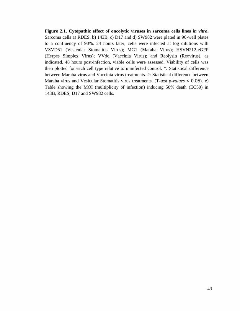
Oncolytic Rhabdoviruses are Effective Against Sarcoma Cell Lines in vitro.
As a first assessment of the potential of OV candidates as treatment for
sarcoma, we compared head to head five promising OV platforms for their capacity to
kill sarcoma cells in vitro. Herpes Simplex Virus-1 (HSVN212-eGFP; HSV), Reovirus
(Reolysin; Reo), Vaccinia virus (VVdd-eGFP; VVdd) and two Rhabdoviruses,
Vesicular Stomatitis Virus (VSVD51-GFP; VSVD51) and Maraba virus (MG1-eGFP;
MG1), were tested in four different sarcoma cell lines. Human osteosarcoma (143B),
canine osteosarcoma (D17), human Ewing’s sarcoma (RD-ES) and human synovial
sarcoma (SW982) were infected in vitro with different multiplicity of infection (MOI)
of each OV. EC50 (effective concentration 50%) values were then determined at 48
hours post-infection (Figure 2.1). While cell lines showed variable sensitivities to
OVs, both rhabdoviruses (MG1 and VSVD51) consistently demonstrated a high
potency in their ability to kill sarcoma cells. However, MG1 was slightly more
effective than VSVD51 for the majority of cell lines tested (Figure 2.1a, c-e), as it
required a lower multiplicity of infection (MOI) to induce more than 50% cell death.
These findings are consistent with the previous report evaluating rhabdoviruses in a
panel of adenocarcinoma cell lines(50) . Although it required a higher MOI to induce
sufficient cell death than for MG1, VVdd also demonstrated potent killing ability in
several sarcoma cell lines. Reovirus and HSV were only effective in RDES Ewing’s
sarcoma cells. Indeed, this cell line was highly sensitive to all OVs tested. In contrast,
SW982 synovial sarcoma cells were the most resistant cell line to OV infection

41
(Figure 2.1d). D17 and 143B osteosarcoma cell lines exhibited an intermediate
sensitivity to OVs (Figure 2.1e), but were mostly refractory to both HSV and Reo.
These data suggests that rhabdovirus-based OV platforms are more efficient at
inducing virus-mediated cell death in vitro in a diverse set of sarcoma cell lines.

42
43
Figure 2.1. Cytopathic effect of oncolytic viruses in sarcoma cells lines in vitro.
Sarcoma cells a) RDES, b) 143B, c) D17 and d) SW982 were plated in 96-well plates
to a confluency of 90%. 24 hours later, cells were infected at log dilutions with
VSVD51 (Vesicular Stomatitis Virus); MG1 (Maraba Virus); HSVN212-eGFP
(Herpes Simplex Virus); VVdd (Vaccinia Virus); and Reolysin (Reovirus), as
indicated. 48 hours post-infection, viable cells were assessed. Viability of cells was
then plotted for each cell type relative to uninfected control. *: Statistical difference
between Maraba virus and Vaccinia virus treatments. #: Statistical difference between
Maraba virus and Vesicular Stomatitis virus treatments. (T-test p-values < 0.05). e)
Table showing the MOI (multiplicity of infection) inducing 50% death (EC50) in
143B, RDES, D17 and SW982 cells.

44
Human sarcoma specimens are susceptible to MG1 infection ex vivo.
We next tested the ability of two of the most effective OV platforms in vitro,
MG1 and VVdd, to productively infect human sarcoma specimens ex vivo. For many
OV platforms, productive infection as defined by an infection that leads to release of
infectious viral progeny, is considered important for the efficacy of OVs in vivo,
particularly in the context of OVs encoding transgenes whose expression is directly
linked to viral replication (e.g. rhabdoviruses) (98, 206, 207) . Therefore,
measurement of virus replication ex vivo is a great alternative to assessing virus-
mediated cancer cell killing since virus-induced cytotoxicity is difficult to accurately
measure in live patient’s specimens(206) . Human specimens freshly collected
following surgery were processed and analyzed according to our previously published
method(205) . Tissues that presented low viability assessed using alamar blue prior to
processing were excluded from the analysis. A total of 21 specimens representing
distinct sarcoma subtypes were subsequently cored and infected ex vivo with MG1
(Supplemental Figure 2) and 29 for VVdd (Supplemental Figure 3). Figure 2.2a shows
the distribution of the sarcoma subtypes for MG-1-infected cores: osteosarcoma (OST,
29%), undifferentiated pleomorphic sarcoma (UPS, 24%), chondrosarcoma (CDS,
14%), leiomyosarcoma (LMS, 10%), rhabdomyosarcoma (RMS, 4%) and other types
of sarcoma (O, 19%). Viral production was determined by plaque assay and expressed
as a fold-change over input. We detected MG1 virus in 95% of the specimens tested
(20 of the 21). Importantly, 86% of the samples (18 of the 21) showed productive
viral replication (Figure 2.2b; virus levels exceeding that of virus input). In contrast,
productive infection with VVdd was much less frequent and therefore eight additional
specimens (3 UPS, 4 O and 1 RMS) were infected only with VVdd. In total, only 58%

45
of specimens infected with VVdd (17 of the 29) had detectable VVdd by plaque assay.
Furthermore, only 24% (7 out of 29) showed evidence of productive infection that, on
average, was several orders of magnitude lower than that observed for MG1
(Supplemental Figure 1). As MG1 expresses eGFP, eGFP expression was used as an
additional measurement of tumor-specific OV infection in MG1- infected sarcoma or
normal tissue specimens, originating from patients with diverse sarcoma etiologies.
Regardless of sarcoma type, eGFP expressed from the MG1 virus was abundantly
detected by fluorescent microscopy in sarcoma tissue, which corroborates and
confirms our findings that MG1 productively infects sarcoma specimens (Figure 2.2c).
As expected, we did not detect GFP expression in normal tissues, indicating virus
replication is restricted to cancer cells. Collectively, these results suggest that MG1 is
more effective than VVdd in its capacity to productively replicate in human sarcoma
tissues ex vivo.

46

47
Figure 2.2. Productive infection of human sarcoma explants by MG1 and VV. a)
Various human sarcoma subtypes including Osteosarcoma (OST), Undifferentiated
Pleomorphic Sarcoma (UPS), Rhabdomyosarcoma (RMS), Leiomyosarcoma (LMS),
Chondrosarcoma (CDS) and other (O) sarcoma explants were process for ex vivo
infection with MG1-eGFP. In percentage b) 24 hours after infection with MG1-eGFP,
explants were homogenized and titered. Fold changes in viral titer compared to input
virus have been plotted for each patients numerated from 1 to 21. c) Green fluorescent
pictures were taken 24 hours after infection using MG1-eGFP of sarcoma tumor and
normal human tissue.
48
MG1 is an efficient therapeutic treatment in vivo.
Our in vitro and ex vivo data led us to select MG1 for further in vivo evaluation
as treatment for sarcoma. Given the potential contribution of the immune response in
eliciting the anti-tumor effects of OV therapy, we tested MG1 on murine S180
metastatic sarcoma cells implanted subcutaneously in immune-competent Balb/C
mice. We first tested whether MG1 could kill S180 cells in vitro (Supplemental Figure
4). Indeed we found that similarly to most human and canine cell lines, S180 cells
were highly susceptible to MG1 killing (EC50 <0.0001). We subsequently tested
whether MG1 could productively infect S180 tumors in vivo after intra- tumoral (i.t.)
or intra-venous (i.v.) injection, as well as ex vivo in tumors collected from tumor-
bearing mice (Figure 2.3). Similar to human specimens, ex vivo infection of tumor
cores induced productive viral infection as depicted by plaque assay (Figure 2.3a) and
eGFP expression (Figure 2.3b). We observed that i.v. injection of MG1 did not lead to
a productive viral replication in the tumor (Figure 2.3a). However, we were able to
detect virus infection after i.t. injection by both plaque assay of homogenized tumor
and immunohistochemistry (Figures 2.3a and c). Therefore, i.t. delivery of the virus
was chosen to assess the effectiveness of MG1 to treat Balb/C mice bearing S180 sub-
cutaneous sarcoma tumors. A schematic representation of the treatment regimen is
showed in Figure 2.3d. Even though spontaneous remissions were observed in 40% of
tumor- bearing mice injected with PBS (6/15), MG1 treatment slowed tumor
progression (Figure 2.3e) and significantly increased the number of cured mice (80%,
11/15; P-value=0.0276; Figure 2.3f). To further investigate whether cured mice could
reject new S180 tumors following S180 re- challenge, S180 tumor cells were re-
implanted (day 90) on the opposite flank of cured mice from both PBS and MG1
49
treated groups (PBS=6; MG1=11; Figure 2.3d). While 100% of spontaneously cured
mice from the PBS group developed tumors a week later, only 27% (3/11) of MG1-
cured mice re-challenged with S180 cells developed tumors (Figure 2.3g) and these
were smaller in size as compared to those from the PBS group (Figure 2.3h).
However, similar percentage tumor take rate was observed in PBS and MG1-cured
mice after challenge with syngeneic 4T1 breast cancer cells compared to naïve PBS
group (Figure 2.3i). Altogether, these results suggest that MG1 delivered i.t.
effectively replicates in S180 murine sarcoma tumors implanted in immunocompetent
hosts, leading to eradication of 80% of tumors and protection from S180- specific re-
challenge in all cured mice.

50

51
Figure 2.3. Maraba virus MG1 infects and replicates in murine sarcoma S180
tumors, leading to persistent anti-tumor effects.
a) Murine S180 sarcoma cells were implanted sub-cutaneously in Balb/c mice
(2x10⁶cells). When tumors reached at least 5x5mm size, mice were injected with 10
8 pfu
MG1 by i.t. or i.v. route. 48 hours later, mice were sacrificed, tumors harvested, and
homogenized for MG1 titration. In parallel, naïve mice were sacrificed and tumors
harvested for ex vivo infection with MG1. 24 hours later, tumors were homogenized for
MG1 titration. b) Fluorescent picture from ex vivo sample 24 hours after infection. c)
S180 sarcoma tumor immunohistochemistry. Tumors were implanted as in a) and
paraffin sections were prepared for rhabdovirus immuno-staining in untreated or MG1
treated mice. Secondary antibody staining (Ctl 2ry Ab) and Haematoxilin & Eosin stain
(H&E) were used as controls Right panel shows the higher magnification of Rhabovirus
immuno-staining from MG1 tumor treated mouse highlighting infected portion of the
tissue as well as corresponding H&E stain. d) Schematic representation of S180 sarcoma
implantation and treatment schedule. S180 were established subcutaneously in
immunocompetent in Balb/c mice. All mice had palpable tumors by day 8. On day 8, 10
and 13, animals were injected i.t. with PBS or MG1 (1x10⁸ pfu/dose). e) Plot graph
representing tumor growth for each mouse corresponding to the PBS and MG1 treated
groups (N=15). f) Kaplan- Meier plot outlining the time to S180 tumor endpoint (1600
mm3) from experiment described in d-e). *p=0.0437 Log Rank test comparing MG1 to
PBS. g) Cured mice from e-f) were re- challenged with S180 (1x10⁶ cells) at day 90 and
percentage take rate was plotted for day 97 (one week post re-challenge). h) Tumor size
was measured at day 97 and plotted. *p=0.0209 two tailed t-test. i) 4T1 re-challenge
percentage tumor takes rate at day 126, 10 days after tumor implantation. No significant
differences were observed measured by unpaired t-test (p> 0.05).

52
Discussion
Sarcoma currently requires a multimodal treatment approach, relying primarily
upon surgical resection, with adjuvant chemotherapy and/or radiation therapy (208).
This aggressive approach is quite taxing for patients. Our study suggests that the
oncolytic rhabdovirus MG1 has potential for the treatment of sarcoma. Among several
other clinically relevant candidate OVs, MG1 was the most potent OV based on in
vitro cell killing across a panel of four sarcoma cell lines. Close behind MG1 was
VSV∆51, another oncolytic rhabdovirus, followed by the vaccinia- based OV
candidate, VVdd. However, from a heterogeneous panel of human sarcoma tissue
processed ex vivo, we observed that MG1 outperformed VVdd in the ability to infect
and replicate in tumor tissue. Overall, we found that the difference between Vaccinia
and MG-1 observed in in vitro cytotoxicity assays was in line with the tissue explant
viral growth data, wherein MG-1 grew more effectively than Vaccinia. However, we
acknowledge there may be a number of normal cells (e.g. immune cells, endothelial
cells, and cancer associated fibroblasts) that could further influence the capacity of
viruses to infect tissue explants.
Lastly, using the immunocompetent murine subcutaneous S180 sarcoma
model, we showed that MG1 treatment led to significantly more durable cures
compared to control. Upon subsequent re-challenge with the same tumor cells, mice
previously treated with MG1 were more readily protected from tumor re-growth. One
caveat is that S180 tumors had a high frequency of spontaneous regression, which has

53
been reported by other groups (209). Also given OV treatment for sarcomas would
likely be assessed in the context of tumor resection, it will be interesting to pursue
subsequent studies characterizing the effect of MG1 administered perioperatively
following tumor resection or amputation in animal models. However, there is a general
paucity in murine sarcoma models, which makes these studies challenging. Given we
have shown that MG1 is effective in D17 canine sarcoma cells, a canine study would
be of particular interest given the naturally high prevalence of osteosarcoma in
dogs(209) and the similarity of clinical of disease progression and treatment in the
veterinary setting.
Finally, while it seems less effective than MG1 and VSVD51, VVdd was also
capable of killing sarcoma cell lines in vitro, and was able to infect a small proportion of
ex vivo human sarcoma samples. However, it is unclear whether the same level of
infection with VVdd is required to elicit an anti-tumor effect as compared to MG1. Our
group has shown in clinical trials that intravenous injection of the vaccinia virus JX-594
(3.10e7pfu.Kg-1
) led to replication of the virus in one patient with leiomyosarcoma,
resulting in >16 weeks disease control by RECIST criteria(201). Hence, this platform
may merit more investigation for sarcoma treatment. Overall, our study open doors to use
viral immunotherapeutic based platform to treat sarcoma. Notably, Maraba virus MG1 is
clearly a promising candidate, which warrants further evaluation either alone or in a
prime-boost strategy.
54
Chapter 3 - Dimethyl Fumarate Potentiates Oncolytic Virotherapy
through NF-kB inhibition
Mohammed Selman1,2
, Paula Ou1,3
, Christopher Rousso1,3
, Anabel Bergeron1,3
, Ramya
Krishnan1,2
, Larissa Pikor1, Andrew Chen
1, Brian A. Keller
1,2, Carolina Ilkow
1,2, John C.
Bell1,2
, Jean-Simon Diallo1,2
1Centre for Innovative Cancer Research, Ottawa Hospital Research Institute, Ottawa,
Ontario, Canada. 2Department of Biochemistry, Microbiology and Immunology,
University of Ottawa, Ontario, Canada. 3Faculty of Science, University of Ottawa,
Ontario, Canada
Accepted November 2017; Published Science Translational Medicine.
Contribution: MS, PO, CR, AB, RK, LP conducted in vitro experiments. MS, AC
performed mouse experiments. MS, BAK processed patient tumor biopsies. BAK, CI
isolated human-derived cell lines. MS, JSD participated in the conception and design of
the study. MS, JSD wrote the manuscript with editorial contributions from LP, RK, BAK.
JSD, JCB supervised the study.
From M. Selman, P. Ou, C. Rousso, A. Bergeron, R. Krishnan, L. Pikor, A. Chen, B. A.
Keller, C. Ilkow, J. C. Bell, J.-S. Diallo, Dimethyl fumarate potentiates oncolytic
virotherapy through NF-kB inhibition. Sci. Transl. Med. 10, eaao1613 (2018). Reprinted
with permission from AAAS.
Reproduction permission : The American Association for the Advancement of Science
(AAAS), the non-profit publisher of the Science family of journals states that authors can
immediately use final works for non-profit purposes.

55
Abstract
Resistance to oncolytic virotherapy renders this treatment ineffective, and it is frequently
associated with failure of tumor cells to get infected. Dimethyl fumarate (DMF), a
common treatment for psoriasis and multiple sclerosis, has been also shown to have
anticancer properties. In the present study, we show that DMF and various fumaric and
maleic acid esters (FMAEs) enhance viral infection of several oncolytic viruses (OV) in
cancer cell lines as well as in human tumor biopsies, improving therapeutic outcomes in
resistant syngeneic and xenograft tumor models. This results in durable responses, even
in models otherwise refractory to OV and drug monotherapies. The ability of DMF to
enhance viral spread is due to its ability to inhibit type I IFN production and response,
which is associated with its ability to block nuclear translocation of transcription factor
NF-kB. This study demonstrates that unorthodox application of approved FDA drugs and
biological agents can lead to improved anti-cancer therapeutic outcomes.

56
Introduction
Deregulated metabolism and defective innate immune response are common
characteristics of transformed cells(74). This makes it possible to genetically engineer or
select oncolytic viruses (OVs), which specifically infect and kill tumor cells without
harming normal tissues. OVs can be generated from various viral backbones using
diverse genetic approaches, and provide capacity for expressing therapeutic or imaging
transgenes. Some of the early OV candidates have finally made their way to the clinic
with the approval of talimogene laherparepvec (or T-Vec) based on herpes simplex virus
1 (HSV-1) for treatment of advanced melanoma (29).
Pre-clinical and clinical trials have showcased the heterogeneity in the therapeutic
response to OV treatment, in which a subset of patient tumors is refractory to infection
with OVs (8, 81, 84, 88). The identification of pharmacological agents that can
functionally enhance OVs to improve therapeutic benefit has been an area of intense
investigation and has been recognized as critical to maximize the therapeutic impact of
OVs in the clinic (8). Combination strategies with checkpoint blockade have shown
promise recently (30, 210, 211) but do not address the frequent resistance of tumors to
viral infection. To this end, strategies aiming to suppress antiviral immune responses, for
example using immunosupressants such as cyclophosphamide, rapamycin, and histone
deacetylase inhibitors, have shown promise in vitro but variable effects in animal models
(108, 186–188). Combinations of OVs with cyclophosphamide in particular are under
clinical evaluation, with favorable safety profiles reported thus far (NCT01598129).

57
Fumaric acid esters (FAE) are a class of compounds with known anti-
inflammatory and neuroprotective effects (126, 189, 190). The mechanisms involved
have yet to be fully elucidated but are thought to be mediated through the activation of
the antioxidative transcription factor nuclear factor (erythroid-derived 2)-like 2 (NRF2)
pathway(189), the inhibition of NF-κB (212), as well as functional depletion of
glutathione (GSH)(213, 214). FAEs (marketed as Fumaderm, Psorinovo) were first
approved as a treatment for psoriasis in Germany. Recently, dimethyl fumarate (DMF),
an FAE marketed as Tecfidera, was approved by the U.S. Food and Drug Administration
(FDA) and the European Medicines Agency for the treatment of relapsing forms of
multiple sclerosis and relapsing-remitting multiple sclerosis (191). Clinical studies on the
long-term use of DMF have not revealed any severe long-term adverse effects (191, 192,
215). Recent reports suggest that DMF has anticancer potential, shown to suppress tumor
growth and metastasis (141, 144, 149–151, 216) in addition to sensitizing tumors to
chemotherapy(144, 145). Furthermore, DMF is currently under clinical evaluation for the
treatment of chronic lymphocytic leukemia and cutaneous T cell lymphoma
(NCT02546440, NCT02784834). Given this and the documented positive effect of
diverse immunosuppressants in combination with OVs, we set out to explore the
possibility of using DMF in combination with oncolytic virotherapy in vitro and in vivo.
58
Results
Dimethyl fumarate enhances tumor-specific viral spread and oncolysis in vitro
Little is known about the effect of DMF on viral infection. We first examined the
impact of DMF (structure displayed in Figure 3.1A) on the growth of oncolytic vesicular
stomatitis virus (VSVΔ51). Notably, VSV and closely related Maraba are currently
undergoing clinical evaluation but face resistance to infection in approximately a third of
cancers (48, 88). 786-0 renal carcinoma cells are highly refractory to VSVΔ51 infection,
however, a 4 hour pretreatment with DMF at doses between 50 and 200 µM increased
VSVΔ51 virus growth at a low multiplicity of infection (MOI) by more than 100-fold
(Figure 3.1B). Virus-encoded GFP expression was also greatly enhanced, as visualized
by microscopy (Figure 3.1C). This effect was observed with pretreatment times up to 24
hours before infection, and after infection up to 8 hours (Supplemental Figure 1A). More
broadly, DMF robustly enhanced infection in a panel of human and murine cancer cell
lines (sarcoma, osteosarcoma, breast, colon, melanoma, ovarian) with a wide range of
sensitivity to VSVΔ51 (Figure 3.1D). DMF also improved viral output of Sindbis virus
and oncolytic herpes virus (HSV-1 mutant N212) after treatment in 786-0 cells (Figure
3.1, E and F). Furthermore, we also tested the effect of DMF on adenoviruses, which in
addition to oncolytic virotherapy have been extensively used as gene therapy vectors
(217). Pretreatment of human lung adenocarcinoma A549 cells with DMF increased
luciferase transgene expression of non-replicating adenovirus, E1A-deleted Ad5, over the
course of 7 days by up to 20-fold (Figure 3.1G).

59
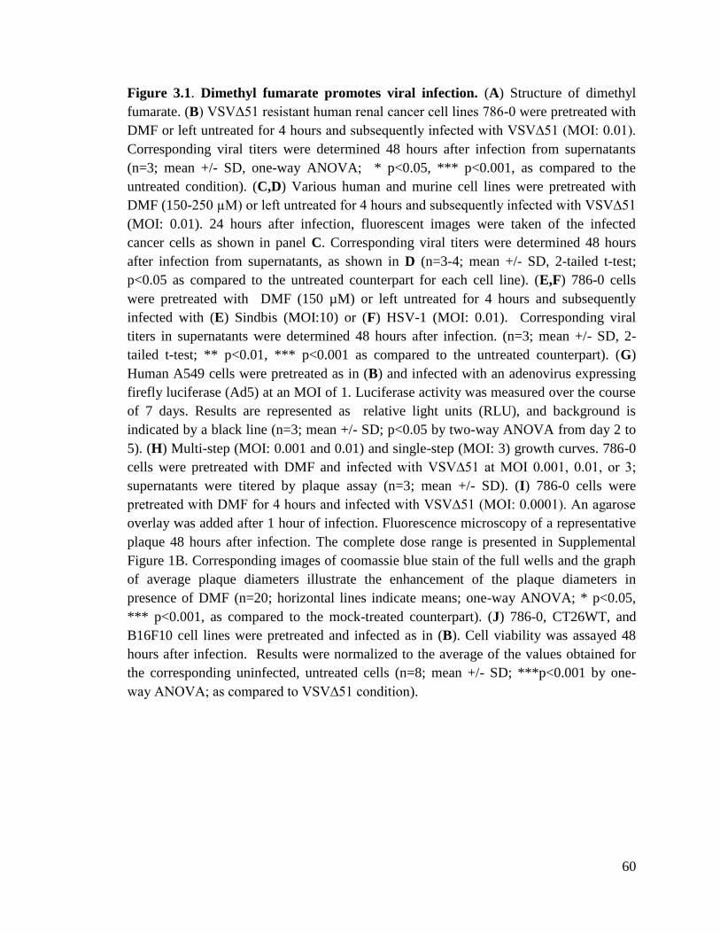
60
Figure 3.1. Dimethyl fumarate promotes viral infection. (A) Structure of dimethyl
fumarate. (B) VSVΔ51 resistant human renal cancer cell lines 786-0 were pretreated with
DMF or left untreated for 4 hours and subsequently infected with VSV∆51 (MOI: 0.01).
Corresponding viral titers were determined 48 hours after infection from supernatants
(n=3; mean +/- SD, one-way ANOVA; * p<0.05, *** p<0.001, as compared to the
untreated condition). (C,D) Various human and murine cell lines were pretreated with
DMF (150-250 µM) or left untreated for 4 hours and subsequently infected with VSV∆51
(MOI: 0.01). 24 hours after infection, fluorescent images were taken of the infected
cancer cells as shown in panel C. Corresponding viral titers were determined 48 hours
after infection from supernatants, as shown in D (n=3-4; mean +/- SD, 2-tailed t-test;
p<0.05 as compared to the untreated counterpart for each cell line). (E,F) 786-0 cells
were pretreated with DMF (150 µM) or left untreated for 4 hours and subsequently
infected with (E) Sindbis (MOI:10) or (F) HSV-1 (MOI: 0.01). Corresponding viral
titers in supernatants were determined 48 hours after infection. (n=3; mean +/- SD, 2-
tailed t-test; ** p<0.01, *** p<0.001 as compared to the untreated counterpart). (G)
Human A549 cells were pretreated as in (B) and infected with an adenovirus expressing
firefly luciferase (Ad5) at an MOI of 1. Luciferase activity was measured over the course
of 7 days. Results are represented as relative light units (RLU), and background is
indicated by a black line (n=3; mean +/- SD; p<0.05 by two-way ANOVA from day 2 to
5). (H) Multi-step (MOI: 0.001 and 0.01) and single-step (MOI: 3) growth curves. 786-0
cells were pretreated with DMF and infected with VSV∆51 at MOI 0.001, 0.01, or 3;
supernatants were titered by plaque assay (n=3; mean +/- SD). (I) 786-0 cells were
pretreated with DMF for 4 hours and infected with VSV∆51 (MOI: 0.0001). An agarose
overlay was added after 1 hour of infection. Fluorescence microscopy of a representative
plaque 48 hours after infection. The complete dose range is presented in Supplemental
Figure 1B. Corresponding images of coomassie blue stain of the full wells and the graph
of average plaque diameters illustrate the enhancement of the plaque diameters in
presence of DMF (n=20; horizontal lines indicate means; one-way ANOVA; * p<0.05,
*** p<0.001, as compared to the mock-treated counterpart). (J) 786-0, CT26WT, and
B16F10 cell lines were pretreated and infected as in (B). Cell viability was assayed 48
hours after infection. Results were normalized to the average of the values obtained for
the corresponding uninfected, untreated cells (n=8; mean +/- SD; ***p<0.001 by one-
way ANOVA; as compared to VSV∆51 condition).

61
Single and multi-step growth curves performed in 786-0 cells revealed that DMF
enhanced the viral output of VSVΔ51 when using a low MOI, but not when using a high
MOI (Figure 3.1H), indicating DMF inhibits mechanisms that impinge on viral spread
rather than virus output per infected cell. To further explore the effect of DMF on virus
spreading, 786-0 cell monolayers were infected with VSVΔ51 and overlaid with agarose
to generate defined plaques of virus replication foci. DMF substantially increased the
average plaque diameter of VSVΔ51 on fluorescent imaging and Coomassie Blue
staining of infected cell monolayers (Figure 3.1I and Supplemental Figure 1B). To further
assess the oncolytic effect of VSVΔ51 in the presence of DMF, we pretreated cancer cells
with DMF before infection with VSVΔ51 at a low MOI, and cell viability was assessed
with the metabolic dye alamarBlue, 48 hours after infection. Combined treatment with
DMF and VSVΔ51 resulted in a decrease in cell viability in human 786-0 as well murine
CT26WT and B16F10 cells (Figure 3.1J).
We further assessed the ability of DMF to enhance VSVΔ51 infection ex vivo in
mouse-derived tissues. Tumor cores from mice subcutaneously implanted with CT26WT
murine colon cancer cells or B16F10 murine melanoma cells, as well as cores from
normal lung, muscle, and spleen were collected and subsequently infected with VSVΔ51-
GFP in the presence or absence of 150 µM DMF. DMF robustly increased the growth of
the virus in CT26WT and B16F10 cores by 31-fold and 13-fold, respectively, but did not
increase virus growth in normal tissue cores (Figure 3.2A). Furthermore, DMF increased
VSVΔ51 infection by > 10-fold ex vivo in primary human melanoma, lung, prostate, and

62
ovarian tumor samples, as observed by plaque assay and microscopy (Figure 3.2, B and
C), as well as in various melanoma and ovarian patient-derived cancer cell lines (Figure
3.2D). Similar to the murine normal tissue cores, DMF did not promote virus growth in
human normal lung and muscle tissues from various patients (Figure 3.2E).

63
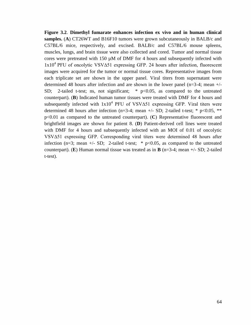
64
Figure 3.2. Dimethyl fumarate enhances infection ex vivo and in human clinical
samples. (A) CT26WT and B16F10 tumors were grown subcutaneously in BALB/c and
C57BL/6 mice, respectively, and excised. BALB/c and C57BL/6 mouse spleens,
muscles, lungs, and brain tissue were also collected and cored. Tumor and normal tissue
cores were pretreated with 150 µM of DMF for 4 hours and subsequently infected with
1x10⁴
PFU of oncolytic VSV∆51 expressing GFP. 24 hours after infection, fluorescent
images were acquired for the tumor or normal tissue cores. Representative images from
each triplicate set are shown in the upper panel. Viral titers from supernatant were
determined 48 hours after infection and are shown in the lower panel (n=3-4; mean +/-
SD; 2-tailed t-test; ns, not significant; * p<0.05, as compared to the untreated
counterpart). (B) Indicated human tumor tissues were treated with DMF for 4 hours and
subsequently infected with 1x10⁴ PFU of VSV∆51 expressing GFP. Viral titers were
determined 48 hours after infection (n=3-4; mean +/- SD; 2-tailed t-test; * p<0.05, **
p<0.01 as compared to the untreated counterpart). (C) Representative fluorescent and
brightfield images are shown for patient 8. (D) Patient-derived cell lines were treated
with DMF for 4 hours and subsequently infected with an MOI of 0.01 of oncolytic
VSV∆51 expressing GFP. Corresponding viral titers were determined 48 hours after
infection (n=3; mean +/- SD; 2-tailed t-test; * p<0.05, as compared to the untreated
counterpart). (E) Human normal tissue was treated as in B (n=3-4; mean +/- SD; 2-tailed
t-test).

65
In addition to DMF, various fumaric and maleic acid esters (FMAEs) exhibit
antiinflammatory and immunomodulatory properties (218). We therefore tested whether
other FAEs and their cis- and trans-isoforms (maleic acid esters) (Figure 3.3, A and B)
have a similar impact to DMF on viral infection of cancer cells. Indeed, the cell-
permeable FAE diethyl fumarate (DEF), and maleic acid esters dimethyl maleate (DMM)
and diethyl maleate (DEM) robustly enhanced VSVΔ51infection, spread, and oncolysis
in 786-0 and CT26WT cells in vitro (Figure 3.3C, Supplemental Figure 2 and S3).
Enhanced infection was also observed with FMAEs in CT26WT tumor cores infected ex
vivo (Figure 3.3D). DMF is rapidly hydrolyzed to monomethyl fumarate (MMF) by
esterases in vivo, and subsequently to fumaric acid (FA) (as displayed in Figure
3.3B)(219). Indeed, MMF is thought to be the active metabolite of DMF in the treatment
of multiple sclerosis(219). In 786-0 cells, MMF also enhanced infection of VSVΔ51 to a
similar extent as DMF, albeit at higher effective doses. Fumaric acid (FA), in contrast
with the cell-permeable esters MMF and DMF, had no impact on viral growth (Figure
3.3C, Supplemental Figure 2). Taken together, our data indicate that DMF and other cell-
permeable FMAEs can dramatically enhance the spread of OVs in both mouse and
human cell lines and cancer tissue explants.

66

67
Figure 3.3. Fumaric and maleic acid esters promote infection by VSVΔ51. (A)
Structures of fumaric acid esters (DEF) and maleic acid esters (DEM, DMM) are
presented. (B) Metabolism of DMF. DMF is hydrolyzed into monomethyl fumarate
(MMF), which in turn is metabolized into fumaric acid (FA). FA subsequently enters the
TCA cycle. (C) 786-0 cells or (D) CT26WT ex vivo tumor cores were pretreated with
various FMAEs and analogues for 4 hours, and subsequently infected with oncolytic
VSV∆51 expressing GFP at (C) an MOI of 0.01 or (D) 1x10⁴
PFU. 24 hours after
infection, we obtained fluorescent images of the infected 786-0 cells or CT26WT tumor
cores. Corresponding viral titers were determined from supernatants 48 hours after
infection (n=3; mean +/- SD; one-way ANOVA; * p<0.05, ** p<0.01, *** p<0.001, as
compared to the untreated counterpart).
68
Dimethyl fumarate improves therapeutic efficacy of oncolytic rhabdoviruses
Because DMF is a clinically approved drug and it broadly and robustly enhanced
the growth and activity of VSVΔ51 in vitro in both human and mouse tumor explants,
and did so preferentially in tumors opposed to normal tissues, we next evaluated the
potential therapeutic benefit of combining DMF with oncolytic VSVΔ51 in vivo. To this
end, we used both syngeneic and xenograft mouse tumor models, in which VSVΔ51 is
ineffective as a monotherapy (119, 220–222). Mouse CT26WT and B16F10 and human
colon cancer HT29 cells were grown subcutaneously in Balb/c, C57BL/6, or nude mice,
respectively. Mice were injected intratumorally with DMF for 4 hours, and subsequently
infected with VSVΔ51 expressing luciferase. With the exception of the B16F10 model,
DMF enhanced virus-associated luciferase gene expression specifically in tumors 24
hours after the first injection of virus, as assessed using an in vivo imaging system (IVIS)
(Figure 3.4, A and B). In all three models, the combination therapy considerably delayed
tumor progression (Figure 3.4C, table S1) and significantly prolonged survival compared
with either monotherapy (combination therapy compared to VSV∆51 alone; CT26WT:
p=0.0008; B16F10: p=0.0039; HT29: p=0.0003) (Figure 3.4D). The combination therapy
produced complete remission in approximately 20% of the mice in both the CT26WT and
B16F10 models. The cured CT26WT-bearing mice that had received the combination
regimen subsequently became immune to re-challenge with the same cancer cells (Figure
3.4E). However, when cured CT26WT-bearing mice were challenged with a foreign
tumor (murine 4T1 breast cancer), cured CT26WT mice grew 4T1 tumors at a similar
rate as the naive mice (Figure 3.4F). Altogether, these results indicate the effective
generation of a specific and long-lasting anti-tumor immunity.

69

70
Figure 3.4. Dimethyl fumarate enhances VSV∆51 therapeutic efficacy in syngeneic
and xenograft tumor models. (A-D) CT26WT, B16F10, and HT29 tumor-bearing mice
were treated intratumorally with the vehicle (DMSO) or 50 (B16F10) or 200 (CT26WT
and HT29) mg/kg of DMF for 4 hours, and subsequently injected with 1x10⁸
PFU of
oncolytic VSV∆51 expressing firefly luciferase or the vehicle (PBS), intratumorally. The
treatment was administered twice or three times, as indicated by arrows in panel C. 24
hours after infection, viral replication was monitored. (A) Representative
bioluminescence images of mice. (B) Quantification of luminescence in photons/sec
(n=10-18; horizontal lines indicate means; ns, not significant; **p < 0.01 by 2-tailed t-
test, as compared to VSV∆51 infected condition; dashed lines represent average
background intensity). (C) Tumor volume (n=9-15; data shown as mean ± SEM; SD
values are indicated in table S1; *p < 0.05, ** p<0.01, ***p<0.001 by two-way ANOVA;
comparing DMF+VSV∆51 to DMSO alone). (D) Survival was monitored over time.
Log-rank (Mantel-Cox) test indicates that the combined treatment significantly prolonged
survival over VSV∆51 alone (CT26WT: n=10-13, p=0.0008; B16F10: n=9-14, p=0.0039;
HT29: n=10-15, p=0.0003). (E) Tumor volume and survival were monitored after re-
implantation of CT26WT in cured and naïve mice from D (n=3-5, mean +/- SD). (F)
Tumor volume was monitored after implantation of 4T1 cells in CT26WT-cured and
naïve mice (n=3, mean +/- SD).

71
Fumaric and maleic acid esters inhibit the antiviral response
To gain further insight into the possible mechanism mediating the enhancement of
OVs by DMF and other FMAEs, microarray gene expression analysis was performed on
786-0 cells 24 hours after infection with VSVΔ51 in the presence or absence of DMF,
DEM, DEF, or DMM. DMF-treated cells had a similar gene expression profile to cells
treated with the other FMAEs tested (Supplemental Figure 4A). Upon infection with
VSVΔ51, multiple antiviral genes were up-regulated as expected; however, DMF led to
the inhibition of many of these (IFITIM1, MX2, GBP4, IFI27, IFNA, CXCL10), and
upregulated various genes, including a number of redox response genes (CYP4F11,
CDK5RAP2, ANXA10 HMOX1, OSGIN1, TXNRD1, AKR1B10, AKR1B15, AKR1C1,
AKR1C2) (Figure 3.5A and B). GOterm analysis revealed that FMAE treatment of
infected 786-0 cells inhibited the response to virus as well as type I IFN signaling (Figure
3.5C, Supplemental Figure 4B ). Consistent with repression of the type I IFN response,
DMF decreased activation (phosphorylation) of both STAT1 and STAT2, 24 hours after
infection (Figure 3.5D). Additionally, the expression of the antiviral protein IFITM1 was
potently repressed by DMF, whereas the VSV viral proteins were increased (Figure
3.5E). Furthermore, DMF enhanced infection of VSVΔ51, but it had no impact on
infection of 786-0 cells by wild-type VSV (wtVSV) (Figure 3.5E). Unlike VSVΔ51,
wtVSV robustly inhibits Type I IFN production(48), and therefore we would expect the
effect of DMF to be redundant in the context of wtVSV if a Type I IFN response is
indeed involved in eliciting the pro-viral effects of DMF.

72

73
Figure 3.5. FMAEs inhibit antiviral cytokine production and response to type I
interferon. (A-C) Lysates of 786-0 cells treated with FMAEs and infected with
VSV∆51 expressing GFP were collected at 24 hpi, and RNA or protein was extracted.
(A) Scatter plot showing the expression of all genes in infected 786-0 in the presence (y-
axis) or absence (x-axis) of DMF. Blue dots represent genes upregulated by DMF during
infection, red dots represent genes downregulated by DMF during infection. (B) Heatmap
showing the expression of the differentially expressed genes belonging to the “response
to virus” GO term. (C) List of top GO terms downregulated by FMAEs during viral
infection. (D) Lysates of 786-0 cells, treated with DMF (150 µM) and infected with
VSV∆51 expressing GFP or left untreated, were collected at 24 hpi and probed for
indicated proteins by western blot. (E) 786-0 cells were pretreated for 4 hours with DMF
(150 µM) or mock treated and infected with VSV∆51 or wtVSV (MOI: 0.01).
Corresponding viral titers were determined from supernatants 48 hours after infection
(n=3; mean +/- SD). (F) 786-0 cells were treated with DMF (150 µM), MMF (1500 µM),
or mock treated for 4 hours and infected with VSVΔ51ΔG at MOI 1. 12 or 16 hpi,
supernatants were collected and used to precondition 786-0 cells for 4 hours, and then the
cells were infected with VSV∆51 or wtVSV. Corresponding viral titers were determined
24 hours after infection from supernatants (n=3; mean +/- SD). Representative
fluorescent images are shown. (G-H) 786-0 cells were treated as in (A). (G) At 16 hpi,
supernatants were collected and assayed by ELISA for IFNβ (n=3; mean +/- SD). (H) At
36 hpi, supernatants were collected and assayed by ELISA for IFNα (n=3; mean +/- SD).
(I) 786-0 cells were treated with FMAEs for 6 hours and with IFNβ or IFNα for 4 hours,
and subsequently infected with VSVΔ51 at MOI 0.1. Corresponding viral titers were
determined from supernatants 48 hours after infection (n=3; mean +/- SD). (J) 786-0
cells were pretreated with 200 µM of DMF for 1 or 4 hours and treated with IFNβ (250
U/ml) for 30 minutes, or left untreated. Cell lysates were extracted and probed for
pSTAT1, STAT1, and β-actin by western blot.

74
To gain mechanistic insight into the effect of FMAEs on the antiviral response,
we examined their ability to protect against virus challenge and IFN-mediated antiviral
signaling. After treatment with FMAEs (or mock), we infected cells with a spread-
deficient version of VSVΔ51 that does not encode the viral G protein (glycoprotein
responsible for virus budding, host cell binding, and virus entry; VSVΔ51ΔG) to
suppress formation of de novo virions(223). Cell supernatants were collected 12 or 16
hour post infection (hpi), and used to pretreat target cells before infection with VSVΔ51
or wtVSV. Our results show that while the supernatant of cells infected with VSVΔ51ΔG
could protect against subsequent viral infection with VSVΔ51 or wtVSV, the addition of
DMF or MMF was able to completely overcome this suppressive effect for both VSVΔ51
and wtVSV (Figure 3.5F). Indeed, we observed through ELISA a decrease in the
production of IFNβ and IFNα after infection in the presence of FMAEs (Figure 3.5, G
and H). Furthermore, treatment of cells with DMF and other FMAEs antagonized the
antiviral effects of Type I IFN pretreatment on VSVΔ51 infection (Figure 3.5I). Taken
together, these data suggest that FMAEs affect antiviral signaling by repressing
production of Type I IFN and downstream signaling through the JAK-STAT pathway.
Consistent with this, Western blotting revealed that in cells conditioned with IFNβ at the
doses which inhibit VSVΔ51 infection, DMF and DEM inhibited STAT1
phosphorylation, which is involved in transcription and response to type I IFN after viral
infection (Figure 3.5J and Supplemental Figure 4C).
Fumaric and maleic acid esters promote infection through NF-κB inhibition
independently of GSH depletion

75
Although our data clearly implicated an effect of multiple FMAEs on the antiviral
response, the molecular chain of events leading to these effects remained unclear. DMF,
DEM, DEF, and MMF share a common α,β-unsaturated carbon, which is attacked by
GSH in a Michael addition reaction and which is implicated in the capacity of these
compounds to deplete cellular GSH and activate the antioxidant response (224). We
therefore tested the impact of dimethyl succinate (DMS) (Figure 3.6A), which lacks this
functional moiety. We found that DMS had no impact on viral output (Figure 3.6, B and
C), nor did the hydrolysed form of DMS, succinate (S). In contrast with FA, DMS, and S,
all other FMAEs were able to deplete GSH (Figure 3.6D). However, the proviral activity
of DMF was still evident after pre-depletion of cellular GSH by culturing cells for 10
days in the presence of buthionine sulfoximine (BSO), an inhibitor of glutamate cysteine
ligase (GCL) required for the synthesis of GSH (Figure 3.6E).

76

77
Figure 3.6. DMF inhibits NF-κB translocation upon infection. (A) Structures of DMS,
MMS, S. (B-C) 786-0 cells were pretreated with the indicated DMF analogues for 4
hours and subsequently infected with oncolytic VSV∆51 expressing GFP at an MOI of
0.01. (B) 24 hours after infection, fluorescent images were taken of the infected 786-0
cells. (C) Corresponding viral titers were determined from supernatants 48 hours after
infection (n=3; mean +/- SD; one-way ANOVA; *** p<0.001, as compared to the
untreated counterpart). (D) GSH concentrations were determined in 786-0 cells after a 4-
hour treatment with FMAEs (n=4; mean +/- SD; one-way ANOVA; *** p<0.001, as
compared to the untreated counterpart). (E) 786-0 cells were grown in the presence or
absence of BSO (2 mM) for 7 days and pretreated with DMF (200 µM) for 4 hours or left
untreated, then infected with oncolytic VSV∆51 expressing GFP at an MOI of 0.01.
Corresponding viral titers were determined from supernatants 48 hours after infection
(n=3; mean +/- SD).(F) Heatmap showing the expression of the differentially expressed
oxidative stress genes. Expression of genes was normalized to values obtained for
untreated, uninfected control. (G) HMOX1 expression quantified by qPCR from 786-0
cells after a 6-hour treatment with FMAEs (n=3; mean +/- SD). (H) siNRF2 knockdown
786-0 cells were treated with DMF or untreated and infected as in (E). Corresponding
viral titers were determined from supernatants 24 hours after infection. RNA was
extracted, and the expression of NRF2 and IFITM1 genes was quantified by qPCR (n=3;
mean +/- SD). (I-J) Cytoplasmic and nuclear protein fractions were extracted from 786-0
cells treated with DMF (200 µM) for 4 hours and (I) subsequently infected with oncolytic
VSV∆51 expressing GFP at an MOI of 1 for 8 hours, or (J) treated with TNFα (30 ng/ml)
for 30 min. Cell lysates were probed for multiple proteins as indicated, by western blot.
(K) 786-0 cells were pretreated with NF-κB inhibitors targeting IKK [IKK16 (10 µM),
TPCA1 (40 µM)] for 4 hours and subsequently co-treated with DMF (150 µM) and
oncolytic VSV∆51 expressing GFP at an MOI of 0.01. Corresponding viral titers were
determined from supernatants 24 hours after infection. (n=3; mean +/- SD; one-way
ANOVA; *** p<0.001, as compared to the untreated counterpart).

78
In parallel with their impact on antiviral gene expression by microarray, FMAEs
also induced robust expression of multiple genes involved in the antioxidant response
(Figure 3.6F). In particular, HMOX1 expression, as determined by real-time PCR, was
consistently up-regulated well over 100-fold by FMAEs (Figure 3.6G), but not by
treatment with FA, DMS, or S (Figure 3.6G). DMF and other FMAEs induce nuclear
translocation of NRF2 via covalent modification of KEAP1, which induces antioxidant
genes(225). This is consistent with our observation of the induction of HMOX1 and other
NRF2 target genes by FMAEs but not FA, DMS, or S (Figure 3.6F and G). To determine
whether the proviral effect of DMF is dependent on NRF2 activity, siRNA knockout
against NRF2 was performed. We found that knockdown of NRF2 did not block the
capacity of DMF to enhance VSVΔ51 infection or inhibit antiviral factor IFITM1
(Figure 3.6H). Furthermore, DMF was able enhance infection in a number of cell lines
harboring KEAP1 mutations (A549(226), CT26WT(227)) (Figure 3.1D). Altogether
these data suggest that the ability of FMAEs to enhance infection requires the α,β-
unsaturated carbon involved in GSH depletion, but that neither GSH depletion nor NRF2
activity are likely key mediators of this phenomenon. Given that DMF inhibits LPS-
stimulated cytokine production by inhibiting NF-κB nuclear entry(228, 229), we looked
at nuclear and cytoplasmic fractionations of infected cells. Probing for NF-κB subunit
p65 revealed that upon infection or TFNα stimulation, DMF inhibits phosphorylation and
translocation of this transcription factor, involved in the transcription of IFNβ particularly
but also in the response to type I IFN (Figure 3.6 I and J). Furthermore, DMF did not
improve infection in cells treated with inhibitors of IκB kinase (IKK) degradation using
chemical compounds IKK16 and TCPA1(230, 231) (Figure 3.6K).

79
Discussion
In this study, we demonstrate that fumaric and maleic acid esters like DMF
enhance OVs such as VSVΔ51 by increasing viral spread and oncolysis in resistant
cancer cells. Furthermore, we show that DMF can overcome the innate immune response
of cancer cells in a manner consistent with its modulation of NF-κB activity. This results
in a decrease in cytokine production and the inhibition of the type I IFN response at
multiple levels, including the NF-κB and JAK-STAT pathways.
FMAEs such as DMF are electrophilic hence they can covalently link to essential
thiol groups on various proteins, including KEAP1, which activates NRF2(189). The
activation of antioxidative transcription factor NRF2 by DMF has been long thought to
be a main mechanism in the treatment of multiple sclerosis (MS), however recent studies
suggest that the anti-inflammatory activity of DMF in MS treatment occurs through
alternative pathways, independent of NRF2 (134, 139, 228). Consistent with this, we
show that enhancement of viral infection by DMF is not mediated by the activation of
NRF2. However, NRF2 signaling may nevertheless play a role in some contexts, given a
recent study implicating its involvement in sulforaphane-mediated enhancement of
infection by VSVΔ51 in prostate cancer cells (232).
DMF blocks the nuclear translocation and DNA binding activity of NF-κB
transactivator subunit p65 upon stimulation with TFNα or lipopolysaccharides (LPS)
(139, 212, 228, 229). Similarly to KEAP1, DMF also covalently binds p65 cysteines, and
in particular cysteine 38 that is essential to block its nuclear translocation and
transcriptional activity (216). Consistent with this, here we show that DMF is able to

80
block the translocation of the NF-κB transcription factor p65 upon viral infection. RNA
virus infection triggers the activation of interferon regulatory factor 3 (IRF3) and NF-κB,
transcription factors downstream of the viral RNA sensors such as RIG-I-like
receptor(233). Both IRF3 and NF-κB are required for the expression of the antiviral
cytokine IFNβ (233). However, DMF does not block the translocation of IRF3, but
inhibits numerous antiviral and proinflammatory cytokines, promoting the spread of viral
infection within the tumor environment. Of note, the wtVSV matrix (M) protein has the
ability to inhibit NF-κB activation, while mutation in the M protein found in VSVΔ51
abrogates this function(234). In addition, oncolytic virus infection can be improved in
vitro by using compounds that inhibit NF-κB signaling through inhibition of IKK for
instance (79, 235) as we have confirmed here. However, the capacity of these compounds
to enhance oncolytic virus activity in vivo has not been assessed, and they are not
approved for human use, in contrast with DMF.
DMF inhibits the cellular response to type I IFN, in part by decreasing IFN
production via the modulation of NF-κB, in addition to the inhibition of STAT1
activation upon IFN stimulation. DMF mediates suppression of STAT1 phosphorylation
in dendritic cells (236), further supporting our findings. However, the mechanism of
suppression of STAT1 by DMF remains unclear. Recent studies found that oxidative
stress has a marked impact on signal transduction through the JAK-STAT pathway,
impairing STAT1 phosphorylation(237, 238). This could suggest a mechanism whereby
ROS induced by DMF indirectly blocks STAT1 phosphorylation and IFN signaling.
Despite an absence of clinical data regarding an increased risk of infections in
patients treated with FAEs(126, 239), recent reports of progressive multifocal
81
leukoencephalopathy (PML), a rare disease of the central nervous system caused by the
polyoma JC virus(240), were associated with long term treatment with DMF in several
patients(241–245). The susceptibility and occurrence of PML in DMF-treated patients
have been widely debated(246–249), however this potential causative mechanism
remains to be explored. Given our observation that DMF is able to inhibit type I IFN
response upon infection, as well as inhibit DC maturation(229), this suggests that long-
term treatment with DMF may increase susceptibility to IFN-sensitive viruses such as
polyomaviruses(250). Further investigation to this end is warranted.
Nevertheless, Tecfidera has been safely used by more than 135,000 patients
worldwide since its approval. Furthermore, Fumaderm is the most frequently prescribed
drug for systemic treatment of psoriasis in Germany. DMF is emerging as a promising
anticancer agent that can inhibit melanoma and colon cancer(129, 149, 150). Here we
show that DMF can also enhance the therapeutic benefit of oncolytic virotherapy in vivo.
The clinical availability of DMF and related FAEs and the recent approval of the first
oncolytic virus for the treatment of melanoma (29, 251) provide a clear path towards
clinical evaluation of this promising combination therapy.
Materials and Methods
Study design. In our hypothesis-driven study, we assessed the therapeutic potential of the
clinically approved drug DMF in combination with oncolytic virotherapy in human and
murine cancer cell lines (in vitro, ex vivo), in human-derived tumor cell lines, and in
82
human tumor specimens. The effect of the combination therapy on viral infection was
assessed by plaque assay. This study was extended to syngeneic and xenograft tumor
models refractory to OV therapy to analyze the effects of combination therapy in vivo. In
all experiments, animals were assigned to various experimental groups at random but
experimenters were not blinded. For survival studies, sample sizes of 9 to 18 mice per
group were used. Mice were euthanized when tumors had reached 1,500 mm3. All
outliers were included in the data analysis.
Drugs, chemicals, and cytokines. Drugs, chemicals, and cytokines and their respective
suppliers and solvents used in this study are listed in table S2.
Cell lines. Cells and their respective suppliers and growth media used in this study are
listed in table S3. Cells were cultured in HyQ high-glucose Dulbecco’s modified Eagle’s
medium (DMEM) (Hyclone) or Roswell Park Memorial Institute (RPMI)-1640 medium
(Corning) supplemented with 10 % fetal calf serum (CanSera) and
penicillin/streptomycin (Gibco). All cell lines were incubated at 37 °C in a 5% CO2
humidified incubator. All cells were tested to ensure they are free of mycoplasma
contamination.
Human-derived cell lines. Ovarian cancer primary cultures were derived from the
ascites of individuals with ovarian cancer during routine paracentesis according to
Ottawa Health Science Network Research Ethics Board (OHSN-REB) protocol number
20140075-01H. These cells were maintained in complete Dulbecco’s Modified Eagle’s
medium supplemented with 10% fetal bovine serum. These cultures have been
characterized and cryopreserved for use as experimental models.

83
Melanoma primary cultures were derived from excised surgical specimens. The surgeries
were performed at the Ottawa Hospital and specimens were taken after the receipt of
patient consent according to the OHSN-REB # 20120559-01. Primary cultures were
maintained in Roswell Park Memorial Institute (RPMI)-1640 medium supplemented with
10% fetal bovine serum. Primary cultures were established after scalpel-mediated
homogenization of tumor specimens and filtration of the homogenate through a 70 μm
nylon mesh cell strainer (ThermoFisher Scientific). Homogenate was maintained in
culture with periodically refreshed medium until sufficient cellular proliferation occurred
for experimental purposes. Both primary melanoma cultures have been characterized and
cryopreserved for use as experimental models.
Viruses and quantification.
Rhabodviruses. The Indiana serotype of VSV (VSVΔ51 or wild type) was used
throughout this study and was propagated in Vero cells. VSVΔ51 expressing GFP or
firefly luciferase are recombinant derivatives of VSVΔ51 described previously (48). All
viruses were propagated on Vero cells and purified on 5–50% Optiprep (Sigma) gradient,
and all virus titers were quantified by the standard plaque assay on Vero cells as
previously described (252). The number of infectious virus particles was expressed as
plaque-forming unit (PFU) per milliliter (ml).
Adenovirus. The Ad5-luciferase (adenovirus serotype 5 expressing firefly luciferase) was
generously provided by Dr. Jack Gauldie (McMaster University).

84
Herpes simplex virus. The HSV-1 N212 expressing GFP (253) was a gift from Dr. Karen
Mossman (McMaster University, Canada). HSV virus titers were quantified by the
standard plaque assay on Vero cells as previously described (253).
Sindbis virus. The Sindbis virus expressing GFP was a gift from Dr. Benjamin tenOever
(Icahn School of Medicine at Mount Sinai, NY, USA). The Sindbis virus was quantified
by the standard plaque assay in Vero cells. Plaques were counted 3 days after infection.
Cell viability assay. The metabolic activity of the cells was assessed using alamarBlue
(Bio-Rad) according to the manufacturer’s protocol. Fluorescence was measured at 590
nm upon excitation at 530 nm using a Fluoroskan Ascent FL (Thermo Labsystems).
Microarray and analysis. 786-0 cells were plated at a density of 1 × 10⁶ in 6-well dishes
and allowed to adhere overnight. The next day, cells were pretreated for 4 hours with
DEM (350 µM), DEF (350 µM), DMM (300 µM), DMF (200 µM), or the vehicle. After
pre-treatment, the cells were infected with VSVΔ51 at an MOI of 0.01 or mock-infected.
24 hours after infection, RNA was collected using an RNA-easy kit (Qiagen). Biological
triplicates were subsequently pooled and RNA quality was measured using Agilent 2100
Bioanalyzer (Agilent Technologies) before hybridization to Affymetrix Human
PrimeView Array (The Centre for Applied Genomics, The Hospital for Sick Children,
Toronto, Canada). Microarray data were processed using Transcriptome Analysis
Console (TAC) 3.0 under default parameters of Gene Level Differential Expression
Analysis. Fold change in gene expression was calculated for each gene in relation to
uninfected, untreated control. Heatmaps of normalized expression values were generated
using R package pheatmap. Volcano plots of gene expression values were generated

85
using R. Gene ontology enrichment analysis was evaluated using GOrilla (254) after
correction for multiple hypothesis testing (Benjamini–Hochberg). Raw and processed
microarray data have been deposited in NCBI-Gene Expression Omnibus database
(GSE97328).
Mouse tumor models.
CT26WT model. Six-week-old female BALB/c mice obtained from Charles River
Laboratories were subcutaneously injected with 5 × 10⁵ syngeneic CT26WT cells
suspended in 100 μl PBS. 11 days after implantation, tumors were treated intratumorally
(i.t.) once with 200 mg/kg of DMF (dissolved in DMSO) or the vehicle alone. 4 hours
later, tumors were intratumorally injected with 1 × 10⁸ PFU (in 25 μl PBS) of VSVΔ51
expressing firefly luciferase.
HT29 model. Six-week-old female BALB/c nude mice were subcutaneously injected
with 5×10⁶ syngeneic HT29 cells suspended in 100 μl serum-free DMEM and 100 µl
Geltrex (Thermo Fisher). When tumors grew to approximately 5 mm x 5 mm (11 days
after implantation), mice were treated intratumorally once with 200 mg/kg of DMF
(dissolved in DMSO) or the vehicle as indicated. Four hours later, tumors were injected
intratumorally with 1 × 10⁸ PFU of VSVΔ51-expressing firefly luciferase.
B16F10 model. Six-week-old female C57BL/6 mice obtained from Charles River
Laboratories were subcutaneously injected with 5 × 10⁵ syngeneic B16F10 cells
suspended in 100 μl PBS. 11 days after implantation, tumors were treated intratumorally
(i.t.) once with 50 mg/kg of DMF (dissolved in DMSO) or the vehicle alone. 4 hours
86
later, tumors were intratumorally injected with 1 × 10⁸ PFU (in 25 μl PBS) of VSVΔ51
expressing firefly luciferase.
Tumor sizes were measured every other day using an electronic caliper. Tumor volume
was calculated as (length2 × width)/2. For survival studies, mice were euthanized when
tumors had reached 1,500 mm3. For in vivo imaging, an IVIS (Perkin Elmer) was used as
described previously(119). The bioluminescent signal intensities for each mouse were
quantified using Living Image v2.50.1 software. Sample size in all animal experiments
was n ≥ 5. Mice were randomized to the different treatment groups according to tumor
size in all experiments. Mice with no palpable tumors on initial treatment day were
excluded from the study. The investigators were not blinded to allocation during
experiments and outcome assessment. All experiments were performed in accordance
with the University of Ottawa Animal Care and Veterinary Services guidelines for animal
care under the protocol OHRI-2265 and OHRI-2264.
Ex vivo mouse model. BALB/c mice were implanted with subcutaneous CT26WT. Mice
were sacrificed after tumors had reached at least 10 mm x 10 mm in size. Tumor, lung,
spleen, and brain tissue were extracted from the mice, cut into 2 mm-thick slices and
cored into 2 mm x 2 mm pieces using a punch biopsy. Each tissue core was incubated in
1 mL of Dulbecco’s Modified Eagle’s Medium (DMEM) supplemented with 10% fetal
bovine serum and 30 mM HEPES at 37 °C in a 5% CO2 humidified incubator. Cores
were treated for 4 hours with the indicated concentrations of chemical compounds.
Subsequently, the cores were infected with VSVΔ51-GFP. GFP images were obtained for
each core 24 hours after infection.

87
Ex vivo human samples. Tumor samples were acquired from consenting individuals
during surgery, and specimens were manipulated as previously described (255). Approval
was granted by the Ottawa Health Science Network Research Ethics Board for all studies
requiring human tissue samples (OHSN-REB # 2003109-01H and OHSN-REB #
20120559-01). Patient provided their written informed consent in accordance with
Declaration of Helsinki guidelines.
Immunoblotting. Cells were pelleted and lysed on ice for 30 minutes using 50 mM
HEPES, pH 7.4, 150 mM NaCl, 10 mM EDTA, 10 mM Na4P2O7, 100 mM NaF, 2 mM
Na3VO4, protease inhibitor cocktail (Roche), and 1% Triton X-100. For nuclear and
cytoplasmic extracts, the NE-PER Nuclear and Cytoplasmic Extraction kit
(ThermoFisher Scientific) was used according to the provided protocol. After protein
determination by Bradford assay (Bio-Rad Protein Assay Solution), 20 μg of clarified cell
lysates were electrophoresed on NuPAGE Novex 4-12% Bis-Tris precast gels
(ThermoFisher Scientific) using the XCell SureLock mini-cell System (ThermoFisher
Scientific) and transferred to nitrocellulose membranes (Hybond-C, Bio-Rad). Blots were
blocked with 5% BSA or milk and probed with antibodies specific for phospho-STAT1
(Tyr701, #9171, Cell Signaling Technology, used at 1:1000), STAT1 (#9172, Cell
Signaling Technology, used at 1:1000), STAT2 (#72604, Cell Signaling Technology,
used at 1:1000), phospho-STAT2 (#88410S, Cell Signaling Technology, used at 1:1000),
IFITM1 (#60074-1-Ig, Proteintech Group, used at 1:1000, in 5% milk), VSV (a gift from
Dr. Earl Brown, used at 1:2000), HMOX1 (#70081, Cell Signaling Technology, used at
1:2000), or β-actin (#4970, Cell Signaling Technology, used at 1:1000). Blots were then
probed with a goat anti-rabbit or mouse peroxidase-conjugated antibodies (Jackson

88
Immunoresearch Labs). Bands were visualized using the Supersignal West Pico
Chemiluminescent substrate (ThermoFisher Scientific).
ELISA. 786-0 cells plated in 12-well dishes were pretreated with compound or the
vehicle for 4 hours, and subsequently infected with VSVΔ51-GFP at the indicated MOI
or left uninfected. Cell supernatants were collected at different times after infection as
indicated. IFNα and IFNβ quantifications were performed using the Verikine Human
IFNα or IFNβELISA kits (PBL Assay Science) as per the manufacturer's instructions.
Absorbance values at 450 nM were measured on a Multiskan Ascent Microplate Reader
(MXT Lab Systems).
Quantitative real-time PCR. 786-0 cells were treated for 6 hours with the indicated
chemical compound or vehicle. Cells were collected and RNA extraction was performed
using the Qiagen RNeasy kit (Qiagen). RNA quantity and purity was assessed using a
NanoDrop ND-1000 spectrophotometer (Thermo Scientific). RNA was converted to
cDNA with RevertAid H Minus First Strand cDNA Synthesis Kit (Thermo Scientific).
Real-time PCR reactions were performed according to the manufacturer’s protocol with
the QuantiTect SYBR Green PCR kit (Qiagen) on a 7500 Fast Real-Time PCR system
(Applied Biosystems). Gene expression was calculated relative to GAPDH or β-actin.
Fold induction was calculated relative to the untreated/uninfected samples for each gene.
List of qPCR primers used:
GAPDH (For -ACAGTCAGCCGCATCTTCTT; Rev-GTTAAAAGCAGCCCTGGTGA)
HMOX1 (For-ACTGCGTTCCTGCTCAACAT; Rev-GGGGCAGAATCTTGCACTTT)
89
NRF2 (For-CAACTACTCCCAGGTTGCCC; Rev-AGTGACTGAAACGTAGCCGA)
IFITM1 (For-CCGTGAAGTCTAGGGACAGG; Rev-
GGTAGACTGTCACAGAGCCG)
Supernatant transfer experiment. 786-0 cells plated in 12-well dishes were pretreated
with FMAEs or the vehicle for 4 hours and subsequently infected with VSVΔ51ΔG-GFP
at an MOI of 1. This virus can infect cells and replicate its genome but does not bud or
spread further because of the lack of the viral G protein, thus preventing release of viral
particles in the supernatant(256). 1 hour after infection, the supernatant was removed to
eliminate residual drug and virus and replenished with growth medium supplemented
with 10% fetal bovine serum. 12 or 16 hours after infection, supernatants were collected
before being transferred to fresh 786-0 cells and processed for further analysis.
siRNA. 786-0 cells plated in 12-well dishes were transfected with small interfering
RNAs (100 nM) against NRF2 (ON-TARGETplus NFE2L2 siRNA #L-003755-00-0005,
GE Dharmacon) or with a non-targeting scrambled siRNA (ON-TARGETplus Non-
targeting Control Pool, #D-001810-10-05, GE Dharmacon). Transfections were carried
out according to the manufacturer's protocol (Oligofectamine, Life Technologies).
Glutathione assay. 786-0 cells plated in a 96-well plate were pretreated with FMAEs or
the vehicle for 4 hours, and the glutathione concentrations were determined using the
GSH-Glo Glutathione Assay kit (Promega) as per the manufacturer's instructions. The
luminescence-based assay is based on the conversion of a luciferin derivative into
luciferin in the presence of glutathione, catalyzed by glutathione S-transferase (GST).
The signal generated in a coupled reaction with firefly luciferase is proportional to the
90
amount of glutathione present in the sample. The assay result was normalized using GSH
standard solution provided with the kit. Luciferase expression was then measured on a
SynergyMx Microplate Reader (BioTek).
Statistics. Statistical significance was calculated using Student’s T-test with Welch's
correction, one-way, or two-way ANOVA test, as indicated in the figure legends. For all
statistical analyses, differences were considered significant when a p-value was below or
equal to 0.05. The log-rank (Mantel-Cox) test was used to determine significant
differences in plots for survival studies. Statistical analyses were performed using
GraphPad Prism 6.0 and Microsoft Excel.
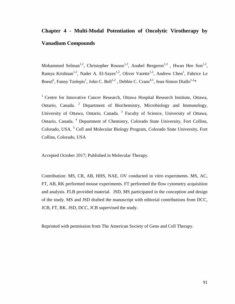
91
Chapter 4 - Multi-Modal Potentiation of Oncolytic Virotherapy by
Vanadium Compounds
Mohammed Selman1,2
, Christopher Rousso1,2
, Anabel Bergeron1,3
, Hwan Hee Son1,2
,
Ramya Krishnan1,2
, Nader A. El-Sayes1,2
, Oliver Varette1,2
, Andrew Chen1, Fabrice Le
Boeuf1, Fanny Tzelepis
1, John C. Bell
1,2 , Debbie C. Crans
4,5, Jean-Simon Diallo
1,2*
1 Centre for Innovative Cancer Research, Ottawa Hospital Research Institute, Ottawa,
Ontario, Canada. 2 Department of Biochemistry, Microbiology and Immunology,
University of Ottawa, Ontario, Canada. 3 Faculty of Science, University of Ottawa,
Ontario, Canada. 4 Department of Chemistry, Colorado State University, Fort Collins,
Colorado, USA. 5 Cell and Molecular Biology Program, Colorado State University, Fort
Collins, Colorado, USA
Accepted October 2017; Published in Molecular Therapy.
Contribution: MS, CR, AB, HHS, NAE, OV conducted in vitro experiments. MS, AC,
FT, AB, RK performed mouse experiments. FT performed the flow cytometry acquisition
and analysis. FLB provided material. JSD, MS participated in the conception and design
of the study. MS and JSD drafted the manuscript with editorial contributions from DCC,
JCB, FT, RK. JSD, DCC, JCB supervised the study.
Reprinted with permission from The American Society of Gene and Cell Therapy.

92
Abstract
Oncolytic viruses (OV) are an emerging class of anticancer bio-therapeutics that induce
antitumor immunity through selective replication in tumor cells. However, the efficacy
of OVs as single agents remains limited. We introduce a strategy that boosts the
therapeutic efficacy of OVs by combining their activity with immuno-modulating, small
molecule protein tyrosine phosphatase inhibitors. We report that vanadium-based
phosphatase inhibitors enhance OV infection in vitro and ex vivo, in resistant tumor cell
lines. Furthermore, vanadium compounds increase anti-tumor efficacy in combination
with OV in several syngeneic tumor models, leading to systemic and durable responses,
even in models otherwise refractory to OV and drug alone. Mechanistically, this involves
subverting the antiviral type I IFN response towards a death-inducing and
proinflammatory type II IFN response, leading to improved OV spread, increased
bystander killing of cancer cells, and enhanced anti-tumor immune-stimulation. Overall,
we showcase a new ability of vanadium compounds to simultaneously maximize viral
oncolysis and systemic anticancer immunity, offering new avenues for the development
of improved immunotherapy strategies.

93
Introduction
Oncolytic viruses (OV) are an emerging class of anticancer bio-therapeutics that
selectively replicate in and lyse tumor cells, without causing damage to normal cells(8,
74). Multiple OVs have shown efficacy in preclinical models of cancer and in clinical
trials(8). Notably, the US Food and Drug Administration (FDA) and European Medicines
Agency (EMA) have recently approved T-VEC (Imlygic) for the treatment of melanoma.
While OVs can lead to profound anticancer responses as single agents, clinical data show
that some patients do not respond to OVs alone and may benefit from combination
therapies(29, 88, 257).
Poor infection of tumors is an important factor in the resistance to OV therapy.
OV spread, oncolysis, and overall therapeutic efficacy can be improved in resistant
tumors among others by using pharmacological compounds that block the cellular innate
antiviral immune response mediated by type I interferon (IFN)(114, 115, 118, 119).
Beyond direct effects on tumor cells, OVs can remodel the tumor microenvironment and
boost anti-tumor immunity by directing immune responses to the tumor niche(11, 211,
258, 259). This immunostimulatory effect can be enhanced by integrating immune
stimulatory genes into the viral genome(221, 260) or by combination with other forms of
immunotherapy such as immune checkpoint inhibitors(30, 194, 195, 261).
Vanadate and other vanadium-based compounds are pan-inhibitors of protein
tyrosine phosphatases (PTPs), with a wide range of biological effects(154, 262).
Clinically, these compounds have been mostly explored for their antidiabetic potential,
demonstrating safety for this indication in phase I/II human trials(263, 264). In more

94
recent years, a number of vanadium compounds were also found to exhibit anticancer
effects in animal models(162, 164, 166, 265–269). Numerous studies suggest that
vanadium compounds impact the immune system(159, 197, 270, 271), for example by
stimulating and activating T-cells(270). However, the mechanism by which vanadium-
based compounds can modulate anticancer immunity has not been investigated.
Given their potential immunostimulatory effects, we assessed the use of selected
vanadium compounds as complementary pharmacological agents to OV–mediated
immunotherapy. Here we demonstrate an unprecedented ability of these compounds to
subvert the type I IFN antiviral response towards a death-inducing and proinflammatory
type II IFN response, culminating in the dramatically improved anticancer activity of
OVs both in vitro and in vivo.

95
Results
Vanadate enhances the spread of oncolytic RNA viruses
Contrasting results have been reported in the few studies that have probed the
effect of vanadate (Figure 4.1a) on viral infection(272, 273). To explore this in the
context of OVs, we tested the impact of orthovanadate on the growth of a small panel of
candidate oncolytic viruses. We included negative single strand RNA viruses Vesicular
Stomatitis Virus (VSVΔ51)(48) and Measles (Schwarz strain)(274), a positive single
strand RNA virus (Sindbis), as well as double-stranded DNA viruses herpes simplex
virus-1 (HSV-1 mutant N212)(275) and vaccinia virus (deleted for thymidine kinase and
expressing GM-CSF)(276) (Figure 4.1b). We found that the growth of RNA viruses
including Measles, Sindbis, and oncolytic VSVΔ51, but not DNA viruses HSV-1 and
vaccinia was enhanced by treatment with vanadate. VSVΔ51 growth was most robustly
enhanced and further testing in VSV-resistant 786-0 renal carcinoma cells revealed that
vanadate increased viral output, as measured by plaque assay, up to ~400-fold over a 10-
1000µM dose range (Figure 4.1c), which was below the median lethal concentration of
vanadate (Figure 4.3a). Use of a GFP-expressing VSVΔ51 revealed a corresponding
increase in the number of GFP positive, infected cells (Figure 4.1d). A similar impact on
viral infection was observed in various human and murine cancer cell lines
(Supplemental Figure 1a,b). Single and multi-step growth curves in 786-0 cells revealed
that vanadate more robustly enhanced the yield of VSVΔ51 at a low multiplicity of
infection (MOI) compared to a high MOI, indicating that vanadate promotes viral spread
(compare Figure 4.1e vs. f). Similarly, in the presence of vanadate, VSVΔ51 produced
larger plaques, based on both the area of GFP-positive cells as well as the cytopathic

96
effect observed upon coomassie blue staining (Supplemental Figure 2). The impact of
vanadate on viral growth was more pronounced with increasing pre-treatment time
(maximum tested 24h), with the effect of vanadate being negligible when provided later
than 12h post infection (Supplemental Figure 1c). Similarly, treatment with vanadate
after infection reduced its negative effect on vaccinia infection but did not lead to
enhancement (Supplemental Figure 1d). Correlating with viral titers and GFP expression
data, Figure 4.1g shows that vanadate increased the production of VSVΔ51 genomes in
infected 786-0 cells as well as in mouse CT26WT colon cancer cells. In contrast, viral
growth was not enhanced in normal adult human GM38 fibroblasts (Figure 4.1h). Using
mouse tissue cores obtained from normal lung, muscle, spleen and tumor tissues obtained
from mice implanted with CT26WT and DBT (glioma) cells, we further evaluated the
impact of vanadate on growth of VSVΔ51-GFP. Fluorescence microscopy images in
Figure 4.1i show that vanadate preferentially increased the growth of the virus in tumor
cores with no impact on normal tissues.

97
98
Figure 4.1. Vanadate enhances VSV∆51 infection in cancer cells but not normal
cells.
(a) Structure of vanadate ion present at pH 7.4. (b-f,h) Resistant human renal cancer cell
lines 786-0, were pretreated with vanadate for 4 hours and subsequently infected with (b-
e,h) VSV∆51 (MOI: 0.01, [150µM]), (b) Measles (MOI: 0.01, [100µM]) , (b)Sindbis
(MOI:10, [150µM]), (b) HSV (MOI: 0.01, [150µM]), (b) Vaccinia (MOI: 0.01,
[200µM]). (b,c) Corresponding viral titers were determined 24 (VSV∆51) or 48
(Measles, Sindbis, HSV, Vaccinia) hours post infection (hpi) from supernatants (b,g,h;
N=3;Error bars indicate SEM; t-test; NS = no statistical significance, ** p<0.001, as
compared to the untreated condition counterparts) (c; N=3; Significance enhancement at
100 - 200µm; p<0.0001 by 1-way ANOVA; as compared to 0µm condition) (d) 24 hpi
fluorescent and phase contrast images were taken of the 786-0 cells treated with mock or
200µM of vanadate . (e) Multi-step, and (f) single-step growth curve of 786-0 pretreated
with vanadate and infected with VSV∆51 (e) MOI: 0.01 or (f) MOI: 3, supernatants were
tittered by plaque assay (N=3; Significant enhancement at 50-200µm at indicated time;
NS, no statistical significance, ** p<0.001, *** p<0.0001 by 2-way ANOVA; as
compared to Mock condition). (g) 24 hpi, RNA was collected from 786-0 and CT26WT,
and expression of VSV-M gene was quantified by qPCR. (h) Normal cell line GM38
were pretreated as in (b) infected with VSV∆51. Corresponding viral titers were
determined 24 hours post infection from supernatants. (i) CT26WT and DBT tumors
cores and BALB/c mouse spleen, muscle, lung, and brain tissue cores were pretreated
with 300µM of vanadate for 4 hours and subsequently infected with 1x10⁴
PFU of
VSV∆51 expressing GFP. 24 hpi fluorescent images were acquired of the tumor and
normal tissue cores. Representative images from each triplicate set are shown.

99
Vanadium compounds have unique viral enhancing properties
Vanadate has been shown to inhibit tyrosine phosphatases, an activity which is
linked to its close structural and electronic analogy to phosphate(262, 277). We therefore
tested whether phosphate could induce similar effects but found various phosphate salts
had no impact on viral growth in contrast to vanadate (Supplemental Figure 3a).
Vanadate is a simple oxometalate. We wanted to investigate whether other oxidized
transition metals could potentially enhance viral growth. We found that neither
aluminum, molybdenum, manganese and tungsten oxides could enhance VSVΔ51 growth
(Figure 4.2a). Chromium trioxide modestly enhanced viral growth but only at a single
dose (10 µM, Figure 4.2a) and was highly toxic in comparison to vanadate (not shown).
The protonation or form of vanadate at different pH had no impact on its activity
(Supplemental Figure 3b). Because this suggested the virus-enhancing effect was linked
to the vanadium itself, we tested other oxidized vanadium complexes, including
metavanadate, vanadium(V) oxytriethoxyde (VOx), vanadium(IV) oxide sulphate (VS)
and bismaltolato oxovanadium(IV) (BMOV) (Figure 4.2b). All of these compounds
effectively enhanced VSVΔ51 growth (Figure 4.2c,d, Fig S3c). Similar results were
obtained using vanadium(IV) tetra-fluoride and vanadium(III) tri-bromide (Figure 4.2e).
Interestingly, vanadate’s pro-viral activity was abrogated upon inclusion of L-Ascorbic
Acid or tiron, both potent metal chelating agents (Figure 4.2f).

100

101
Figure 4.2 Viral enhancement is dependent on Vanadium.
786-0 cells were pretreated for 4 hours with indicated concentrations of (a) various
oxidized transitional metals, (c-e) vanadates from various sources prepared in a pH 7.4
buffer. In a, c-e, cells were subsequently infected with oncolytic VSV∆51 expressing
GFP at an MOI of 0.01. (a,d,e) Corresponding viral titer were determined 24 hours post
infection from supernatants (N=3). (c) Corresponding GFP positive cell counts 24 hours
post infection. (b) Structure of vanadium oxometalates are illustrated. (f) 786-0 were
pretreated with 200µM of vanadate or mock and treated with chelating agent ascorbic
acid (L-AA) or Tiron, then infected with VSV∆51 (MOI : 0.01). Corresponding viral
titers were determined 24 hours post infection from supernatants (N=3). (Error bars
indicate SEM. NS, no statistical significance; *p < 0.05, **p < 0.01, ***p < 0.001 by 1
way ANOVA analysis; as compared to 0µm or Mock condition)
102
Vanadate enhances virus and cytokine-induced death
Overall, our data suggested that vanadium compounds enhance the spread of a
subset of RNA-based OVs in cancer cells. Next we examined whether this extended to
impact oncolytic activity. Figure 3.3a shows indeed that while vanadate had cytotoxic
effects on its own at high doses, cancer cell death was vastly enhanced upon co-infection
with a low MOI of VSVΔ51, which is otherwise innocuous to 786-0 cells. A similar
effect was observed in various human cancer cell lines, and with various vanadium
compounds (Supplemental Figure 4). The mode of cell death was found to exhibit
characteristics of apoptosis as determined by flow cytometry following staining with
Annexin V and 7-AAD (Figure 4.3b). Furthermore, we found that vanadate increased
transcription of proapoptotic factors PUMA and Noxa that are normally induced by p53
over the course of viral infection (278, 279) (Figure 4.3c). Interestingly, the impact of
vanadate on cell death was independent of its promotion of viral spread since increased
786-0 cell death was also observed using UV-inactivated VSV, as well as a G-less (ΔG)
version of VSVΔ51 devoid of its capacity to produce glycoprotein (Figure 4.3d). Indeed,
G-less VSVΔ51 can infect cells and replicate its genome but does not bud or spread
further. Altogether, this suggested the potential involvement of virus-induced bystander
killing and so we tested whether antiviral IFN normally secreted following infection of
786-0 cells by VSVΔ51 could produce similar effects in combination with vanadate.
Indeed, Figures 3e-f show that Type I IFN (α and β), could lead to cytotoxicity in 786-0
cells in the presence of vanadate. Similar effects were obtained when challenging cells
with poly I:C, a toll like receptor 3 (TLR3) agonist.

103

104
Figure 4.3. Vanadate facilitates virus-induced type I interferon and ROS mediated
cell death.
(a) 786-0 cells were pretreated with a range of concentrations of vanadate for 4 hours and
were subsequently infected with VSV∆51 expressing GFP at an MOI of 0.01. 786-0 cell
viability was assayed in cells 24 hours post infection. Results were normalized to the
average of the values obtained for the corresponding uninfected, untreated cells (N=4;
Error bars indicate SEM; Significance decrease in viability at 16 - 1000µm; ***p<0.0001
by 2 way ANOVA; as compared to uninfected condition). (b) 786-0 were pretreated with
vanadate (100µM) for 4 hours and subsequently infected with oncolytic VSV∆51
expressing GFP at an MOI of 0.01. 24 hours post induction of cell death was determined
by annexin V and 7-aminoactinomycin D (7-AAD) staining. Numbers indicate the
percentage in each quadrant. (c) Cell lysates of 786-0 treated with vanadate(100µM) and
VSV∆51 expressing GFP were collected at indicated time points, RNA was collected,
and expression of PUMA and Noxa genes was quantified by qPCR (N=3; Error bars
indicate SEM; **p < 0.01, ***p<0.0001 by 2 way ANOVA; comparing VSV∆51
condition to vanadate+VSV∆51 condition). (d-e) 786-0 cells were pretreated with
vanadate for 4 hours and subsequently infected with VSV∆51, UV-inactivated VSV∆51,
VSV∆51∆G, or treated with IFNa, IFNb or PolyI:C. Cell viability was assayed 48 hours
post infection or treatment. Corresponding cell morphology is presented in panel (f). (g-i)
786-0 cells were co-treated with vanadate and N-acetyl-L-cysteine (NAC) for 4 hours and
infected with VSV∆51 expressing GFP at an MOI of 0.01 (g,i) or with IFNa, IFNb or
PolyI:C (h) and cell viability was assayed 48 hours post infection or treatment. In (i),
viral titers were determined 24 hours post infection from supernatants (N=3; Error bars
indicate SEM; ***p<0.001 by 2way ANOVA; as compared to mock treated condition).
(j-k) MRC5 (j) and GM38 (k) normal cells were pre-treated with vanadate for 4 hours
and subsequently infected with VSV∆51 or treated with IFNa, IFNb or PolyI:C. For (d, e,
g, h, j, k) cell viability results were normalized to the average of the values obtained for
the corresponding uninfected, untreated cells (N=4; Error bars indicate SEM; NS, no
statistical significance, ***p<0.0001 by 2-way ANOVA).
105
The possibility that the effects of vanadate on virus-induced death could be
reactive oxygen species (ROS) -mediated was investigated next. To evaluate this, we
infected cells treated with vanadate and increasing concentrations of N-Acetyl Cysteine
(NAC), which increases cellular glutathione and reduces the levels of cellular ROS.
Increasing NAC antagonized vanadate’s ability to enhance virus-induced death (Figure
4.3g). Similar results were observed when cells were treated with vanadate and with Type
I IFN (α and β) or poly I:C (Figure 4.3h). However, the highest dose of NAC did not
abrogate vanadate’s ability to increase viral spread (Figure 4.3i), again suggesting that the
cytotoxic and virus-enhancing activities of vanadate are distinct. In addition, the observed
enhancement of cell death mediated by Type I IFN, poly I:C, and VSVΔ51 by vanadate
was not observed in normal adult GM38 or MRC5 human embryonic lung fibroblasts
(Figure 4.3j,k).
Vanadate enhances the oncolytic activity of VSVΔ51 and anti-tumor immunity in vivo
Given the observation that vanadate enhanced both spread and oncolytic activity
of VSVΔ51 in vitro, we wondered if the combination of VSVΔ51 and vanadate could
have anti-cancer effects in mouse models of cancer. Reports in the literature suggest that
vanadate has immunomodulatory properties(159), hence we performed our experiments
in a panel of immunocompetent syngeneic mouse tumor models refractory to VSVΔ51
infection. In mice with established CT26WT, 4T1 (breast cancer) and DBT tumors, one
single intratumoral injection of vanadate and VSVΔ51 expressing luciferase robustly
enhanced virus-associated luciferase gene expression compared to virus alone, as

106
assessed by an in vivo imaging system (IVIS, Figure 4.4a,b). Likewise, vanadyl sulfate
(commonly used as a bodybuilding supplement) as well as BMOV evaluated pre-
clinically for treatment of diabetes(264, 280), both robustly increased VSVΔ51 viral
growth in the DBT model over the course of 3 days post infection (Figure 4.4d,e).
Vanadate and VSVΔ51 combination treatment led to significantly improved survival of
DBT, CT26WT, and 4T1 tumor-bearing mice compared to the monotherapies (Figure
4.4c). Approximately 80% of DBT tumor-bearing mice and 20% of CT26WT tumor-
bearing mice presented complete remission after combination treatment. (Figure 4.4c).

107
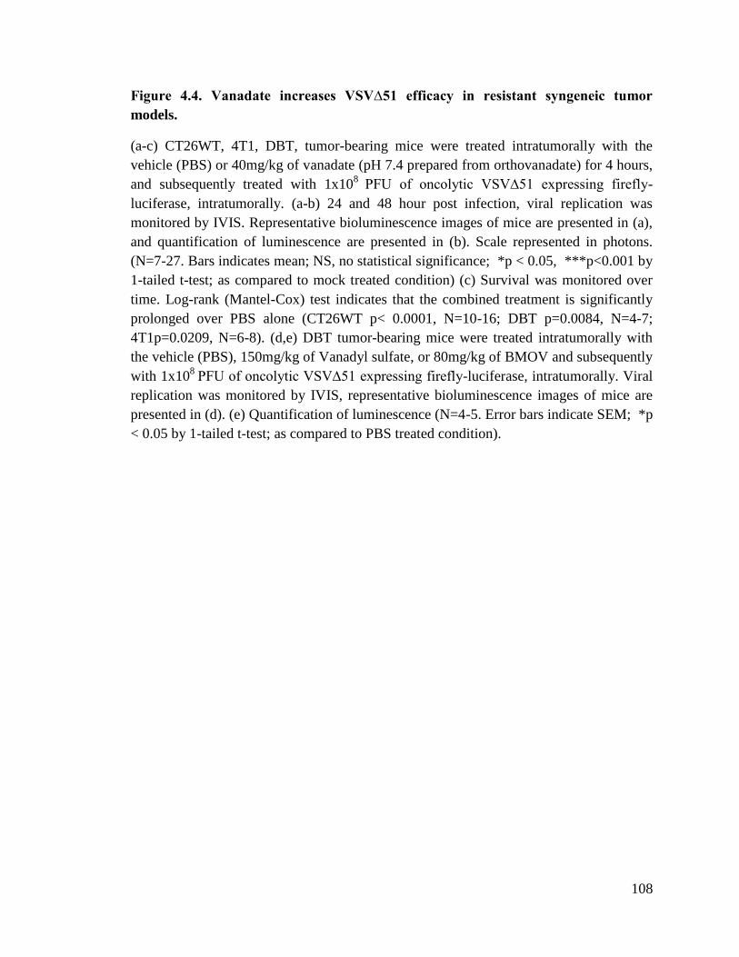
108
Figure 4.4. Vanadate increases VSV∆51 efficacy in resistant syngeneic tumor
models.
(a-c) CT26WT, 4T1, DBT, tumor-bearing mice were treated intratumorally with the
vehicle (PBS) or 40mg/kg of vanadate (pH 7.4 prepared from orthovanadate) for 4 hours,
and subsequently treated with 1x10⁸
PFU of oncolytic VSV∆51 expressing firefly-
luciferase, intratumorally. (a-b) 24 and 48 hour post infection, viral replication was
monitored by IVIS. Representative bioluminescence images of mice are presented in (a),
and quantification of luminescence are presented in (b). Scale represented in photons.
(N=7-27. Bars indicates mean; NS, no statistical significance; *p < 0.05, ***p<0.001 by
1-tailed t-test; as compared to mock treated condition) (c) Survival was monitored over
time. Log-rank (Mantel-Cox) test indicates that the combined treatment is significantly
prolonged over PBS alone (CT26WT p< 0.0001, N=10-16; DBT p=0.0084, N=4-7;
4T1p=0.0209, N=6-8). (d,e) DBT tumor-bearing mice were treated intratumorally with
the vehicle (PBS), 150mg/kg of Vanadyl sulfate, or 80mg/kg of BMOV and subsequently
with 1x10⁸
PFU of oncolytic VSV∆51 expressing firefly-luciferase, intratumorally. Viral
replication was monitored by IVIS, representative bioluminescence images of mice are
presented in (d). (e) Quantification of luminescence (N=4-5. Error bars indicate SEM; *p
< 0.05 by 1-tailed t-test; as compared to PBS treated condition).

109
Immune profiling of CT26WT tumors indicated an enhanced leukocyte infiltration with
significantly increased T cells (Figure 4.5a), including IFNγ producing CD8+ T cells
(Figure 4.5a), in mice treated with the combination of vanadate and VSVΔ51 compared
to the monotherapies. This suggested that induction and/or recruitment of T cells to the
tumors is improved in the presence of vanadate combined with VSVΔ51, which could
contribute to tumor control. Indeed, we observed a correlation between the amount of T
cell infiltration and tumor regression (Figure 4.5b) in mice from the combined therapy
group with the higher responders (HR) presenting increased infiltration compared to
lower responders (LR), even though the enhancement of virus-associated luciferase gene
expression was similar between them (Figure 4.5c). This suggests that the amount of
tumor infection is not the key determinant for maximum T cell infiltration and indicates
an additional need to create a milieu that promotes T cell infiltration following infection.
Furthermore, mice that were able to completely eliminate CT26WT tumors (Fig 4.4c)
subsequently became immune to rechallenge with the same cancer cells (Fig 4.5d),
indicating that combination therapy leads to long term anti-tumor immunity.

110

111
Figure 4.5. Vanadate /VSV∆51 co-treatment triggers T-cell infiltration and anti-
tumor immunity.
(a-c) CT26WT tumor-bearing mice were treated intratumorally with the vehicle (PBS) or
40mg/kg of vanadate (pH 7.4 prepared from orthovanadate) for 4 hours, and subsequently
treated with 1x10⁸
PFU of oncolytic VSV∆51 expressing firefly-luciferase,
intratumorally. The Vanadate + VSV∆51 group was divided in 2 groups, High and Low
responders (HR and LR), based on median tumor size 10 days post treatment, as shown
in (b). Viral replication was monitored 24 hour post infection; quantification of
luminescence is presented in (c) (N=5). Tumor volume 10 days post treatment are shown
in (b) (N=5). (a) Percentage of CD45+ cells; CD3+ cells of total CD45+ cells; IFNγ
expressing CD8+ cells in each tumor was quantified by flow cytometry, 10 days post
treatment. (N=4-5; Error bars indicate SEM; * p<0.05, ** p<0.001, ***p<0.0001, by 1-
way ANOVA). (d) Survival was monitored after re-implantation of CT26WT in cured
and naïve mice from Figure 3.4c (N=3-5). (e) Immunocompetent mice and (f) nude mice
bearing the CT26LacZ tumor were treated intratumorally with the vehicle (PBS) or
40mg/kg of vanadate for 4 hours, and subsequently treated with 1x10⁸
PFU of oncolytic
VSV∆51 expressing firefly-luciferase, intratumorally. Log-rank (Mantel-Cox) test
indicates that survival in the combined treatment is significantly prolonged over VSV∆51
alone in the immunocompetent mouse model alone (Immunocompetent mice p=0.0506,
N=6-8; Nude mice no statistical significance, N=4-10). (g) Schematic representation of
treatment schedule for bilateral DBT tumors. (h) Representative bioluminescence images
of mice are presented. (i) Growth of treated (right flank) and distant (left flank) DBT
tumors (N=4-7).

112
Next, we investigated the effect of vanadate in the CT26LacZ tumor model,
which we have previously shown to be significantly more susceptible to infection with
VSVΔ51(281). Here vanadate somewhat decreased virus-associated luminescence
(Supplemental Figure 5a), in line with the observation that vanadate did not enhance viral
growth in cultured CT26LacZ cells (Supplemental Figure 1a,b). However, the
combination of vanadate and VSVΔ51 led to a significant improvement of survival over
the monotherapies in this model, reaching nearly 90% complete remissions (Figure 4.5e).
While enhanced bystander killing as observed in Fig 3 in part could explain this
phenomenon, we wondered whether enhanced adaptive immune responses could play a
role in generating such a high cure rate with a single intratumoral dose of vanadate and
VSVΔ51. We therefore performed these experiments in athymic nude mice that are
devoid of T-cells. Remarkably, while VSVΔ51 alone still delayed tumor progression and
led to cures, the combination effect was completely abrogated in this context (Figure
4.5f), albeit virus-associated luminescence was not generally affected (Supplemental
Figure 5b). Likewise, the combination therapy did not lead to enhanced efficacy in the
HT29 tumors implanted in athymic nude (Supplemental Figure 6c) even though
enhancement of VSVΔ51 viral growth was observed (Supplemental Figure 6a,b). These
results strongly support an important role of T cell-mediated protection observed in the
above-described models and the role of vanadate in eliciting an improved T cell response
when combined with VSVΔ51. However, the improved efficacy of vanadate was not
observed when the CT26LacZ tumors were infected with non-spreading VSVΔ51
(VSVΔ51ΔG) (Supplemental Figure 5c), indicating that viral growth is essential for the
combination therapy effect.

113
To further evaluate the role of the anti-tumor immune response induced by
vanadate / virus treatment in tumor control, we implanted immunocompetent mice with
bilateral DBT tumors and injected only the right tumors with the combination of
VSVΔ51 and vanadate (or monotherapies / PBS) (Figure 4.5g). Interestingly, we found
that while virus-associated luminescence was uniquely enhanced by vanadate in the
injected tumors on the right side, the tumors on the left also shrunk following the
combination therapy (Fig 4.5h,i). Although some tumors did regress following treatment
with vanadate, all of the uninjected (left) tumors did not regress. VSVΔ51 monotherapy
did not induce tumor regression in the injected (right) or untreated (left) tumors. Taken
together, these results reveal that vanadium compounds have the ability to elicit a robust,
systemic protective anti-tumor immune response when combined with VSVΔ51.
Vanadate inhibits the type I Interferon response and potentiates a proinflammatory
response via type II interferon.
Our in vitro results indicated that both OV spread and bystander killing in cancer
cells can be enhanced by vanadate. While this likely contributes towards improving OV
efficacy in vivo, our data suggested that a critical component of the therapeutic efficacy
associated to the combination regimen in immunocompetent models involves the
generation of anti-tumor T-cell responses. To further understand the cell-autonomous
molecular mechanisms involved in stimulating this response while being able to enhance
infection, a microarray analysis was performed in vitro. We first looked at the gene
expression profiles of 786-0 cells 24 h following VSVΔ51 or mock infection in the

114
presence and absence of vanadate (prepared from solutions of orthovanadate or
metavanadate). In line with our demonstration that vanadate stimulates an anti-tumor
immune response, gene set-enrichment analyses using GOrilla revealed that vanadate
alone induced inflammatory responses, and immune system processes; which were
further potentiated in combination with VSVΔ51 (Supplemental Figure 7a). Uniquely,
the infection of vanadate treated cells led to the increased expression of a number of
proinflammatory cytokines (CCL8, CCL3, IL6, TNF, IFNβ, CCL5) and many genes
typically induced by type II IFN (IFNγ), including chemokines such as CXCL9,
CXCL10, and CXCL11 (Figure 4.6b,c and Supplemental Figure 7b,c and Table S1).
Among others, CXCL9 plays a key role in leukocyte trafficking and its mRNA was up-
regulated by more than 100,000 fold during viral infection of vanadate-treated cells
compared to mock (Figure 4.6a). We further validated the vanadate-mediated increase in
mRNA expression of IFNγ induced chemokines such as CXCL9 in mouse CT26WT cells
which were used for our in vivo models, as well as its secretion by ELISA in various
human cancer cell lines (Supplemental Figure 7c,d). While many genes typically induced
by IFNγ were up-regulated upon infection of vanadate-treated cells, IFNγ itself was not
up-regulated at any time-point post infection under any condition tested in 786-0 (Figure
4.6a,b). On the other hand, IFNβ mRNA was up-regulated by more than 10-fold 24h
following infection (Figure 4.6a) in vanadate treated cells compared to mock.
Surprisingly, genes typically induced by Type I IFN were either unaffected or decreased
in these conditions (Figure 4.6b,c). Indeed, genes induced by Type I IFN, such as MX2
and IFITM1 with known antiviral function against rhabdoviruses, were robustly
downregulated as early as 8h post-infection (Figure 4.6a). Furthermore, protein

115
expression levels of IFITM1 was potently repressed by vanadate in infected cells 16 and
24h following infection (Supplemental Figure 8).

116

117
Figure 4.6. Vanadate re-wires antiviral type I IFN signaling towards pro-
inflammatory type II IFN response.
(a-c) Cell lysates of 786-0 treated with vanadate and VSV∆51 expressing GFP were
collected at indicated time points. RNA was extracted and probed for expression of (a)
ifnβ, ifnγ, mx2, ifitm1 and cxcl9 genes by qPCR (N=3; Error bars indicate SEM; **p <
0.01, ***p<0.0001 by 2 way ANOVA; comparing the VSV∆51 condition to the
Vanadate+VSV∆51 condition). (b) RNA was used for gene expression microarray
analysis. Data is normalized to untreated control, indicating log2 fold change in gene
expression of genes documented to be induced by Type I or Type II IFN. (Bars indicate
mean; 1-way ANOVA; NS, no statistical significance, *** p<0.0001, comparing
combination treatment to virus alone condition), (c) Heatmap showing the expression
levels of the differentially expressed IFN stimulated genes. Expression of genes was
normalized to values obtained for mock-treated, infected control. Color scale indicates
log2 fold change. (d) 786-0 cells were pretreated with vanadate (1000µM) for 4 hours
and challenged with IFNβ (100U/ml). One hour later, fractionated cell lysates were
probed for pSTAT1, STAT1, pSTAT2, STAT2 and actin. (e) Immunofluorescence for
pSTAT1 and pSTAT2 was performed in 786-0 cells treated with vanadate (1000µM) for
4 hours and with human IFNβ (1000U) for 1 hour. Quantification of the average nuclear
fluorescence associated to pSTAT1 or pSTAT2 in each condition (N=30; Bars indicate
mean; 1-way ANOVA, * p<0.05, ** p<0.001, *** p<0.0001, as compared to the mock
condition counterpart). Microscopy images can be found in Figures S9-11.
118
Importantly, IFNβ and IFNγ bind to distinct receptors and lead to differential
activation of STAT1 and STAT2 (Signal Transducer and Activator of Transcription)
transcription factors. Phosphorylation of STATs leads to their dimerization and nuclear
translocation to activate transcription of IFN-stimulated genes (ISGs). Some of these
genes are regulated by both type I and type II IFNs, whereas others are selectively
regulated by one or the other. Type I IFNs induce the phosphorylation of both STAT1
and STAT2, leading to the formation of the ISGF3 complex composed of a STAT1-
STAT2 heterodimer and IRF9 that binds specific promoter regions known as IFN-
stimulated response elements (ISREs); while type II IFN primarily induce the
phosphorylation of STAT1, leading to the formation of STAT1-STAT1 homodimers that
bind IFNγ-activated-sequence (GAS) elements(56). Consistent with a shift from a type I
towards a type II IFN response, vanadate treatment inhibited the IFNβ-induced
phosphorylation of STAT2 and reduced its nuclear accumulation but did not similarly
affect STAT1 as observed by Western blot (Figure 4.6d). Supporting this idea,
immunofluorescence also revealed that whereas activated STAT1 translocated to the
nucleus following infection of vanadate-treated cells (Figure 4.6e, Supplemental Figure
9), STAT2 remained mostly in the cytoplasm (Figure 4.6e, Supplemental Figure 10,11).
Remarkably, this suggests that vanadate enhances OV activity through a previously
unappreciated mechanism that converts a predominantly antiviral type I IFN response
into a type II IFN response, through the preferential repression of STAT2 activation. This
signal “rewiring” leads to up-regulation of proinflammatory cytokines and chemokines
that favour the generation of a T cell dependent anti-tumor response.

119
Discussion
OV therapy as a standalone treatment can be highly effective but treatment
resistance remains a frequent occurrence. This is attributed to a number of factors,
including a need to robustly infect tumors and ensure the initiation of a robust anti-tumor
immune response(8). The latter has prompted many investigators to evaluate OVs in
combination with immune checkpoint inhibitors; a strategy has been shown to be
effective by several groups in both preclinical models and clinical trials(30, 193–196).
However this specific combination strategy is not uniformly effective(282).
We show here that vanadium compounds can provide a significant therapeutic
benefit in CT26WT and other various aggressive, treatment-refractory, murine tumor
models when combined with OVs, leading to enhanced anti-tumor T-cell responses
mediated by the induction of a Type II Interferon-like response in infected cancer cells.
The availability of clinically-advanced vanadium candidates such as vanadate, vanadyl
sulfate and bis(ethylmaltolato)oxovanadium(IV) (BEOV)(263, 264, 283) that have been
used in the context of diabetes will greatly facilitate the testing of such combination
regimens in human cancer patients. In addition, oncolytic rhabdoviruses derived from
VSV and closely related Maraba are currently undergoing clinical evaluation, including
in combination with immune checkpoint blockade (NCT02923466, NCT02879760).
Altogether, this lays the groundwork for rapid evaluation of our novel approach in
humans. However, while many OVs including T-Vec are currently delivered
intratumorally in the clinic(284), it will be relevant to further explore alternative
regimens and formulations to abrogate the need for intratumoral injection of vanadium
120
compounds ahead of virus. Oral administration of vanadium compounds is possible and
has been extensively tested in the treatment of type II diabetes(285).
In the present work, we tested a range of different vanadium salts and compounds.
These compounds are known to undergo different hydrolytic conversions in
solution(154). Orthovanadate, metavanadate and vanadium(V) oxytriethoxide all result in
a solution of H2VO4- at physiological pH, while the solutions prepared from vanadyl
sulfate and vanadium(IV) fluoride will result in a solution of aqueous V(IV) and
V(V)(154), and those prepared from bis(maltolato)oxovanadium(IV) will contain both
V(IV) and V(V) maltolato complexes(280). Importantly, all of these compounds show a
robust capacity to enhance OV activity (Figure 4.2c-e). This suggests significant
flexibility in the design of new vanadium-based compounds that may be tailored for use
in combination with OVs. Furthermore, these results are consistent with various forms of
vanadate binding tightly to several phosphatases with similar Ki values(262).
Protein tyrosine phosphatases are important regulator of cellular processes (286,
287) and may have complex functions during the course of viral infection. Here we found
that the pan-tyrosine phosphatase inhibitor vanadate, while enhancing all of the RNA
viruses in our panel (Rhabdovirus, Alphavirus, Paramyxoviruses), substantially inhibits
infection of DNA viruses like vaccinia and HSV. These observations showcase the
selective effects of vanadium compounds on RNA viruses, which do not encode viral
phosphatases, in contrast with both DNA viruses tested where they serve as important
virulence factors(288, 289). Notably, the RNA viruses tested generally lack the ability to
counteract Type I IFN signaling. When using wild-type VSV, which more effectively
overcomes the type I IFN response compared to VSVΔ51, we found that the positive

121
impact of vanadate was comparatively reduced in vitro (Supplemental Figure 12).
Numerous drugs and compounds have been shown to increase the efficacy of OVs by
inhibition of Type I IFN production or signalling(108, 114, 118, 188). The ability of
vanadium compounds to convert the antiviral Type I IFN to a pro-inflammatory Type II
IFN response, leading not only to better OV spread but also to greater anti-tumor immune
stimulation during OV treatment is to our knowledge unprecedented.
Previously vanadate has been reported to promote death induced by addition of
type I IFN(290), supporting our observations (Figure 4.3). Interestingly, we found that
antioxidants could abrogate the priming of apoptosis by vanadate (Figure 4.3g,h),
suggesting a potential role of vanadate-induced ROS(291) in promoting death initiated by
Type I IFN. A role for ROS in modulating IFN activity has also been previously
suggested(292), however, this mechanism is unlikely to be at play here given antioxidant
treatment did not abrogate enhancement of viral growth (Figure 4.3i). Instead, we found
that STAT1 and STAT2 are differently activated by Type I IFN in vanadate-treated cells,
ultimately favouring the accumulation of STAT1 in the nucleus (Figure 4.6d-e,
Supplemental Figure 9-11). This mechanism is in line with activation of Type II IFN
target genes that rely on STAT1-STAT1 homodimers for their transcription, even in
absence of IFN-γ. This could also explain the enhanced capacity for Type I IFN to induce
cell death (Figure 4.3e-g), given STAT1 has been shown to promote cell death by
interacting with TRADD, HDACs, or p53 to increase the expression of target genes such
as Noxa and Puma as observed in this study (Figure 4.3c)(293, 294). Notably, type I IFN,
such as IFNα (INTRON® A) is widely used for the treatment of renal cancer, although
therapeutic results remain limited(295). Further studies investigating the impact of

122
vanadium compounds on this and other anti-cancer strategies that aim to kill cancer cells
through type I IFN or through recruitment of T-cells to the tumor site may be warranted.

123
Materials and Methods
Drugs, chemicals and cytokines. Drugs, chemicals and cytokines and their respective
supplier and solvent used in this study are listed in Table S2. The aqueous chemistry of
some of these vanadium compounds lead to conversion of the original compound into
vanadate(296) under the condition of the studies, and this is also indicated in Table S2.
Cell lines. Cells and their respective supplier and growth media used in this study are
listed in Table S3. Cells were cultured in HyQ high-glucose Dulbecco’s modified Eagle’s
medium (GE Healthcare Life Sciences Hyclone, Logan, UT) supplemented with 10 %
fetal calf serum (CanSera, Etobicoke, Canada), HEPES, penicillin/streptomycin (Gibco
Life Technologies, Waltham, MA). All cell lines were incubated at 37 °C in a 5% CO2
humidified incubator. All cells were tested to ensure they are free of mycoplasma
contamination.
Viruses and quantification.
Rhabodviruses.The Indiana serotype of VSV (VSV∆51 or wild type) was used
throughout this study and was propagated in Vero cells. VSV∆51-expressing GFP or
firefly luciferase are recombinant derivatives of VSV∆51 described previously(48). All
viruses were propagated on Vero cells and purified on 5–50% Optiprep (Sigma-Aldrich,
St. Louis, MO) gradient and all virus titres were quantified by the standard plaque assay
on Vero cells as previously described(252).

124
Herpes simplex virus. The HSV-1 N212 expressing GFP(253) was a gift from Dr. Karen
Mossman (McMaster University, Canada). HSV virus titres were quantified by the
standard plaque assay on Vero cells as previously described(253).
Measles virus. The Measles virus (MV) expressing GFP (Schwartz strain) was a gift from
Dr. Guy Ungerechts (Ottawa Hospital Research Institute, Canada). MV virus titres were
quantified on Vero cells.
Sindbis virus. The Sindbis virus expressing GFP was a gift from Dr.Benjamin tenOever
(Icahn School of Medicine at Mount Sinai, NY, USA). The Sindbis virus was quantified
by the standard plaque assay in Vero cells. Plaques were counted 3 days post-infection.
Vaccinia virus. Vaccinia virus (Wyeth strain deleted for thymidine kinase and expressing
GM-CSF) was quantified by plaque assay in U20S cells as described previously(201).
Cell viability assay. The metabolic activity of the cells was assessed using alamarBlue
(Bio-Rad, Mississauga, Canada) according to the manufacturer’s protocol. Treated and/or
infected cells in a 96-well plate (Corning, Manassas, VA) were treated, at indicated time,
with 10µL of alamarBlue in each well and incubated for 2 to 4 hours. Fluorescence was
measured at 590nm upon excitation at 530nm using a Fluoroskan Ascent FL (Thermo
Labsystems, Beverly, MA).
Microarray and analysis. The 786-0 cells were plated at a density of 1 × 10⁶ in 6-well
dishes and allowed to adhere overnight. The next day, cells were pretreated for 4hours
with orthovanadate(150µM), metavanadadate (150µM) or the vehicle. Following pre-

125
treatment, the cells were infected with VSVΔ51 at an MOI of 0.01 or left uninfected.
Twenty-four hours post infection, RNA was collected using an RNA-easy kit (Qiagen,
Toronto, Canada). Biological triplicates were subsequently pooled and RNA quality was
measured using Agilent 2100 Bioanalyzer (Agilent Technologies, Mississauga, Canada)
before hybridization. Hybridized to Affymetrix Human PrimeView Array was performed
by The Centre for Applied Genomics, The Hospital for Sick Children, Toronto, Canada.
Microarray data was processed using Transcriptome Analysis Console (TAC) 3.0 under
default parameters of Gene Level Differential Expression Analysis. Fold change in gene
expression was calculated for each gene in relation to uninfected, untreated control.
Heatmap of normalized expression values were generated using R package pheatmap.
Volcano plots of gene expression values were generated using R. Gene ontology
enrichments analysis was evaluated using GOrilla(254) following correction for multiple
hypothesis testing (Benjamini–Hochberg). Raw and processed microarray data have been
deposited in the NCBI-Gene Expression Omnibus database (GSE97327).
Mouse tumor models. CT26WT, CT26LacZ, DBT, 4T1 models. Six-week-old female
BALB/c mice obtained from Charles River Laboratories (Senneville, Canada) were given
subcutaneous tumors by injecting 3 × 10⁵ syngeneic CT26WT, CT26-LacZ, or DBT
cells, or 2 × 10⁵ 4T1 cells, suspended in 100μl PBS. 10 days (4T1), 11 days (CT26WT,
CT26LacZ) or 13 days (DBT) post implantation, tumors were treated intratumorally once
with a chemical compound (dissolved in PBS) or the vehicle as indicated. Four hours
later, tumors were injected intratumorally with 1 × 10⁸ p.f.u. (in 25 μl PBS) of the
indicated virus. Tumor sizes were measured every other day using an electronic caliper.
Numerical ear tagging system enabled unbiased data collection. Tumor volume was

126
calculated as =(length2 × width)/2. For survival studies, mice were culled when tumors
had reached 1,500 mm3. For in vivo imaging, an IVIS (Perkin Elmer) was used as
described previously(119). Quantification of the bioluminescent signal intensities in each
mouse was measured using Living Image® v2.50.1 software. Mice were randomized to
the different treatment groups according to tumor size in all experiments. Mice with no
palpable tumors on initial treatment day were excluded from study. The investigators
were not blinded to allocation during experiments and outcome assessment.
HT29 model. Six-week-old CD1 nude mice were given subcutaneous tumors by injecting
1×10⁶ syngeneic HT29 cells suspended in 100 μl serum-free DMEM. When tumors grew
to approximately 5 mm x 5 mm (between 18 – 25 days post-implantation), mice were
treated intratumorally once with a chemical compound (dissolved in PBS) or the vehicle
as indicated. Four hours later, tumors were injected intratumorally with 1 × 10⁸ PFU of
the indicated virus. Tumor dimensions were measured every other day with electronic
calipers.
All experiments were performed in accordance with the University of Ottawa Animal
Care and Veterinary Services guidelines for animal care under the protocol OHRI-2265
and OHRI-2264.
Ex vivo tumor model. BALB/c mice were implanted with subcutaneous CT26WT or
DBT cells. Mice were sacrificed after tumors had reached at least 10mm x 10mm in size.
Tumor, lung, spleen and brain tissue were extracted from the mice, cut into 2 mm thick
slices and cored into 2mm x 2mm pieces using a punch biopsy. Each tissue core was
incubated in 1 mL of Dulbecco’s Modified Eagle’s Medium (DMEM) supplemented with

127
10% fetal bovine serum, 30 mM HEPES and were incubated at 37 °C in a 5% CO2
humidified incubator. Cores were treated for 4 hours with indicated concentration of
chemical compound. Subsequently the cores were then infected VSVΔ51-GFP. GFP
pictures were taken for each core 24 hours post infection.
Flow cytometry.
Cell death staining. The 786-0 cells were plated in 6-well dishes, and treated as indicated.
24hour post infection, cells were collected and stain with Annexin V and 7-AAD
according to the manufacturer's protocol, using the APC Annexin V Apoptosis Detection
Kit with 7-AAD (BioLegend, San Diego, CA). Collected samples were analysed by flow
cytometry on a BD LSRFortessa (data analysed with the FlowJo software).
Tumor-infiltrating lymphocytes. 10 days post treatment, BALB/c tumor bearing mice
were sacrificed and the tumors were collected and dissociated using the Tumor
dissociation Kit-mouse (Miltenyi Biotec, Auburn, CA), according to the manufacturer’s
instructions. Red blood cells were lysed using ACK lysis buffer. Upon resuspension in
R10 buffer (RPMI, 10% FBS), the cells were counted and 1.5e6 cells per condition were
stained. Cells were then stained with the FVS780 (BD Biosciences, San Jose, CA)
viability, for 15min at room temperature. After washes, cells were incubated with anti-
CD16/32 in 0.5% BSA/PBS at 4°C to block nonspecific Ab interaction with Fc receptors.
For surface staining, cells were incubated with combinations of anti-CD45-BV786, anti-
CD3-AF700 and anti-CD8 PE-CF594 (BD Biosciences) for 30 minutes at 4oC. Cells
were then washed twice and resuspended in 1% PFA buffer for analysis. For Intracellular

128
staining, cells were incubated in the presence of golgiplug for 5h, and stained with anti-
IFNγ-PE according to the manufacturer's protocol, using the BD Cytofix/Cytoperm
Fixation /Permeabilization Solution Kit (BD Biosciences). Collected samples were
analysed by flow cytometry on a BD LSRFortessa and data analysed with the FlowJo
software (TreeStar).
Immunoblotting. Cells were pelleted and lysed on ice for 30 minutes using 50 mM
HEPES, pH 7.4, 150 mM NaCl, 10 mM EDTA, 10 mM Na4P2O7, 100 mM NaF, 2 mM
Na3VO4, protease inhibitor cocktail (Roche) and 1% Triton X-100. For nuclear and
cytoplasmic extracts the NE-PER™ Nuclear and Cytoplasmic Extraction kit
(ThermoFisher Scientific, Rockford IL) was used according to the provided protocol.
Following protein determination by Bradford assay (Bio-Rad Protein Assay Solution),
20 μg of clarified cell lysates were electrophoresed on NuPAGE® Novex® 4-12% Bis-
Tris precast Gels (ThermoFisher Scientific) using the XCell SureLock® mini-cell System
(ThermoFisher Scientific) and transferred on nitrocellulose membranes (Hybond-C, Bio-
Rad). Blots were blocked with 5% BSA or milk and probed with antibodies specific for
phospho-Stat1 (Tyr701, #9171, Cell Signalling Technology, used at 1:1000) and Stat1
(#9172, Cell Signalling Technology, used at 1:1000), Stat2 (#72604, Cell Signalling
Technology, used at 1:1000), phospho-Stat2 (#88410S, Cell Signalling Technology, used
at 1:1000), IFITM1 (#60074-1-Ig, Proteintech Group, used at 1:1000), VSV (a gift from
Dr Earl Brown, used at 1:2000) or β-Actin (#4970, Cell Signalling Technology, used at
1:1000). Blots were then probed with a goat anti-rabbit or mouse peroxidase-conjugated

129
antibodies (Jackson Immunoresearch Labs, West Grove, PA). Bands were visualized
using the Supersignal West Pico Chemiluminescent substrate (ThermoFisher Scientific).
All uncropped western blots are available in Supplemental Figure 13.
Immunofluorescence. Cells were cultured on coverslips prior to treatment with vanadate
and human IFNβ subsequently. Following 1h incubation with IFNβ , the cells were
washed with cold PBS and fixed using ice-cold methanol:acetone (1:1). Blocking buffer
(5% FBS, 0.3% triton, PBS) and antibody dilution buffer were used (1% bovine serum
albumin, 0.3% triton, PBS). The cells were stained using a rabbit anti-phospho-Stat1
(Tyr701, #9171, Cell Signalling Technology, used at 1:500), Stat2 (#72604, Cell
Signalling Technology, used at 1:200), or phospho-Stat2 (#88410S, Cell Signalling
Technology, used at 1:200) and subsequently with a goat anti-rabbit-488 secondary
antibody (#A-11008, Life Technologies). Prolong gold anti-fade with 4’,6-diamidino-2-
phenylindole (Molecular Probes) was used to mount the coverslips onto slides. The
images were captured using the EVOS microscope (ThermoFisher Scientific).
Quantification for nuclear:cytoplasmic ratio or the average nuclear fluorescence was
performed on ImageJ.
Quantitative real-time PCR. 786-0 or CT26WT cells were pretreated for 4 h with
chemical compound or the vehicle , and were infected with VSVΔ51 at MOI 0.01 or left
uninfected. 24 hours post infection, cells were collected and RNA extraction was
performed using the Qiagen RNeasy kit (Qiagen). RNA quantity and purity was assessed
using a NanoDrop ND-1000 spectrophotometer (Thermo Scientific) RNA was converted

130
to cDNA with RevertAid H Minus First Strand cDNA Synthesis Kit (Thermo Scientific).
Real-time PCR reactions were performed according to the manufacturer’s protocol with
the QuantiTect SYBR Green PCR kit (Qiagen) on a 7500 Fast Real-Time PCR system
(Applied Biosystems, Foster City, CA). Gene expression relative to GAPDH or b-actin.
Fold induction was calculated relative to the untreated/uninfected samples for each gene.
List of qPCR primers used in this study are listed in Table S4.
ELISA. 786-0 cells plated in 12-well dishes, were pretreated with drug or the vehicle for
4 h, and subsequently infected with VSVΔ51-GFP at indicated MOI or left uninfected.
Cell supernatants were collected at different times post infection as indicated. IFN alpha
and IFN beta quantification was performed using the Verikine Human IFN alpha or IFN
beta ELISA kit (PBL Assay Science, Piscataway, NJ) by following the manufacturer's
instructions. Absorbance values at 450 nM were measured on a Multiskan Ascent
Microplate Reader (MXT Lab Systems).
Cytokine Array. Supernatants from treated 786-0 cells were assayed screened with the
RayBio® Cytokine Antibody Arrays - Human Cytokine Antibody Array System 3
(RayBiotech, Norcross, GA). The assay was performed according to the manufacturer's
instructions. Data were analyzed using ImageJ and Analysis Tool for AAH-CYT-3
(RayBiotech).
Statistics. Statistical significance was calculated using Student’s T-test, one-way or two
way ANOVA test as indicated in the figure legends. The Log-rank (Mantel-Cox) test was
used to determine significant differences in plots for survival studies. Error bars represent

131
standard error of the mean. Statistical analyses were performed using GraphPad Prism 6.0
and Excel.
132
Chapter 5 - General discussion
Cancer is a diverse, heterogeneous disease that requires novel approaches for its
effective treatment. Over the past two decades, oncolytic virotherapy has emerged as a
promising treatment alternative to the current standard of care for cancer patients.
Nevertheless, efficacy in clinical trial remains limited. In Chapter 2 we have emphasized
the heterogeneity of cancers, and in particular sarcomas, in their capacity to be infected
by oncolytic viruses. Importantly, studies in Chapters 3 and 4 have contributed multiple
new ways to improve the efficacy of oncolytic virotherapy in the face of this
heterogeneity.
Crosstalk between type I and II interferon pathways
The balance between the type I and type II interferon pathways plays a critical
role in the coordination of the innate and adaptive immune systems (56, 297, 298). Type
I IFN leads to the assembly of the ISGF3 complex; composed of STAT1, STAT2, and
IRF9. Type II IFN leads the homodimerization of STAT1 to form the GAF complex.
ISGF3 and GAF bind respectively to the IRSE and GAS regulatory elements upstream of
type I and II IFN inducible genes, and activate the expression of distinct sets of antiviral
genes. While stimulation with type I IFN, leads to a type I IFN response inducing an
antiviral state(57); we demonstrate in Chapter 4 that vanadate can convert the type I IFN
mediated antiviral response to a proinflammatory type II IFN response, resulting in

133
increased viral infection and in a greater anti-tumor immune stimulation during OV
treatment.
Several molecular and pharmacological approaches have been studied for their
ability to suppress or activate the interferon signaling pathways (299). However, this
pharmacological conversion of a type I IFN response to a type II IFN response is to our
knowledge unprecedented. A similar phenotype was observed in a previous study
revealing that the antiviral response of IκB kinase epsilon (IKKε)-deficient cells showed
a reduced type I IFN response and an enhanced type II IFN response, rendering cells less
resistant to viral infection (300). IKKε was shown to reduce STAT1 homodimerization
and stabilized the interaction between STAT1 and STAT2, leading to shift the balance of
STAT1 from GAF to ISGF3 transcriptional complex.
Type I IFN alone has also been observed to signal through GAS, this crossover in
signaling is modulated by the SHP-1 phosphatase that is known to target Tyk2 for
dephosphorylation. Overexpression of SHP-1 was shown to suppress type I IFN signaling
through GAS, while expression of a mutant SHP-1 reversed that effect, indicating the
role of SHP-1 in regulating the crossovers between the type I and type II IFN signaling
pathway (301).
Vanadate is not known to inhibit kinases such as IKKε, but its capacity to inhibit
phosphatases is well documented. Given this, SHP-1 activity may be an important target
of vanadium compounds, and further investigation is warranted to see if it is involved in
134
the phenomena presented in Chapter 4. Because of its inhibition of cellular phosphatase,
vanadate is predicted to increase phosphorylation levels of cellular protein. Surprisingly
however, we found that vanadate suppresses STAT2 phosphorylation (Tyr690) upon
stimulation with IFNβ, while leaving STAT1 phosphorylation intact. This suppression of
STAT2 bolsters the shift of STAT1 from the ISGF3 to GAF transcriptional complex,
inducing a potent proinflammatory response. Similar observations were reported in
STAT2 deficient cells (302). Furthermore, STAT2 deficient mice also exhibit increased
susceptibility to viral infection and enhanced replication of vesicular stomatitis virus
(303). The molecular mechanism underlying the suppression of STAT2 activity by
vanadate was not determined but could involve the indirect regulation of upstream
kinases by phosphatases for example.
Generally, regulatory processes that affect STAT2 also impact STAT1,
simultaneously altering both type I and II IFN signaling (304, 305). To our knowledge, a
phosphatase dephosphorylating STAT2 directly has yet to be identified (306).
Identification of a regulator of STAT2 activity that is decoupled from that of STAT1-
may provide clues to the regulation of IFN signaling by vanadate. Recently, threonine
387 was identified as novel phosphorylation site of STAT2 that negatively regulates the
formation of the ISGF3 complex. The majority of STAT2 in cells is constitutively
phosphorylated on threonine 387, while a cyclin-dependent kinase is hypothesized to
mediate its phosphorylation. Mutation of this novel site potentiated the expression of
IFN-stimulated genes, and was more effective in protecting cells against viral infection
(307). The ability of vanadate to inhibit tyrosine phosphatases is well documented, but

135
some reports also demonstrate the capacity of vanadium compounds to inhibit
serine/threonine phosphatase activity of various proteins (308–310). Whether vanadate
affects threonine 387 phosphorylation site is currently unknown, but may give us
indication of how vanadate suppresses STAT2 activity.
The antiapoptotic and proapoptotic effects of vanadate
Studies on the modulation of apoptosis reveal the intricate effect of vanadate on
cell death. Upon induction of cell death using radiation, vanadate was shown to block the
induction of apoptosis, by suppressing p53 transactivation and suppressing the
conformational change of Bax normally resulting in loss of mitochondrial membrane
potential and activation of caspases (311). Furthermore, the suppression of p53-mediated
apoptosis by vanadate resulted in the protection of mice from lethal dose of radiation
(312, 313). This mechanism by which vanadate can suppress apoptosis is not well
understood, but was shown to be independent of its effect on PTPs (311). Vanadate may
be useful for inhibiting DNA damage-induced apoptosis, as a radio-protector, but may
hinder the efficacy of radiation in cancer treatment.
In contrast to the reported negative effect of vanadate on radiation therapy,
various vanadium compounds were described as anti-cancers agents because of their
capacity to induce cell death via several mechanisms on their own. The generation of
reactive oxygen species by vanadate, was shown to induce cell cycle arrest (314) and
apoptosis in several cancer models (315). Intriguingly this was found to be dependent on
the transactivation of p53 (316). Furthermore, vanadate induced caspase-dependent

136
apoptosis in thyroid papillary carcinoma cell lines through the PI3K/Akt/mTOR
cascade(317). Dioxovanadium complexes were shown to inhibit cancer cell growth by
activation of the receptor interacting protein kinase 3 (RIPK3), leading to the induction of
necroptosis, an inflammatory programmed cell death (318).
In Chapter 4, we found that vanadate promoted cell death upon exposure to
cytokine. Exogenous type I IFN or its induction by poly I:C or viral infection has little to
no effect of cell viability in the cancer cell lines used in our study. However in the
presence of vanadate, these cytokines and TLR agonists led to a potent proapoptotic
response. Vanadate has previously been reported to facilitate IFNα-mediated apoptosis
(319), supporting our observations. This effect was found to be specific to type I IFN, as
vanadate did not trigger apoptosis upon TFNα stimulation (319). Furthermore vanadate
induced caspase 3 activity upon IFN treatment, however it did not require p53 to induced
IFN mediated apoptosis (319). Analogously, we found that vanadate potentiated cell
death upon infection in several human tumor cell defective in p53 activity (p53 mutant
cell lines: JIMT1, OVCA8, MDA-MB-231; HPV positive cell line: Hela). In the study by
Gamero et al., cells in which components of the JAK-STAT pathway (STAT1, STAT2,
JAK1, and TYK2) were knocked down did not undergo apoptosis in the presence
vanadate and IFNα, suggesting STAT dependent pathway leading to apoptosis (319).
They found, as we did here, that antioxidants could abrogate the priming of apoptosis by
vanadate. This suggests that interferon can mediate apoptosis through the generation of
ROS. Several reports have suggested a critical role for ROS in modulating interferon
mediated apoptosis (320–323).

137
Expanding OV tropism
Each virus utilizes one or several specific cell surface receptors to gain entry, and
the clinical indications will likely depend on the presence of specific viral entry receptors
on the cancer cell (10). For example the Coxsackie virus and rhinoviruses are known to
rely on cell surface entry receptor ICAM1 (324, 325). Coxsackievirus (CAVATAK™),
an RNA virus, was shown to exhibit oncolytic activity both in vitro and in preclinical
animal models. Its efficacy is limited by low expression of ICAM1 in most tumors.
Interestingly, we show that vanadate can potentiate expression of ICAM1 upon infection
(Chapter 4, Figure 4.6); similar results were observed with different vanadium compound
(326). Chapter 3 and 4 focuses on improving OV therapy in part by boosting viral spread,
but a recent report suggests that improving entry or attachment of OV to cancer cells, in
addition to viral spread, can drastically improve OV infection (115). It would be of
interest to explore and assess the potential of vanadium compounds to further expand
tropism and improve efficacy of OVs utilizing ICAM1 as a port of entry into the cancer
cell by testing them in combination with Coxsackie virus or rhinoviruses(327).
Improving Oncolytic rhabdovirus therapy in the treatment of Sarcoma
Our studies in chapter 2 as well as those of others have suggested that OVs are a
promising therapeutic avenue for the treatment of sarcoma (176, 183, 328–331). Our
studies particularly bolster the potential for oncolytic rhabdoviruses such as oncolytic
VSV and MG1 (180, 332, 333). Single-agents have shown limited promise in clinical

138
sarcoma models and viral tumor penetration is particularly complex in osteosarcomas
because of to the general anatomy of bone tissues (328). Interestingly, in humans, once
absorbed, vanadium compounds accumulate preferentially in bone, with a half-life of 5
days, compared to half a day in blood (334). The deposition of vanadium in bone is due
to its structural similarity with phosphate (334). Furthermore, numerous novel vanadium
compounds were shown to inhibit cell proliferation and differentiation of various human
osteosarcoma cell lines (265, 335–337). Hence, to further improve oncolytic virotherapy
in sarcoma and osteosarcoma, and given the biological capacity of vanadium compounds,
evaluation of the therapeutic potential of using vanadium compounds in combination
with oncolytic VSV for the treatment of sarcoma may lead to a more robust treatment.
Cancer therapy targeting the NF-κB pathway
The NF-κB pathway in cancer cells promotes tumor initiation and development
by maintaining a chronic inflammatory microenvironment, by preventing apoptosis,
regulating tumor angiogenesis, promoting tumor metastasis and remodeling of the tumor
metabolism (142). Targeting NF-κB as a cancer therapy has been extensively investigated
in the past decades (338). Interestingly, constitutive activation of the NF-κB pathway
occurs in almost 70% of pancreatic ductal adenocarcinoma (PDA) cases, promoting its
tumorigenesis (339). The molecular mechanisms underlying NF-κB activation in PDA
has been linked to the G12D mutation in KRAS (340). Most cancer therapies have shown
little efficacy in treating PDA, making it one of the most lethal abdominal malignancies
(341). Considering that oncolytic VSV has shown some efficacy in pancreatic ductal

139
adenocarcinoma model in vitro (22), investigating the benefits of the combination therapy
with NF-κB inhibitor DMF with oncolytic rhabdoviruses as described in chapter 3, may
lead to development of new treatment strategies for PDA.
Treatment delivery
In both the case of the treatment of multiple sclerosis with DMF or of type II
diabetes with vanadium compounds, the drugs are formulated for oral administration.
However, in our studies in Chapter 3 and 4, we opted for intratumoral delivery of
vanadium compounds and DMF to ensure co-delivery of virus and drug. We considered
that oral gavage may limit the delivery of DMF or vanadate and consequently their ability
to enhance viral replication within the tumor, because of to the physical barriers that the
drug must face before reaching the tumor (such as digestive system, liver), and poor
tumor vascularization. Furthermore, given that currently approved oncolytic viruses (such
as T-Vec) are administered intratumorally, we considered it relevant to deliver the drug
by the same route. Nevertheless it will be important to explore oral administration routes
and other dosing regimens ahead of clinical trials.
Treatment schedules and dosing vary in the different in vivo tumor models
employed in our studies owing to their baseline characteristics, which are different on
several levels (drug class, toxicity of the drug, mouse strain, cancer origin, species, and
baseline sensitivity to monotherapies). For example, the estimated lethal dose in 50% of
animals (LD50) for vanadate in mice is 60 mg/kg, while the LD50 for DMF in female

140
mice is 990 mg/kg when administered intraperitoneally. Furthermore, variation in the
dosing regimen was based either on toxicity (in the case of vanadate), or because of
varying efficacies of the single treatment, which had to be taken into account in order to
detect potential synergies (in the case of DMF). In Chapter 2, 3, and 4, the dosing for
VSVΔ51 was 1e8 PFU/injection for all tumor models given we know this dose (with
anywhere from 1-3 injections within a period of 1 week) to be modestly effective on its
own in the all OV-resistant models presented. Overall, we feel this is rather positive
given it indicates flexibility in the potential dosing and scheduling regimen used, which
would need to be altered when translating from mouse models to human trials.
Beyond oncolytic immunotherapy
In chapters 3 and 4, we have identified and characterized the mechanism by which
pharmacological drugs based on vanadium or FMAEs are able to break resistance to
oncolytic virotherapy. Interestingly, recent studies have described the mechanism of
treatment resistance to checkpoint inhibitors, arguably the most impactful cancer
immunotherapy in recent history (342).
Similarly to oncolytic viruses, prolonged IFN signaling leads to resistance to
immune checkpoint blockade using anti-PD1 (343). Persistent IFN signaling allows
tumors to acquire STAT1-related epigenomic changes and increases expression of ISGs
and ligands for multiple T-cell inhibitory receptors. Both type I and II IFN signaling
results in overactivation of T-cell, and can lead to resistance to anti-PD1 (343).
Interestingly, Benci et al. demonstrate that resistance to anti-PD1 can be overcome

141
pharmacologically using JAK inhibitors. We showed in Chapter 3 that fumaric acid esters
such as DMF can inhibit IFN production and response in cancer cells. Furthermore,
studies have shown the profound effect of DMF on the activity of T-cells(135).
Therefore, DMF may prove to be an efficient way to overcome resistance to anti-PD1
treatment. Future assessment of this therapeutic combination may lead to promising
treatment alternative for cancer patients.
In contrast to anti-PD1 treatment, non-responders to anti-CTLA-4 treatment in
patients with metastatic melanoma were found to have tumors with genomic defects in
genes involved in the type II IFN signaling pathway (344). Additionally, mice bearing
melanoma tumors with knockdown of IFN-γ receptor 1 (IFNGR1) have diminished
response to anti-CTLA-4 therapy (344). Interestingly, we show that vanadate can
potentiate type II IFN response, upon viral infection. Similarly, a vanadium compound
was found to boost IFN-γ signaling, resulting in increased STAT1 transcriptional activity
(326). Hence, the molecular mechanisms underlying resistance to anti-CTLA4, leads to
the hypothesis that vanadium compound could break resistance and further potentiate the
effect of checkpoint inhibitor anti-CTLA4.
Concluding thoughts
The most successful cancer therapies are those that target more than one aspect of
tumor biology. A tremendous effort in the field of cancer treatment has been devoted to
the development and evaluation of multi-modal therapies, for instance, through

142
bifunctional antibodies simultaneously targeting PD-L1 and TGF-β, combination of
different immune checkpoint inhibitors, or the combination of checkpoint inhibitors with
antibody-drug conjugates, oncolytic viruses, or with antiangiogenic therapy (196, 345–
350). We show that our newly identified VSes promote viral spread, cytotoxicity and in
the case of vanadium compounds, we also demonstrate an enhanced antitumor immunity.
Therefore combining viral sensitizers such as vanadium compounds or DMF with OVs
are promising multifunctional therapeutic candidates.
Our work also showcases the capacity to chemically re-wire the interferon
signaling pathways leading to more effective oncolytic virotherapy. This novel interferon
re-wiring may also contribute in the advancement of treatments of bacterial infection,
skin lesions or immune disorders such as chronic granulomatous disease (297, 351).
Overall this work has contributed to the cancer therapy field by identifying novel
drug treatment strategies to overcome resistance to oncolytic virotherapy, in addition to
identifying oncolytic rhabdoviruses as a promising treatment for sarcoma.

143
Appendices I – Chapter 2 Supplemental information
Supplemental Figure 1: Various human sarcoma subtypes including Osteosarcoma
(OST), Undifferentiated Pleomorphic Sarcoma (UPS), Rhabdomyosarcoma (RMS),
Leiomyosarcoma (LMS), Chondrosarcoma (CDS) and other (O) sarcoma explants were
process for ex vivo infection with VVdd-eGFP. 48 hours after infection with VVdd,
explants were homogenized and titered. Fold changes in viral titer compared to input
virus have been plotted for each patient with a specific number. Patient number #1 in
Maraba plot correspond to patient number #1 in vaccinia plot. Note: Some patients have
been processed only for Vaccinia or for Maraba virus.
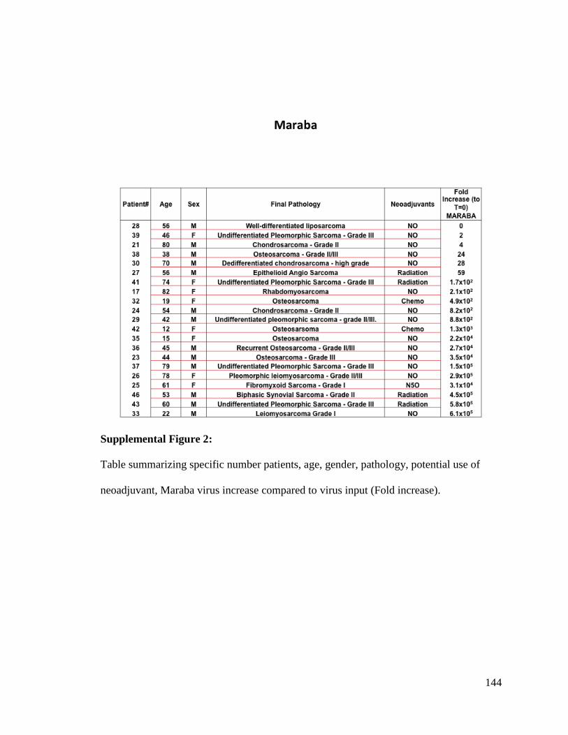
144
Supplemental Figure 2:
Table summarizing specific number patients, age, gender, pathology, potential use of
neoadjuvant, Maraba virus increase compared to virus input (Fold increase).

145
Supplemental Figure 3:
Table summarizing specific number patients, age, gender, pathology, potential use of
neoadjuvant, Vaccinia virus increase compared to virus input (Fold increase).

146
Supplemental Figure 4:
S180 sarcoma murine tumor cell line was plated in 96 wells plate and infected with
Maraba virus at various MOI. Cell viability have been assessed using Alamar Blue assay
24 hours after infection and plotted.
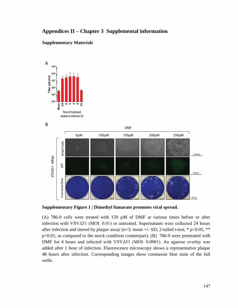
147
Appendices II – Chapter 3 Supplemental information
Supplementary Materials
Supplementary Figure 1 | Dimethyl fumarate promotes viral spread.
(A) 786-0 cells were treated with 150 µM of DMF at various times before or after
infection with VSV∆51 (MOI: 0.01) or untreated. Supernatants were collected 24 hours
after infection and titered by plaque assay (n=3; mean +/- SD; 2-tailed t-test; * p<0.05, **
p<0.01, as compared to the mock condition counterpart). (B) 786-0 were pretreated with
DMF for 4 hours and infected with VSV∆51 (MOI: 0.0001). An agarose overlay was
added after 1 hour of infection. Fluorescence microscopy shows a representative plaque
48 hours after infection. Corresponding images show coomassie blue stain of the full
wells.

148
Supplementary Figure 2 | Fumaric and maleic acid esters enhance VSV∆51 infection
in 786-0 and CT26WT cancer cell lines.
(A) 786-0 cells were treated with various FMAEs at the indicated concentrations for 4
hours and subsequently infected with VSV∆51 (MOI: 0.01). 16, 20, and 24 hours after

149
infection, fluorescent and phase contrast images were taken of the infected 786-0 cells.
(B) 786-0 and CT26WT cells were treated as in A, supernatants were collected 48 hours
after infection and titered by plaque assay (n=3; mean +/- SD).
Supplementary Figure 3 | Fumaric and maleic acid esters enhance VSV∆51 spread
in cancer cells.
(A-B) 786-0 were pretreated for 4 hours with various FMAEs and subsequently infected
with VSV∆51 expressing GFP. An agarose overlay was added after 1 hour of infection.
(A) Fluorescence microscopy of a representative plaque 24 hours after infection.
Corresponding images of coomassie blue stain of the full well and (B) a graph showing
average plaque diameter illustrate the enhancement of the plaque diameters in presence of
DMF (n=20; horizontal lines indicate means; one-way ANOVA; *** p<0.001, as
compared to the mock condition counterpart).

150
Supplementary Figure 4 | Fumaric and maleic acid esters inhibit response to type I
interferon.
(A-B) 786-0 cells were treated with FMAEs and infected with VSV∆51 expressing GFP,
and subsequently RNA was extracted at 24 hpi. (A) Heatmap shows the expression of all
the differentially expressed genes. (B) Summary of gene ontology relationships,
indicating GO terms significantly (p-value scale indicated) downregulated by FMAEs
during viral infection. (C) 786-0 cells were pretreated with DEM (100 to 500 µM) for 4
hours and treated with IFNβ (250 U/ml) for 30 minutes. Proteins were extracted and
subsequently probed for pSTAT1, STAT1, and β-actin by western blot.
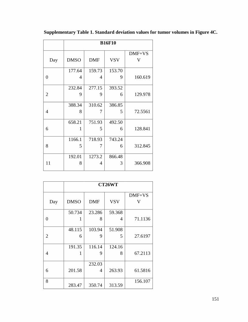
151
Supplementary Table 1. Standard deviation values for tumor volumes in Figure 4C.
B16F10
Day DMSO DMF VSV
DMF+VS
V
0
177.64
4
159.73
4
153.70
9 160.619
2
232.84
9
277.15
9
393.52
6 129.978
4
388.34
8
310.62
7
386.85
5 72.5561
6
658.21
1
751.93
5
492.50
6 128.841
8
1166.1
5
718.93
7
743.24
6 312.845
11
192.01
8
1273.2
4
866.48
3 366.908
CT26WT
Day DMSO DMF VSV
DMF+VS
V
0
50.734
1
23.286
8
59.368
4 71.1136
2
48.115
6
103.94
9
51.908
5 27.6197
4
191.35
1
116.14
9
124.16
8 67.2113
6 201.58
232.03
4 263.93 61.5816
8 283.47 350.74 313.59
156.107

152
7 9 5
12
686.48
4
523.45
4
751.55
1 373.339
HT29
Day DMSO DMF VSV
DMF+VS
V
0
45.418
5 30.366
43.164
2 46.0185
2
65.077
1 23.311 34.218 29.6766
4
57.373
4
23.109
9
117.19
3 118.603
6
93.800
9
42.567
5
117.25
2 119.602
8
95.169
7
37.364
9
160.15
9 82.4382
12
149.75
9
70.526
3
261.30
5 55.8793
14 186.6
78.852
9
318.84
6 135.389
19
254.34
5
129.08
9 421.99 189.064
22
338.37
7
201.38
7
443.70
2 152.971
27
374.75
5
229.59
6
379.34
1 194.364
33 629.57 524.04 494.45
246.646

153
7 5 3
38
435.18
7
673.15
3
684.56
7 260.016
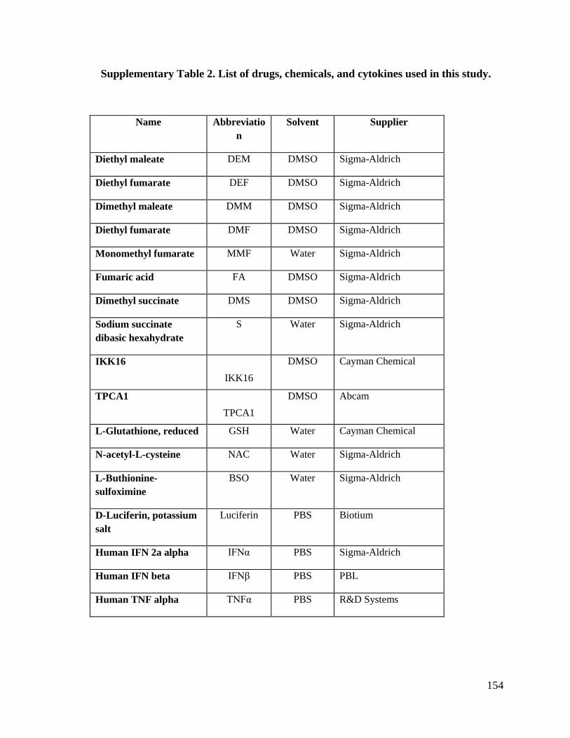
154
Supplementary Table 2. List of drugs, chemicals, and cytokines used in this study.
Name Abbreviatio
n
Solvent Supplier
Diethyl maleate DEM DMSO Sigma-Aldrich
Diethyl fumarate DEF DMSO Sigma-Aldrich
Dimethyl maleate DMM DMSO Sigma-Aldrich
Diethyl fumarate DMF DMSO Sigma-Aldrich
Monomethyl fumarate MMF Water Sigma-Aldrich
Fumaric acid FA DMSO Sigma-Aldrich
Dimethyl succinate DMS DMSO Sigma-Aldrich
Sodium succinate
dibasic hexahydrate
S Water Sigma-Aldrich
IKK16
IKK16
DMSO Cayman Chemical
TPCA1
TPCA1
DMSO Abcam
L-Glutathione, reduced GSH Water Cayman Chemical
N-acetyl-L-cysteine NAC Water Sigma-Aldrich
L-Buthionine-
sulfoximine
BSO Water Sigma-Aldrich
D-Luciferin, potassium
salt
Luciferin PBS Biotium
Human IFN 2a alpha IFNα PBS Sigma-Aldrich
Human IFN beta IFNβ PBS PBL
Human TNF alpha TNFα PBS R&D Systems

155
Supplementary Table 3. List of cell lines used in this study.
Cell Line Organism Tissue Cell type
Growth
media Vendor
Catalog
number
Vero
African green
monkey Kidney DMEM ATCC CCL-81
786-0 human Kidney
Renal cell
adenocarcinoma DMEM ATCC CRL-1932
A549 human Lung Carcinoma RPMI ATCC CCL-185
HT29 human Colon
Colorectal
Adenocarcinoma DMEM ATCC HTB-38
M14 human Skin Melanoma DMEM *
OVCA433 human Ovary Adenocarcinoma RPMI *
SKOV3 human Ovary Adenocarcinoma RPMI ATCC HTB-77
CT26WT mouse Colon Carcinoma DMEM ATCC CRL-2638
EKVX human Lung
Non-small cell lung
carcinoma RPMI *
76-9 Mouse Rhabdomyosarcoma RPMI **
HT1080 human Fibrosarcoma DMEM *
H226 human Lung
Squamous cell lung
carcinoma RPMI ATCC CRL-5826
B16F10 mouse Skin Melanoma DMEM ATCC CRL-6475
EMT6 mouse Breast Carcinoma DMEM ATCC CRL-2755
K7M2 Mouse
Bone
*derived
from
metastatic
site (lung) Osteosarcoma DMEM ATCC CRL-2836
ATCC - American Type Culture Collection; None of the cell lines listed are present in
the commonly misidentified cell lines database maintained by ICLAC. * The M14,
OVCA433, HT1080 and EKVX cell lines were provided by Dr. John Bell (Ottawa
Hospital Research Institute, Ottawa, Ontario, Canada). ** The rhabdomyosarcoma cell
156
lines were generously provided by Dr. Martin Holcik (Children’s Hospital of Eastern
Ontario Research Institute (CHEO), Ottawa, ON, Canada).
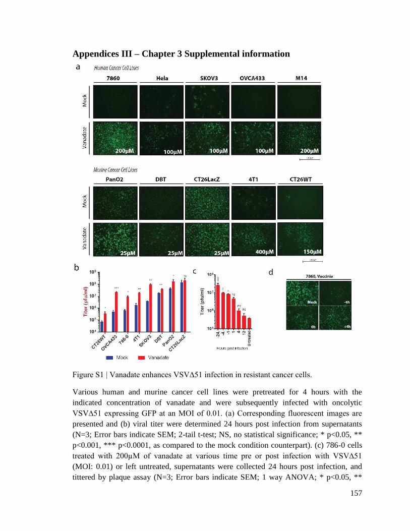
157
Appendices III – Chapter 3 Supplemental information
Figure S1 | Vanadate enhances VSV∆51 infection in resistant cancer cells.
Various human and murine cancer cell lines were pretreated for 4 hours with the
indicated concentration of vanadate and were subsequently infected with oncolytic
VSV∆51 expressing GFP at an MOI of 0.01. (a) Corresponding fluorescent images are
presented and (b) viral titer were determined 24 hours post infection from supernatants
(N=3; Error bars indicate SEM; 2-tail t-test; NS, no statistical significance; * p<0.05, **
p<0.001, *** p<0.0001, as compared to the mock condition counterpart). (c) 786-0 cells
treated with 200µM of vanadate at various time pre or post infection with VSV∆51
(MOI: 0.01) or left untreated, supernatants were collected 24 hours post infection, and
tittered by plaque assay (N=3; Error bars indicate SEM; 1 way ANOVA; * p<0.05, **

158
p<0.001, *** p<0.0001, as compared to the untreated condition counterpart). (d) 786-0
cells treated with 100µM of vanadate at various time pre or post infection with the
vaccinia virus expressing GFP (MOI: 0.01) or left untreated. Corresponding fluorescent
images are presented.

159

160
Figure S2 | Vanadate enhances VSV∆51 spread in cancer cells.
Various human and murine cancer cell lines were pretreated for 4 hours with the
indicated concentration of vanadate and were subsequently infected with oncolytic
VSV∆51 expressing GFP, an agarose overlay was added after 1 hour of infection. (a)
Fluorescence microscopy of a representative plaque 24 hour after infection. (b)
Corresponding image of coomassie blue stain of the full well in (a), and (c) average
plaque diameter of (b) illustrating the enhancement of the plaque diameters in presence of
vanadate. (N=10; Bars indicate mean; ND, not detected; 2-tail t-test; ** p<0.001, ***
p<0.0001, as compared to the mock condition counterpart)

161
Figure S3 | Viral enhancement is dependent on Vanadium.
(a,b) 786-0 or (c) CT2WT were pretreated for 4 hours with various concentration (a) of
phosphate salts or pyrophosphate, (b) vanadate solutions at various pH values, (c)
solution of various vanadium compounds and were subsequently infected with oncolytic
VSV∆51 expressing GFP at an MOI of 0.01. (a,b) Corresponding viral titer were
determined 24 hours post infection from supernatants (N=3). (c) Corresponding GFP
positive cell counts 24 hours post infection. Error bars indicate SEM.

162
Figure S4 | Vanadate enhances viral oncolysis in various human cancer cell lines.
(a) Various human cancer cell lines were pretreated for 4 hours with a range of
concentration of vanadate and were subsequently infected with oncolytic VSV∆51
expressing GFP at an MOI of 0.01. Cell viability was assayed in 786-0 cells 48 hours
post infection. Results were normalized to the average of the values obtained for the
corresponding uninfected, untreated cells (N=4). Error bars indicate SEM .(b) 786-0
were pretreated for 4 hours with a range of concentration of various vanadate based
compounds and were subsequently infected with oncolytic VSV∆51 expressing GFP at
an MOI of 0.01. Cell viability was assayed in 786-0 cells 24 hours post infection. Results
were normalized to the average of the values obtained for the corresponding uninfected,
untreated cells (N=4), error bars indicate SEM.
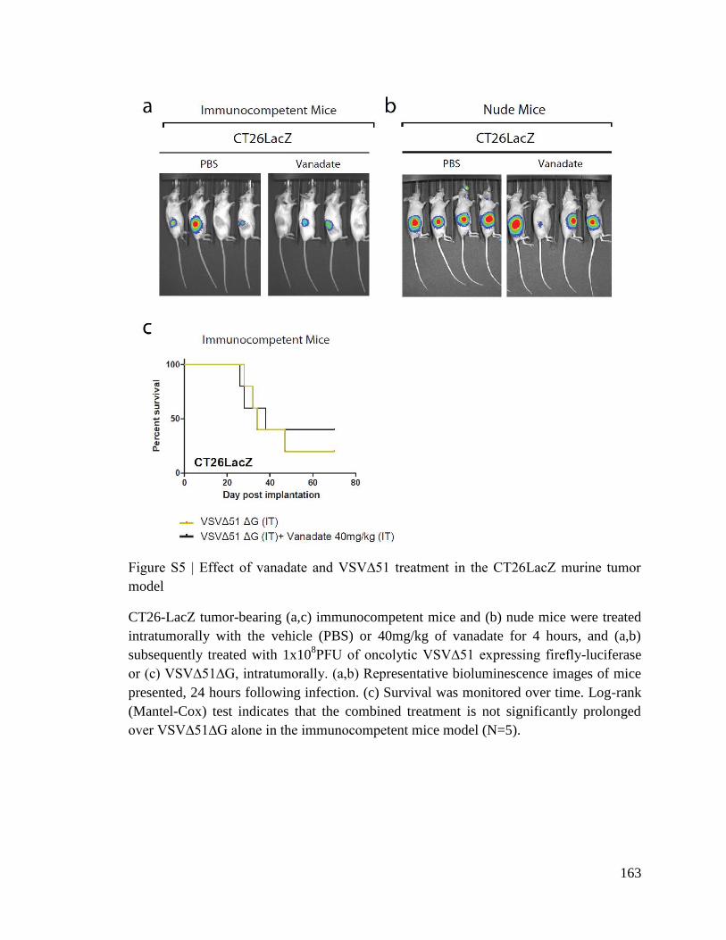
163
Figure S5 | Effect of vanadate and VSV∆51 treatment in the CT26LacZ murine tumor
model
CT26-LacZ tumor-bearing (a,c) immunocompetent mice and (b) nude mice were treated
intratumorally with the vehicle (PBS) or 40mg/kg of vanadate for 4 hours, and (a,b)
subsequently treated with 1x10⁸PFU of oncolytic VSV∆51 expressing firefly-luciferase
or (c) VSV∆51∆G, intratumorally. (a,b) Representative bioluminescence images of mice
presented, 24 hours following infection. (c) Survival was monitored over time. Log-rank
(Mantel-Cox) test indicates that the combined treatment is not significantly prolonged
over VSV∆51∆G alone in the immunocompetent mice model (N=5).

164
Figure S6 | Effect of vanadate and VSV∆51 treatment in the HT29 human tumor
xenograft model
(a-c) HT29 tumor-bearing mice were treated intratumorally with the vehicle (DMSO) or
40mg/kg of vanadate for 4 hours, and subsequently treated with 1x10⁸PFU of oncolytic
VSV∆51 expressing firefly-luciferase, intratumorally. 24, 48 and 72 hours post infection,
viral replication was monitored. Representative bioluminescence images of mice are
presented. (b) Quantification of luminescence. Scale represented in photons. (N=4-10.
Bars indicates mean; *p < 0.05, by 1-tailed t-test; as compared to DMSO treated
condition). (c) Survival was monitored over time. Log-rank (Mantel-Cox) test indicates
that the combined treatment does not significantly prolong survival over DMSO
treatment or virus alone (N=4-10).

165
Figure S7 | Vanadate potentiates immune system processes and increases cytokine
expression following VSV∆51 infection of cancer cells.
(a-d) The 786-0 and (c) CT26WT cells were pretreated for 4 hours with vehicle (buffer
pH 7.4), or with vanadate (from a solutions prepared from orthovanadate or
metavanadate) and infected with VSV∆51 (MOI: 0.01), or left uninfected. 24 hours post
infection RNA was extracted and supernatants were collected. (a,b) RNA was
subsequently processed for hybridization on an Affymetrix Human PrimeView Array
(N=1, pooled biological triplicate for each experimental condition), or (c) processed for
qPCR quantification. (a) Upregulated GOterm graphed in presence of vanadate with or
without infection. (b) Heatmap showing the expression levels of the differentially
expressed cytokines and chemokines. Expression of genes was normalized to values
obtained for untreated, uninfected control. Hierarchical clustering of genes from all
samples was also performed. In the heatmap, red indicates relatively higher expression
and blue indicates relatively lower expression relative to untreated, uninfected control
(Log2 fold change). (b) Gene expression of various cytokines and chemokines in 786-0
and CT26wt, quantified by qPCR. (d) Quantified CXCL9 from supernatant of 786-0,
SKOV3 and OVCA433, 24hour following infection as in (a).

166
Figure S8 | Effect of vanadate on expression of antiviral proteins IFITM1 during
infection.
Cell lysates of 786-0 treated with vanadate and VSV∆51 expressing GFP was collected at
indicated time points. Protein was extracted and probed for IFITM1, VSV and actin by
western blot.
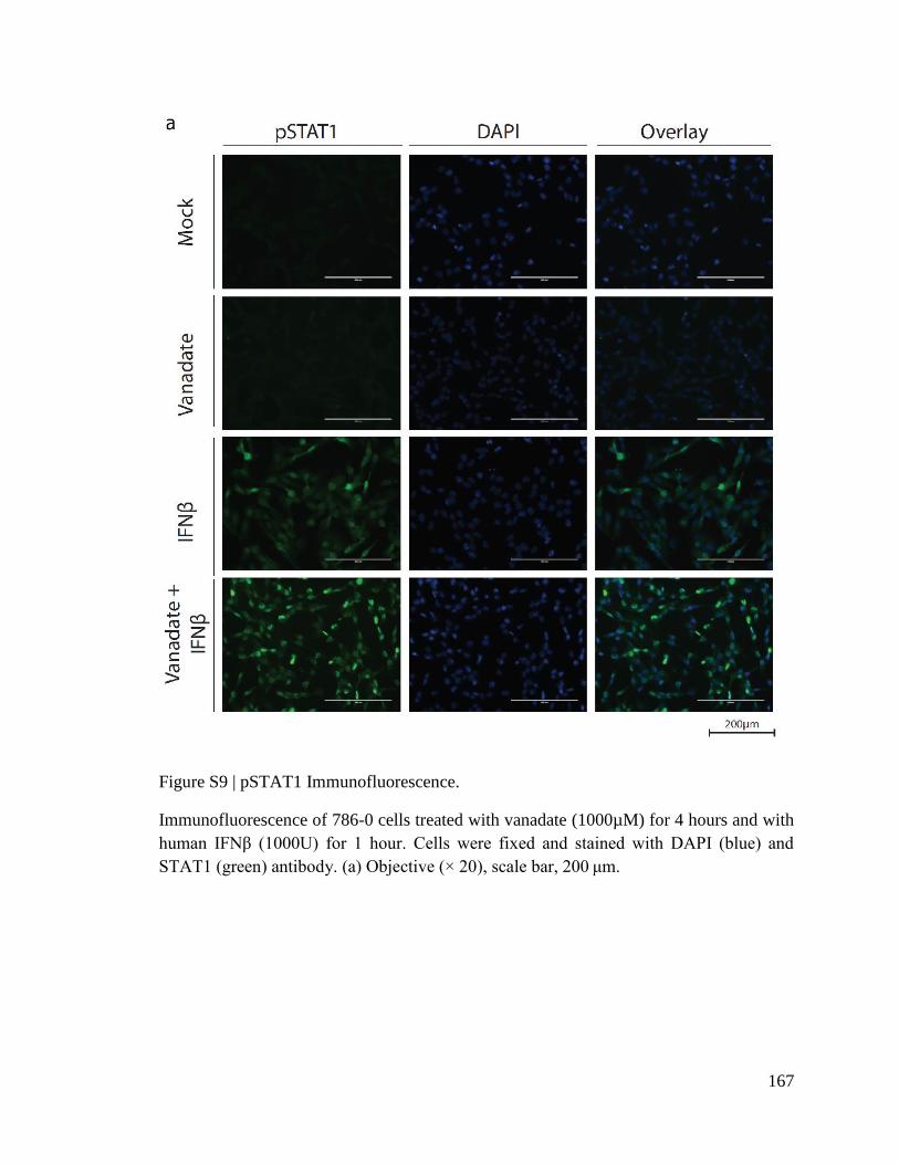
167
Figure S9 | pSTAT1 Immunofluorescence.
Immunofluorescence of 786-0 cells treated with vanadate (1000µM) for 4 hours and with
human IFNβ (1000U) for 1 hour. Cells were fixed and stained with DAPI (blue) and
STAT1 (green) antibody. (a) Objective (× 20), scale bar, 200 μm.

168

169
Figure S10 | STAT2 Immunofluorescence.
Immunofluorescence of 786-0 cells treated with vanadate (1000µM) for 4 hours and with
human IFNβ (1000U) for 1 hour. Cells were fixed and stained with DAPI (blue) and
STAT2 (green) antibody. (a) Objective (× 20), scale bar, 200 μm. (b) Quantification of
nuclear:cytoplasmic ratio and the average nuclear fluorescence in each condition (N=30;
Bars indicate mean; 1-way ANOVA, * p<0.05, ** p<0.001, *** p<0.0001, as compared
to the mock condition counterpart.)

170

171
Figure S11 | pSTAT2 Immunofluorescence.
Immunofluorescence of 786-0 cells treated with vanadate (1000µM) for 4 hours and with
human IFNβ (1000U) for 1 hour. Cells were fixed and stained with DAPI (blue) and
pSTAT2 (green) antibody. (a) Objective (× 20), scale bar, 200 μm. (b) Quantification of
nuclear:cytoplasmic ratio in each condition (N=30; Bars indicate mean; 1-way ANOVA,
* p<0.05, ** p<0.001, *** p<0.0001, as compared to the mock condition counterpart.)

172
Figure S12 | Effect of Vanadate on wtVSV infection .
The 786-0 cells were pretreated for 4 hours with vanadate or mock treated and infected
with VSV∆51 or wtVSV (MOI: 0.01). Corresponding viral titer were determined 24
hours post infection from supernatants (N=3).

173
Figure S13 | Uncropped western blots.
Uncropped western blots for (a) Figure 4.6d and (b) Supplemental Figure 8.

174
Table S1 | Human Cytokine Array.
The 786-0 cells were pretreated for 4 hours with vanadate and were subsequently infected
with oncolytic VSV∆51 expressing GFP at an MOI of 0.01. 24 hours following infection,
cytokine array was performed. Normalized (with Background Subtraction) values to
positive and negative control sample are indicated in table below.
Condition
Cytokine Mock Van VSV VAN+VSV
ENA-78 149.75 246.14 77.64 274.71
G-CSF 0.00 22.47 0.00 0.00
GM-CSF 147.25 765.83 0.00 148.63
GRO 974.25 1,551.16 830.82 1,447.05
GRO-alpha 100.25 66.08 197.37 1.34
I-309 206.75 110.35 73.00 0.00
IL-1alpha 118.25 93.17 82.27 0.00
IL-1beta 254.25 228.96 257.24 165.36
IL-2 190.75 58.81 0.00 246.93
IL-3 184.25 426.20 332.17 689.09
IL-4 65.75 129.18 0.00 133.40
IL-5 0.00 13.22 0.00 97.84
IL-6 4,663.75 11,150.84 13,903.79 13,375.43
IL-7 25.75 159.91 89.22 78.43
IL-8 16,791.25 15,279.01 16,587.83 14,039.57
IL-10 172.75 152.31 18.54 99.34
IL12-p40 206.75 92.18 91.15 156.70
IL-13 1.75 0.00 201.62 0.00
IL-15 0.00 37.33 176.13 30.32
IFN-gamma 63.75 42.62 265.35 4.63
MCP-1 6,617.75 8,770.74 8,352.24 9,187.08
MCP-2 0.00 232.92 0.00 468.61
MCP-3 0.00 91.52 66.05 309.07
M-CSF 299.75 665.40 932.40 979.19
MDC 180.25 250.76 259.95 523.58
MIG 0.00 0.00 0.00 53.03
MIP-1-delta 41.75 103.08 111.24 299.51
RANTES 738.25 519.04 10,671.28 9,910.98
SCF 278.75 129.84 225.96 264.85
SDF-1 334.25 270.26 234.84 383.46
TARC 185.75 382.92 396.68 390.33
TGF-beta 1 64.75 163.21 295.87 15.98
TNF-alpha 239.75 116.96 0.00 511.03
TNF-beta 0.00 156.60 0.00 178.21
EGF 0.00 476.75 307.45 502.37
IGF-1 0.00 364.42 412.90 424.39
Angiogenin 1,108.75 1,259.10 2,373.11 1,651.11
Oncostatin M 155.75 680.59 656.24 1,008.47
TPO 125.75 128.52 242.95 338.95
VEGF 232.75 594.03 357.67 509.54
PDGF-BB 313.25 293.05 179.61 177.02
Leptin 62.25 185.35 130.94 142.06
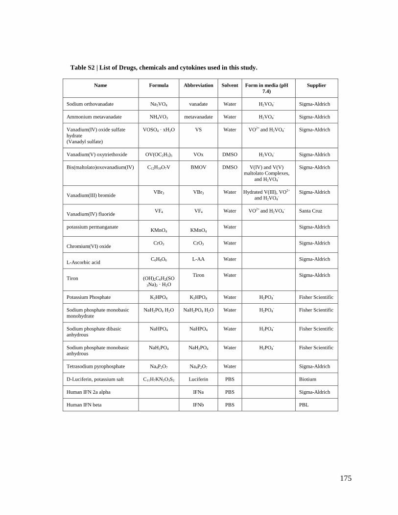
175
Table S2 | List of Drugs, chemicals and cytokines used in this study.
Name Formula Abbreviation Solvent Form in media (pH
7.4)
Supplier
Sodium orthovanadate Na3VO4 vanadate Water H2VO4- Sigma-Aldrich
Ammonium metavanadate NH4VO3 metavanadate Water H2VO4- Sigma-Aldrich
Vanadium(IV) oxide sulfate
hydrate
(Vanadyl sulfate)
VOSO4 · xH2O VS Water VO2+ and H2VO4- Sigma-Aldrich
Vanadium(V) oxytriethoxide OV(OC2H5)3 VOx DMSO H2VO4- Sigma-Aldrich
Bis(maltolato)oxovanadium(IV) C12H10O7V BMOV DMSO V(IV) and V(V) maltolato Complexes,
and H2VO4-
Sigma-Aldrich
Vanadium(III) bromide VBr3 VBr3 Water Hydrated V(III), VO2+
and H2VO4-
Sigma-Aldrich
Vanadium(IV) fluoride VF4 VF4 Water VO2+ and H2VO4
- Santa Cruz
potassium permanganate KMnO4 KMnO4
Water Sigma-Aldrich
Chromium(VI) oxide CrO3 CrO3 Water Sigma-Aldrich
L-Ascorbic acid C6H8O6 L-AA Water Sigma-Aldrich
Tiron (OH)2C6H2(SO
3Na)2 · H2O
Tiron Water Sigma-Aldrich
Potassium Phosphate K2HPO4 K2HPO4 Water H2PO4- Fisher Scientific
Sodium phosphate monobasic
monohydrate
NaH2PO4 H2O NaH2PO4 H2O Water H2PO4- Fisher Scientific
Sodium phosphate dibasic anhydrous
NaHPO4 NaHPO4 Water H2PO4- Fisher Scientific
Sodium phosphate monobasic anhydrous
NaH2PO4 NaH2PO4 Water H2PO4- Fisher Scientific
Tetrasodium pyrophosphate Na4P2O7 Na4P2O7 Water Sigma-Aldrich
D-Luciferin, potassium salt C11H7KN2O3S2 Luciferin PBS Biotium
Human IFN 2a alpha IFNa PBS Sigma-Aldrich
Human IFN beta IFNb PBS PBL

176
Table S3 | List of cell lines used in this study.
Cell Line Organism Tissue Cell type Growth
media Vendor
Catalog
number
Vero
African
green monkey Kidney DMEM ATCC CCL-81
786-O Human Kidney
Renal cell
adenocarcin
oma DMEM ATCC CRL-1932
A549 Human Lung Carcinoma RPMI ATCC CCL-185
GM38 Human Normal
Fibroblast DMEM
Coriell
Institute for
Medical research GM00038
HeLa Human Cervix Adenocarcin
oma DMEM ATCC CCL2
HT29 Human Colon
Colorectal
Adenocarcin
oma DMEM ATCC HTB-38
JIMT-1 Human Breast Carcinoma DMEM DSMZ ACC-589
M14 Human Skin Melanoma DMEM *
MCF7 Human Breast
Adenocarcin
oma DMEM ATCC HTB-22
MDA-MB-
231 Human Breast
Adenocarcin
oma DMEM ATCC HTB-26
OVCA433 Human Ovary
Adenocarcin
oma RPMI *
OVCAR8 Human Ovary
Adenocarcin
oma RPMI *
SKOV3 Human Ovary
Adenocarcin
oma RPMI ATCC HTB-77
4T1 Mouse Breast Carcinoma DMEM ATCC CRL-2539
CT26LacZ
(CT26.CL25
) Mouse Colon Carcinoma DMEM ATCC CRL-2639
CT26WT Mouse Colon Carcinoma DMEM ATCC CRL-2638
PanO2 Mouse Pancreas Carcinoma DMEM *
DBT Mouse Brain Glioma DMEM **
ATCC - American Type Culture Collection (Manassas, VA); DSMZ- Deutsche Sammlung von Mikroorganismen und
Zellkulturen (Braunschweig, Germany). None of the cell lines listed was present in the commonly misidentified cell
lines database maintained by ICLAC. * The M14, OVCA433, OVCAR8, PanO2 cell lines were a generous gift from
Dr. John Bell (Ottawa Hospital Research Institute, Ottawa, Ontario, Canada). ** The DBT cell line was a generous gift
from Robert C. Rostomily, (University of Washington, School of Medicine, Seattle, WA, USA).

177
Table S4 | List of primers used in this study.
Model Gene Forward Primer (5'->3') Reverse Primer (5'->3')
VSV M ATACTCAGATGTGGCAGCCG GATCTGCCAATACCGCTGGA
Human CXCL9 AGTGCAAGGAACCCCAGTAG AGGGCTTGGGGCAAATTGTT
CCL8 TGCTGAAGCTCACACCCTTG GGAAACTGAATCTGGCTGAGCA
CCL3 TTCCGTCACCTGCTCAGAAT CAGCAGCAAGTGATGCAGAGA
IL6 ACCCCCAATAAATATAGGACTGGA GAAGGCGCTTGTGGAGAAGG
CXCL8 ACCGGAAGGAACCATCTCAC GGCAAAACTGCACCTTCACAC
TNF GCTGCACTTTGGAGTGATCG GAGGGTTTGCTACAACATGGG
CXCL10 CTGAGCCTACAGCAGAGGAAC AGGTACTCCTTGAATGCCACTT
CXCL11 CAGCATTTCTACTCCTTCCAAGA TGGGGAAGCCTTGAACAACT
CCL5 GCAGTCGTCCACAGGTCAAG TCTTCTCTGGGTTGGCACAC
IFNβ CATTACCTGAAGGCCAAGGA CAGCATCTGCTGGTTGAAGA
IFNγ TCTTTTGGATGCTCTGGTCA TTCAGCTCTGCATCGTTTTG
GAPDH ACAGTCAGCCGCATCTTCTT GTTAAAAGCAGCCCTGGTGA
MX2 GAACGTGCAGCGAGCTTGTC AAGGCTTGTGGGCCTTAGAC
IFITM1 CCGTGAAGTCTAGGGACAGG GGTAGACTGTCACAGAGCCG
Mouse CXCL9 CAGTGTGGAGTTCGAGGAACC TTTGTTGCAATTGGGGCTTGG
CCL3 CCATATGGAGCTGACACCCC TCAGGAAAATGACACCTGGCT
IL6 TCCTCTCTGCAAGAGACTTCC GGTCTGTTGGGAGTGGTATCC
CXCL11 CAGCTGCTCAAGGCTTCCTTA CAACTTTGTCGCAGCCGTTA
CCL5 CTGCTGCTTTGCCTACCTCT CGAGTGACAAACACGACTGC
IFNβ CAGTGTGGAGTTCGAGGAACC TTTGTTGCAATTGGGGCTTGG
beta Actin AGGTCTCAAACATGATCTG AGGTATCCTGACCCTGAAG
178
References
1. D. Hanahan, R. A. Weinberg, Hallmarks of cancer: the next generation, Cell 144, 646–674
(2011).
2. D. M. Pardoll, The blockade of immune checkpoints in cancer immunotherapy, Nat. Rev.
Cancer 12, 252–264 (2012).
3. Canadian Cancer Statistics 2012: (611042012-001) (2012), doi:10.1037/e611042012-001.
4. C. C. Statistics, Canadian Cancer Society’s Advisory Committee On Cancer Statistics. in,
Canadian Cancer Society, Canadian Cancer Society (2015).
5. C. Dalba, D. Klatzmann, C. R. Logg, N. Kasahara, Beyond oncolytic virotherapy: replication-
competent retrovirus vectors for selective and stable transduction of tumors, Curr. Gene Ther. 5,
655–667 (2005).
6. J. K.-K. Li, Oncolytic bluetongue viruses: promise, progress, and perspectives, Front.
Microbiol. 2, 46 (2011).
7. C. Levaditi, S. Nicolau, Affinite du virus herpetique pour les neoplasmes epitheliaux, CR Soc.
Biol 87, 498–500 (1922).
8. S. J. Russell, K.-W. Peng, J. C. Bell, Oncolytic virotherapy, Nat. Biotechnol. 30, 658–670
(2012).
9. M. Liang, Clinical Development of Oncolytic Viruses in China, Curr. Pharm. Biotechnol. 13,
1852–1857 (2012).
10. F. J. Kohlhapp, H. L. Kaufman, Molecular Pathways: Mechanism of Action for Talimogene
Laherparepvec, a New Oncolytic Virus Immunotherapy, Clin. Cancer Res. 22, 1048–1054
(2016).
11. B. D. Lichty, C. J. Breitbach, D. F. Stojdl, J. C. Bell, Going viral with cancer immunotherapy,
Nat. Rev. Cancer 14, 559–567 (2014).
12. H. H. Wong, N. R. Lemoine, Y. Wang, Oncolytic Viruses for Cancer Therapy: Overcoming
the Obstacles, Viruses 2, 78–106 (2010).
13. J. Hughes, P. Wang, G. Alusi, H. Shi, Y. Chu, J. Wang, V. Bhakta, I. McNeish, A. McCart,
N. R. Lemoine, Y. Wang, Lister strain vaccinia virus with thymidine kinase gene deletion is a
tractable platform for development of a new generation of oncolytic virus, Gene Ther. 22, 476–
484 (2015).

179
14. J. K. Kaufmann, S. Bossow, C. Grossardt, S. Sawall, J. Kupsch, P. Erbs, J. C. Hassel, C. von
Kalle, A. H. Enk, D. M. Nettelbeck, G. Ungerechts, Chemovirotherapy of malignant melanoma
with a targeted and armed oncolytic measles virus, J. Invest. Dermatol. 133, 1034–1042 (2013).
15. T. Nakamura, S. J. Russell, Engineering oncolytic measles viruses for targeted cancer therapy,
Molecular Targeting in Oncology (2008) (available at
http://link.springer.com/content/pdf/10.1007/978-1-59745-337-0.pdf#page=434).
16. J. K. Fan, N. Wei, M. Ding, J. F. Gu, X. R. Liu, B. H. Li, R. Qi, W. D. Huang, Y. H. Li, X. Q.
Xiong, J. Wang, R. S. Li, X. Y. Liu, Targeting Gene-ViroTherapy for prostate cancer by DD3-
driven oncolytic virus-harboring interleukin-24 gene, Int. J. Cancer 127, 707–717 (2010).
17. W. Zhang, K. Ge, Q. Zhao, X. Zhuang, Z. Deng, L. Liu, J. Li, Y. Zhang, Y. Dong, Y. Zhang,
S. Zhang, B. Liu, A novel oHSV-1 targeting telomerase reverse transcriptase-positive cancer cells
via tumor-specific promoters regulating the expression of ICP4, Oncotarget 6, 20345–20355
(2015).
18. F. Garrido, N. Aptsiauri, E. M. Doorduijn, A. M. Garcia Lora, T. van Hall, The urgent need to
recover MHC class I in cancers for effective immunotherapy, Curr. Opin. Immunol. 39, 44–51
(2016).
19. E. A. Chiocca, S. D. Rabkin, Oncolytic viruses and their application to cancer
immunotherapy, Cancer Immunol Res 2, 295–300 (2014).
20. R. J. Prestwich, K. J. Harrington, H. S. Pandha, R. G. Vile, A. A. Melcher, F. Errington,
Oncolytic viruses: a novel form of immunotherapy, Expert Rev. Anticancer Ther. 8, 1581–1588
(2008).
21. Z. S. Guo, Z. Liu, S. Kowalsky, M. Feist, P. Kalinski, B. Lu, W. J. Storkus, D. L. Bartlett,
Oncolytic Immunotherapy: Conceptual Evolution, Current Strategies, and Future Perspectives,
Front. Immunol. 8, 555 (2017).
22. A. M. Murphy, D. M. Besmer, M. Moerdyk-Schauwecker, N. Moestl, D. A. Ornelles, P.
Mukherjee, V. Z. Grdzelishvili, Vesicular stomatitis virus as an oncolytic agent against pancreatic
ductal adenocarcinoma, J. Virol. 86, 3073–3087 (2012).
23. A. Arvin, G. Campadelli-Fiume, E. Mocarski, P. S. Moore, B. Roizman, R. Whitley, K.
Yamanishi, Human Herpesviruses: Biology, Therapy, and Immunoprophylaxis (Cambridge
University Press, 2007).
24. D. K. B. Rao, Herpes Viruses – An Overview, iosrphr 4, 39–41 (2014).
25. M. Aghi, R. L. Martuza, Oncolytic viral therapies - the clinical experience, Oncogene 24,
7802–7816 (2005).
26. K. J. Looker, A. S. Magaret, M. T. May, K. M. E. Turner, P. Vickerman, S. L. Gottlieb, L. M.
Newman, Global and Regional Estimates of Prevalent and Incident Herpes Simplex Virus Type 1
Infections in 2012, PLoS One 10, e0140765 (2015).
27. S. Varghese, S. D. Rabkin, Oncolytic herpes simplex virus vectors for cancer virotherapy,
Cancer Gene Ther. 9, 967–978 (2002).
28. J. C. C. Hu, R. S. Coffin, C. J. Davis, N. J. Graham, N. Groves, P. J. Guest, K. J. Harrington,
180
N. D. James, C. A. Love, I. McNeish, L. C. Medley, A. Michael, C. M. Nutting, H. S. Pandha, C.
A. Shorrock, J. Simpson, J. Steiner, N. M. Steven, D. Wright, R. C. Coombes, A phase I study of
OncoVEXGM-CSF, a second-generation oncolytic herpes simplex virus expressing granulocyte
macrophage colony-stimulating factor, Clin. Cancer Res. 12, 6737–6747 (2006).
29. R. H. I. Andtbacka, H. L. Kaufman, F. Collichio, T. Amatruda, N. Senzer, J. Chesney, K. A.
Delman, L. E. Spitler, I. Puzanov, S. S. Agarwala, M. Milhem, L. Cranmer, B. Curti, K. Lewis,
M. Ross, T. Guthrie, G. P. Linette, G. A. Daniels, K. Harrington, M. R. Middleton, W. H. Miller
Jr, J. S. Zager, Y. Ye, B. Yao, A. Li, S. Doleman, A. VanderWalde, J. Gansert, R. S. Coffin,
Talimogene Laherparepvec Improves Durable Response Rate in Patients With Advanced
Melanoma, J. Clin. Oncol. 33, 2780–2788 (2015).
30. A. Ribas, R. Dummer, I. Puzanov, A. VanderWalde, R. H. I. Andtbacka, O. Michielin, A. J.
Olszanski, J. Malvehy, J. Cebon, E. Fernandez, J. M. Kirkwood, T. F. Gajewski, L. Chen, K. S.
Gorski, A. A. Anderson, S. J. Diede, M. E. Lassman, J. Gansert, F. S. Hodi, G. V. Long,
Oncolytic Virotherapy Promotes Intratumoral T Cell Infiltration and Improves Anti-PD-1
Immunotherapy, Cell 170, 1109–1119.e10 (2017).
31. D. M. Knipe, P. M. Howley, Fields’ Virology (Lippincott Williams & Wilkins, 2007).
32. G. McFadden, Poxvirus tropism, Nat. Rev. Microbiol. 3, 201–213 (2005).
33. W. M. Chan, G. McFadden, Oncolytic Poxviruses, Annu Rev Virol 1, 119–141 (2014).
34. T.-C. Liu, E. Galanis, D. Kirn, Clinical trial results with oncolytic virotherapy: a century of
promise, a decade of progress, Nat. Clin. Pract. Oncol. 4, 101–117 (2007).
35. A. E. Merrick, E. J. Ilett, A. A. Melcher, JX-594, a targeted oncolytic poxvirus for the
treatment of cancer, Curr. Opin. Investig. Drugs 10, 1372–1382 (2009).
36. B.-H. Park, T. Hwang, T.-C. Liu, D. Y. Sze, J.-S. Kim, H.-C. Kwon, S. Y. Oh, S.-Y. Han, J.-
H. Yoon, S.-H. Hong, A. Moon, K. Speth, C. Park, Y.-J. Ahn, M. Daneshmand, B. G. Rhee, H.
M. Pinedo, J. C. Bell, D. H. Kirn, Use of a targeted oncolytic poxvirus, JX-594, in patients with
refractory primary or metastatic liver cancer: a phase I trial, Lancet Oncol. 9, 533–542 (2008).
37. H. Fukuhara, Y. Ino, T. Todo, Oncolytic virus therapy: A new era of cancer treatment at
dawn, Cancer Sci. 107, 1373–1379 (2016).
38. J. Heo, T. Reid, L. Ruo, C. J. Breitbach, S. Rose, M. Bloomston, M. Cho, H. Y. Lim, H. C.
Chung, C. W. Kim, J. Burke, R. Lencioni, T. Hickman, A. Moon, Y. S. Lee, M. K. Kim, M.
Daneshmand, K. Dubois, L. Longpre, M. Ngo, C. Rooney, J. C. Bell, B.-G. Rhee, R. Patt, T.-H.
Hwang, D. H. Kirn, Randomized dose-finding clinical trial of oncolytic immunotherapeutic
vaccinia JX-594 in liver cancer, Nat. Med. 19, 329–336 (2013).
39. K. L. Norman, K. Hirasawa, A.-D. Yang, M. A. Shields, P. W. K. Lee, Reovirus oncolysis:
the Ras/RalGEF/p38 pathway dictates host cell permissiveness to reovirus infection, Proc. Natl.
Acad. Sci. U. S. A. 101, 11099–11104 (2004).
40. J. Downward, Targeting RAS signalling pathways in cancer therapy, Nat. Rev. Cancer 3, 11–
22 (2003).
41. J. E. Strong, M. C. Coffey, D. Tang, P. Sabinin, P. W. Lee, The molecular basis of viral

181
oncolysis: usurpation of the Ras signaling pathway by reovirus, EMBO J. 17, 3351–3362 (1998).
42. D. G. Morris, X. Feng, L. M. DiFrancesco, K. Fonseca, P. A. Forsyth, A. H. Paterson, M. C.
Coffey, B. Thompson, REO-001: A phase I trial of percutaneous intralesional administration of
reovirus type 3 dearing (Reolysin®) in patients with advanced solid tumors, Invest. New Drugs
31, 696–706 (2013).
43. J. Gong, E. Sachdev, A. C. Mita, M. M. Mita, Clinical development of reovirus for cancer
therapy: An oncolytic virus with immune-mediated antitumor activity, World J Methodol 6, 25–
42 (2016).
44. A. A. V. Albertini, E. Baquero, A. Ferlin, Y. Gaudin, Molecular and cellular aspects of
rhabdovirus entry, Viruses 4, 117–139 (2012).
45. D. Finkelshtein, A. Werman, D. Novick, S. Barak, M. Rubinstein, LDL receptor and its
family members serve as the cellular receptors for vesicular stomatitis virus, Proc. Natl. Acad.
Sci. U. S. A. 110, 7306–7311 (2013).
46. D. Blondel, G. Maarifi, S. Nisole, M. K. Chelbi-Alix, Resistance to Rhabdoviridae Infection
and Subversion of Antiviral Responses, Viruses 7, 3675–3702 (2015).
47. M. Ahmed, M. O. McKenzie, S. Puckett, M. Hojnacki, L. Poliquin, D. S. Lyles, Ability of the
matrix protein of vesicular stomatitis virus to suppress beta interferon gene expression is
genetically correlated with the inhibition of host RNA and protein synthesis, J. Virol. 77, 4646–
4657 (2003).
48. D. F. Stojdl, B. D. Lichty, B. R. tenOever, J. M. Paterson, A. T. Power, S. Knowles, R.
Marius, J. Reynard, L. Poliquin, H. Atkins, E. G. Brown, R. K. Durbin, J. E. Durbin, J. Hiscott, J.
C. Bell, VSV strains with defects in their ability to shutdown innate immunity are potent systemic
anti-cancer agents, Cancer Cell 4, 263–275 (2003).
49. D. F. Stojdl, B. Lichty, S. Knowles, R. Marius, H. Atkins, N. Sonenberg, J. C. Bell,
Exploiting tumor-specific defects in the interferon pathway with a previously unknown oncolytic
virus, Nat. Med. 6, 821–825 (2000).
50. J. Brun, D. McManus, C. Lefebvre, K. Hu, T. Falls, H. Atkins, J. C. Bell, J. A. McCart, D.
Mahoney, D. F. Stojdl, Identification of genetically modified Maraba virus as an oncolytic
rhabdovirus, Mol. Ther. 18, 1440–1449 (2010).
51. J. Zhang, L.-H. Tai, C. S. Ilkow, A. A. Alkayyal, A. A. Ananth, C. T. de Souza, J. Wang, S.
Sahi, L. Ly, C. Lefebvre, T. J. Falls, K. B. Stephenson, A. B. Mahmoud, A. P. Makrigiannis, B.
D. Lichty, J. C. Bell, D. F. Stojdl, R. C. Auer, Maraba MG1 virus enhances natural killer cell
function via conventional dendritic cells to reduce postoperative metastatic disease, Mol. Ther.
22, 1320–1332 (2014).
52. J. G. Pol, L. Zhang, B. W. Bridle, K. B. Stephenson, J. Rességuier, S. Hanson, L. Chen, N.
Kazdhan, J. L. Bramson, D. F. Stojdl, Y. Wan, B. D. Lichty, Maraba virus as a potent oncolytic
vaccine vector, Mol. Ther. 22, 420–429 (2014).
53. D. J. Jonker, S. J. Hotte, A. R. Abdul Razak, D. J. Renouf, B. Lichty, J. C. Bell, J. Powers, C.
J. Breitbach, D. F. Stojdl, K. B. Stephenson, J. L. Bramson, J. Hummel, C. G. Lemay, J.-C. Cutz,
J. Wells, R. Eady, X. Sun, D. Tu, J. Dancey, Phase I study of oncolytic virus (OV) MG1
182
maraba/MAGE-A3 (MG1MA3), with and without transgenic MAGE-A3 adenovirus vaccine
(AdMA3) in incurable advanced/metastatic MAGE-A3-expressing solid tumours: CCTG
IND.214, J. Clin. Orthod. 35, e14637–e14637 (2017).
54. A. S. Aitken, D. G. Roy, M.-C. Bourgeois-Daigneault, Taking a Stab at Cancer; Oncolytic
Virus-Mediated Anti-Cancer Vaccination Strategies, Biomedicines 5 (2017),
doi:10.3390/biomedicines5010003.
55. A. S. Aitken, D. G. Roy, N. T. Martin, S. Sad, J. C. Bell, M.-C. Bourgeois-Daigneault, Brief
Communication; A Heterologous Oncolytic Bacteria-Virus Prime-Boost Approach for Anticancer
Vaccination in Mice, J. Immunother. (2017), doi:10.1097/CJI.0000000000000208.
56. L. C. Platanias, Mechanisms of type-I- and type-II-interferon-mediated signalling, Nat. Rev.
Immunol. 5, 375–386 (2005).
57. L. B. Ivashkiv, L. T. Donlin, Regulation of type I interferon responses, Nat. Rev. Immunol.
14, 36–49 (2014).
58. J. R. Schoenborn, C. B. Wilson, in Advances in Immunology, (Academic Press, 2007), vol.
96, pp. 41–101.
59. J. W. Schoggins, C. M. Rice, Interferon-stimulated genes and their antiviral effector
functions, Curr. Opin. Virol. 1, 519–525 (2011).
60. M. Rieder, K.-K. Conzelmann, in Advances in Virus Research, A. C. Jackson, Ed. (Academic
Press, 2011), vol. 79, pp. 91–114.
61. S. Balachandran, P. C. Roberts, L. E. Brown, H. Truong, A. K. Pattnaik, D. R. Archer, G. N.
Barber, Essential role for the dsRNA-dependent protein kinase PKR in innate immunity to viral
infection, Immunity 13, 129–141 (2000).
62. J. Verhelst, P. Hulpiau, X. Saelens, Mx proteins: antiviral gatekeepers that restrain the
uninvited, Microbiol. Mol. Biol. Rev. 77, 551–566 (2013).
63. F. McNab, K. Mayer-Barber, A. Sher, A. Wack, A. O’Garra, Type I interferons in infectious
disease, Nat. Rev. Immunol. 15, 87–103 (2015).
64. M. J. Lenardo, C. M. Fan, T. Maniatis, D. Baltimore, The involvement of NF-kappa B in
beta-interferon gene regulation reveals its role as widely inducible mediator of signal
transduction, Cell 57, 287–294 (1989).
65. K. Honda, T. Taniguchi, IRFs: master regulators of signalling by Toll-like receptors and
cytosolic pattern-recognition receptors, Nat. Rev. Immunol. 6, 644–658 (2006).
66. J. Wang, S. H. Basagoudanavar, X. Wang, E. Hopewell, R. Albrecht, A. García-Sastre, S.
Balachandran, A. A. Beg, NF-κB RelA Subunit Is Crucial for Early IFN-β Expression and
Resistance to RNA Virus Replication, The Journal of Immunology 185, 1720–1729 (2010).
67. M. G. Santoro, A. Rossi, C. Amici, NF-kappaB and virus infection: who controls whom,
EMBO J. 22, 2552–2560 (2003).
68. S. Ghosh, M. J. May, E. B. Kopp, NF-kappa B and Rel proteins: evolutionarily conserved
mediators of immune responses, Annu. Rev. Immunol. 16, 225–260 (1998).
183
69. A. Oeckinghaus, M. S. Hayden, S. Ghosh, Crosstalk in NF-κB signaling pathways, Nat.
Immunol. 12, 695–708 (2011).
70. C. E. Stewart, R. E. Randall, C. S. Adamson, Inhibitors of the interferon response enhance
virus replication in vitro, PLoS One 9, e112014 (2014).
71. B. S. Parker, J. Rautela, P. J. Hertzog, Antitumour actions of interferons: implications for
cancer therapy, Nat. Rev. Cancer 16, 131–144 (2016).
72. R. J. Critchley-Thorne, D. L. Simons, N. Yan, A. K. Miyahira, F. M. Dirbas, D. L. Johnson,
S. M. Swetter, R. W. Carlson, G. A. Fisher, A. Koong, S. Holmes, P. P. Lee, Impaired interferon
signaling is a common immune defect in human cancer, Proc. Natl. Acad. Sci. U. S. A. 106,
9010–9015 (2009).
73. B. N. Bidwell, C. Y. Slaney, N. P. Withana, S. Forster, Y. Cao, S. Loi, D. Andrews, T.
Mikeska, N. E. Mangan, S. A. Samarajiwa, N. A. de Weerd, J. Gould, P. Argani, A. Möller, M. J.
Smyth, R. L. Anderson, P. J. Hertzog, B. S. Parker, Silencing of Irf7 pathways in breast cancer
cells promotes bone metastasis through immune escape, Nat. Med. 18, 1224–1231 (2012).
74. C. S. Ilkow, S. L. Swift, J. C. Bell, J.-S. Diallo, From scourge to cure: tumour-selective viral
pathogenesis as a new strategy against cancer, PLoS Pathog. 10, e1003836 (2014).
75. B. D. Lichty, A. T. Power, D. F. Stojdl, J. C. Bell, Vesicular stomatitis virus: re-inventing the
bullet, Trends Mol. Med. 10, 210–216 (2004).
76. K.-X. Zhang, Y. Matsui, B. A. Hadaschik, C. Lee, W. Jia, J. C. Bell, L. Fazli, A. I. So, P. S.
Rennie, Down-regulation of type I interferon receptor sensitizes bladder cancer cells to vesicular
stomatitis virus-induced cell death, Int. J. Cancer 127, 830–838 (2010).
77. K. P. Kotredes, A. M. Gamero, Interferons as inducers of apoptosis in malignant cells, J.
Interferon Cytokine Res. 33, 162–170 (2013).
78. S. Indraccolo, Interferon-alpha as angiogenesis inhibitor: learning from tumor models,
Autoimmunity 43, 244–247 (2010).
79. M. Cataldi, N. R. Shah, S. A. Felt, V. Z. Grdzelishvili, Breaking resistance of pancreatic
cancer cells to an attenuated vesicular stomatitis virus through a novel activity of IKK inhibitor
TPCA-1, Virology 485, 340–354 (2015).
80. V. Saloura, L.-C. S. Wang, Z. G. Fridlender, J. Sun, G. Cheng, V. Kapoor, D. H. Sterman, R.
N. Harty, A. Okumura, G. N. Barber, R. G. Vile, M. J. Federspiel, S. J. Russell, L. Litzky, S. M.
Albelda, Evaluation of an attenuated vesicular stomatitis virus vector expressing interferon-beta
for use in malignant pleural mesothelioma: heterogeneity in interferon responsiveness defines
potential efficacy, Hum. Gene Ther. 21, 51–64 (2010).
81. Y.-P. Liu, L. Suksanpaisan, M. B. Steele, S. J. Russell, K.-W. Peng, Induction of antiviral
genes by the tumor microenvironment confers resistance to virotherapy, Sci. Rep. 3, 2375 (2013).
82. S. Berchtold, J. Lampe, T. Weiland, I. Smirnow, S. Schleicher, R. Handgretinger, H.-G.
Kopp, J. Reiser, F. Stubenrauch, N. Mayer, N. P. Malek, M. Bitzer, U. M. Lauer, Innate immune
defense defines susceptibility of sarcoma cells to measles vaccine virus-based oncolysis, J. Virol.
87, 3484–3501 (2013).

184
83. V. Monsurrò, S. Beghelli, R. Wang, S. Barbi, S. Coin, G. Di Pasquale, S. Bersani, M.
Castellucci, C. Sorio, S. Eleuteri, A. Worschech, J. A. Chiorini, P. Pederzoli, H. Alter, F. M.
Marincola, A. Scarpa, Anti-viral state segregates two molecular phenotypes of pancreatic
adenocarcinoma: potential relevance for adenoviral gene therapy, J. Transl. Med. 8, 10 (2010).
84. I. Liikanen, V. Monsurrò, L. Ahtiainen, M. Raki, T. Hakkarainen, I. Diaconu, S. Escutenaire,
O. Hemminki, J. D. Dias, V. Cerullo, A. Kanerva, S. Pesonen, D. Marzioni, M. Colombatti, A.
Hemminki, Induction of interferon pathways mediates in vivo resistance to oncolytic adenovirus,
Mol. Ther. 19, 1858–1866 (2011).
85. Y. Sun, Tumor microenvironment and cancer therapy resistance, Cancer Lett. 380, 205–215
(2016).
86. M. J. V. Vähä-Koskela, F. Le Boeuf, C. Lemay, N. De Silva, J.-S. Diallo, J. Cox, M. Becker,
Y. Choi, A. Ananth, C. Sellers, S. Breton, D. Roy, T. Falls, J. Brun, A. Hemminki, A. Hinkkanen,
J. C. Bell, Resistance to two heterologous neurotropic oncolytic viruses, Semliki Forest virus and
vaccinia virus, in experimental glioma, J. Virol. 87, 2363–2366 (2013).
87. F. J. Zemp, B. A. McKenzie, X. Lun, L. Maxwell, K. M. Reilly, G. McFadden, V. Wee Yong,
P. A. Forsyth, Resistance to Oncolytic Myxoma Virus Therapy in Nf1−/−/Trp53−/− Syngeneic
Mouse Glioma Models Is Independent of Anti-Viral Type-I Interferon, PLoS One 8, e65801
(2013).
88. F. Le Boeuf, M. Selman, H. Hee Son, A. Bergeron, A. Chen, J. Tsang, D. Butterwick, R.
Arulanandam, N. E. Forbes, F. Tzelepis, J. C. Bell, J. Werier, H. Abdelbary, J.-S. Diallo,
Oncolytic Maraba virus MG1 as a treatment for Sarcoma, Int. J. Cancer (2017),
doi:10.1002/ijc.30813.
89. M. Vähä-Koskela, A. Hinkkanen, Tumor Restrictions to Oncolytic Virus, Biomedicines 2,
163–194 (2014).
90. R. Bilbao, M. Bustos, P. Alzuguren, M. J. Pajares, M. Drozdzik, C. Qian, J. Prieto, A blood--
tumor barrier limits gene transfer to experimental liver cancer: the effect of vasoactive
compounds, Gene Ther. 7 (2000) (available at
https://www.researchgate.net/profile/Pilar_Alzuguren/publication/12218545_Bilbao_R_Bustos_
M_Alzuguren_P_Pajares_MJ_Drozdzik_M_Qian_C_Prieto_JA_blood-
tumor_barrier_limits_gene_transfer_to_experimental_liver_cancer_the_effect_of_vasoactive_co
mpounds_Gene_Ther_7_1824-1832/links/57a3642c08ae3f4529220a02/Bilbao-R-Bustos-M-
Alzuguren-P-Pajares-MJ-Drozdzik-M-Qian-C-Prieto-JA-blood-tumor-barrier-limits-gene-
transfer-to-experimental-liver-cancer-the-effect-of-vasoactive-compounds-Gene-Ther-7-1824-
18.pdf).
91. T. D. McKee, P. Grandi, W. Mok, G. Alexandrakis, N. Insin, J. P. Zimmer, M. G. Bawendi,
Y. Boucher, X. O. Breakefield, R. K. Jain, Degradation of fibrillar collagen in a human
melanoma xenograft improves the efficacy of an oncolytic herpes simplex virus vector, Cancer
Res. 66, 2509–2513 (2006).
92. N. Kuriyama, H. Kuriyama, C. M. Julin, K. Lamborn, M. A. Israel, Pretreatment with
protease is a useful experimental strategy for enhancing adenovirus-mediated cancer gene
therapy, Hum. Gene Ther. 11, 2219–2230 (2000).
93. S. Schäfer, S. Weibel, U. Donat, Q. Zhang, R. J. Aguilar, N. G. Chen, A. A. Szalay, Vaccinia

185
virus-mediated intra-tumoral expression of matrix metalloproteinase 9 enhances oncolysis of PC-
3 xenograft tumors, BMC Cancer 12, 366 (2012).
94. C.-S. Hong, W. Fellows, A. Niranjan, S. Alber, S. Watkins, J. B. Cohen, J. C. Glorioso, P.
Grandi, Ectopic matrix metalloproteinase-9 expression in human brain tumor cells enhances
oncolytic HSV vector infection, Gene Ther. 17, 1200–1205 (2010).
95. W. Hou, P. Sampath, J. J. Rojas, S. H. Thorne, Oncolytic Virus-Mediated Targeting of PGE 2
in the Tumor Alters the Immune Status and Sensitizes Established and Resistant Tumors to
Immunotherapy, Cancer Cell 30, 108–119 (2016).
96. G. Fulci, N. Dmitrieva, D. Gianni, E. J. Fontana, X. Pan, Y. Lu, C. S. Kaufman, B. Kaur, S.
E. Lawler, R. J. Lee, C. B. Marsh, D. J. Brat, N. van Rooijen, A. O. Stemmer-Rachamimov, A. S.
Rachamimov, F. H. Hochberg, R. Weissleder, R. L. Martuza, E. A. Chiocca, Depletion of
peripheral macrophages and brain microglia increases brain tumor titers of oncolytic viruses,
Cancer Res. 67, 9398–9406 (2007).
97. N. E. Forbes, R. Krishnan, J.-S. Diallo, Pharmacological modulation of anti-tumor immunity
induced by oncolytic viruses, Front. Oncol. 4, 191 (2014).
98. J.-S. Diallo, F. Le Boeuf, F. Lai, J. Cox, M. Vaha-Koskela, H. Abdelbary, H. MacTavish, K.
Waite, T. Falls, J. Wang, R. Brown, J. E. Blanchard, E. D. Brown, D. H. Kirn, J. Hiscott, H.
Atkins, B. D. Lichty, J. C. Bell, A high-throughput pharmacoviral approach identifies novel
oncolytic virus sensitizers, Mol. Ther. 18, 1123–1129 (2010).
99. M. Kim, M. Nitschké, B. Sennino, P. Murer, B. J. Schriver, A. M. Bell, A. Subramanian, C.
E. McDonald, J. Wang, H. Cha, M.-C. Bourgeois-Daigneault, D. H. Kirn, J. C. Bell, N. De Silva,
C. J. Breitbach, D. M. McDonald, Amplification of oncolytic vaccinia virus widespread tumor
cell killing by sunitinib through multiple mechanisms, Cancer Res. (2017), doi:10.1158/0008-
5472.CAN-15-3308.
100. R. Garza-Morales, K. Yaddanapudi, R. Perez-Hernandez, E. Riedinger, K. M. McMasters,
H. Shirwan, E. Yolcu, J. G. Gomez-Gutierrez, Temozolomide renders murine cancer cells
susceptible to oncolytic adenovirus replication and oncolysis, Cancer Biol. Ther. , 0 (2017).
101. G. Herbein, D. Wendling, Histone deacetylases in viral infections, Clin. Epigenetics 1, 13–
24 (2010).
102. H.-M. Chang, M. Paulson, M. Holko, C. M. Rice, B. R. G. Williams, I. Marié, D. E. Levy,
Induction of interferon-stimulated gene expression and antiviral responses require protein
deacetylase activity, Proc. Natl. Acad. Sci. U. S. A. 101, 9578–9583 (2004).
103. T. Katsura, S. Iwai, Y. Ota, H. Shimizu, K. Ikuta, Y. Yura, The effects of trichostatin A on
the oncolytic ability of herpes simplex virus for oral squamous cell carcinoma cells, Cancer Gene
Ther. 16, 237–245 (2009).
104. T.-C. Liu, P. Castelo-Branco, S. D. Rabkin, R. L. Martuza, Trichostatin A and oncolytic
HSV combination therapy shows enhanced antitumoral and antiangiogenic effects, Mol. Ther. 16,
1041–1047 (2008).
105. H. MacTavish, J.-S. Diallo, B. Huang, M. Stanford, F. Le Boeuf, N. De Silva, J. Cox, J. G.
Simmons, T. Guimond, T. Falls, J. A. McCart, H. Atkins, C. Breitbach, D. Kirn, S. Thorne, J. C.

186
Bell, Enhancement of vaccinia virus based oncolysis with histone deacetylase inhibitors, PLoS
One 5, e14462 (2010).
106. T. L.-A. Nguyên, H. Abdelbary, M. Arguello, C. Breitbach, S. Leveille, J.-S. Diallo, A.
Yasmeen, T. A. Bismar, D. Kirn, T. Falls, Others, Chemical targeting of the innate antiviral
response by histone deacetylase inhibitors renders refractory cancers sensitive to viral oncolysis,
Proceedings of the National Academy of Sciences 105, 14981–14986 (2008).
107. S. Kaur, L. Lal, A. Sassano, B. Majchrzak-Kita, M. Srikanth, D. P. Baker, E. Petroulakis, N.
Hay, N. Sonenberg, E. N. Fish, L. C. Platanias, Regulatory effects of mammalian target of
rapamycin-activated pathways in type I and II interferon signaling, J. Biol. Chem. 282, 1757–
1768 (2007).
108. T. Alain, X. Lun, Y. Martineau, P. Sean, B. Pulendran, E. Petroulakis, F. J. Zemp, C. G.
Lemay, D. Roy, J. C. Bell, G. Thomas, S. C. Kozma, P. A. Forsyth, M. Costa-Mattioli, N.
Sonenberg, Vesicular stomatitis virus oncolysis is potentiated by impairing mTORC1-dependent
type I IFN production, Proc. Natl. Acad. Sci. U. S. A. 107, 1576–1581 (2010).
109. X. Fu, L. Tao, A. Rivera, X. Zhang, Rapamycin enhances the activity of oncolytic herpes
simplex virus against tumor cells that are resistant to virus replication, Int. J. Cancer 129, 1503–
1510 (2011).
110. Z. K. Jiang, M. Johnson, D. L. Moughon, J. Kuo, M. Sato, L. Wu, Rapamycin enhances
adenovirus-mediated cancer imaging and therapy in pre-immunized murine hosts, PLoS One 8,
e73650 (2013).
111. B. K. Jha, I. Polyakova, P. Kessler, B. Dong, B. Dickerman, G. C. Sen, R. H. Silverman,
Inhibition of RNase L and RNA-dependent protein kinase (PKR) by sunitinib impairs antiviral
innate immunity, J. Biol. Chem. 286, 26319–26326 (2011).
112. B. K. Jha, B. Dong, C. T. Nguyen, I. Polyakova, R. H. Silverman, Suppression of antiviral
innate immunity by sunitinib enhances oncolytic virotherapy, Mol. Ther. 21, 1749–1757 (2013).
113. S. Verstovsek, H. Kantarjian, R. A. Mesa, A. D. Pardanani, J. Cortes-Franco, D. A. Thomas,
Z. Estrov, J. S. Fridman, E. C. Bradley, S. Erickson-Viitanen, K. Vaddi, R. Levy, A. Tefferi,
Safety and efficacy of INCB018424, a JAK1 and JAK2 inhibitor, in myelofibrosis, N. Engl. J.
Med. 363, 1117–1127 (2010).
114. D. Escobar-Zarate, Y.-P. Liu, L. Suksanpaisan, S. J. Russell, K.-W. Peng, Overcoming
cancer cell resistance to VSV oncolysis with JAK1/2 inhibitors, Cancer Gene Ther. 20, 582–589
(2013).
115. S. A. Felt, G. N. Droby, V. Z. Grdzelishvili, Ruxolitinib and Polycation Combination
Treatment Overcomes Multiple Mechanisms of Resistance of Pancreatic Cancer Cells to
Oncolytic Vesicular Stomatitis Virus, J. Virol. 91 (2017), doi:10.1128/JVI.00461-17.
116. F. B. Yebdri, J. Van Grevenynghe, V. A. Tang, M.-L. Goulet, J. H. Wu, D. F. Stojdl, J.
Hiscott, R. Lin, Triptolide-mediated inhibition of interferon signaling enhances vesicular
stomatitis virus-based oncolysis, Mol. Ther. 21, 2043–2053 (2013).
117. D. V. Titov, B. Gilman, Q.-L. He, S. Bhat, W.-K. Low, Y. Dang, M. Smeaton, A. L.
Demain, P. S. Miller, J. F. Kugel, J. A. Goodrich, J. O. Liu, XPB, a subunit of TFIIH, is a target

187
of the natural product triptolide, Nat. Chem. Biol. 7, 182–188 (2011).
118. R. Arulanandam, C. Batenchuk, O. Varette, C. Zakaria, V. Garcia, N. E. Forbes, C. Davis,
R. Krishnan, R. Karmacharya, J. Cox, A. Sinha, A. Babawy, K. Waite, E. Weinstein, T. Falls, A.
Chen, J. Hamill, N. De Silva, D. P. Conrad, H. Atkins, K. Garson, C. Ilkow, M. Kærn, B.
Vanderhyden, N. Sonenberg, T. Alain, F. Le Boeuf, J. C. Bell, J.-S. Diallo, Microtubule
disruption synergizes with oncolytic virotherapy by inhibiting interferon translation and
potentiating bystander killing, Nat. Commun. 6, 6410 (2015).
119. M. H. Dornan, R. Krishnan, A. M. Macklin, M. Selman, N. El Sayes, H. H. Son, C. Davis,
A. Chen, K. Keillor, P. J. Le, C. Moi, P. Ou, C. Pardin, C. R. Canez, F. Le Boeuf, J. C. Bell, J. C.
Smith, J.-S. Diallo, C. N. Boddy, First-in-class small molecule potentiators of cancer virotherapy,
Sci. Rep. 6, 26786 (2016).
120. D. J. Mahoney, C. Lefebvre, K. Allan, J. Brun, C. A. Sanaei, S. Baird, N. Pearce, S.
Grönberg, B. Wilson, M. Prakesh, A. Aman, M. Isaac, A. Mamai, D. Uehling, R. Al-Awar, T.
Falls, T. Alain, D. F. Stojdl, Virus-tumor interactome screen reveals ER stress response can
reprogram resistant cancers for oncolytic virus-triggered caspase-2 cell death, Cancer Cell 20,
443–456 (2011).
121. S. T. Beug, V. A. Tang, E. C. LaCasse, H. H. Cheung, C. E. Beauregard, J. Brun, J. P.
Nuyens, N. Earl, M. St-Jean, J. Holbrook, H. Dastidar, D. J. Mahoney, C. Ilkow, F. Le Boeuf, J.
C. Bell, R. G. Korneluk, Smac mimetics and innate immune stimuli synergize to promote tumor
death, Nat. Biotechnol. 32, 182–190 (2014).
122. D.-S. Kim, H. Dastidar, C. Zhang, F. J. Zemp, K. Lau, M. Ernst, A. Rakic, S. Sikdar, J.
Rajwani, V. Naumenko, D. R. Balce, B. W. Ewanchuk, P. Taylor, R. M. Yates, C. Jenne, C.
Gafuik, D. J. Mahoney, Smac mimetics and oncolytic viruses synergize in driving anticancer T-
cell responses through complementary mechanisms, Nat. Commun. 8, 344 (2017).
123. A. Atwan, J. R. Ingram, R. Abbott, M. J. Kelson, T. Pickles, A. Bauer, V. Piguet, in
Cochrane Database of Systematic Reviews, (2015).
124. D. Smith, Fumaric acid esters for psoriasis: a systematic review, Ir. J. Med. Sci. 186, 161–
177 (2017).
125. E. D. Deeks, Dimethyl Fumarate: A Review in Relapsing-Remitting MS, Drugs 76, 243–254
(2016).
126. L. Kappos, R. Gold, D. H. Miller, D. G. Macmanus, E. Havrdova, V. Limmroth, C. H.
Polman, K. Schmierer, T. A. Yousry, M. Yang, M. Eraksoy, E. Meluzinova, I. Rektor, K. T.
Dawson, A. W. Sandrock, G. N. O’Neill, BG-12 Phase IIb Study Investigators, Efficacy and
safety of oral fumarate in patients with relapsing-remitting multiple sclerosis: a multicentre,
randomised, double-blind, placebo-controlled phase IIb study, Lancet 372, 1463–1472 (2008).
127. N. H. R. Litjens, E. van Strijen, C. van Gulpen, H. Mattie, J. T. van Dissel, H. B. Thio, P. H.
Nibbering, In vitro pharmacokinetics of anti-psoriatic fumaric acid esters, BMC Pharmacol. 4, 22
(2004).
128. U. Mrowietz, K. Asadullah, Dimethylfumarate for psoriasis: more than a dietary curiosity,
Trends Mol. Med. 11, 43–48 (2005).
188
129. Z. Al-Jaderi, A. A. Maghazachi, Utilization of Dimethyl Fumarate and Related Molecules
for Treatment of Multiple Sclerosis, Cancer, and Other Diseases, Front. Immunol. 7, 278 (2016).
130. S. Menegon, A. Columbano, S. Giordano, The Dual Roles of NRF2 in Cancer, Trends Mol.
Med. 22, 578–593 (2016).
131. Q. Ma, Role of nrf2 in oxidative stress and toxicity, Annu. Rev. Pharmacol. Toxicol. 53,
401–426 (2013).
132. K. Itoh, N. Wakabayashi, Y. Katoh, T. Ishii - Genes to …, 2003, Keap1 regulates both
cytoplasmic‐ nuclear shuttling and degradation of Nrf2 in response to electrophiles, Wiley Online
Library (2003) (available at http://onlinelibrary.wiley.com/doi/10.1046/j.1365-
2443.2003.00640.x/full).
133. T. Suzuki, H. Motohashi, M. Yamamoto, Toward clinical application of the Keap1-Nrf2
pathway, Trends Pharmacol. Sci. 34, 340–346 (2013).
134. U. Schulze-Topphoff, M. Varrin-Doyer, K. Pekarek, C. M. Spencer, A. Shetty, S. A. Sagan,
B. A. C. Cree, R. A. Sobel, B. T. Wipke, L. Steinman, R. H. Scannevin, S. S. Zamvil, Dimethyl
fumarate treatment induces adaptive and innate immune modulation independent of Nrf2,
Proceedings of the National Academy of Sciences 113, 4777–4782 (2016).
135. M. M. Blewett, J. Xie, B. W. Zaro, K. M. Backus, A. Altman, J. R. Teijaro, B. F. Cravatt,
Chemical proteomic map of dimethyl fumarate-sensitive cysteines in primary human T cells, Sci.
Signal. 9, rs10 (2016).
136. N. Traverso, R. Ricciarelli, M. Nitti, B. Marengo, A. L. Furfaro, M. A. Pronzato, U. M.
Marinari, C. Domenicotti, Role of glutathione in cancer progression and chemoresistance, Oxid.
Med. Cell. Longev. 2013, 972913 (2013).
137. T. J. Schmidt, M. Ak, U. Mrowietz, Reactivity of dimethyl fumarate and methylhydrogen
fumarate towards glutathione and N-acetyl-L-cysteine--preparation of S-substituted thiosuccinic
acid esters, Bioorg. Med. Chem. 15, 333–342 (2007).
138. I. Kastrati, M. I. Siklos, E. L. Calderon-Gierszal, L. El-Shennawy, G. Georgieva, E. N.
Thayer, G. R. J. Thatcher, J. Frasor, Dimethyl Fumarate Inhibits the Nuclear Factor κB Pathway
in Breast Cancer Cells by Covalent Modification of p65 Protein, J. Biol. Chem. 291, 3639–3647
(2016).
139. G. O. Gillard, B. Collette, J. Anderson, J. Chao, R. H. Scannevin, D. J. Huss, J. D. Fontenot,
DMF, but not other fumarates, inhibits NF-κB activity in vitro in an Nrf2-independent manner, J.
Neuroimmunol. 283, 74–85 (2015).
140. I. Lastres-Becker, A. J. García-Yagüe, R. H. Scannevin, M. J. Casarejos, S. Kügler, A.
Rábano, A. Cuadrado, Repurposing the NRF2 Activator Dimethyl Fumarate as Therapy Against
Synucleinopathy in Parkinson’s Disease, Antioxid. Redox Signal. 25, 61–77 (2016).
141. X. Xie, Y. Zhao, C.-Y. Ma, X.-M. Xu, Y.-Q. Zhang, C.-G. Wang, J. Jin, X. Shen, J.-L. Gao,
N. Li, Z.-J. Sun, D.-L. Dong, Dimethyl fumarate induces necroptosis in colon cancer cells
through GSH depletion/ROS increase/MAPKs activation pathway, Br. J. Pharmacol. 172, 3929–
3943 (2015).

189
142. Y. Xia, S. Shen, I. M. Verma, NF-κB, an active player in human cancers, Cancer Immunol
Res 2, 823–830 (2014).
143. J. P. Nicolay, K. Müller-Decker, A. Schroeder, M. Brechmann, M. Möbs, C. Géraud, C.
Assaf, S. Goerdt, P. H. Krammer, K. Gülow, Dimethyl fumarate restores apoptosis sensitivity and
inhibits tumor growth and metastasis in CTCL by targeting NF-κB, Blood 128, 805–815 (2016).
144. T. Valero, S. Steele, K. Neumüller, A. Bracher, H. Niederleithner, H. Pehamberger, P.
Petzelbauer, R. Loewe, Combination of dacarbazine and dimethylfumarate efficiently reduces
melanoma lymph node metastasis, J. Invest. Dermatol. 130, 1087–1094 (2010).
145. B. Gu, L. M. DeAngelis, Enhanced cytotoxicity of bioreductive antitumor agents with
dimethyl fumarate in human glioblastoma cells, Anticancer Drugs 16, 167–174 (2005).
146. F. Li, G. Sethi, Targeting transcription factor NF-kappaB to overcome chemoresistance and
radioresistance in cancer therapy, Biochim. Biophys. Acta 1805, 167–180 (2010).
147. K. D. Held, E. R. Epp, E. P. Clark, J. E. Biaglow, Effect of dimethyl fumarate on the
radiation sensitivity of mammalian cells in vitro, Radiat. Res. 115, 495–502 (1988).
148. W. G. Kirlin, J. Cai, M. J. DeLong, E. J. Patten, D. P. Jones, Dietary compounds that induce
cancer preventive phase 2 enzymes activate apoptosis at comparable doses in HT29 colon
carcinoma cells, J. Nutr. 129, 1827–1835 (1999).
149. N. E. B. Saidu, G. Noé, O. Cerles, L. Cabel, N. Kavian-Tessler, S. Chouzenoux, M.
Bahuaud, C. Chéreau, C. Nicco, K. Leroy, B. Borghese, F. Goldwasser, F. Batteux, J. Alexandre,
Dimethyl Fumarate Controls the NRF2/DJ-1 Axis in Cancer Cells: Therapeutic Applications,
Mol. Cancer Ther. 16, 529–539 (2017).
150. R. Loewe, T. Valero, S. Kremling, B. Pratscher, R. Kunstfeld, H. Pehamberger, P.
Petzelbauer, Dimethylfumarate impairs melanoma growth and metastasis, Cancer Res. 66,
11888–11896 (2006).
151. I. Kaluzki, I. Hrgovic, T. Hailemariam-Jahn, M. Doll, J. Kleemann, E. M. Valesky, S.
Kippenberger, R. Kaufmann, N. Zoeller, M. Meissner, Dimethylfumarate inhibits melanoma cell
proliferation via p21 and p53 induction and bcl-2 and cyclin B1 downregulation, Tumour Biol.
37, 13627–13635 (2016).
152. Y. Yamazoe, M. Tsubaki, H. Matsuoka, T. Satou, T. Itoh, T. Kusunoki, Y. Kidera, Y.
Tanimori, K. Shoji, H. Nakamura, M. Ogaki, S. Nishiura, S. Nishida, Dimethylfumarate inhibits
tumor cell invasion and metastasis by suppressing the expression and activities of matrix
metalloproteinases in melanoma cells, Cell Biol. Int. 33, 1087–1094 (2009).
153. W. H. Schlesinger, E. M. Klein, A. Vengosh, Global biogeochemical cycle of vanadium,
Proc. Natl. Acad. Sci. U. S. A. 114, E11092–E11100 (2017).
154. D. C. Crans, J. J. Smee, E. Gaidamauskas, L. Yang, The chemistry and biochemistry of
vanadium and the biological activities exerted by vanadium compounds, Chem. Rev. 104, 849–
902 (2004).
155. D. Rehder, in Interrelations between Essential Metal Ions and Human Diseases, Metal Ions
in Life Sciences. (Springer, Dordrecht, 2013), pp. 139–169.

190
156. E. Irving, A. W. Stoker, Vanadium Compounds as PTP Inhibitors, Molecules 22 (2017),
doi:10.3390/molecules22122269.
157. J. C. Pessoa, S. Etcheverry, D. Gambino, Vanadium compounds in medicine, Coord. Chem.
Rev. 301, 24–48 (2015).
158. T. Scior, J. Antonio Guevara-Garcia, Q.-T. Do, P. Bernard, S. Laufer, Why Antidiabetic
Vanadium Complexes are Not in the Pipeline of “Big Pharma” Drug Research? A Critical
Review, Curr. Med. Chem. 23, 2874–2891 (2016).
159. O. Tsave, S. Petanidis, E. Kioseoglou, M. P. Yavropoulou, J. G. Yovos, D. Anestakis, A.
Tsepa, A. Salifoglou, Role of Vanadium in Cellular and Molecular Immunology: Association
with Immune-Related Inflammation and Pharmacotoxicology Mechanisms, Oxid. Med. Cell.
Longev. 2016, 4013639 (2016).
160. V. Imbert, J. F. Peyron, D. Farahi Far, B. Mari, P. Auberger, B. Rossi, Induction of tyrosine
phosphorylation and T-cell activation by vanadate peroxide, an inhibitor of protein tyrosine
phosphatases, Biochem. J 297 ( Pt 1), 163–173 (1994).
161. C. Huang, M. Ding, J. Li, S. S. Leonard, Y. Rojanasakul, V. Castranova, V. Vallyathan, G.
Ju, X. Shi, Vanadium-induced Nuclear Factor of Activated T Cells Activation through Hydrogen
Peroxide, J. Biol. Chem. 276, 22397–22403 (2001).
162. A. M. Evangelou, Vanadium in cancer treatment, Crit. Rev. Oncol. Hematol. 42, 249–265
(2002).
163. K. Takenaga, Suppression of metastatic potential of high-metastatic Lewis lung carcinoma
cells by vanadate, an inhibitor of tyrosine phosphatase, through inhibiting cell-substrate adhesion,
Invasion Metastasis 16, 97–106 (1996).
164. L. G. Naso, I. Badiola, J. Marquez Clavijo, M. Valcarcel, C. Salado, E. G. Ferrer, P. A. M.
Williams, Inhibition of the metastatic progression of breast and colorectal cancer in vitro and in
vivo in murine model by the oxidovanadium(IV) complex with luteolin, Bioorg. Med. Chem. 24,
6004–6011 (2016).
165. S. Lamouille, J. Xu, R. Derynck, Molecular mechanisms of epithelial–mesenchymal
transition, Nat. Rev. Mol. Cell Biol. 15, nrm3758 (2014).
166. S. Petanidis, E. Kioseoglou, K. Domvri, P. Zarogoulidis, J. M. Carthy, D. Anestakis, A.
Moustakas, A. Salifoglou, In vitro and ex vivo vanadium antitumor activity in (TGF-β)-induced
EMT. Synergistic activity with carboplatin and correlation with tumor metastasis in cancer
patients, Int. J. Biochem. Cell Biol. 74, 121–134 (2016).
167. L. Novotny, S. B. Kombian, Vanadium: Possible Use in Cancer Chemoprevention and
Therapy, J. Can. Res. Updates 3, 97–102 (2014).
168. H. J. Thompson, N. D. Chasteen, L. D. Meeker, Dietary vanadyl (IV) sulfate inhibits
chemically-induced mammary carcinogenesis, Carcinogenesis 5, 849–851 (1984).
169. A. Bishayee, S. Oinam, M. Basu, M. Chatterjee, Vanadium chemoprevention of 7,12-
dimethylbenz(a)anthracene-induced rat mammary carcinogenesis: probable involvement of
representative hepatic phase I and II xenobiotic metabolizing enzymes, Breast Cancer Res. Treat.
191
63, 133–145 (2000).
170. S. Manna, S. Das, M. Chatterjee, M. Janarthan, M. Chatterjee, Combined supplementation of
vanadium and fish oil suppresses tumor growth, cell proliferation and induces apoptosis in
DMBA-induced rat mammary carcinogenesis, J. Cell. Biochem. 112, 2327–2339 (2011).
171. R. S. Ray, M. Basu, B. Ghosh, K. Samanta, M. Chatterjee, Vanadium, a versatile
biochemical effector in chemical rat mammary carcinogenesis, Nutr. Cancer 51, 184–196 (2005).
172. T. J. Bocklage, R. Quinn, B. Schmit, C. Verschraegen, Bone and Soft Tissue Tumors: A
Multidisciplinary Review with Case Presentations (JP Medical Ltd, 2014).
173. L. Mirabello, R. J. Troisi, S. A. Savage, Osteosarcoma incidence and survival rates from
1973 to 2004: data from the Surveillance, Epidemiology, and End Results Program, Cancer 115,
1531–1543 (2009).
174. R. Gorlick, in Pediatric and Adolescent Osteosarcoma, Cancer Treatment and Research.
(Springer, Boston, MA, 2009), pp. 467–478.
175. J. Y. Ou, H. Spraker-Perlman, A. C. Dietz, R. R. Smits-Seemann, S. Kaul, A. C. Kirchhoff,
Conditional survival of pediatric, adolescent, and young adult soft tissue sarcoma and bone tumor
patients, Cancer Epidemiol. 50, 150–157 (2017).
176. M. Garcia-Moure, N. Martinez-Vélez, A. Patiño-García, M. M. Alonso, Oncolytic
adenoviruses as a therapeutic approach for osteosarcoma: A new hope, Journal of Bone Oncology
(2016), doi:10.1016/j.jbo.2016.12.001.
177. T. Sasaki, H. Tazawa, J. Hasei, T. Kunisada, A. Yoshida, Y. Hashimoto, S. Yano, R.
Yoshida, F. Uno, S. Kagawa, Y. Morimoto, Y. Urata, T. Ozaki, T. Fujiwara, Preclinical
evaluation of telomerase-specific oncolytic virotherapy for human bone and soft tissue sarcomas,
Clin. Cancer Res. 17, 1828–1838 (2011).
178. J. C. Paglino, A. N. van den Pol, Vesicular stomatitis virus has extensive oncolytic activity
against human sarcomas: rare resistance is overcome by blocking interferon pathways, J. Virol.
85, 9346–9358 (2011).
179. S. Atsumi, A. Matsumine, H. Toyoda, R. Niimi, T. Iino, T. Nakamura, T. Matsubara, K.
Asanuma, Y. Komada, A. Uchida, A. Sudo, Oncolytic virotherapy for human bone and soft tissue
sarcomas using live attenuated poliovirus, Int. J. Oncol. 41, 893–902 (2012).
180. T. Kubo, S. Shimose, T. Matsuo, J. Fujimori, T. Sakaguchi, M. Yamaki, K. Shinozaki, S. L.
C. Woo, M. Ochi, Oncolytic vesicular stomatitis virus administered by isolated limb perfusion
suppresses osteosarcoma growth, J. Orthop. Res. 29, 795–800 (2011).
181. S. Kubo, K. Haga, A. Tamamoto, D. J. Palmer, P. Ng, Adenovirus–retrovirus hybrid vectors
achieve highly enhanced tumor transduction and antitumor efficacy in vivo, Molecular (2011)
(available at https://www.nature.com/mt/journal/v19/n1/full/mt2010182a.html).
182. S. Sugiura, F. Goshima, H. Takakuwa, T. Sata, T. Nakashima, Y. Nishiyama, Treatment of
solid sarcomas in immunocompetent mice with novel, oncolytic herpes simplex viruses,
Otolaryngol. Head Neck Surg. 130, 470–478 (2004).
183. C. K. Lettieri, P. Hingorani, E. A. Kolb, Progress of oncolytic viruses in sarcomas, Expert

192
Rev. Anticancer Ther. 12, 229–242 (2012).
184. R. Vile, D. Ando, D. Kirn, The oncolytic virotherapy treatment platform for cancer: unique
biological and biosafety points to consider, Cancer Gene Ther. 9, 1062–1067 (2002).
185. T. Delaunay, M. Violland, N. Boisgerault, S. Dutoit, V. Vignard, C. Münz, M. Gannage, B.
Dréno, K. Vaivode, D. Pjanova, N. Labarrière, Y. Wang, E. A. Chiocca, F. L. Boeuf, J. C. Bell,
P. Erbs, F. Tangy, M. Grégoire, J.-F. Fonteneau, Oncolytic viruses sensitize human tumor cells
for NY-ESO-1 tumor antigen recognition by CD4+ effector T cells, Oncoimmunology , 00–00
(2017).
186. G. Fulci, L. Breymann, D. Gianni, K. Kurozomi, S. S. Rhee, J. Yu, B. Kaur, D. N. Louis, R.
Weissleder, M. A. Caligiuri, E. A. Chiocca, Cyclophosphamide enhances glioma virotherapy by
inhibiting innate immune responses, Proc. Natl. Acad. Sci. U. S. A. 103, 12873–12878 (2006).
187. N. Hasegawa, M. Abei, K. K. Yokoyama, K. Fukuda, E. Seo, R. Kawashima, Y. Nakano, T.
Yamada, K. Nakade, H. Hamada, Y. Obata, I. Hyodo, Cyclophosphamide enhances antitumor
efficacy of oncolytic adenovirus expressing uracil phosphoribosyltransferase (UPRT) in
immunocompetent Syrian hamsters, Int. J. Cancer 133, 1479–1488 (2013).
188. T. L.-A. Nguyên, H. Abdelbary, M. Arguello, C. Breitbach, S. Leveille, J.-S. Diallo, A.
Yasmeen, T. A. Bismar, D. Kirn, T. Falls, V. E. Snoulten, B. C. Vanderhyden, J. Werier, H.
Atkins, M. J. V. Vähä-Koskela, D. F. Stojdl, J. C. Bell, J. Hiscott, Chemical targeting of the
innate antiviral response by histone deacetylase inhibitors renders refractory cancers sensitive to
viral oncolysis, Proc. Natl. Acad. Sci. U. S. A. 105, 14981–14986 (2008).
189. R. A. Linker, D.-H. Lee, S. Ryan, A. M. van Dam, R. Conrad, P. Bista, W. Zeng, X.
Hronowsky, A. Buko, S. Chollate, G. Ellrichmann, W. Brück, K. Dawson, S. Goelz, S. Wiese, R.
H. Scannevin, M. Lukashev, R. Gold, Fumaric acid esters exert neuroprotective effects in
neuroinflammation via activation of the Nrf2 antioxidant pathway, Brain 134, 678–692 (2011).
190. P. Albrecht, I. Bouchachia, N. Goebels, N. Henke, H. H. Hofstetter, A. Issberner, Z. Kovacs,
J. Lewerenz, D. Lisak, P. Maher, A.-K. Mausberg, K. Quasthoff, C. Zimmermann, H.-P. Hartung,
A. Methner, Effects of dimethyl fumarate on neuroprotection and immunomodulation, J.
Neuroinflammation 9, 163 (2012).
191. C. B. Burness, E. D. Deeks, Dimethyl fumarate: a review of its use in patients with
relapsing-remitting multiple sclerosis, CNS Drugs 28, 373–387 (2014).
192. J. J. Hoefnagel, H. B. Thio, R. Willemze, Long‐ term safety aspects of systemic therapy
with fumaric acid esters in severe psoriasis, British Journal of (2003) (available at
http://onlinelibrary.wiley.com/doi/10.1046/j.1365-2133.2003.05433.x/full).
193. I. Puzanov, M. M. Milhem, D. Minor, O. Hamid, A. Li, L. Chen, M. Chastain, K. S. Gorski,
A. Anderson, J. Chou, H. L. Kaufman, R. H. I. Andtbacka, Talimogene Laherparepvec in
Combination With Ipilimumab in Previously Untreated, Unresectable Stage IIIB-IV Melanoma,
J. Clin. Oncol. 34, 2619–2626 (2016).
194. W. Shen, M. M. Patnaik, A. Ruiz, S. J. Russell, K.-W. Peng, Immunovirotherapy with
vesicular stomatitis virus and PD-L1 blockade enhances therapeutic outcome in murine acute
myeloid leukemia, Blood 127, 1449–1458 (2016).
193
195. Z. Liu, R. Ravindranathan, P. Kalinski, Z. S. Guo, D. L. Bartlett, Rational combination of
oncolytic vaccinia virus and PD-L1 blockade works synergistically to enhance therapeutic
efficacy, Nat. Commun. 8, 14754 (2017).
196. A. Samson, K. J. Scott, D. Taggart, E. J. West, E. Wilson, G. J. Nuovo, S. Thomson, R.
Corns, R. K. Mathew, M. J. Fuller, T. J. Kottke, J. M. Thompson, E. J. Ilett, J. V. Cockle, P. van
Hille, G. Sivakumar, E. S. Polson, S. J. Turnbull, E. S. Appleton, G. Migneco, A. S. Rose, M. C.
Coffey, D. A. Beirne, F. J. Collinson, C. Ralph, D. Alan Anthoney, C. J. Twelves, A. J. Furness,
S. A. Quezada, H. Wurdak, F. Errington-Mais, H. Pandha, K. J. Harrington, P. J. Selby, R. G.
Vile, S. D. Griffin, L. F. Stead, S. C. Short, A. A. Melcher, Intravenous delivery of oncolytic
reovirus to brain tumor patients immunologically primes for subsequent checkpoint blockade, Sci.
Transl. Med. 10 (2018), doi:10.1126/scitranslmed.aam7577.
197. T. Mustelin, T. Vang, N. Bottini, Protein tyrosine phosphatases and the immune response,
Nat. Rev. Immunol. 5, 43–57 (2005).
198. W. J. A. J. Hendriks, A. Elson, S. Harroch, A. W. Stoker, Protein tyrosine phosphatases:
functional inferences from mouse models and human diseases, FEBS J. 275, 816–830 (2008).
199. K. M. Doody, A. Bourdeau, M. L. Tremblay, T-cell protein tyrosine phosphatase is a key
regulator in immune cell signaling: lessons from the knockout mouse model and implications in
human disease, Immunol. Rev. 228, 325–341 (2009).
200. R. G. Maki, A. A. Jungbluth, S. Gnjatic, G. K. Schwartz, D. R. D’Adamo, M. L. Keohan, M.
J. Wagner, K. Scheu, R. Chiu, E. Ritter, J. Kachel, I. Lowy, L. J. Old, G. Ritter, A Pilot Study of
Anti-CTLA4 Antibody Ipilimumab in Patients with Synovial Sarcoma, Sarcoma 2013, 168145
(2013).
201. C. J. Breitbach, J. Burke, D. Jonker, J. Stephenson, A. R. Haas, L. Q. M. Chow, J. Nieva, T.-
H. Hwang, A. Moon, R. Patt, A. Pelusio, F. Le Boeuf, J. Burns, L. Evgin, N. De Silva, S.
Cvancic, T. Robertson, J.-E. Je, Y.-S. Lee, K. Parato, J.-S. Diallo, A. Fenster, M. Daneshmand, J.
C. Bell, D. H. Kirn, Intravenous delivery of a multi-mechanistic cancer-targeted oncolytic
poxvirus in humans, Nature 477, 99–102 (2011).
202. S. Kubo, K. Haga, A. Tamamoto, D. J. Palmer, P. Ng, H. Okamura, N. Kasahara,
Adenovirus-retrovirus hybrid vectors achieve highly enhanced tumor transduction and antitumor
efficacy in vivo, Mol. Ther. 19, 76–82 (2011).
203. J. H. Kim, J. Y. Oh, B. H. Park, D. E. Lee, J. S. Kim, H. E. Park, M. S. Roh, J. E. Je, J. H.
Yoon, S. H. Thorne, D. Kirn, T. H. Hwang, Systemic armed oncolytic and immunologic therapy
for cancer with JX-594, a targeted poxvirus expressing GM-CSF, Mol. Ther. 14, 361–370 (2006).
204. C. M. Sanfilippo, J. A. Blaho, ICP0 gene expression is a herpes simplex virus type 1
apoptotic trigger, J. Virol. 80, 6810–6821 (2006).
205. J.-S. Diallo, D. Roy, H. Abdelbary, N. De Silva, J. C. Bell, Ex vivo infection of live tissue
with oncolytic viruses, J. Vis. Exp. (2011), doi:10.3791/2854.
206. F. Le Boeuf, J.-S. Diallo, J. A. McCart, S. Thorne, T. Falls, M. Stanford, F. Kanji, R. Auer,
C. W. Brown, B. D. Lichty, K. Parato, H. Atkins, D. Kirn, J. C. Bell, Synergistic interaction
between oncolytic viruses augments tumor killing, Mol. Ther. 18, 888–895 (2010).

194
207. F. Le Bœuf, C. Batenchuk, M. Vähä-Koskela, S. Breton, D. Roy, C. Lemay, J. Cox, H.
Abdelbary, T. Falls, G. Waghray, H. Atkins, D. Stojdl, J.-S. Diallo, M. Kærn, J. C. Bell, Model-
based rational design of an oncolytic virus with improved therapeutic potential, Nat. Commun. 4,
1974 (2013).
208. E. G. Demicco, R. G. Maki, D. C. Lev, A. J. Lazar, New therapeutic targets in soft tissue
sarcoma, Adv. Anat. Pathol. 19, 170–180 (2012).
209. J. L. Rowell, D. O. McCarthy, C. E. Alvarez, Dog models of naturally occurring cancer,
Trends Mol. Med. 17, 380–388 (2011).
210. N. M. Durham, K. Mulgrew, K. McGlinchey, N. R. Monks, H. Ji, R. Herbst, J. Suzich, S. A.
Hammond, E. J. Kelly, Oncolytic VSV Primes Differential Responses to Immuno-oncology
Therapy, Mol. Ther. 25, 1917–1932 (2017).
211. D. Zamarin, R. B. Holmgaard, S. K. Subudhi, J. S. Park, M. Mansour, P. Palese, T.
Merghoub, J. D. Wolchok, J. P. Allison, Localized oncolytic virotherapy overcomes systemic
tumor resistance to immune checkpoint blockade immunotherapy, Sci. Transl. Med. 6, 226ra32
(2014).
212. R. Loewe, W. Holnthoner, M. Gröger, M. Pillinger, F. Gruber, D. Mechtcheriakova, E.
Hofer, K. Wolff, P. Petzelbauer, Dimethylfumarate Inhibits TNF-Induced Nuclear Entry of NF-
κB/p65 in Human Endothelial Cells, The Journal of Immunology 168, 4781–4787 (2002).
213. J. C. U. Lehmann, J. J. Listopad, C. U. Rentzsch, F. H. Igney, A. von Bonin, H. H.
Hennekes, K. Asadullah, W.-D. F. Docke, Dimethylfumarate induces immunosuppression via
glutathione depletion and subsequent induction of heme oxygenase 1, J. Invest. Dermatol. 127,
835–845 (2007).
214. S. X. Lin, L. Lisi, C. Dello Russo, P. E. Polak, A. Sharp, G. Weinberg, S. Kalinin, D. L.
Feinstein, The anti-inflammatory effects of dimethyl fumarate in astrocytes involve glutathione
and haem oxygenase-1, ASN Neuro 3 (2011), doi:10.1042/AN20100033.
215. A. Atwan, J. R. Ingram, R. Abbott, M. J. Kelson, T. Pickles, A. Bauer, V. Piguet, Oral
fumaric acid esters for psoriasis, Cochrane Database Syst. Rev. , CD010497 (2015).
216. I. Kastrati, M. I. Siklos, E. L. Calderon-Gierszal, L. El-Shennawy, G. Georgieva, E. N.
Thayer, G. R. J. Thatcher, J. Frasor, Dimethyl Fumarate Inhibits the Nuclear Factor κB Pathway
in Breast Cancer Cells by Covalent Modification of p65 Protein, J. Biol. Chem. 291, 3639–3647
(2016).
217. C. Sheridan, Gene therapy finds its niche, Nat. Biotechnol. 29, 121–128 (2011).
218. D. Moharregh-Khiabani, R. A. Linker, R. Gold, M. Stangel, Fumaric Acid and its esters: an
emerging treatment for multiple sclerosis, Curr. Neuropharmacol. 7, 60–64 (2009).
219. R. Bomprezzi, Dimethyl fumarate in the treatment of relapsing–remitting multiple sclerosis:
an overview, Ther. Adv. Neurol. Disord. (2015), doi:10.1177/1756285614564152.
220. C. G. Lemay, J. L. Rintoul, A. Kus, J. M. Paterson, V. Garcia, T. J. Falls, L. Ferreira, B. W.
Bridle, D. P. Conrad, V. A. Tang, J.-S. Diallo, R. Arulanandam, F. Le Boeuf, K. Garson, B. C.
Vanderhyden, D. F. Stojdl, B. D. Lichty, H. L. Atkins, K. A. Parato, J. C. Bell, R. C. Auer,

195
Harnessing oncolytic virus-mediated antitumor immunity in an infected cell vaccine, Mol. Ther.
20, 1791–1799 (2012).
221. M.-C. Bourgeois-Daigneault, D. G. Roy, T. Falls, K. Twumasi-Boateng, L. E. St-Germain,
M. Marguerie, V. Garcia, M. Selman, V. A. Jennings, J. Pettigrew, S. Amos, J.-S. Diallo, B.
Nelson, J. C. Bell, Oncolytic vesicular stomatitis virus expressing interferon-σ has enhanced
therapeutic activity, Molecular Therapy - Oncolytics 3, 16001 (2016).
222. M. Selman, C. Rousso, A. Bergeron, H. H. Son, R. Krishnan, N. A. El-Sayes, O. Varette, A.
Chen, F. Le Boeuf, F. Tzelepis, J. C. Bell, D. Crans, J.-S. Diallo, Multi-Modal Potentiation of
Oncolytic Virotherapy by Vanadium Compounds, Mol. Ther. 0 (2017),
doi:10.1016/j.ymthe.2017.10.014.
223. C. S. Robison, M. A. Whitt, The membrane-proximal stem region of vesicular stomatitis
virus G protein confers efficient virus assembly, J. Virol. 74, 2239–2246 (2000).
224. L. B. Sullivan, E. Martinez-Garcia, H. Nguyen, A. R. Mullen, E. Dufour, S. Sudarshan, J. D.
Licht, R. J. Deberardinis, N. S. Chandel, The proto-oncometabolite fumarate binds glutathione to
amplify ROS-dependent signaling, Mol. Cell 51, 236–248 (2013).
225. M. S. Brennan, M. F. Matos, B. Li, X. Hronowski, B. Gao, P. Juhasz, K. J. Rhodes, R. H.
Scannevin, Dimethyl Fumarate and Monoethyl Fumarate Exhibit Differential Effects on KEAP1,
NRF2 Activation, and Glutathione Depletion In Vitro, PLoS One 10, e0120254 (2015).
226. B. E. Hast, E. W. Cloer, D. Goldfarb, H. Li, P. F. Siesser, F. Yan, V. Walter, N. Zheng, D.
N. Hayes, M. B. Major, Cancer-derived mutations in KEAP1 impair NRF2 degradation but not
ubiquitination, Cancer Res. 74, 808–817 (2014).
227. J. C. Castle, M. Loewer, S. Boegel, J. de Graaf, C. Bender, A. D. Tadmor, V. Boisguerin, T.
Bukur, P. Sorn, C. Paret, M. Diken, S. Kreiter, Ö. Türeci, U. Sahin, Immunomic, genomic and
transcriptomic characterization of CT26 colorectal carcinoma, BMC Genomics 15, 190 (2014).
228. V. A. McGuire, T. Ruiz-Zorrilla Diez, C. H. Emmerich, S. Strickson, M. S. Ritorto, R. V.
Sutavani, A. Weiβ, K. F. Houslay, A. Knebel, P. J. Meakin, I. R. Phair, M. L. J. Ashford, M.
Trost, J. S. C. Arthur, Dimethyl fumarate blocks pro-inflammatory cytokine production via
inhibition of TLR induced M1 and K63 ubiquitin chain formation, Sci. Rep. 6, 31159 (2016).
229. H. Peng, M. Guerau-de-Arellano, V. B. Mehta, Y. Yang, D. J. Huss, T. L. Papenfuss, A. E.
Lovett-Racke, M. K. Racke, Dimethyl fumarate inhibits dendritic cell maturation via nuclear
factor κB (NF-κB) and extracellular signal-regulated kinase 1 and 2 (ERK1/2) and mitogen
stress-activated kinase 1 (MSK1) signaling, J. Biol. Chem. 287, 28017–28026 (2012).
230. P. L. Podolin, J. F. Callahan, B. J. Bolognese, Y. H. Li, K. Carlson, T. G. Davis, G. W.
Mellor, C. Evans, A. K. Roshak, Attenuation of murine collagen-induced arthritis by a novel,
potent, selective small molecule inhibitor of IkappaB Kinase 2, TPCA-1 (2-
[(aminocarbonyl)amino]-5-(4-fluorophenyl)-3-thiophenecarboxamide), occurs via reduction of
proinflammatory cytokines and antigen-induced T cell Proliferation, J. Pharmacol. Exp. Ther.
312, 373–381 (2005).
231. R. Waelchli, B. Bollbuck, C. Bruns, T. Buhl, J. Eder, R. Feifel, R. Hersperger, P. Janser, L.
Revesz, H.-G. Zerwes, A. Schlapbach, Design and preparation of 2-benzamido-pyrimidines as
inhibitors of IKK, Bioorg. Med. Chem. Lett. 16, 108–112 (2006).

196
232. D. Olagnier, R. R. Lababidi, S. B. Hadj, A. Sze, Y. Liu, S. D. Naidu, M. Ferrari, Y. Jiang, C.
Chiang, V. Beljanski, M.-L. Goulet, E. V. Knatko, A. T. Dinkova-Kostova, J. Hiscott, R. Lin,
Activation of Nrf2 Signaling Augments Vesicular Stomatitis Virus Oncolysis via Autophagy-
Driven Suppression of Antiviral Immunity, Mol. Ther. (2017), doi:10.1016/j.ymthe.2017.04.022.
233. J. Hiscott, Convergence of the NF-κB and IRF pathways in the regulation of the innate
antiviral response, Cytokine Growth Factor Rev. 18, 483–490 (2007).
234. A. J. Varble, C. D. Ried, W. J. Hammond, K. A. Marquis, M. C. Woodruff, M. C. Ferran,
The vesicular stomatitis virus matrix protein inhibits NF-κB activation in mouse L929 cells,
Virology 499, 99–104 (2016).
235. Z. Du, M. A. Whitt, J. Baumann, J. M. Garner, C. L. Morton, A. M. Davidoff, L. M. Pfeffer,
Inhibition of type I interferon-mediated antiviral action in human glioma cells by the IKK
inhibitors BMS-345541 and TPCA-1, J. Interferon Cytokine Res. 32, 368–377 (2012).
236. K. Ghoreschi, J. Brück, C. Kellerer, C. Deng, H. Peng, O. Rothfuss, R. Z. Hussain, A. R.
Gocke, A. Respa, I. Glocova, N. Valtcheva, E. Alexander, S. Feil, R. Feil, K. Schulze-Osthoff, R.
A. Rupec, A. E. Lovett-Racke, R. Dringen, M. K. Racke, M. Röcken, Fumarates improve
psoriasis and multiple sclerosis by inducing type II dendritic cells, J. Exp. Med. 208, 2291–2303
(2011).
237. D. Di Bona, M. Cippitelli, C. Fionda, C. Cammà, A. Licata, A. Santoni, A. Craxì, Oxidative
stress inhibits IFN-α-induced antiviral gene expression by blocking the JAK–STAT pathway, J.
Hepatol. 45, 271–279 (2006/8).
238. N. Kaur, B. Lu, R. K. Monroe, S. M. Ward, S. W. Halvorsen, Inducers of oxidative stress
block ciliary neurotrophic factor activation of Jak/STAT signaling in neurons, J. Neurochem. 92,
1521–1530 (2005).
239. R. Gold, L. Kappos, D. L. Arnold, A. Bar-Or, G. Giovannoni, K. Selmaj, C. Tornatore, M.
T. Sweetser, M. Yang, S. I. Sheikh, K. T. Dawson, DEFINE Study Investigators, Placebo-
controlled phase 3 study of oral BG-12 for relapsing multiple sclerosis, N. Engl. J. Med. 367,
1098–1107 (2012).
240. B. J. Brew, N. W. S. Davies, P. Cinque, D. B. Clifford, A. Nath, Progressive multifocal
leukoencephalopathy and other forms of JC virus disease, Nat. Rev. Neurol. 6, 667–679 (2010).
241. D. J. Nieuwkamp, J.-L. Murk, B. W. van Oosten, C. H. P. Cremers, J. Killestein, M. C.
Viveen, W. Van Hecke, D. W. Frijlink, M. P. Wattjes, PML in Dutch MS Patients Consortium,
PML in a patient without severe lymphocytopenia receiving dimethyl fumarate, N. Engl. J. Med.
372, 1474–1476 (2015).
242. U. Ermis, J. Weis, J. B. Schulz, PML in a patient treated with fumaric acid, N. Engl. J. Med.
368, 1657–1658 (2013).
243. B. W. van Oosten, J. Killestein, F. Barkhof, C. H. Polman, M. P. Wattjes, PML in a patient
treated with dimethyl fumarate from a compounding pharmacy, N. Engl. J. Med. 368, 1658–1659
(2013).
244. N. Dammeier, V. Schubert, T.-K. Hauser, A. Bornemann, F. Bischof, Case report of a
patient with progressive multifocal leukoencephalopathy under treatment with dimethyl fumarate,

197
BMC Neurol. 15, 108 (2015).
245. T. Rosenkranz, M. Novas, C. Terborg, PML in a patient with lymphocytopenia treated with
dimethyl fumarate, N. Engl. J. Med. 372, 1476–1478 (2015).
246. E. E. Longbrake, R. T. Naismith, B. J. Parks, G. F. Wu, A. H. Cross, Dimethyl fumarate-
associated lymphopenia: Risk factors and clinical significance, Mult Scler J Exp Transl Clin 1
(2015), doi:10.1177/2055217315596994.
247. K. Lehmann-Horn, H. Penkert, P. Grein, U. Leppmeier, S. Teuber-Hanselmann, B. Hemmer,
A. Berthele, PML during dimethyl fumarate treatment of multiple sclerosis: How does
lymphopenia matter?, Neurology 87, 440–441 (2016).
248. M. T. Sweetser, K. T. Dawson, C. Bozic, Manufacturer’s response to case reports of PML,
N. Engl. J. Med. 368, 1659–1661 (2013).
249. D. Dubey, B. C. Kieseier, H. P. Hartung, B. Hemmer, C. Warnke, T. Menge, W. A. Miller-
Little, O. Stuve, Dimethyl fumarate in relapsing–remitting multiple sclerosis: rationale,
mechanisms of action, pharmacokinetics, efficacy and safety, Expert Rev. Neurother. 15, 339–
346 (2015).
250. J. K. G. Co, S. Verma, U. Gurjav, L. Sumibcay, V. R. Nerurkar, Interferon- alpha and - beta
restrict polyomavirus JC replication in primary human fetal glial cells: implications for
progressive multifocal leukoencephalopathy therapy, J. Infect. Dis. 196, 712–718 (2007).
251. P. A. Ott, F. S. Hodi, Talimogene Laherparepvec for the Treatment of Advanced Melanoma,
Clin. Cancer Res. 22, 3127–3131 (2016).
252. J.-S. Diallo, M. Vähä-Koskela, F. Le Boeuf, J. Bell, Propagation, purification, and in vivo
testing of oncolytic vesicular stomatitis virus strains, Methods Mol. Biol. 797, 127–140 (2012).
253. P. T. Sobol, J. E. Boudreau, K. Stephenson, Y. Wan, B. D. Lichty, K. L. Mossman, Adaptive
antiviral immunity is a determinant of the therapeutic success of oncolytic virotherapy, Mol. Ther.
19, 335–344 (2011).
254. E. Eden, R. Navon, I. Steinfeld, D. Lipson, Z. Yakhini, GOrilla: a tool for discovery and
visualization of enriched GO terms in ranked gene lists, BMC Bioinformatics 10, 48 (2009).
255. J.-S. Diallo, D. Roy, H. Abdelbary, N. De Silva, J. C. Bell, Ex vivo infection of live tissue
with oncolytic viruses, J. Vis. Exp. (2011), doi:10.3791/2854.
256. A. Roberts, L. Buonocore, R. Price, J. Forman, J. K. Rose, Attenuated vesicular stomatitis
viruses as vaccine vectors, J. Virol. 73, 3723–3732 (1999).
257. S. E. Lawler, E. A. Chiocca, Oncolytic Virus-Mediated Immunotherapy: A Combinatorial
Approach for Cancer Treatment, J. Clin. Oncol. 33, 2812–2814 (2015).
258. C. S. Ilkow, M. Marguerie, C. Batenchuk, J. Mayer, D. Ben Neriah, S. Cousineau, T. Falls,
V. A. Jennings, M. Boileau, D. Bellamy, D. Bastin, C. T. de Souza, A. Alkayyal, J. Zhang, F. Le
Boeuf, R. Arulanandam, L. Stubbert, P. Sampath, S. H. Thorne, P. Paramanthan, A. Chatterjee,
R. M. Strieter, M. Burdick, C. L. Addison, D. F. Stojdl, H. L. Atkins, R. C. Auer, J.-S. Diallo, B.
D. Lichty, J. C. Bell, Reciprocal cellular cross-talk within the tumor microenvironment promotes
oncolytic virus activity, Nat. Med. 21, 530–536 (2015).

198
259. H. Kalkavan, P. Sharma, S. Kasper, I. Helfrich, A. A. Pandyra, A. Gassa, I. Virchow, L.
Flatz, T. Brandenburg, S. Namineni, M. Heikenwalder, B. Höchst, P. A. Knolle, G. Wollmann, D.
von Laer, I. Drexler, J. Rathbun, P. M. Cannon, S. Scheu, J. Bauer, J. Chauhan, D. Häussinger, G.
Willimsky, M. Löhning, D. Schadendorf, S. Brandau, M. Schuler, P. A. Lang, K. S. Lang,
Spatiotemporally restricted arenavirus replication induces immune surveillance and type I
interferon-dependent tumour regression, Nat. Commun. 8, 14447 (2017).
260. D. Zamarin, R. B. Holmgaard, J. Ricca, T. Plitt, P. Palese, P. Sharma, T. Merghoub, J. D.
Wolchok, J. P. Allison, Intratumoral modulation of the inducible co-stimulator ICOS by
recombinant oncolytic virus promotes systemic anti-tumour immunity, Nat. Commun. 8, 14340
(2017).
261. D. Saha, R. L. Martuza, S. D. Rabkin, Macrophage Polarization Contributes to Glioblastoma
Eradication by Combination Immunovirotherapy and Immune Checkpoint Blockade, Cancer Cell
32, 253–267.e5 (2017).
262. C. C. McLauchlan, B. J. Peters, G. R. Willsky, Vanadium–phosphatase complexes:
Phosphatase inhibitors favor the trigonal bipyramidal transition state geometries, Coord. Chem.
Rev. (2015) (available at http://www.sciencedirect.com/science/article/pii/S0010854514003555).
263. K. H. Thompson, J. Lichter, C. LeBel, M. C. Scaife, J. H. McNeill, C. Orvig, Vanadium
treatment of type 2 diabetes: a view to the future, J. Inorg. Biochem. 103, 554–558 (2009).
264. K. H. Thompson, C. Orvig, Vanadium in diabetes: 100 years from Phase 0 to Phase I, J.
Inorg. Biochem. 100, 1925–1935 (2006).
265. A. Bishayee, A. Waghray, M. A. Patel, M. Chatterjee, Vanadium in the detection, prevention
and treatment of cancer: the in vivo evidence, Cancer Lett. 294, 1–12 (2010).
266. J. Costa Pessoa, Thirty years through vanadium chemistry, J. Inorg. Biochem. 147, 4–24
(2015).
267. C. Rozzo, D. Sanna, E. Garribba, M. Serra, A. Cantara, G. Palmieri, M. Pisano, Antitumoral
effect of vanadium compounds in malignant melanoma cell lines, J. Inorg. Biochem. 174, 14–24
(2017).
268. J.-X. Wu, Y.-H. Hong, X.-G. Yang, Bis(acetylacetonato)-oxidovanadium(IV) and sodium
metavanadate inhibit cell proliferation via ROS-induced sustained MAPK/ERK activation but
with elevated AKT activity in human pancreatic cancer AsPC-1 cells, J. Biol. Inorg. Chem. 21,
919–929 (2016).
269. O. Clark, I. Park, A. Di Florio, A.-C. Cichon, S. Rustin, R. Jugov, R. Maeshima, A. W.
Stoker, Oxovanadium-based inhibitors can drive redox-sensitive cytotoxicity in neuroblastoma
cells and synergise strongly with buthionine sulfoximine, Cancer Lett. 357, 316–327 (2015).
270. J. J. O’Shea, D. W. McVicar, T. L. Bailey, C. Burns, M. J. Smyth, Activation of human
peripheral blood T lymphocytes by pharmacological induction of protein-tyrosine
phosphorylation, Proc. Natl. Acad. Sci. U. S. A. 89, 10306–10310 (1992).
271. M. Ustarroz-Cano, I. Garcia-Pelaez, S. Cervantes-Yepez, N. Lopez-Valdez, T. I. Fortoul,
Thymic cytoarchitecture changes in mice exposed to vanadium, J. Immunotoxicol. 14, 9–14
(2017).
199
272. Y. J. Gordon, E. Romanowski, J. Berman, P. Vikoren, L. S. Lin, D. Schlessinger, T.
Araullo-Cruz, Vanadate promotes reactivation and iontophoresis-induced ocular shedding of
latent HSV-1 W in different host animals, Curr. Eye Res. 9, 1015–1021 (1990).
273. F. Yamamoto, H. Fujioka, M. Iinuma, M. Takano, K. Maeno, Y. Nagai, Y. Ito, Enhancement
of Newcastle Disease Virus-Induced Fusion of Mouse L Cells by Sodium Vanadate, Microbiol.
Immunol. 28, 75–83 (1984).
274. R. Veinalde, C. Grossardt, L. Hartmann, M.-C. Bourgeois-Daigneault, J. C. Bell, D. Jäger,
C. von Kalle, G. Ungerechts, C. E. Engeland, Oncolytic measles virus encoding interleukin-12
mediates potent anti-tumor effects through T cell activation, Oncoimmunology 0, 00–00.
275. K. L. Mossman, P. F. Macgregor, J. J. Rozmus, A. B. Goryachev, A. M. Edwards, J. R.
Smiley, Herpes simplex virus triggers and then disarms a host antiviral response, J. Virol. 75,
750–758 (2001).
276. K. A. Parato, C. J. Breitbach, F. Le Boeuf, J. Wang, C. Storbeck, C. Ilkow, J.-S. Diallo, T.
Falls, J. Burns, V. Garcia, F. Kanji, L. Evgin, K. Hu, F. Paradis, S. Knowles, T.-H. Hwang, B. C.
Vanderhyden, R. Auer, D. H. Kirn, J. C. Bell, The oncolytic poxvirus JX-594 selectively
replicates in and destroys cancer cells driven by genetic pathways commonly activated in cancers,
Mol. Ther. 20, 749–758 (2012).
277. M. J. Gresser, A. S. Tracey, P. J. Stankiewicz, The interaction of vanadate with tyrosine
kinases and phosphatases, Adv Prot Phosphatases 4, 35–57 (1987).
278. C. Lallemand, B. Blanchard, M. Palmieri, P. Lebon, E. May, M. G. Tovey, Single-stranded
RNA viruses inactivate the transcriptional activity of p53 but induce NOXA-dependent apoptosis
via post-translational modifications of IRF-1, IRF-3 and CREB, Oncogene 26, 328–338 (2007).
279. A. Takaoka, S. Hayakawa, H. Yanai, D. Stoiber, H. Negishi, H. Kikuchi, S. Sasaki, K. Imai,
T. Shibue, K. Honda, T. Taniguchi, Integration of interferon-alpha/beta signalling to p53
responses in tumour suppression and antiviral defence, Nature 424, 516–523 (2003).
280. C. Orvig, P. Caravan, L. Gelmini, N. Glover, F. G. Herring, H. Li, J. H. McNeill, S. J.
Rettig, I. A. Setyawati, Reaction chemistry of BMOV, bis(maltolato)oxovanadium(IV), a potent
insulin mimetic agent, J. Am. Chem. Soc. 117, 12759–12770 (1995).
281. J. J. Ruotsalainen, M. U. Kaikkonen, M. Niittykoski, M. W. Martikainen, C. G. Lemay, J.
Cox, N. S. De Silva, A. Kus, T. J. Falls, J.-S. Diallo, F. Le Boeuf, J. C. Bell, S. Ylä-Herttuala, A.
E. Hinkkanen, M. J. Vähä-Koskela, Clonal variation in interferon response determines the
outcome of oncolytic virotherapy in mouse CT26 colon carcinoma model, Gene Ther. 22, 65–75
(2015).
282. K. G. Shim, S. Zaidi, J. Thompson, T. Kottke, L. Evgin, K. R. Rajani, M. Schuelke, C. B.
Driscoll, A. Huff, J. S. Pulido, R. G. Vile, Inhibitory Receptors Induced by VSV
Viroimmunotherapy Are Not Necessarily Targets for Improving Treatment Efficacy, Mol. Ther.
25, 962–975 (2017).
283. D. C. Crans, Antidiabetic, Chemical, and Physical Properties of Organic Vanadates as
Presumed Transition-State Inhibitors for Phosphatases, J. Org. Chem. 80, 11899–11915 (2015).
284. P. K. Bommareddy, A. Patel, S. Hossain, H. L. Kaufman, Talimogene Laherparepvec (T-
200
VEC) and Other Oncolytic Viruses for the Treatment of Melanoma, Am. J. Clin. Dermatol. 18, 1–
15 (2017).
285. D. M. Smith, R. M. Pickering, G. T. Lewith, A systematic review of vanadium oral
supplements for glycaemic control in type 2 diabetes mellitus, QJM 101, 351–358 (2008).
286. S. Li, M. Zhu, R. Pan, T. Fang, Y.-Y. Cao, S. Chen, X. Zhao, C.-Q. Lei, L. Guo, Y. Chen,
C.-M. Li, E. Jokitalo, Y. Yin, H.-B. Shu, D. Guo, The tumor suppressor PTEN has a critical role
in antiviral innate immunity, Nat. Immunol. 17, 241–249 (2016).
287. N. K. Tonks, Protein tyrosine phosphatases: from genes, to function, to disease, Nat. Rev.
Mol. Cell Biol. 7, 833–846 (2006).
288. J.-Y. Wang, C.-L. Yeh, H.-C. Chou, C.-H. Yang, Y.-N. Fu, Y.-T. Chen, H.-W. Cheng, C.-Y.
F. Huang, H.-P. Liu, S.-F. Huang, Y.-R. Chen, Vaccinia H1-related phosphatase is a phosphatase
of ErbB receptors and is down-regulated in non-small cell lung cancer, J. Biol. Chem. 286,
10177–10184 (2011).
289. B. He, M. Gross, B. Roizman, The gamma(1)34.5 protein of herpes simplex virus 1
complexes with protein phosphatase 1alpha to dephosphorylate the alpha subunit of the
eukaryotic translation initiation factor 2 and preclude the shutoff of protein synthesis by double-
stranded RNA-activated protein kinase, Proc. Natl. Acad. Sci. U. S. A. 94, 843–848 (1997).
290. A. M. Gamero, A. C. Larner, Vanadate facilitates interferon alpha-mediated apoptosis that is
dependent on the Jak/Stat pathway, J. Biol. Chem. 276, 13547–13553 (2001).
291. C. Huang, Z. Zhang, M. Ding, J. Li, J. Ye, S. S. Leonard, H. M. Shen, L. Butterworth, Y.
Lu, M. Costa, Y. Rojanasakul, V. Castranova, V. Vallyathan, X. Shi, Vanadate induces p53
transactivation through hydrogen peroxide and causes apoptosis, J. Biol. Chem. 275, 32516–
32522 (2000).
292. A. R. Simon, U. Rai, B. L. Fanburg, B. H. Cochran, Activation of the JAK-STAT pathway
by reactive oxygen species, Am. J. Physiol. 275, C1640–52 (1998).
293. H. S. Kim, M.-S. Lee, STAT1 as a key modulator of cell death, Cell. Signal. 19, 454–465
(2007).
294. P. A. Townsend, T. M. Scarabelli, S. M. Davidson, R. A. Knight, D. S. Latchman, A.
Stephanou, STAT-1 interacts with p53 to enhance DNA damage-induced apoptosis, J. Biol.
Chem. 279, 5811–5820 (2004).
295. R. M. Bukowski, Immunotherapy in renal cell carcinoma, Oncology 13, 801–10; discussion
810, 813 (1999).
296. A. Levina, D. C. Crans, P. A. Lay, Speciation of metal drugs, supplements and toxins in
media and bodily fluids controls in vitro activities, Coord. Chem. Rev. (2017),
doi:10.1016/j.ccr.2017.01.002.
297. R. M. B. Teles, T. G. Graeber, S. R. Krutzik, D. Montoya, M. Schenk, D. J. Lee, E.
Komisopoulou, K. Kelly-Scumpia, R. Chun, S. S. Iyer, E. N. Sarno, T. H. Rea, M. Hewison, J. S.
Adams, S. J. Popper, D. A. Relman, S. Stenger, B. R. Bloom, G. Cheng, R. L. Modlin, Type I
Interferon Suppresses Type II Interferon–Triggered Human Anti-Mycobacterial Responses,

201
Science 339, 1448–1453 (2013).
298. A. Deczkowska, K. Baruch, M. Schwartz, Type I/II Interferon Balance in the Regulation of
Brain Physiology and Pathology, Trends Immunol. 37, 181–192 (2016).
299. G. Miklossy, T. S. Hilliard, J. Turkson, Therapeutic modulators of STAT signalling for
human diseases, Nat. Rev. Drug Discov. 12, 611–629 (2013).
300. S.-L. Ng, B. A. Friedman, S. Schmid, J. Gertz, R. M. Myers, B. R. Tenoever, T. Maniatis,
IκB kinase epsilon (IKK(epsilon)) regulates the balance between type I and type II interferon
responses, Proc. Natl. Acad. Sci. U. S. A. 108, 21170–21175 (2011).
301. W. Min, J. S. Pober, D. R. Johnson, Original Contributions-Vascular Biology-Interferon
Induction of TAP1: The Phosphatase SHP-1 Regulates Crossover Between the IFN-a/b and the
IFN-g Signal-Transduction Pathways, Circ. Res. 83, 815–823 (1998).
302. J. Ho, C. Pelzel, A. Begitt, M. Mee, H. M. Elsheikha, D. J. Scott, U. Vinkemeier, STAT2 Is
a Pervasive Cytokine Regulator due to Its Inhibition of STAT1 in Multiple Signaling Pathways,
PLoS Biol. 14, e2000117 (2016).
303. C. Park, S. Li, E. Cha, C. Schindler, Immune response in Stat2 knockout mice, Immunity 13,
795–804 (2000).
304. H. C. Steen, A. M. Gamero, STAT2 phosphorylation and signaling, JAKSTAT 2, e25790
(2013).
305. K. Blaszczyk, H. Nowicka, K. Kostyrko, A. Antonczyk, J. Wesoly, H. A. R. Bluyssen, The
unique role of STAT2 in constitutive and IFN-induced transcription and antiviral responses,
Cytokine Growth Factor Rev. 29, 71–81 (2016).
306. F.-D. Böhmer, K. Friedrich, Protein tyrosine phosphatases as wardens of STAT signaling,
JAKSTAT 3, e28087 (2014).
307. Y. Wang, J. Nan, B. Willard, X. Wang, J. Yang, G. R. Stark, Negative regulation of type I
IFN signaling by phosphorylation of STAT2 on T387, EMBO J. , e201694834 (2016).
308. M. Morioka, K. Fukunaga, T. Kawano, S. Hasegawa, K. Korematsu, Y. Kai, J. Hamada, E.
Miyamoto, Y. Ushio, Serine/threonine phosphatase activity of calcineurin is inhibited by sodium
orthovanadate and dithiothreitol reverses the inhibitory effect, Biochem. Biophys. Res. Commun.
253, 342–345 (1998).
309. R. G. Uhrig, A.-M. Labandera, J. Muhammad, M. Samuel, G. B. Moorhead, Rhizobiales-
like Phosphatase 2 from Arabidopsis thaliana Is a Novel Phospho-tyrosine-specific Phospho-
protein Phosphatase (PPP) Family Protein Phosphatase, J. Biol. Chem. 291, 5926–5934 (2016).
310. D. Parra-Diaz, Q. Wei, E. Y. Lee, L. Echegoyen, D. Puett, Binding of vanadium (IV) to the
phosphatase calcineurin, FEBS Lett. 376, 58–60 (1995).
311. A. Morita, J. Zhu, N. Suzuki, A. Enomoto, Y. Matsumoto, M. Tomita, T. Suzuki, K.
Ohtomo, Y. Hosoi, Sodium orthovanadate suppresses DNA damage-induced caspase activation
and apoptosis by inactivating p53, Cell Death Differ. 13, 499–511 (2006).
312. A. Morita, S. Yamamoto, B. Wang, K. Tanaka, N. Suzuki, S. Aoki, A. Ito, T. Nanao, S.

202
Ohya, M. Yoshino, J. Zhu, A. Enomoto, Y. Matsumoto, O. Funatsu, Y. Hosoi, M. Ikekita,
Sodium orthovanadate inhibits p53-mediated apoptosis, Cancer Res. 70, 257–265 (2010).
313. B. Wang, K. Tanaka, A. Morita, Y. Ninomiya, K. Maruyama, K. Fujita, Y. Hosoi, M. Nenoi,
Sodium orthovanadate (vanadate), a potent mitigator of radiation-induced damage to the
hematopoietic system in mice, J. Radiat. Res. 54, 620–629 (2013).
314. Z. Zhang, C. Huang, J. Li, S. S. Leonard, R. Lanciotti, L. Butterworth, X. Shi, Vanadate-
induced cell growth regulation and the role of reactive oxygen species, Arch. Biochem. Biophys.
392, 311–320 (2001).
315. T. M. F. Günther, M. R. Kviecinski, C. C. Baron, K. B. Felipe, M. S. Farias, F. O. da Silva,
N. C. F. Bücker, C. T. Pich, E. A. Ferreira, D. W. Filho, J. Verrax, P. B. Calderon, R. C. Pedrosa,
Sodium orthovanadate associated with pharmacological doses of ascorbate causes an increased
generation of ROS in tumor cells that inhibits proliferation and triggers apoptosis, Biochem.
Biophys. Res. Commun. 430, 883–888 (2013).
316. C. Huang, Z. Zhang, M. Ding, J. Li, J. Ye, S. S. Leonard, H. M. Shen, L. Butterworth, Y.
Lu, M. Costa, Y. Rojanasakul, V. Castranova, V. Vallyathan, X. Shi, Vanadate induces p53
transactivation through hydrogen peroxide and causes apoptosis, J. Biol. Chem. 275, 32516–
32522 (2000).
317. A. P. Gonçalves, A. Videira, P. Soares, V. Máximo, Orthovanadate-induced cell death in
RET/PTC1-harboring cancer cells involves the activation of caspases and altered signaling
through PI3K/Akt/mTOR, Life Sci. 89, 371–377 (2011).
318. S. R. Rana, V. McCaffrey, B. J. Rabquer, Vanadium Complex Induced Cancer Cell Death
via RIPK3 Activated Necroptosis, The FASEB Journal 31, 876.6–876.6 (2017).
319. A. M. Gamero, A. C. Larner, Vanadate Facilitates Interferon α-mediated Apoptosis That Is
Dependent on the Jak/Stat Pathway, J. Biol. Chem. 276, 13547–13553 (2001).
320. Y. Watanabe, O. Suzuki, T. Haruyama, T. Akaike, Interferon-gamma induces reactive
oxygen species and endoplasmic reticulum stress at the hepatic apoptosis, J. Cell. Biochem. 89,
244–253 (2003).
321. S. Rakshit, B. S. Chandrasekar, B. Saha, E. S. Victor, S. Majumdar, D. Nandi, Interferon-
gamma induced cell death: Regulation and contributions of nitric oxide, cJun N-terminal kinase,
reactive oxygen species and peroxynitrite, Biochim. Biophys. Acta 1843, 2645–2661 (2014).
322. H. Y. Yim, Y. Yang, J.-S. Lim, M. S. Lee, D.-E. Zhang, K. I. Kim, The mitochondrial
pathway and reactive oxygen species are critical contributors to interferon-α/β-mediated
apoptosis in Ubp43-deficient hematopoietic cells, Biochem. Biophys. Res. Commun. 423, 436–
440 (2012).
323. J. P. Parody, M. P. Ceballos, A. D. Quiroga, D. E. Frances, C. E. Carnovale, G. B. Pisani, M.
L. Alvarez, M. C. Carrillo, FoxO3a modulation and promotion of apoptosis by interferon-α2b in
rat preneoplastic liver, Liver Int. 34, 1566–1577 (2014).
324. C. Xiao, C. M. Bator, V. D. Bowman, E. Rieder, Y. He, B. Hébert, J. Bella, T. S. Baker, E.
Wimmer, R. J. Kuhn, M. G. Rossmann, Interaction of coxsackievirus A21 with its cellular
receptor, ICAM-1, J. Virol. 75, 2444–2451 (2001).

203
325. J. Ylä-Pelto, L. Tripathi, P. Susi, Therapeutic Use of Native and Recombinant Enteroviruses,
Viruses 8, 57 (2016).
326. M. Audette, L. Larouche, I. Lussier, N. Fugère, Stimulation of the ICAM-1 gene
transcription by the peroxovanadium compound [bpV(Pic)] involves STAT-1 but not NF-κB
activation in 293 cells : Regulation of ICAM-1 transcription by bpV(Pic), Eur. J. Biochem. 268,
1828–1836 (2001).
327. H. Ganjian, C. Zietz, D. Mechtcheriakova, D. Blaas, R. Fuchs, ICAM-1 Binding
Rhinoviruses Enter HeLa Cells via Multiple Pathways and Travel to Distinct Intracellular
Compartments for Uncoating, Viruses 9 (2017), doi:10.3390/v9040068.
328. P. Hingorani, V. Sampson, C. Lettieri, E. A. Kolb, Oncolytic viruses for potential
osteosarcoma therapy, Adv. Exp. Med. Biol. 804, 259–283 (2014).
329. A. Ketola, A. Hinkkanen, F. Yongabi, P. Furu, A.-M. Määttä, T. Liimatainen, R. Pirinen, M.
Björn, T. Hakkarainen, K. Mäkinen, J. Wahlfors, R. Pellinen, Oncolytic Semliki forest virus
vector as a novel candidate against unresectable osteosarcoma, Cancer Res. 68, 8342–8350
(2008).
330. P. Hingorani, W. Zhang, J. Lin, L. Liu, C. Guha, E. A. Kolb, Systemic administration of
reovirus (Reolysin) inhibits growth of human sarcoma xenografts, Cancer 117, 1764–1774
(2011).
331. T. D. Pencavel, M. J. Wilkinson, D. C. Mansfield, A. A. Khan, R. Seth, E. M.
Karapanagiotou, V. Roulstone, R. J. Aguilar, N. G. Chen, A. A. Szalay, A. J. Hayes, K. J.
Harrington, Isolated limb perfusion with melphalan, tumour necrosis factor-alpha and oncolytic
vaccinia virus improves tumour targeting and prolongs survival in a rat model of advanced
extremity sarcoma, Int. J. Cancer 136, 965–976 (2015).
332. F. Le Boeuf, M. Selman, H. Hee Son, A. Bergeron, A. Chen, J. Tsang, D. Butterwick, R.
Arulanandam, N. E. Forbes, F. Tzelepis, J. C. Bell, J. Werier, H. Abdelbary, J.-S. Diallo,
Oncolytic Maraba virus MG1 as a treatment for Sarcoma, Int. J. Cancer (2017),
doi:10.1002/ijc.30813.
333. J. C. Paglino, A. N. van den Pol, Vesicular stomatitis virus has extensive oncolytic activity
against human sarcomas: rare resistance is overcome by blocking interferon pathways, J. Virol.
85, 9346–9358 (2011).
334. D. A. Barrio, S. B. Etcheverry, Vanadium and bone development: putative signaling
pathways, Can. J. Physiol. Pharmacol. 84, 677–686 (2006).
335. M. S. Molinuevo, A. M. Cortizo, S. B. Etcheverry, Vanadium(IV) complexes inhibit
adhesion, migration and colony formation of UMR106 osteosarcoma cells, Cancer Chemother.
Pharmacol. 61, 767–773 (2008).
336. I. E. León, N. Butenko, A. L. Di Virgilio, C. I. Muglia, E. J. Baran, I. Cavaco, S. B.
Etcheverry, Vanadium and cancer treatment: Antitumoral mechanisms of three
oxidovanadium(IV) complexes on a human osteosarcoma cell line, J. Inorg. Biochem. 134, 106–
117 (2014).
337. I. E. León, J. F. Cadavid-Vargas, A. Resasco, F. Maschi, M. A. Ayala, C. Carbone, S. B.

204
Etcheverry, In vitro and in vivo antitumor effects of the VO-chrysin complex on a new three-
dimensional osteosarcoma spheroids model and a xenograft tumor in mice, J. Biol. Inorg. Chem.
21, 1009–1020 (2016).
338. V. Baud, M. Karin, Is NF-kappaB a good target for cancer therapy? Hopes and pitfalls, Nat.
Rev. Drug Discov. 8, 33–40 (2009).
339. L. Prabhu, R. Mundade, M. Korc, P. J. Loehrer, T. Lu, Critical role of NF-κB in pancreatic
cancer, Oncotarget 5, 10969–10975 (2014).
340. J. Ling, Y. ’an Kang, R. Zhao, Q. Xia, D.-F. Lee, Z. Chang, J. Li, B. Peng, J. B. Fleming, H.
Wang, J. Liu, I. R. Lemischka, M.-C. Hung, P. J. Chiao, KrasG12D-induced IKK2/β/NF-κB
activation by IL-1α and p62 feedforward loops is required for development of pancreatic ductal
adenocarcinoma, Cancer Cell 21, 105–120 (2012).
341. A. Stathis, M. J. Moore, Advanced pancreatic carcinoma: current treatment and future
challenges, Nat. Rev. Clin. Oncol. 7, 163–172 (2010).
342. P. Sharma, S. Hu-Lieskovan, J. A. Wargo, A. Ribas, Primary, Adaptive, and Acquired
Resistance to Cancer Immunotherapy, Cell 168, 707–723 (2017).
343. J. L. Benci, B. Xu, Y. Qiu, T. J. Wu, H. Dada, C. Twyman-Saint Victor, L. Cucolo, D. S. M.
Lee, K. E. Pauken, A. C. Huang, T. C. Gangadhar, R. K. Amaravadi, L. M. Schuchter, M. D.
Feldman, H. Ishwaran, R. H. Vonderheide, A. Maity, E. J. Wherry, A. J. Minn, Tumor Interferon
Signaling Regulates a Multigenic Resistance Program to Immune Checkpoint Blockade, Cell 167,
1540–1554.e12 (2016).
344. J. Gao, L. Z. Shi, H. Zhao, J. Chen, L. Xiong, Q. He, T. Chen, J. Roszik, C. Bernatchez, S.
E. Woodman, P.-L. Chen, P. Hwu, J. P. Allison, A. Futreal, J. A. Wargo, P. Sharma, Loss of IFN-
γ Pathway Genes in Tumor Cells as a Mechanism of Resistance to Anti-CTLA-4 Therapy, Cell
167, 397–404.e9 (2016).
345. Y. Lan, D. Zhang, C. Xu, K. W. Hance, B. Marelli, J. Qi, H. Yu, G. Qin, A. Sircar, V. M.
Hernández, M. H. Jenkins, R. E. Fontana, A. Deshpande, G. Locke, H. Sabzevari, L. Radvanyi,
K.-M. Lo, Enhanced preclinical antitumor activity of M7824, a bifunctional fusion protein
simultaneously targeting PD-L1 and TGF-β, Sci. Transl. Med. 10 (2018),
doi:10.1126/scitranslmed.aan5488.
346. P. Müller, M. Kreuzaler, T. Khan, D. S. Thommen, K. Martin, K. Glatz, S. Savic, N.
Harbeck, U. Nitz, O. Gluz, M. von Bergwelt-Baildon, H. Kreipe, S. Reddy, M. Christgen, A.
Zippelius, Trastuzumab emtansine (T-DM1) renders HER2+ breast cancer highly susceptible to
CTLA-4/PD-1 blockade, Sci. Transl. Med. 7, 315ra188 (2015).
347. E. Allen, A. Jabouille, L. B. Rivera, I. Lodewijckx, R. Missiaen, V. Steri, K. Feyen, J.
Tawney, D. Hanahan, I. P. Michael, G. Bergers, Combined antiangiogenic and anti-PD-L1
therapy stimulates tumor immunity through HEV formation, Sci. Transl. Med. 9 (2017),
doi:10.1126/scitranslmed.aak9679.
348. M. Schmittnaegel, N. Rigamonti, E. Kadioglu, A. Cassará, C. Wyser Rmili, A. Kiialainen,
Y. Kienast, H.-J. Mueller, C.-H. Ooi, D. Laoui, M. De Palma, Dual angiopoietin-2 and VEGFA
inhibition elicits antitumor immunity that is enhanced by PD-1 checkpoint blockade, Sci. Transl.
Med. 9 (2017), doi:10.1126/scitranslmed.aak9670.

205
349. E. Nolan, P. Savas, A. N. Policheni, P. K. Darcy, F. Vaillant, C. P. Mintoff, S. Dushyanthen,
M. Mansour, J.-M. B. Pang, S. B. Fox, Kathleen Cuningham Foundation Consortium for
Research into Familial Breast Cancer (kConFab), C. M. Perou, J. E. Visvader, D. H. D. Gray, S.
Loi, G. J. Lindeman, Combined immune checkpoint blockade as a therapeutic strategy for
BRCA1-mutated breast cancer, Sci. Transl. Med. 9 (2017), doi:10.1126/scitranslmed.aal4922.
350. M. Selman, P. Ou, C. Rousso, A. Bergeron, R. Krishnan, L. Pikor, A. Chen, B. A. Keller, C.
Ilkow, J. C. Bell, J.-S. Diallo, Dimethyl fumarate potentiates oncolytic virotherapy through NF-
κB inhibition, Sci. Transl. Med. 10, eaao1613 (2018).
351. P. R. Errante, J. B. Frazão, A. Condino-Neto, The use of interferon-gamma therapy in
chronic granulomatous disease, Recent Pat. Antiinfect. Drug Discov. 3, 225–230 (2008).