determination of the disulfide bond arrangement of human
TRANSCRIPT

Protein Science (1997), 61308-1315. Cambridge University Press. Printed in the USA. Copyright 0 1997 The Protein Society
Determination of the disulfide bond arrangement of human respiratory syncytial virus attachment (G) protein by matrix-assisted laser desorption/ionization time-of-flight mass spectrometry
JEFFREY J. GORMAN,' BETTINA L. FERGUSON,' DAN SPEELMAN?3 AND JOHN MILLS4 'Biomolecular Research Institute, 343 Royal Parade, Parkville, Vic. 3052 Australia 'American Cyanamid, Lederle-Praxis Biologicals Division, Bailey Road, West Henrietta, New York, 14586-9728 4Macfarlane Burnet Centre for Medical Research, Yarra Bend Road, Fairfield, Vic. 3078 Australia
(RECEIVED December 16, 1996; ACCEFTED March 11, 1997)
Abstract
The attachment protein or G protein of the A2 strain of human respiratory syncytial virus (RSV) was digested with trypsin and the resultant peptides separated by reverse-phase high-performance liquid chromatography (HPLC). One tryptic peptide produced a mass by matrix-assisted laser desorption/ionization (MALDI) time-of-flight (TOF) mass spectrometry (MS) corresponding to residues 152-187 with the four Cys residues of the ectodomain (residues 173, 176, 182, and 186) in disulfide linkage and absence of glycosylation. Sub-digestion of this tryptic peptide with pepsin and thermolysin produced peptides consistent with disulfide bonds between Cys173 and Cys186 and between Cys176 and Cys182. Analysis of ions produced by post-source decay of a peptic peptide during MALDI-TOF-MS revealed frag- mentation of peptide bonds with minimal fission of an inter-chain disulfide bond. Ions produced by this unprecedented MALDI-induced post-source fragmentation corroborated the existence of the disulfide arrangement deduced from mass analysis of proteolysis products. These findings indicate that the ectodomain of the G protein has a non-glycosylated subdomain containing a "cystine noose."
Keywords: attachment (G) protein; disulfides; glycosylation; mass spectrometry; respiratory syncytial virus
Respiratory syncytial viruses (RSV) are serious respiratory patho- gens of humans and animals, particularly young children (Collins et al., 1996). RSV belongs to the Pneumovirus genus of the Purumyxoviridue family of single-strand negative-sense RNA vi- ruses that contains other serious human pathogens such as parain- fluenza, mumps, and measles viruses in other genera (Collins et al., 1996). RSV has a specific membrane glycoprotein (G pro- tein) that mediates attachment of virions to cells. However, the mechanism of RSV attachment or identity of the cellular receptor for the G protein are not defined (Markwell, 1991).
The gene for the RSV strain A2 G protein encodes a potential primary translation product of 298 amino acids with a M, of 32,588 (Satake et al., 1985; Wertz et al., 1985), but the native protein has an apparent molecular weight of 80,000-90,000 (Levine, 1977; Gruber & Levine, 1983; Lambert & Pons, 1983; Gruber & Levine,
Reprint requests to: Jeffrey J. Gorman, Biomolecular Research Institute, 343 Royal Parade, Parkville, Vic. 3052 Australia; e-mail: jeff.gorman@ bioresi.com.au.
3Present address: Wyeth-Lederle Vaccines and Pediatrics, 4300 Oak Park, Sanford, North Carolina 27330.
1985; Lambert, 1988) as estimated by electrophoresis in polyacryl- amide gels containing sodium dodecylsulfate (SDS-PAGE). This anomaly has been attributed to the influence of a high composition of both 0- and N-linked oligosaccharides (Gruber & Levine, 1985; Lambert, 1988).
The G protein is a type I1 integral membrane protein with its relatively conserved N-terminus (residues 1-38) located inside the viral envelope, the transmembrane region (residues 39-66) is also conserved. In contrast, the ectodomain (232 residues) has two regions of marked sequence variation that contain most of the potential sites for glycosylation (Collins, 1991; Collins et al., 1996). These two variable regions are separated by a central region (res- idues 149-197) that is highly conserved within subgroups and contains four closely positioned Cys residues (Fig. 1) that are conserved in all RSV sequences (Satake et al., 1985; Wertz et al., 1985; Johnson et al., 1987; Lerch et al., 1990; Sullender et al., 1990; Alansari & Potgeiter, 1993). This region also has a sequence of 13 amino acids, including two of the conserved Cys residues (Fig. l), which is identical in all wild-type isolates of RSV that infect humans (Satake et al., 1985; Wertz et al., 1985; Johnson et al., 1987; Sullender et al., 1990, 1991; Cane et al., 1991; Collins, 1991, 1996; Sullender & Wertz, 1991; Garcia et al., 1994).
1308

Disuljides of respiratory syncytial virus G protein 1309
'7 '7 'T '7 'T '4 KQRQNKPPSKPNNDFHFEVFNFVPCSICSNNPTCWAICKRIPNKKPGKK Human A
-S~SKN--K--KD-Y------------- G--QL-KS---T--SN--K-- Human 6
NPSGSI--ENHQDHNN-QTLPY----T-EG-LA-LSL-HIETER-SRA Bovine
SSQKSN-SEIQQDYSDFQILPY---N--EGDSA-LSL-QDRSESILD-A Ovine
Fig. 1. Amino acid sequences encompassing residues 149-197 of the G proteins of A (Satake et al., 1985; Wertz et al., 1985) and B (Johnson et a]., 1987; Sullender et al., 1990, 1991) subtypes of human RSV, bovine RSV (Lerch et al., 1990), and ovine RSV (Alansari & Potgeiter, 1993).
It has been suggested that the conserved portion of the ecto- domain may be involved in ligand interactions with a cellular receptor for the G protein (Johnson et al., 1987; Collins, 1991, 1996). However, the disulfide linkage pattern of this region of the isolated protein remains to be elucidated. Furthermore, the occu- pancy status of potential glycosylation sites in the conserved se- quence or around the cysteine residues of the ectodomain is undefined.
The present report describes MALDI-TOF-MS analysis of the disulfide bonding pattern and glycosylation status of the conserved Cys-containing subdomain of human RSV G protein. Observation of ions produced by hitherto unreported post-source fragmentation of peptide bonds of an inter-chain disulfide-bonded peptide with
L
minimal disulfide bond cleavage assisted determination of the di- sulfide linkages.
Results
Disulfide arrangement revealed by analysis of proteolytic peptides of the native G protein
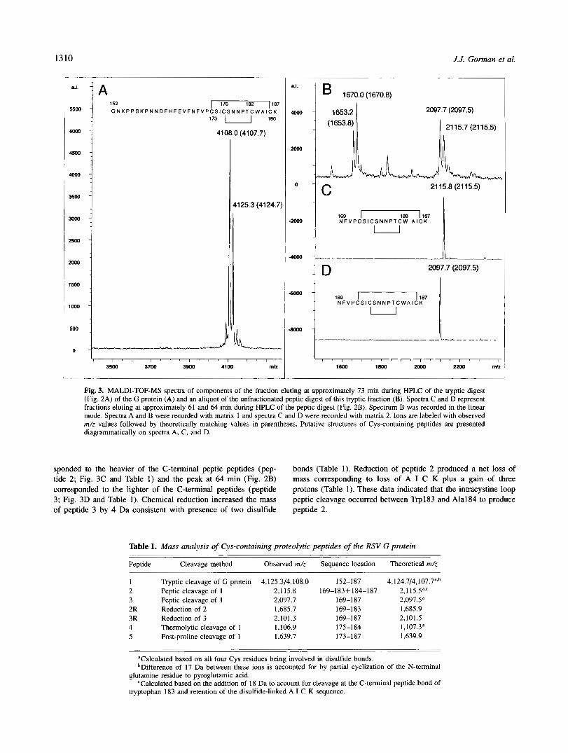
Reverse-phase high-performance liquid chromatography of tryptic digests of the G protein (Fig. 2A) revealed few discrete peaks of absorbance at 214 nm and a series of small, poorly resolved, peaks. The peak at 73 min produced intense ion signals at m/z values of 4,108.0 and 4,125.3 (Fig. 3A) consistent with residues 152-187 of the G protein (peptide 1; Table I), taking into account oxidation of the four Cys residues of this sequence to disulfides (Table 1) and partial conversion of the N-terminal Gln152 to pyroglutamic acid. These data also show that Asn179, Thrl81, Ser157, Ser174, and Ser177 are not glycosylated.
An unfractionated peptic digest of residues 152-187 (peptide 1) revealed ions at m/z = 1,653.2 and m/z = 1,670.0 representing residues 152-165, and ions at m/z = 2,097.7 and m/z = 2,115.7 representing residues 169-1 87, allowing for peptide bond cleavage within a cystine loop of the heavier peptide (peptides 2 and 3; Fig. 3B and Table 1). The N-terminal peptic peptides were detected at 52 and 52.5 min in chromatograms (Fig. 2B) of the digest (mass analysis data not shown), the peak at 61 min (Fig. 2B) corre-
I I I I I I I I
10 20 30 40 50 60 70 80
Time (min)
.1
.05
- Q
C E
s Q!. -.05 a, V c m 0 -.1 v)
e
-.I5
-2
I 2
4 1 I
5
I I I I I I I I 10 20 30 40 50 60 70 80
Time (min)
1 Fig. 2. HPLC separation of peptides produced by tryptic digestion of 40 p g of RSV strain A2 G protein (A) and by further digestion of tryptic peptide 1 using pepsin (B), thermolysin (C), and post-proline cleavage enzyme (D). The fraction eluting at 73 min in chromatogram A was depleted of CH3CN with a stream of high punty nitrogen and further digested with: pepsin (100 p L of the fraction plus 10 pL of a 1 mg/mL solution of pepsin in 5% (v/v) formic acid for two hours at 37 "C); thermolysin (45 pL of the fraction plus 45 p L of 0.1 M N h H C 0 3 , 11 p L of 0.01 M CaClz and 10 pL of a 1 mg/mL solution thermolysin in 0.1 M N%HC03 for 2 h at 37 "C); or post-proline cleavage enzyme (45 p L of the fraction plus 20 p L of 0.1 M N h H C 0 3 and 45 pL of a 0.1 mg/mL solution of the enzyme in 0.1 M ammonium acetate for 2 h at pH 6.5 and 37°C).
1310 J.J. Gorman et al.
5500 -
5000 -
4500 -
4ooo-
3500 -
3000 -
2500 -
2wo-
1500 -
1000 -
500 -
152 I 176 182 1187 Q N K P P S K P N N D F H F E V F N F V P C S I C S N N P T C W A I C K
173 I I 186
4108.0 (4107.7)
a.i. 4 1670.0 (1670.8)
1653.2
0
2097.7 (2097.5)
I 21 15.7 (21 15.5)
21 15.8 (21 15.5)
183 187 N F V P C S I C S N N P T C W A l C K
U
4wo -
: D 2097.7 (2097.5)
6000 - 169 - 187 N F V P C S I C S N N P T C W A I C K
U
a m -
Fig. 3. MALDI-TOF-MS spectra of components of the fraction eluting at approximately 73 min during HPLC of the tryptic digest (Fig. 2A) of the G protein (A) and an aliquot of the unfractionated peptic digest of this tryptic fraction (B). Spectra C and D represent fractions eluting at approximately 61 and 64 min during HPLC of the peptic digest (Fig. 2B). Spectrum B was recorded in the linear mode. Spectra A and B were recorded with matrix 1 and spectra C and D were recorded with matrix 2. Ions are labeled with observed m/z values followed by theoretically matching values in parentheses. Putative structures of Cys-containing peptides are presented diagrammatically on spectra A, C, and D.
sponded to the heavier of the C-terminal peptic peptides (pep- bonds (Table 1). Reduction of peptide 2 produced a net loss of tide 2; Fig. 3C and Table 1) and the peak at 64 min (Fig. 2B) mass corresponding to loss of A I C K plus a gain of three corresponded to the lighter of the C-terminal peptides (peptide protons (Table 1). These data indicated that the intracystine loop 3; Fig. 3D and Table 1). Chemical reduction increased the mass peptic cleavage occurred between Trp183 and Ala184 to produce of peptide 3 by 4 Da consistent with presence of two disulfide peptide 2.
Table 1. Mass analysis of Cys-containing proteolytic peptides of the RSV G protein
Peptide Cleavage method Observed m/z Sequence location Theoretical m/z
1 Tryptic cleavage of G protein 4,125.3/4,108.0 152-187 4,124.7/4,107.7a,b 2 Peptic cleavage of 1 2.1 15.8 169-183+184-187 2,l 15.5a,c 3 Peptic cleavage of 1 2,097.7 169-187 2,097.5" 2R Reduction of 2 1,685.7 169-183 1,685.9 3R Reduction of 3 2,101.3 169-187 2,101.5 4 Thermolytic cleavage of 1 1,106.9 175-1 84 1,107.3a 5 Post-proline cleavage of 1 1,639.7 173-187 1,639.9
aCalculated based on all four Cys residues being involved in disulfide bonds. bDifference of 17 Da between these ions is accounted for by partial cyclization of the N-terminal
CCalculated based on the addition of 18 Da to account for cleavage at the C-terminal peptide bond of glutamine residue to pyroglutamic acid.
tryptophan 183 and retention of the disulfide-linked A I C K sequence.

Disuljides of respiratory syncytial virus G protein 1311
A thermolytic digest of peptide 1 contained ions at m/z values of 912.9 and 1,106.9, plus the sodium and potassium adducts of this ion (Fig. 4A), consistent with cleavages prior to Phel70, Ile175, and Ile185. The ion at m/z = 912.9 is consistent with disulfide bridging between Cys173 and Cys186 (residues 170-174 linked to residues 185-187). and the ion at m/z = 1,106.9 is consistent with disulfide bridging between Cys176 and Cys182 (residues 177- 184). Reverse-phase HPLC of the thermolytic digest revealed nu- merous peaks (Fig. 2C), most of which were derived from the enzyme preparation as they were also present in a chromatogram of a thermolysin control digest lacking tryptic peptide 1 (data not shown). The fraction at approximately 54 min was not present in the control digest and yielded an ion consistent with the peptide containing the Cys176 to Cys182 loop (peptide 4; Fig 4B and Table 1). Automated Edman degradation sequencing produced an N-terminal sequence of Ile-Xaa-Ser-Asn, which is consistent with the identification of this peptide by mass analysis.
Post-proline cleavage enzyme cleaved after Pro172 of the iso- lated peptide 1 to produce peptide 5 (Fig. 2D and Table 1) but failed to cleave at the intradisulfide loop Prol80, which is consis- tent with inaccessibility of Prol80.
Disulfide determination by mass spectrometric-based sequence analysis of peptide 2
Analysis of peptic peptide 2 using a-cyano4hydroxycinnamic acid as a matrix for MALDI and laser irradiance above threshold
1 75 1 84 I C S N N P T C W A
U j 1106.9 (1 107.3)
i ' 7 F " P L - T :
1 912.9 (914.2)
4000 I MNa'
I
MK'
i
-4000 I i l -,+ -1-
900 loo0 1100 1200 mlz
Fig. 4. MALDI-TOF-MS spectra of an unfractionated thermolytic digest of tryptic peptide 1 (A) and the thermolytic peptide eluting at approxi- mately 54 min during HPLC (Fig. 2C) of the thermolytic digest (B). Matrix 2 was used to record both spectra.
produced abundant post-source fragment ions (Fig. 5 and Table 2). Fragment ions 2 to 4 (Figs. 5A and Table 2), inclusive, are b-type ions, which are independent of possible disulfide bonding arrange- ments, but fragment ions 5 to 7, inclusive, indicated disulfide linkage between Cys173 and Cysl86 due to retention of the mass of the A I C K sequence with fragment ions arising from cleavage at Cys173, Ser174, and Ile175 of the larger peptide chain. These ions were complemented by the occurrence of y-type fragment ions 8 to 1 1, which also bear the mass of the A I C K sequence and subsequent failure to observe this mass accompanying fragment ions 12 to 14.
Symmetric and asymmetric cleavage of the interchain disulfide was indicated by fragment ions 15 and 16, respectively (Fig. 5B and Table 2). In addition, the A I C K sequence was observed in linkage with a series of internal cleavage fragments of the larger peptide chain as represented by fragment ions 17 to 20, inclusive (Fig. 5B and Table 2). Symmetric cleavage of the interchain di- sulfide was also apparent for b6, 214, and y14 fragments of the larger peptide chain to produce fragment ions 21 to 23, respec- tively (Fig. 5C and Table 2). Fragment ion 24 corresponded to y-type cleavage of alanine of the A I C K sequence in disulfide linkage with the y14 ion of the larger peptide chain (Fig. 5C and
'- I ' MH+
I
Bm
B I S S I
q ~ m 1 N F V P C S I C S N N P T C W
B I S S I
q ~ m 1 N F V P C S I C S N N P T C W
IS MH+
m -
ll. m rm om 8m ,100 llxl r r m l l m 1 . m - "a
Fig. 5. Post-source decay fragment ion spectrum of peptic peptide 2 (Figs. 2B and 3C). Fragment ions are numbered according to designations in the text and Table 2 or labeled (s) to indicate differences in 18 Da or 28 Da from assigned ions or (u) to indicate unassigned ions and are pre- sented on three different panels containing the same spectrum.

1312 J.J. Gonnan et al.
Table 2). Many of the remaining ions in the spectrum could be accounted for as satellites (s) to defined ions formed due to loss of water or cleavage on either side of a backbone carbonyl (Fig. 5). For example, the ion observed at m/z = 234.3 represented the a2 ion of peptide 2 due to cleavage of the carbonyl from the b2 fragment (m/z = 262.2). Despite the propensity for post-source decay to generate unassignable ions, only two relatively intense ions (u) in relevant parts of the spectrum of peptide 2 could not be assigned on the basis of the proposed disulfide linkage pattern (Fig. 5C). These unassigned ions may have been derived from complex cleavages involving functional groups on both peptide chains and/or cleavages within the intrachain disulfide.
Discussion
The present findings indicate that the four Cys residues of the RSV G protein ectodomain are located in a non-glycosylated region of the protein and exist in a predominant stable configuration with Cys173 linked to Cys186 and Cys176 linked to Cys182 by disul- fide bonds (Fig. 6). This disulfide pattern was evident from the well-established approach of measurement of masses of proteolyt- ically derived peptides to identify disulfide-linked sequences (Mor- ris & Pucci, 1985; Yazdanparast et al., 1986, 1987; Stewart et al., 1992).
A second mass spectrometric approach also indicated the pres- ence of the same disulfide configuration, which involved analysis
Table 2. Fragment ions of peptic peptide 2 of native RSV G protein
m/z
Observedb
1 2 3 4 5 6 7 8 9
10 I 1 12 13 14 15 16 17 18 19 20 21 22 23 24
- 262.4 2 0.2 361.4 t 0.3 458.6 5 0.2 992.9 t 0.5
1,080.6 5 0.2 1,193.5 t 0.6 2,001.2 5 0.8 1,854.3 t 1.1 1,755.2 2 1 1,658.9 t 1 1.123.1 t 0.3 1.036.4 t 0.6
923.1 t 0.6 434.4 5 0.4 466.7 t 0.1 535.5 5 0.2 632.7 t 0.1 719.9 5 0.4 832.9 t 0.5 648.9 t 0.1
1.55 I .2 2 0.5 1,571.1 2 0.1 1,929.9 t 0.5
Predicted Fragment type"
115.0 262.3 b2 361.4 b3 458.5 b4 993.2 b5
1,080.3 b6 1,193.5 b7 2.001.4 Y 14 1,854.2 Y13
1,658.0 Yll 1,123.3 Y 10 1,036.2 Y9
923.1 Y8 434.6 s-s 466.6 S - C M 535.7 Internal 632.8 Internal 719.9 Internal 833.1 Internal 648.7 b6 + S-S
1.55 1.8 7.14 + S-S 1,570.9 y14 + S-S +2H 1,930.3 y14 + yl(A1CK)
-
1.755. I Y 12
'Backbone cleavages have been assigned to the larger of the peptide chains of peptic peptide 2 except for ion 24. in which a y-type cleavage has also been accounted for in the smaller A I C K chain.
bAverage values obtained from three different determinations.
173 lea
H2N-1 C I""
. . "" L!gL "" ~ """ .. - Cca-l
I II 111 IV V 176 182
Fig. 6. Diagrammatic representation of the human RSV A2 strain G pro- tein. The proposed domain structure consists of a cytoplasmic domain, with a single Cys residue ( I ) , followed by a transmembrane domain (11) and putatively heavily glycosylated regions (I11 and V ) of the ectodomain sepa- rated by a non-glycosylated subdomain containing the "cystine noose" (V).
of fragment ions of peptic peptide 2 produced by raising the laser irradiance above threshold and using cy-cyano-4-hydroxycinnamic acid as a matrix. Analysis of such metastable ions produced by post-source decay during MALDI-TOF-MS is a useful means of obtaining sequence information on peptides (Spengler et al., 1992; Kaufmann et al., 1993, 1994). Furthermore, analysis of fragment ions produced during high-energy (Bean & Cam, 1992) or low- energy (Bauer et al., 1993) collisional activation of disulfide- linked peptides in tandem double-focusing sector or biple quadrupole mass spectrometers, respectively, has revealed disulfide-linked frag- ment ions enabling assignment of linkage positions. Inter-chain disulfide bonds have previously been shown to be labile during MALDI (Zhou et al., 1993; Patterson & Katta, 1994; Crimmins et al.. 1995; Hemling et al.. 1996). particularly using high laser irradiances, which caused prompt fragmentation of the disulfide bond in the source as seen in both linear (Patterson & Katta, 1994; Crimmins et al., 1995) and reflector modes of operation (Patterson & Katta, 1994; Hemling et al.. 1996). However, data were obtained in the present study that were consistent with survival of the di- sulfide linkage even at the higher laser irradiances required for MALDI-induced post-source decay of peptide bonds. Asymmetric cleavage of the disulfide that was observed in the present study is also a feature of high energy fragmentation (Bean & Carr, 1992) or low-energy fragmentation (Bauer et al.. 1993) of ions generated by other modes of ionization and analysis. Although asymmetric di- sulfide bond fission has been reported to occur during MALDI- TOF-MS of interchain disulfide-linked peptides (Zhou et al., 1993; Katta et al., 1995), it is often resisted by such peptides in favor of symmetric cleavage (Patterson & Katta, 1994; Crimmins et al., 1995). The reason(s) for the successful use of fragmentation anal- ysis of disulfide linkages in the present study, as opposed to indi- cations of earlier MALDI-TOF-MS studies (Patterson & Katta, 1994; Crimmins et al., 1995; Katta et al., 1995; Hemling et al., 1996) is not clear at present. It may have been due to unusual lability of the backbone bonds of peptic peptide 2 or unusual stability of the disulfides of this peptide. Alternatively, subtle in- strumental or sample preparation procedures may have given rise to collisions that were different in nature than obtained in earlier studies. However, it cannot be overlooked that the observations may be a more general feature of MALDI-induced post-source decay, but insufficient experimentation has been performed to es- tablish this phenomenon.
Regardless of the generality of the present observations, analysis of metastable ions produced by peptic peptide 2 revealed the same disulfide pattern deduced by analysis of proteolytic peptides. Fur- thermore, one of the other potential disulfide configurations (Cys173 linked to Cysl76 and Cysl82 linked to Cysl86) would have been expected to undergo facilitated cleavage at Pro180 (Hunt et al., 1986) to liberate fragment ions containing intra-chain disulfide bonds from peptic peptide 2. Despite the fact that substantial

Disulfides of respiratory syncytial virus G protein 1313
fragmentation was observed at Pro172, no intra-chain disulfide- containing fragments were observed corresponding to cleavage at Prol80. Although fragmentation is not generally observed within intra-chain disulfide loops (Bean & Carr, 1992; Katta et al., 1995; Suckau et al., 1996), it is possible that cleavage initiated at Pro180 within the Cys176 to Cys182 loop (Bean & Carr, 1992) contrib- uted to the unassigned post-source decay fragments of peptic pep- tide 2.
Recent reports (Langedijk et al., 1996) have produced evidence that a shorter cysteine containing synthetic peptide from the cor- responding region of bovine RSV (residues 158-189) also folds spontaneously to form two disulfide bonds. NMR of this bovine RSV peptide (Doreleijers et al., 1996) showed it to contain a rigid “cystine noose” structure (Lapthom et al., 1995) constrained by the same disulfide arrangement as defined herein for native human RSV G protein. However, the sequence flanking the disulfide loops, corresponding to the conserved sequence of human RSV G pro- teins (Fig. l), failed to reveal a stable structure in these NMR experiments. We have demonstrated that a synthetic peptide cor- responding to residues 149-197 of human RSV G protein folded spontaneously to form the correct disulfides, as evident by mass spectrometry (Gorman et al., unpubl. obs.). It will be interesting to see if the longer human RSV sequence adopts a more ordered structure by NMR.
The conserved nature of the 13-residue sequence adjacent to and including 2 cysteines of the “cystine nooses” in G proteins of all human isolates of RSV, conservation of the cysteine residues in all RSV G proteins, and their presence as a “cystine noose” in both human and bovine isolates suggest a role for this subdomain in binding of RSV to susceptible cells. Furthermore, immunological studies have shown that this subdomain is a sub-group-specific immuno-dominant region of the G protein (Norrby et al., 1987; Akerlind-Stopner et al., 1990) that elicits neutralising antibodies due to natural infection (Norrby et al., 1987). Immunization of mice with this subdomain in synthetic peptide form (Trudel et al., 1991) or as part of expressed proteins (Olmstead et al., 1989; Sinard et al., 1995; Sullender et al., 1990) protected them from challenge with live RSV. Immunological recognition of this sub- domain is dependent upon intact disulfides (Akerlind-Stopner et al., 1990) and is subject to mutation in virus variants generated by propagation in the presence of neutralizing monoclonal anti- bodies (Rueda et al., 1994). Neutralizing antibodies that recognize this sub-domain also block binding of native G protein to RSV susceptible cells (Feldman & Hendry, 1994).
The “cystine noose” motif is known to exist with biological significance in other receptor binding polypeptides such as endo- thelin and human chorionic gonadotropin (Lapthom et al., 1995) and a similar cystine loop forms an immuno-dominant domain of HIV gp41 (Oldstone et a]., 1991). Experiments are in progress to determine whether synthetic residues 149-197 of human RSV G protein binds to RSV susceptible cells and, if so, to assess the relative contributions of the conserved sequence and the “cystine noose.”
Materials and methods
Peptide isolation
Strain A2 RSV G protein was isolated by immunoaftinity chro- matography with modifications to an existing procedure (Walsh et al., 1984) to include immunoaftinity columns specific for RSV
F and nucleocapsid proteins prior to a final G protein antibody column and elution from the affinity column with potassium thio- cyanate. Lyophilized G protein samples were reconstituted in suf- ficient 0.1 M NI&HC03 to result in a final concentration of 0.01 % (v/v) Triton X-100, 0.14 M NaCI, and 10 mh4 phosphate buffer (pH 7.2 in the absence of w H C O 3 ) . N-Ethylmaleimide was also added to a final concentration of 1 mM as a precaution against disulfide bond interchange and oxidation of cysteine residues to disulfides. Digestion of the intact protein was performed for 4 h at 37 “C using two additions of 1% (w/w) of sequencing grade trypsin (Boehringer-Mannheim) with the second addition made at 2 h. Sub-digestion of peptide fragments isolated by HPLC was achieved with pepsin (Boehringer-Mannheim) or thermolysin (Calbiochem) or post-proline cleavage enzyme (Seikagaku Corporation, Tokyo) as described in the legends to Figure 2.
Proteolytic fragments were isolated by reverse-phase HPLC using slight variations of a previously described protocol (Gorman et al., 1990). These involved use of a 2.1 mm X 25 cm column of octadecasilica (Vydac), a flow rate of 150 pL/min, and a linear gradient, from 0.1% (v/v) aqueous trifluoroacetic acid to 80% (v/v) aqueous CH3CN containing 0.09% (v/v) trifluoroacetic acid, developed over 90 min. Gradients were generated using a Hewlett Packard chromatography system comprised of a 1090M solvent delivery system under the control of a DOS Chemstation and elu- tion of peptides was monitored at 214 nm using a 1090 diode array detector.
Analytical methods
Matrix-assisted laser desorption/ionization time-of-flight (MALDI- TOF) mass spectrometry was performed using a Bruker Reflex mass spectrometer (Bruker-Franzen Analytik, GMBH, Bremen, Germany) as described previously (Gorman et al., 1996). Samples were prepared by mixing with an equal proportion of a 10 mg/mL solution of a-cyano-4-hydroxycinnamic acid (Beavis et al., 1992) in 50% (v/v) C2H50H/CH3CN (matrix 1) or a 10 mg/mL solution of 2,6-dihydroxyacetophenone in 50% (v/v) C2H50H/CH3CN con- taining 0.1 M di-ammonium hydrogen citrate (matrix 2) (Gorman et al., 1996; Pitt and Gorman, 1996) or samples were reduced with tris(carboxyethy1)-phosphine in 0.2 M aqueous di-ammonium hy- drogen citrate prior to mixing with 2,6-dihydroxyacetophenone and deposition on a MALDI target (Gonnan et al., 1996). Except where specifically noted, spectra were recorded in reflectron mode.
Analysis of metastable ions arising from post-source decay of peptic peptide 2 prepared in matrix 1 was performed using 20 step-wise decrements in the reflectron potential and increasing the laser irradiance to optimize production of ions in each voltage window (Spengler et al., 1992; Kaufmann et al., 1993, 1994). Masses were assigned to metastable ions by reference to a cali- bration table created by determining the behavior of metastable ions of known mass, produced from adrenocorticotropic hormone residues 18-39, at various reflectron potentials (Rouse et al., 1995). Data were acquired at a digitization rate of 250 MHz. Assembly of the individual spectra for each reflectron voltage onto a continuous mass scale was performed using Bruker FAST software routines within the Bruker XMASS software package.
Theoretical masses of fragment ions resulting from post-source decay were calculated using a commercially available program (MacBiospec 1 .O. 1 from Perkin-Elmer-Sciex, Foster City, CA) that computes fragment ion m/z values according to defined fragmen- tation pathways (Roepstorff & Fohlman, 1984; Johnson et al.,

1314 J.J. Gorman et al.
1988). In the case of disulfide-linked peptides, the smaller chain was treated as a modification to the half cystine residues of the larger chain (Bean & Cam, 1992). Step-wise amino acid sequence analysis of peptides was performed by automated Edman degra- dation (Edman & Begg, 1967) using a Hewlett Packard GlOOOA solid-phase protein sequenator.
Acknowledgments
We are grateful for the support of American Cyanamid Company, Lederle- Praxis Biologicals Division through supply of RSV G protein used in these studies. Constructive review of the manuscript by Neil McKern, James Pitt, and Mark Peeples is also greatly appreciated. This work was supported in part by a grant from the National Health and Medical Research Council of Australia.
References Akerlind-Stopner B, Utter G, Mufson MA, Orvell C, Lemer RA, Norrby E.
1990. A subgroup-specific antigenic site in the G protein of respiratory syncytial virus forms a disulphide loop. J Virol 645143-5148.
Alansari H, Potgeiter ND. 1993. Nucleotide sequence analysis of the ovine respiratory syncytial virus G glycoprotein gene. Virology 196:873-877.
Bauer M, Sun Y, Degenhardt C, Kozikowski B. 1993. Assignment of all four disulfide bridges in echistatin. J Protein Chem 12:759-764.
Bean MF, Can SA. 1992. Characterization of disulfide bond position proteins and sequence analysis of cystine-bridged peptides by tandem mass spec- trometry. Anal Biochem 201:216-226
Beavis RC, Chaudhary T, Chait BT. 1992. Alpha-cyano-4-hydroxycinnamic acid as a matrix for matrix-assisted laser desorption mass spectrometry. Organ Mass Spectrom 27156-158.
Cane PA, Matthews DA, Pringle CR. 1991, Identification of variable domains of the attachment (G) of subgroup A respiratory syncytial viruses. J Gen V i r 0 1 72:2091-2096.
Collins PL. 1991. The molecular biology of human respiratory syncytial virus (RSV) of the genus Pneumovirus. In: Kingsbury DW, ed. The paramyxo- viruses. New York: Plenum Press. pp 103-161.
Collins PL, McIntosh K, Chanock RM. 1996. Respiratory syncytial virus. In: Fields BN, Knipe DM, Howley PM, eds. Fields virology. Philadelphia: Lippincott-Raven Publishers. pp 13 13-1 35 1.
Crimmins DL, Saylor M, Rush J, Thoma RS. 1995. Facile, in situ matrix- assisted laser desorption ionization-mass spectrometry analysis and assign- ment of disulfide pairings in heteropeptide molecules. Anal Biochem 226355- 361.
Doreleijers JF, Langedijk JPM, Hard K, Boelens R, Rullmann JAC, Schaaper WM, van Oirschot JT, Kaptein R. 1996. Solution structure of the immuno- dominant region of protein G of bovine respiratory syncytial virus. Biochem 35:14684-14688.
Edman P, Begg G. 1967. A protein sequenator. Eur J Biochem 1 :80-91. Feldman SA, Hendry RM. 1994. Characterization of the human respiratory
syncytial virus G glycoprotein binding domain using synthetic peptides. Am Soc Virol 13:158.
Garcia 0, Martin M, Dopazo J, Arbiza J, Frabasile S, Russi J , Hortal M, Perez-Brena P, Martinez I, Garcia-Barreno B, Melero JA. 1994. Evolution- ary pattern of human respiratory syncytial virus (subgroup A): Cocirculating lineages and correlation of genetic and antigenic changes in the G glyco- protein. J Virol 685448-5459.
Gorman JJ, Corino GL, Shiell BJ. 1990. Role of mass spectrometry in mapping strain variation and post-translational modifications of viral proteins. Bio- med Environ Mass Spectrom 19:646-654.
Gorman JJ, Ferguson BL, Nguyen TN. 1996. Use of 2.6-dihydroxyacetophenone for analysis OF Fragile peptides, disulfide bonding and small proteins by matrix-assisted laser desorption/ionization. Rapid Commun Mass Spectrom 10:529-536.
Gruber C, Levine S. 1983. Respiratory syncytial virus polypeptides: The enve- lope associated proteins. J Gen Virol 64:825-832.
Gruher C, Levine S. 1985. Respiratory syncytial virus polypeptides. IV. The
Hemling ME, Mentzer MA, Capiau C, Carr SA. 1996. A multifaceted strategy oligosaccharides of the glycoproteins. J Gen Virul 66:417-432.
for the characterization of recombinant gD-2, a potent herpes vaccine. In:
Totowa, NJ: Humana Press. pp 307-33 1. Burlingame AL, Can SA eds. Mass spectrometry in the biological sciences.
Hunt DF, Yates JR 111, Shabanowitz J, Winston S, Hauer CR. 1986. Protein sequencing by tandem mass spectrometry. Proc Nut1 Acad Sci USA 83:6233- 6237.
Johnson PR, Spriggs MK, Olmsted RA, Collins PL. 1987. The G glycoprotein of human respiratory syncytial viruses of subgroups A and B: Extensive sequence divergence between antigenically related proteins. Pmc Natl Acad Sci USA 84:5625-5629.
Johnson RS, Martin SA, Biemann K. 1988. Collision induced fragmentation of (M + H ) + ions of peptides. Side chain specific sequence ions. Int J Mass
Katta V, Liu N, Meng S-Y, Zamborelli T, Mayer J, Lu H, Hara S. 1995. Disulfide Spectrom ton Process 86: 137-154.
structure study of EGF domain of Neu differentiation factor. Proc ASMS Conf Mass Spectrom Allied Top 43:1277.
Kaufmann R, Kirsch D, Spengler B. 1994. Sequencing of peptides in a time- of-flight mass spectrometer: Evaluation of post source decay following matrix-assisted laser desorption ionisation (MALDI). Int J Mass Spectrom Ion Proc 131355-385.
Kaufmann R, Spengler B, Lutzenkirchen F. 1993. Mass spectrometric sequenc- ing of linear peptides by product-ion analysis in a reflectron time-of-flight mass spectrometer using matrix-assisted laser desorption ionization. Rapid Commun Mass Spectrom 7902-910.
Lambert DM. 1988. Role of oligosaccharides in the structure and function of respiratory syncytial virus glycoproteins. Virology 164458-466.
Lambert DM, Pons MW. 1983. Respiratory syncytial virus glycoproteins. Vi- rology 130:204-214.
Langedijk JPM, Schapper WMM, Meloen RH, van Oirschot JT. 1996. Proposed three-dimensional model for the attachment protein G of respiratory syn- cytial virus. J Gen Virol 771249-1257.
Lapthom AJ, Janes RW, Isaacs N W , Wallace BA. 1995. Cystine nooses and protein specificity. Nut Struct B i d 2:266-268.
Lerch RA, Anderson K, Wertz GW. 1990. Nucleotide sequence analysis and expression from recombinant vectors demonstrate that the attachment pro- tein G of bovine respiratory syncytial virus is distinct from that of human respiratory syncytial virus. J Virol 64359-5569.
Levine S. 1977. Polypeptides of respiratory syncytial virus. J Vir01 21:427-431. Markwell MA. 1991. New frontiers opened by the exploration of host cell
receptors. In: Kingshury DW, ed. The paramyxoviruses. New York: Plenum Press. pp 407-426.
Moms HR, Pucci P. 1985. A new method for rapid assignment of S-S bridges
Norrby E, Mufson MA, Alexander H, Houghten RA, Lemer RA. 1987. Site- in proteins. Biochem Biophys Res Commun 126:1122-1128.
directed serology with synthetic peptides representing the large glycopro- tein G of respiratory syncytial virus. Proc Natl Acad Sei USA 84:6572- 6576.
Oldstone MBA, Tishon A, Lewiki H, Dyson HJ, Feher VA, Assa-Munt N, Wright PE. 1991. Mapping the anatomy of the immunodominant domain of the human immunodeficiency virus gp41 transmembrane protein: Peptide conformational analysis using monoclonal antibodies and proton nuclear magnetic resonance spectroscopy. J Vir01 65: 1727-1734.
Olmstead RA, Murphy BR, Lawrence LA, Elango N, Moss B, Collins PL. 1989. Processing, surface expression, and immunogenicity of carboxy-terminally
63:411-420. truncated mutants of G protein of human respiratory syncytial virus. J V i r 0 1
Pitt JJ, Gorman JJ. 1996. Matrix-assisted laser desorption/ionization time-of- flight mass spectrometry of sialylated glycopeptides and proteins using 2.6-dihydroxyacetophenone as a matrix. Rapid Commun Mass Spectrom 101786-1788.
Patterson SD, Katta V. 1994. Prompt fragmentation of disulfide-linked peptides during matrix-assisted laser desorption ionization mass spectrometry. Anal Chem 66:3727-3732
Roepstorff P, Fohlman J. 1984. Proposal for a common nomenclature for se-
Rouse JC, Yu w, Martin SA. 1995. A comparison of the fragmentation obtained quence ions in mass spectra of peptides. Biomed Mass Spectrom 11 :601.
from a reflector matrix-assisted laser desorption-ionization time-of-flight and a tandem four sector mass spectrometer. J Am Soc Mass Spectrom 6822-835.
Rueda P, Garcia-Barreno B, Melero JA. 1994. Loss of conserved cysteine res- idues in the attachment (G) glycoprotein of two human respiratory syncytial
tions). Virol 198653-662. virus escape mutants that contain multiple A-G substitutions (hypermuta-
Satake M, Coligan JE, Elango N, Norrby E, Venkatesan S. 1985. Respiratory syncytial virus envelope glycoprotein (G) has a novel structure. Nucleic Acids Res 21:7795-7812.
Simard C, Nadon F, Seguin C, Trudel M. 1995. Evidence that the amino acid
constitutes a major part of the polypeptide domain that is involved in the region 124-203 of glycoprotein G from the respiratory syncytial virus (RSV)
protection against RSV infection. Antiviral Res 28:303-3 15. Spengler B, Kirsch D, Kaufmann R, Jaeger E. 1992. Peptide sequencing by
matrix-assisted laser desorption mass spectrometry. Rapid Commun Mass
Stewart AE, Raffioni S, Chaudhary T, Chait BT, Luporini P, Bradshaw RA. Spectrom 6:105-108.

Disulfides of respiratoly syncytial virus G protein 1315
1992. The disulfide bond pairing of the pheromones Er-1 and Er-2 of the ciliated protozoan Euplotes raikovi. Protein Sei 1:777-785
Suckau D. Holle A, Resemann A. 1996. Biopolymer sequencing by MALDI- TOF-MS. Proc ASMS Conf Mass Spectrom Allied Top 44:1339.
Sullender WM, Anderson K, Wertz GW. 1990. The respiratory syncytial virus subgroup B attachment glycoprotein: Analysis of sequence, expression from a recombinant vector, and evaluation as an immunogen against homologous
Sullender WM, Mufson MA, Anderson, LA Wertz GW. 1991. Genetic diversity and heterologous subgroup virus challenge. Virology 178: 195-203.
of the attachment protein of subgroup B respiratory syncytial viruses. J Virol 655425-5434.
Sullender MW, Wertz GW. 1991. The unusual attachment glycoprotein of the respiratory syncytial viruses: Structure, maturation, and role in immunity. In: Kingshury DW, ed. The paramyxoviruses. New York: Plenum Press. pp 383-406.
Trudel M, Nadon F, Seguin C, Binz H. 1991. Protection of BALB/c mice from respiratory syncytial virus infection by immunization with a synthetic pep- tide derived from the G glycoprotein. Virology 185749-757.
Walsh EE, Schlesinger JJ, Brandriss MW. 1984. Purification and characteriza- tion of GP90, one of the envelope proteins of respiratory syncytial virus. J Gen virol65:761-767.
Wertz GW, Collins PL, Huang Y, Gruber C, Levine S, Ball LA. 1985. Nucle- otide sequence of the G protein gene of human respiratory syncytial virus reveals an unusual type of viral membrane protein. Proc Natl Acad Sci USA 82:4075-4079.
Yazdanparast R, Andrews P, Smith DL, Dixon JE. 1986. A new approach for detection and assignment of disulfide bonds in peptides. Anal Biochem 153:348-353.
Yazdanparast R, Andrews P, Smith DL, Dixon JE. 1987. Assignment of disulfide bonds in proteins by fast atom bombardment mass spectrometry. J Biol Chem 262:2507-25 13.
Zhou J, Poppe-Schriemer N, Standing KG, Westmore JB. 1993. Cleavage of interchain disulfide bonds following matrix-assisted laser desorption. Int J Mass Spectrom Ion Proc 126: 115-1 22.