er membrane-localized oxidoreductase ero1 is required … · er membrane-localized oxidoreductase...
TRANSCRIPT
ER membrane-localized oxidoreductase Ero1is required for disulfide bond formationin the rice endospermYayoi Ondaa, Toshihiro Kumamarub, and Yasushi Kawagoea,1
aDivision of Plant Sciences, National Institute of Agrobiological Sciences, 2-1-2 Kannondai, Tsukuba, Ibaraki 305-8602, Japan; and bInstitute of GeneticResources, Faculty of Agriculture, Kyushu University, Hakozaki, Fukuoka 812-8581, Japan
Edited by Bob B. Buchanan, University of California, Berkeley, CA, and approved July 1, 2009 (received for review April 22, 2009)
The developing endosperm of rice (Oryza sativa, Os) synthesizes alarge amount of storage proteins on the rough (r)ER. The majorstorage proteins, glutelins and prolamins, contain either intra orintermolecular disulfide bonds, and oxidative protein folding is nec-essary for the sorting of the proteins to the protein bodies. Here, weinvestigated an electron transfer pathway for the formation ofprotein disulfide bonds in the rER of the rice endosperm, focusing onthe roles of the thiol-disulfide oxidoreductase, OsEro1. Confocalmicroscopic analysis revealed that N-glycosylated OsEro1 is localizedto the rER membrane in the subaleurone cells, and that targeting ofOsEro1 to the rER membrane depends on the N-terminal region fromMet-1 to Ser-55. The RNAi knockdown of OsERO1 inhibited theformation of native disulfide bonds in the glutelin precursors (pro-glutelins) and promoted aggregation of the proglutelins throughnonnative intermolecular disulfide bonds in the rER. Inhibition of theformation of native disulfide bonds was also observed in the seeds ofthe esp2 mutant, which lacks protein disulfide isomerase-like(PDIL)1;1, but shows enhanced OsEro1 expression. We detected thegeneration of H2O2 in the rER of the WT subaleurone cells, whereasthe rER-derived H2O2 levels decreased markedly in EM49 homozygousmutant seeds, which have fewer sulfhydryl groups than the WTseeds. Together, we propose that the formation of native disulfidebonds in proglutelins depends on an electron transfer pathwayinvolving OsEro1 and OsPDIL.
protein disulfide isomerase � seed storage protein � esp2 �hydrogen peroxide � protein body
The developing endosperm of rice (Oryza sativa, Os) synthesizesa large amount of disulfide-bond-rich proteins, including 2
major groups of storage proteins, the acid- or alkaline-solubleglutelins and the alcohol-soluble prolamins. During seed develop-ment, these storage proteins are synthesized on the rough (r)ERand translocated into the rER lumen, where oxidative proteinfolding proceeds. Disulfide bond formation is necessary for thesorting of the proteins to the protein bodies (PBs) (1). The assemblyof prolamins into the ER-derived PBs (PB-I) is stabilized byintermolecular disulfide bonds, whereas the glutelin precursors(proglutelins) form intramolecular disulfide bonds before beingtransported via the Golgi from the ER to the protein storagevacuole (PSV, also designated PB-II) (2–4).
Protein disulfide isomerase (PDI) is the principal catalyst fordisulfide-linked protein folding in the ER lumen, functioning as thedirect donor of disulfide bonds to nascent polypeptides by athiol-disulfide exchange reaction (5). The activity of PDI dependson 2 pairs of Cys residues, each of which is found in the CGHC motifwithin a thioredoxin-like redox-active domain (5). The rice genomeencodes 7 OsPDI-like (L) proteins containing 2 thioredoxin-likeactive domains with the CxxC motif, OsPDIL1;1-1;4 andOsPDIL2;1-2;3 (6). OsPDIL1;1 is an orthologue of Zea mays(Zm)PDIL1;1, which is by far the most highly expressed in themaize endosperm (6). Studies on the OsPDIL1;1-knockout ricemutant, esp2, showed that OsPDIL1;1 has an important role for thesegregation of prolamins and proglutelins in the ER (7). Glutathi-
one constitutes the major small-molecule redox buffer in the ER.The redox state of the ER is more oxidative than that of the cytosolin eukaryotic cells. The ratio of reduced glutathione to oxidizedform ([glutathione (GSH)]/[oxidized glutathione (GSSG)]) rangesfrom 1:1 to 3:1 in the ER, whereas it ranges from 30:1 to 100:1 inthe cytosol (8). In the endosperm cells, which are devoted to thesynthesis of disulfide-rich storage proteins, large amounts of reduc-ing equivalents flux from the cytosol into the ER in the form of Cysresidues during the synthesis of storage proteins, and storageproteins are sequestered from the ER lumen to the PBs. A keyquestion is how the endosperm cells constantly establish the ERredox environment favorable for regeneration of oxidized PDIL;thus, facilitating oxidative folding of nascent polypeptides.
The flavoenzyme Ero1p was first identified in yeast by mutationstudies of the temperature-sensitive conditional mutant ero1-1,which fails to support protein disulfide bond formation in the ERand consequently accumulates misfolded proteins (9, 10). Ero1psupplies oxidizing equivalents for disulfide bond formation in theER, by relaying the oxidizing power from molecular oxygen to thereduced yeast Pdi1p (11–13). Ero1p directly oxidizes the active siteof Pdi1p by thiol-disulfide exchange reactions with the oxidizedshuttle Cys pair, Cys-100-Cys-105, the reduced form of which isreoxidized by the active-site Cys pair, Cys-352-Cys-355 (14–16).Ero1 proteins of different species characterized to date are alllocalized to the ER. However, the mechanisms for their ERretention appear to differ; yeast Ero1p is tightly associated with theER membrane (9, 10), whereas human Ero1� and Ero1� areretained in the ER by covalent interactions with PDI and ERp44(17). The cDNA clones encoding plant homologues of yeast Ero1p,AERO1 and AERO2, have been isolated from Arabidopsis (Ara-bidopsis thaliana) (18). In vitro translation analysis showed thatthese Arabidopsis Ero1 homologues are glycoproteins (18), buttheir localization in the cell has not been characterized.
In this study, we investigated the roles of OsEro1 protein as asource of protein disulfide bonds in the endosperm. The 3nendosperm cells are unique; the endosperm exhibits a high level ofenzymatic activity to synthesize a vast amount of storage disulfide-rich proteins during early seed development; and the endosperm,but not the adjacent 2n embryo, is destined for programmed celldeath during the subsequent seed desiccation and maturationphase. We studied the OsEro1 localization in the subaleurone cellsby confocal microscopic and biochemical analyses, and demon-strated that the N-terminal region of OsEro1 functions as a rERmembrane-targeting signal. We produced OsERO1-knockdownrice plants by inducing RNAi under the control of an endosperm-
Author contributions: Y.O. and Y.K. designed research; Y.O., T.K., and Y.K. performedresearch; Y.O. and Y.K. analyzed data; and Y.O. and Y.K. wrote the paper.
The authors declare no conflict of interest.
This article is a PNAS Direct Submission.
1To whom correspondence should be addressed. E-mail: [email protected].
This article contains supporting information online at www.pnas.org/cgi/content/full/0904429106/DCSupplemental.
14156–14161 � PNAS � August 18, 2009 � vol. 106 � no. 33 www.pnas.org�cgi�doi�10.1073�pnas.0904429106

specific promoter, and examined the effects on the formation ofdisulfide bonds and OsPDIL expression. We detected the genera-tion of H2O2 in the rER in correlation with the oxidation ofsulfhydryl groups, and will discuss its physiological role in seedmaturation.
ResultsSubcellular Localization of OsEro1 in the Endosperm SubaleuroneCells. A rice homologue (OsERO1) of yeast ERO1 was identified byBLAST search of the rice genomic sequence and the EST database.OsERO1 is predicted to encode a polypeptide of 474-aa residuescontaining 2 pairs of Cys residues, Cys-134-Cys-139 and Cys-391-Cys-394, which are suggested to be catalytically active sites in yeastEro1p (14–16). OsEro1 shows 71 and 56% identity to ArabidopsisAERO1 and Physcomitrella patens Ero1, respectively, and 36 and29% identity to human Ero1� and yeast Ero1p, respectively.
OsEro1 does not contain an obvious ER retention signal such asthe C-terminal tetrapeptide KDEL. We examined subcellular lo-calization of OsEro1 in the subaleurone cells of the rice endospermduring early seed development (for the subaleurone layers exam-ined, see Fig. S1). First, we identified the rER structure in thesubaleurone cells by using GFP-fused OsPDIL1;1 and Cherry-fusedSec61� (the single membrane-spanning � subunit of rice Sec61) asa rER lumen marker and a rER membrane marker, respectively.PDI is a highly abundant resident protein of the ER lumen (5). TheSec61��� complex is the essential core of the protein translocationmachinery in the ER membrane and is tightly associated withmembrane-bound ribosome (19). Confocal microscopic analysisrevealed that spGFP-OsPDIL1;1 was evenly distributed within thedilated rER, and Cherry-Sec61� was localized to the boundary ofthe dilated rER (Fig. 1A). Note that PB-I was formed within thedilated rER (Fig. 1B; Fig. S2). By immunofluorescence microscopywith the anti-OsEro1 antibody, we observed that OsEro1 was
mostly colocalized with GFP-Sec61� (Fig. 1C), which suggests thatOsEro1 is associated with the rER membrane.
Characterization of the N-Terminal Region of OsEro1 on rER Mem-brane Targeting. The algorithm TMpred (20) predicts that OsEro1contains a single transmembrane domain (TMD; Ala-37 to Ser-55)with a score of 2436 (Fig. S3A). By comparison, the yeast Ero1p andhuman Ero1� TMpred scores are lower (786 and 1806, respec-tively). To examine the role of the putative TMD of OsEro1 insubcellular localization, we expressed each of 3 chimeric genesencoding modified GFPs in rice endosperm cells: the full length ofOsEro1 (Met-1 to Ile-474) followed by GFP (OsEro1-GFP), theN-terminal truncated form of OsEro1 (Ser-56 to Ile-474) fused tospGFP (spGFP-OsEro1�N), and the N-terminal 75-aa residuesfrom OsEro1 followed by GFP (OsEro1�C-GFP) (Fig. 2 A and B).Confocal microscopic analysis of the subaleurone cells revealed thatOsEro1-GFP was localized to the rER membrane (Fig. 2C). Incontrast, deletion of the N-terminal 55-aa residues of OsEro1(spGFP-OsEro1�N) led to dispersion of the GFP fusion proteinevenly throughout the rER lumen (Fig. 2D). OsEro1�C-GFP wastargeted to the rER membrane (Fig. 2E).
OsEro1 contains 2 potential N-glycosylation sites at Asn-381 andAsn-422. The treatment of seed extracts with either Endo H or
Fig. 1. OsEro1 localizes to the rER membrane in the subaleurone cells. (A)Confocal fluorescence images of the subaleurone cells (10 DAF) expressingspGFP-OsPDIL1;1 and Cherry-Sec61�. (B) Confocal fluorescence images of thesubaleurone cells (10 DAF) expressing GFP-Sec61�. PB-I was labeled withRhodamine. (C) GFP-Sec61�-expressing subaleurone cells (14 DAF) were la-beled with the anti-OsEro1 antibody and then with the Rhodamine-conjugated anti-rabbit antibody. Arrowheads and arrows indicate the rER andPB-I, respectively. (Scale bar, 5 �m.)
Fig. 2. The N-terminal region of OsEro1 is necessary and sufficient for rERmembrane localization. (A) Schematic representation of constructs. Blue boxes,OsEro1; red boxes, predicted TMD of OsEro1 (Ala-37 to Ser-55); yellow lines,catalytic active sites of OsEro1 (Cys-134-Cys-139 and Cys-391-Cys-394); greenboxes, GFP. (B) Extracts from seeds (10 DAF) expressing each of 3 chimeric genesencodingOsEro1-GFP (lane1), spGFP-OsEro1�N(lane2),andOsEro1�C-GFP (lane3) were subjected to SDS/PAGE, followed by Western blot analysis with anti-GFPantibody. The predicted molecular mass of OsEro1-GFP, spGFP-OsEro1�N, andOsEro1�C-GFP are 80, 78, and 34 kDa, respectively. (C–E) Confocal fluorescenceimages of the subaleurone cells (10 DAF) expressing OsEro1-GFP (C), spGFP-OsEro1�N (D), and OsEro1�C-GFP (E). PB-I was labeled with Rhodamine. Arrow-heads indicate the rER. The fluorescence signals of DsRed-Sec61� are visible in C,but those of DsRed-Sec61� (D) and Cherry-Sec61� (E) are not detectable. (Scalebar, 5 �m.)
Onda et al. PNAS � August 18, 2009 � vol. 106 � no. 33 � 14157
PLA
NT
BIO
LOG
Y
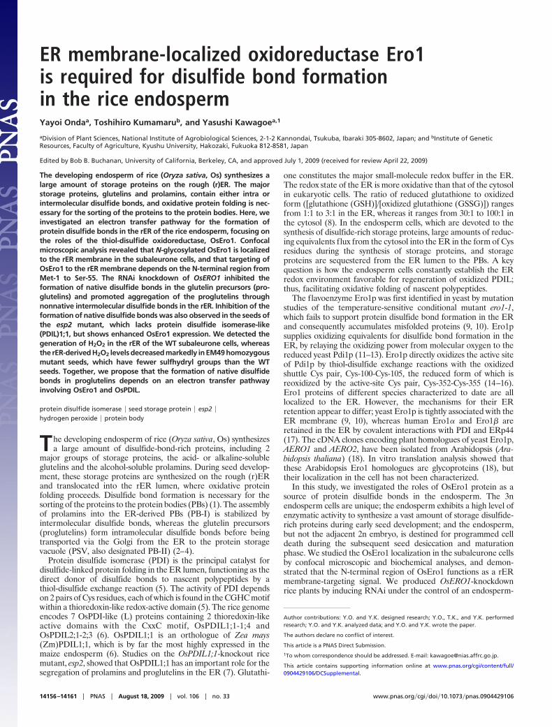
PNGase F caused a mobility shift of a polypeptide that reacted withthe anti-OsEro1 antibody (see panel Ero1 in Fig. 3A), confirmingthat OsEro1 is glycosylated. We analyzed the properties of mem-brane binding of OsEro1 by fractionation of cell extracts undervarious conditions. Both OsEro1 and GFP-Sec61� were recoveredprimarily in the membrane pellet fraction (Fig. 3B), and weresolubilized from the membrane pellet by detergent (1% TritionX-100), but not by high salt (1 M NaCl) or by an alkaline pH (pH 11)(Fig. 3B). OsEro1-GFP, which was also glycosylated (Fig. 3A), andOsEro1�C-GFP showed membrane-binding properties similar tothose of the endogenous OsEro1 (Fig. 3B). In contrast, spGFP-OsEro1�N, which was also glycosylated (Fig. 3A), was recoveredprimarily in the soluble fraction (Fig. 3B). Together, these resultsindicate that the N-terminal region (Met-1 to Ser-55) of OsEro1 isnecessary and sufficient for targeting the protein to the rERmembrane.
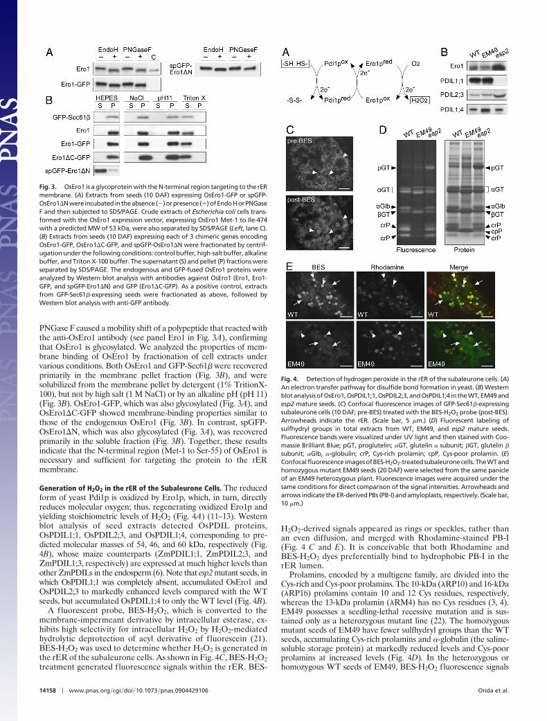
Generation of H2O2 in the rER of the Subaleurone Cells. The reducedform of yeast Pdi1p is oxidized by Ero1p, which, in turn, directlyreduces molecular oxygen; thus, regenerating oxidized Ero1p andyielding stoichiometric levels of H2O2 (Fig. 4A) (11–13). Westernblot analysis of seed extracts detected OsPDIL proteins,OsPDIL1;1, OsPDIL2;3, and OsPDIL1;4, corresponding to pre-dicted molecular masses of 54, 46, and 60 kDa, respectively (Fig.4B), whose maize counterparts (ZmPDIL1;1, ZmPDIL2;3, andZmPDIL1;3, respectively) are expressed at much higher levels thanother ZmPDILs in the endosperm (6). Note that esp2 mutant seeds, inwhich OsPDIL1;1 was completely absent, accumulated OsEro1 andOsPDIL2;3 to markedly enhanced levels compared with the WTseeds, but accumulated OsPDIL1;4 to only the WT level (Fig. 4B).
A fluorescent probe, BES-H2O2, which is converted to themembrane-impermeant derivative by intracellular esterase, ex-hibits high selectivity for intracellular H2O2 by H2O2-mediatedhydrolytic deprotection of acyl derivative of fluorescein (21).BES-H2O2 was used to determine whether H2O2 is generated inthe rER of the subaleurone cells. As shown in Fig. 4C, BES-H2O2treatment generated fluorescence signals within the rER. BES-
H2O2-derived signals appeared as rings or speckles, rather thanan even diffusion, and merged with Rhodamine-stained PB-I(Fig. 4 C and E). It is conceivable that both Rhodamine andBES-H2O2 dyes preferentially bind to hydrophobic PB-I in therER lumen.
Prolamins, encoded by a multigene family, are divided into theCys-rich and Cys-poor prolamins. The 10-kDa (�RP10) and 16-kDa(�RP16) prolamins contain 10 and 12 Cys residues, respectively,whereas the 13-kDa prolamin (�RM4) has no Cys residues (3, 4).EM49 possesses a seedling-lethal recessive mutation and is sus-tained only as a heterozygous mutant line (22). The homozygousmutant seeds of EM49 have fewer sulfhydryl groups than the WTseeds, accumulating Cys-rich prolamins and �-globulin (the saline-soluble storage protein) at markedly reduced levels and Cys-poorprolamins at increased levels (Fig. 4D). In the heterozygous orhomozygous WT seeds of EM49, BES-H2O2 fluorescence signals
Fig. 3. OsEro1 is a glycoprotein with the N-terminal region targeting to the rERmembrane. (A) Extracts from seeds (10 DAF) expressing OsEro1-GFP or spGFP-OsEro1�Nwere incubatedintheabsence(�)orpresence(�)ofEndoHorPNGaseF and then subjected to SDS/PAGE. Crude extracts of Escherichia coli cells trans-formed with the OsEro1 expression vector, expressing OsEro1 Met-1 to Ile-474with a predicted MW of 53 kDa, were also separated by SDS/PAGE (Left, lane C).(B) Extracts from seeds (10 DAF) expressing each of 3 chimeric genes encodingOsEro1-GFP, OsEro1�C-GFP, and spGFP-OsEro1�N were fractionated by centrif-ugation under the following conditions: control buffer, high-salt buffer, alkalinebuffer, and Triton X-100 buffer. The supernatant (S) and pellet (P) fractions wereseparated by SDS/PAGE. The endogenous and GFP-fused OsEro1 proteins wereanalyzed by Western blot analysis with antibodies against OsEro1 (Ero1, Ero1-GFP, and spGFP-Ero1�N) and GFP (Ero1�C-GFP). As a positive control, extractsfrom GFP-Sec61�-expressing seeds were fractionated as above, followed byWestern blot analysis with anti-GFP antibody.
Fig. 4. Detection of hydrogen peroxide in the rER of the subaleurone cells. (A)An electron transfer pathway for disulfide bond formation in yeast. (B) WesternblotanalysisofOsEro1,OsPDIL1;1,OsPDIL2;3,andOsPDIL1;4 in theWT,EM49andesp2 mature seeds. (C) Confocal fluorescence images of GFP-Sec61�-expressingsubaleurone cells (10 DAF; pre-BES) treated with the BES-H2O2 probe (post-BES).Arrowheads indicate the rER. (Scale bar, 5 �m.) (D) Fluorescent labeling ofsulfhydryl groups in total extracts from WT, EM49, and esp2 mature seeds.Fluorescence bands were visualized under UV light and then stained with Coo-massie Brilliant Blue; pGT, proglutelin; �GT, glutelin � subunit; �GT, glutelin �
subunit; �Glb, �-globulin; crP, Cys-rich prolamin; cpP, Cys-poor prolamin. (E)Confocalfluorescence imagesofBES-H2O2-treatedsubaleuronecells.TheWTandhomozygous mutant EM49 seeds (20 DAF) were selected from the same panicleof an EM49 heterozygous plant. Fluorescence images were acquired under thesame conditions for direct comparison of the signal intensities. Arrowheads andarrows indicate the ER-derived PBs (PB-I) and amyloplasts, respectively. (Scale bar,10 �m.)
14158 � www.pnas.org�cgi�doi�10.1073�pnas.0904429106 Onda et al.

appeared in the rER more strongly (see panel WT, arrowheads, inFig. 4E) than in the amyloplasts (see panel WT, arrows, in Fig. 4E;for amyloplast identification, see Fig. S4). However, in the EM49homozygous mutant seeds, the BES-H2O2 fluorescence intensity inthe rER was weak at levels similar to those observed in theamyloplasts (see panel EM49, arrowheads and arrows, in Fig. 4E).These results indicate that the generation of H2O2 in the rERcorrelates with the oxidation of sulfhydryl groups.
Effects of RNAi Knockdown of OsERO1 on the Formation of DisulfideBonds in Proglutelins. To investigate the function of OsEro1 in thesubaleurone cells, we obtained OsERO1-knockdown lines (ero1)with severely decreased levels of OsEro1 in seeds by inducingRNAi in the endosperm (Fig. 5A). Note that BiP, the major ER
chaperone, was strongly induced in the ero1 seeds (Fig. 5A).Compared with the WT seeds, the ero1 seeds accumulatedproglutelins at a higher level (Fig. 5 A and B; Fig. S5). The WTsubaleurone cells formed 2 distinct types of PBs, PB-I and PB-II,which were differentially stained with Rhodamine (Fig. 5C).Rhodamine labels PB-I (1–2 �m) as intense signals due to thepreference of Rhodamine for binding hydrophobic prolaminpolypeptides (see arrowheads in Fig. 5C; Fig. S2) (23). The ricetonoplast intrinsic protein OsTIP3 is specifically localized to themembrane of PB-II (3–4 �m) (Fig. S6A) (24). Rhodamineweakly and evenly stained the matrix of PB-II (see arrows in Fig.5C; Fig. S6B), which was distinguished from the dilated rER (Fig.S6 C–F). The ero1 seeds failed to form the typical PB-I andPB-II, and instead, formed abnormal aggregates of Rhodamine-stained small particles (see panel ero1 in Fig. 5D), as observedin esp2 mutant seeds (see panel esp2 in Fig. 5D).
Proglutelins (57 kDa) acquire intramolecular disulfide bonds inthe ER before being transported to PB-II and proteolyticallyprocessed into the acidic (�; 37–39 kDa) and basic (�; 22–23 kDa)subunits in PB-II (2). Proglutelins accumulated at a markedly lowerlevel in the WT seeds than did the � and � subunits (Fig. 6A, laneWT, T). By comparison, proglutelins accumulated to a higher levelin the ero1 seeds than did the � and � subunits (Fig. 6A, lane ero1,T), which was also the case for the esp2 seeds (Fig. 6A, lane esp2,T). The proglutelins were efficiently extracted from the WT seedsinto the supernatant without a reducing agent (Fig. 6A, lane WT,S). Note that extraction of the proglutelins from the ero1 and esp2seeds required a reducing agent (Fig. 6A, lanes ero1 and esp2, P).Although proglutelins also accumulated to a higher level in seedswith enhanced expression of SEC12 (PGT1-SEC12; Fig. S7 A andB), the PGT1-SEC12 seeds showed proglutelin extraction into thesupernatant without a reducing agent (Fig. S7C). The 57-kDaproglutelins (see arrowhead in Fig. 6B) and larger apparent mo-lecular mass (Fig. 6B, asterisks) were extracted from the nonre-duced pellet fractions (NR-P; Fig. 6 A and B Upper) of the ero1 andesp2 seeds in a DTT concentration-dependent manner (Fig. 6B,lanes ero1 and esp2), whereas those were hardly detectable in theWT fractions (Fig. 6B, lanes WT). To further examine whether theNR-P fractions from the ero1 and esp2 seeds contain proglutelinswith nonnative intermolecular disulfide bonds, we used a modified
Fig. 5. The RNAi knockdown of OsERO1 in seeds. Total extracts of the WT andero1 seeds (17 DAF) were subjected to SDS/PAGE, followed by Western blotanalysis with antibodies against OsEro1, BiP, and glutelin (A) or CoomassieBrilliant Blue staining (B); pGT, proglutelin; �GT, glutelin � subunit; �GT, glutelin� subunit. (C) Confocal fluorescence images of the subaleurone cells (17 DAF)expressing OsTIP3-GFP (PB-II membrane marker), stained with Rhodamine. (D)Confocal fluorescence images of Rhodamine-stained subaleurone cells (17 DAF)from the WT, ero1, and esp2 seeds. Arrowheads and arrows indicate PB-I andPB-II, respectively. (Scale bar, 5 �m.)
Fig. 6. Proglutelins form aggregates through nonnative intermolecular disulfide bonds in the ero1 seeds. (A) Proteins were extracted from the WT, ero1, andesp2 mature seeds, as in schematic representation (Upper). The T, S, and P fractions were subjected to SDS/PAGE, followed by Coomassie Brilliant Blue staining;pGT, proglutelin; �GT, glutelin � subunit; �GT, glutelin � subunit; crP, Cys-rich prolamin; cpP, Cys-poor prolamin. (B) The NR-P fractions were homogenized inthe presence of varying concentrations of DTT (0, 1, 3, and 10 mM) for 30 min at 25 °C. After centrifugation, the resulting supernatants (P0, P1, P3, and P10; Upper)were subjected to SDS/PAGE, followed by Western blot analysis with anti-glutelin antibody. (C) The P3 fractions from the ero1 and esp2 seed extracts weresubjected to 2D SDS/PAGE, followed by Western blot analysis with anti-glutelin antibody. (D) Western blot analysis of OsEro1, OsPDIL1;1, OsPDIL2;3, andOsPDIL1;4 in the WT and ero1 mature seeds (fractions T in A).
Onda et al. PNAS � August 18, 2009 � vol. 106 � no. 33 � 14159
PLA
NT
BIO
LOG
Y

2D SDS/PAGE separation (25), in which the NR-P proteins werepartially reduced at 3 mM DTT in the first dimension, and thenreduced at 200 mM DTT in the second dimension. The separationof the ero1 seed extracts recovered the proglutelins in a broadmolecular mass range below the diagonal line, which was also thecase for esp2 (Fig. 6C), indicating that the proglutelins formaggregates through nonnative intermolecular disulfide bonds in theero1 and esp2 seeds.
DiscussionPathway for Disulfide Bond Formation Involving OsEro1. This reportprovides key insights into how plant cells establish the redoxenvironment in the ER for the formation of protein disulfide bonds.The RNAi knockdown of OsERO1 and knockout of OsPDIL1;1 ledto aggregation of the proglutelins through nonnative intermoleculardisulfide bonds (Figs. 5D and 6C), which demonstrates that OsEro1and OsPDIL1;1 are essential for the formation of the correctpattern of disulfide bonds in the subaleurone cells. Yeast Ero1pactivity is rapidly modulated by a redox-dependent feedback systemthrough 2 noncatalytic Cys pairs (Cys-90-Cys-349 and Cys-150-Cys-295); reduction of the regulatory disulfide bonds increases Ero1pactivity for the promotion of disulfide bond formation by directoxidation of Pdi1p (26). On the basis of sequence similarity, the Cyspair Cys-90-Cys-349 of Ero1p is conserved, but the Cys-150-Cys-295equivalent is missing in OsEro1 (Fig. S3B). Recent studies revealedthat human Ero1� possesses a different mode of regulation, inwhich Cys-94 functions as a molecular switch by forming an activedisulfide bond with Cys-99 (shuttle Cys pair) or a regulatorydisulfide bond with Cys-131 (27). Such a feedback system may existin OsEro1 regulation, because Cys-134, Cys-139, and Cys-167 ofOsEro1 appear to correspond to Cys-94, Cys-99, and Cys-131 ofEro1�, respectively (Fig. S3B).
The changes in the ER redox environment induce a set ofproteins involved in protein folding in the ER, including Ero1p (9,10), Pdi1p (28), and BiP (7). In the esp2 seeds, OsEro1 wasup-regulated (Fig. 4B), presumably in response to the accumulationof misfolded proglutelins. Interestingly, in the ero1 seeds, whichexhibited the unfolded protein response-related induction of BiP(Fig. 5A), the protein levels of OsPDIL1;1 markedly decreased,whereas OsPDIL2;3 and OsPDIL1;4 accumulated to enhanced andWT levels, respectively (Fig. 6D), as observed in the esp2 seeds (Fig.4B). OsPDIL1;1 and OsPDIL1;4 show a similar organization of the2 redox-active (a and a�) and 2 redox-inactive (b and b�) domainsin the order a-b-b�-a�, which is similar to the order in yeast Pdi1pand human PDI, whereas the domains of OsPDIL2;3 are in theorder a-a�-b, which is similar to the order in human P5. Themechanism for the different responses of these OsPDILs to thechanges in the rER redox environment is not clear, but OsPDIL1;1,OsPDIL2;3, and OsPDIL1;4 may have distinct functions in thesubaleurone cells.
Although OsEro1 activity is essential for the formation of nativedisulfide bonds in the subaleurone cells, the proteins neverthelessformed native and nonnative disulfide bond pairings in the ero1seeds (Fig. 6), indicating that the alternative electron transferpathways for disulfide bond formation operate in the subaleuronecells. A possible pathway may involve quiescin-sulfhydryl oxidase,which has been identified in higher eukaryotes, and can catalyze thedirect oxidation of a wide range of unfolded proteins withoutadditional partners in vitro (29).
N-Terminal Region-Dependent Localization of OsEro1 to the rERMembrane. The typical ER of higher plants is composed of anetwork of the flat lamellar and tubular cisternae throughout thecytoplasm, corresponding to the rough and smooth ER, respectively(30). Unlike the typical rER structures found in higher plants, therER lumen (spGFP-OsPDIL1;1) and membrane marker (Cherry-Sec61�) proteins revealed the dilated rER structures in the sub-aleurone cells during early stages of seed development (Fig. 1A).
Such a unique structure of the dilated rER allowed us to distinguishthe membrane-bound OsEro1 from luminal proteins such asOsPDIL1;1 by confocal microscopic analysis (Figs. 1 and 2). In vivomutational analyses demonstrated that the N-terminal region(Met-1 to Ser-55) of OsEro1 functions as a rER membrane-targeting signal (Figs. 2 and 3). The C-terminal region (Ser-56 toIle-474) of OsEro1 contains the highly conserved amino acidresidues essential for catalytic activity (Cys-134, Cys-139, Cys-391,and Cys-394), for FAD binding (Trp-230, His-261, and Arg-298),and for N-glycosylation (Asn-381 and Asn-422), which are allexpected to occur within the rER lumen. We predict that OsEro1has a type II topology with the N-terminal TMD (Ala-37 to Ser-55)and the C-terminal region (Ser-56 to Ile-474) exposed to the rERlumen.
Biochemical analyses revealed that yeast Ero1p is tightly associ-ated with the luminal face of the ER membrane (9), requiring theC-terminal tail of 127-aa residues (31). By comparison, humanEro1�, which lacks the C-terminal domain tail found in Ero1p,contains a signal peptide of 23-aa residues (31, 32), and is retainedin the ER by covalent interactions with PDI and ERp44 (17).OsEro1�C-GFP, which contains a sequence rich in Ala and Proresidues (Met-1 to Trp-36) and the putative TMD (Ala-37 toSer-55), was localized to the rER membrane (Figs. 2E and 3B),suggesting that the N-terminal region of OsEro1 (Met-1 to Ser-55)may also act as a rER-retention signal.
Possible Roles of Hydrogen Peroxide in Seed Maturation. The en-dosperm cells undergo induction of desiccation and programmedcell death during the late stages of seed maturation, the progressionof which is triggered by ethylene signaling (33). Overexpression ofEro1p in yeast cells causes a depletion of GSH and increasedgeneration of reactive oxygen species, which contributes to celldeath (34, 35). The major intracellular sources of H2O2 in higherplants during normal metabolism include the Mehler reaction inchloroplasts, the mitochondrial electron transport chain, and theperoxisomal respiratory pathway. The present study suggests thatdisulfide bond formation in the rER lumen may lead to theproduction of substantial amounts of H2O2 (Fig. 4 C–E). Interest-ingly, wheat PER1, which has thiol-specific antioxidant activity toscavenge H2O2, is expressed in the aleurone and embryo made upof viable cells in the mature seeds, but not in the endospermdestined for cell death (36). High concentrations of H2O2 in therER, if not scavenged promptly, can cause peroxidation of themembrane lipid, which may gradually deteriorate the membraneintegrity and lead to the leakage of small molecules, including water(37). It should be noted that the amount of H2O2 in the rERsignificantly decreased in the homozygous mutant seeds of EM49(Fig. 4E). The homozygous mutant seeds of EM49 contained ahigher ratio of water (65 � 3%, n � 6) than the WT seeds (33 �3%, n � 6) at the same stage [20 days after flowering (DAF)] ofseed development, whereas the mature seeds of the homozygousEM49 mutant and WT contained a similar water content (8.5 � 0.2and 8.7 � 0.4, respectively; n � 6). It is possible that H2O2 in therER can function as a signal for inducing programmed endospermcell death or can directly damage the rER membrane. If thishypothesis is correct, the synthesis of a large amount of storageproteins, which supply amino acids for germination and initialseedling growth, may also contribute to programmed endospermcell death and seed desiccation by the concomitant generation ofH2O2.
Materials and MethodsPlasmid Construction and Rice Transformation. The binary vectors (listed inTable S1) were generated by the Gateway system (Invitrogen) and used totransform rice, as described in SI Materials and Methods. The plasmid forOsERO1 RNAi contains inverted repeat of a sequence encoding OsEro1 Val-119to Lys-410. A peptide derived from rice �-globulin, Met-1 to Ser-27, was addedto sGFP (S65T) (38), designated spGFP. PCR primers are listed in Table S2.
14160 � www.pnas.org�cgi�doi�10.1073�pnas.0904429106 Onda et al.

Suborganellar Fractionation and Deglycosylation Analysis. Rice seeds (10 DAF)werehomogenizedin10volumes(vol/wt)ofbufferA(50mMHepes-KOH,pH7.5,5 mM EDTA, and protease inhibitors; Roche Applied Science). After centrifuga-tion (10,000 � g, 20 min, 4 °C), the resulting supernatant was treated with eachsolution of buffer A, high-salt buffer (1 M NaCl in buffer A), alkaline buffer (0.1M Na2CO3, pH 11, in buffer A), and Triton X-100 buffer (1% [vol/vol] Triton X-100in buffer A). After incubation on ice for 1 h, the samples were fractionated byultracentrifugation at 100,000 � g for 1 h at 4 °C. Supernatants were collectedand mixed with SDS sample buffer, and membrane pellets were solubilized in theSDS sample buffer at the same final volume as the supernatants. Proteins inthe supernatant (for spGFP-OsEro1�N) and NaCl-treated membrane pellet (forthe endogenous OsEro1 and OsEro1-GFP) fractions were incubated in the pres-ence of Endo H or PNGase F (NEB) according to the manufacturer’s instructions.For details, see SI Materials and Methods.
Protein Extraction from Rice Seeds and Fluorescent Labeling of SulfhydrylGroups. Total proteins were extracted from rice seeds in 20% (vol/vol) glycerol,4% (wt/vol) SDS, 6 M urea, 100 mM DTT, and 50 mM Tris�HCl, pH 6.8 (200 �Lper 10 mg of mature seed or 700 �L per 1 developing seed). Fluorescentlabeling of sulfhydryl groups was performed as described in SI Materials andMethods.
For protein fractionation (Fig. 6A), proteins were extracted from ricemature seeds in nonreducing buffer B [10% (vol/vol) glycerol, 4% (wt/vol) SDS,8 M urea, and 50 mM Tris�HCl, pH 6.8; 700 �L per seed] by vigorous shaking for2.5 h at 25 °C. The homogenate was centrifuged at 20,400 � g for 10 min at25 °C, and supernatants were collected (fraction S). The resulting pellets(fraction NR-P) were homogenized in buffer B containing 0.1 M DTT for 30 minat 25 °C, and the soluble fractions were collected by centrifugation as de-scribed above (fraction P). Total proteins (fraction T) were extracted frommature seeds in buffer B containing 0.1 M DTT. The S fractions were reducedwith 0.1 M DTT before SDS/PAGE analysis.
SDS/PAGE and Western Blot Analysis. The NR-P fractions were homogenized inbuffer B (350 �L per 10 mg of seed; for details, see SI Materials and Methods),and aliquots were incubated in the presence of varying concentrations of DTT(0, 1, 3, and 10 mM) for 30 min at 25 °C. After centrifugation (10,000 � g, 10min, 25 °C), the resulting supernatants (fractions P0, P1, P3, and P10) weresubjected to SDS/PAGE (5–20% acrylamide gradient; Fig. 6B). The P3 fractionswere subjected to 2D SDS/PAGE (5–20% acrylamide gradient; Fig. 6C) asdescribed previously (25). The gel lane was immersed in SDS sample buffercontaining 200 mM DTT for 30 min at 25 °C before SDS/PAGE in the seconddimension. To visualize the diagonal line, a reducing agent-free proteinmolecular marker (Invitrogen) was used.
Proteins were separated by SDS/PAGE (10–20% acrylamide, unless other-wise indicated), electroblotting to a polyvinylidene difluoride membrane(Atto) was followed by immunodetection using specific antibodies. Antibod-ies specific for OsEro1, OsPDIL2;3, and OsPDIL1;4 were raised against frag-ments derived from OsEro1, Arg-57 to Ile-474 (prepared as described in SIMaterials and Methods), OsPDIL2;3, Ser-150 to Ala-166, and OsPDIL1;4, Asp-132 to Gly-147. The antibodies against OsPDIL1;1, BiP, and glutelin � subunitwere prepared previously (1, 7). Anti-GFP antibody was purchased from MBL.The antigen-antibody complex was visualized with horseradish peroxidase-conjugated secondary antibodies and enhanced chemiluminescence (GEHealthcare).
Confocal Laser Scanning Microscopy. Rhodamine labeling of PBs, immunoflu-orescent staining, and detection of intracellular H2O2 generation were per-formed as described in SI Materials and Methods. The fluorescence images ofthe subaleurone cells were analyzed with a confocal laser scanning micro-scope with laser beams of wavelengths 488 and 543 nm (TCS SP2 AOBS; Leica).The data were processed using Adobe Photoshop CS3.
ACKNOWLEDGMENTS. This work was supported by the Research and Devel-opment Program for New Bio-Industry Initiatives from the Bio-Oriented Tech-nology Research Advanced Institution.
1. Kawagoe Y, et al. (2005) The critical role of disulfide bond formation in protein sortingin the endosperm of rice. Plant Cell 17:1141–1153.
2. Yamagata H, Sugimoto T, Tanaka K, Kasai Z (1982) Biosynthesis of storage proteins indeveloping rice seeds. Plant Physiol 70:1094–1100.
3. Ogawa M, et al. (1987) Purification of protein body-I of rice seed and its polypeptidecomposition. Plant Cell Physiol 28:1517–1527.
4. Mitsukawa N, et al. (1999) Amino acid sequencing and cDNA cloning of rice seedstorage proteins, the 13kDa prolamins, extracted from type I protein bodies. PlantBiotechnol 16:103–113.
5. Wilkinson B, Gilbert HF (2004) Protein disulfide isomerase. BBA-Proteins Proteom1699:35–44.
6. Houston NL, et al. (2005) Phylogenetic analyses identify 10 classes of the proteindisulfide isomerase family in plants, including single-domain protein disulfide isomer-ase-related proteins. Plant Physiol 137:762–778.
7. Takemoto Y, et al. (2002) The rice mutant esp2 greatly accumulates the glutelinprecursor and deletes the protein disulfide isomerase. Plant Physiol 128:1212–1222.
8. Hwang C, Sinskey AJ, Lodish HF (1992) Oxidized redox state of glutathione in theendoplasmic reticulum. Science 257:1496–1502.
9. Frand AR, Kaiser CA (1998) The ERO1 gene of yeast is required for oxidation of proteindithiols in the endoplasmic reticulum. Mol Cell 1:161–170.
10. Pollard MG, Travers KJ, Weissman JS (1998) Ero1p: A novel and ubiquitous protein withan essential role in oxidative protein folding in the endoplasmic reticulum. Mol Cell1:171–182.
11. Frand AR, Kaiser CA (1999) Ero1p oxidizes protein disulfide isomerase in a pathway fordisulfide bond formation in the endoplasmic reticulum. Mol Cell 4:469–477.
12. Tu BP, Weissman JS (2002) The FAD- and O2-dependent reaction cycle of Ero1-mediatedoxidative protein folding in the endoplasmic reticulum. Mol Cell 10:983–994.
13. Gross E, et al. (2006) Generating disulfides enzymatically: Reaction products andelectron acceptors of the endoplasmic reticulum thiol oxidase Ero1p. Proc Natl Acad SciUSA 103:299–304.
14. Frand AR, Kaiser CA (2000) Two pairs of conserved cysteines are required for theoxidative activity of Ero1p in protein disulfide bond formation in the endoplasmicreticulum. Mol Biol Cell 11:2833–2843.
15. Gross E, Kastner DB, Kaiser CA, Fass D (2004) Structure of Ero1p, source of disulfidebonds for oxidative protein folding in the cell. Cell 117:601–610.
16. Sevier CS, Kaiser CA (2006) Disulfide transfer between two conserved cysteine pairsimparts selectivity to protein oxidation by Ero1. Mol Biol Cell 17:2256–2266.
17. Otsu M, et al. (2006) Dynamic retention of Ero1� and Ero1� in the endoplasmicreticulum by interactions with PDI and ERp44. Antioxid Redox Signal 8:274–282.
18. Dixon DP, Van Lith M, Edwards R, Benham A (2003) Cloning and initial characterizationof the Arabidopsis thaliana endoplasmic reticulum oxidoreductins. Antioxid RedoxSignal 5:389–396.
19. Gorlich D, Rapoport TA (1993) Protein translocation into proteoliposomes reconsti-tuted from purified components of the endoplasmic reticulum membrane. Cell 75:615–630.
20. Hofmann K, Stoffel W (1993) TMbase-a database of membrane spanning proteinssegments. Biol Chem Hoppe-Seyler 374:166.
21. Maeda H, et al. (2004) Fluorescent probes for hydrogen peroxide based on a non-oxidative mechanism. Angew Chem Int Ed 43:2389–2391.
22. Matsusaka H, Kumamaru T, Ogawa M, Satoh H (2003) Advances in Rice Genetics, edsKhush GS, Brar DS, Hardy B (International Rice Research Institute, Manila, Philippines),pp 441–444.
23. Hamada S, et al. (2003) Dual regulated RNA transport pathways to the cortical regionin developing rice endosperm. Plant Cell 15:2265–2272.
24. Takahashi H, et al. (2004) Differential localization of tonoplast intrinsic proteins on themembrane of protein body type II and aleurone grain in rice seeds. Biosci BiotechnolBiochem 68:1728–1736.
25. Yano H, Wong JH, Lee YM, Cho MJ, Buchanan BB (2001) A strategy for theidentification of proteins targeted by thioredoxin. Proc Natl Acad Sci USA 98:4794 –4799.
26. Sevier CS, et al. (2007) Modulation of cellular disulfide-bond formation and the ERredox environment by feedback regulation of Ero1. Cell 129:333–344.
27. Appenzeller-Herzog C, Riemer J, Christensen B, Sorensen ES, Ellgaard L (2008) A noveldisulphide switch mechanism in Ero1� balances ER oxidation in human cells. EMBO J27:2977–2987.
28. Cox JS, Shamu CE, Walter P (1993) Transcriptional induction of genes encoding endo-plasmic reticulum resident proteins requires a transmembrane protein kinase. Cell73:1197–1206.
29. Rancy PC, Thorpe C (2008) Oxidative protein folding in vitro: A study of the cooperationbetween quiescin-sulfhydryl oxidase and protein disulfide isomerase. Biochemistry47:12047–12056.
30. Staehelin LA (1997) The plant ER: A dynamic organelle composed of a large number ofdiscrete functional domains. Plant J 11:1151–1165.
31. Pagani M, Pilati S, Bertoli G, Valsasina B, Sitia R (2001) The C-terminal domain of yeastEro1p mediates membrane localization and is essential for function. FEBS Lett 508:117–120.
32. Pagani M, et al. (2000) Endoplasmic reticulum oxidoreductin 1-L� (ERO1-L�), a humangene induced in the course of the unfolded protein response. J Biol Chem 275:23685–23692.
33. Young TE, Gallie DR, DeMason DA (1997) Ethylene-mediated programmed cell deathduring maize endosperm development of wild-type and shrunken2 genotypes. PlantPhysiol 115:737–751.
34. Cuozzo JW, Kaiser CA (1999) Competition between glutathione and protein thiols fordisulphide-bond formation. Nat Cell Biol 1:130–135.
35. Haynes CM, Titus EA, Cooper AA (2004) Degradation of misfolded proteins preventsER-derived oxidative stress and cell death. Mol Cell 15:767–776.
36. Stacy RA, Nordeng TW, Culianez-Macia FA, Aalen RB (1999) The dormancy-relatedperoxiredoxin anti-oxidant, PER1, is localized to the nucleus of barley embryo andaleurone cells. Plant J 19:1–8.
37. Sattler SE, et al. (2006) Nonenzymatic lipid peroxidation reprograms gene expressionand activates defense markers in Arabidopsis tocopherol-deficient mutants. Plant Cell18:3706–3720.
38. Niwa Y, Hirano T, Yoshimoto K, Shimizu M, Kobayashi H (1999) Non-invasive quanti-tative detection and applications of non-toxic, S65T-type green fluorescent protein inliving plants. Plant J 18:455–463.
Onda et al. PNAS � August 18, 2009 � vol. 106 � no. 33 � 14161
PLA
NT
BIO
LOG
Y