development and manufacturing of scaffold-less constructs
TRANSCRIPT

Development and Manufacturing of Scaffold-less Constructs for Tendon/Ligament Repair
by
Michael J. Smietana
A dissertation submitted in partial fulfillment of the requirements for the degree of
Doctor of Philosophy (Biomedical Engineering)
in the University of Michigan 2014
Doctoral Committee:
Associate Professor Lisa M. Larkin, Co-Chair Professor Ellen M. Arruda, Co-Chair Assistant Professor Kenneth M. Kozloff Associate Professor Jan P. Stegemann

© 2014 Michael J. Smietana All Rights Reserved

ii
Dedication
To my family for all their love and support

iii
Table of Contents
Dedication ............................................................................................................. ii
List of Tables ..................................................................................................... viii
List of Figures ..................................................................................................... ix
Abstract .............................................................................................................. xv
Chapter 1 Introduction ........................................................................................ 1
1.1 Specific Aims ................................................................................................ 1
1.2 Rotator Cuff Injury, Repair, and Existing Limitations .................................... 3
1.2.1 Rotator Cuff Injury ................................................................................. 3
1.2.2 Current Rotator Cuff Repair Techniques ............................................... 5
1.2.3 Rotator Cuff Repair: Augmentation with Biologic Scaffolds ................... 5
1.2.4 ACL Injury and Repair ........................................................................... 7
1.3 Tendon/Ligament to Bone Interface - Enthesis ............................................ 9
1.3.1 Enthesis Structure and Function ........................................................... 9
1.3.2 Enthesis Healing Following Repair ...................................................... 12
1.3.3 Native Enthesis Development ............................................................. 14
1.4 Strategies to Improve Tendon-Bone Healing In Vivo ................................. 16
1.4.1 Growth Factor Therapies ..................................................................... 16
1.4.2 Cell-Based Therapies .......................................................................... 18
1.5 In Vitro Scaffold-Based Strategies for Enthesis Repair .............................. 20
1.5.1 Stratified Scaffold Designs .................................................................. 20

iv
1.5.2 Graded Scaffold Designs ..................................................................... 21
1.6 Scaffold-less Tissue Engineering – STEL Approach ................................. 23
1.7 Summary .................................................................................................... 24
Chapter 2 Utilization of Scaffold-less Constructs for Tendon-Bone Interface Regeneration ...................................................................................................... 27
2.1 Introduction ................................................................................................ 27
2.2 Materials & Methods .................................................................................. 29
2.2.1 Animal Model & Design ....................................................................... 29
2.2.2 Preparation of Solutions and Media .................................................... 30
2.2.3 Bone Marrow Isolation and Cell Expansion ......................................... 30
2.2.4 Preparation of Self-Organized Constructs ........................................... 31
2.2.5 Surgical Technique .............................................................................. 32
2.2.6 Biomechanical Testing ........................................................................ 33
2.2.7 Histomorphometric Analysis ................................................................ 33
2.3 Results ....................................................................................................... 35
2.3.1 Gross Morphology ............................................................................... 35
2.3.2 Biomechanical Testing ........................................................................ 35
2.3.3 Histomorphometric Analysis ................................................................ 37
2.4 Discussion .................................................................................................. 44
Chapter 3 Standardization of Existing Methodologies for Reproducible Construct Fabrication ........................................................................................ 48
3.1 Introduction ................................................................................................ 48
3.2 Background/Motivation ............................................................................... 49
3.2.1 Current BLB Manufacturing Process ................................................... 52
3.3 Objective .................................................................................................... 55
v
3.4 Methods ..................................................................................................... 56
3.4.1 Preparation of Solutions and Media .................................................... 56
3.4.2 Preparation of a Novel Plate to Eliminate the Loss of Monolayers ...... 57
3.4.3 Bone Marrow Aspiration and Isolation Technique ............................... 58
3.4.4 Optimization and Standardization of a Protocol for the Expansion of
MSCs ............................................................................................................ 59
3.4.5 Formation of Ligament Constructs ...................................................... 60
3.4.6 Mechanical Analysis ............................................................................ 60
3.4.7 Histological Analysis ............................................................................ 61
3.5 Results ....................................................................................................... 62
3.5.1 Bone Marrow Aspiration and Isolation Technique ............................... 62
3.5.2 Expansion of MSCs with Standardized Passage Densities ................. 63
3.5.3 Reproducibility of MSC Expansion Protocol ........................................ 64
3.5.4 Fabrication of Ligament Constructs Utilizing Standardized BLB
Protocol ......................................................................................................... 65
3.5.5 Mechanical Analysis of Constructs ...................................................... 66
3.5.6 Construct Histology ............................................................................. 66
3.6 Discussion .................................................................................................. 67
Chapter 4 Characterization of a MSC Population Required for Scaffold-less Construct Fabrication ........................................................................................ 72
4.1 Introduction ................................................................................................ 72
4.2 Methods ..................................................................................................... 74
4.2.1 Formation of Ligament and Bone Constructs from hMSCs ................. 74
4.2.2 Cell and Tissue Preservation for RNA Extraction ................................ 76
4.2.3 Histological Analysis of Tissue Constructs .......................................... 76

vi
4.2.4 Gene Expression Analysis ................................................................... 77
4.3 Results ....................................................................................................... 79
4.3.1 Formation of Human Bone and Ligament Constructs ......................... 79
4.3.2 Mechanical Properties of Ligament hMSC Constructs ........................ 80
4.3.3 Characterization of the Human-Derived Tissue Constructs ................ 80
4.3.4 RT-PCR Results .................................................................................. 81
4.4 Discussion .................................................................................................. 85
Chapter 5 Bioreactor Manufacturing of Multi-Phasic Tissue Constructs ..... 88
5.1 Introduction ................................................................................................ 88
5.2 Background/Motivation ............................................................................... 88
5.3 Significance ................................................................................................ 93
5.4 Experimental Design .................................................................................. 93
5.5 Step 1: Elimination of Manual BLB Manufacturing Processes ................... 94
5.6 Methods ..................................................................................................... 95
5.6.1 Isolation of MSCs ................................................................................ 95
5.6.2 Expansion and Cryopreservation of MSCs .......................................... 96
5.6.3 Thawing and Plating of Cells ............................................................... 96
5.6.4 Preparation of Bone Constructs .......................................................... 97
5.6.5 Preparation of Ligament Culture Plates ............................................... 97
5.7 Results ....................................................................................................... 98
5.7.1 Sequential Formation of BLB Constructs Within Single Vessels ......... 98
5.7.2 Formation of Large Diameter Tissue Constructs ................................. 99
5.8 Discussion ................................................................................................ 100
5.9 Step 2: Bioreactor Development .............................................................. 101

vii
5.9.1 Bioreactor Design .............................................................................. 101
5.9.2 Bioreactor Design Criteria ................................................................. 102
5.9.3 Performance Requirements of Bioreactor ......................................... 103
5.9.4 Cell-Culture for Use in Bioreactor ...................................................... 103
5.9.5 Flip-Plate Bioreactor Design Summary ............................................. 103
5.9.6 Detailed Flip-Plate Design and Description of Use ............................ 104
5.10 Construct Evaluation Methodologies ...................................................... 113
5.10.1 Mechanical Testing .......................................................................... 113
5.10.2 Histological Analysis ........................................................................ 114
5.10.3 PCR Analysis – Y Chromosome ...................................................... 114
5.11 Results ................................................................................................... 114
5.11.1 Flip-Plate Construct Formation ........................................................ 114
5.12 Construct Validation ............................................................................... 116
5.12.1 Mechanics ....................................................................................... 116
5.12.2 Histology .......................................................................................... 117
5.12.3 PCR ................................................................................................. 118
5.13 Discussion .............................................................................................. 119
5.13.1 Design Elements ............................................................................. 119
5.13.2 Performance Requirements ............................................................. 121
5.14 Conclusion ............................................................................................. 122
Chapter 6 Thesis Conclusions and Future Work .......................................... 124
6.1 Conclusions .............................................................................................. 124
6.2 Future Work.............................................................................................. 125
References ....................................................................................................... 127
viii
List of Tables
Table 1 Comparative Analysis of MSC Yields ..................................................... 63
Table 2 Donor and Lot Details of Cells Used for Construct Formation ................ 75
Table 3 TaqMan Gene Expression Assays (Applied Biosystems) used for qPCR
............................................................................................................................. 78

ix
List of Figures
Figure 1.1 Rotator Cuff Anatomy A) Native shoulder, and B) Full-thickness
supraspinatus tendon tear. (http://www.smith-nephew.com/patient/health-
concerns/sports-injuries/shoulder-injuries/rotator-cuff-tear/) ................................. 4
Figure 1.2 Anatomy of the Knee A) Native ACL, and B) Torn ACL
(http://orthoinfo.aaos.org/topic.cfm?topic=a00549) ............................................... 8
Figure 1.3 Structural and Compositional Zonal Arrangement of the Enthesis
(http://omicsonline.org/2161-0533/2161-0533-S1-003.php?aid=4930) ............... 10
Figure 1.4 Safranin O Staining of RC Enthesis A) Native RC enthesis, and B) RC
enthesis following suture repair. .......................................................................... 13
Figure 1.5 Scaffold-Less BLB Construct .............................................................. 24
Figure 2.1 Ligament/Bone Interface of BLB Construct Explant A) H&E showing
ligament integration into bone through Sharpey’s fibers (arrow) and fibrocartilage-
like region at 2-months, B) Alizarin Red staining of ligament -bone interface
showing mineralization gradient at 2 months, and C) Femoral insertion of
explanted BLB graft at 9-months. Construct diameter significantly increased in
size from initial construct diameter (approx. 3mm). ............................................. 29
Figure 2.2 In vitro Tissue Engineered Constructs A) Construct prior to
implantation and B) Construct inserted into humeral bone tunnel during repair of
acute rotator cuff tear. .......................................................................................... 32
Figure 2.3 Mechanics of Tendon-Bone Interface Following Acute and Chronic
Injury Repair A) Stiffness of contralateral (CL), suture repaired, and construct
repaired shoulders and B) Load to Failure of contra-lateral (CL), suture repaired,

x
and construct repaired shoulders. (#) Denotes statistical significance compared
to CL shoulder (P<0.05). Data are shown as mean ± S.E. for each group. ......... 37
Figure 2.4 Collagen Organization at Enthesis Following Acute and Chronic Injury
Repair. No significant differences were observed between acute and chronic
injury groups. (#) Denotes statistical significance compared to contralateral (CL)
shoulder (P<0.05). Data are shown as mean ± S.E. for each group. .................. 38
Figure 2.5 Representative Collagen Organization of Enthesis A) Contralateral
shoulder, B) Acute suture repair, C) Acute construct repair, D) Chronic suture
repair, and E) Chronic construct repair. Images are of picrosirius red stained
sections under polarized light and imaged at X40 magnification. ........................ 39
Figure 2.6 Fibrocartilage Formation at the Enthesis Following Acute and Chronic
Injury Repair. Suture repair of chronically injured shoulders had significantly less
fibrocartilage formation compared to both contralateral shoulder and construct
repaired specimens. No significant differences were observed between acute
and chronic injury groups. (#) Denotes statistical significance compared to
contralateral (CL) shoulder (P<0.05). (##) Denotes statistical significance
compared to acute suture repair (P<0.05). (###) Denotes statistical significance
compared to chronic construct repair (P<0.05). Data are shown as mean ± S.E.
for each group. ..................................................................................................... 40
Figure 2.7 Representative Fibrocartilage Formation at Enthesis. Representative
images of safranin O stained A) Contralateral, B) Acute Suture Repair, C) Acute
Construct Repair, (D) Chronic Suture Repair, and E) Chronic Construct Repair
sections. Area of metachromasia (depicted as red) in each image was outlined
for semi quantitative analysis. Images are shown at X40 magnification. ............. 41
Figure 2.8 Representative Structure at Enthesis. Representative images of H&E
stained A) Contralateral, B) Acute Suture Repair, C) Acute Construct Repair, D)
Chronic Suture Repair, and E) Chronic Construct Repair sections. Suture

xi
repaired shoulders were more disorganized and fibrous than contralateral or
construct repaired shoulders. Images are shown at X40 magnification. ............. 43
Figure 3.1 In vitro Tissue Constructs A) BLB graft pinned at an appropriate length
for use in ACL reconstruction. Suture integrated into the bone regions (ends) of
the construct is applied immediately prior to surgery to introduce and anchor the
construct within the knee. B) Ligament constructs manually formed. Variability in
pin placement resulted in varied construct lengths and geometries rendering the
tissue unsuitable for ACL reconstruction. ............................................................ 55
Figure 3.2 Novel “Pin Plate” Capture System. Shown polystyrene tissue culture
dish is 100 mm in diameter. Distance between pins is 60 mm. ........................... 58
Figure 3.3 Determination of Construct Diameter. Image depicts the technique
used for determination of construct diameter during mechanical testing. Lengths
of 10 randomly spaced vertical lines were determined for each specimen tested.
............................................................................................................................. 61
Figure 3.4 Average Cell Yield and Plate Confluence. A-C) Average cell yield/cm2
and representative confluence at 5k, 10k, and 20k cells/cm2 passage densities,
and D) Plate confluence following plating and 8 days of culture. ........................ 64
Figure 3.5 Cell Yield Following Passaging. Data represents average cell
yield/cm2 ± S.E. .................................................................................................... 64
Figure 3.6 Ligament Construct Formation A) Construct captured via pin plate at
1-week post-delamination and B) Construct diameter 2 weeks following
delamination. Data are represented as mean ± S.E. for each donor. .................. 65
Figure 3.7 Construct Mechanics. Data are presented as mean ± standard error.
(#) denotes significance compared to donor 1. .................................................... 66
Figure 3.8 Histological Staining of Ligament Constructs Top Row: H&E of general
morphology for all respective donors, Middle Row: Picrosirius Red staining
imaged under polarized light (yellow/red). More collagen alignment was present

xii
in constructs from Donor 1. Bottom Row: Collagen type 1 (red) /DAPI (blue)
immunostaining. All images were taken at X40 magnification. Scale bar = 200
µm. ....................................................................................................................... 67
Figure 4.1 Experimental Design for hMSC Construct Analysis. Temporal gene
expression was analyzed at day 0, 4, 8, and 12 time points that corresponded to
various aspects of the construct manufacturing process. Mechanical and
histological analyses were performed exclusively on day 15 (3 days following roll-
up) constructs. ..................................................................................................... 76
Figure 4.2 hMSC Ligament Construct. Dimensions of 3D tissue are
approximately 20 mm x 0.5 mm x 0.5 mm ........................................................... 79
Figure 4.3 Mechanics of hMSC Ligament Constructs. All tests are shown until
failure. .................................................................................................................. 80
Figure 4.4 Histology of Human-Derived Constructs A) H&E (X4), B) Collagen-
1/DAPI immunostaining (X4), C) Alizarin staining (X40) of ligament constructs,
and D) Alizarin staining of bone construct (X40) ................................................. 81
Figure 4.5 Relative Gene Expression of hMSC Ligament Constructs. (#) denotes
significance with respect to undifferentiated MSCs. ............................................ 83
Figure 4.6 Relative Gene Expression of hMSC Bone Constructs. (#) denotes
significance with respect to undifferentiated MSCs. ............................................ 84
Figure 5.1 Custom Pin Plates A) 8-well Bone Plate, and B) 4-well Ligament Plate.
The locations of the posts were precisely aligned between the bone and ligament
plates, facilitating the efficient transfer of uniform bone constructs onto ligament
plates. .................................................................................................................. 97
Figure 5.2 BLB Delamination. Bone regions were captured and surrounded by
delaminating ligament monolayer. The culture surface for each of the 4 wells is
21.8 cm2. .............................................................................................................. 99

xiii
Figure 5.3 Pin Plate Construct 1 Week Post Delamination. Top: Construct
fabricated on 100 mm plate. Bottom: Construct fabricated on 150 mm plate .... 100
Figure 5.4 Flip Plate Bioreactor System. Approximate dimensions are 7 in x 6 in x
2 inches. ............................................................................................................ 102
Figure 5.5 Exploded view of the Flip-Plate bioreactor design ............................ 109
Figure 5.6 Translation of bone constructs within Flip Plate bioreactor. Diagrams
A, B, C, and D are cross-sectional views that show the capturing and co-culture
process. ............................................................................................................. 110
Figure 5.7 Mechanism for tissue translations of the Flip-Plate device shown in a
perspective view. Shown as component 3 of Figure 5.5. .................................. 111
Figure 5.8 Formation of BLB construct within the Flip-Plate bioreactor. Diagrams
A and B are cross-sectional views of the BLB captured on suture constraints. . 112
Figure 5.9 BLB on tensile testing set-up ............................................................ 113
Figure 5.10 Translation of Bone Constructs in Flip-Plate Bioreactor A) Capturing
of bone constructs, B) Inversion of device, C) Translation of constructs followed
by subsequent, and D) seeding of ligament cells. As a reference, dimensions of
bioreactor device are 7 x 6 x 2 inches. .............................................................. 115
Figure 5.11 Formation of BLB construct within Bioreactor. A) Delamination of
ligament, B) Captured BLB construct shown with device opened, and C) BLB
construct after removal from bioreactor and placed into separate dish. As a
reference, construct length is 6 cm for each image. .......................................... 116
Figure 5.12 Failure Stress-Strain Data of Manual vs. Bioreactor Formed
Constructs. Strains are until failure of ligament region of the BLB construct. .... 117
Figure 5.13 Histological Staining of Bioreactor fabrication BLB Constructs A)
Alizarin staining of bone region (X4), B) Alizarin staining of bone region (X20), C)
xiv
Alizarin staining of ligament region, D) Col-1/DAPI of bone region (X4), E) Col-
1/DAPI of bone region (X20), and F) Col-1/DAPI of ligament region. ................ 118
Figure 5.14 PCR Data of Bioreactor Formed BLB Constructs. Bone regions were
positive for male cell DNA while ligament regions comprised of female cells were
not. ..................................................................................................................... 118

xv
Abstract
Soft tissues, such as rotator cuff tendons and the anterior cruciate ligament
(ACL), integrate with the subchondral bone through a complex multi-tissue
interface that functions to minimize the formation of stress concentrations and
enable the efficient transfer of load between tendon or ligament and bone.
Current rotator cuff tendon and ACL repair techniques, requiring the
reattachment of the tendon/ligament to its original bony footprint, fail to
regenerate this interface. Instead, the repaired insertion site transitions from
tendon/ligament to bone through a disorganized, fibrovascular scar tissue with
weak mechanical properties, leaving it prone to failure and compromising long-
term clinical outcomes. To improve tendon-bone integration following rotator cuff
repairs, the objective of this thesis was to utilize a scaffold-less tissue engineered
construct to promote the regeneration of the tendon-bone interface and develop
a reproducible, automated manufacturing system to facilitate the advancement of
the construct towards clinical use. Matrix organization and mechanical properties
of the regenerated enthesis were evaluated in both acute (immediate repair) and
chronic (repair 4 weeks post injury) supraspinatus tear rat models. Utilization of
tissue-engineered constructs resulted in superior enthesis regeneration
compared to current mechanical fixation techniques.
Next, to enhance the reproducibility and uniformity of existing multi-phasic
scaffold-less construct fabrication methodologies, protocol standards and a novel
delamination system were developed and later extrapolated for use with human
derived constructs. The novel construct fabrication methods yielded an increased
number of engineered constructs of consistent size and mechanical properties.
Temporal gene expression confirmed the commitment of human derived
constructs toward tendon and bone-like tissues. Lastly, to facilitate the eventual

xvi
large-scale commercial production of our multiphasic tissues, a novel semi-
closed bioreactor system was developed and validated. The use of the bioreactor
successfully facilitated the co-culture and integration of two distinct tissue types
in a single chamber without any direct user manipulation. The findings described
in this thesis will lead to the development of a new soft-tissue-to-bone repair
strategy to improve functional tendon/ligament repair outcomes and provide the
framework for expediting the clinical and commercial translation of our tissue
engineering technologies.

1
Chapter 1 Introduction
1.1 Specific Aims
Rotator cuff injuries are one of the most common orthopedic disorders affecting
the shoulder with approximately 75,000 repair procedures performed each year
in the United States 169. Rotator cuff tears typically involve the supraspinatus
tendon at the tendon-bone insertion (enthesis) and are often the cause of
significant pain and weakness. Due to the complex anatomy of the shoulder,
degenerative changes within the joint and relative hypo-vascularity of the tendon,
large tears (>3 cm) do not heal on their own and often require surgical
intervention to improve symptomatic injuries. Current primary repair techniques
utilize suture to reattach and securely fix the ruptured tendon to its bony
insertion. Failure rates, however, with these techniques are unacceptably high
(20-94%) and are likely due, at least in part, to the inability to regenerate the
native tissue properties of the enthesis during repair. Healing at the enthesis,
with current fixation methods, forms a notably weaker, less-organized
fibrovascular tissue rather than a mechanically and compositionally graded tissue
interface. The organized heterogeneity of the enthesis serves to minimize strain
concentrations at this interface and enables load transfer between the soft tissue
and bone. Failure to promote native enthesis regeneration can leave the repair
site prone to failure and compromise long-term clinical outcomes. As such, there
is a need for tissue engineering strategies to recapitulate the enthesis to improve
functional outcomes following repair.

2
Our lab has established a scaffold-less method for engineering 3D tissue
constructs derived from mesenchymal stem cells (MSCs). In vitro, MSCs are
differentiated towards tendon/ligament and bone pathways to form early stage
and initially extensible constructs that rapidly remodel in vivo to form mature,
functional replacement tissues. The combination of these two distinct tissue
types in co-culture forms a bi-phasic construct that when used in a sheep ACL
repair model quickly integrates with the host tissue and re-establishes a
functional enthesis 105. The success of our constructs to regenerate the
tendon/ligament-bone interface of the ACL prompted the investigation of our
tissue in a rotator cuff repair model to enhance regeneration of the native tendon
insertion site thereby improving repair outcomes.
Additionally, for effective translation of this technology for clinical use, well-
characterized and reproducible manufacturing processes must be established
that are suited for large-scale commercial production and are compliant with
current FDA regulations. Following the development of suitable methodologies,
incorporation of these practices into a closed and automated bioreactor system
can provide a means to generate efficient, reproducible, and safe constructs in a
cost-effective way. The consideration and transition of current laboratory
methodologies towards translational manufacturing strategies early in the
developmental process can significantly lessen the technical, regulatory, and
commercial hurdles plaguing the field of tissue engineering and regenerative
medicine.
As such, the goal of this thesis was to design and utilize our scaffold-less
constructs to improve enthesis regeneration following rotator cuff repair and to
develop a standardized process and manufacturing system for closed,
commercially relevant, construct production. To achieve this goal, I have
completed the following aims:

3
Aim 1: Evaluate the efficacy of scaffold-less constructs to regenerate the
enthesis following acute and chronic rotator cuff repair.
Aim 2: Develop standardized and clinically relevant methodologies for
uniform and reproducible construct fabrication.
Aim 3: Design, build, and evaluate a commercially relevant bioreactor
system for closed, automated bone-ligament-bone (BLB) construct
fabrication.
The purpose of this thesis is to significantly advance the commercialization of this
technology and hopefully hasten its clinical availability for the treatment of
musculoskeletal injuries involving the ACL and rotator cuff tendons.
1.2 Rotator Cuff Injury, Repair, and Existing Limitations
1.2.1 Rotator Cuff Injury
The rotator cuff functions to stabilize the glenohumeral joint and control shoulder
movement. The rotator cuff is comprised of four muscles and associated
tendons that include the supraspinatus, infraspinatus, teres minor and
subscapularis (Figure 1.1A). Rotator cuff tendon tears are one of the most
common injuries affecting the shoulder, with over 75,000 repair procedures
performed annually in the United States 169. Rotator cuff tears most commonly
involve partial thickness or full thickness rupture of the supraspinatus tendon
near the tendon-to-bone interface of the humeral head 78 (Figure 1.1B).

4
Figure 1.1 Rotator Cuff Anatomy A) Native shoulder, and B) Full-thickness supraspinatus tendon tear. (http://www.smith-nephew.com/patient/health-concerns/sports-injuries/shoulder-injuries/rotator-cuff-tear/)
The two main mechanisms of rotator cuff injury are acute and chronic tears.
Acute tears account for less than 10% of all rotator cuff tears and arise as a
result of sudden high-impact motions, or are usually sustained following a fall or
shoulder dislocation 12. Most rotator cuff tears (~90%) are chronic in nature and
occur as the result of repetitive wear over time 161. Patient age has been
implicated as a key contributing factor for the increased frequency and size of
chronic rotator cuff tears in the elderly population 20,75,144,161. A cadaveric study
showed that 30% of people over age 60 displayed signs of degenerative tears
compared to only 6% of those younger than 60 90.
Rotator cuff injuries typically involve one or more tendon tears leading to a loss of
function, instability, pain, and compromised joint mechanics 78. Due to the hypo-
vascularization and degenerative weakening of the tendon with age, as well as,
the complex anatomy and extensive range of motion associated with the
shoulder joint, tears of the rotator cuff tendons do not heal 58,60,108,143,144,182.
Treatment of rotator cuff injuries can be managed conservatively with anti-
inflammatory medication, steroid injections, and physical therapy aimed to
reduce pain and restore strength 23. However, for those with large tears (>3cm),
persistent pain, or shoulder weakness, surgical intervention is typically
recommended to restore joint mechanics 89.
A B

5
1.2.2 Current Rotator Cuff Repair Techniques
The current standard of treatment for symptomatic rotator cuff tears includes
early primary anatomic repair followed by controlled rehabilitation 59. Repair
procedures most commonly involve arthroscopic tendon-bone fixation techniques
that rely on suture materials and suture anchors 43,120,172. Regardless of the
specific suture configuration used, the goal of the surgery is to anatomically
secure the ruptured tendon to its anatomic footprint to improve shoulder strength
and function 4,168,172. While the repair of acute, small to medium tears have had
suitable outcomes with existing techniques, repair of chronic, large (3-5cm) or
massive tears (>5cm, 2 tendons) have been associated with significant failure
rates 7,118,144.
Repairs of chronic rotator cuff injuries often fail to form a robust attachment at the
tendon-to-bone repair site, making it prone to continued failure 166. Excessive
tension placed on the initial repair site can gradually pull the healing tendon away
from the bone, forming a gap that can result in complete repair failure, or a re-
tear 26. Clinical studies have reported unacceptably high re-tear failure rates
ranging from 20-94% 53,60 depending on the size of tear 33,66,79, tissue quality 51,67,68,79,85,87,137,138, and time between injury and repair 11,12,19,46,55,74,76. Chronic
injuries in particular present additional challenges that include significant
retraction of detached tendon, poor quality of remaining tendon, limited
vascularization, muscle atrophy, fatty infiltration, and osteolysis at the insertion
site that can further complicate repair outcomes 33,67,72,187. To reduce the high
occurrence of re-tears and improve tendon repair outcomes, several tissue-
engineering strategies designed to promote tendon-bone integration and/or
augment mechanics at the repair site have been developed 7,40,136.
1.2.3 Rotator Cuff Repair: Augmentation with Biologic Scaffolds
To improve primary repair outcomes of large rotator cuff defects, several
commercially available biologic and synthetic scaffold devices have been utilized
to mechanically reinforce and enhance the body’s own healing potential for
6
integrating the tendon to bone 39,147. A recent publication by Ricchetti et al.,
extensively reviews the current basic science and clinical understanding of
several ECM scaffolds currently used to enhance tendon attachment to bone 136.
Extracellular matrix (ECM)-derived scaffolds are the most widely used scaffolds
for rotator cuff repair and are commonly derived from acellularized small
intestinal sub-mucosa (SIS) or dermis tissue, from both allogeneic and
xenogeneic sources 8,35,41. Used as patches or overlays to augment existing
suture techniques, these scaffolds aim to provide temporary mechanical support
to “off-load” the repair site at early time points until sufficient tendon attachment
has occurred 42,96,97. However, variations in the structural composition of the
scaffold (i.e. collagen crosslinking) can greatly affect the matrix degradation rates
in vivo9. The failure to match the scaffold’s degradation rates with a sufficient
amount and/or quality of new matrix production can result in a weakening of the
scaffold’s overall mechanical properties and limit its usefulness for repair.
In a recent study performed by Derwin et al., four commercially available ECM
scaffolds were evaluated at time of repair using a canine infraspinatus tear model 41. The results of this study showed that the elastic moduli of all four matrices
were initially an order of magnitude lower than the native canine tendon and,
upon implantation, rapidly decreased their mechanical properties as a result of
premature graft resorption. This suggests that the mechanical role of these
scaffolds for use as an augmenting device and their ability to enhance tendon-
bone attachment may be limited. Additionally, the high compliance associated
with these materials would likely result in considerable stretching of the material
under physiologic muscle loading and limit its use in primary rotator cuff repair 41.
In addition to providing a mechanical structure, the ECM scaffold’s natural
composition, unique 3D structure, and ability for remodeling in vivo may provide
a chemical and structural environment capable of instructing host cells to
enhance healing at the site of repair. However, despite encouraging results as a

7
graft patch in animal models, sub-optimal results were observed in human trials 80,149,171. In a randomized control trial performed by Iannotti et al., augmentation
with small intestinal submucosa (SIS) failed to improve frequency of re-tear,
tendon healing, or clinical outcome scores 80. The sub-optimal results have been
primarily attributed to a mismatch in mechanical properties, rapid matrix
remodeling found in the chronically degenerated shoulder joint environment, and
lack of functional tendon-to-bone integration 41.
More recently, free biceps tendon autografts have been used in repair,
particularly in exceptionally large retracted tears, in which anatomic re-
attachment is impossible 32,117,121,135. However, the use of autogenic tissues,
especially from most at-risk elderly patients, is limited by tissue availability, donor
site morbidity, and poor tissue quality. Additionally, the availability, associated
costs of processing, and risk of immune response from residual genetic material
further diminishes the potential use of allogeneic or xenogeneic grafts and
scaffold tissues for rotator cuff repair 139.
To date, the utilization of tendon grafts and ECM derived materials to improve
function and integration at the tendon-bone attachment site has encountered
limited clinical success 31,41. Alternative strategies and factors are needed to
improve the biological fixation of the tendon to the bone to improve overall
stability and functionality of the repair.
1.2.4 ACL Injury and Repair
The anterior cruciate ligament (ACL) connects the femur to the tibia and is the
most commonly injured ligament of the knee with an estimated 200,000 injuries
occurring each year in the US, especially in younger female athletes 18 (Figure
1.2). The ACL mediates load transfer and provides overall knee stability by
limiting excessive anterior tibial translation and internal knee rotation. The ACL
does not heal on its own following injury due in part to a lack of vascularization.
Surgical intervention is often required to restore knee stability. Over 100,000

8
reconstructive surgeries are performed yearly in the US, with expenditures
exceeding 5 billion dollars 25,124,170.
Figure 1.2 Anatomy of the Knee A) Native ACL, and B) Torn ACL (http://orthoinfo.aaos.org/topic.cfm?topic=a00549)
Current reconstruction techniques utilize allogeneic or autogenic tendon grafts
secured into anatomically placed tibial and femoral bone tunnels to restore ACL
function 116. Grafts most commonly used include the hamstring tendon (HT) graft
and the bone-patellar tendon-bone (BPTB) graft isolated from the central third of
the patellotibial junction. However, the higher risk of revision surgery with HT
grafts and the ability of BPTB grafts, to more effectively integrate with
subchondral bone of the tunnels have made BPTB grafts the current gold-
standard for repair 50,125.
Despite favorable outcomes following ACL repair, graft failure rates have been
reported as high as 25% due in part to poor graft fixation 37,45,134. Long-term,
follow-up studies of ACL reconstructed patients have shown continued knee
laxity and only a 63% return to pre-injury activity 6. Additionally, the incidence of
early-onset osteoarthritis (OA) within 7-14 years has been reported as high as
50% 95,129,145; and more recently, a study by Barenius et al., revealed a 3-fold
increase in the prevalence of OA after ACL reconstruction compared to the
contralateral knee 10.
A B

9
Compared to the native ACL, current tendon graft tissues exhibit vastly different
mechanical and viscoelastic properties that often exceed the native tissue
stiffness and strength 105,139. Recent evidence suggests that an overly stiff ECM
can shield the endogenous cells or infiltrating host cells within these structures
from strains necessary for proper graft-bone integration and growth of new
ligamentous tissue 49,64,111,127. Suboptimal tissue mechanics, limited tissue
availability, and donor-site morbidity associated with the use of autologous grafts,
as well as the additional risk of disease transmission with allogeneic tissues, has
led to the development of novel tissue-engineering graft alternatives for ACL
repair.
Current tissue engineering strategies for ACL repair often rely on the use of
exogenous scaffold materials to match the native tissue properties 127. Again, the
over-design of these graft materials for initial strength and stiffness, compounded
with variable degradation rates and risk of immune rejection, limit their ability to
integrate with native bone and regenerate the damaged ligament tissue.
Mechanical fixation of tendon grafts to bone is commonly performed with
interference screws, which often fail to preserve or re-establish the anatomic
insertion site following surgery. Consequently, the resultant attachment site is
considered the weak point during early post-operative healing and is prone to
pullout failure under normal knee motion 100,113,140,142. Regeneration of the native
tissue interface is important for improving graft strength and maturation of healing
tissue and is critical for restoring normal knee kinematics and improving long-
term outcomes following repair 101.
1.3 Tendon/Ligament to Bone Interface - Enthesis
1.3.1 Enthesis Structure and Function
Tendons (connecting muscle to bone) and ligaments (connecting bone to bone)
attach to the bone via an indirect or direct insertion site 16. Indirect insertions,

10
also known as fibrous insertions, superficially attach the tendon or ligament to the
bone via the periosteum and are characterized by collagenous fibers known as
Sharpey’s fibers that traverse directly from the tendon or ligament deep into the
bone 126. Fibrous attachments are often broad, distributing force over a large
area and reducing stress concentration at the interface. Examples of indirect
entheses include the medial collateral ligament (MCL) and the deltoid tendon
insertion of the humerus.
Direct insertion sites are characterized by a heterogeneous and regional
distribution of cells, matrix, and mineral content 100,148,155,166. The composition and
structure of this insertion site is shared by both rotator cuff tendons of the
shoulder and cruciate ligaments of the knee and are considered biochemically
and morphologically similar 36,179. Direct entheses connect the tendon or ligament
to bone via a characteristic fibrocartilage region over a length of approximately
100µm-1mm. 180. This interface, also referred to as a fibrocartilaginous insertion,
exhibits a functionally graded zonal arrangement composed of four regions that
include: 1) tendon or ligament, 2) un-mineralized fibrocartilage, 3) mineralized
fibrocartilage, and 4) bone (Figure 1.3) 15,16,180.
Figure 1.3 Structural and Compositional Zonal Arrangement of the Enthesis (http://omicsonline.org/2161-0533/2161-0533-S1-003.php?aid=4930)

11
The tendon or ligament (zone 1) consists of elongated, spindle-shaped
fibroblasts embedded in a highly aligned extracellular matrix (ECM) composed of
primarily type I collagen with small amounts of type III and V collagen, elastin,
and proteoglycans such as decorin 88. The un-mineralized fibrocartilage region
(zone 2) contains ovoid fibrochondrocytes and consists of type II, and III collagen
with small amounts of type I, IX, and X collagen along with the proteoglycans
aggrecan and decorin 165. The mineralized fibrocartilage region (zone 3) contains
hypertrophic fibrochondrocytes and significant amounts of type I, II and X
collagen and aggrecan. Calcium phosphate crystals and mineral deposits are
also contained within this region 148,155. Collagen fibril alignment is more
disorganized here compared to both tendon and un-mineralized fibrocartilage
regions 164. The border between mineralized and non-mineralized
fibrocartilaginous regions is identified by a ‘‘tidemark’’, a basophilic line that
represents the mineralization front. The last region, bone (zone 4), consists of
osteoblasts, osteocytes and osteoclasts and is mainly composed of type 1
collagen and a large mineral content 165.
The mechanical properties of bone and tendon are significantly different. Bone is
stiff with a modulus of 20 GPa whereas tendon is extensible with a stiffness
around 200 MPa 162. The presence of a fibrocartilaginous transition region is
designed to reduce stress concentrations that would otherwise form at the
interface of two dissimilar materials 150,164.
The varied mechanical properties across the tendon/ligament insertion site are
related to gradations in collagen fiber orientation and mineral content. Highly
aligned collagen fibers become more disorganized across the insertion as the
fibers angularly integrate with the bone, reducing tissue stiffness (Figure 4) 164.
This structural gradation results in a region between tendon and bone that is
approximately half as stiff as the tendon 100. The compliance of the region
between tendon and bone may serve to dissipate energy during load transfer and
help prevent injury 61,100. Nearer to the bone region, the increasing mineral
12
content of the insertion site causes stiffening of the collagen fibers and increases
tissue stiffness approaching that of the bone 61.
Interdigitation of the fibrocartilage region with the bone increases the surface
area of the attachment and, along with its ability to resist changes in cross-
sectional area, is thought to reduce stress concentrations at this interface 61.
Additionally, the structural and mechanical differences across this zone gradually
dissipate fiber bending and stretch-induced narrowing of the tendon that can
arise during movement away from the tendon-bone junction 14,17,150. As a result,
the likelihood of failure of the fibrocartilaginous zone, if healthy, is drastically
reduced with failure occurring more often in the tendon/ligament region or
subchondral bone 57.
1.3.2 Enthesis Healing Following Repair
The structure and function of the native enthesis is essential for mediating load at
the tendon/ligament bone interface. As such, its regeneration following repair is
critical for optimal outcomes. Failure to regenerate may leave the attachment site
prone to failure, limiting clinical outcomes such as the return of normal shoulder
function 61,93.
Despite efforts to improve tendon-bone healing, current rotator cuff repair
practices involving suture with or without scaffold augmentation fail to restore the
native enthesis 101,141. Instead, current mechanical fixation techniques develop a
disorganized fibrovascular tissue with inferior mechanical properties between the
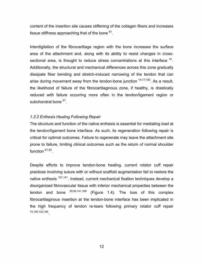
tendon and bone 29,56,141,166 (Figure 1.4). The loss of this complex
fibrocartilaginous insertion at the tendon-bone interface has been implicated in
the high frequency of tendon re-tears following primary rotator cuff repair 74,100,122,164.
13
Figure 1.4 Safranin O Staining of RC Enthesis A) Native RC enthesis, and B) RC enthesis following suture repair.
Formation of this fibrous tissue following repair occurs over three stages:
inflammation (0-7 days), repair (5-14 days), and remodeling (>14 days) 100. First,
in the inflammation stage, a substantial infiltration of fibroblast and inflammatory
cells occurs at the site of injury and increases angiogenesis. The ECM contains
some cartilage-like cells and is composed of primarily fibronectin,
glycosaminoglycans, and type III collagen 163. Collagen fiber orientation and
tensile mechanical properties of the tendon are significantly decreased, resulting
in no integration with bone and the lack of a fibrocartilage insertion zone.
During the repair stage, collagen fibers begin to integrate with the bone but
overall tissue quality remains poor and disorganized compared to native 63. The
tissue is composed of granulation tissue and contains chondrocyte-like cells and
fibroblasts 56. Several growth factors are up-regulated to induce cell proliferation
and matrix deposition at the site of repair including fibroblast growth factor basic
(FGF-b), bone morphogenetic proteins (BMP-12 -13, and -14), platelet derived
growth factor-B (PDGF-B), and transforming growth factor-B1 (TGF-B1) 181.
In the remodeling stage, the newly formed tissue remains disorganized and
hypercellular 62. Following 4 weeks repair in a rat model, the tendon had
integrated with bone but no fibrocartilage region was observed 62. At 8 weeks, the
repair site was more organized than previous time points but did not resemble
the graded fibrocartilage structure of the native enthesis 163. Although structural
B A

14
properties reached two thirds of normal levels, the material properties of the
tissue remained an order of magnitude weaker than in control groups 29. By 16
weeks, the healing tissue still had not functionally integrated with the bone. The
tendon attached to the bone through a fibrovascular scar with a sharp boundary
between soft tissue and bone and was structurally and compositionally different
than the native enthesis tissue 3,101.
Like the rotator cuff, repair of the ACL with current grafting techniques also fails
to recreate a fibrocartilaginous enthesis. Instead, the graft often heals with a
fibrovascular scar at the graft-tunnel interface that exhibits poor mechanical
stability and remains a primary cause of ACL graft failure 2. Furthermore,
suboptimal long-term ACL outcomes and increased risk of OA may be due in part
to poor graft-bone integration and an inability to effectively transfer load 48. As
such, the re-establishment of a native-like enthesis is crucial for improving the
long-term success and functionality of the reconstructed knee.
As described, current surgical procedures for both rotator cuff and ACL repairs
fail to regenerate the compositional, geometrical, and biomechanical properties
of the native enthesis to the level needed for normal joint function and tissue
regeneration 126. As a result, researchers have begun to design new strategies
focused on recreating the functional properties of the native enthesis to improve
repair success and improve long-term outcomes 98,154.
1.3.3 Native Enthesis Development
Understanding the biological and mechanical processes regulating the
development of the native tendon-bone interface may be useful in identifying new
tissue engineering strategies for improving enthesis repair. In a recent in situ
hybridization study by Galatz et al., the spatial and temporal expression of ECM
genes involved in rotator cuff development were examined utilizing a murine
model 52. The study reports the formation of tendon and bone masses at 15.5
days post-conception but a transition zone was not observed until 7 days after

15
birth. Furthermore, significant enthesis maturation did not occur until day 21
postnatally. The formation of a direct insertion site is believed to occur through
endochondral ossification. The tendon/ligament initially attaches to the hyaline
cartilage of bone and as ossification occurs, a fibrocartilaginous region develops
at the tendon-bone interface. The formation of this fibrocartilage zone is due to
fibroblast metaplasia as a result of mechanical stimuli at the insertion site 83,167.
The influence that mechanical load has on enthesis formation during post-natal
development underlines the important role mechanical stimuli may play in
regenerating the insertion site during repair.
In a separate study, muscle loading during tendon enthesis development has
been shown to significantly impact insertion maturation in a murine model 77.
Botulinum toxin injection, which causes muscle paralysis, resulted in less
mineralization, impaired fibrocartilage formation, diminished collagen alignment,
and inferior mechanical properties compared to saline controls at post-natal time
points 77. However, paralysis at early time points did not result in significant
differences in enthesis structure suggesting that mechanical load is not needed
for initiation of enthesis development but is a critical factor in directing enthesis
maturation 77.
In addition to mechanical cues, chemical factors are also known to be important
in enthesis development. Proteins including Indian hedgehog (Ihh) and
parathyroid hormone-related protein (PTHrP) have been implicated in regulating
chondrocyte hypertrophy and mineralization100,162. Graded expression of these
factors at the tendon-bone interface may regulate spatial gradients between
mineral and un-mineralized fibrocartilage zones162. Transcription factors SOX-9
and scleraxis (Scx) are associated with chondrogenesis and tenogenesis
respectively 152,160. Cells forming the insertion site originate from both SOX-9 and
Scx lineages and the localization of these genes at the tendon-bone interface
defines the tendon/fibrocartilage transition 160. Scx expression at this site has
16
also been shown to regulate BMP-4 expression in tendon cells. Inhibition of
BMP-4 expression in Scx positive cells failed to initiate enthesis development 21.
The combination of these works demonstrates that the development of a
functionally graded enthesis is driven by and responsive to both biologic and
mechanical signals. As such, tissue-engineering strategies targeting these
signaling pathways post-injury may be useful for stimulating the required cells,
ECM, and mechanical properties necessary for native-like repair.
1.4 Strategies to Improve Tendon-Bone Healing In Vivo
Recent tissue-engineering approaches to improve soft-tissue/bone healing at the
tendon-bone interface are typically designed to augment current repair
techniques in vivo. Treatments often rely on the singular or combined use of
growth factors and cells with existing suture or scaffold-augmented fixation
techniques to modulate the repair site and facilitate the formation of the native
insertion site. These biologic approaches have been extensively reviewed by
several researchers3,13,114. Examples of specific applications as they relate to
rotator cuff repair are described below:
1.4.1 Growth Factor Therapies
To enhance rotator cuff healing in vivo, the application of various growth factors
capable of modulating the biology at site of repair have been investigated 114.
Fibroblast growth factor basic (FGF-2) is known to stimulate angiogenesis as well
as influence matrix synthesis and is up-regulated during early stages of tendon
healing 3,71. Local application of FGF-2 in a fibrin sealant following acute injury
and repair of the supraspinatus tendon in a rat model resulted in accelerated
healing at the tendon-bone interface compared to the fibrin sealant alone in the
contralateral shoulder 81. At 2 weeks, histological scorings of repair sites
demonstrated improved cellularity, vascularity, and fiber maturity and were
correlated with improved mechanical strength when compared to fibrin only

17
treatment. However, differences were not observed at the later 4 and 6 week
time points. Additionally, despite the improvement in attachment strength, a
fibrocartilaginous region characteristic of the native tendon-bone interface was
not recreated.
In a similar study by the same group, FGF-2 fibrin sealant was applied to a rat
tendon-bone interface and repaired with a commercially available acelluarized
dermal graft (GraftJacket)82. At 2 weeks, repair with FGF-2 showed similar
histological scores and strength compared to graft repairs without growth factor.
However at 6 and 12 weeks, repair with FGF-2 formed a cartilaginous tissue at
the tendon-bone interface accompanied with improved repair failure strength.
Despite the presence of fibrocartilage, the insertion site still did not recreate the
complexity of the direct insertion found in the unoperated control shoulders.
TGF-B isoforms have also been shown to alter healing at the tendon-bone
interface with varied results 84. Application of TGF-B1 (and suppression of
isoforms 2 and 3) at repair sites resulted in increased fibrous scar tissue
formation and a decrease in mechanical properties; while the use of TGF-B3 and
suppression of isoforms 1 and 2, resulted in no significant change compared to
controls. In a separate study, however, prolonged exposure to TGF-B3 in a
heparin/fibrin-based delivery system positively enhanced the structural and
material properties of the tendon-bone interface following repair 107. Decreased
scar formation and increased fibrocartilage formation, collagen alignment, and
interface strength have also been reported with the use TGF-B3 administered at
site of repair within osteoconductive calcium-phosphate matrix86.
In vivo, growth factor signaling pathways may be regulated and may interact with
each other to coordinate control of cell function. FGF-2, for example, is known to
alter gene expression of several growth downstream factors including TGF-B and
BMP-2 13. As such, signaling of a single growth factor is unlikely to mediate

18
native-like tissue regeneration alone and has led researchers to the use of
platelet rich plasma (PRP) therapies to improve enthesis repair.
PRP, isolated from autologous blood, forms a dense plasma matrix that can be
applied at the repair site. PRP is known to contain several growth factors that
have been shown to play a critical role in the tendon healing process including:
TGF-B, bFGF, PDGF, vascular endothelial growth factor, connective tissue
growth factor, and epidermal growth factor 3. In a recent study by Ersen et al., the
use of PRP in a rat supraspinatus tear model resulted in improved load to failure
and stiffness at the repair site 47. However, histological differences were not
observed between groups repair treated with and without PRP.
While the use of growth factor based strategies commonly results in increased
matrix synthesis and structural improvements at the repair site, the resultant
tissue often consists of fibrous scar tissue rather than an organized, fibrocartilage
gradient characteristic of the native enthesis. Further optimization related to the
mode of delivery, timing of release, and ideal concentrations is needed for these
strategies to be successful. As such, surgeons continue to rely on existing suture
fixation techniques with sub-optimal outcomes. Alternative cell-based therapies,
capable of sensing and responding to the local chemical and mechanical
environments, may be needed to improve enthesis repair.
1.4.2 Cell-Based Therapies
One of the most common cells used in current cell-based therapies is the
mesenchymal stem cell (MSC). MSCs are characterized by their unique ability to
self-renew and differentiate in vitro into several different specialized tissue
lineages including bone, tendon, cartilage, muscle, ligament, and adipose tissue 27,184. MSCs can be easily isolated from an autologous or allogeneic bone
marrow aspiration and are considered to be anti-inflammatory and/or immuno-
modulatory upon implantation. More recently, resident MSC populations have
also been harvested from adipose tissues which may be a more accessible

19
donor source for MSC isolation 44. In vivo, MSCs are capable of directly
participating in the healing response or signaling local or distant host processes.
The use of MSCs to improve tendon-bone healing following rotator cuff injury has
recently been examined in a rat model 69. Following the detachment of the
supraspinatus tendon, bone marrow derived MSC contained in a fibrin sealant
carrier were injected into the repair site and the tendon subsequently repaired
with suture 69. Results, at both 2 and 4 week time points, showed no histological
difference in cartilage formation, collagen orientation, failure strength, or stiffness
of the enthesis compared to controls 69. Additional signals or differentiation
factors are likely needed for effective interface repair. Undifferentiated MSC
alone may not be enough to stimulate the desired regenerative response.
In a follow-up study also performed by Gulotta et al., MSCs were transduced with
adenoviral-mediated scleraxis, a transcription factor implicated in tenogenesis
and native enthesis development, before being integrated into a fibrin sealant
carrier and injected into the repair site70. Despite the lack of histological
improvement at 2 weeks following repair, increases in both load to failure and
stiffness were observed compared to non-transduced MSC injected controls. At 4
weeks, the biomechanics continued to improve, and histologically, the formation
of fibrocartilage was observed at the tendon-bone interface. However, the
organization of this tissue and amount of fibrocartilage present was still
significantly less than in the native enthesis. Nonetheless, the application of
differentiated MSCs may be a useful tool for inducing the native healing response
following rotator cuff repair and holds promise for future cell-based strategies
utilizing these cells.
Despite the potential of the described in vivo techniques to improve tissue
healing at the tendon-bone interface, many of these strategies continue to rely on
existing suture-fixation techniques and frequently fail to re-create the native
anatomy of the rotator cuff enthesis. The inability to restore this interface

20
compromises repair success and long-term clinical outcomes following
tendon/ligament injury 99. As such, novel tissue engineering approaches capable
of regenerating the graded fibrocartilaginous structure and mechanics of the
native enthesis are needed for orthopedic repair.
1.5 In Vitro Scaffold-Based Strategies for Enthesis Repair
To improve regeneration of the tendon-bone insertion site following repair,
several in vitro tissue-engineering strategies have been developed to replicate
the functionally graded properties of the enthesis for subsequent implantation in
vivo 186. These approaches mimic the zonal arrangement of the native tissue by
imploring stratified or graded scaffold designs described in the following sections 154.
1.5.1 Stratified Scaffold Designs
Stratified scaffold designs for enthesis regeneration typically consist of various
cells, growth factors, or other signals continuously arranged in pre-defined
regions specific for the generation of distinct tissue types. The goals of stratified
scaffold designs are to integrate these regions and form a graded
fibrocartilaginous transition zone following implantation. Work by Spalazzi et al.,
successfully produced a co-cultured 3D scaffold composed of three distinct but
continuous phases arranged in succession. The first phase served as the tendon
region and consisted of poly- (lactic-co-glycolic acid) (PLGA) (10:90) matrix
seeded with ACL derived fibroblasts 158,159. The second region was the
fibrocartilaginous region composed of PLGA (85:15) and seeded with
chondrocytes. The third region served as the bone region and was composed of
sintered PLGA (85:10) and 45S5 glass microspheres and was seeded with
osteoblasts. Deposition of collagen 1 was observed throughout the tendon and
fibrocartilage regions with significant amounts of mineral in the bone region.
Subcutaneous implantation of the constructs resulted in continuity across all
three phases with mineralized tissue in the bone regions and distinct

21
fibrocartilage and tendon regions 156,157. The results of these studies
demonstrated the ability of a multi-phasic construct consisting of appropriate cells
and matrix composition to form a functionally graded tissue following
implantation. However, the ability of this stratified scaffold to integrate with the
host or grafted tissue in a tendon/ligament repair model has not been evaluated.
Moreover, the utilization of scaffold materials with variable degradation rates and
mechanical properties may shield the cells from signals required for optimal
tissue regeneration upon implantation.
Paxton et al. describes an alternative scaffold design for the creation of a whole
multi-tissue construct composed of an artificial ligament mid-substance attached
between two osteoconductive anchors 123. Fibroblasts seeded into a fibril gel are
guided through cell-mediated tension around two bio-resorbable bone cement
anchors (Brushite) producing ligament/bone-like tissue interfaces. Increasing the
collagen content of the ligament region, through the addition of growth factors
(ascorbic acid, proline, and TGF-B), led to an increase in the attachment strength
at this interface. The ability of this strategy to form a ligament mid-substance and
two relevant ligament/bone interfaces allows this technology to directly replace
the damaged tissue at the site of repair eliminating the need to integrate with the
damaged tendon/ligament tissue or replacement tendon graft. However, the
ability of this construct to regenerate a fibrocartilaginous enthesis following
implantation has not been evaluated.
1.5.2 Graded Scaffold Designs
Opposed to stratified scaffold designs that rely on integration of distinct, pre-
defined regions upon implantation to recreate the graded properties of the
enthesis, graded scaffold designs utilize spatially arranged biochemical or
biophysical signals to gradually control cell differentiation and matrix synthesis in
vitro. In a study performed by Phillis et al., fibroblasts were seeded onto a
scaffold substrate conjugated with a linearly graded increase of a bone
transcription factor, Runx2. Regions of low Runx2 concentration were associated

22
with more tendon-like phenotype, while higher concentrations trans-differentiated
towards osteoblast-like cells 128. The results showed a gradual change in
phenotype over the length of the scaffold. However, the formation of a
fibrocartilaginous transition was not recreated.
In a similar study, MSC were cultured onto a polymeric scaffold to engineer
composite osteochondral tissues 174. Silk microspheres formed two inversely
proportional linear gradients across the construct controlling the delivery of BMP-
2 and IGF-1. The proximal and distal ends of the scaffold contained the highest
concentrations of BMP-2 and IGF-1, respectively, which both decreased across
the construct length. This gradual change of local growth factor release resulted
in graded differences of calcium and glycosaminoglycan (GAG) deposition,
characteristic of bone and cartilage regions, respectively. Collagen type-I, II, and
X gene expressions were also similarly graded across the two construct regions,
mimicking the native composition of the osteochondral interface. The results of
this study validate the ability of controlled growth factor release to generate
tissue gradients in vitro and may be applicable in generation of other tissue
interfaces such as the tendon-bone interface.
Biophysical gradients can also be used to form functionally graded interface
tissues in vitro 92,186. In a study by Li et al., a mineralization gradient of calcium
phosphate was incorporated into electrospun PLGA nanofibers 91. The formation
of this gradient was associated with stiffness variations across the scaffold
length. Seeding this scaffold with adipose-derived MSCs resulted in the graded
expression of osteogenic cell markers, with regions of highest stiffness and
mineral content being most bone-like. Interestingly, formation of this osteogenic
gradient was achieved without osteogenic inducing culture medium suggesting
that compositional and mechanical gradations alone may be sufficient to promote
cell differentiation and influence cell behavior.

23
Each of the scaffold-based strategies described emphasize the influence local
environmental factors (generated in vitro or following implantation) have on the
formation of graded tissue properties. However, use of these strategies (stratified
or graded in design) is limited as they are routinely designed to match the native
properties of the tendon, bone, and/or insertion site, potentially shielding the
repair site from signals necessary for proper enthesis regeneration. Additionally,
the degeneration of the scaffold material often does not match the rate of new
tissue synthesis and can impair the rate and course of new tissue development
and maturation. Furthermore, the use of exogenous scaffolding materials can
trigger a foreign body reaction leading to material rejection and/or increased
amounts of fibrotic scar tissue at site of repair.
Due to these imitations and the inability of current scaffold-techniques to fully
regenerate native enthesis following repair, alternative tissue engineering
strategies are needed. A scaffold-less method to engineer an initially compliant
multi-phasic tissue graft, composed of physiologically relevant tissue interfaces
and capable of sensing and responding to local environmental signals, may be
well suited for regeneration of the native functional tendon-bone interface.
1.6 Scaffold-less Tissue Engineering – STEL Approach
Previous work from our lab, the Skeletal Tissue Engineering Laboratory (STEL),
has demonstrated the successful fabrication of a multiphasic bone-ligament-bone
(BLB) graft for ligament repair, without the use of exogenous or stiff, scaffolding
materials 104,105. Regionally distinct areas of bone and ligament form viable tissue
interfaces in vitro that are capable of quickly remodeling upon implantation.
(Figure 1.5)

24
Figure 1.5 Scaffold-Less BLB Construct
The utilization of this initially compliant graft in both medial collateral (MCL) and
anterior cruciate ligament (ACL) repair models has shown that the graft quickly
incorporates into native tissue and develops a viable enthesis shortly following
repair 105. Significant improvements in construct size, composition, morphology,
and mechanics were observed at increased ACL recovery time points, as
described in the work of Ma et al. 106. Briefly, BLB outcomes, following 6 months
recovery, restored approximately 52% of native tissue modulus and were
histologically similar to the native ACL tissue. Results at later 9 month recovery
time points compared to existing autologous patella tendon graft techniques,
demonstrated superior tissue regeneration and knee mechanics 106.
However, despite the success of this BLB tissue in an ACL repair model, the
translation of these results to a rotator cuff repair model is unknown. Additionally,
to further the advancement of this technology towards clinical use, a regulatory
compliant, standardized, and commercially viable manufacturing process is
needed. Achievement of these goals could significantly streamline the long-term
success of this tissue engineering product for both tendon and ligament repair.
1.7 Summary
The approach, outlined in the aforementioned aims, seeks to advance the
commercial manufacturing and clinical translation of a compliant multi-phasic
tissue construct for tendon/ligament repair. The use of a compliant scaffold-free
graft that can respond to local mechanical and biological environments has the
potential to improve tendon/ligament-to-bone regeneration and subsequent repair
outcomes following rotator cuff and ACL injury. However, despite considerable
25
amounts of research in the field and the development of numerous tissue-
engineering strategies, there are relatively few products currently available to
patients or in late-stage clinical trials. This slow progress in development is due
part to a lack of proficiency in translating bench-scale methods to robust, cost-
effective manufacturing processes that comply with current good manufacturing
processes (CGMP) guidelines and ensure the reproducibility, efficacy, and safety
of the final tissue engineering product for human use.
Utilizing a rat model, Chapter 2 of this thesis, will evaluate the efficacy of our
scaffold-less construct to regenerate the native enthesis following an acute, and
more clinically relevant, chronic, full thickness supraspinatus tear. It is
hypothesized that utilization of our construct will regenerate an enthesis that
more closely resembles the native structural properties compared to current
suture repair techniques.
Next, Chapter 3 and Chapter 4 will describe the advancement of existing
laboratory protocols for the production of uniform, reproducible, and clinically
relevant scaffold-less constructs. The development of such a well-characterized
system will provide consistent, high-quality constructs and lessen the difficulty for
adapting fabrication methodologies into CGMP-compliant and scalable
manufacturing processes required for human use.
Chapter 5 will describe the translation of the reproducible bench-scale
manufacturing processes of Chapter 3 into a novel bioreactor system for semi-
closed, automated BLB fabrication. The development of this bioreactor will
minimize contamination risk, eliminate user-variability, and provide means to
economically scale construct production for commercial construct use.
In summary, this thesis describes a novel treatment strategy for the regeneration
of the tendon/ligament-to-bone interface, which may be critical for improving
current rotator cuff repair outcomes following injury. These findings may provide

26
a new paradigm for the treatment of other musculoskeletal injuries requiring the
reattachment of soft-tissue-to-bone. Furthermore, the approach outlined in this
report for scaffold-less construct manufacturing demonstrates the practicality of
employing translatable and scalable processes early in the product development
stage as a means to overcome existing challenges in the commercialization and
clinical advancement of new tissue engineering technologies.

27
Chapter 2 Utilization of Scaffold-less Constructs for Tendon-Bone
Interface Regeneration
2.1 Introduction
Rotator cuff (RC) tears are one of the most common orthopedic disorders in the
US with over 70,000 repair procedures performed annually 169. Tears of the
rotator cuff tendons often fail to heal spontaneously and are frequently the cause
of significant shoulder pain and impaired shoulder function 114. Current repair
techniques utilize an open or arthroscopic suture technique to reattach the
tendon to bone. Failure rates, however, especially in full thickness large (3-5cm)
or massive tears (>5cm, 2 tendons), are unacceptably high (20-94%) 53,108.
Complications including tendon retraction, muscle atrophy, and fatty infiltration
have been known to limit the successfulness of the repair 33,67,72,187. Additionally,
healing of the tendon-bone insertion (enthesis) with current techniques often
results in a notably weaker fibrovascular scar rather than a normal enthesis and
may explain the high prevalence of repair failures 26.
The native enthesis consists of a compositionally and mechanically graded
fibrocartilage transition zone designed to minimize stress concentrations at the
tendon-bone interface. Using a rat supraspinatus rotator cuff model,
Thomopoulos et al., demonstrated that the biology and zonal arrangement of the
enthesis is not regenerated following rotator cuff repair 166. Rather, a sharp
boundary was histologically observed, with disorganized collagen fibers and a
fibrocartilaginous region, representing a significant deviation from the native
enthesis anatomy. The structural repair and regeneration of a native enthesis

28
and return of normal biomechanics is paramount for improving functional
outcomes and reducing the risk of recurrent structural failure after repair. As such,
several tissue-engineering approaches have been developed to improve the
biological healing and regeneration of the enthesis following RC repair.
To improve the healing response at the enthesis, Derwin et al. described several
commercially available extracellular matrix (ECM) scaffolds currently used
clinically for rotator cuff repair 41. While the inherent biochemical properties of
these scaffold materials have the potential to modulate host processes and
influence the biology of the repair, the scaffold’s properties, which are degraded
over time, often do not match the rate of new extracellular matrix production.
Additionally, a structurally native fibrocartilage enthesis often fails to regenerate
and, as such, alternative strategies are needed to improve enthesis repair of
rotator cuff injuries.
More recently, stratified and functionally graded scaffold-based approaches have
been developed to pre-engineer the native tendon/ligament-to-bone interface in
vitro 154,186. In these studies, the spatial distribution of mechanical properties,
biological factors, and/or cell populations are used to direct the formation of
gradients designed to mimic the tendon insertion site in vitro or following
subcutaneous implantation. Evaluation of these graded scaffolds in an in vivo
rotator cuff repair model, however, has not been performed, and the degradation
characteristics of the scaffold and biocompatibility has not been defined to date.
As an alternative to current scaffold based approaches, our lab has developed an
initially compliant, scaffold-less tissue engineered bone-ligament-bone construct
that relies on the local biologic and mechanical environments to direct the
regeneration of the tendon or ligament-to-bone insertion. Previous work in sheep
ACL and rat MCL repair models have shown that the osteogenic regions of our
construct quickly integrate with the host tissue and rapidly remodel forming a
graded tendon/ligament-to-bone transition that undergoes hypertrophy to match

29
the native footprint (Figure 2.1).
Figure 2.1 Ligament/Bone Interface of BLB Construct Explant A) H&E showing ligament integration into bone through Sharpey’s fibers (arrow) and fibrocartilage-like region at 2-months, B) Alizarin Red staining of ligament -bone interface showing mineralization gradient at 2 months, and C) Femoral insertion of explanted BLB graft at 9-months. Construct diameter significantly increased in size from initial construct diameter (approx. 3mm).
While the results of these studies in the ACL are encouraging, it is unclear if they
can be extrapolated to a rotator cuff model. The objective of this study was to
evaluate the efficacy of our osteogenic scaffold-less constructs to regenerate the
native matrix organization and mechanical strength of the enthesis in both acute
(immediate repair) and chronic (repair 4 weeks post injury) supraspinatus tears in
a rat model. We hypothesized that utilization of the tissue-engineered tendon
construct would better regenerate the fibrocartilage transition zone, collagen
organization, and tendon attachment strength of the native enthesis in both acute
and chronic tear repairs compared to suture repair alone.
2.2 Materials & Methods
2.2.1 Animal Model & Design
The University of Michigan Institutional Animal Care and Use Committee
(IACUC) approved this study. Female Fisher 344 rats, approximately 150-200 g
in mass, were used in this study. For recreating an acute supraspinatus tendon
tear, right shoulders underwent a full thickness supraspinatus tenectomy using a
transacromial approach 29. Chronic supraspinatus tendon tears were created by
encasing the detached end of the supraspinatus tendon in a small piece of
A B C

30
silicone tubing and allowing the animal to recover for 4 weeks. The encasement
with the silicone tubing prevented attachment to the surrounding connective
tissue so that the cut end of the tendon could easily be identified and repaired at
a later date. The left contralateral shoulders were used as non-surgical controls
for each treatment group. Both acute and chronic tendon tears were repaired
with either a Mason-Allen suture technique (suture repair group) or a modified
trans-osseous technique utilizing a tissue engineered construct (construct repair
group). At 8 weeks (n=43) following repair of the tendon, animals were
euthanized with an overdose of sodium pentobarbital and assessed for
biomechanical and structural repair of the injury site.
2.2.2 Preparation of Solutions and Media
All media and solutions were prepared and stored at 4°C prior to use. Media
were warmed in a heated water bath to 37°C before utilized in the culture of cells.
Media were prepared, as previously described, with slight modifications 104,153.
Growth medium (GM) consisted of Dulbecco’s Modified Eagle Medium (DMEM;
Gibco, Rockville, MD, Cat# 10565-042) supplemented with 20% Fetal Bovine
Serum (FBS; Gibco, Rockville, MD, Cat# 10437-028) and 2% antibiotic-
antimycotic (Sigma, St. Louis, MO, Cat# A9909). Osteogenic GM consisted of
GM supplemented with 600ng/ml bFGF, and 40 ng/ml dexamethasone.
Differentiation medium (DM) consisted of DMEM supplemented with 7% Horse
Serum Albumin (HS, Gibco, Rockville, MD, Cat# 16050-122) and 2% antibiotic-
antimycotic. Osteogenic DM consisted of DM, plus the addition of 0.13 mg/ml
ascorbic acid-2 phosphate, 0.05 mg/ml L-proline, 400 ng/ml dexamethasone and
2 ng/ml transforming growth factor-beta (TGF-b; Peprotech, Rocky Hill, NJ, Cat#
100-21).
2.2.3 Bone Marrow Isolation and Cell Expansion
Bone marrow stromal cells were isolated and expanded according to a previously
described protocol 104,153. Briefly, rat femurs were removed from the animal and
cut at the proximal and distal metaphysis. The marrow contents were extracted

31
through centrifugation (900 g for 10 min) and cultured on 100 mm tissue culture
treated polystyrene plates (BD Falcon). Plates were fed osteogenic GM and
incubated at 37°C, 95% humidity, and 5% CO2. After 72 hours, plates were
rinsed with DPBS and fed with fresh GM, removing the non-adherent cells. The
remaining adherent cell population was cultured to 80% confluence, at which
time cells were enzymatically removed from the 100 mm plate using a 0.25%
trypsin-EDTA solution (Gibco), passaged, and seeded onto 100 mm plates at a
density of 5 x 10³ cells/cm2. Following 3-4 passages, the cultured cells were
utilized for construct fabrication.
2.2.4 Preparation of Self-Organized Constructs
Methods for creating scaffold-less three-dimensional tissues have been
previously described by our lab 104,105,153. Bone-like constructs, instead of
tendon/ligament-like constructs, were used in this study based on their ability to
specifically regenerate the tendon-bone interface in an ACL repair model. Briefly,
MSCs plated onto 100 mm cell culture dishes at 21x103 cells/cm2 and suspended
in 8 mls of osteogenic GM supplemented with 0.13 mg/ml ascorbic acid-2
phosphate and 0.05 mg/ml L-proline. The dishes were placed in a 37°C, 5% CO2
incubator and medium changed every 2–3 days. After approximately 5 days, the
cells became confluent and osteogenic differentiation medium (DM) was
substituted for GM to induce monolayer delamination. As the monolayer
delaminated, it was transferred to a Sylgard coated dish and constrained
between two minutien pins placed approximately 60 mm apart. The monolayer
was fully self-assembled into a 60 mm long cylindrical construct within 1-2 days.
One week following delamination, constructs were approximately 0.9 mm in
diameter and cut into 6 mm long segments for use in rotator cuff repairs (Figure
2.2A). The medium was again changed every 2-3 days until the construct was
used for implantation.
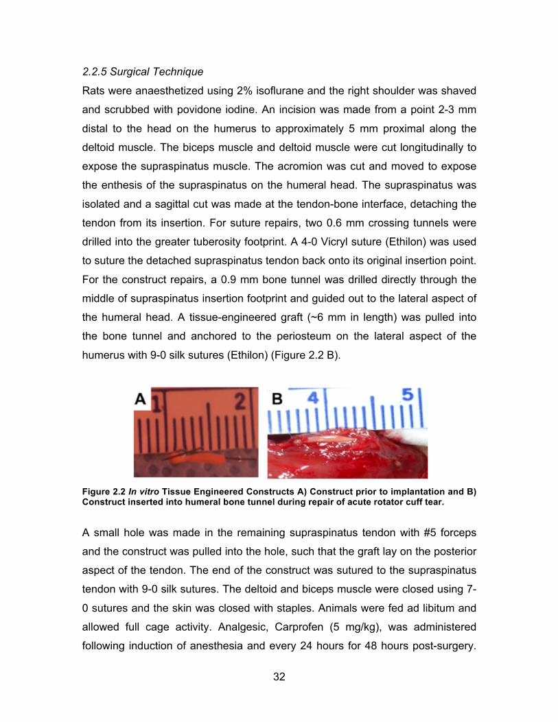
32
2.2.5 Surgical Technique
Rats were anaesthetized using 2% isoflurane and the right shoulder was shaved
and scrubbed with povidone iodine. An incision was made from a point 2-3 mm
distal to the head on the humerus to approximately 5 mm proximal along the
deltoid muscle. The biceps muscle and deltoid muscle were cut longitudinally to
expose the supraspinatus muscle. The acromion was cut and moved to expose
the enthesis of the supraspinatus on the humeral head. The supraspinatus was
isolated and a sagittal cut was made at the tendon-bone interface, detaching the
tendon from its insertion. For suture repairs, two 0.6 mm crossing tunnels were
drilled into the greater tuberosity footprint. A 4-0 Vicryl suture (Ethilon) was used
to suture the detached supraspinatus tendon back onto its original insertion point.
For the construct repairs, a 0.9 mm bone tunnel was drilled directly through the
middle of supraspinatus insertion footprint and guided out to the lateral aspect of
the humeral head. A tissue-engineered graft (~6 mm in length) was pulled into
the bone tunnel and anchored to the periosteum on the lateral aspect of the
humerus with 9-0 silk sutures (Ethilon) (Figure 2.2 B).
Figure 2.2 In vitro Tissue Engineered Constructs A) Construct prior to implantation and B) Construct inserted into humeral bone tunnel during repair of acute rotator cuff tear.
A small hole was made in the remaining supraspinatus tendon with #5 forceps
and the construct was pulled into the hole, such that the graft lay on the posterior
aspect of the tendon. The end of the construct was sutured to the supraspinatus
tendon with 9-0 silk sutures. The deltoid and biceps muscle were closed using 7-
0 sutures and the skin was closed with staples. Animals were fed ad libitum and
allowed full cage activity. Analgesic, Carprofen (5 mg/kg), was administered
following induction of anesthesia and every 24 hours for 48 hours post-surgery.
33
Eight weeks following surgical repair of the tendon the animals were euthanized
with sodium pentobarbital and both shoulders were removed for biomechanical
and histological analyses.
2.2.6 Biomechanical Testing
Biomechanical testing was performed on freshly dissected supraspinatus
tendons while still attached to the humerus. Prior to testing, the supraspinatus
muscle belly was carefully removed from the supraspinatus tendon and the cross
sectional area of the tendon was measured using a digital micrometer. To
prevent the fracture of the humeral physis during mechanical testing, the
humerus was potted with polymer into a custom-designed grip. The specimen
was then placed into a Dynamic Mechanical Analyzer (RSA-III, TA Instruments)
for uniaxial tensile testing. The end of the tendon was secured to a screw grip
using sandpaper and cyanoacrylate. The specimen was attached to a 35 N load
cell and aligned uniaxially. The specimen was loaded to failure at a rate of 0.3
mm/sec or approximately 0.10/sec. Samples were tested at room temperature
and were continuously moistened via a PBS drip throughout testing. The ultimate
load-to-failure and location of failure (enthesis or tendon mid-substance) were
recorded. The stiffness for each specimen was calculated from the linear region
of the load-deformation curve. Statistical analysis was performed by two-way
ANOVA with significance as P < 0.05. Data from the contralateral shoulders were
combined for each injury group. Data are presented as mean ± standard error.
2.2.7 Histomorphometric Analysis
Following recovery, the supraspinatus tendon with the humerus still attached was
carefully dissected from the surrounding tissue under a microscope leaving the
supraspinatus muscle intact. Following dissection, samples were fixed in 10%
neutral buffered formalin for 48 hours, decalcified in EDTA for 48 hours, and
subsequently washed in phosphate-buffered saline solution. The samples were
dehydrated and embedded in paraffin for sectioning. To facilitate comparisons
between groups, an orientation of 90° between the humerus and supraspinatus

34
muscle was maintained for each sample during embedding. Serial sections 12-
µm thick, containing the repaired tendon and greater tuberosity of the humerus,
were cut in the coronal plane and stained with picosirius red, safranin-O, and
hematoxylin and eosin (H&E). Using an Olympus B light microscope, digital
images of the stained slides were taken using a SPOT RT camera (Diagnostic
Instruments, Sterling Heights, Michigan). Five representative slides for each stain
and specimen were prepared. Each slide was evaluated using polarized and
non-polarized light and analyzed by 3 different people.
To evaluate collagen organization in the repaired tendon enthesis, sections
stained with picrosirius red were imaged under monochromatic polarized light.
The birefringence of collagen fibers was semi-quantitatively analyzed, based on
brightness, to determine differences in collagen deposition and maturation at the
site of repair 34,86. Five representative tissue sections across the full width of the
tendon-bone interface were analyzed for each shoulder. The polarization plane
was rotated until maximum brightness was obtained to account for variations in
specimen orientation on the slide. Digital images were taken of each tissue
section at X10 magnification during a single imaging session under identical
imaging parameters. The images were imported into Image J software and
underwent 8-bit digitalization. Non-collagenous material was shown as black
(zero) and collagenous material was assigned a gray scale value from 1-255.
Higher gray-scale values signify more organized and mature collagen. Using the
Image J software, ten rectangular areas (50 x 50 µm) were randomly selected in
the tendon directly adjacent to the tendon-bone interface. Mean gray scale
values for each region were averaged for each specimen to obtain a brightness
value. Statistical analysis of specimen brightness was performed by two-way
ANOVA with P value < 0.05 considered significant. Data are presented as mean
± standard error.
The area of fibrocartilaginous material at the tendon-bone interface was
assessed by use of safranin O stained sections. Safranin O stains sulfated
35
proteoglycans reddish-purple, a property referred to as metachromasia. Digital
images of the stained sections were imported into Image J and areas of
metachromasia were meticulously outlined and measured using the Image J
software at a X4 magnification (National Institutes of Health, Bethesda, Maryland,
USA). Measured areas were averaged for suture and construct repaired
shoulders and contralateral controls. Statistical analysis of total area of
fibrocartilage was performed by two-way ANOVA with P value < 0.05 considered
significant. Data are presented as mean ± standard error.
2.3 Results
2.3.1 Gross Morphology
At the time of necropsy, there was continuity between the tendon and bone in all
acute and chronic repairs with no detectable differences between groups. All
animals appeared healthy and showed no signs of infection. The cross-sectional
area of the enthesis was measured and was not significantly different between
each group in either acute or chronic injury repairs (3.4±0.8 mm2).
2.3.2 Biomechanical Testing
Minimums of 5 samples for each treatment group were tested uniaxially until
failure. In the acute injury repair groups, four of the suture-repaired specimens
(n=8) failed at the enthesis, while the rest of the suture-repaired specimens and
all the construct-repaired specimens (n=5) failed at the tendon mid-substance. In
the chronic injury repair group, three suture-repaired specimens (n=5) failed at
the enthesis while the rest of the suture-repaired specimens and the entire
construct repair group (n=5) failed at the tendon mid-substance. All non-operative
contralateral shoulders failed at the tendon mid-substance during testing.
Following 8 weeks recovery, acute and chronic injuries repaired with either
suture or construct techniques displayed no differences in stiffness between
groups. All repair groups were significantly less stiff than their respective contra-
36
lateral shoulders and not significantly different from each other (Figure 2.3). The
mean stiffness of the tendon-bone interface in the acute injury repair groups was
as follows: contra-lateral (23.2±1.5 N/mm, n=13), suture repair (12.9±0.8 N/mm,
n=8), and construct repair (14.0±0.7 N/mm, n=5). The suture and construct
repaired shoulders achieved 56% and 60% of native contralateral stiffness
respectively. The mean stiffness of the tendon-bone interface in the chronic injury
repair groups was as follows: contra-lateral (21.7±1.2 N/mm, n=10), suture repair
(9.3±1.6 N/mm, n=5), and construct repair (10.1±0.6 N/mm, n=5). The suture and
construct repaired shoulders achieved 43% and 46% of native contralateral
stiffness, respectively.
The maximum load-to-failure was not significantly different between acute and
chronic injuries repair with suture or tissue constructs. Each group was
significantly less than its respective non-operative contralateral shoulder and not
significantly different from the other group (Figure 2.3). The maximum load-to-
failure for acute injury repair was as follows: contra-lateral (25.0±1.7 N, n=13),
suture repair (18.8±1.3 N, n=8), and construct repair (17.0±2.8 N, n=5). This
corresponded to approximately 75% and 68% of the contralateral failure load for
suture and construct repairs, respectively. Maximum load to failure for chronic
injury repair groups was as follows: contra-lateral (22.9±1.4 N, n=10), suture
repair (14.0±2.3 N, n=5), and construct repair (12.9±2.1 N, n=5), respectively.
The suture and construct repaired shoulders achieved 61% and 56% of
contralateral shoulder load to failure, respectively.

37
Figure 2.3 Mechanics of Tendon-Bone Interface Following Acute and Chronic Injury Repair A) Stiffness of contralateral (CL), suture repaired, and construct repaired shoulders and B) Load to Failure of contra-lateral (CL), suture repaired, and construct repaired shoulders. (#) Denotes statistical significance compared to CL shoulder (P<0.05). Data are shown as mean ± S.E. for each group.
2.3.3 Histomorphometric Analysis
Collagen birefringence was semi-quantitatively analyzed, based on brightness, to
evaluate collagen organization at the tendon-bone interface. Five specimens
were analyzed for each treatment group. Contralateral shoulders of acute and
chronic injured shoulders were not significantly different and were combined for
analysis. Suture and construct repaired shoulders, although significantly more
disorganized (shown in picrosirius red staining) than contralateral shoulders,
were not significantly different to each other in either acute or chronic injury
groups (Figure 2.4). Additionally, although repaired chronic injuries trended
towards lower gray scale values, no significant difference in collagen
organization was observed between repaired acutely or chronically injured
shoulders. Suture repairs of acute injuries had a mean brightness gray scale
value of 70±1 gray scale units, while construct repairs had value of 70±4 gray
scale units. Chronic injuries repaired with suture and construct techniques had
gray scale values of 58±5 and 65±4 gray scale units, respectively. Non-operative
contralateral shoulders had an average brightness value of 95±6 gray scale
units. Representative images from each treatment and control group can be seen
in Figure 2.5.
Acute Injury Chronic Injury0
10
20
30
40S
tiffn
ess
(N/m
m)
Stiffness of Tendon-Bone Interface
Suture Repair
# #
# #
Construct RepairCL
Acute Injury Chronic Injury0
10
20
30
40
Load
-to-
Failu
re (N
)
Load-to-Failure of Tendon-Bone Interface
Suture Repair
##
# #
Construct RepairCL
A B

38
Figure 2.4 Collagen Organization at Enthesis Following Acute and Chronic Injury Repair. No significant differences were observed between acute and chronic injury groups. (#) Denotes statistical significance compared to contralateral (CL) shoulder (P<0.05). Data are shown as mean ± S.E. for each group.
Acute Injury Chronic Injury0
50
100
150
Big
htne
ss G
ray
Scal
e Va
lue
(0-2
55)
Collagen Organization at Enthesis
CL
## ##
Suture Repair Construct Repair

39
Figure 2.5 Representative Collagen Organization of Enthesis A) Contralateral shoulder, B) Acute suture repair, C) Acute construct repair, D) Chronic suture repair, and E) Chronic construct repair. Images are of picrosirius red stained sections under polarized light and imaged at X40 magnification.
A B
C D
E
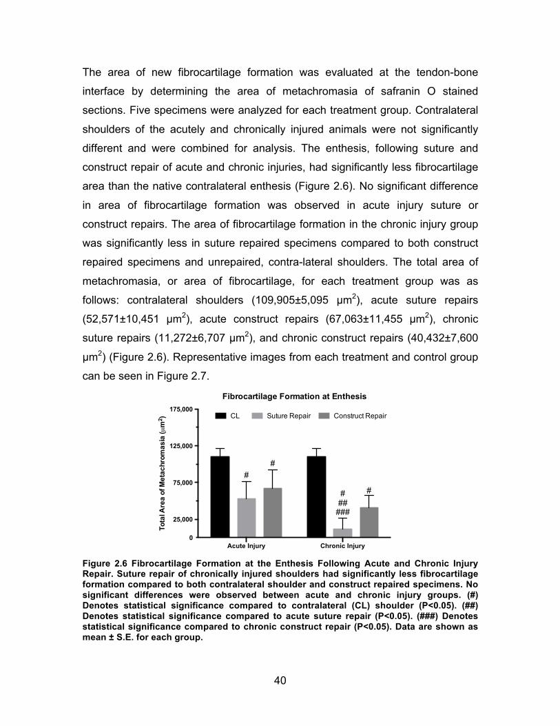
40
The area of new fibrocartilage formation was evaluated at the tendon-bone
interface by determining the area of metachromasia of safranin O stained
sections. Five specimens were analyzed for each treatment group. Contralateral
shoulders of the acutely and chronically injured animals were not significantly
different and were combined for analysis. The enthesis, following suture and
construct repair of acute and chronic injuries, had significantly less fibrocartilage
area than the native contralateral enthesis (Figure 2.6). No significant difference
in area of fibrocartilage formation was observed in acute injury suture or
construct repairs. The area of fibrocartilage formation in the chronic injury group
was significantly less in suture repaired specimens compared to both construct
repaired specimens and unrepaired, contra-lateral shoulders. The total area of
metachromasia, or area of fibrocartilage, for each treatment group was as
follows: contralateral shoulders (109,905±5,095 µm2), acute suture repairs
(52,571±10,451 µm2), acute construct repairs (67,063±11,455 µm2), chronic
suture repairs (11,272±6,707 µm2), and chronic construct repairs (40,432±7,600
µm2) (Figure 2.6). Representative images from each treatment and control group
can be seen in Figure 2.7.
Figure 2.6 Fibrocartilage Formation at the Enthesis Following Acute and Chronic Injury Repair. Suture repair of chronically injured shoulders had significantly less fibrocartilage formation compared to both contralateral shoulder and construct repaired specimens. No significant differences were observed between acute and chronic injury groups. (#) Denotes statistical significance compared to contralateral (CL) shoulder (P<0.05). (##) Denotes statistical significance compared to acute suture repair (P<0.05). (###) Denotes statistical significance compared to chronic construct repair (P<0.05). Data are shown as mean ± S.E. for each group.
Acute Injury Chronic Injury
25,000
75,000
125,000
175,000
0
Tota
l Are
a of
Met
achr
omas
ia (µ
m2 )
Fibrocartilage Formation at Enthesis
CL Suture Repair Construct Repair
###
#
#
###
#

41
Figure 2.7 Representative Fibrocartilage Formation at Enthesis. Representative images of safranin O stained A) Contralateral, B) Acute Suture Repair, C) Acute Construct Repair, (D) Chronic Suture Repair, and E) Chronic Construct Repair sections. Area of metachromasia (depicted as red) in each image was outlined for semi quantitative analysis. Images are shown at X40 magnification.
A B
C D
E

42
H&E staining of the tendon-bone interface allowed for qualitative evaluation of
the entheses repair. Acute and chronic tendon injuries repaired with suture had
less area of fibrocartilage formation and fewer organized collagen fibers than
both construct-repaired and contralateral shoulders. The attachment of tendon to
bone utilizing our tissue constructs resembled a more organized, native
appearance. Suture repaired shoulders in both acute and chronic injury models
had a more abrupt, fibrous interface at tendon-bone transition site, especially in
suture-repaired specimens of chronic injuries. Representative images from each
treatment and control group can be seen in Figure 2.8.

43
Figure 2.8 Representative Structure at Enthesis. Representative images of H&E stained A) Contralateral, B) Acute Suture Repair, C) Acute Construct Repair, D) Chronic Suture Repair, and E) Chronic Construct Repair sections. Suture repaired shoulders were more disorganized and fibrous than contralateral or construct repaired shoulders. Images are shown at X40 magnification.
A B
C D
E

44
2.4 Discussion
Tissue engineering to improve the architecture and structural integrity of the
healing enthesis after rotator cuff repair offers future promise to improve
outcomes in patients. Bone-like constructs, instead of tendon/ligament-like
constructs, were used in this study based on their ability to specifically
regenerate the tendon-bone interface in an ACL repair model. Future work
involving the use of multi-phasic tissues (with both bone and tendon/ligament
tissue) may further enhance the properties of the tendon mid-substance but was
beyond the scope of this experiment. The objectives of this current study were to
evaluate the efficacy of our scaffold-less tissue engineered tendon construct to
regenerate the native tendon-bone interface of the supraspinatus following full-
thickness acute and chronic injury in a rat model. Furthermore, we tested the
hypothesis that tendon-bone repair utilizing our scaffold-less construct could
strengthen enthesis healing and improve collagen organization and fibrocartilage
formation compared to suture fixation alone.
In the acute injury repair model, our results demonstrate that there were positive
effects with the utilization of our scaffold-less constructs. While no significant
differences in structural mechanics were observed, failure occurred more
frequently at the tendon-bone interface in suture-repaired, acutely injured
shoulders. Histologically, qualitative differences in general morphology revealed
more native histological architecture by 8 weeks post-operatively. In tendons with
chronic injury, repairs utilizing our tissue engineered construct resulted in a more
native-like enthesis. Despite similar biomechanical properties with load-to-failure
testing, suture repaired specimens of chronic injuries failed more frequently at
tendon-bone interface. However, the structure of the construct repaired enthesis
appeared qualitatively more native-like in appearance and quantitatively more
fibrocartilage was observed at the tendon insertion site compared to suture
repairs.

45
The formation of fibrocartilage at the tendon-bone insertion site has an important
role in the force transmission and energy dissipation between two tissues with
vastly different properties. This complex interface is seldom recreated with
current fixation techniques and often results in repair failure. The healing of the
enthesis following repair is known to be influenced by mechanical cues. In
separate studies performed by Galatz and Hettrich et al., complete removal of
load following repair via immobilization and botulinum toxin application to
supraspinatus muscles resulted in decreased structural properties and
decreased ECM production at the tendon-bone interface 54,77. As such, suture
techniques that rely on rigid mechanical fixation of tendon to bone may impede
signals that may be necessary for proper enthesis regeneration resulting in a
more fibrous tissue interface. The unique ability of our initially compliant construct
to sense and respond to local mechanical cues and biological factors has led to
improved structure and fibrocartilage formation at the enthesis.
The biological composition of our graft may also contribute to repair of tendon-
bone interface. Recent research has shown that application of MSCs, with the
addition of appropriate growth factors, can enhance tendon-bone healing of the
repair 70. Our constructs are fabricated from MSCs and driven towards an
osteogenic lineage, via the addition of previously described growth factors. As
such, enthesis regeneration at a repair site may be due to the unique cellular and
molecular signals provided by the cells of our construct that are necessary for
facilitating the proper host-mediated response. Alternately, the implanted
construct may be actively forming a new interface tissue in vivo. Current studies
are underway to evaluate the origin of the regenerated enthesis tissue.
Although more successful at regenerating the enthesis than current suture
techniques, the excessively high strains at the site of repair may have limited
repair outcomes. High tensile loads, especially in chronically retracted tendons,
can lead to elongation of the tendon, reducing overall muscle length and
compromising shoulder function. Though the length of tendon following repair

46
was not evaluated in this study, gross morphological changes of the
supraspinatus tendon and muscle were not observed. Nonetheless, due to the
compliance of our tissue, the primary benefit of our construct may be initially
biologic and a reliance on compositional properties of the graft until sufficient
tissue mechanical development can occur. As such, initial load sharing at the
repair site utilizing our construct as a biological in-lay may enhance repair
outcomes.
There were several limitations to this study. Mechanical testing evaluated
stiffness and load-to-failure of a single test. Cyclic loading, a more clinically
representative measure, was not performed. Furthermore, material properties
utilizing tissue level strains of the repaired tissue were not evaluated due to
technical difficulties and may explain the lack of significant differences in
mechanics despite histological differences in tissue composition and location of
failure. Assessment of the material properties of the regenerated tissue (i.e.
moduli) utilizing tissue level strains, rather than only geometric properties
(stiffness and load at failure) of the tissue, may better evaluate the quality of the
regenerated tissue independent of size. Additionally, the creation of a chronic
supraspinatus injury in the rat may not be fully representative of the human
condition. Healing in the rodent model is relatively robust and may not simulate
the difficulties experienced in structure failure after large and massive repairs in
human patients. Additionally, as this study focused specifically on enthesis
regeneration, important components of rotator cuff injury specific to the tendon
mid-substance and muscle such as muscle atrophy, fatty infiltration, quality of
tendon, etc., were not evaluated.
In conclusion, repair of acute supraspinatus injuries in a rat model with
engineered constructs more accurately recapitulated the appearance of the
native enthesis but was not significantly different biomechanically compared to
suture techniques. Utilization of scaffold-less constructs for repair of chronic
rotator cuff injuries was found to reduce scar formation and increase the area of

47
fibrocartilage formation at the tendon-bone interface over suture-only repairs.
Further investigation is needed to optimize the ideal tissue properties and
surgical technique for encouraging maximum enthesis regeneration in vivo.
48
Chapter 3 Standardization of Existing Methodologies for
Reproducible Construct Fabrication
3.1 Introduction
The previous chapter evaluated the use of scaffold-less tissue constructs for
regeneration of the native tendon-bone interface following acute and chronic
rotator cuff injury and repair. As with rotator cuff repair procedures, current ACL
repair procedures also often fail to regenerate the native fibrocartilaginous
enthesis and have been implicated in poor long-term outcomes and pre-mature
graft failures. However, unlike repairs of the supraspinatus tendon that require re-
attachment of the damaged tissue to its original insertion site, current strategies
to repair the ACL replace the entire ACL complex within the knee. Current ACL
replacement grafts are secured to bone tunnels drilled through the femur and
tibia using interference screws, span the intra-articular space, and provide
immediate knee stability. These ACL replacement grafts from cadaveric allografts
or autografts from the patient’s patellar tendon or hamstrings tendon have
several disadvantages. The grafts often do not fully integrate with the native
tissue within bone tunnel and fail to restore full function to the knee. Additionally,
a mismatch of mechanical properties exists between the tendon replacement
graft and the native ACL 105. This mismatch of mechanical properties may play a
role in the early onset of OA. As such, a multi-phasic graft capable of integrating
with both bone tunnels and regenerating the native properties of the ligament
mid-substance would be advantageous.

49
As described in Chapter 1, our lab has tissue engineered a compliant BLB graft
consisting of viable ligamentous mid-substance tissue, osteogenic tissue at each
end, and viable entheses in between. In both rat MCL and sheep ACL repair
models the graft induces endogenous bone and ligament regeneration, rapidly
develops biochemically relevant and biomechanically compatible interfaces with
native tissue, and restores the native anatomy and function to the knee 105. As an
“off-the-shelf” allogeneic ACL graft, our tissue-engineered graft could eliminate
donor site morbidity from autologous tendon harvesting and improve the
structural and mechanical outcomes compared to current grafting techniques105.
It is clear that utilization of this tissue engineering approach has tremendous
potential to improve musculoskeletal tissue repair and regeneration.
A significant amount of work remains before we can use our current lab-based
tissue engineering processes for large-scale clinical use. Our hands-on, labor
intensive laboratory methods must be advanced to more efficient protocols
capable of producing consistently uniform, reproducible constructs that meet the
current industry regulations within the health care field. The commercialization of
our BLB construct, specifically for ACL repair, will be used for the remainder of
this thesis as an application-specific model for the translational process of tissue
engineering products.
3.2 Background/Motivation
The Food and Drug Administration (FDA) regulates all engineered tissue and
regenerative medicine products. The FDA oversees the pre-market approvals
and conducts post-market surveillance of existing products. The focus of the FDA
is to ensure the safety and effectiveness of new products through clinical trials
and to oversee the manufacturer’s processes validation in accordance with
current good manufacturing practice (CGMP) regulations. Overall these
regulations require exquisite definition and control of the product and fabrication
process 177.

50
Failure to adequately transition from a labor-intensive laboratory process
performed by expert technicians to an automated industrialized production
method is a major hurdle in the translation of all current tissue engineering
technologies. Though process validation may be performed in parallel with
clinical trials, the consideration of designing a process that is compliant with
existing FDA regulations is essential to avoiding late-stage product failures and
costly product development cycles. Implementing FDA compliant technologies
early in the research stage has the potential to shorten the duration of the FDA
approval process and hasten the availability of our tissue-engineered grafts for
widespread clinical use.
Tissue engineering products, in particular, are among the most complex products
to manufacture132. Tissues are composed of various living cells and ECM
components that are constantly changing and remodeling in response to
environmental cues. Due to the inherent heterogeneity of cells, the FDA requires
creation of tissue end product specifications and the demonstration of a
consistent manufacturing process. As such, extensive work in the tissue
engineering/regenerative medicine field is needed to design and implement
novel, high-quality manufacturing systems capable of generating sufficient
amounts of data needed to establish statistical confidences of variability, define
product specifications, and demonstrate overall process consistency.
CGMP requires that each step of the manufacturing process be designed and
confirmed such that in-process materials and the final-end product(s) are
produced consistently and reliably, and meet their predetermined quality
attributes. The manufacturer must evaluate the process performance and control
batch-to-batch variability within the system. To ensure process consistency, in-
process specifications must be established to monitor variability in the
characteristics of process materials or end products. As described by CGMP
regulations, “in-process specifications shall be derived from previous acceptable

51
process averages and process variability estimates where possible and
determined by the application of suitable statistical procedures where
appropriate” (See FDA Section 2.11.110(b)).
Therefore, as a first step for our technology, a robust, well-established
manufacturing method must be designed at an early stage to provide information
and knowledge about product reproducibility and to define the process to identify
and control for variability. To eliminate or reduce the variability associated with
our multi-step process, critical steps reliant on technician skill should be
minimized or eliminated. In addition, the ability to systematically collect data
during the process is critical to establishing baseline averages, to evaluating the
sources, degree, and impact that procedural variation has on manufacturing
process, and ultimately to determining the attributes of the end product. Only
after process variability has been minimized, controlled, and properly
characterized can in-process specifications be defined minimizing any remaining
variation in proportion to the risk it represents to the process and final product.
Once appropriate controls have been established, the process can be evaluated
under CGMP compliant conditions to demonstrate that the commercial
manufacturing process is capable of consistently producing a quality product.
Additionally, from a manufacturing standpoint, understanding the process
variation can lead to more efficient manufacturing systems that improve
production volumes and reduce failure rates.
The process of obtaining FDA approval for products in the field of tissue
engineering regenerative medicine is complex and the specific requirements for
process validation and regulatory approval are not clearly defined. Due to the
broad range of medical technologies being developed, new products are
generally evaluated on a case-by-case basis. Thus, the specific information that
is needed as part of an FDA regulatory submission to demonstrate sufficient
process control is unknown. Guidance documents provided by the FDA outline
the FDA’s current thinking on various topics (including process validation) and

52
serve as useful guidelines for industry. These documents incorporate principles
and approaches that manufacturers can use to validate manufacturing processes
for human, animal, drug, or biological products under current CGMP regulations.
3.2.1 Current BLB Manufacturing Process
For the purposes of this study, I will focus exclusively on the fabrication of the
ligament portion of our BLB construct, the region that replaces the damaged ACL
tissue within the intra-articular space. The stiffness of this region directly
determines the graft’s ability to accommodate strain under normal knee loading
and the fabrication of this region is currently the most difficult aspect of the BLB
fabrication process to reproduce. Thus, the goal of this study is to design
strategies to enhance construct production and to characterize the properties of
the ligament region during fabrication. The methods developed here to establish
a reproducible protocol for production of the ligament-region of the BLB graft will
be useful as a guide for improving production of the bone-region in future
research.
The original process used for BLB fabrication has been described by Ma et al.,
with slight modifications 104,105. Briefly, the methods used in the Ma paper to
harvest bone marrow for the fabrication of ligament tissue required the sacrifice
of a donor animal, removal of the whole femoral bone, and extraction of the
entire content of femoral bone marrow. Obviously, the removal of the entire
femur and the extraction of the entire marrow contents is not a practical method
for obtaining cellular material and should be replaced with more clinically relevant
bone marrow aspiration techniques. In the previous fabrication process, the
entire amount of extracted marrow was plated onto four polystyrene dishes and
MSCs were isolated by their plastic adherence properties. This method of bone
marrow extraction led to low primary MSC yields and often necessitated
significant and unpredictable amounts of time in culture to achieve suitable cell
quantities for passaging. The execution of these process steps at irregular
intervals added an unnecessary variable to the protocol and reduced the overall
53
efficiency of the process. The previous methodology resulted in differences in the
amount, cellularity, and composition of bone marrow collected for tissue
fabrication. Standardization of the original isolation procedure was needed to
appropriately evaluate variability between and within donor samples and recover
reliable cell volumes at defined time points.
In addition to standardizing the bone marrow isolation methods, the MSC
differentiation and expansion methods associated with our original BLB protocol
also required improvement to obtain sufficient quantities of a consistent starting
cell population prior to seeding. Existing methodologies induced an unknown
amount of MSCs toward a fibrogenic lineage and passaged these cells every
other day at a 1:3 liquid volume ratio until sufficient numbers of ligament cells
were achieved. Failure to rigorously monitor the plate density to the desired 80%
confluence mark between passages led to significant variability in the number of
recovered cells. This error in cell number was exacerbated in successive
passages. The recovery of low cell quantities during passaging led to a decrease
in the number of constructs that could be fabricated, and reduced the overall
efficiency of the process. Optimal culture conditions for passaging MSCs typically
occur at 80% confluency 115. High cell densities during passaging were typically
indicative of a plate that was well beyond 80% confluence. The resultant cell-cell
contact at these high cell densities may lead to changes in cell phenotype,
genetics, and construct structure and function that can introduce significant
variability into the manufacturing process. Thus, we determined that it was
imperative to standardize the methodologies used to expand and induce the
MSCs to ligament cells during passaging and to maintain the cells at each
passage as close to 80% confluence as possible, to minimize cell-to-cell contact
and provide adequate cell qualities for tissue fabrication. The establishment of
set passage densities would allow for accurate predictions of appropriate times
between passages necessary to obtain consistent starting cell numbers.
In addition to the issues associated with obtaining and expanding the MSC
population to a consistent fibrogenic lineage, we recognized that our current

54
methodologies for fabricating 3D BLB constructs could also be improved. The
process for capturing and constraining the spontaneously delaminating ligament
monolayers into uniform cylindrical constructs, in particular, was unpredictable,
technically challenging, led to a significant loss of constructs, and reduced the
overall number of constructs produced. Briefly, in our original method, once a
hyper-confluent monolayer was formed and a sufficient amount of extracellular
matrix was produced, plates were switched to a low-serum medium and fed TGF-
β to induce intercellular tension and spontaneous contraction of the monolayer.
These resulted in the spontaneous delamination of the monolayer that facilitates
the formation of a 3D cylindrical construct. During delamination, the monolayer
had to be delicately transferred onto a syglard-coated plate and pinned.
Constraint pins were applied to the monolayer and the delaminating tissue
aligned along a singular axis of the pins allowing for the remodeling of the
construct into a 3-D tissue (Figure 3.1).
The delamination process occurs spontaneously. The inability to continuously
monitor and manipulate the tissue at the beginning of the delamination process
led to the loss of a significant number of constructs. Failure to constrain the
monolayers at an appropriate length before significant tissue contraction
occurred resulted in tissues of an unsuitable length for use as a replacement
graft (>6cm) (Figure 3.1). Additionally, variation in the placement of the pins
often led to uneven tension during tissue contraction and resulted in construct
thinning, abnormal construct geometry, and eventual failure in a substantial
number of constructs (Figure 3.1).
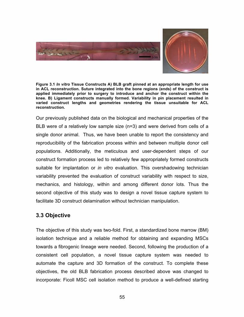
55
Figure 3.1 In vitro Tissue Constructs A) BLB graft pinned at an appropriate length for use in ACL reconstruction. Suture integrated into the bone regions (ends) of the construct is applied immediately prior to surgery to introduce and anchor the construct within the knee. B) Ligament constructs manually formed. Variability in pin placement resulted in varied construct lengths and geometries rendering the tissue unsuitable for ACL reconstruction.
Our previously published data on the biological and mechanical properties of the
BLB were of a relatively low sample size (n=3) and were derived from cells of a
single donor animal. Thus, we have been unable to report the consistency and
reproducibility of the fabrication process within and between multiple donor cell
populations. Additionally, the meticulous and user-dependent steps of our
construct formation process led to relatively few appropriately formed constructs
suitable for implantation or in vitro evaluation. This overshadowing technician
variability prevented the evaluation of construct variability with respect to size,
mechanics, and histology, within and among different donor lots. Thus the
second objective of this study was to design a novel tissue capture system to
facilitate 3D construct delamination without technician manipulation.
3.3 Objective
The objective of this study was two-fold. First, a standardized bone marrow (BM)
isolation technique and a reliable method for obtaining and expanding MSCs
towards a fibrogenic lineage were needed. Second, following the production of a
consistent cell population, a novel tissue capture system was needed to
automate the capture and 3D formation of the construct. To complete these
objectives, the old BLB fabrication process described above was changed to
incorporate: Ficoll MSC cell isolation method to produce a well-defined starting
1cm

56
cell material from a clinically relevant bone-marrow aspiration, consistent
passage densities to limit significant cell-cell contact known to alter cell
phenotype and function, and a novel capture system to automatically capture the
monolayer delamination and allow the formation of a 3D construct on the plate.
We hypothesized that the incorporation of these changes would allow for the
fabrication of more consistent and reproducible tissue constructs with defined
end-product specifications and marginal variability. The information and
knowledge gained from the addition of these methodologies will be useful in
generating the large number of constructs needed to establish an acceptable
baseline average and to statistically evaluate the process variation.
3.4 Methods
3.4.1 Preparation of Solutions and Media
We did not perceive the media and growth factors to be a significant source of
variation in the fabrication process. Solutions used for cell processing were
prepared as described in previous published work, with slight modifications 104,153. Briefly, all media and solutions were prepared and stored at 4°C prior to
use. Media were warmed in a heated water bath to 37°C before use for the
culture of cells. Media were prepared, as described previously, with slight
modifications 104,153. Growth medium (GM) consisted of Dulbecco’s Modified
Eagle Medium (DMEM; Gibco, Rockville, MD, Cat# 10565-042) supplemented
with 20% Fetal Bovine Serum (FBS; Gibco, Rockville, MD, Cat# 10437-028) and
2% antibiotic-antimycotic (Sigma, St. Louis, MO, Cat# A9909). Fibrogenic GM
consisted of GM supplemented with only 600ng/ml bFGF, 0.13 mg/ml ascorbic
acid-2 phosphate and 0.05 mg/ml L-proline. Differentiation medium (DM)
consisted of DMEM supplemented with 7% Horse Serum Albumin (HS, Gibco,
Rockville, MD, Cat# 16050-122) and 2% antibiotic-antimycotic. Fibrogenic DM
consisted of DM, plus the addition of 0.13 mg/ml ascorbic acid-2 phosphate, 0.05
mg/ml L-proline, and 2 ng/ml transforming growth factor-beta (TGF-b; Peprotech,
Rocky Hill, NJ, Cat# 100-21).
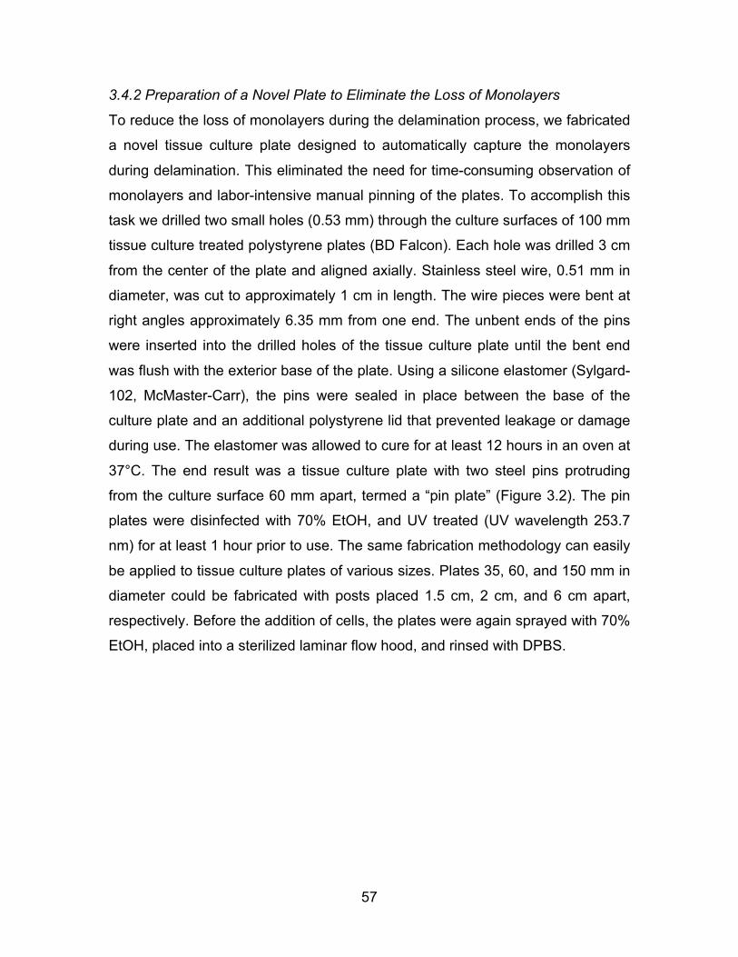
57
3.4.2 Preparation of a Novel Plate to Eliminate the Loss of Monolayers
To reduce the loss of monolayers during the delamination process, we fabricated
a novel tissue culture plate designed to automatically capture the monolayers
during delamination. This eliminated the need for time-consuming observation of
monolayers and labor-intensive manual pinning of the plates. To accomplish this
task we drilled two small holes (0.53 mm) through the culture surfaces of 100 mm
tissue culture treated polystyrene plates (BD Falcon). Each hole was drilled 3 cm
from the center of the plate and aligned axially. Stainless steel wire, 0.51 mm in
diameter, was cut to approximately 1 cm in length. The wire pieces were bent at
right angles approximately 6.35 mm from one end. The unbent ends of the pins
were inserted into the drilled holes of the tissue culture plate until the bent end
was flush with the exterior base of the plate. Using a silicone elastomer (Sylgard-
102, McMaster-Carr), the pins were sealed in place between the base of the
culture plate and an additional polystyrene lid that prevented leakage or damage
during use. The elastomer was allowed to cure for at least 12 hours in an oven at
37°C. The end result was a tissue culture plate with two steel pins protruding
from the culture surface 60 mm apart, termed a “pin plate” (Figure 3.2). The pin
plates were disinfected with 70% EtOH, and UV treated (UV wavelength 253.7
nm) for at least 1 hour prior to use. The same fabrication methodology can easily
be applied to tissue culture plates of various sizes. Plates 35, 60, and 150 mm in
diameter could be fabricated with posts placed 1.5 cm, 2 cm, and 6 cm apart,
respectively. Before the addition of cells, the plates were again sprayed with 70%
EtOH, placed into a sterilized laminar flow hood, and rinsed with DPBS.

58
Figure 3.2 Novel “Pin Plate” Capture System. Shown polystyrene tissue culture dish is 100 mm in diameter. Distance between pins is 60 mm.
3.4.3 Bone Marrow Aspiration and Isolation Technique
To shift to a more clinically relevant method for obtaining MSCs, bone marrow
was aspirated from the iliac crest of sheep using a Monoject Illinois needle
(Sherwood Medical Company, St. Louis, Mo). With the animal under general
anesthesia induced by intravenous propofol and sustained with inhalation of
isoflurane in oxygen, bone marrow aspirates were collected using heparinized
needles and dispensed into EDTA blood collection tubes (BD, San Jose, Ca) for
processing. The marrow was passed through a 100 µm filter to remove solid
debris before it was consolidated into a single 50 ml conical. An equivalent
volume of DPBS was added to the bone marrow. Next, the diluted bone marrow
solution was slowly added to a 15 ml layer of Ficoll-Paque™ Premium (GE
Healthcare, Munich, Germany) contained within a separate conical and
centrifuged (AccuSpin FR; Beckman Coulter Inc., Fullerton, CA) at 600 g for 30
minutes @ 25°C. Following centrifugation, the upper layer of the mixture
containing plasma and platelets was discarded and the mononuclear cell (MNC)
layer, containing the resident MSC population, was transferred into a new sterile
conical. The cells were suspended in at least three times volume of DPBS and
centrifuged at 500 g for 10 minutes. The supernatant was removed and an
equivalent volume of ACK lysis buffer was added to the pellet volume to lyse any
remaining red blood cells. The conical was then filled with DPBS and centrifuged
at 400 g for 5 min. The supernatant was again removed and the MSC pellet was
re-suspended in 20 ml of GM. Viable cells were counted via trypan blue
59
(Invitrogen) exclusion staining and a Countess automated cell-counting system
(GE Healthcare). Viable cells were immediately plated at a 40,000 cells/cm2
density to approximate the original protocol (approximately 80% plate confluency
within 5-15 days).
3.4.4 Optimization and Standardization of a Protocol for the Expansion of MSCs
To optimize and standardize the methods for MSC cell expansion, the following
protocol was established. First, the adherent MSC population was allowed to
attach for 3 days before the plate was rinsed with DPBS to remove any
remaining debris or contaminating non-adherent cell populations. Next, MSCs
were induced towards a fibrogenic lineage and expanded to sufficient numbers
by feeding fresh GM supplemented with 600 ng/ml bFGF every 2 days. Eight
days after plating plate were approximately 50-80% confluent and the plate-
adherent cell population was passaged.
Approximately 12 µl/cm2 of trypsin-0.25% EDTA was added to the culture plate
surface before the culture plate was placed into a tissue culture incubator at
37°C, 5% CO2, and 95% humidity for 10 minutes. The trypsin was neutralized by
the addition of GM comprised of 20% FBS. The cell suspension was removed
from the tissue culture plate and centrifuged at a speed of 1500 rpm for 5 min.
Following centrifugation, the supernatant was removed and the viable cells were
counted in triplicate via trypan dye exclusion using a Countess automated cell
counter. Cells were re-plated at a density of 5,000, 10,000, and 20,000 cells/cm2
and passaged approximately every 48 hours to achieve the ideal plate
confluency (80%) for successive passaging and minimizing cell-to-cell contact. At
passage three, cells were collected and used for construct fabrication. Statistical
analysis of recovered cell yields was performed with a one-way ANOVA and
P<0.05 considered significant. Bartlett's test with P<0.05 was used to evaluate
the equality of variances between groups.

60
3.4.5 Formation of Ligament Constructs
The protocol for the formation of the 3-D ligament tissue was not altered from
previously published papers 105. Briefly, cells were seeded onto 100 mm
polystyrene pin plates at a density of 21,000 cells/cm2. Fibrogenic GM was
exchanged every other day for 8 days until a confluent monolayer was formed. At
that time, the medium was replaced with fibrogenic DM and cultured another 5
days until spontaneous delamination occurred. The plates were kept in a tissue
culture incubator at 37°C, 5% CO2, and 95% humidity and DM was exchanged
every 2 days. Following delamination, the tissues were maintained in fibrogenic
DM and allowed to mature between the constraint pins for 2 weeks before
analysis. Partially delaminated constructs were manually rolled upon pin plates to
maintain consistent time in culture prior to mechanical and histological testing.
3.4.6 Mechanical Analysis
Mechanical testing of the tissue-engineered constructs was performed with a
custom-built tensile testing system with a 2.5 N load cell (Applied Biosystems).
Constructs were manually removed from pin plates and submerged in a warmed
DPBS bath within the testing system. Construct ends were secured to the
apparatus using two custom designed Velcro compression grips. The grips were
incrementally lengthened to approximate the original length of the construct upon
the pins of the culture dish. Sixty-micron diameter beads were painted onto the
surface of the tissue for post analysis digital image correlation (DIC) to
extrapolate the tissue-level strain during testing. The construct was loaded until
failure occurred at a rate of 0.01/sec. Synchronized force and image recordings
were complied from a Basler A102fc digital video camera using LabVIEW.
Cross-sectional area measurements were extracted from a microscope image
captured immediately prior to loading. The image was imported into Image J and
pixels calibrated to scale. Vertical lines were drawn at 10 randomly chosen
regions along the length of the construct to account for sampling variation of the
construct’s diameter (Figure 3.3). Approximating the construct as a cylinder, the

61
cross-sectional area was determined and utilized for calculations of stress and
subsequent data analysis. Statistical analysis of average construct diameters
was performed via one-way ANOVA test with a P<0.05 considered significant. A
Bartlett's test with P<0.05 was used to evaluate equality of variances between
groups.
Figure 3.3 Determination of Construct Diameter. Image depicts the technique used for determination of construct diameter during mechanical testing. Lengths of 10 randomly spaced vertical lines were determined for each specimen tested.
Metamorph software was used for DIC and tracking. The tangent modulus over a
strain range of 0.05-0.07 was determined by calculating the secondary slope of
the nominal stress (load/cross-sectional area) versus nominal strain (change in
separation of image data/ initial separation) response curve. Statistical analysis
was performed via one-way ANOVA with P<0.05 considered significant. A
Bartlett's test with P<0.05 was used to evaluate equality of variances between
groups.
3.4.7 Histological Analysis
The constructs used for histological analyses were immediately fixed in 4%
paraformaldehyde (PFA) for 1 hour while secured to pin plates. The fixative was
replaced with a 15% sucrose solution for 1 hour at 4°C followed by a 30%
sucrose solution overnight (>12 hrs). The constructs were then transferred into
TBS medium, frozen over dry ice, and stored at -80°C until needed. Samples
were sectioned with a cryostat to obtain 12 µm longitudinal sections that were
adhered to Superfrost Plus microscopy slides until used for staining. The general
morphology of the tissue sections was analyzed with H&E staining. Picrosirius
red staining under polarized light was used to detect collagen birefringence within

62
the sections. Birefringence under identical imaging parameters is indicative of
collagen alignment. All tissue sections were imaged at x10 magnification using
an inverted light microscope (Nikon 23500).
Immunofluorescent staining with specific antibodies was performed to detect the
presence of collagen type I within the engineered tissues. Frozen sections were
fixed with ice-cold methanol for 10 min and subsequently rinsed 3 times with
Phosphate Buffered Saline (PBS). Sections were blocked for 30 min with PBS-
0.05% Tween20 (PBST) containing 20% calf serum (PBST-S) at room
temperature. Sections were incubated overnight at 4°C with the primary
antibodies in PBST-S. The concentration of rabbit anti-rat collagen type 1
(Abcam) was 10 mg/ml. Sections were incubated at room temperature for one
hour with Cy3-conjugated anti-mouse or anti-rabbit antibody (Jackson
ImmunoResearch Lab., West Grove, PA). Nuclei were stained incubation with a
DAPI solution (Sigma, St. Louis, MO) in PBST for 5 min. The sections were
examined and photographed with a confocal microscope (Olympus A) with dual
excitation wavelengths of 488 nm and 568 nm.
3.5 Results
3.5.1 Bone Marrow Aspiration and Isolation Technique
To standardize the collection and isolation of our initial MSC population, a Ficoll
density centrifugation technique was used to isolate the mononuclear cell fraction
from a clinically relevant bone marrow aspiration harvested from the iliac crest of
sheep. Comparative analyses of cell yields (cells/cm2) isolated from four different
BM donors are shown in Table 1. The plastic adherent MSCs were recovered
following 8 days in culture. The coefficient of variance (standard deviation/mean)
of MNCs and recovered MSCs was 28% and 74%, respectively. The relatively
high variability between donors observed among MSCs recovered following 8
days in culture, suggested differences in BM composition or MSC growth. Cells
isolated from donors 2-4 visually appeared less than 80% confluent during
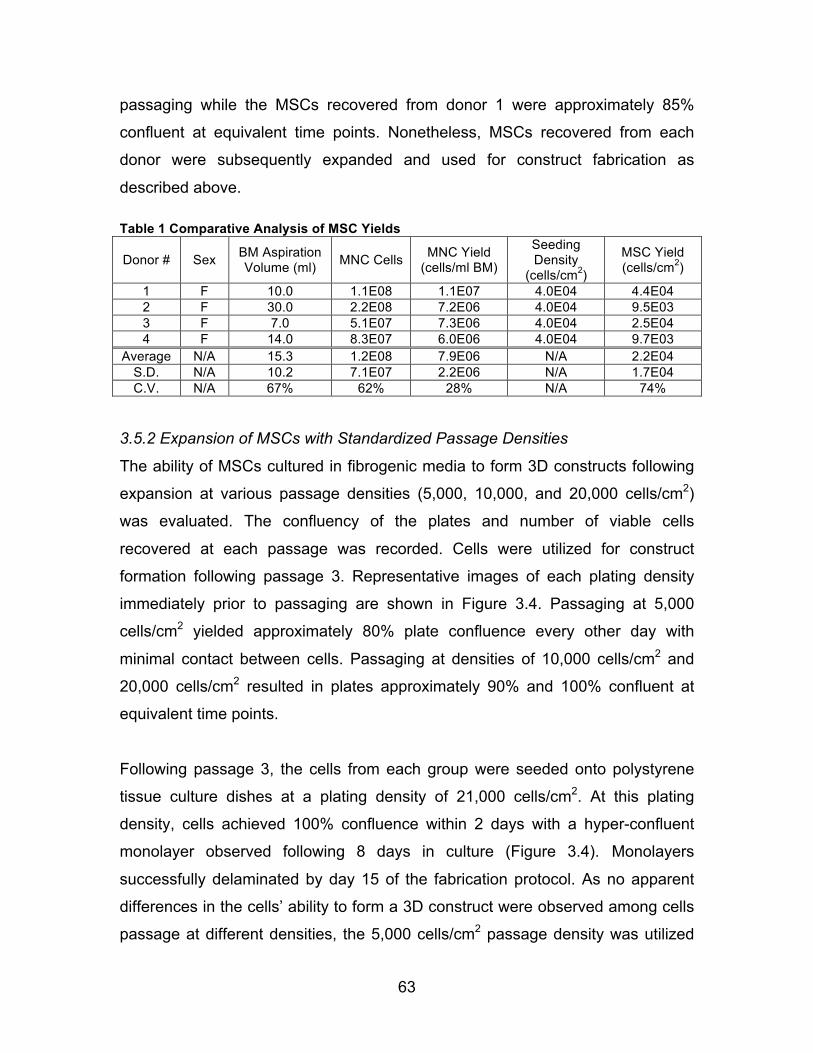
63
passaging while the MSCs recovered from donor 1 were approximately 85%
confluent at equivalent time points. Nonetheless, MSCs recovered from each
donor were subsequently expanded and used for construct fabrication as
described above. Table 1 Comparative Analysis of MSC Yields
Donor # Sex BM Aspiration Volume (ml) MNC Cells MNC Yield
(cells/ml BM)
Seeding Density
(cells/cm2)
MSC Yield (cells/cm2)
1 F 10.0 1.1E08 1.1E07 4.0E04 4.4E04 2 F 30.0 2.2E08 7.2E06 4.0E04 9.5E03 3 F 7.0 5.1E07 7.3E06 4.0E04 2.5E04 4 F 14.0 8.3E07 6.0E06 4.0E04 9.7E03
Average N/A 15.3 1.2E08 7.9E06 N/A 2.2E04 S.D. N/A 10.2 7.1E07 2.2E06 N/A 1.7E04 C.V. N/A 67% 62% 28% N/A 74%
3.5.2 Expansion of MSCs with Standardized Passage Densities
The ability of MSCs cultured in fibrogenic media to form 3D constructs following
expansion at various passage densities (5,000, 10,000, and 20,000 cells/cm2)
was evaluated. The confluency of the plates and number of viable cells
recovered at each passage was recorded. Cells were utilized for construct
formation following passage 3. Representative images of each plating density
immediately prior to passaging are shown in Figure 3.4. Passaging at 5,000
cells/cm2 yielded approximately 80% plate confluence every other day with
minimal contact between cells. Passaging at densities of 10,000 cells/cm2 and
20,000 cells/cm2 resulted in plates approximately 90% and 100% confluent at
equivalent time points.
Following passage 3, the cells from each group were seeded onto polystyrene
tissue culture dishes at a plating density of 21,000 cells/cm2. At this plating
density, cells achieved 100% confluence within 2 days with a hyper-confluent
monolayer observed following 8 days in culture (Figure 3.4). Monolayers
successfully delaminated by day 15 of the fabrication protocol. As no apparent
differences in the cells’ ability to form a 3D construct were observed among cells
passage at different densities, the 5,000 cells/cm2 passage density was utilized
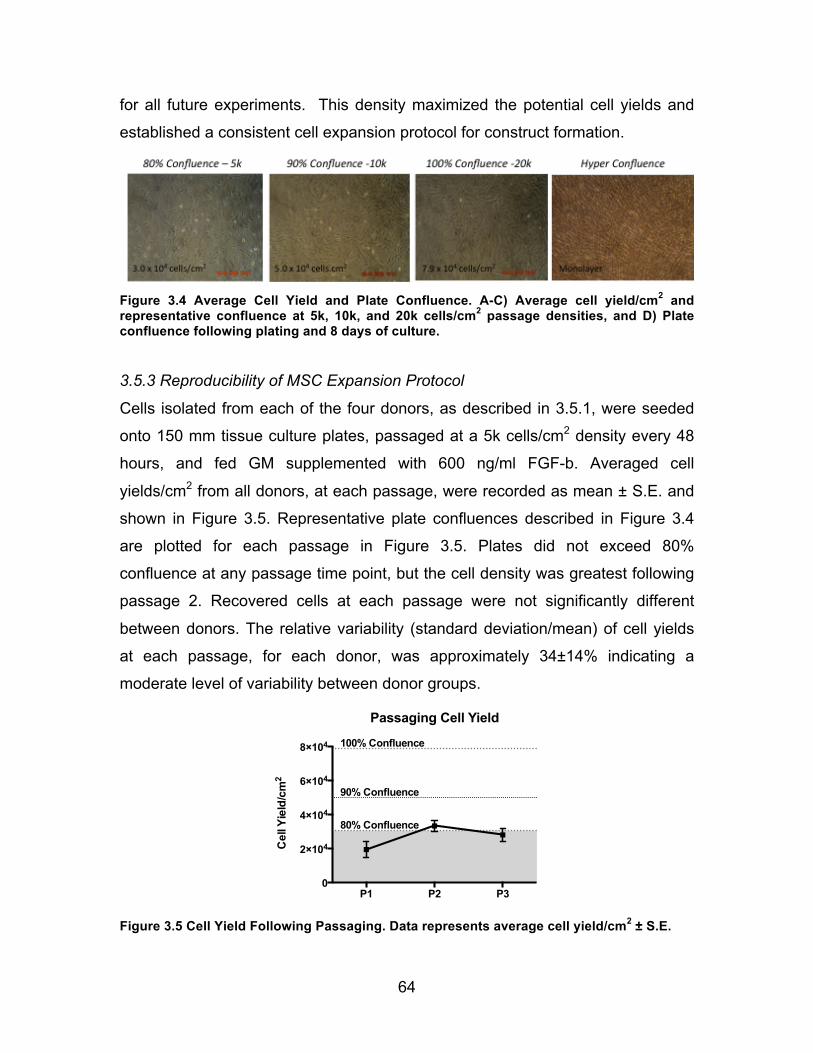
64
for all future experiments. This density maximized the potential cell yields and
established a consistent cell expansion protocol for construct formation.
Figure 3.4 Average Cell Yield and Plate Confluence. A-C) Average cell yield/cm2 and representative confluence at 5k, 10k, and 20k cells/cm2 passage densities, and D) Plate confluence following plating and 8 days of culture.
3.5.3 Reproducibility of MSC Expansion Protocol
Cells isolated from each of the four donors, as described in 3.5.1, were seeded
onto 150 mm tissue culture plates, passaged at a 5k cells/cm2 density every 48
hours, and fed GM supplemented with 600 ng/ml FGF-b. Averaged cell
yields/cm2 from all donors, at each passage, were recorded as mean ± S.E. and
shown in Figure 3.5. Representative plate confluences described in Figure 3.4
are plotted for each passage in Figure 3.5. Plates did not exceed 80%
confluence at any passage time point, but the cell density was greatest following
passage 2. Recovered cells at each passage were not significantly different
between donors. The relative variability (standard deviation/mean) of cell yields
at each passage, for each donor, was approximately 34±14% indicating a
moderate level of variability between donor groups.
Figure 3.5 Cell Yield Following Passaging. Data represents average cell yield/cm2 ± S.E.
P1 P2 P30
2×10⁴
4×10⁴
6×10⁴
8×10⁴
80% Confluence
90% Confluence
100% Confluence
Cel
l Yie
ld/c
m2
Passaging Cell Yield
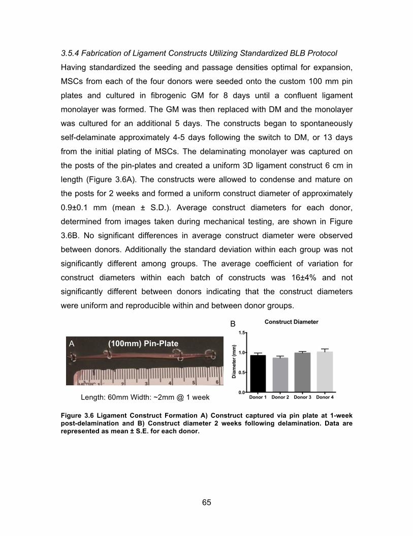
65
3.5.4 Fabrication of Ligament Constructs Utilizing Standardized BLB Protocol
Having standardized the seeding and passage densities optimal for expansion,
MSCs from each of the four donors were seeded onto the custom 100 mm pin
plates and cultured in fibrogenic GM for 8 days until a confluent ligament
monolayer was formed. The GM was then replaced with DM and the monolayer
was cultured for an additional 5 days. The constructs began to spontaneously
self-delaminate approximately 4-5 days following the switch to DM, or 13 days
from the initial plating of MSCs. The delaminating monolayer was captured on
the posts of the pin-plates and created a uniform 3D ligament construct 6 cm in
length (Figure 3.6A). The constructs were allowed to condense and mature on
the posts for 2 weeks and formed a uniform construct diameter of approximately
0.9±0.1 mm (mean ± S.D.). Average construct diameters for each donor,
determined from images taken during mechanical testing, are shown in Figure
3.6B. No significant differences in average construct diameter were observed
between donors. Additionally the standard deviation within each group was not
significantly different among groups. The average coefficient of variation for
construct diameters within each batch of constructs was 16±4% and not
significantly different between donors indicating that the construct diameters
were uniform and reproducible within and between donor groups.
Figure 3.6 Ligament Construct Formation A) Construct captured via pin plate at 1-week post-delamination and B) Construct diameter 2 weeks following delamination. Data are represented as mean ± S.E. for each donor.
Donor 1 Donor 2 Donor 3 Donor 40
1
2
3
4
5
Tang
ent M
odul
us (M
Pa)
Tangent Stiffness
## #
Donor 1 Donor 2 Donor 3 Donor 40.0
0.5
1.0
1.5
Dia
met
er (m
m)
Construct Diameter
Length: 60mm Width: ~2mm @ 1 week
(100mm) Pin-Plate A
B
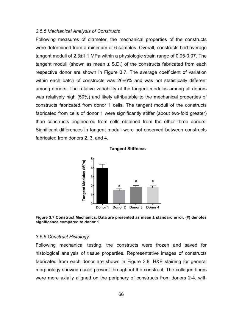
66
3.5.5 Mechanical Analysis of Constructs
Following measures of diameter, the mechanical properties of the constructs
were determined from a minimum of 6 samples. Overall, constructs had average
tangent moduli of 2.3±1.1 MPa within a physiologic strain range of 0.05-0.07. The
tangent moduli (shown as mean ± S.D.) of the constructs fabricated from each
respective donor are shown in Figure 3.7. The average coefficient of variation
within each batch of constructs was 26±6% and was not statistically different
among donors. The relative variability of the tangent modulus among all donors
was relatively high (50%) and likely attributable to the mechanical properties of
constructs fabricated from donor 1 cells. The tangent moduli of the constructs
fabricated from cells of donor 1 were significantly stiffer (about two-fold greater)
than constructs engineered from cells obtained from the other three donors.
Significant differences in tangent moduli were not observed between constructs
fabricated from donors 2, 3, and 4.
Figure 3.7 Construct Mechanics. Data are presented as mean ± standard error. (#) denotes significance compared to donor 1.
3.5.6 Construct Histology
Following mechanical testing, the constructs were frozen and saved for
histological analysis of tissue properties. Representative images of constructs
fabricated from each donor are shown in Figure 3.8. H&E staining for general
morphology showed nuclei present throughout the construct. The collagen fibers
were more axially aligned on the periphery of constructs from donors 2-4, with
Donor 1 Donor 2 Donor 3 Donor 40
1
2
3
4
5
Tang
ent M
odul
us (M
Pa)
Tangent Stiffness
## #
Donor 1 Donor 2 Donor 3 Donor 40.0
0.5
1.0
1.5
Dia
met
er (m
m)
Construct Diameter
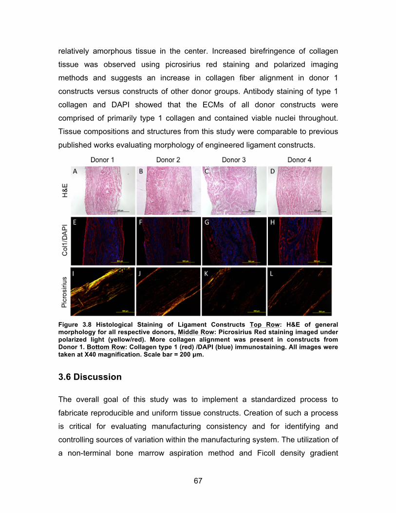
67
relatively amorphous tissue in the center. Increased birefringence of collagen
tissue was observed using picrosirius red staining and polarized imaging
methods and suggests an increase in collagen fiber alignment in donor 1
constructs versus constructs of other donor groups. Antibody staining of type 1
collagen and DAPI showed that the ECMs of all donor constructs were
comprised of primarily type 1 collagen and contained viable nuclei throughout.
Tissue compositions and structures from this study were comparable to previous
published works evaluating morphology of engineered ligament constructs.
Figure 3.8 Histological Staining of Ligament Constructs Top Row: H&E of general morphology for all respective donors, Middle Row: Picrosirius Red staining imaged under polarized light (yellow/red). More collagen alignment was present in constructs from Donor 1. Bottom Row: Collagen type 1 (red) /DAPI (blue) immunostaining. All images were taken at X40 magnification. Scale bar = 200 µm.
3.6 Discussion
The overall goal of this study was to implement a standardized process to
fabricate reproducible and uniform tissue constructs. Creation of such a process
is critical for evaluating manufacturing consistency and for identifying and
controlling sources of variation within the manufacturing system. The utilization of
a non-terminal bone marrow aspiration method and Ficoll density gradient

68
isolation technique allowed for a consistent number of plastic-adherent MSCs to
be isolated from a known amount of starting material. Additionally, considering
multiple aspirations can be performed from a single donor, these methodologies
greatly improve the amount of cells and the number of resultant constructs that
can be fabricated from a given BM donation.
As the new BLB fabrication method can be performed from a routine bone
marrow aspiration, commercial manufacturing can be performed as an
autologous or allogeneic process. An autogenic manufacturing process would
treat each graft as a patient-specific product. While the risk of immune rejection
is eliminated with autogenic grafting, the variation between the starting cell
sources obtained from patient specific bone marrow aspirations can lead to large
variations in the tissue-engineered end products and make it difficult to meet the
established manufacturing process controls and end-product specifications
necessary to demonstrate the end products are acceptable for use. In contrast,
an allogeneic process, while having some risk of immune rejection, would allow
for many constructs to be made from a single well-characterized donor source
and is a more commercially viable approach for manufacturing our grafts.
Although the use of the Ficoll isolation technique improved upon our existing
methodology by providing an increased number of adherent MSCs, significant
variability still existed in the resultant cell isolates from each donor. Despite each
plate having an equivalent numbers of mononuclear cells seeded initially onto the
cell culture plates (40,000 cells/cm2), the cell yields following 8 days of culture
were significantly different. This finding is consistent with the work of other
researchers that have demonstrated variability in MSC yields even between two
aspirations taken from the same donor during the same procedure 28. This finding
suggests that the initial plating density may not be critical for isolating a well-
defined cell population119 and that amplification of cells with successive passages
may be sufficient to expand and isolate a purified MSC source. However, even if
a purified MSC population can be expanded through passaging, characterization

69
of this population will be required by the FDA to confirm the degree of purity and
identity of the cell population and to evaluate the variability related to
differentiation potential, growth rates, etc. that may exist between cells obtained
from different donors or the same donor at different time points.
We found that the newly established passage density of 5,000 cells/cm2
consistently expanded the isolated cell populations ex vivo to 80% confluence,
and prevented over confluence that can potentially alter the cells’ phenotype.
Recent work suggests that lower passage densities may reduce population
doubling times of MSCs 119. Thus, future work investigating the optimal
parameters for maximizing cell yields in the shortest amount of time will be
beneficial to improving our process efficacy and reducing overall costs.
The fabrication of individual constructs utilizing our novel pin-plate design has
significantly streamlined the manufacturing process. The rate of success for
capturing and constraining the delaminating monolayer to an appropriate length
significantly improved from about 30% with the old protocol to 100% with the
novel pin-plate approach. In a few constructs, the contraction of the tissues
around the pins post-delamination, led to very thin constructs that tore off the
pins. Future studies will need to investigate factors that influence tissue roll-up
including growth factors, pin locations, and time in culture, to improve the
longevity of our 3D construct fabrication process.
The use of the pin-plates decreased labor and eliminated the variability
associated with using a technician. This allowed for more accurate evaluation of
variability between and within constructs engineered from different cell
populations. Additionally, because a technician is not required to transfer
monolayers to a new plate and manually constrain them with pins, a single
technician can fabricate many more constructs in the same amount of time. The
increased ability of the grafts to survive in vitro will be critical for increasing the
sample sizes per batch required to adequately characterize the resultant tissue.

70
The rate-limiting step of the construct manufacturing process now shifts to our
ability to efficiently exchange and add media to each plate. Therefore, future
experiments were designed to include an automated bioreactor system, capable
of feeding multiple plates at once.
The relative variability in the average tangent modulus of engineered constructs
was relatively high (~50%). This was in part due to the significantly higher
modulus of construct fabricated from donor 1 cells compared to those made from
cells of the other donors. Exclusion of donor 1 constructs from analysis of BLB
mechanics shows that variability among donors is greatly reduced with an
average tangent modulus of 1.7±0.2 MPa and a relative variability of across all
donors of 12%. While general morphology was similar across all groups, donor 1
qualitatively exhibited more aligned collagen as seen in polarized images of
picrosirius red staining. This increase in collagen alignment may have led to
increased mechanics of these tissues.
While ECM variability may explain the mechanical differences of our constructs,
the source of this variation is unknown and likely related to variations in the initial
cell populations obtained from each donor. To control for this variation, further
identification and characterization of the cell population immediately prior to
plating is needed to establish a purified starting population required for construct
fabrication. While it may be impossible to completely eliminate heterogeneity
within the starting cell population, the identification of specific cell properties or
ranges of cell properties indicative of successful end-product performance, will
be useful for improving process efficiency and construct quality. Defining
acceptable cell criteria will also be important for CGMP compliant process
validation that will require the identification and control of variation with the
manufacturing procedures.
Despite differences observed in final construct properties among donor cell
populations, significant differences in cell yields during passaging were not

71
observed. Alternative methods for characterizing cells during the construct
fabrication process, such as relative gene expression analysis, may be needed to
identify additional sources of variability that may exist between donor
populations. Additional information about the donor (health history and age of the
animal), may also be useful in identifying sources of variations that can influence
the process results. In addition to adequate cell characterization, the
identification of suitable construct properties and specifications, indicative of the
efficacy of tissue regeneration in vivo is a significant challenge in the field of
tissue engineering. Systematic data collection of much larger sample sizes within
a tightly controlled, well-characterized manufacturing system is needed to help
identify new in vitro targets that are more representative of the construct’s
therapeutic effect in vivo.
In conclusion, the novel methods described in this chapter have led to a
standardized process for fabricating uniform and reproducible constructs. Future
studies designed to optimize our process will concentrate on investigating plating
densities, expression of ligamentous markers, and construct mechanics.
Additionally, our newly introduced methods demonstrate the standardization of
only the ligament part of the BLB manufacturing process. Cells grown under
osteogenic culture conditions may have vastly different growth rates and
properties. Thus, we will need to develop custom protocols to standardize
passage densities, culture time, and growth conditions to optimize their
osteogenic potential in vitro.

72
Chapter 4 Characterization of a MSC Population Required for
Scaffold-less Construct Fabrication
4.1 Introduction
Despite the standardization of our MSC isolation and expansion methodologies,
the mechanical properties of constructs fabricated from the bone marrow
obtained from different donors varied. This variation was likely attributable to
subtle differences in the starting cell populations of the extracted MSCs. Thus, it
is necessary to further define and control the starting cell population in order to
validate our manufacturing process, ensure a more consistent and well-defined
end product, and comply with current CGMP regulations. Additionally, to improve
the translational applications of our BLB technology, the ability to produce human
derived constructs using our current fabrication method should be evaluated.
Within our current ligament manufacturing protocol, MSCs cultured in fibrogenic
media attach to, proliferate, and differentiate towards a tendon/ligament-like
lineage over successive passages before being seeded to form a self-
delaminating monolayer. However, extensive characterization of these cells
immediately prior to final seeding has not been performed and requires sufficient
analysis to evaluate and define the composition, function, and ligamentous
differentiation of this starting MSC population.
Human MSCs (hMSCs) have been shown to differentiate into a variety of tissues
including tendon, bone, muscle, and fat. However, cell surface similarities
between MSCs and tendon fibroblasts and the lack of a specific tendon marker

73
make characterizing the differentiated tendon phenotype difficult. The use of
gene expression data can be used to describe differences in the regulation of
tendon-related ECM production even though they are not directly indicative of
protein quantity. Work by several authors has utilized a combination of gene
targets, including Col-1, Col-3, TenC, and scleraxis, to identify induction of MSCs
toward a tendon-like lineage 27,183,185. Scleraxis, although expressed in other cell
types such as osteocytes and chondrocytes, is considered highly specific for
ligament differentiation and believed to also regulate tenomodulin and Col-1
expression. The expression of Runx 2 and alkaline phosphatase (ALPL) is
commonly associated with differentiation to an osteogenic lineage and can be
used as a negative control to confirm the commitment towards ligament versus
bone.
The characterization of human MSCs is not trivial. MSCs isolated from bone
marrow aspirations have been shown to be few in number with roughly 1:10,000
nucleated cells attaching to the culture dish 30. Additionally, contamination of the
MSCs with plastic adherent hematopoietic debris complicates the isolation of a
purified MSC population. As such, successive expansion of the extracted MSCs
is required to amplify and purify cells for subsequent characterization assays. In
addition to their plastic adherence properties, MSCs are characterized by leptin
receptors (CD45, 95, 105) and their ability to differentiate towards multiple
lineages. Due to the increasing use of bone marrow derived MSCs in tissue
engineering applications and regenerative cell therapies, several companies and
researchers have begun investigating strategies for large-scale MSC production
using CGMP compliant conditions. The use of sophisticated methods of
manufacturing and characterization that are in accordance with international
standards for MSC identification (i.e. plastic-adherence, cell surface markers,
and differentiation potential) and allow for a well-defined cell population to be
produced, are well beyond the capabilities of our laboratory.

74
Fortunately for us, well-defined MSCs are commercially available from
companies such as Lonza and Rooster Bio. The utilization of these MSCs
eliminates a significant part of the manufacturing process that involves the
expansion and characterization of a consistent MSC starting population. It
eliminates the extensive testing required to identify and describe the composition
and phenotype of MSCs isolated with our current techniques. Additionally, the
time saved by utilizing a ready-made, purified cell source would shorten
fabrication time and improve the overall process efficiency. However, we have
not tested the ability of an undifferentiated MSC population to differentiate to
tendon-like lineage and directly form a 3D construct via our existing
manufacturing protocol.
The objective of this study was therefore to utilize a commercially available and
well-characterized hMSC population as our starting material and evaluate the
ability of the hMSC to differentiate to tendon and form a 3D construct using our
current BLB fabrication process. If successful, this will expedite the translational
application capabilities by identifying a clinically relevant, regulatory compliant,
pre-manufactured starting material for ligament/tendon construct fabrication. The
characterization of the ligamentous MSC lineage throughout the ligament
fabrication process via gene expression will be used to establish process
validation criteria of the in-process intermediates, and will more accurately define
the specifications and properties of our graft.
4.2 Methods
4.2.1 Formation of Ligament and Bone Constructs from hMSCs
First, human MSCs (Lonza and Rooster Bio) were removed from liquid nitrogen
storage and placed in a 37°C water bath. Immediately following the thawing
process, the entire volume of cells and media was transferred to a sterile 15 ml
conical containing at least 3 times the volume of warmed GM. Next, the cell
suspension was centrifuged at 1500 rpm for 5 min at 37°C. The supernatant was

75
aspirated and remaining cells were re-suspended in fresh GM and counted.
Viable cells were seeded onto sterile polystyrene tissue culture plates at a
density of 21,000 cells/cm2. Plates were immediately induced towards bone and
ligament linages through the addition of osteogenic and fibrogenic media,
respectively. Both hMSCs manufacturers provided extensive characterization of
the starting cell populations. De-identified characteristics of the donor and lot
information of cells are described below in Table 2.
Table 2 Donor and Lot Details of Cells Used for Construct Formation Manufacturer Patient Sex Patient Age Lot # Passage Frozen
Lonza Male 23 056 P2
Rooster Bio Male 30 007 7.2 Pop. Doubling
Rooster Bio Female 33 012 7.6 Pop. Doubling
At various defined stages of our fabrication process, cells and tissue were
harvested for subsequent qPCR analysis at 0, 4, 8, and 12 days. These time
points correspond to the initial plating, growth phase, differentiation phase, and
3-D roll-up phase of the self-delamination tissue fabrication process, respectively.
An additional 3 constructs were fabricated and allowed to remain in a 3D
configuration in vitro until day 15 for subsequent mechanical and histological
analyses of the formed 3D tissue. A diagram of the experimental set-up and
sample size for each lot of donor cells is shown in Figure 4.1.

76
Figure 4.1 Experimental Design for hMSC Construct Analysis. Temporal gene expression was analyzed at day 0, 4, 8, and 12 time points that corresponded to various aspects of the construct manufacturing process. Mechanical and histological analyses were performed exclusively on day 15 (3 days following roll-up) constructs.
4.2.2 Cell and Tissue Preservation for RNA Extraction
Isolation of ribonucleic acid (RNA) was performed on cell and tissue isolates, at
0, 4, 8, and 12 days. Day 0 cell suspensions, directly from the manufacturer,
were immediately placed in a 2 ml cryovial and centrifuged at 1500 rpm for 5 min
at 25°C. Following centrifugation, the supernatant was removed and the cryovial
was submerged in liquid nitrogen for a minimum of 30 seconds to freeze the
resultant cell pellet. Cells and tissue recovered at 4, 8, and 12 day time points
were first trypsinized from the culture surface, before being placed into 2 ml
cryovials and centrifuged at 1500 rpm for 5 min at 25°C. Following removal of the
supernatant, the resultant cell/tissue concentration was frozen in liquid nitrogen
for at least 30 sec. All cryovials were stored at -80°C following freezing until
required for further processing.
4.2.3 Histological Analysis of Tissue Constructs
Histological analysis of the constructs was performed to investigate the general
morphology and distribution of collagen and mineral within the constructs.
Methods for histological and immunohistochemical staining have been described
previously in section 3.4.7. Briefly, at 3 days post 3D construct formation (day
15), constructs were rinsed with phosphate buffered saline (PBS, Sigma-Aldrich)
and flash frozen in tissue freezing medium. Sample sections (10 µm, Milipore
Cryostat) were prepared for histological and immunohistochemical analyses.

77
Tissue structure and collagen distribution were determined using H&E and
collagen-1/DAPI immunohistochemical staining, respectively. Deposition and
distribution of calcified mineral was determined with Alizarin Red S staining 130.
4.2.4 Gene Expression Analysis
Total cellular RNA was isolated by lysis in Trizol (Qiagen) followed by a one-step
phenol chloroform-isoamyl alcohol extraction. RNA isolation was performed on
undifferentiated MSCs (n=1 sample, per donor) and cells harvested at days 4, 8,
and 12 of the BLB fabrication process (n=3 samples, per time point and donor)
by using a miRNeasy Kit (Qiagen) and treating with DNase I (Qiagen).
Osteogenic constructs were simultaneously fabricated and evaluated at
equivalent day 4, 8, and 12 time points (n=1 sample, per time point and donor).
The amount and integrity of RNA were quantified using a bio-analyzer system
(Nanodrop 2000). A total of 200 ng of RNA was reverse-transcribed using an RT2
HT First Stand kit (Qiagen). The amount of cDNA in each sample was analyzed
using a Nanodrop system, and 200 ng of the cDNA template was used for each
qPCR reaction. The cDNA was amplified in a CFX96 real time thermal cycler
(BioRad) using a Taqman gene expression assay kit. The level of expression of
mRNA transcripts of each target gene was calculated as 2-(dCt) and normalized to
GAPDH using methods of Schmittgen and Livak 94. A list of TaqMan® Gene
Expression Assays used in this experiment is shown in

78
Table 3. The qPCR reactions were run in duplicate and results are presented as
averaged target gene expression (mean ± standard error) normalized to GAPDH
for all donors.
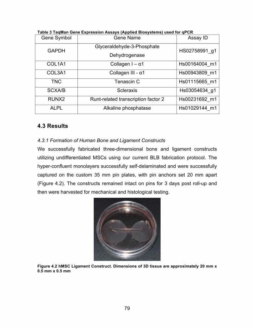
79
Table 3 TaqMan Gene Expression Assays (Applied Biosystems) used for qPCR Gene Symbol Gene Name Assay ID
GAPDH Glyceraldehyde-3-Phosphate
Dehydrogenase HS02758991_g1
COL1A1 Collagen I – α1 Hs00164004_m1
COL3A1 Collagen III - α1 Hs00943809_m1
TNC Tenascin C Hs01115665_m1
SCXA/B Scleraxis Hs03054634_g1
RUNX2 Runt-related transcription factor 2 Hs00231692_m1
ALPL Alkaline phosphatase Hs01029144_m1
4.3 Results
4.3.1 Formation of Human Bone and Ligament Constructs
We successfully fabricated three-dimensional bone and ligament constructs
utilizing undifferentiated MSCs using our current BLB fabrication protocol. The
hyper-confluent monolayers successfully self-delaminated and were successfully
captured on the custom 35 mm pin plates, with pin anchors set 20 mm apart
(Figure 4.2). The constructs remained intact on pins for 3 days post roll-up and
then were harvested for mechanical and histological testing.
Figure 4.2 hMSC Ligament Construct. Dimensions of 3D tissue are approximately 20 mm x 0.5 mm x 0.5 mm

80
4.3.2 Mechanical Properties of Ligament hMSC Constructs
Ligament constructs were mechanically tested utilizing a custom built
tensiometer as described earlier. Briefly, the constructs were removed from pins
and tested 3 days following formation of a 3D construct. The stress vs. strain
data for each sample are shown in Figure 4.3. Strain data were obtained through
digital image correlation of markers on the tissue and calculated using methods
described in section 3.4.6. The maximum tangent modulus was calculated for
each sample by computing the slope of the nominal stress vs. nominal strain
curve. The mean tangent modulus was 12.0 ± 0.2 kPa. No significant differences
in the mechanical properties of the tissues were observed between cell lots (n>2
per donor). Due to the limited number of bone samples, the bone constructs were
not mechanically analyzed.
Figure 4.3 Mechanics of hMSC Ligament Constructs. All tests are shown until failure.
4.3.3 Characterization of the Human-Derived Tissue Constructs
Morphologically the constructs were similar in appearance to previously
described constructs derived from ovine cells. The constructs were composed
primarily of Col-1 with viable nuclei present throughout the tissue (Figure 4.4B).
The H&E staining showed that the constructs were composed of relatively
amorphous tissue with more highly aligned collagen regions observed on the
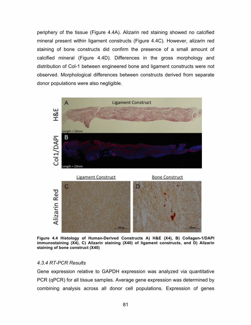
81
periphery of the tissue (Figure 4.4A). Alizarin red staining showed no calcified
mineral present within ligament constructs (Figure 4.4C). However, alizarin red
staining of bone constructs did confirm the presence of a small amount of
calcified mineral (Figure 4.4D). Differences in the gross morphology and
distribution of Col-1 between engineered bone and ligament constructs were not
observed. Morphological differences between constructs derived from separate
donor populations were also negligible.
Figure 4.4 Histology of Human-Derived Constructs A) H&E (X4), B) Collagen-1/DAPI immunostaining (X4), C) Alizarin staining (X40) of ligament constructs, and D) Alizarin staining of bone construct (X40)
4.3.4 RT-PCR Results
Gene expression relative to GAPDH expression was analyzed via quantitative
PCR (qPCR) for all tissue samples. Average gene expression was determined by
combining analysis across all donor cell populations. Expression of genes

82
typically associated with tendon and bone differentiation was analyzed and
compared over time in culture (days 0, 4, 8, and 12) and is shown in Figure 4.5.
Relative gene expression of tendon differentiation markers Col-1, Ten-C, and
Scx were significantly increased at day 8 compared to undifferentiated MSC
starting cell populations. While the expression of Col-3 was increased by day 8
(P=0.07), the increased expression was not significant until day 12. The
expressions of ligament markers Col-1, Ten-C, and Scx peaked at day 8 before
dropping at day 12 to undifferentiated (day 0) expression levels. Additionally,
expressions of Runx-2 and ALPL within our ligament constructs were significantly
less at days 8 and 12 compared to undifferentiated MSC populations. The down-
regulation of these bone-specific markers at day 8 and 12 time points further
confirms the induction of the MSCs towards a tendon/ligament lineage.
The average relative gene expression of bone constructs was also evaluated
(qPCR) at days 0, 4, 8, and 12 of culture (Figure 4.6). Expression of Col-1 and
Col-3 increased with increased time in culture with approximately a two-fold
increase compared to the undifferentiated MSC expression levels at day 0. The
increase in Col-1 and Col-3 markers is consistent with an increased production of
ECM material necessary for 3D tissue formation. As expected, significant
increases in the expression of ligament-specific markers Ten-C and Scx were not
observed in MSCs induced to an osteogenic lineage. Conversely, expression of
bone-specific markers Runx 2 and ALPL did increase over time in the MSCs
induced to an osteogenic lineage.

83
Figure 4.5 Relative Gene Expression of hMSC Ligament Constructs. (#) denotes significance with respect to undifferentiated MSCs.
Relative Gene Expression of Ligament Constructs
MSC Day 4 Day 8 Day 120.0
1.0
2.0
3.0
4.0
5.0
Rel
ativ
e Ex
pres
sion
to G
APD
H
Collagen 1
#
MSC Day 4 Day 8 Day 120.0
0.1
0.2
0.3
0.4
0.5
Tenacin C
Rel
ativ
e Ex
pres
sion
to G
APD
H #
MSC Day 4 Day 8 Day 120.00
0.01
0.02
0.03
0.04
Runx2
Rel
ativ
e Ex
pres
sion
to G
APD
H
##
MSC Day 4 Day 8 Day 120.0
0.5
1.0
1.5
Collagen 3
Rel
ativ
e Ex
pres
sion
to G
APD
H
#
MSC Day 4 Day 8 Day 120.000
0.002
0.004
0.006
0.008R
elat
ive
Expr
essi
on to
GA
PDH
Scleraxis
#
MSC Day 4 Day 8 Day 120.00
0.01
0.02
0.03
0.04
ALPL
Rel
ativ
e Ex
pres
sion
to G
APD
H
##

84
Figure 4.6 Relative Gene Expression of hMSC Bone Constructs. (#) denotes significance with respect to undifferentiated MSCs.
Relative Expression of Bone Constructs
MSC Day 4 Day 8 Day 120.0
2.0
4.0
6.0
Collagen 1
Rel
ativ
e Ex
pres
sion
to G
APD
H
#
MSC Day 4 Day 8 Day 120.00
0.05
0.10
0.15
0.20
0.25
Rel
ativ
e Ex
pres
sion
to G
APD
H
Tenacin C
#
MSC Day 4 Day 8 Day 120.00
0.01
0.02
0.03
0.04
Runx2
Rel
ativ
e Ex
pres
sion
to G
APD
H
MSC Day 4 Day 8 Day 120.0
1.0
2.0
3.0
4.0
Collagen 3
Rel
ativ
e Ex
pres
sion
to G
APD
H
#
MSC Day 4 Day 8 Day 120.000
0.001
0.002
0.003
0.004R
elat
ive
Expr
essi
on to
GA
PDH
Scleraxis
MSC Day 4 Day 8 Day 120.00
0.02
0.04
0.06
0.08
ALPL
Rel
ativ
e Ex
pres
sion
to G
APD
H

85
4.4 Discussion
The results of this study validate the use of our manufacturing process to
engineer 3D tendon and bone constructs starting from a well-characterized
commercially available cell source. Both tendon and bone tissues, fabricated
directly from an undifferentiated MSC population and expanded to ligamentous or
osteogenic lineages during the fabrication process, successfully formed 3D BLB
constructs. The prevention of the labor intensive cell passaging steps in this
newly developed manufacturing protocol reduces construct fabrication time by
removing the need to define and characterize specific tendon and bone cell
populations prior to use within our system and eliminating the need for continual
monitoring by a highly trained technician.
The ability of MSCs to differentiate towards a ligament phenotype in culture was
demonstrated through the analysis of gene expression data normalized to
GAPDH. No significant changes in relative gene expression were observed
between undifferentiated MSCs and MSCs induced to a ligamentous lineage for
4 days. However, following 8 days in fibrogenic media, the cells recovered from
the confluent monolayer had a higher gene expression of ligament-specific
markers of Col-1, Col-3, TenC, and Scx compared to the undifferentiated cells.
Expression of bone-specific markers Runx2 and ALPL were also down-regulated
at the 8-day time point. Within our fabrication process, the day 8 time point is
believed to be associated with an increased synthesis of ECM as the cells form a
confluent monolayer with sufficient matrix integrity to allow for self-delamination.
Following peak ligament-specific gene expression at day 8, the average
expressions of Col-1, TenC, and Scx in cells cultured for 12 days were reduced
to levels observed in the undifferentiated MSCs. Following the formation of a 3D
construct at day 12, the protein synthesis of the cells may have switched from
high rates of ECM synthesis to maintenance of existing ECM. It is important to
note that gene expression is not always indicative of protein expression. In fact,

86
despite the finding that gene expression at 12 days was equivalent to that of
undifferentiated MSCs, the overall amounts of tendon proteins within the
developing tissue may have been increased. Histological staining with Col-
1/DAPI confirmed a high abundance of Col-1 within the tendon portion of the
constructs despite a decrease in relative gene expression of Col-1 at day 12.
MSCs cultured in osteogenic media demonstrated a decrease in ligament-
specific makers and an increase in bone-specific markers (Runx-2 and ALPL)
with time. Histologically small amounts of mineral were seen within the bone-
differentiated engineered tissue. However, the relatively small amount of
mineralization suggests that additional time in vitro may be required to elicit more
substantial bone-like phenotypical changes in the bone region of the engineered
tissue.
The earliest time point suitable to correlate the mechanics of our 3D tissues with
gene expression immediately following delamination (day 12), was at three days
following roll-up (or day 15 of fabrication process) because mechanical analysis
of the material properties of our tissue requires accurate construct dimensions.
Tissue monolayers immediately following delamination are amorphous and
require sufficient time in culture to contract and uniformly align between the two
constraint pins. Evaluation of the constructs before a uniform diameter had been
established would have yielded inaccurate mechanical results. The moduli of our
constructs at this three-day time point were significantly more compliant than
previously reported tissue properties described in Chapter 2. This suggests that
significant changes in ECM structure and composition may occur within tissues
with increased time in culture.
While the average relative gene expression with time in culture was similar
between hMSC donors, the degree of relative gene expression varied. Despite
these differences between groups, observable changes in construct histology or
mechanics were not observed. However, more detailed assays looking at protein

87
content within our constructs may be needed to further characterize ligamentous
differentiation and construct properties among donor samples.
Continued evaluation of our fabrication process utilizing a well-defined cell
population is needed to identify and control sources of variation within the
manufacturing system. The use of standardized markers of gene expression may
provide suitable specification necessary to demonstrate production consistency
and graft effectiveness in vivo.
In summary, use of characterized, undifferentiated, bone marrow derived hMSCs
is an acceptable starting cell population for the fabrication of scaffold-less
constructs. As these MSCs are capable of differentiating towards both
ligamentous and osteogenic lineages during the construct formation process,
significant time associated with expansion and characterization of a pre-
differentiated tendon or bone-like MSC population can be eliminated.
Additionally, sufficient quantities of these well-characterized cells can be readily
obtained from CGMP compliant manufacturers, making them an ideal input
material for our translational manufacturing process.

88
Chapter 5 Bioreactor Manufacturing of Multi-Phasic Tissue
Constructs
5.1 Introduction
In addition to establishing a robust protocol capable of producing uniform
constructs reproducibly, significant hurdles remain with respect to commercial
manufacturing of an engineered product. Transitioning the laboratory
concept/methods into a well-characterized medical product/process is a
significant challenge, commonly underestimated by researchers in tissue
engineering and regenerative medicine fields. In addition to meeting regulatory
requirements to ensure consistency and safety of any product, the manufacturing
system must be scalable and cost-effective to be sufficiently economically viable
to displace current treatment options. Addressing manufacturing issues related to
quality control, cost-effectiveness, and large-scale production early in the
research stage, can overcome problems that typically slow development, limit
investment, and escalate costs limiting the clinical translation and availability of
the treatment to patients in need.
5.2 Background/Motivation
Despite significant advancements in the field of tissue engineering, relatively few
products have realized commercial success as tissue substitutes 22,102,103.
Several challenges still remain for translating tissue-engineering technologies
from bench to bedside. In addition to demonstrating clinical effectiveness, cost-
effective manufacturing processes are needed that comply with the current

89
quality and safety regulations 1,175. Failure to adequately consider the likely cost
of manufacturing early in the developmental stage has been recognized as a
limiting factor for commercial success of technologies in the field 5. Traditional
manual laboratory tissue culture techniques are often not economical at large
clinical scales and have inherent variability and contamination risks. However,
due to their widespread use, simplicity, minimal development time, and low initial
costs, these processes are still used to rapidly advance into clinical trials and the
marketplace 24,38.
For example, Carticel™, recently acquired by Aastrom Biosciences, Inc. (Ann
Arbor, MI), is an autologous cell transplantation product used to repair articular
cartilage injures in the knee. Carticel™ is manufactured via manual techniques
performed by trained technicians in a serologic clean room. Briefly, cells are
isolated from the patient and expanded on tissue culture flasks to a sufficient
number needed for therapeutic use 110. This process requires a large number of
manual labor-intensive manipulation steps. A manual process, though perhaps
more feasible and economical on a small-scale during early process
development, is generally regarded as high risk due to the increased potential for
contamination 112,132. Additionally, inherent differences in processing techniques
between individuals, especially with technically challenging methods, can lead to
process inconsistencies and end product variability 177. Furthermore, as
production demand increases, scale-up will require multiple manual processes to
be performed in parallel, requiring additional technicians and generating
substantial labor costs. As a result, the overall cost of production of such
products tends to be high and can limit clinical success when the cost-benefit of
the product is evaluated against competing and often simpler therapeutic
strategies 133. The risk of commercial failure can be greatly reduced by
considering the economic viability of the product throughout the developmental
process 176,178. Automating manual tissue-culture processes through the use of
bioreactor systems can provide a means to standardize the culture process,
tightly control culture environment, remove user-dependent operation, and

90
establish a cost-effective tissue manufacturing process to meet large-scale
clinical demand 24,175.
Automated bioreactors have long been used in tissue-engineering manufacturing
and typically involve streamlining traditional 2D cell culture processes (cell
selection, expansion, differentiation, etc.) 65,131,173. While important, these cell
manufacturing processes are often only the starting point for many tissue
engineering strategies that utilize well characterized cell populations for tissue
growth. Thus there is a need to foster automated bioreactor-based systems fit for
the development of reproducible 3D engineered constructs. A closed,
standardized, and operator-independent bioreactor system would have the
potential to ensure safety and regulatory compliance and enable cost-effective,
large-scale, in vitro tissue production 1,151.
Recently, Advanced Tissue Sciences developed an automated system for large
scale manufacturing of their human fibroblast derived dermal substitute,
Dermagrat™ (currently manufactured by Organogenesis, Inc. (Canton, MA)).
Dermagrat™ consists of living cryopreserved allogeneic dermal fibroblasts
seeded onto a bio-absorbable polyglactin mesh scaffold used for treatment of
chronic skin wounds such as diabetic foot ulcers 109. The entire manufacturing is
performed in a closed bioreactor bag, eliminating user-dependent variability and
shielding the culture process from risk of contamination. The bag also serves as
a transport container. Following injection of cells into the system, the bioreactor is
only externally manipulated until the graft is thawed and utilized in the operating
room.
In addition to minimizing contamination risk, a closed bioreactor system can
precisely control and maintain the tissue culture environment. Tissue growth and
development is highly responsive to environmental factors (pH, temperature,
nutrient concentration, etc.), and as such, the entire fabrication process is critical
to the integrity of the final product and must maintain a high level of control to
meet the rigorous current good manufacturing practice (CGMP) standards

91
required by the Food and Drug Administration (FDA) to ensure tissue safety and
manufacturing consistency (Code of Federal Regulations, CFR 21).
The current BLB open manufacturing process is a labor-intensive one consisting
of a multiple steps in an open-environment, to manually combine several tissue
phases and form the multi-phasic graft. This labor-intensive process significantly
limits the technician’s BLB manufacturing capacity and is one of the greatest
factors impacting cost of production with scale.
Current automated bioreactor systems for 3D tissue fabrication are product
specific and/or not commercially available. As no bioreactor currently exists for
the fabrication of our unique BLB construct in a single vessel, a novel bioreactor
must be developed. As with current automated manufacturing systems, the
bioreactor design should be user independent, only externally manipulated,
sterile, and scalable. It must also recreate the unique multi-step formation
process incorporating bone and ligament tissues engineered from distinctive cell
populations into a single construct. As such, the current protocol developed in
Chapter 3, consisting of Ficoll isolated cells, standardized passage density, and a
novel pin-plate method for capturing spontaneous monolayer delamination, must
first be modified as it still includes several technician dependent steps limiting the
automation and scale of the process.
Firstly, the protocol for BLB fabrication described in Chapter 3 requires the
simultaneous culture of bone and ligament monolayers. A trained technician, in a
separate tissue culture plate, performs the combination of these distinct tissue
types following their delamination. To facilitate formation of a single BLB
construct within a single-closed vessel, the formation of the BLB construct must
occur sequentially within the same chamber. Secondly, to engineer BLB
constructs of sufficient size (>4 cm diameter) for use as an ACL graft
replacement, at least four constructs must be manually transferred by a
technician into a new plate and aligned in parallel to allow fusion of the grafts

92
with each other to form a single uniform construct. This labor-intensive step of
the fabrication process must be eliminated to facilitate construct fabrication within
a closed, hands-off bioreactor system. The utilization of larger tissue culture
plates with increased surface area may allow for larger monolayers and resultant
3D construct diameters following delamination. Thirdly, for the sequential
formation of bone and ligament tissue within a single vessel, the timing of cells
introduced into both the bone and ligament fabrication process will have to be
modified. To maintain a consistent starting cell population for tissue formation as
determined in Chapter 3, bone and ligament cells must be expanded and
cryogenically preserved at appropriate time points (passage 3 for ligament and
passage 4 for bone). The efficacy of cryogenically recovered cells in forming a
robust, self-delaminating monolayer and BLB construct has not been evaluated
by our laboratory. The utilization of a cryogenically preserved allogeneic cell line
will also allow for a consistent and well-characterized population of cells to be
used for all experiments and will eliminate donor variability between tissues.
Following these process changes, the BLB manufacturing protocol will be
replicated in a closed, automated bioreactor system, resulting in a continuous
multi-tissue constructs without any internal manipulation by the technician. As no
such system currently exists, the goal of this study was to design, build, and test
a novel automated bioreactor to fabricate a single ACL graft in a closed-system
chamber.
To minimize contamination and technician variability, and to provide economy of
scale, the bioreactor was designed to be closed, automated, modular, scalable,
and to require minimal external manipulation. Bioreactor success was measured
by the ability of the user to form a BLB construct without opening the bioreactor
or manually manipulating the tissue. Bi-phasic structure of the construct was
confirmed by chromosomal detection of bone cells (male) within expected areas
of the construct. BLB construct structure and function were evaluated
histologically (H&E, picrosirius red, alizarin red) and mechanically (tangent
modulus, extensibility) at 1-week, post-3D construct formation. The histological

93
and mechanical properties of the current BLB fabrication process were used as
the standards for comparison, since the current protocol described in Chapter 3
generates ACL grafts that effectively regenerate ligament in vivo (data not
shown).
5.3 Significance
This study addresses the significant manufacturing and bioprocessing challenges
inherent to the commercialization and translation of tissue engineered products
from laboratory to clinic. In addition to demonstrating clinical efficacy and meeting
regulatory standards, new tissue engineered products need to be cost-effective
manufacturing processes to deliver the cost-benefit ratio needed to compete with
alternative therapeutic options. Automated, closed bioreactor systems can
provide the means for the development of standardized, cost-effective, and safe
manufacturing of engineered tissues for clinical applications. Current bioreactor
systems for 3D tissue fabrication are highly product-specific and often not
commercially available. Therefore, a novel bioreactor design is required for
sterile BLB graft fabrication and can fill an important knowledge gap in the field.
The development of an automated manufacturing system during an early
research stage has the potential to significantly lessen the technical, regulatory,
and commercial barriers that tissue-engineering technologies are likely to
encounter in the future pre-market stages. This development may also serve as a
model or platform for other tissue systems or tissue-engineering technologies to
streamline their associated laboratory processes into safe, reproducible, suitably
scaled tissues for clinical studies.
5.4 Experimental Design
The development and design of a novel bioreactor culture system for closed,
automated BLB manufacturing can be divided into two steps. The first step is to
eliminate the manual steps associated with the current BLB fabrication
processes. In particular, the user-dependent combination of separate bone and
94
ligament tissues into a single vessel and the fusion of multiple BLB constructs to
achieve one construct with a diameter of suitable size for ACL reconstruction,
must be eliminated. The second step is to design and validate a closed,
automated, bioreactor system to recreate the adjusted manufacturing protocol.
Process methodologies were performed utilizing ovine-derived cell populations
due to the large amount of knowledge associated with their ability to form a multi-
phasic BLB construct. To eliminate timing issues associated with the sequential
formation of multiple tissues, cryopreserved cell populations were used to form
multi-phasic BLB constructs. Male and female cell populations were used to
make bone and ligament tissues, respectively, to confirm the co-culturing of
multiple tissue types following construct formation.
5.5 Step 1: Elimination of Manual BLB Manufacturing Processes
Previously described methods for BLB fabrication require the simultaneous
culture of bone and ligament monolayers in separate culture dishes. Forming the
actual multi-phasic BLB graft requires the timed, manual transfer of two 3D bone
constructs onto a confluent ligament monolayer prior to the monolayer’s
delamination. The delicate timing and difficulty associated with manually
performing this process make it difficult to replicate in a closed bioreactor design.
The sequential preparation of bone and ligament tissues in a single culture
vessel would greatly eliminate the technical complexities of this process and
simplify the bioreactor design. However, the ability to form a confluent,
spontaneously delaminating ligament monolayer from a single fibrogenic cell
suspension while in the presence of pre-formed bone constructs had not been
evaluated by anyone to the best of our knowledge.
The current process of BLB construct fabrication requires the manual
combination and fusion of multiple 3D BLB constructs to form a construct of
suitable diameter for use as a graft in ACL reconstruction. Again, from a design
95
perspective, it would be advantageous to eliminate this manual combination step
and fabricate a single construct of sufficient size for implantation within a single
vessel. The inclusion of these protocol changes to the current BLB fabrication
process will simplify the design of a closed, “hands-off” bioreactor system.
5.6 Methods
5.6.1 Isolation of MSCs
Bone marrow was aspirated from the iliac crest of sheep using a Monoject Illinois
needle (Sherwood Medical Company, St. Louis, Mo) with the animal under
general anesthesia induced by intravenous propofol and sustained with
inhalation of isoflurane in oxygen. A minimum of 15 ml of bone marrow volume
was collected using heparinized needles and dispensed into EDTA blood
collection tubes (BD, San Jose, Ca.) for processing.
The marrow was filtered through a 100 mm filter to remove solid debris and
consolidated into a single 50 ml conical. An equivalent volume of Dulbecco’s
Phosphate-Buffered Saline (DPBS) pH 7.2 (Gibco BRL Cat# 14190-144) was
added. The marrow solution was slowly added to a 15 ml layer of Ficoll-Paque
Premium (GE Healthcare, Munich, Germany) contained within a separate conical
and centrifuged (AccuSpin FR; Beckman Coulter Inc., Fullerton, CA) at 600 g for
30 minutes at 25°C.
The upper layer containing plasma and platelets was discarded, and
mononuclear cell layers containing the MSC population were transferred into a
new sterile conical. The cells were suspended in at least three times their volume
of DPBS and centrifuged at 500 g for 10 minutes. The supernatant was removed,
and an equivalent volume of ACK lysis buffer was added to the pellet volume,
lysing any remaining red blood cells. The conical was filled with DPBS and
centrifuged at 400 g for 5 min. The supernatant was again removed, and the
MSC pellet was re-suspended in 20 ml of growth medium (GM; DMEM

96
supplemented with 20% FBS and 2% ABAM). Cells were counted utilizing a
Countess automated counting system and immediately plated at a density of
40,000 cells/cm2 in a 150 mm tissue culture dish.
5.6.2 Expansion and Cryopreservation of MSCs
The adherent cell population was allowed to attach for 3 days before the plate
was rinsed with DPBS to remove any remaining debris or contaminating non-
adherent cell populations. Following plating, cells were immediately cultured
towards either an osteogenic or ligamentous lineages. Osteogenic growth
medium consisted of GM supplemented with 600 ng/ml basic fibroblast growth
factor (bFGF; Peprotech), and 400 ng/ml dexamethasone (dex; Sigma-Aldrich).
Ligamentous growth medium consisted of GM supplemented with only bFGF.
Plates were fed either bone or ligament medium every other day. At
approximately 50% confluence, the plate-adherent MSC populations were
trypsinized (trypsin-0.25% EDTA), combined into a single conical, and passaged
at a density of 5,000 cells/cm2 every other day. Following passage three,
ligamentous derived MSCs (ligament cells) were trypsinized and cryogenically
preserved. MSCs induced to an osteogenic lineage (bone cells) were passaged
and cryopreserved after passage four. Cells were cryogenically preserved at a
concentration of 5x10⁶ cells/ml in GM+10% DMSO (Sigma-Aldrige). One ml
volumes were aliquoted into 2 ml cryovials (Nalgene) and slowly frozen
(approximately 1°C/hr) overnight to -80°C. The vial was then transferred into a
liquid nitrogen freezer for long-term storage.
5.6.3 Thawing and Plating of Cells
As needed, cells were removed from liquid nitrogen storage and placed in a 37°C
water bath. Immediately after the medium turned to liquid, the entire volume of
cells and medium was transferred to a sterile 15 ml conical containing at least 3
times their volume of warmed GM. The cell suspension was centrifuged at 1500
rpm for 5 min at 37°C. The supernatant was aspirated and remaining cells were
re-suspended in fresh GM and counted. Cell viability was assessed with trypan

97
blue dye (Life Technologies). Recovered cells utilized for tissue formation
contained greater than 90% viable cells. Cells were seeded onto sterile
polystyrene tissue culture plates at a viable cell density of 21,000 cells/cm2. Bone
plates were fed GM supplemented with 400 ng/ml dex, 13 mg/ml ascorbic acid, 5
mg/ml L-proline, and 600 ng/ml bFGF. The ligament medium was similar to the
GM, lacking only the dex.
5.6.4 Preparation of Bone Constructs
Bone cells were first seeded onto 8-well rectangular tissue culture treated
polystyrene plates (NUNC Plastics) modified with two metal posts aligned
longitudinally and centered within the plate 15 mm apart (Figure 5.1A). Medium
was changed every 2-3 days. Cells became confluent after approximately 12
days. After 5 days in culture, the dishes had become hyper-confluent, forming a
monolayer sheet consisting of cells and cell-generated ECM. At this time, the
medium was switched to differentiation medium (DM; supplemented with 7% HS
and 2% ABAM) supplemented with 10µl/ml dex, 10µl/ml ascorbic acid/proline,
and 20µl/ml TGF-B. After approximately 1-2 days in culture, spontaneous de-
lamination of the monolayer occurred. The capturing of the shrinking monolayer
between the two constraining posts formed 3D cylindrical bone constructs.
Figure 5.1 Custom Pin Plates A) 8-well Bone Plate, and B) 4-well Ligament Plate. The locations of the posts were precisely aligned between the bone and ligament plates, facilitating the efficient transfer of uniform bone constructs onto ligament plates.
5.6.5 Preparation of Ligament Culture Plates
Bone constructs were transferred at fixed length onto identical post locations in a
sterile 4 well-rectangular tissue culture treated polystyrene plate (NUNC Plastics)
B A

98
(Figure 5.1B). The arrangement of the wells identically matched to 8-well bone
plates to ensure each ligament well had two bone constructs separated by a 30
mm distance centered along the long axis of the well. Ligament cells were
seeded into each well at a density of 21,000 cells/cm2. Cells became confluent
within 1-2 days and formed a hyper-confluent robust monolayer after 8 days in
culture. Cells were fed ligament GM every 2-3 days. After 8 days, the medium
was switched to ligament DM that induced the spontaneous delamination of the
monolayer within 3-5 days.
5.7 Results
5.7.1 Sequential Formation of BLB Constructs Within Single Vessels
Monolayers derived from both bone and ligament cryopreserved MSC lineages
successfully formed and spontaneously delaminated, demonstrating the ability of
cryogenically preserved cells to form BLB grafts. Additionally, seeding of
ligament cells following transfer of bone constructs to the ligament culture
surface did not interfere with ligament monolayer formation and resulted in the
successful co-culture of ligament and bone required for BLB formation. The
delaminating ligament monolayers fully surrounded the bone constructs,
capturing them within the newly formed BLB construct (Figure 5.2). The diameter
of the ligament region was noticeably thin (0.3+0.1 mm, n=5) with approximately
half of the constructs failing in the ligament mid-substance after 2 weeks. The
results demonstrate the ability to form a BLB sequentially in the same dish by
first adding bone constructs and then seeding ligament cells.

99
Figure 5.2 BLB Delamination. Bone regions were captured and surrounded by delaminating ligament monolayer. The culture surface for each of the 4 wells is 21.8 cm2.
5.7.2 Formation of Large Diameter Tissue Constructs
Larger pin plates were fabricated utilizing 150 mm tissue culture plates, with a
culture surface area of 182 cm2, and two metal posts located 50mm apart
centered and aligned uni-axially. As a control, 100 mm pin-plates, with a culture
surface area of 82 cm2, were also fabricated as described in Chapter 3. Plates
were UV treated for at least 1 hour, sprayed with 70% ETOH, and rinsed with
approximately 20 ml DPBS prior to use. Ligament cells were seeded onto 100
mm and 150 mm pin-plates and grown into a confluent monolayer, and
spontaneously delaminated into a 3D construct. Construct diameters were
compared at a one-week time point following delamination, as shown in Figure
5.3. The 100 mm pin-plate formed a construct approximately 2 mm in diameter
(n=3) while constructs fabricated in the 150 mm pin-plates formed constructs
about 5 mm in diameter (n=3). Pin locations were identical in each of the plates.

100
Figure 5.3 Pin Plate Construct 1 Week Post Delamination. Top: Construct fabricated on 100 mm plate. Bottom: Construct fabricated on 150 mm plate
5.8 Discussion
Adding ligament derived MSCs to individual wells, containing strategically placed
pre-formed 3D bone constructs, formed a BLB construct. This is in sharp contrast
to previous methodologies that require the combination of 3D bone constructs
onto confluent ligament monolayers to form the multi-phasic tissue. The results
demonstrate the feasibility of forming BLB constructs within a single well by
sequentially seeding cryogenically preserved bone and ligament cells. This
process adaptation eliminates the need for technicians to combine bone and
ligament tissues to form a BLB. As cells instead of 3D tissues are input into the
system, the process can easily be performed sterilely. The elimination of
technician dependent steps allows for the process to be automated and
performed in a closed system, which is essential for use within a bioreactor.
The use of cryogenically preserved allogeneic cells also allows for large-scale
expansion of cells that can be well characterized and used as needed for graft
fabrication. This is a significant advantage for process scalability compared to
fresh cells, as the starting cell population can be screened and evaluated before
investing time and resources into large volume construct production. The
regulatory perspective requires a well-defined starting cell population. Utilization
101
of allogeneic cells has the advantage of performing this characterization only
once. Autogenic cells must be characterized for each donor significantly
increasing the economy of scale and limiting the commercialization potential.
The fabrication of larger diameter grafts formed on the 150 mm plates compared
to 100 mm plates reveals the ability to control graft size through the manipulation
of culture surface area. This process eliminates the need for manual fusion of
multiple constructs by a technician, again, a technician-dependent step that can
impede the development of efficient, automated manufacturing processes,
suitable for bioreactor incorporation. Further evaluation of the change of
construct diameter, passive construct tension, and tissue mechanics must be
evaluated further. The described methods demonstrate essential protocol
adaptations that were needed to facilitate fabrication of BLB constructs within
single well, closed, bioreactor systems. The following sections will describe the
development of that system.
5.9 Step 2: Bioreactor Development
5.9.1 Bioreactor Design Following the removal of user-dependent steps from the fabrication process, the
end multi-phasic tissue graft must be replicated within a closed bioreactor
system. As no current bioreactor system exists, a novel device must be
designed, fabricated, and tested to confirm that a viable multi-phasic BLB graft
can be fabricated by user-independent methods within a closed system. The
bioreactor device shown in Figure 5.4 below was designed based on the
following design elements and performance requirements.

102
Figure 5.4 Flip Plate Bioreactor System. Approximate dimensions are 7 in x 6 in x 2 inches.
5.9.2 Bioreactor Design Criteria • Minimizes Contamination Risk
o Closed system – completely “hands-off” internal manipulation,
beginning with the injection of starting material and ending with
final construct formation
o Potential for inline monitoring and media sampling
o Container suitable for construct preservation, packaging, and
shipping
o Consists of FDA compliant materials that are capable of being
easily sterilized (autoclavable, ethylene oxide)
! Or the use of disposable elements that can be replaced if
they come into contact with cell, media, or wastes
• Eliminates Manual, User-Dependent Processes
o Automated – capable of being controlled without no reliance on a
technician
o Only external manipulation
! Eliminates user variability and construct consistency
o Simple to use, easy to operate, and the construct is exposed only
when needed for patient use
• Scales Economically
o Automated – reduce manufacturing time and increase capacity
103
o Modular – capable of incorporating multiple cartridges into larger
centrally controlled unit
o Small - Relatively small overall volume to minimize required space.
5.9.3 Performance Requirements of Bioreactor
• Replicate Current Self-delaminating BLB Fabrication Process
o Cells, media, nutrients can be added and waste removed on a
regular basis
o As suture will be primary method of introducing during ACL repair
surgery, suture must be made available and easily integrated into
construct.
• Form a Continuous Multi-phasic BLB Construct within a Single Device
5.9.4 Cell-Culture for Use in Bioreactor
Bone and ligament MSC populations were isolated, expanded, and cryogenically
preserved as described in section 5.6. For use within the bioreactor, bone cells
were fabricated from male ovine bone marrow while ligament cells were derived
from female ovine bone marrow. The use of gender-specific cell populations
allowed for identification of male cell migration between bone and ligament
regions and verification of co-culturing of two distinct cell populations within a
single BLB graft.
5.9.5 Flip-Plate Bioreactor Design Summary The final bioreactor design, termed a “Flip-Plate”, consists of an enclosed
chamber, multiple monolayer growth modules, a suture capture system for
constraining the monolayers during delamination and maturation, and a
mechanism for enabling tissue co-culturing through magnetic translation of
tissue. The Flip-Plate bioreactor can be used for the fabrication and co-culture of
multiple tissue types. As an example, the use of this device for the creation of a
BLB construct will be described.

104
BLB constructs are made from MSCs differentiated to osteogenic and fibrogenic
lineages and integrated within the bioreactor to form a continuous multi-tissue
construct with sufficient structural integrity and mechanical compliance to
withstand physiological loading when used as a graft for ACL repair without the
inclusion of exogenous scaffolding. As the culture surface of the bioreactor could
not be visualized under a microscope, ligament only and bone only constructs
were fabricated on 100 mm and 60 mm pin plates, respectively, and used as
process controls. BLB constructs formed within the bioreactor were evaluated
histologically and mechanically and compared to manually fabricated bone and
ligament tissues. The details of one or more aspects of the Flip-Plate device are
described in the accompanying drawings and descriptions below. Other features,
objectives, and advantages of the invention will be apparent from the detailed
description and drawings.
5.9.6 Detailed Flip-Plate Design and Description of Use Referring to Figure 5.5, cell differentiation, monolayer growth, capturing, and co-
culturing required to make the BLB are accomplished in bioreactor 40. Though
this bioreactor is designed for the fabrication of scaffold-free BLB tissue
constructs, other cell types (i.e. muscle, nerve, etc.) consisting of spontaneously
or substrate controlled tissue monolayer delamination can be used. Current cell
culture bioreactors are for the most part designed for non-adherent cell
suspensions or expansions of cells 131,146,173. These devices do not provide the
means of capturing and co-culturing delaminated monolayers of multiple tissue
types that are required for the formation of the multi-phasic BLBs.
Bone monolayer growth is carried out in module 1, located and sealed into part 25 of bioreactor 40. Module 1 may be segmented into individual wells 88 to
form multiple monolayers of various shapes and sizes by the addition of silicone
barriers 99 to the culture substrate 11. Other barrier materials such as Teflon
that do not allow for significant cell adhesion may also be used. Bioreactor 40

105
includes manual or automatic translation elements designed to facilitate bone
monolayer capture (suture 22) and transfer into co-culture (component 3). Manipulation of suture 22 arrangements though culture substrate 11 and
tension (controlled via component 3) can be used to control construct length and
graft tension within delaminated constructs. During bone monolayer formation,
MSCs are seeded onto culture substrate 11 of module 1, which preferably
consists of a sterile polystyrene culture surface coated with or without cell
adhesion proteins such as laminin.
Media exchange of module 1 is controlled manually with sterile injection of
media in an open-environment or automatically with perfused media. The
connecting plate 26 contains fittings 5 designed to add and remove media
contained in each segmented well of module 1. MSCs of the bone monolayer
growth process contained in module 1 are seeded at an initial density of
approximately 21,000 cells/cm2. Seeding density may not be critical to the
success of this invention, affecting only rate of monolayer formation.
Cells proliferate after seeding, forming a monolayer, and differentiate towards an
osteogenic lineage. While additional serum-containing cell culture media and
growth factors may be used, a preferred growth medium to optimize cell
proliferation and promote robust osteogenic ECM production consists of DMEM
supplemented with 20% fetal bovine serum and antibiotics, along with the
addition of dex, FGF-b, and ascorbic acid/ L-proline. Growth conditions that must
be maintained in module 1 include a pH of about 7.4, a medium temperature of
37°C, and a sterile air environment consisting of up to 20% O2. The medium
must be exchanged at sufficient rate and volume to prevent depletion of critical
media constituents by the growing cells; preferably 50% replaced every two
days.
In reference to Figure 5.6, following sufficient ECM matrix production by the
seeded cells, a confluent bone monolayer 9 is formed on the culture substrate 11 after approximately 5 days at the described seeding density (Figure 5.6A).

106
Growth-promoting medium is replaced with differentiation medium consisting of
DMEM, supplemented with 7% horse serum, antibiotic, transforming growth
factor-beta (TGF-b), ascorbic acid / L-proline, and dex to induce cell
differentiation, integration, and passive tension that leads to subsequent
spontaneous delamination of confluent tissue monolayer. Bone monolayer 9
delamination within module 1 is captured on sutures 22, forming a 3D bone construct 10 as shown in Figure 5.6B. Sutures 22 transverse modules 1 and 2
and are held in tension by anchoring spools 7 (shown in Figure 5.7). The bone
constructs 10 are allowed to mature on suture 22; the tissue continues to
contract and reorganize the ECM structure while constrained under tension. This
maturation phase occurs over approximately 2 days. However longer or shorter
times are possible. To facilitate translation and co-culture of the bone
constructs 10, the bioreactor 40 is inverted (Figure 5.6C). Following the
release of tension in the sutures 22 by loosening the anchoring spools 7, both
bone constructs are translated to ligament culture substrate 20 in module 2
and secured within part 32 (Figure 5.6D). Manual or automated translation of the
suture via magnets 8 contained within component 3 facilitates this transfer
(Figure 5.7). In reference to Figure 5.7, tension is re-established in sutures 22
(not shown) by pulling the magnets 8 after the anchoring spool 7 has been re-
tightened.
The sutures 22 required for translation reside within tubes (not shown) used to
minimize sharp edges and friction during translation. These tubes reside in
grooves 16 of parts 25 and 32. The grooves align the sutures to ensure uniaxial
insertion across modules 1 and 2 and locate insertion into component 3 to
interface with the anchoring spools 7 and magnets 8. Sliding grooves for
magnets 8 are located in part 37. Plates 34 and 35 secure the tubing into the
grooves 16 and anchor parts 25 and 32 to component 3, attached to
connecting plate 26.
107
Following translation of bone constructs, MSCs are seeded onto the ligament culture substrate 20 of module 2 at an initial density of approximately 21,000
cells/cm2. The osteogenic medium is replaced with ligament growth medium to
promote fibroblastic differentiation and production of ligamentous ECM. This
process requires approximately 8 days in culture. The ligamentous medium
consists of DMEM supplemented with 20% fetal bovine serum and antibiotics,
along with the addition of FGF-b, and ascorbic acid/ L-proline. Ligament medium
is exchanged via fittings 5 attached to the connecting plate 26, at 50% of
volume every 2 days. Environmental conditions required for ligament growth
within module 2 are identical to module 1.
Following formation of a ligament monolayer 55 upon the ligament culture substrate 20 (Figure 5.8A), the medium is switched to differentiation medium to
further induce matrix production and cell-matrix tension. The ligamentous
differentiation medium is composed of DMEM, supplemented with 7% horse
serum, antibiotic, transforming growth factor-beta (TGF-b), and ascorbic acid / L-
proline. As the ligament monolayer 55 spontaneously delaminates, sutures 22
capture it, surrounding the bone constructs 10 on substrate 20. The end result
is a multi-phasic BLB construct 50 that continues to mature (remodel,
condense, and develop ECM) over time in culture (Figure 5.8B). This maturation
phase requires approximately 1 week, although shorter and longer periods are
possible.
The resulting BLB construct can be grafted into the patient at this point or if
desired, further conditioned mechanically through the physical manipulation of
sutures 22. If implanted, the incorporated sutures may be utilized to secure the
graft into the patient during the surgical procedure. It is preferable that a fresh
BLB construct be used within 3 weeks following formation, as cells and
appropriate graft tension are difficult to maintain in culture for extended periods of
times. Alternatively, freezing, acellularizing, or other means of preservation may
be used to preserve the construct. As will be understood by those skilled in the

108
art, a skilled practitioner may make many changes in the apparatus described
above without departing from the spirit or scope of the device.

109
Figure 5.5 Exploded view of the Flip-Plate bioreactor design
Bioreactor 40
2
99
11 25
88
22
26
5
32
1
3
20
16
34
35
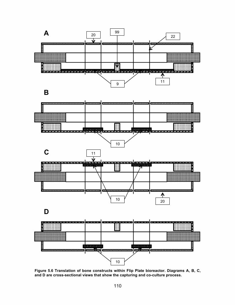
110
Figure 5.6 Translation of bone constructs within Flip Plate bioreactor. Diagrams A, B, C, and D are cross-sectional views that show the capturing and co-culture process.
D
C
B
A
10
9 11
99 22
10
10
20
20
11

111
Figure 5.7 Mechanism for tissue translations of the Flip-Plate device shown in a perspective view. Shown as component 3 of Figure 5.5.
Component 3
7
3
8
37
70

112
Figure 5.8 Formation of BLB construct within the Flip-Plate bioreactor. Diagrams A and B are cross-sectional views of the BLB captured on suture constraints.
B
A
10 55
50
22

113
5.10 Construct Evaluation Methodologies
Following successful construct formation, constructs were removed from the
bioreactor and evaluated as described below:
5.10.1 Mechanical Testing
Following delamination of the ligament monolayer, the BLB constructs were
allowed to fuse, forming a uniform cylindrical construct constrained between the
tensioned sutures. Upon removal of the construct from the bioreactor, separate
parts of the construct were allocated for mechanics or histological and molecular
biology analysis. Approximately half of bone/ligament complex was taken for
histology and molecular biology. The rest was attached to a custom-built tensile
tester and submerged in DPBS for subsequent tensile testing. The testing set-up
can be seen in Figure 5.9. Constructs were secured using custom Velcro grips
and incrementally lengthened to approximate the original length within the
reactor. Sixty-micron beads were painted onto the surface of the tissue for post
analysis digital image correlation to extrapolate the tissue-level strain during
testing. The construct was pulled until failure at a rate of 0.01 /sec. Load and
strain at failure was recorded. Figure 5.9 is an image of the described testing set-
up of the ligament region of the BLB construct. The missing bone region on the
right, shown by lack of suture, indicates the region removed for PCR analysis.
Figure 5.9 BLB on tensile testing set-up
114
5.10.2 Histological Analysis
For staining, unfixed samples were placed into TBS medium (Triangle Biological
Sciences), frozen in cold isopentane, and stored at -80°C until needed. Fixed
samples were placed in 4% paraformaldehyde (PFA) for 1 hour. The samples
were then placed into 15% sucrose solution for 1 hr at 4°C and then overnight
(>12 hrs) in 30% sucrose solution also at 4°C. The constructs were then
transferred into TBS medium and frozen over dry ice and stored at -80°C until
needed. Samples were cut to obtain cross and longitudinal sections with a
cryostat at a thickness of 12 µm, adhered to Superfrost Plus microscopy slides,
and used for staining. Sections were stained for general morphology
observations with hematoxylin and eosin (H&E). Mineral content of the tissue
was determined via alizarin red staining (Sigma), and collagen alignment with
picro-sirius red stain.
5.10.3 PCR Analysis – Y Chromosome
Genomic DNA was isolated from respective bone and ligament regions of the
BLB tissue samples by proteinase K digestion followed by ethanol precipitation.
A PCR-based assay for the ovine-specific Y-chromosome repeat sequence
Ucd043 was used to determine the presence of male cells in BLB explant
samples 73. Duplex PCR was performed using SCUcd043.FWD/SCUcd043. REV
primers to amplify Ucd043 together with P1- 5EZ/P2-3EZ primers to amplify the
ZFY/ZFX locus. P1-5EZ and P2-3EZ primers also provided an internal control for
amplification. Sensitivity of the assay was assessed with a dilution panel of 100,
25, and 5 pg of male ovine DNA. Subsequent experimental reactions were
carried out with 25 ng of genomic DNA template.
5.11 Results
5.11.1 Flip-Plate Construct Formation
The ability of the Flip-Plate bioreactor to successfully produce a BLB construct
without user-dependent manipulation has been demonstrated using the

115
bioreactor and processes detailed above. Briefly, MSCs induced towards an
osteogenic lineage were sterilely delivered into individual wells within module 1 of
the device and fed nutrients and growth factors specific to bone formation. A
bone monolayer is formed, and as it self-delaminated, a constraint system guided
its formation into two 3D cylindrical bone constructs (Figure 5.10A).
Figure 5.10 Translation of Bone Constructs in Flip-Plate Bioreactor A) Capturing of bone constructs, B) Inversion of device, C) Translation of constructs followed by subsequent, and D) seeding of ligament cells. As a reference, dimensions of bioreactor device are 7 x 6 x 2 inches.
Following formation of bone constructs in module 1, the bioreactor cartridge was
inverted, and the bone constructs were translated via magnets along guides to
the adjacent unseeded culture surface (Figure 5.10 B, C). Additional MSCs
induced towards ligamentous lineage were added to the culture surface (Figure
5.10D), fed nutrients and fibrogenic growth factors, and grown to confluence,
A B
C D

116
forming a ligament monolayer. At confluence, the ligament monolayer
delaminated from the substrate, surrounded the bone constructs (Figure 5.11A),
and formed a bone-ligament-bone construct that could be removed from the
bioreactor and utilized as an ACL graft (Figure 5.11 B, C).
Figure 5.11 Formation of BLB construct within Bioreactor. A) Delamination of ligament, B) Captured BLB construct shown with device opened, and C) BLB construct after removal from bioreactor and placed into separate dish. As a reference, construct length is 6 cm for each image.
5.12 Construct Validation
5.12.1 Mechanics
The ligament region of bioreactor BLB constructs and ligament only controls
were mechanically tested until failure at one week following formation. Stress vs.
strain data are shown in Figure 5.12. Ligament constructs, formed with the manual
process of Aim 2, had a modulus of 0.38±0.12 MPa (n=3) at a physiological
strain range of 0.05-0.07. Bioreactor constructs were similar, with an average
tissue modulus of 0.32±0.11 MPa (n=3). The mechanics of the bone regions
were not evaluated. The relative standard deviation within each group was
similar to that reported in Chapter 3, approximately 30%. The diameter of the
bioreactor constructs were 1.4±0.6 mm for BLBs (n=3) and 0.91±0.03 mm for
ligament only constructs captured on pin-plates (n=3).
A B C

117
Figure 5.12 Failure Stress-Strain Data of Manual vs. Bioreactor Formed Constructs. Strains are until failure of ligament region of the BLB construct.
5.12.2 Histology
Alizarin red staining shows mineral nodules within the bone regions of the graft
that were absent from the ligament region. This was evidence of a bi-modal
tissue distribution within the BLB graft (Figure 5.13 A-C). Mineralization was
comparable to bone and ligament constructs cultured seperately in osteogenic
and fibrogenic media, respectively, demonstrating a capacity of the tissue to
maintain a differentiated state while in co-culture. The construct was composed
of primarily type 1 collagen and contained viable nuclei thoughout (Figure 5.13 D-
F).
0.00
0.05
0.10
0.15
0.20
0 0.05 0.1 0.15 0.2 0.25 0.3
Stress (M
Pa)
Strain
Manual vs. Bioreactor Construct Mechanics
Man1 Man2 Man3 FPC1 FPC2 FPC3

118
Figure 5.13 Histological Staining of Bioreactor fabrication BLB Constructs A) Alizarin staining of bone region (X4), B) Alizarin staining of bone region (X20), C) Alizarin staining of ligament region, D) Col-1/DAPI of bone region (X4), E) Col-1/DAPI of bone region (X20), and F) Col-1/DAPI of ligament region.
5.12.3 PCR
The PCR analysis demonstrates that co-culture of multiple tissues within a
continuous tissue construct was successful. Male Y chromosome was detected
in only the bone regions of the construct (Figure 5.14). Female tissue was
detected throughout the construct and most highly expressed in the central
ligament region (n=2). Both bone and ligament regions contained significant
amounts of DNA.
Figure 5.14 PCR Data of Bioreactor Formed BLB Constructs. Bone regions were positive for male cell DNA while ligament regions comprised of female cells were not.

119
5.13 Discussion
5.13.1 Design Elements
The Flip-Plate bioreactor design successfully meets all previously described
design criteria.
Minimize Contamination Risk The Flip-Plate is designed to be a fully closed system. Although not utilized for
the initial proof-of-concept prototype, inclusion of sterile inline attachments for
media exchange to the inlet and outlet fittings of the bioreactor device can
completely seal the device from the external environment. Being a closed
system, perfusion is required for the infusion of temperature and gas controlled
media. The use of a perfusion system allows for precise control of culture
parameters and the potential for continuous system monitoring and in-line media
sampling. Such a system can greatly improve the safety of the device and
comply with current CGMP standards.
However, the inclusion of a perfusion system introduces additional variables into
the system that can alter cell adherence and growth. It also adds an increased
level of complexity to the device requiring additional costs and labor time to
implement. For these reasons, the fittings of the bioreactor were left open,
exposed to the local environmental condition, creating a semi-closed system.
Use of the bioreactor as a semi-closed system still significantly limited external
exposure to contamination. This permitted the bioreactor to fully replicate the pre-
defined BLB fabrication methodologies to allow cell growth within a sterile
incubator at 37C, 95% humidity, and 5%CO2 and media exchange within a sterile
laminar flow hood. The semi-closed bioreactor still provides reduced
contamination risk by eliminating direct user-contact with cells and tissue. Cell
injection and media exchange are performed only through sterile injection ports
via the syringe push-pull system. Despite being slightly exposed to the external
environment, no contamination was observed with use of this device. Once the

120
cells are injected into the device, the two culture surfaces are not taken apart
until the final tissue graft has been created and matured.
The entire device is fabricated from FDA compliant materials. Aside from the
disposable cell culture surfaces, the entire system can be sterilized by traditional
sterilization techniques (autoclaving or ethylene oxide) and reused. Further UV
treating of the device following assembly in a laminar flow hood further ensures
the sterility of the device prior to use. When fully closed, the device can be used
as a container for tissue preservation, storage, and shipping. The overall goal is
to deliver the preserved graft within the device so that it can be stored and
opened when needed by the surgeon.
Eliminate Manual, User-Depended Processes Eventually, the bioreactor system will be designed for complete automation. As
opposed to our current manual, user-dependent technology utilized in previous
BLB fabrication methods, all physical manipulations were performed on the
exterior of the device. This eliminated the user-variability associated with forming
the 3D BLB graft during the tissue combination process. Smooth and efficient
translation of the tissue was achieved by utilizing a magnet to pull the sutures.
Though other modes of movement are feasible, the utilization of a magnetic field
to provide translation allows the translational mechanism to be completely sealed
from the external environment. The end result is a much more consistent
manufacturing process not reliant on technician skill.
The eventual attachment of the bioreactor cartridge into a larger mechanical
system will allow for automated magnetic manipulation and inversion, or flipping,
of the device. If used in combination with an automatic perfusion system, larger
quantities of grafts can be fabricated with minimal need for a technician.
121
Economically Scalable The lack of reliance upon technician skill or labor through design of an automated
system would greatly improve the economic viability of the device with scale. The
bioreactor is modular, meaning several bioreactor cartridge systems can be
connected in parallel to a single media reservoir and manifold system. This
allows for the production of multiple grafts with little additional costs. The Flip-
Plate device was also designed to have a relatively small footprint to maximize
space and reduce infrastructure costs with large-scale manufacturing. The
current dimensions were chosen due to ease of manufacturing. Small profile
devices can be achieved through more skillful machining processes such as
injection molding.
5.13.2 Performance Requirements
The Flip-Plate bioreactor successfully produced a construct that was
morphologically similar to previous work 105, thus demonstrating the successful
adaptation of our manual BLB fabrication methods into a semi-closed automated
process. The entire construct was continuous with compositionally different
tissue at each end. Mineralized matrix was observed in the bone regions of the
graft despite the removal of dexamethasone and prolonged exposure to ligament
growth medium. Mechanically, despite being similar to the ligament-only controls,
the modulus of the ligament region of the graft was approximately an order of
magnitude lower than previously data described in section 3.5.5. The reason for
this is likely due to the difference in maturation time of only 1 week in the present
study. Further studies will investigate whether additional time in vitro may
improve matrix remodeling and graft mechanics at later time points.
Size disparity was also observed between experiments. Larger 5 mm and 2 mm
diameter constructs formed during stage 1 at one-week time points in 100 mm
and 150 mm plates, respectively, were not reproduced during Flip-Plate
bioreactor experiments. Flip-Plate BLB constructs were approximately 2-3 mm in
diameter. Inherent variability in cell ECM synthesis and remodeling likely account

122
for these differences. Previous work has demonstrated the ability of the BLB
construct to significantly grow in size in vivo approximating the native tissue
dimensions. Despite this feature, the in vitro construct must still maintain a
consistent and robust diameter to provide sufficient structural integrity during the
implantation procedure. Therefore, strategies to maintain construct size, such as
alteration of culture surface area, the removal of TGF-B, or release of suture
tension, may be needed to prevent excessive tissue condensation and better
preserve construct diameter and viability.
Delamination and 3D tissue formation within the Flip-Plate bioreactor was
facilitated through the use of tensioned sutures, rather than conventional pin-
plates described previously. During both trans-osseous rotator cuff repair and
anatomic ACL reconstructive surgery, the tissue-engineered graft is sutured to
the periosteum of the bone. The inclusion of the suture during the fabrication
process provides adequate material to secure the ends of the graft and anchor
the construct during surgery. Inclusion of alternative suture configurations may
enhance suture-graft integration and provide additional stability during
implantation and fixation to native tissue. Additionally, through automated
manipulation of suture tension, mechanical conditioning of the graft could be
performed to modulate the maturation and physical properties of the tissue graft.
5.14 Conclusion
In conclusion, the work outlined in this aim demonstrates the feasibility of
converting traditional bench-scale tissue culture techniques into a closed,
automated bioreactor system. In addition, this work describes and validates a
novel bioreactor design for the fabrication of scaffold-free, multi-phasic, co-
cultured tissue grafts for musculoskeletal repair. The application of bioreactors
for the generation of tissue-engineered products can lower contamination risks
and reduce user-dependency, providing a more consistent and economically
scalable manufacturing process. The development of cost-effective, scalable,

123
and regulatory compliant manufacturing procedures early in the research stages
of product development and before pre-clinical and clinical trials can significantly
lower the technical, regulatory, and financial hurdles limiting the current
translation of products within the tissue engineering field.
124
Chapter 6 Thesis Conclusions and Future Work
6.1 Conclusions
The objectives of this thesis were to evaluate the efficacy of our scaffold-less
tissue engineered constructs to regenerate the rotator cuff enthesis following
repair and to advance our existing construct fabrication methodologies to more
standardized and commercially relevant manufacturing processes. To
accomplish these objectives, the research of this thesis was guided by three
aims. Aim 1 focused on the repair of full thickness rotator cuff tears following
acute and chronic injury, utilizing our scaffold-less 3D construct methodologies.
As described in Chapter 2, use of our engineered tissues improved the
regeneration of the rotator cuff enthesis compared to existing suture repair
techniques. The re-establishment of the native enthesis is critical for modulating
loads at the tendon/bone interface and its regeneration may lead to the
improvement of repair outcomes following rotator cuff repair.
Aim 2 focused on the standardization of construct fabrication methodologies for
uniform, reproducible, and clinically relevant tissue production. As described in
Chapter 3, the use of a non-terminal bone marrow aspiration technique, a Ficoll
cell isolation method, a defined passaging density, and a novel monolayer
capturing “pin-plate”, greatly improved the consistency and efficiency of our
manufacturing process. Furthermore, the application of these processes using
human derived MSCs successfully demonstrated the feasibly of our methods to
fabricate ligament and bone tissues from a clinically relevant cell source without
the use of exogenous scaffolding (Chapter 4). The addition of these
125
manufacturing processes is a significant step towards the translation of these
scaffold-less technologies for human use and enables a more efficient process
for construct production.
However, to further expedite the commercialization of this technology and to
comply with existing FDA manufacturing guidelines, a novel bioreactor system
was designed, built, and evaluated as described in Aim 3. Because no existing
bioreactor systems existed for the closed, automated manufacturing of multiple
tissues within a single chamber, a new Flip-Plate bioreactor device was
developed (Chapter 5). The successful fabrication of a BLB construct within a
semi-closed chamber, without the need for direct tissue manipulation by a
technician, allows for scalable tissue production needed for large scale BLB graft
manufacturing. By addressing issues related to the development and
manufacturing of tissue engineering technologies early in the manufacturing
process, our ability to overcome current challenges that limit the development of
new medical products in the field of regenerative medicine is enhanced.
6.2 Future Work
Future work related to this thesis will focus on the continued evaluation of our
constructs in a rotator cuff repair model. Specifically, the safety and efficacy of
the constructs following implantation must be evaluated as required by the FDA.
While the results of Aim 1 were encouraging in a rat rotator cuff repair model, the
construct’s utility in a large animal model will need to be demonstrated.
Additionally, the toxicological response and in vivo function of the graft must be
investigated before proceeding into clinical trials.
The manufacturing process of our scaffold-less constructs must also be further
validated. While the described methodologies are a significant improvement over
existing protocols, they lack the sample size and validation needed to effectively
demonstrate the safety and reproducibly of the manufacturing process to the

126
FDA. Moreover, having optimized production of only the ligament region of our
BLB graft, future work is needed to optimize methods related to the formation of
scaffold-less bone tissues. To facilitate commercial manufacturing of BLB
constructs, several fully automated Flip-Plate bioreactors must be built in order to
simultaneously produce a large number of constructs at an economical cost.
Production of these constructs will be vital for characterizing the attributes of our
tissues and establishing product specifications for our BLB grafts. They will also
be needed to support future large-scale FDA process validation studies and the
widespread clinical use of our product. The research of this thesis presents a
new enthesis repair strategy to improve the treatment of musculoskeletal injuries
involving the ACL and rotator cuff tendons. By overcoming the current
translational challenges related to product development and industrialized
manufacturing early in the research process, the work of this thesis will hopefully
provide a framework for expediting the clinical and commercial translation of
other tissue engineering products in the field.

127
References
1. Abbasalizadeh S, Baharvand H. Technological progress and challenges
towards cGMP manufacturing of human pluripotent stem cells based
therapeutic products for allogeneic and autologous cell therapies.
Biotechnol. Adv. 2013;31:1600–1623.
doi:10.1016/j.biotechadv.2013.08.009
2. Akhtar MA, Bhattacharya R, Ohly N, Keating JF. Revision ACL
reconstruction - causes of failure and graft choices. Br. J. Sports Med.
2011 Nov 10;45(15):A15–A16. doi:10.1136/bjsports-2011-090606.49
3. Angeline ME, Rodeo SA. Biologics in the Management of Rotator Cuff
Surgery. Clin. Sports Med. 2012;31:645–663.
doi:10.1016/j.csm.2012.07.003
4. Apreleva M, Ozbaydar M, Fitzgibbons PG, Warner JJP. Rotator cuff tears:
the effect of the reconstruction method on three-dimensional repair site
area. Arthroscopy. 2002;18(5):519–26. doi:10.1053/jars.2002.32930
5. Archer R, Williams DJ. Why tissue engineering needs process engineering.
Nat. Biotechnol. 2005;23(11):1353–1355.
6. Ardern CL, Webster KE, Taylor NF, Feller JA. Return to sport following
anterior cruciate ligament reconstruction surgery: a systematic review and
meta-analysis of the state of play. Br. J. Sports Med. 2011 Jun;45(7):596–
606. doi:10.1136/bjsm.2010.076364
7. Aurora A, McCarron J, Iannotti JP, Derwin K. Commercially available
extracellular matrix materials for rotator cuff repairs: state of the art and

128
future trends. J. Shoulder Elbow Surg. 2007;16:S171–8.
doi:10.1016/j.jse.2007.03.008
8. Badylak SF. The extracellular matrix as a scaffold for tissue reconstruction.
Semin. Cell Dev. Biol. 2002 Oct;13(5):377–83.
9. Barber FA, Herbert MA, Coons DA. Tendon augmentation grafts:
biomechanical failure loads and failure patterns. Arthroscopy. 2006
May;22(5):534–8. doi:10.1016/j.arthro.2005.12.021
10. Barenius B, Ponzer S, Shalabi A, Bujak R, Norlén L, Eriksson K. Increased
risk of osteoarthritis after anterior cruciate ligament reconstruction: a 14-
year follow-up study of a randomized controlled trial. Am. J. Sports Med.
2014 May;42(5):1049–57. doi:10.1177/0363546514526139
11. Bartolozzi A, Andreychik D, Ahmad S. Determinants of outcome in the
treatment of rotator cuff disease. Clin. Orthop. Relat. Res. 1994
Nov;(308):90–7.
12. Bassett RW, Cofield RH. Acute tears of the rotator cuff. The timing of
surgical repair. Clin. Orthop. Relat. Res. 1983 May;(175):18–24.
13. Bedi A, Maak T, Walsh C, Rodeo SA, Grande D, Dines DM, et al.
Cytokines in rotator cuff degeneration and repair. J. Shoulder Elb. Surg.
2012;21:218–227. doi:10.1016/j.jse.2011.09.020
14. Benjamin M, Kumai T, Milz S, Boszczyk BM, Boszczyk AA, Ralphs JR. The
skeletal attachment of tendons--tendon “entheses”. Comp. Biochem.
Physiol. A. Mol. Integr. Physiol. 2002;133:931–945. doi:10.1016/S1095-
6433(02)00138-1
15. Benjamin M, McGonagle D. Entheses: Tendon and ligament attachment
sites. Scand. J. Med. Sci. Sport. 2009;19:520–527. doi:10.1111/j.1600-
0838.2009.00906.x

129
16. Benjamin M, Toumi H, Ralphs JR, Bydder G, Best TM, Milz S. Where
tendons and ligaments meet bone: Attachment sites (’entheses') in relation
to exercise and/or mechanical load. J. Anat. 2006;208:471–490.
doi:10.1111/j.1469-7580.2006.00540.x
17. Benjamin M. Anatomy and biochemistry of entheses. Ann. Rheum. Dis.
2000 Dec 1;59(12):995–999. doi:10.1136/ard.59.12.995
18. Bicer EK, Lustig S, Servien E, Selmi TAS, Neyret P. Current knowledge in
the anatomy of the human anterior cruciate ligament. Knee Surg. Sports
Traumatol. Arthrosc. 2010 Aug;18(8):1075–84. doi:10.1007/s00167-009-
0993-8
19. Bishop J, Klepps S, Lo IK, Bird J, Gladstone JN, Flatow EL. Cuff integrity
after arthroscopic versus open rotator cuff repair: a prospective study. J.
Shoulder Elbow Surg. 2006;15(3):290–9. doi:10.1016/j.jse.2005.09.017
20. Björnsson HC, Norlin R, Johansson K, Adolfsson LE. The influence of age,
delay of repair, and tendon involvement in acute rotator cuff tears:
structural and clinical outcomes after repair of 42 shoulders. Acta Orthop.
2011 Apr;82(2):187–92. doi:10.3109/17453674.2011.566144
21. Blitz E, Viukov S, Sharir A, Shwartz Y, Galloway JL, Pryce BA, et al. Bone
ridge patterning during musculoskeletal assembly is mediated through SCX
regulation of Bmp4 at the tendon-skeleton junction. Dev. Cell. 2009
Dec;17(6):861–73. doi:10.1016/j.devcel.2009.10.010
22. Bouchie A. Tissue engineering firms go under. Nat. Biotechnol.
2002;20:1178–1179. doi:10.1038/nbt1202-1178
23. Boykin RE, Heuer HJD, Vaishnav S, Millett PJ. Rotator cuff disease –
basics of diagnosis and treatment. Rheumatol. Reports. 2010 Mar 8;2(1).
doi:10.4081/rr.2010.e1

130
24. Brandenberger R, Burger S, Campbell A, Fong T, Lapinskas E, Rowley JA.
Cell Therapy Bioprocessing. Bio Process Int. 2011;
25. Brown CH, Carson EW. Revision anterior cruciate ligament surgery. Clin.
Sports Med. 1999 Jan;18(1):109–71.
26. Burkhart SS, Johnson TC, Wirth MA, Athanasiou KA. Cyclic loading of
transosseous rotator cuff repairs: tension overload as a possible cause of
failure. Arthroscopy. 1997 Apr;13(2):172–6.
27. Canseco JA, Kojima K, Penvose AR, Ross JD, Obokata H, Gomoll AH, et
al. Effect on Ligament Marker Expression by Direct-Contact Co-culture of
Mesenchymal Stem Cells and Anterior Cruciate Ligament Cells. Tissue
Eng. Part A. 2012;:120924061154007. doi:10.1089/ten.tea.2012.0030
28. Caplan AI. Mesenchymal stem cells. J. Orthop. Res. 1991 Sep;9(5):641–
50. doi:10.1002/jor.1100090504
29. Carpenter JE, Thomopoulos S, Flanagan CL, DeBano CM, Soslowsky LJ.
Rotator cuff defect healing: a biomechanical and histologic analysis in an
animal model. J. Shoulder Elbow Surg. 1998;7(6):599–605.
30. Castro-Malaspina H, Gay RE, Resnick G, Kapoor N, Meyers P, Chiarieri D,
et al. Characterization of human bone marrow fibroblast colony-forming
cells (CFU-F) and their progeny. Blood. 1980 Aug;56(2):289–301.
31. Chen J, Xu J, Wang A, Zheng M. Scaffolds for tendon and ligament repair:
review of the efficacy of commercial products. Expert Rev. Med. Devices.
2009 Jan;6(1):61–73. doi:10.1586/17434440.6.1.61
32. Cho NS, Yi JW, Rhee YG. Arthroscopic biceps augmentation for avoiding
undue tension in repair of massive rotator cuff tears. Arthroscopy. 2009
Feb;25(2):183–91. doi:10.1016/j.arthro.2008.09.012
131
33. Cofield RH, Parvizi J, Hoffmeyer PJ, Lanzer WL, Ilstrup DM, Rowland CM.
Surgical repair of chronic rotator cuff tears. A prospective long-term study.
J. Bone Joint Surg. Am. 2001 Jan;83-A(1):71–7.
34. Cohen DB, Kawamura S, Ehteshami JR, Rodeo SA. Indomethacin and
celecoxib impair rotator cuff tendon-to-bone healing. Am. J. Sports Med.
2006;34:362–369. doi:10.1177/0363546505280428
35. Coons DA, Alan Barber F. Tendon graft substitutes-rotator cuff patches.
Sports Med. Arthrosc. 2006 Sep;14(3):185–90.
36. Cooper RR, Misol S. Tendon and ligament insertion. A light and electron
microscopic study. J. Bone Joint Surg. Am. 1970 Jan;52(1):1–20.
37. Crawford SN, Waterman BR, Lubowitz JH. Long-term failure of anterior
cruciate ligament reconstruction. Arthroscopy. 2013 Sep;29(9):1566–71.
doi:10.1016/j.arthro.2013.04.014
38. Davie NL, Brindley D, Culme-Seymour EJ, Mason C. Streamlining Cell
Therapy Manufacture. Bio Process Int. 2012;
39. Dejardin LM, Arnoczky SP, Ewers BJ, Haut RC, Clarke RB. Tissue-
engineered rotator cuff tendon using porcine small intestine submucosa.
Histologic and mechanical evaluation in dogs. Am. J. Sports Med.
2001;29(2):175–84.
40. Derwin KA, Badylak SF, Steinmann SP, Iannotti JP. Extracellular matrix
scaffold devices for rotator cuff repair. J. Shoulder Elb. Surg. 2010;19:467–
476. doi:10.1016/j.jse.2009.10.020
41. Derwin KA, Baker AR, Spragg RK, Leigh DR, Iannotti JP. Commercial
Extracellular Matrix Scaffolds for Rotator Cuff Tendon Repair:
Biomechanical, Biochemical, and Cellular Properties. J Bone Jt. Surg Am.
2006;88:2665–2672. doi:10.2106/JBJS.E.01307

132
42. Derwin KA, Codsi MJ, Milks RA, Baker AR, McCarron JA, Iannotti JP.
Rotator cuff repair augmentation in a canine model with use of a woven
poly-L-lactide device. J. Bone Joint Surg. Am. 2009 May;91(5):1159–71.
doi:10.2106/JBJS.H.00775
43. Dugas JR, Campbell DA, Warren RF, Robie BH, Millett PJ. Anatomy and
dimensions of rotator cuff insertions. J. Shoulder Elbow Surg.
2002;11(5):498–503.
44. Eagan MJ, Zuk PA, Zhao K-W, Bluth BE, Brinkmann EJ, Wu BM, et al. The
suitability of human adipose-derived stem cells for the engineering of
ligament tissue. J. Tissue Eng. Regen. Med. 2011 Sep 22;6:702–709.
doi:10.1002/term.474
45. Van Eck CF, Schkrohowsky JG, Ramirez C, Irrgang JJ, Fu FH, Working Z.
Failure Rate and Predictors of Failure after Anatomic ACL Reconstruction
with Allograft (SS-61). Arthrosc. J. Arthrosc. Relat. Surg. 2011
May;27(5):e62–e63. doi:10.1016/j.arthro.2011.03.065
46. Ellman H, Hanker G, Bayer M. Repair of the rotator cuff. End-result study
of factors influencing reconstruction. J. Bone Joint Surg. Am. 1986
Oct;68(8):1136–44.
47. Ersen A, Demirhan M, Atalar AC, Kapicioğlu M, Baysal G. Platelet-rich
plasma for enhancing surgical rotator cuff repair: evaluation and
comparison of two application methods in a rat model. Arch. Orthop.
Trauma Surg. 2014 Mar;134(3):405–11. doi:10.1007/s00402-013-1914-3
48. Fleming BC, Hulstyn MJ, Oksendahl HL, Fadale PD. Ligament Injury,
Reconstruction and Osteoarthritis. Curr. Opin. Orthop. 2005
Oct;16(5):354–362.

133
49. Freeman JW, Woods MD, Laurencin CT. Tissue engineering of the anterior
cruciate ligament using a braid-twist scaffold design. J. Biomech. 2007
Jan;40(9):2029–36. doi:10.1016/j.jbiomech.2006.09.025
50. Fu FH, Bennett CH, Ma CB, Menetrey J, Lattermann C. Current trends in
anterior cruciate ligament reconstruction. Part II. Operative procedures and
clinical correlations. Am. J. Sports Med. 2000;28(1):124–30.
51. Fuchs B, Weishaupt D, Zanetti M, Hodler J, Gerber C. Fatty degeneration
of the muscles of the rotator cuff: assessment by computed tomography
versus magnetic resonance imaging. J. Shoulder Elbow Surg.
1999;8(6):599–605.
52. Galatz L, Rothermich S, VanderPloeg K, Petersen B, Sandell L,
Thomopoulos S. Development of the supraspinatus tendon-to-bone
insertion: localized expression of extracellular matrix and growth factor
genes. J. Orthop. Res. 2007 Dec;25(12):1621–8. doi:10.1002/jor.20441
53. Galatz LM, Ball CM, Teefey SA, Middleton WD, Yamaguchi K. The
outcome and repair integrity of completely arthroscopically repaired large
and massive rotator cuff tears. J. Bone Joint Surg. Am. 2004 Mar;86-
A(2):219–24.
54. Galatz LM, Charlton N, Das R, Kim HM, Havlioglu N, Thomopoulos S.
Complete removal of load is detrimental to rotator cuff healing. J. Shoulder
Elbow Surg. 2009;18(5):669–75. doi:10.1016/j.jse.2009.02.016
55. Galatz LM, Griggs S, Cameron BD, Iannotti JP. Prospective longitudinal
analysis of postoperative shoulder function : a ten-year follow-up study of
full-thickness rotator cuff tears. J. Bone Joint Surg. Am. 2001 Jul;83-
A(7):1052–6.

134
56. Galatz LM, Sandell LJ, Rothermich SY, Das R, Mastny A, Havlioglu N, et
al. Characteristics of the rat supraspinatus tendon during tendon-to-bone
healing after acute injury. J. Orthop. Res. 2006;24:541–550.
doi:10.1002/jor.20067
57. Gao J, Räsänen T, Persliden J, Messner K. The morphology of ligament
insertions after failure at low strain velocity: an evaluation of ligament
entheses in the rabbit knee. J. Anat. 1996 Aug;189 ( Pt 1:127–33.
58. Gartsman GM. Massive, irreparable tears of the rotator cuff. Results of
operative debridement and subacromial decompression. J. Bone Joint
Surg. Am. 1997 May;79(5):715–21.
59. Gartsman GM. All arthroscopic rotator cuff repairs. Orthop. Clin. North Am.
2001 Jul;32(3):501–10, x.
60. Gazielly DF, Gleyze P, Montagnon C. Functional and anatomical results
after rotator cuff repair. Clin. Orthop. Relat. Res. 1994 Jul;(304):43–53.
61. Genin GM, Kent A, Birman V, Wopenka B, Pasteris JD, Marquez PJ, et al.
Functional grading of mineral and collagen in the attachment of tendon to
bone. Biophys. J. 2009 Aug 19;97(4):976–85.
doi:10.1016/j.bpj.2009.05.043
62. Gimbel JA, Van Kleunen JP, Lake SP, Williams GR, Soslowsky LJ. The
role of repair tension on tendon to bone healing in an animal model of
chronic rotator cuff tears. J. Biomech. 2007 Jan;40(3):561–8.
doi:10.1016/j.jbiomech.2006.02.010
63. Gimbel JA, Van Kleunen JP, Mehta S, Perry SM, Williams GR, Soslowsky
LJ. Supraspinatus tendon organizational and mechanical properties in a
chronic rotator cuff tear animal model. J. Biomech. 2004 May;37(5):739–
49. doi:10.1016/j.jbiomech.2003.09.019
135
64. Goh JC-H, Ouyang H-W, Teoh S-H, Chan CKC, Lee E-H. Tissue-
engineering approach to the repair and regeneration of tendons and
ligaments. Tissue Eng. 2003 Jan;9 Suppl 1:S31–44.
doi:10.1089/10763270360696969
65. Goltry K, Hampson B, Venturi N, Bartel R. Large-scale production of adult
stem cells for clinical use. In: Lakshmipathy U, Chesnut J, Thyagarajan B,
editors. Emerging Technology Platforms for Stem Cells. John Wiley and
Sons, Inc; 2009. p. 153–168.
66. Gore DR, Murray MP, Sepic SB, Gardner GM. Shoulder-muscle strength
and range of motion following surgical repair of full-thickness rotator-cuff
tears. J. Bone Joint Surg. Am. 1986 Mar;68(2):266–72.
67. Goutallier D, Postel J-M, Gleyze P, Leguilloux P, Van Driessche S.
Influence of cuff muscle fatty degeneration on anatomic and functional
outcomes after simple suture of full-thickness tears. J. Shoulder Elbow
Surg. 2003;12(6):550–4. doi:10.1016/S1058274603002118
68. Goutallier D, Postel JM, Bernageau J, Lavau L, Voisin MC. Fatty muscle
degeneration in cuff ruptures. Pre- and postoperative evaluation by CT
scan. Clin. Orthop. Relat. Res. 1994 Jul;(304):78–83.
69. Gulotta L V, Kovacevic D, Ehteshami JR, Dagher E, Packer JD, Rodeo SA.
Application of bone marrow-derived mesenchymal stem cells in a rotator
cuff repair model. Am. J. Sports Med. 2009;37:2126–2133.
doi:10.1177/0363546509339582
70. Gulotta L V, Kovacevic D, Packer JD, Deng XH, Rodeo SA. Bone marrow-
derived mesenchymal stem cells transduced with scleraxis improve rotator
cuff healing in a rat model. Am. J. Sports Med. 2011;39:1282–1289.
doi:10.1177/0363546510395485

136
71. Gulotta L V., Rodeo SA. Growth Factors for Rotator Cuff Repair. Clin.
Sports Med. 2009;28:13–23. doi:10.1016/j.csm.2008.09.002
72. Gumucio JP, Korn MA, Saripalli AL, Flood MD, Phan AC, Roche SM, et al.
Aging-associated exacerbation in fatty degeneration and infiltration after
rotator cuff tear. J. Shoulder Elbow Surg. 2014 Jan;23(1):99–108.
doi:10.1016/j.jse.2013.04.011
73. Gutiérrez-Adán A, Cushwa WT, Anderson GB, Medrano JF. Ovine-specific
Y-chromosome RAPD-SCAR marker for embryo sexing. Anim. Genet.
1997 Apr;28(2):135–8.
74. Harryman DT, Mack LA, Wang KY, Jackins SE, Richardson ML, Matsen
FA. Repairs of the rotator cuff. Correlation of functional results with integrity
of the cuff. J. Bone Joint Surg. Am. 1991 Aug;73(7):982–9.
75. Hattrup SJ. Rotator cuff repair: relevance of patient age. J. Shoulder Elbow
Surg. 1995;4(2):95–100.
76. Hersche O, Gerber C. Passive tension in the supraspinatus
musculotendinous unit after long-standing rupture of its tendon: a
preliminary report. J. Shoulder Elbow Surg. 1998;7(4):393–6.
77. Hettrich CM, Rodeo SA, Hannafin JA, Ehteshami J, Shubin Stein BE. The
effect of muscle paralysis using Botox on the healing of tendon to bone in a
rat model. J. Shoulder Elb. Surg. 2011;20:688–697.
doi:10.1016/j.jse.2010.09.016
78. Iannotti J. Full-Thickness Rotator Cuff Tears: Factors Affecting Surgical
Outcome. J. Am. Acad. Orthop. Surg. 1994 Mar;2(2):87–95.
79. Iannotti JP, Bernot MP, Kuhlman JR, Kelley MJ, Williams GR.
Postoperative assessment of shoulder function: a prospective study of full-
thickness rotator cuff tears. J. Shoulder Elbow Surg. 1996;5(6):449–57.

137
80. Iannotti JP, Codsi MJ, Kwon YW, Derwin K, Ciccone J, Brems JJ. Porcine
small intestine submucosa augmentation of surgical repair of chronic two-
tendon rotator cuff tears. A randomized, controlled trial. J. Bone Joint Surg.
Am. 2006 Jun;88(6):1238–44. doi:10.2106/JBJS.E.00524
81. Ide J, Kikukawa K, Hirose J, Iyama K ichi, Sakamoto H, Fujimoto T, et al.
The effect of a local application of fibroblast growth factor-2 on tendon-to-
bone remodeling in rats with acute injury and repair of the supraspinatus
tendon. J. Shoulder Elb. Surg. 2009;18:391–398.
doi:10.1016/j.jse.2009.01.013
82. Ide J, Kikukawa K, Hirose J, Iyama K ichi, Sakamoto H, Mizuta H. The
Effects of Fibroblast Growth Factor-2 on Rotator Cuff Reconstruction With
Acellular Dermal Matrix Grafts. Arthrosc. - J. Arthrosc. Relat. Surg.
2009;25:608–616. doi:10.1016/j.arthro.2008.11.011
83. Killian ML, Cavinatto L, Galatz LM, Thomopoulos S. The role of
mechanobiology in tendon healing. J. Shoulder Elb. Surg. 2012;21:228–
237. doi:10.1016/j.jse.2011.11.002
84. Kim HM, Galatz LM, Das R, Havlioglu N, Rothermich SY, Thomopoulos S.
The role of transforming growth factor beta isoforms in tendon-to-bone
healing. Connect. Tissue Res. 2011;52:87–98.
doi:10.3109/03008207.2010.483026
85. Kirschenbaum D, Coyle MP, Leddy JP, Katsaros P, Tan F, Cody RP.
Shoulder strength with rotator cuff tears. Pre- and postoperative analysis.
Clin. Orthop. Relat. Res. 1993 Mar;(288):174–8.
86. Kovacevic D, Fox AJ, Bedi A, Ying L, Deng X-H, Warren RF, et al.
Calcium-phosphate matrix with or without TGF-β3 improves tendon-bone
healing after rotator cuff repair. Am. J. Sports Med. 2011;39:811–819.
doi:10.1177/0363546511399378
138
87. Kronberg M, Wahlström P, Broström LA. Shoulder function after surgical
repair of rotator cuff tears. J. Shoulder Elbow Surg. 1997;6(2):125–30.
88. Kumagai J, Sarkar K, Uhthoff HK, Okawara Y, Ooshima A.
Immunohistochemical distribution of type I, II and III collagens in the rabbit
supraspinatus tendon insertion. J. Anat. 1994 Oct;185 ( Pt 2:279–84.
89. Lähteenmäki HE, Hiltunen A, Virolainen P, Nelimarkka O. Repair of full-
thickness rotator cuff tears is recommended regardless of tear size and
age: a retrospective study of 218 patients. J. Shoulder Elbow Surg.
2007;16(5):586–90. doi:10.1016/j.jse.2007.04.001
90. Lehman C, Cuomo F, Kummer FJ, Zuckerman JD. The incidence of full
thickness rotator cuff tears in a large cadaveric population. Bull. Hosp. Jt.
Dis. 1995 Jan;54(1):30–1.
91. Li X, Xie J, Lipner J, Yuan X, Thomopoulos S, Xia Y. Nanofiber scaffolds
with gradations in mineral content for mimicking the tendon-to-bone
insertion site. Nano Lett. 2009;9:2763–2768. doi:10.1021/nl901582f
92. Liu W, Lipner J, Xie J, Manning CN, Thomopoulos S, Xia Y. Nanofiber
scaffolds with gradients in mineral content for spatial control of
osteogenesis. ACS Appl. Mater. Interfaces. 2014;6:2842–2849.
doi:10.1021/am405418g
93. Liu YX, Thomopoulos S, Birman V, Li JS, Genin GM. Bi-material
attachment through a compliant interfacial system at the tendon-to-bone
insertion site. Mech. Mater. 2012;44:83–92.
doi:10.1016/j.mechmat.2011.08.005
94. Livak KJ, Schmittgen TD. Analysis of relative gene expression data using
real-time quantitative PCR and the 2(-Delta Delta C(T)) Method. Methods.
2001 Dec;25(4):402–8. doi:10.1006/meth.2001.1262
139
95. Lohmander LS, Englund PM, Dahl LL, Roos EM. The long-term
consequence of anterior cruciate ligament and meniscus injuries:
osteoarthritis. Am. J. Sports Med. 2007 Oct;35(10):1756–69.
doi:10.1177/0363546507307396
96. Longo UG, Lamberti A, Maffulli N, Denaro V. Tendon augmentation grafts:
A systematic review. Br. Med. Bull. 2010;94:165–188.
doi:10.1093/bmb/ldp051
97. Longo UG, Lamberti A, Petrillo S, Maffulli N, Denaro V. Scaffolds in tendon
tissue engineering. Stem Cells Int. 2012 Jan;2012:517165.
doi:10.1155/2012/517165
98. Lu HH, Jiang J. Interface tissue engineering and the formulation of
multiple-tissue systems. Adv. Biochem. Eng. Biotechnol. 2006;102:91–111.
doi:10.1007/b138509
99. Lu HH, Subramony SD, Boushell MK, Zhang X. Tissue engineering
strategies for the regeneration of orthopedic interfaces. Ann. Biomed. Eng.
2010;38:2142–2154. doi:10.1007/s10439-010-0046-y
100. Lu HH, Thomopoulos S. Functional attachment of soft tissues to bone:
development, healing, and tissue engineering. Annu. Rev. Biomed. Eng.
2013;15:201–26. doi:10.1146/annurev-bioeng-071910-124656
101. Lui P, Zhang P, Chan K, Qin L. Biology and augmentation of tendon-bone
insertion repair. J. Orthop. Surg. Res. 2010;5:59. doi:10.1186/1749-799X-
5-59
102. Lysaght MJ, Hazlehurst AL. Tissue Engineering: The End of the Beginning.
Tissue Eng. 2004;10:309–320. doi:10.1089/107632704322791943
103. Lysaght MJ, Jaklenec A, Deweerd E. Great expectations: private sector
activity in tissue engineering, regenerative medicine, and stem cell

140
therapeutics. Tissue Eng. Part A. 2008;14:305–315.
doi:10.1089/tea.2007.0267
104. Ma J, Goble K, Smietana M, Kostrominova T, Larkin L, Arruda EM.
Morphological and functional characteristics of three-dimensional
engineered bone-ligament-bone constructs following implantation. J.
Biomech. Eng. 2009;131:101017. doi:10.1115/1.4000151
105. Ma J, Smietana MJ, Kostrominova TY, Wojtys EM, Larkin LM, Arruda EM.
Three-Dimensional Engineered Bone–Ligament–Bone Constructs for
Anterior Cruciate Ligament Replacement. Tissue Eng. Part A.
2011;00(00):103–116. doi:10.1089/ten.tea.2011.0231
106. Ma J. Jinjin Thesis. 2012;
107. Manning CN, Kim HM, Sakiyama-Elbert S, Galatz LM, Havlioglu N,
Thomopoulos S. Sustained delivery of transforming growth factor beta
three enhances tendon-to-bone healing in a rat model. J. Orthop. Res.
2011 Jul;29(7):1099–105. doi:10.1002/jor.21301
108. Mansat P, Cofield RH, Kersten TE, Rowland CM. Complications of rotator
cuff repair. Orthop. Clin. North Am. 1997 Apr;28(2):205–13.
109. Marston WA, Hanft J, Norwood P, Pollak R. The efficacy and safety of
Dermagraft in improving the healing of chronic diabetic foot ulcers: results
of a prospective randomized trial. 2003. doi:10.2337/diacare.26.6.1701
110. Martin I, Simmons PJ, Williams DF. Manufacturing challenges in
regenerative medicine. Sci. Transl. Med. 2014;6:232fs16.
doi:10.1126/scitranslmed.3008558
111. Mascarenhas R, MacDonald PB. Anterior cruciate ligament reconstruction:
a look at prosthetics--past, present and possible future. Mcgill J. Med. 2008
Jan;11(1):29–37.

141
112. Mason C, Hoare M. Regenerative medicine bioprocessing: building a
conceptual framework based on early studies. Tissue Eng. 2007;13:301–
311. doi:10.1089/ten.2007.13.ft-307
113. Moffat KL, Wang INE, Rodeo SA, Lu HH. Orthopedic Interface Tissue
Engineering for the Biological Fixation of Soft Tissue Grafts. Clin. Sports
Med. 2009;28:157–176. doi:10.1016/j.csm.2008.08.006
114. Montgomery SR, Petrigliano FA, Gamradt SC. Biologic augmentation of
rotator cuff repair. Curr. Rev. Musculoskelet. Med. 2011;4:221–230.
doi:10.1007/s12178-011-9095-6
115. Mosna F, Sensebé L, Krampera M. Human bone marrow and adipose
tissue mesenchymal stem cells: a user’s guide. Stem Cells Dev.
2010;19:1449–1470. doi:10.1089/scd.2010.0140
116. Murray MM. Current Status and Potential of Primary ACL Repair. Clin.
Sports Med. 2009;28:51–61. doi:10.1016/j.csm.2008.08.005
117. Nassos JT, Chudik SC. Arthroscopic rotator cuff repair with biceps tendon
augmentation. Am. J. Orthop. (Belle Mead. NJ). 2009 Jun;38(6):279–81.
118. Neer CS, Craig E V, Fukuda H. Cuff-tear arthropathy. J. Bone Joint Surg.
Am. 1983 Dec;65(9):1232–44.
119. Neuhuber B, Swanger SA, Howard L, Mackay A, Fischer I. Effects of
plating density and culture time on bone marrow stromal cell
characteristics. Exp. Hematol. 2008 Sep;36(9):1176–85.
doi:10.1016/j.exphem.2008.03.019
120. Nho SJ, Delos D, Yadav H, Pensak M, Romeo AA, Warren RF, et al.
Biomechanical and biologic augmentation for the treatment of massive
rotator cuff tears. Am. J. Sports Med. 2010 Mar;38(3):619–29.
doi:10.1177/0363546509343199

142
121. Obma PR. Free biceps tendon autograft to augment arthroscopic rotator
cuff repair. Arthrosc. Tech. 2013 Jan;2(4):e441–5.
doi:10.1016/j.eats.2013.07.002
122. Paxton JZ, Barr K, Grover LM. Current Progress in Enthesis Repair:
Strategies for Interfacial Tissue Engineering. Orthop. Muscular Syst.
2012;S1:1–13. doi:10.4172/2161-0533.S1-003
123. Paxton JZ, Donnelly K, Keatch RP, Baar K, Grover LM. Factors affecting
the longevity and strength in an in vitro model of the bone-ligament
interface. Ann. Biomed. Eng. 2010;38:2155–2166. doi:10.1007/s10439-
010-0044-0
124. Pennisi E. Tending tender tendons. Science. 2002 Mar 8;295(5557):1011.
doi:10.1126/science.295.5557.1011
125. Persson A, Fjeldsgaard K, Gjertsen J-E, Kjellsen AB, Engebretsen L, Hole
RM, et al. Increased risk of revision with hamstring tendon grafts compared
with patellar tendon grafts after anterior cruciate ligament reconstruction: a
study of 12,643 patients from the Norwegian Cruciate Ligament Registry,
2004-2012. Am. J. Sports Med. 2014 Mar;42(2):285–91.
doi:10.1177/0363546513511419
126. Petersen W, Laprell H. Insertion of autologous tendon grafts to the bone: a
histological and immunohistochemical study of hamstring and patellar
tendon grafts. Knee Surg. Sports Traumatol. Arthrosc. 2000;8:26–31.
doi:10.1007/s001670050006
127. Petrigliano FA, McAllister DR, Wu BM. Tissue engineering for anterior
cruciate ligament reconstruction: a review of current strategies.
Arthroscopy. 2006;22:441–451. doi:10.1016/j.arthro.2006.01.017

143
128. Phillips JE, Burns KL, Le Doux JM, Guldberg RE, García AJ. Engineering
graded tissue interfaces. Proc. Natl. Acad. Sci. U. S. A. 2008 Aug
26;105(34):12170–5. doi:10.1073/pnas.0801988105
129. Von Porat A, Roos EM, Roos H. High prevalence of osteoarthritis 14 years
after an anterior cruciate ligament tear in male soccer players: a study of
radiographic and patient relevant outcomes. Ann. Rheum. Dis. 2004
Mar;63(3):269–73.
130. Puchtler H, Meloan SN, Terry MS. On the history and mechanism of
alizarin and alizarin red S stains for calcium. J. Histochem. Cytochem.
1969 Feb;17(2):110–24.
131. Ratcliffe A, Niklason LE. Bioreactors and bioprocessing for tissue
engineering. Ann. N. Y. Acad. Sci. 2002;961:210–215. doi:10.1111/j.1749-
6632.2002.tb03089.x
132. Ratcliffe E, Thomas RJ, Williams DJ. Current understanding and
challenges in bioprocessing of stem cell-based therapies for regenerative
medicine. Br. Med. Bull. 2011;100:137–155. doi:10.1093/bmb/ldr037
133. Rayment EA, Williams DJ. Concise review: Mind the gap: Challenges in
characterizing and quantifying cell- and tissue-based therapies for clinical
translation. Stem Cells. 2010;28:996–1004. doi:10.1002/stem.416
134. Reinhardt KR, Hetsroni I, Marx RG. Graft selection for anterior cruciate
ligament reconstruction: a level I systematic review comparing failure rates
and functional outcomes. Orthop. Clin. North Am. 2010 Apr;41(2):249–62.
doi:10.1016/j.ocl.2009.12.009
135. Rhee YG, Cho NS, Lim CT, Yi JW, Vishvanathan T. Bridging the gap in
immobile massive rotator cuff tears: augmentation using the tenotomized

144
biceps. Am. J. Sports Med. 2008 Aug;36(8):1511–8.
doi:10.1177/0363546508316020
136. Ricchetti ET, Aurora A, Iannotti JP, Derwin KA. Scaffold devices for rotator
cuff repair. J. Shoulder Elb. Surg. 2012;21:251–265.
doi:10.1016/j.jse.2011.10.003
137. Riley GP, Harrall RL, Constant CR, Chard MD, Cawston TE, Hazleman BL.
Glycosaminoglycans of human rotator cuff tendons: changes with age and
in chronic rotator cuff tendinitis. Ann. Rheum. Dis. 1994 Jun;53(6):367–76.
138. Riley GP, Harrall RL, Constant CR, Chard MD, Cawston TE, Hazleman BL.
Tendon degeneration and chronic shoulder pain: changes in the collagen
composition of the human rotator cuff tendons in rotator cuff tendinitis. Ann.
Rheum. Dis. 1994 Jun;53(6):359–66.
139. Robertson A, Nutton RW, Keating JF. Current trends in the use of tendon
allografts in orthopaedic surgery. J. Bone Joint Surg. Br. 2006
Aug;88(8):988–92. doi:10.1302/0301-620X.88B8.17555
140. Robertson DB, Daniel DM, Biden E. Soft tissue fixation to bone. Am. J.
Sports Med. 1986;14(5):398–403.
141. Rodeo SA, Potter HG, Kawamura S, Turner AS, Kim HJ, Atkinson BL.
Biologic augmentation of rotator cuff tendon-healing with use of a mixture
of osteoinductive growth factors. J. Bone Joint Surg. Am. 2007
Nov;89(11):2485–97. doi:10.2106/JBJS.C.01627
142. Rodeo SA, Suzuki K, Deng XH, Wozney J, Warren RF. Use of recombinant
human bone morphogenetic protein-2 to enhance tendon healing in a bone
tunnel. Am. J. Sports Med. 1999;27(4):476–88.
143. Rokito AS, Zuckerman JD, Gallagher MA, Cuomo F. Strength after surgical
repair of the rotator cuff. J. Shoulder Elbow Surg. 1996;5(1):12–7.

145
144. Romeo AA, Hang DW, Bach BR, Shott S. Repair of full thickness rotator
cuff tears. Gender, age, and other factors affecting outcome. Clin. Orthop.
Relat. Res. 1999 Oct;(367):243–55.
145. Roos EM. Joint injury causes knee osteoarthritis in young adults. Curr.
Opin. Rheumatol. 2005 Mar;17(2):195–200.
146. Dos Santos FF, Andrade PZ, Da Silva CL, Cabral JMS. Bioreactor design
for clinical-grade expansion of stem cells. Biotechnol. J. 2013;8:644–654.
doi:10.1002/biot.201200373
147. Schlegel TF, Hawkins RJ, Lewis CW, Motta T, Turner AS. The effects of
augmentation with Swine small intestine submucosa on tendon healing
under tension: histologic and mechanical evaluations in sheep. Am. J.
Sports Med. 2006 Feb;34(2):275–80. doi:10.1177/0363546505279912
148. Schwartz AG, Pasteris JD, Genin GM, Daulton TL, Thomopoulos S.
Mineral Distributions at the Developing Tendon Enthesis. PLoS One.
2012;7. doi:10.1371/journal.pone.0048630
149. Sclamberg SG, Tibone JE, Itamura JM, Kasraeian S. Six-month magnetic
resonance imaging follow-up of large and massive rotator cuff repairs
reinforced with porcine small intestinal submucosa. J. Shoulder Elbow
Surg. 2004;13(5):538–41. doi:10.1016/S1058274604001193
150. Shaw HM, Benjamin M. Structure-function relationships of entheses in
relation to mechanical load and exercise: Review. Scand. J. Med. Sci.
Sport. 2007;17:303–315. doi:10.1111/j.1600-0838.2007.00689.x
151. Shaw R. Industrializing Stem Cell Production. Bio Process Int. 2010;:10–
15.

146
152. Shearn JT, Kinneberg KR, Dyment NA, Galloway MT, Kenter K, Wylie C, et
al. Tendon tissue engineering: progress, challenges, and translation to the
clinic. J. Musculoskelet. Neuronal Interact. 2011;11:163–173.
153. Smietana MJ, Syed-Picard FN, Ma J, Kostrominova T, Arruda EM, Larkin
LM. The effect of implantation on scaffoldless three-dimensional
engineered bone constructs. Vitr. Cell. Dev. Biol. - Anim. 2009;45:512–522.
doi:10.1007/s11626-009-9216-3
154. Smith L, Xia Y, Galatz LM, Genin GM, Thomopoulos S. Tissue-Engineering
Strategies for the Tendon/Ligament-to-Bone Insertion. Connect. Tissue
Res. 2012;53:95–105. doi:10.3109/03008207.2011.650804
155. Spalazzi JP, Boskey AL, Pleshko N, Lu HH. Quantitative Mapping of Matrix
Content and Distribution across the Ligament-to-Bone Insertion. PLoS
One. 2013;8. doi:10.1371/journal.pone.0074349
156. Spalazzi JP, Dagher E, Doty SB, Guo XE, Rodeo SA, Lu HH. In vivo
evaluation of a tri-phasic composite scaffold for anterior cruciate ligament-
to-bone integration. In: Annual International Conference of the IEEE
Engineering in Medicine and Biology - Proceedings. 2006. p. 525–
528.doi:10.1109/IEMBS.2006.259296
157. Spalazzi JP, Dagher E, Doty SB, Guo XE, Rodeo SA, Lu HH. In vivo
evaluation of a multiphased scaffold designed for orthopaedic interface
tissue engineering and soft tissue-to-bone integration. In: Journal of
Biomedical Materials Research - Part A. 2008. p. 1–
12.doi:10.1002/jbm.a.32073
158. Spalazzi JP, Doty SB, Moffat KL, Levine WN, Lu HH. Development of
controlled matrix heterogeneity on a triphasic scaffold for orthopedic
interface tissue engineering. Tissue Eng. 2006;12:3497–3508.
doi:10.1089/ten.2006.12.ft-286
147
159. Spalazzi JP, Vyner MC, Jacobs MT, Moffat KL, Lu HH. Mechanoactive
scaffold induces tendon remodeling and expression of fibrocartilage
markers. In: Clinical Orthopaedics and Related Research. 2008. p. 1938–
1948.doi:10.1007/s11999-008-0310-8
160. Sugimoto Y, Takimoto A, Akiyama H, Kist R, Scherer G, Nakamura T, et al.
Scx+/Sox9+ progenitors contribute to the establishment of the junction
between cartilage and tendon/ligament. Development. 2013;140:2280–8.
doi:10.1242/dev.096354
161. Tempelhof S, Rupp S, Seil R. Age-related prevalence of rotator cuff tears
in asymptomatic shoulders. J. Shoulder Elbow Surg. 1999;8(4):296–9.
162. Thomopoulos S, Genin GM, Galatz LM. The development and
morphogenesis of the tendon-to-bone insertion - what development can
teach us about healing -. J. Musculoskelet. Neuronal Interact. 2010;10:35–
45.
163. Thomopoulos S, Hattersley G, Rosen V, Mertens M, Galatz L, Williams
GR, et al. The localized expression of extracellular matrix components in
healing tendon insertion sites: an in situ hybridization study. J. Orthop.
Res. 2002 May;20(3):454–63. doi:10.1016/S0736-0266(01)00144-9
164. Thomopoulos S, Marquez JP, Weinberger B, Birman V, Genin GM.
Collagen fiber orientation at the tendon to bone insertion and its influence
on stress concentrations. J. Biomech. 2006;39:1842–1851.
doi:10.1016/j.jbiomech.2005.05.021
165. Thomopoulos S, Williams GR, Gimbel JA, Favata M, Soslowsky LJ.
Variation of biomechanical, structural, and compositional properties along
the tendon to bone insertion site. J. Orthop. Res. 2003;21:413–419.
doi:10.1016/S0736-0266(03)00057-3
148
166. Thomopoulos S, Williams GR, Soslowsky LJ. Tendon to bone healing:
differences in biomechanical, structural, and compositional properties due
to a range of activity levels. J. Biomech. Eng. 2003 Feb;125(1):106–13.
167. Thomopoulos S. The role of mechanobiology in the attachment of tendon
to bone. IBMS Bonekey. 2011;8:271–285. doi:10.1138/20110515
168. Tuoheti Y, Itoi E, Yamamoto N, Seki N, Abe H, Minagawa H, et al. Contact
area, contact pressure, and pressure patterns of the tendon-bone interface
after rotator cuff repair. Am. J. Sports Med. 2005 Dec;33(12):1869–74.
doi:10.1177/0363546505278256
169. Vitale MA, Vitale MG, Zivin JG, Braman JP, Bigliani LU, Flatow EL. Rotator
cuff repair: an analysis of utility scores and cost-effectiveness. J. Shoulder
Elbow Surg. 2007;16(2):181–7. doi:10.1016/j.jse.2006.06.013
170. Vunjak-Novakovic G, Altman G, Horan R, Kaplan DL. Tissue engineering
of ligaments. Annu. Rev. Biomed. Eng. 2004;6:131–156.
doi:10.1146/annurev.bioeng.6.040803.140037
171. Walton JR, Bowman NK, Khatib Y, Linklater J, Murrell GAC. Restore
orthobiologic implant: not recommended for augmentation of rotator cuff
repairs. J. Bone Joint Surg. Am. 2007 Apr;89(4):786–91.
doi:10.2106/JBJS.F.00315
172. Waltrip RL, Zheng N, Dugas JR, Andrews JR. Rotator cuff repair. A
biomechanical comparison of three techniques. Am. J. Sports Med.
2003;31(4):493–7.
173. Wang T, Gardner B, Lin Z, Rubenson J, Wang A, Kirk TB, et al. Bioreactor
design for tendon/ligament engineering. Tissue Eng. Part B Rev.
2012;:121017044711003. doi:10.1089/ten.TEB.2012.0295

149
174. Wang X, Wenk E, Zhang X, Meinel L, Vunjak-Novakovic G, Kaplan DL.
Growth factor gradients via microsphere delivery in biopolymer scaffolds
for osteochondral tissue engineering. J. Control. Release. 2009;134:81–90.
doi:10.1016/j.jconrel.2008.10.021
175. Wendt D, Riboldi SA, Cioffi M, Martin I. Potential and bottlenecks of
bioreactors in 3D cell culture and tissue manufacturing. Adv. Mater.
2009;21:3352–3367. doi:10.1002/adma.200802748
176. Williams DJ, Sebastine IM. Tissue engineering and regenerative medicine:
manufacturing challenges. IEE Proc. Nanobiotechnol. 2005;152:207–210.
doi:10.1049/ip-nbt:20050001
177. Williams DJ, Thomas RJ, Hourd PC, Chandra A, Ratcliffe E, Liu Y, et al.
Precision manufacturing for clinical-quality regenerative medicines. Philos.
Trans. R. Soc. A Math. Phys. Eng. Sci. 2012;370:3924–3949.
doi:10.1098/rsta.2011.0049
178. Williams DJ. Overcoming manufacturing and scale-up challenges. Regen.
Med. 2011 Nov;6(6 Suppl):67–9. doi:10.2217/rme.11.63
179. Woo SL, Buckwalter JA. AAOS/NIH/ORS workshop. Injury and repair of
the musculoskeletal soft tissues. Savannah, Georgia, June 18-20, 1987. J.
Orthop. Res. 1988 Jan;6(6):907–31. doi:10.1002/jor.1100060615
180. Wopenka B, Kent A, Pasteris JD, Yoon Y, Thomopoulos S. The tendon-to-
bone transition of the rotator cuff: a preliminary Raman spectroscopic study
documenting the gradual mineralization across the insertion in rat tissue
samples. Appl. Spectrosc. 2008 Dec;62(12):1285–94.
doi:10.1366/000370208786822179
181. Würgler-Hauri CC, Dourte LM, Baradet TC, Williams GR, Soslowsky LJ.
Temporal expression of 8 growth factors in tendon-to-bone healing in a rat

150
supraspinatus model. J. Shoulder Elbow Surg. 2007;16(5 Suppl):S198–
203. doi:10.1016/j.jse.2007.04.003
182. Yamanaka K, Matsumoto T. The joint side tear of the rotator cuff. A
followup study by arthrography. Clin. Orthop. Relat. Res. 1994
Jul;(304):68–73.
183. Yates EW, Rupani A, Foley GT, Khan WS, Cartmell S, Anand SJ.
Ligament tissue engineering and its potential role in anterior cruciate
ligament reconstruction. Stem Cells Int. 2012;doi:10.1155/2012/438125
184. Yilgor C, Yilgor Huri P, Huri G. Tissue engineering strategies in ligament
regeneration. Stem Cells Int. 2012;doi:10.1155/2012/374676
185. Zhang L, Tran N, Chen HQ, Kahn CJF, Marchal S, Groubatch F, et al.
Time-related changes in expression of collagen types I and III and of
tenascin-C in rat bone mesenchymal stem cells under co-culture with
ligament fibroblasts or uniaxial stretching. Cell Tissue Res. 2008;332:101–
109. doi:10.1007/s00441-007-0564-6
186. Zhang X, Bogdanowicz D, Erisken C, Lee NM, Lu HH. Biomimetic scaffold
design for functional and integrative tendon repair. J. Shoulder Elb. Surg.
2012;21:266–277. doi:10.1016/j.jse.2011.11.016
187. Zumstein MA, Jost B, Hempel J, Hodler J, Gerber C. The clinical and
structural long-term results of open repair of massive tears of the rotator
cuff. J. Bone Joint Surg. Am. 2008 Nov;90(11):2423–31.
doi:10.2106/JBJS.G.00677