dietary cholesterol exacerbates hepatic steatosis and … · 52: 1626–1635. supplementary key...
TRANSCRIPT

1626 Journal of Lipid Research Volume 52, 2011
Copyright © 2011 by the American Society for Biochemistry and Molecular Biology, Inc.
This article is available online at http://www.jlr.org
The obesity epidemic has led to a dramatic increase in the incidence of the metabolic syndrome, insulin resis-tance, and type 2 diabetes. Nonalcoholic fatty liver disease (NAFLD) is a liver disorder strongly associated with obe-sity, type 2 diabetes, and insulin resistance ( 1 ). The spec-trum of changes in the liver in NAFLD ranges from simple, noninfl ammatory triglyceride accumulation in hepatocytes (“simple” steatosis or fatty liver) to steatosis with infl amma-tion and fi brosis (steatohepatitis), which occasionally pro-gresses to cirrhosis, end-stage liver disease, and hepato-cellular carcinoma ( 2 ). Thus, NAFLD has emerged as a substantial public health concern and is now considered to be the hepatic manifestation of the metabolic syndrome ( 3 ).
Triglyceride accumulation in hepatocytes increases vul-nerability of the liver to secondary insults through effects of cytokines or oxidative stress ( 2 ). While triglyceride ac-cumulation is believed to occur initially (the “fi rst hit”), it is postulated that progression to more advanced stages of NAFLD, including infl ammation, fi brosis (NASH), and cirrhosis, requires a “second hit” superimposed upon he-patic steatosis. This second hit may include genetic suscep-tibility, dietary factors, or environmental stressors. However, the precise molecular signals that trigger this change have not yet been identifi ed.
Several dietary and genetic mouse models have been used to study the pathogenesis of NAFLD. Of these, the
Abstract Non-alcoholic fatty liver disease (NAFLD), the hepatic manifestation of the metabolic syndrome, can prog-ress to steatohepatitis (NASH) and advanced liver disease. Mechanisms that underlie this progression remain poorly understood, partly due to lack of good animal models that resemble human NASH. We previously showed that several metabolic syndrome features that develop in LDL receptor-defi cient (LDLR � / � ) mice fed a diabetogenic diet are worsened by dietary cholesterol. To test whether dietary cholesterol can alter the hepatic phenotype in the metabolic syndrome, we fed LDLR � / � mice a high-fat, high-carbohydrate diabe-togenic diet (DD) without or with added cholesterol (DDC) . Both groups of mice developed obesity and insulin resis-tance. Hyperinsulinemia, dyslipidemia, hepatic triglyceride, and alanine aminotransferase (ALT) elevations were greater with DDC. Livers of DD-fed mice showed histological changes resembling NAFLD, including steatosis and modest fi brotic changes; however, DDC-fed animals developed micro- and macrovesicular steatosis, infl ammatory cell foci, and fi brosis resembling human NASH. Dietary cholesterol also exacerbated hepatic macrophage infi ltration, apopto-sis, and oxidative stress. Thus, LDLR � / � mice fed diabe-togenic diets may be useful models for studying human NASH. Dietary cholesterol appears to confer a second “hit” that results in a distinct hepatic phenotype characterized by increased infl ammation and oxidative stress. — Subramanian, S., L. Goodspeed, S. Wang, J. Kim, L. Zeng, G. N. Ioannou, W. G. Haigh, M. M. Yeh, K. V. Kowdley, K. D. O’Brien, S. Pennathur, and A. Chait. Dietary cholesterol exacerbates hepatic steatosis and infl ammation in obese LDL receptor-defi cient mice. J. Lipid Res. 2011. 52: 1626–1635.
Supplementary key words fatty liver • metabolic syndrome • oxy-sterols • apoptosis • oxidative stress • low density lipoprotein • oxidized fatty acids
This work was supported by National Institutes of Health Grants P30 DK-035816 (A.C. and S.S.), P01 HL-092969 (A.C.), DK-082841 (S.P.), DK-089503 (S.P.), and a mentor-based American Diabetes Association postdoctoral fellowship award (S.S.). Its contents are solely the responsibility of the authors and do not necessarily represent the offi cial views of the National Institutes of Health.
Manuscript received 7 April 2011 and in revised form 17 June 2011.
Published, JLR Papers in Press, June 20, 2011 DOI 10.1194/jlr.M016246
Dietary cholesterol exacerbates hepatic steatosis and infl ammation in obese LDL receptor-defi cient mice
Savitha Subramanian , 1, * ,† Leela Goodspeed , * ,† Shari Wang , * ,† Jinkyu Kim , § Lixia Zeng , §§ George N. Ioannou , ** W. Geoffrey Haigh , ** Matthew M. Yeh , †† Kris V. Kowdley , ** Kevin D. O’Brien , § Subramaniam Pennathur , §§ and Alan Chait * ,†
Diabetes and Obesity Center of Excellence* and Divisions of Metabolism, Endocrinology & Nutrition, † Cardiology, § Gastroenterology,** Department of Medicine, and Department of Pathology, †† University of Washington , Seattle, WA ; and Division of Nephrology, §§ Department of Internal Medicine, University of Michigan Medical School , Ann Arbor, MI
Abbreviations: ACC, acetyl CoA carboxylase; ACOX, acyl CoA oxi-dase; ALT, alanine aminotransferase; CPT1 � , carnitine palmitoyl transferase-1 � ; DD, diabetogenic diet; DDC, diabetogenic diet with cholesterol; DGAT1, diacylglcerol:acyl transferase-1; H and E, hema-toxylin and eosin; HETE, hydroxyeicosatetraenoic acid; HODE, hy-droxyoctadecadienoic acid; IL-6, interleukin-6; LDLR, LDL receptor; MCD, methionine-choline-defi cient; NAFLD, non-alcoholic fatty liver disease; NASH, non-alcoholic steatohepatitis; PGC1 � , PPAR- � coactiva-tor 1 � ; SAA1.1/2.1 serum amyloid A1.1/2.1; SREBP, sterol regulatory binding protein; TBARS, thiobarbituric acid reactive substance; TNF � , tumor necrosis factor- � ; TUNEL, terminal deoxynucleotidyl transfer-ase nick-end labeling.
1 To whom correspondence should be addressed. e-mail: [email protected]
by guest, on Septem
ber 6, 2018w
ww
.jlr.orgD
ownloaded from

Dietary cholesterol and steatohepatitis 1627
cholesterol (DDC, F4997, Bio-serv). Details of these diets have been published previously ( 9 ). Diets were free of added antioxi-dants and were changed approximately every three days. Diets were stored in a similar fashion to regular chow diet without any added precautions. Both high-fat diets were stored in small plas-tic containers and stored at � 70°C until processing for measure-ment of oxysterols. Mice were maintained in a temperature- and light-controlled facility and received the diets ad libitum for a total of 24 weeks. Blood was collected after a 4 h fast from the retro-orbital sinus on the day of euthanasia. Livers were rapidly excised after perfusion with 10% phosphate buffered saline and either fi xed in 10% formalin for histological examination or snap-frozen in liquid nitrogen and stored at � 70°C until further analysis. For tissue oxysterol and lipid peroxide analysis, approxi-mately 200 mg of liver was placed in antioxidant solution [10 � M butylhydroxytoluene (BHT) and diethylene triamine pentaacetic acid in 95% ethanol] and frozen at � 70°C until further analysis. This project was approved by the Animal Care and Use Commit-tee of the University of Washington.
Analytical procedures Plasma and liver cholesterol and triglycerides were measured
using colorimetric kits. Plasma insulin was measured using an ELISA kit (Millipore, Billerica, MA). Alanine aminotransferase (ALT) was measured using an autoanalyzer through the Nutrition & Obesity Research Center at the University of Washington. Free fatty acid levels were measured colorimetrically (NEFA-C test kit, Wako, Richmond, VA). Liver lipid extraction was performed us-ing a modifi ed Folch technique ( 14 ). Hepatic thiobarbituric acid reactive substances (TBARS) concentration was measured in ho-mogenates from 200 mg of liver, as described previously ( 15 ).
Liver histology and immunohistochemistry Formalin-fi xed livers embedded in paraffi n wax were sectioned
and stained with hematoxylin and eosin (H and E) or Masson’s trichrome stains for histological analyses. Liver morphology was evaluated by a hepatopathologist (M.M.Y.) in a blinded manner. Macrophages were detected in liver sections immunohistochemi-cally using a rat monoclonal antibody against Mac2 (titer 1:2500, Cedarlane Laboratories, Burlington, NC). Liver fi brosis was quanti-fi ed in trichrome-stained sections of the liver. Area quantifi cation for MAC2 and fi brosis was performed on digital images of immu-nostained liver sections using image analysis software (Image-Pro Plus, Media Cybernetics, Bethesda, MD). Liver cell apoptosis was assessed using the terminal deoxynucleotidyl transferase nick-end labeling (TUNEL) assay according to the manufacturer’s instruc-tions (ApopTag Peroxidase In Situ Apoptosis Detection Kit, Mil-lipore). To determine the number of apoptotic hepatocytes, liver sections were quantifi ed by counting the number of TUNEL-positive cells in 30 random microscopic fi elds (20×). Results are ex-pressed as number of TUNEL-positive cells per fi eld magnifi cation.
Real-time quantitative PCR analysis Total RNA was extracted from 100 mg of liver tissue using TRI
reagent (Sigma-Aldrich, St. Louis, MO) according to the manu-facturer’s protocol. After spectroscopic quantifi cation, 2 µg of RNA was reverse-transcribed, and cDNA thus obtained was ana-lyzed by real-time quantitative PCR by standard protocols using the ABI 7900HT instrument in our laboratory. Primer and FAM probes for individual genes were purchased from Applied Biosys-tems (Assay-on-Demand, Life Technologies, Carlsbad, CA). Rela-tive quantities of mRNA were calculated with GAPDH used as the reference gene, and the amount of target gene was calculated using the � � Ct formula. Levels of the reference gene were not altered in the three animal groups.
methionine-choline defi cient (MCD) diet has been used widely to induce a dietary model of NASH in rodents ( 4 ). This diet rapidly leads to intrahepatic lipid accumulation, with cell injury and cell death due to impaired synthesis of phosphotidylcholine, an essential component of lipopro-tein phospholipid ( 4 ). However, this dietary model results in decreased plasma glucose and insulin levels, improved insulin sensitivity, and weight loss, and therefore, a meta-bolic phenotype that is very different from that seen in metabolic syndrome in humans. This greatly limits extrap-olation of fi ndings in the MCD model to human steato-hepatitis. Additional rodent models of NAFLD have involved dietary manipulations in genetically mutant mice, such as leptin-defi cient (ob/ob), leptin receptor-defi cient (db/db) ( 5 ), and apoE2 knock-in mice ( 6 ). In these models, high-fat diet feeding induces insulin resistance and triglyceride accumulation in the liver. However, these models lack in-fl ammation and/or fi brosis that are required to fulfi ll the criteria of NASH ( 4 ). Models in which caloric overload by intragastric and enteral feeding are used to induce hepatic steatosis and steatohepatitis ( 7 ) show signifi cant variability in the features of NAFLD observed.
Cholesterol feeding can induce several features of the metabolic syndrome, such as dyslipidemia and insulin re-sistance ( 8, 9 ). In animal models, dietary cholesterol ap-pears to be an important risk factor for hepatic steatosis and progression to steatohepatitis ( 10, 11 ). However, wide variations in cholesterol content in diets (0.2-2%) and lack of development of steatosis and/or obesity limit extrapola-tion of these animal models to human NASH ( 8, 10–12 ).
The LDL receptor-defi cient (LDLR � / � ) mouse devel-ops many features of the metabolic syndrome when fed a diet rich in saturated fat and refi ned carbohydrates (“dia-betogenic” diet). These include obesity, insulin resistance, and dyslipidemia, as well as local (adipose tissue), systemic infl ammation and atherosclerosis ( 9, 13 ). We previously showed that addition of a small amount of added choles-terol (0.15%) to a diet rich in saturated fat and refi ned carbohydrate increases insulin resistance, adipose tissue infl ammation, chronic systemic infl ammation, and athero-sclerosis in LDLR � / � mice ( 9 ). In the present study, we used LDLR � / � mice fed diabetogenic diets without or with added cholesterol to investigate the effects of dietary cholesterol on the hepatic phenotype in the metabolic syndrome. We show that the LDLR � / � mouse is an attractive rodent model to study changes occurring in the liver in NAFLD, and that dietary cholesterol plays an important role in hepatic fat accumulation, infl ammation, and fi brosis characteristic of NASH. Importantly, an increase in lipid peroxidation products in the liver suggests that oxidative stress is in-volved in the pathogenesis of steatohepatitis in this model.
METHODS
Animals and diets Adult (10-week-old) male LDL receptor-defi cient mice on a
C57BL/6 background were fed rodent chow, a “diabetogenic” high-fat diet (DD, 35.5% carbohydrate and 36.6% fat, F1850, Bio-serv, Frenchtown, NJ) or a diabetogenic diet with 0.15% added
by guest, on Septem
ber 6, 2018w
ww
.jlr.orgD
ownloaded from

1628 Journal of Lipid Research Volume 52, 2011
tion of dietary cholesterol (DDC) worsened hyperinsuline-mia without substantially worsening dyslipidemia. In the present study, we confi rmed these fi ndings ( Table 1 ) and found equivalent weight gain in the DD and DDC groups. Liver weights were higher in both DD and DDC groups than in chow-fed animals ( Table 1 ). Hypertriglyceridemia and hypercholesterolemia were not different between the obese LDLR � / � animals on the DD and DDC diets. Circu-lating free fatty acids were increased in both diabetogenic diet-fed groups but were higher in the DDC group ( P < 0.001 versus chow, P < 0.01 versus DD; Table 1 ). Hepatic triglyceride content was increased in DD animals ( P < 0.01 versus chow; Table 1 ) but was higher in the DDC animals ( P < 0.001). Hepatic cholesterol levels were elevated only in the DDC group ( Table 1 ). Circulating FFA levels corre-lated with hepatic triglyceride levels ( r = 0.66, P = 0.02) and plasma ALT levels ( r = 0.72, P = 0.01; Fig. 1A , B ). Thus, several metabolic alterations that have been implicated in the development of NAFLD were worsened by dietary cho-lesterol in obese LDLR � / � mice.
Liver morphology is altered in LDLR � / � mice by the addition of cholesterol to a diabetogenic diet
To determine the effect of dietary cholesterol on hepatic morphology, we performed histological examination of the livers. In general, the histopathologic features required for a diagnosis of NASH in humans include macrovesicular steatosis (hepatocyte fat accumulation), lobular infl amma-tion, fi brosis around hepatocytes and hepatic sinusoids, and hepatocyte ballooning ( 16 ). Analysis of H and E-stained liver sections from chow-fed animals revealed normal liver histology without lipid accumulation in hepatocytes. In contrast, the DD group demonstrated diffuse macrovesic-ular steatosis in a nonzonal pattern ( Fig. 2A ). Infl ammation and fi brosis were seen only to a limited extent in the DD group. In the DDC animals, steatosis was both macrovesicu-lar as well as microvesicular, with a predominant centrilobu-lar distribution. Moderate infl ammatory cell foci ( Fig. 2B ) and intrasinusoidal and pericellular fi brosis (Fig. 2C) were seen to a greater extent in the DDC group. Quantifi cation of fi brosis in trichrome-stained sections revealed increased
LC/ESI/MS/MS analysis of multiple species of lipid peroxidation products
Liver samples ( � 50 mg) were homogenized in 500 � l of PBS with 100 µM butylated hydroxytoluene and 1 µM diethylene tet-raamino pentaacetic acid (pH 7.0), followed by addition of an internal standard cocktail containing isotopically labeled 9(S)-HODE-d4, 13-(S)HODE-d4, 5-(S)HETE-d8, 12-(S)HETE-d8, 15-(S)HETE-d8, 20-(S)HETE-d6, arachidonic acid-d8, and linoleic acid-d4. The samples were incubated with an excess amount of sodium borohydride for 5 min, subjected to base hydrolysis with 1M KOH in methanol under nitrogen for 1 h at 40°C. The reac-tion was terminated by the addition of 2 ml of 10% acetic acid, lipids were extracted with chloroform/ethyl acetate (4:1, v/v), and the organic layers were pooled and dried.
Samples were subjected to reverse phase LC for hydroxyocta-decadienoic acid (HODE), hydroxyeicosatetraenoic acid (HETE), and arachidonic acid analysis utilizing an Agilent 1200 LC system (Santa Clara, CA). Mass spectrometric experiments were per-formed using an Agilent technologies 6410 Triple Quadrupole system equipped with an electrospray source. Quantifi cation of oxidized fatty acids and their precursors were performed by com-paring peak areas of the analyte of interest and their correspond-ing isotopically labeled internal standard. The levels of the oxidized fatty acid (HODEs and HETEs) were normalized to pre-cursor fatty acids (linoleic and arachidonic acids), respectively. 8-iso-prostaglandin-F2 � was measured by monitoring transition of the m/z 353 to 193 and m/z 357 to 193 to quantify native 8-iso-prostaglandin-F2 � and its isotopically labeled internal standard 8-iso-prostaglandin-F2 � -d4. Levels were normalized to aracha-donic acid, the precursor fatty acid.
Determination of cholesterol oxidation products (oxysterols) by GC/MS
Liver and diet samples ( � 200 mg) were homogenized in a glass Tenbroeck homogenizer in 1 ml PBS and 1 ml aqueous 2% acetic acid (to inhibit lipolytic enzymes). The homogenate was extracted with chloroform/methanol (2:1) containing 100 ppm of BHT as an antioxidant. After centrifugation to separate the phases, the lower (chloroform) layer was removed and evaporated to dryness under nitrogen. Two parts methanol per one part 6M KOH were added to the residue to saponify in the dark over-night. Sterols were extracted three times with diethyl ether and water. The extract was dried under nitrogen and derivatized with N,O-bis(trimethylsilyl) trifl uoroacetamide in pyridine and heated at 60°C for 1 h. The sample was dried under nitrogen and taken up in hexane for analysis by GC/MS.
Statistical analyses Data were analyzed using GraphPad Prism 5 (GraphPad Soft-
ware Inc., La Jolla, CA) and are presented as means and standard errors. ANOVA (ANOVA) with Bonferroni post hoc testing was used to detect differences among groups. Spearman’s correlative quotient was used to calculate associations. P < 0.05 was consid-ered statistically signifi cant.
RESULTS
Hepatic triglyceride accumulation increases with the addition of cholesterol to a high-fat, high-carbohydrate (diabetogenic) diet
We previously showed that LDLR � / � mice fed a diabeto-genic diet (DD) for 24 weeks gained weight, became hy-perinsulinemic, and developed dyslipidemia, which was manifested as high triglycerides and cholesterol ( 9 ). Addi-
TABLE 1. Metabolic variables in LDLR � / � mice after 24 weeks on the different diets
Metabolic Variable Chow DD DDC
Total body weight at end of study(g)
30.7 ± 1.5 52.7 ± 3.4 c 52.2 ± 1.6 c
Liver weight (g) 1.4 ± 0.4 2.9 ± 0.5 b 3.2 ± 0.4 b Epididymal fat pad weight (g) 0.2 ± 0.1 1.6 ± 0.2 c 1.9 ± 0.2 c Plasma insulin(ng/ml) 0.7 ± 0.2 3.9 ± 1.3 b 6.2 ± 1.5 b , d Cholesterol (mg/dl) 284 ± 38 821 ± 139 b 875 ± 47 c Triglycerides (mg/dl) 108 ± 16 463 ± 166 b 511 ± 139 c Free fatty acids (nmol/l) 0.15 ± 0.1 0.8 ± 0.3 b 1.0 ± 0.3 b , e Hepatic triglycerides (mg/g) 58 ± 14 72 ± 38 135 ± 62 b , d Hepatic cholesterol (mg/g) 16.2 ± 3 27 ± 10 38 ± 5 a ALT (U/l) 44 ± 11 172 ± 50 b 244 ± 106 c , d
Values represent means ± SD (n = 5-8 per group). a P < 0.05 versus chow. b P < 0.01 versus chow. c P < 0.001 versus chow. d P < 0.05 versus DD. e P < 0.01 versus DD.
by guest, on Septem
ber 6, 2018w
ww
.jlr.orgD
ownloaded from

Dietary cholesterol and steatohepatitis 1629
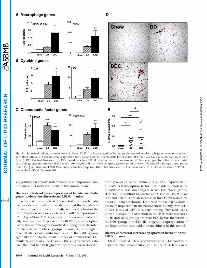
hepatocytes or resident macrophages otherwise known as Kupffer cells. Since macrophages have been implicated as a critical player in obesity-associated insulin resistance, we investigated the effects of dietary cholesterol on hepatic macrophages. Expression of the macrophage-specifi c genes F4/80, CD11b (not shown), and Mac2 were in-creased in the DDC group ( Fig. 3A ). Expression levels of the pro-infl ammatory cytokines TNF � and IL-6 mRNA also were increased in the DDC group ( Fig. 3B ). Increased ex-pression of MCP-1, a chemotactic factor for monocyte macrophages and hepatic stellate cells, was observed in both groups ( Fig. 3C ). SAA1.1 and SAA2.1 are homolo-gous isoforms of liver-derived proteins that are elevated in chronic infl ammatory states ( 17 ) and have chemotactic activity ( 18 ). Expression of SAA1.1/SAA2.1 was increased in both groups but was signifi cant only in the DDC group ( Fig. 3C ). The trend was similar in the DD group but did not achieve statistical signifi cance, likely because of the small number of animals per group. Immunohistochemical analysis of liver sections for the pan-macrophage marker MAC2 showed increased staining in the DD group, but was greater in the DDC group ( Fig. 3D, E ). Taken together, these fi ndings suggest that the addition of dietary choles-terol induces a greater degree of infl ammation in the liver. Moreover, these fi ndings parallel changes that we showed previously in intra-abdominal (visceral) adipose tissue ( 9 ),
fi brotic areas in the DDC livers (chow 0.05%, DD 0.13%, and DDC 0.21% of total area, P < 0.05 chow versus DDC). Although neither group had hepatocyte ballooning, a fea-ture seen in progressive human NASH, obese LDLR � / � mice showed several features suggestive of human NASH, which were heightened with dietary cholesterol.
Dietary cholesterol alters hepatic infl ammation in obese, insulin-resistant LDLR � / � mice
Hepatic infl ammation, a key aspect of NASH, is widely believed to be mediated through cytokines released from
Fig. 1. Plasma free fatty acids play a key role in hepatic steatosis in obese mice. Free fatty acid levels (FFA) correlate strongly with (A) hepatic triglyceride content and (B) plasma ALT, a marker of hepatocellular damage. DD, fi lled circles; DDC, fi lled diamonds.
Fig. 2. Dietary cholesterol worsens histological features of NASH in obese LDLR � / � mice. Representative images from chow, DD, and DDC groups showing (A) steatosis (fat accumulation within hepatocytes) in H and E stained sections (inset 40× magnifi cation) and (B) infl ammatory foci in H and E sections (arrows in-dicate infl ammatory cell clusters; inset 60× magnifi cation). C: Fibrosis in Masson’s trichrome-stained sec-tions for collagen which stains blue (arrows indicate areas of fi brosis).
by guest, on Septem
ber 6, 2018w
ww
.jlr.orgD
ownloaded from
1630 Journal of Lipid Research Volume 52, 2011
Fig. 3. Increased infl ammation in livers of obese LDLR � / � mice is magnifi ed by dietary cholesterol. A: Macrophage gene expression Emr1 and Mac2 mRNA. B: Cytokine gene expression for Tnf a and Il6 . C: Chemotactic factor genes Mcp1 and Saa1.1/2.1 . Chow diet, open bars (n = 8); DD, hatched bars (n = 10); DDC, solid bars (n = 10). D: Representative immunostained photomicrographs of livers stained with macrophage-specifi c antibody MAC2 (red) (40× magnifi cation; n = 5-8 per group; arrows point to areas of increased staining around portal triad). E: Quantifi cation of MAC2 staining. Chow, fi lled squares; DD, fi lled circles; DDC, fi lled diamonds. * P < 0.05 versus chow; ** P < 0.01 versus chow; # P < 0.05 versus DD.
suggesting that hepatic infl ammation is an important com-ponent of diet-induced obesity in this mouse model.
Dietary cholesterol alters expression of hepatic metabolic genes in obese, insulin-resistant LDLR � / � mice
To evaluate the effects of dietary cholesterol on hepatic triglyceride accumulation, we determined the hepatic ex-pression of genes involved in fatty acid metabolism in the liver. No differences were detected in mRNA expression of FAS ( Fig. 4A ) or ACC (not shown), two genes involved in fatty acid synthesis. Expression of SREBP1c, a transcription factor that activates genes involved in lipogenesis, trended upwards in both obese groups of animals, although it reached statistical signifi cance only in the DDC group, again likely due to the small number of animals ( Fig. 4A ). Similarly, expression of DGAT1, the enzyme which cata-lyzes the fi nal step in triglyceride synthesis, was reduced in
both groups of obese animals ( Fig. 4A ). Expression of SREBP2, a transcription factor that regulates cholesterol biosynthesis, was unchanged across the three groups ( Fig. 4A ). In contrast to several other studies ( 19, 20 ), we were not able to show an increase in liver CD36 mRNA ex-pression (data not shown). Disturbed fatty acid � -oxidation has been implicated in the pathogenesis of fatty liver ( 21 ). mRNA levels of CPT1 � , a rate-limiting fatty acid trans-porter involved in � -oxidation in the liver, were increased in DD and DDC groups, whereas PGC1 � was increased in the DDC group only ( Fig. 4B ), suggesting upregulation of the hepatic fatty acid oxidation machinery in this model.
Dietary cholesterol increases apoptosis in livers of obese LDLR � / � mice
Elevations in ALT levels occur with NAFLD secondary to hepatocellular infl ammation and injury. ALT levels were
by guest, on Septem
ber 6, 2018w
ww
.jlr.orgD
ownloaded from

Dietary cholesterol and steatohepatitis 1631
Fig. 4. Altered expression of hepatic metabolic genes in obese LDLR � / � fed dietary cholesterol. A: Expression of genes involved in fatty acid synthesis: Fas, Srebp1c, Dgat1, and Srebp2 . B: Genes involved in fatty acid oxidation: Cpt1 a , Acox1, and Pgc1 � . Chow diet, open bars (n = 8); DD, hatched bars (n = 10); DDC, solid bars (n = 10). * P < 0.05 versus chow; ** P < 0.01 versus chow; *** P < 0.001 versus chow.
increased in the DD group ( P < 0.01 versus chow; Table 1 ) but more so in the DDC group ( P < 0.001 versus chow, P < 0.05 versus DD), suggesting a greater degree of hepatocyte injury with the addition of dietary cholesterol. ALT levels correlated strongly with hepatic triglyceride levels ( r = 0.85, P = 0.0008). TUNEL staining revealed a few apoptotic cells in the livers of mice fed DD, which further increased in the DDC group of mice ( Fig. 5A ). The number of apoptotic cells correlated with hepatic triglycerides ( r = 0.78, P = 0.0006; Fig. 5B ) and with ALT levels ( r = 0.69, P = 0.02; Fig. 5C ).
Increased hepatic oxidative stress in obese mice fed added dietary cholesterol
Oxidative stress with increased production of reactive oxygen species has been observed in animal models and human NASH ( 22, 23 ). Lipids in the steatotic liver are tar-gets of oxidation, and oxidative metabolites of the major fatty acids in vivo, arachidonic acid and linoleic acid, are potent infl ammatory mediators. Total levels of HETE and HODE account for the majority of stable oxidation prod-ucts of arachidonic acid and linoleic acid, respectively ( 24 ). To obtain a comprehensive analysis of lipid peroxi-dation, we analyzed structurally distinct oxidized fatty acid moieties, including HODEs and HETEs and their precur-sor fatty acids, from liver. Levels of 9-HODE and 13-HODE, products of linoleic acid oxidation, were signifi cantly in-creased in the DDC group ( Fig. 6A ). 13-HODE levels were
highly correlated with hepatic triglyceride levels ( r = 0.90, P = 0.0002; Fig. 6C ). Modest elevations in several arachi-donic acid oxidation products were observed in both obese groups. Levels of 8-iso-prostaglandin-F2 � , derived from free radical-mediated oxidation of arachidonic acid, trended higher in both DD and DDC groups than in chow-fed animals, but the difference did not reach statistical sig-nifi cance ( Fig. 6D ). Hepatic TBARS, a crude measure of lipid peroxidation products, also was increased in the DDC group (chow 1.39 ± 0.13, DD 2.90 ± 1.74, DDC 5.98 ± 3.6 nmol/mg protein, P < 0.05 chow versus DDC; data not shown), confi rming our fi ndings with mass spectrometry.
Cholesterol oxidative products (oxysterols) generated by auto-oxidation or enzymatic/nonenzymatic peroxidation of sterols also have been implicated in apoptosis and liver injury. As dietary cholesterol can be modifi ed by oxidation into oxysterols, we measured liver and dietary oxysterols such as 5,6, � -epoxy cholesterol, 5,6, � -epoxy cholesterol, and 7-ketocholesterol. Levels of these oxysterols were un-detectable in the diet (data not shown). However, hepatic levels of � - and � -epoxy-cholesterol and 7-ketocholesterol were increased in the livers of mice on both diabetogenic diets, with � -epoxy-cholesterol being higher in the DDC group ( Fig. 6B ). Taken together, these results reveal evi-dence of increased degree of oxidative stress in the obese LDLR � / � mice that received the diabetogenic diet with added cholesterol.
by guest, on Septem
ber 6, 2018w
ww
.jlr.orgD
ownloaded from

1632 Journal of Lipid Research Volume 52, 2011
the diabetogenic diet without added cholesterol devel-oped histological changes that closely resembled key fea-tures seen in human NAFLD, such as infl ammation and fi brosis. Addition of dietary cholesterol resulted in wors-ened hepatic infl ammation and fi brosis. Thus our data implicates dietary cholesterol as an important determinant of the severity of phenotypic changes that occur with pro-gression of NAFLD.
Features of NAFLD in mouse models often do not fully mimic those seen in humans ( 4 ). Although the methionine-
DISCUSSION
We previously showed that the addition of cholesterol to a diet rich in saturated fat and refi ned carbohydrate (dia-betogenic diet) worsens adipose tissue infl ammation, insu-lin resistance, and systemic infl ammation in LDLR � / � mice ( 9 ). Thus, this is a good model to study features of the metabolic syndrome. In the present study, we used the LDLR � / � mouse to evaluate the effect of these diets on the hepatic phenotype of the metabolic syndrome. Mice fed
Fig. 5. Hepatocyte apoptosis in obese LDLR � / � fed dietary cholesterol. A: TUNEL staining of liver sec-tions. Chow diet, open bars (n = 8); DD, hatched bars (n = 10); DDC, solid bars (n = 10). * P < 0.05 versus chow; ** P < 0.01 versus DD; *** P < 0.001 versus chow. B: The number of apoptotic cells correlates with he-patic triglyceride content and (C) with ALT levels, a marker of hepatocellular damage. DD, fi lled circles; DDC, fi lled diamonds.
Fig. 6. Evidence of increased oxidative stress in LDLR � / � mice is amplifi ed by dietary cholesterol. A: Lipid peroxidation as measured by mass spectrometry. Levels of HODEs and 5-HETE were increased in the DDC animals. B: 8-isoprostaglandin-F2 � levels in livers of lean and obese mice. C: 13-HODE levels strongly correlate with hepatic triglyceride content. DD, fi lled circles; DDC, fi lled diamonds. D: Cholesterol oxidative products (oxysterol) levels in liver were increased in obese mice. Chow diet, open bars (n = 8); DD, hatched bars (n = 10); DDC, solid bars (n = 10). * P < 0.05 versus chow; ** P < 0.01 versus chow; *** P < 0.001 versus chow; † P < 0.01 versus DD. 7KC, 7-ketocholesterol; � epoxy, 5,6, � -epoxy cholesterol; � -epoxy, 5,6, � -epoxy cholesterol.
by guest, on Septem
ber 6, 2018w
ww
.jlr.orgD
ownloaded from

Dietary cholesterol and steatohepatitis 1633
stress, mitochondrial dysfunction, and upregulation of pro-infl ammatory cytokines ( 5 ). In this study, we have be-gun to address several of these potential contributors. Cir-culating FFA levels are increased in the metabolic syndrome and NAFLD in humans, and plasma levels correlate with disease severity ( 32 ). In our study, plasma FFAs correlated with hepatic triglyceride levels, suggesting that circulating FFAs are an important source of hepatic triglycerides. Studies in rodents and humans have established fatty acid transporter CD36 as an important contributor to the pathogenesis of insulin resistance and NASH ( 19, 20 ). However, we were unable to confi rm these fi ndings in our study. One of the most consistent fi ndings in the present study is that changes in the liver were exacerbated by the addition of cholesterol to the diet. Dietary cholesterol has been shown to induce hepatic infl ammation in several studies. While other groups have evaluated the role of di-etary cholesterol in NASH, several important differences exist between our studies and those of others. For exam-ple, short-term feeding studies in mice using 2% dietary cholesterol showed that hepatic infl ammation developed without steatosis ( 10 ). A hamster model of diet-induced obesity showed steatosis and insulin resistance without sig-nifi cant weight gain ( 8 ). Sprague-Dawley rats fed a high-fat diet with 2% cholesterol showed increased steatosis, in-fl ammation, and mitochondrial changes ( 33 ). Large doses of cholesterol in the diet prevented weight gain and were toxic to the liver, especially in conjunction with cholate ( 12 ). We used the LDLR � / � mouse model fed a diabeto-genic diet without or with 0.15% cholesterol, an amount that appeared to be well tolerated and did not blunt weight gain. Our use of lower, nontoxic doses of cholesterol al-lowed us to gain additional insight into mechanisms that might be more relevant to the metabolic syndrome in hu-mans. Genome-based studies have shown that cholesterol feeding induced a wide range of alterations in hepatic metabolic and infl ammatory genes, dependent upon the amount of cholesterol in the diet ( 34, 35 ).
The exact mechanisms by which dietary cholesterol in-duces hepatic infl ammation and oxidative stress in our study are unclear and require further investigation. Cyto-toxicity of free cholesterol is well established. A recent study showed that an important mechanism for choles-terol-mediated liver injury was the sensitization of hepato-cyte mitochondria to cytokine-mediated injury ( 36 ). Other potential mechanisms include endoplasmic reticular stress-mediated apoptosis ( 37 ). Apoptotic cell death is a central feature of lipotoxic liver injury ( 38 ), and hepatocyte apop-tosis correlates with disease severity in NASH ( 39 ). In our model, although apoptosis occurred in livers of mice on both diabetogenic diets, dietary cholesterol increased apoptosis. Dietary cholesterol also increased circulating FFAs, which can trigger a pro-infl ammatory response and induce lipoapoptosis ( 40 ).
Oxidative stress is a well-recognized mechanism contrib-uting to disease progression in NAFLD ( 41–43 ). We de-tected a signifi cant increase in certain subsets of lipid peroxidative products, such as 9-HODE, 13-HODE, and 5-HETE, in mice fed dietary cholesterol. Generation of
choline-defi cient diet commonly used to study NAFLD in mice results in hepatic fi brosis and infl ammation, this model does not replicate human NASH effectively, as mice on this diet lose weight and become more insulin sensitive ( 25 ), in contrast to the phenotype that occurs in the meta-bolic syndrome. Caloric excess by high-fat and intragastric feeding seems to have many features of NASH, but techni-cal challenges and failure to demonstrate reproducible results limit its widespread use ( 4 ). LDLR � / � mice develop obesity, dyslipidemia, and insulin resistance similar to that seen in the metabolic syndrome in humans. We now show that infl ammatory changes also occur in the liver, similar to what we previously showed in adipose tissue ( 9 ). In line with a recent report ( 26 ), we found several histopathologic features reminiscent of human NAFLD in LDLR � / � mice fed a diabetogenic diet, with exacerbation by the addition of dietary cholesterol. Thus, LDLR � / � mice that develop obesity on a diet enriched in refi ned carbohydrate and saturated fat with added cholesterol have histological fea-tures that mimic NASH, together with a metabolic pheno-type similar to that seen in obese humans. This model is, therefore, particularly attractive for studying the patho-genesis of liver disease in the metabolic syndrome, espe-cially the role of dietary cholesterol.
A key feature in obesity associated with insulin resistance is adipose tissue infl ammation, with accumulation of mac-rophages in fat depots in mice and humans ( 27, 28 ). We previously showed that macrophages accumulated in the visceral adipose tissue of LDLR � / � mice fed these diabeto-genic diets and that the extent of macrophage accumula-tion and adipose tissue infl ammatory gene expression were increased by cholesterol supplementation of the dia-betogenic diet ( 9 ). Here, we demonstrate that parallel changes occur in the livers of obese LDLR � / � mice, rais-ing the possibility that similar mechanisms leading to in-fl ammation and insulin resistance might be operative in both adipose tissue and liver. In this study, obese mice showed signifi cant infl ammatory changes in the liver, in-cluding increased expression of macrophage-related genes and an increase in the number of MAC2-positive cells on immunostaining. While these effects were modest in mice that received the diabetogenic diet, they were amplifi ed by addition of cholesterol to the diet. We also observed in-creased expression of MCP-1, a macrophage chemotactic factor, although expression levels were increased equally in both DD and DDC groups, suggesting that other chemo-tactic factors may play a role in liver monocyte-macrophage accumulation. Similarly, macrophage accumulation was not decreased in livers of MCP-1-defi cient mice, although these mice were not obese nor did they have fatty livers or insulin resistance ( 29 ).
Traditionally, the two-hit hypothesis has been proposed for progression of simple steatosis to NASH ( 30 ). Triglyc-eride accumulation in hepatocytes is widely accepted as an initial prerequisite for the development of NAFLD, which predisposes to cellular injury ( 31 ). Dietary cholesterol has been proposed as a second hit ( 8, 10 ). Other second in-sults that have been implicated in inflammatory liver damage and fi brogenesis seen in NASH include oxidative
by guest, on Septem
ber 6, 2018w
ww
.jlr.orgD
ownloaded from

1634 Journal of Lipid Research Volume 52, 2011
3 . Marchesini , G. , M. Brizi , G. Bianchi , S. Tomassetti , E. Bugianesi , M. Lenzi , A. J. McCullough , S. Natale , G. Forlani , and N. Melchionda . 2001 . Nonalcoholic fatty liver disease: a feature of the metabolic syndrome. Diabetes . 50 : 1844 – 1850 .
4 . Larter , C. Z. , and M. M. Yeh . 2008 . Animal models of NASH: get-ting both pathology and metabolic context right. J. Gastroenterol. Hepatol. 23 : 1635 – 1648 .
5 . Browning , J. D. , and J. D. Horton . 2004 . Molecular mediators of hepatic steatosis and liver injury. J. Clin. Invest. 114 : 147 – 152 .
6 . Shiri-Sverdlov , R. , K. Wouters , P. J. van Gorp , M. J. Gijbels , B. Noel , L. Buffat , B. Staels , N. Maeda , M. van Bilsen , and M. H. Hofker . 2006 . Early diet-induced non-alcoholic steatohepatitis in APOE2 knock-in mice and its prevention by fi brates. J. Hepatol. 44 : 732 – 741 .
7 . Schattenberg , J. M. , and P. R. Galle . 2010. Animal models of non-alcoholic steatohepatitis: of mice and man. Dig. Dis. 28 : 247 – 254 .
8 . Basciano , H. , A. E. Miller , M. Naples , C. Baker , R. Kohen , E. Xu , Q. Su , E. M. Allister , M. B. Wheeler , and K. Adeli . 2009 . Metabolic effects of dietary cholesterol in an animal model of insulin resis-tance and hepatic steatosis. Am. J. Physiol. Endocrinol. Metab. 297 : E462 – E473 .
9 . Subramanian , S. , C. Y. Han , T. Chiba , T. S. McMillen , S. A. Wang , A. Haw 3rd , E. A. Kirk , K. D. O’Brien , and A. Chait . 2008 . Dietary cholesterol worsens adipose tissue macrophage accumulation and atherosclerosis in obese LDL receptor-defi cient mice. Arterioscler. Thromb. Vasc. Biol. 28 : 685 – 691 .
10 . Wouters , K. , P. J. van Gorp , V. Bieghs , M. J. Gijbels , H. Duimel , D. Lutjohann , A. Kerksiek , R. van Kruchten , N. Maeda , B. Staels , et al . 2008 . Dietary cholesterol, rather than liver steatosis, leads to hepatic infl ammation in hyperlipidemic mouse models of nonalco-holic steatohepatitis. Hepatology . 48 : 474 – 486 .
11 . Kainuma , M. , M. Fujimoto , N. Sekiya , K. Tsuneyama , C. Cheng , Y. Takano , K. Terasawa , and Y. Shimada . 2006 . Cholesterol-fed rabbit as a unique model of nonalcoholic, nonobese, non-insulin-resistant fatty liver disease with characteristic fi brosis. J. Gastroenterol. 41 : 971 – 980 .
12 . Matsuzawa , N. , T. Takamura , S. Kurita , H. Misu , T. Ota , H. Ando , M. Yokoyama , M. Honda , Y. Zen , Y. Nakanuma , et al . 2007 . Lipid-induced oxidative stress causes steatohepatitis in mice fed an atherogenic diet. Hepatology . 46 : 1392 – 1403 .
13 . Schreyer , S. A. , C. Vick , T. C. Lystig , P. Mystkowski , and R. C. LeBoeuf . 2002 . LDL receptor but not apolipoprotein E defi ciency increases diet-induced obesity and diabetes in mice. Am. J. Physiol. Endocrinol. Metab. 282 : E207 – E214 .
14 . Folch , J. , M. Lees , and G. H. Sloane Stanley . 1957 . A simple method for the isolation and purifi cation of total lipides from animal tis-sues. J. Biol. Chem. 226 : 497 – 509 .
15 . Ohkawa , H. , N. Ohishi , and K. Yagi . 1979 . Assay for lipid peroxides in animal tissues by thiobarbituric acid reaction. Anal. Biochem. 95 : 351 – 358 .
16 . Kleiner , D. E. , E. M. Brunt , M. Van Natta , C. Behling , M. J. Contos , O. W. Cummings , L. D. Ferrell , Y. C. Liu , M. S. Torbenson , A. Unalp-Arida , et al . 2005 . Design and validation of a histological scoring system for nonalcoholic fatty liver disease. Hepatology . 41 : 1313 – 1321 .
17 . Chait , A. , C. Y. Han , J. F. Oram , and J. W. Heinecke . 2005 . Thematic review series: The immune system and atherogenesis. Lipoprotein-associated infl ammatory proteins: markers or mediators of cardio-vascular disease? J. Lipid Res. 46 : 389 – 403 .
18 . Uhlar , C. M. , and A. S. Whitehead . 1999 . Serum amyloid A, the ma-jor vertebrate acute-phase reactant. Eur. J. Biochem. 265 : 501 – 523 .
19 . Greco , D. , A. Kotronen , J. Westerbacka , O. Puig , P. Arkkila , T. Kiviluoto , S. Laitinen , M. Kolak , R. M. Fisher , A. Hamsten , et al . 2008 . Gene expression in human NAFLD. Am. J. Physiol. Gastrointest. Liver Physiol. 294 : G1281 – G1287 .
20 . Koonen , D. P. , R. L. Jacobs , M. Febbraio , M. E. Young , C. L. Soltys , H. Ong , D. E. Vance , and J. R. Dyck . 2007 . Increased hepatic CD36 expression contributes to dyslipidemia associated with diet-induced obesity. Diabetes . 56 : 2863 – 2871 .
21 . Reddy , J. K. , and M. S. Rao . 2006 . Lipid metabolism and liver in-fl ammation. II. Fatty liver disease and fatty acid oxidation. Am. J. Physiol. Gastrointest. Liver Physiol. 290 : G852 – G858 .
22 . Seki , S. , T. Kitada , T. Yamada , H. Sakaguchi , K. Nakatani , and K. Wakasa . 2002 . In situ detection of lipid peroxidation and oxida-tive DNA damage in non-alcoholic fatty liver diseases. J. Hepatol. 37 : 56 – 62 .
such lipid peroxides, either enzymatic or free radical-me-diated, are established markers of oxidative stress ( 44 ) and refl ect the balance between pro-oxidant and antioxidant mechanisms within the tissue. As in our mouse model, in-creased levels of free-radical-mediated linoleic acid oxi-dation products, namely, 9- and 13-HODEs, have been detected in human NASH ( 44 ). In vitro evidence suggests that HODEs can stimulate apoptosis in monocytes ( 45 ). We also found increased 5-HETE levels and a statistically nonsignifi cant trend in 8-iso-prostaglandin-F2 � derived from free-radical lipid peroxidation. The strong correla-tion between hepatic 13-HODE and triglyceride levels pro-vides evidence for a possible role for enzymatic pathways involving 15-lipoxygenase. Thus, these fi ndings are consis-tent with the notion that both free-radical and enzymatic oxidative pathways contribute to the formation of oxidized fatty acids in this model. In addition to these fatty acid by-products, cholesterol can undergo auto-oxidation to oxys-terols. Early studies indicated that substantial amounts of oxysterols can be present in the diet ( 46 ); however, oxy-sterols can also be formed endogenously by enzymatic con-version of cholesterol to a variety of cholesterol oxidation products ( 47 ). In our model, we found increased levels of several oxysterols in liver from both groups of mice fed the diabetogenic diets, although we were unable to detect ox-ysterols in the diet. Oxysterols have been shown to be cyto-toxic ( 48 ), and accumulation of oxysterols in the liver may have contributed to the increased hepatocyte apoptosis seen in our model. In vitro evidence suggests that certain oxysterols can trigger pro-infl ammatory and profi brotic changes in hepatocytes ( 49 ). Hypercholesterolemic apoE � / � mice have high hepatic oxysterol levels and increased sus-ceptibility to liver injury and fi brosis ( 49 ). Thus, dietary cholesterol may have contributed to the fi brosis seen in these mice by conversion to oxysterols in vivo.
In conclusion, the LDLR � / � mouse has many character-istics seen in human NAFLD/NASH and, therefore, may serve as a model for studying the early and late changes that occur in the liver in obesity. We also showed that di-etary cholesterol exacerbated the progression from simple steatosis to steatohepatitis by worsening hepatic infl amma-tion. While the two-hit hypothesis fi nds wide acceptance in this process, in our model, dietary cholesterol appears to play a role in both triglyceride accumulation (fi rst hit) and cytotoxicity, which may be a prelude to macrophage accu-mulation and hepatic infl ammation, as changes in the liver mirror those seen in adipose tissue. Progression from simple steatosis to steatohepatitis might be the result of a complex interplay of multiple pathogenic factors, such as hyperlipidemia, circulating FFAs, increased oxidized fatty acids, and oxysterols, all of which contribute to infl amma-tory changes and cytotoxicity in the liver.
REFERENCES
1 . Marchesini , G. , S. Moscatiello , S. Di Domizio , and G. Forlani . 2008 . Obesity-associated liver disease. J. Clin. Endocrinol. Metab. 93 : S74 – S80 .
2 . Farrell , G. C. , and C. Z. Larter . 2006 . Nonalcoholic fatty liver dis-ease: from steatosis to cirrhosis. Hepatology . 43 : S99 – S112 .
by guest, on Septem
ber 6, 2018w
ww
.jlr.orgD
ownloaded from

Dietary cholesterol and steatohepatitis 1635
23 . Yang , S. , H. Zhu , Y. Li , H. Lin , K. Gabrielson , M. A. Trush , and A. M. Diehl . 2000 . Mitochondrial adaptations to obesity-related oxi-dant stress. Arch. Biochem. Biophys. 378 : 259 – 268 .
24 . Yoshida , Y. , M. Hayakawa , and E. Niki . 2005 . Total hydroxyoctadec-adienoic acid as a marker for lipid peroxidation in vivo. Biofactors . 24 : 7 – 15 .
25 . Rinella , M. E. , and R. M. Green . 2004 . The methionine-choline defi cient dietary model of steatohepatitis does not exhibit insulin resistance. J. Hepatol. 40 : 47 – 51 .
26 . Gupte , A. A. , J. Z. Liu , Y. Ren , L. J. Minze , J. R. Wiles , A. R. Collins , C. J. Lyon , D. Pratico , M. J. Finegold , S. T. Wong , et al. 2010 . Rosiglitazone attenuates age- and diet-associated nonalcoholic steatohepatitis in male low-density lipoprotein receptor knockout mice. Hepatology . 52: 2001–2011.
27 . Weisberg , S. P. , D. McCann , M. Desai , M. Rosenbaum , R. L. Leibel , and A. W. Ferrante , Jr . 2003 . Obesity is associated with macrophage accumulation in adipose tissue. J. Clin. Invest. 112 : 1796 – 1808 .
28 . Xu , H. , G. T. Barnes , Q. Yang , G. Tan , D. Yang , C. J. Chou , J. Sole , A. Nichols , J. S. Ross , L. A. Tartaglia , et al . 2003 . Chronic infl amma-tion in fat plays a crucial role in the development of obesity-related insulin resistance. J. Clin. Invest. 112 : 1821 – 1830 .
29 . Kassel , K. M. , G. L. Guo , O. Tawfi k , and J. P. Luyendyk . Monocyte chemoattractant protein-1 defi ciency does not affect steatosis or infl ammation in livers of mice fed a methionine-choline-defi cient diet. Lab Invest . 90: 1794–1804.
30 . Day , C. P. , and O. F. James . 1998 . Steatohepatitis: a tale of two “hits”? Gastroenterology . 114 : 842 – 845 .
31 . Choi , S. S. , and A. M. Diehl . 2008 . Hepatic triglyceride synthesis and nonalcoholic fatty liver disease. Curr. Opin. Lipidol. 19 : 295 – 300 .
32 . Nehra , V. , P. Angulo , A. L. Buchman , and K. D. Lindor . 2001 . Nutritional and metabolic considerations in the etiology of nonal-coholic steatohepatitis. Dig. Dis. Sci. 46 : 2347 – 2352 .
33 . Xu , Z. J. , J. G. Fan , X. D. Ding , L. Qiao , and G. L. Wang . 2010 . Characterization of high-fat, diet-induced, non-alcoholic steato-hepatitis with fi brosis in rats. Dig. Dis. Sci. 55 : 931 – 940 .
34 . Kleemann , R. , L. Verschuren , M. J. van Erk , Y. Nikolsky , N. H. Cnubben , E. R. Verheij , A. K. Smilde , H. F. Hendriks , S. Zadelaar , G. J. Smith , et al . 2007 . Atherosclerosis and liver infl ammation induced by increased dietary cholesterol intake: a combined tran-scriptomics and metabolomics analysis. Genome Biol. 8 : R200 .
35 . Vergnes , L. , J. Phan , M. Strauss , S. Tafuri , and K. Reue . 2003 . Cholesterol and cholate components of an atherogenic diet in-duce distinct stages of hepatic infl ammatory gene expression. J. Biol. Chem. 278 : 42774 – 42784 .
36 . Mari , M. , F. Caballero , A. Colell , A. Morales , J. Caballeria , A. Fernandez , C. Enrich , J. C. Fernandez-Checa , and C. Garcia-Ruiz . 2006 . Mitochondrial free cholesterol loading sensitizes to TNF- and Fas-mediated steatohepatitis. Cell Metab. 4 : 185 – 198 .
37 . Feng , B. , P. M. Yao , Y. Li , C. M. Devlin , D. Zhang , H. P. Harding , M. Sweeney , J. X. Rong , G. Kuriakose , E. A. Fisher , et al . 2003 . The en-
doplasmic reticulum is the site of cholesterol-induced cytotoxicity in macrophages. Nat. Cell Biol. 5 : 781 – 792 .
38 . Malhi , H. , and G. J. Gores . 2008 . Molecular mechanisms of lipo-toxicity in nonalcoholic fatty liver disease. Semin. Liver Dis. 28 : 360 – 369 .
39 . Feldstein , A. E. , A. Canbay , P. Angulo , M. Taniai , L. J. Burgart , K. D. Lindor , and G. J. Gores . 2003 . Hepatocyte apoptosis and fas expres-sion are prominent features of human nonalcoholic steatohepati-tis. Gastroenterology . 125 : 437 – 443 .
40 . Malhi , H. , S. F. Bronk , N. W. Werneburg , and G. J. Gores . 2006 . Free fatty acids induce JNK-dependent hepatocyte lipoapoptosis. J. Biol. Chem. 281 : 12093 – 12101 .
41 . Oliveira , C. P. , L. C. da Costa Gayotto , C. Tatai , B. I. Della Bina , M. Janiszewski , E. S. Lima , D. S. Abdalla , F. P. Lopasso , F. R. Laurindo , and A. A. Laudanna . 2002 . Oxidative stress in the pathogenesis of nonalcoholic fatty liver disease, in rats fed with a choline-defi cient diet. J. Cell. Mol. Med. 6 : 399 – 406 .
42 . Roskams , T. , S. Q. Yang , A. Koteish , A. Durnez , R. DeVos , X. Huang , R. Achten , C. Verslype , and A. M. Diehl . 2003 . Oxidative stress and oval cell accumulation in mice and humans with al-coholic and nonalcoholic fatty liver disease. Am. J. Pathol. 163 : 1301 – 1311 .
43 . Videla , L. A. , R. Rodrigo , J. Araya , and J. Poniachik . 2004 . Oxidative stress and depletion of hepatic long-chain polyunsaturated fatty ac-ids may contribute to nonalcoholic fatty liver disease. Free Radic. Biol. Med. 37 : 1499 – 1507 .
44 . Feldstein , A. E. , R. Lopez , T. A. Tamimi , L. Yerian , Y. M. Chung , M. Berk , R. Zhang , T. M. McIntyre , and S. L. Hazen . 2010. Mass spectrometric profi ling of oxidized lipid products in human nonal-coholic fatty liver disease and nonalcoholic steatohepatitis. J. Lipid Res. 51 : 3046 – 3054 .
45 . Hampel , J. K. , L. M. Brownrigg , D. Vignarajah , K. D. Croft , A. M. Dharmarajan , J. M. Bentel , I. B. Puddey , and B. B. Yeap . 2006 . Differential modulation of cell cycle, apoptosis and PPARgamma2 gene expression by PPARgamma agonists ciglitazone and 9-hy-droxyoctadecadienoic acid in monocytic cells. Prostaglandins Leukot. Essent. Fatty Acids . 74 : 283 – 293 .
46 . Addis , P. B. 1986 . Occurrence of lipid oxidation products in foods. Food Chem. Toxicol. 24 : 1021 – 1030 .
47 . Brown , A. J. , and W. Jessup . 2009 . Oxysterols: sources, cellular stor-age and metabolism, and new insights into their roles in choles-terol homeostasis. Mol. Aspects Med. 30 : 111 – 122 .
48 . Shibata , N. , and C. K. Glass . 2010. Macrophages, oxysterols and ath-erosclerosis. Circ. J. 74 : 2045 – 2051 .
49 . Ferre , N. , M. Martinez-Clemente , M. Lopez-Parra , A. Gonzalez-Periz , R. Horrillo , A. Planaguma , J. Camps , J. Joven , A. Tres , F. Guardiola , et al . 2009 . Increased susceptibility to exacerbated liver injury in hypercholesterolemic ApoE-defi cient mice: potential in-volvement of oxysterols. Am. J. Physiol. Gastrointest. Liver Physiol. 296 : G553 – G562 .
by guest, on Septem
ber 6, 2018w
ww
.jlr.orgD
ownloaded from