digestive and liver disease auto 1.pdf · autoimmune pancreatitis: a challenging diagnostic puzzle...
TRANSCRIPT

R
A
Ea
b
c
d
e
a
ARAA
KPAPP
1
cp
otp[n
ddtedMcahbr
1d
Digestive and Liver Disease 42 (2010) 92–98
Contents lists available at ScienceDirect
Digestive and Liver Disease
journa l homepage: www.e lsev ier .com/ locate /d ld
eview Article
utoimmune pancreatitis: A challenging diagnostic puzzle for clinicians
. Buscarinia,∗, L. Frullonib, S. De Lisi c, M. Falconid, P.A. Testonie, A. Zambelli a
Gastroenterology Department, Maggiore Hospital, Largo Dossena 2, 26013 Crema, ItalyDepartment of Biomedical and Surgical Sciences, University of Verona, Verona, ItalyCattedra & Unità Operativa di Gastroenterologia, Di.Bi.M.I.S., University of Palermo, Palermo, ItalyDepartment of Anesthesiological and Surgical Sciences, University of Verona, Verona, ItalyGastroenterology Department, I.R.C.C.S San Raffaele, University Vita e Salute San Raffaele, Milan, Italy
r t i c l e i n f o
rticle history:eceived 1 August 2009ccepted 27 August 2009vailable online 4 October 2009
a b s t r a c t
Autoimmune pancreatitis is a form of pancreatitis with autoimmune stigmata that may present as eitherfocal or diffuse gland involvement. In focal forms, autoimmune pancreatitis shares demographic, clinical,biochemical and imaging features with pancreatic cancer. Since autoimmune pancreatitis is a benigndisease and steroid therapy can rapidly resolve symptoms, improve radiological findings and avoid
eywords:ancreatitisutoimmuneancreatic biopsyancreatic imaging
unnecessary surgery, the current clinical challenge is how to differentiate autoimmune pancreatitis frompancreatic neoplasia.
Even though definitive diagnosis of the disease is difficult, several diagnostic criteria have been pro-posed and progress has been made in imaging studies. The management of this unique form of pancreatitisshould, therefore, be handled in centres with knowledge of all aspects of the disease.
This article briefly reviews clinical aspects of autoimmune pancreatitis with a focus on its diagnostict.Gast
imaging and managemen© 2009 Editrice
. Introduction
Autoimmune pancreatitis (AIP) is a form of pancreatitis withlinical, serological and histological features of an autoimmunerocess.
The term AIP (proposed by Yoshida in 1995 [1]) replacedlder designations whose diversity reflects the heterogeneity ofhis disease: primary chronic pancreatitis [2], chronic sclerosingancreatitis, non-alcoholic duct-destructive chronic pancreatitis3], lymphoplasmacytic sclerosing pancreatitis [4,5] and duct-arrowing chronic pancreatitis [6].
The first descriptions of AIP from Asian studies included onlyiffuse forms of the disease that involved the entire gland [7,8]. Aiffuse narrowing of the main pancreatic duct was in fact “manda-ory” in the Japanese diagnostic criteria of AIP [7,8], and a diffusenlargement of the pancreas with narrowing of the main pancreaticuct was defined as “essential” in the Korean diagnostic criteria [8].ore recently, some authors reported that AIP may be radiologi-
ally classified into focal and diffuse forms [9–11]. For the first time,
focal enlargement of the gland, “occasionally with a mass and/orypo-attenuation rim,” has been included as a possible form of AIPy the 2008 Joint Korean and Japanese Consensus [12]. A recentadiological review from the USA stressed that focal forms of AIP,∗ Corresponding author. Tel.: +39 0373 280320; fax: +39 0373 280654.E-mail address: [email protected] (E. Buscarini).
590-8658/$36.00 © 2009 Editrice Gastroenterologica Italiana S.r.l. Published by Elsevieroi:10.1016/j.dld.2009.08.006
roenterologica Italiana S.r.l. Published by Elsevier Ltd. All rights reserved.
designated as “atypical,” represent up to 40% of AIP cases [13]. In arecent Italian series of 87 patients with AIP, the focal form occurredin 63% of patients [11].
From a clinical point of view, focal forms of AIP are of particularinterest since they share many clinical and imaging features withpancreatic carcinoma, yet they have a benign course and can beeasily managed conservatively using steroid therapy, resulting indramatic improvements.
2. Epidemiology
AIP is thought to be a relatively rare condition, but its actualincidence and prevalence are still unknown. Since there is nointernational agreement about diagnostic criteria and no sero-logical markers have yet been identified, it may be significantlyunderreported. AIP accounts for 4–6% of all chronic pancreatitiscases [14,15], and up to 23% of pancreatic resections for presumedmalignancy are done for AIP [16]. Series conducted in Italy, Asiaand the USA obtained somewhat different epidemiological results,however the discrepancies are probably related to the differentdiagnostic criteria used and to the inclusion of focal forms of thedisease in the Italian series [11–16].
Patient age varies widely (30–70 years), but most are older than50 years. AIP affects men more often than women (ratio 2:1) [14].
Association with other autoimmune diseases, particularly ofthe gastrointestinal tract, seems to be quite common. In partic-ular, ulcerative colitis represented up to 30% of the associated
Ltd. All rights reserved.
and Li
abiaap
3
tptat
tHeHhts[
ltlbcUtac7mmmscc
lotb
4
mppptroTsabpsJtp
E. Buscarini et al. / Digestive
utoimmune diseases in an Italian series [14]. The associationetween AIP and ulcerative colitis has been further confirmed
n a recent USA study [17]. However, it is still unclear whetherutoimmune diseases represent only an association or if theyre an extrapancreatic manifestation of a systemic autoimmunerocess that is possibly IgG4-mediated.
. Etiopathogenesis and laboratory findings
The term AIP was introduced solely because of the condi-ion’s dramatic response to steroid therapy. However, autoimmuneathogenesis has not yet been demonstrated since specific autoan-ibodies have not yet been discovered. Some serological features ofutoimmunity may be present in AIP, but they are not specific forhe disease.
Similarly to other autoimmune diseases, in the Japanese popula-ion a relationship with the HLA haplotype DRB1*0405–DQB1*0401LA has been reported [18]. Currently, elevated serum IgG4 lev-ls are considered to be the sole serological hallmark of AIP.amano et al. found that serum IgG4 levels were significantlyigher in patients with AIP than in healthy subjects. By con-rast, in patients with pancreatic cancer and chronic pancreatitis,erum IgG4 levels were similar to those of normal subjects19].
After this initial report, many other papers have been pub-ished on the diagnostic value of serum IgG4 in AIP. However,hey report lower sensitivity and specificity [20–23]. In particu-ar, Ghazale et al. confirmed that serum IgG4 are elevated in AIP,ut they also underlined that 10% of patients with pancreatic can-er may have a non-specific increase in serum IgG4 (<2-fold) [20].sing a >140 mg/dL cut-off, similar to that used by Hamano et al.,
hese authors reported a sensitivity of 76%, a specificity of 93%nd a positive predictive value (PPV) of only 36%. Using a higherut-off (280 mg/dL), the specificity and PPV increased (99% and5%, respectively), but the sensitivity was only 53% [20]. A recenteta-analysis confirmed that serum IgG4 may be useful as an AIParker, but the heterogeneity of the published studies do not per-it an assessment of the real accuracy of the test [23]. Further
tudies are necessary to evaluate the exact value of IgG4 for dis-riminating AIP from other autoimmune diseases and pancreaticancer.
Additional serological markers of AIP are autoantibodies againstactoferrin and carbonic anhydrase II, and these are found in mostf the organs involved in the systemic form of AIP [24]. However,hese antibodies, often found in AIP patients at low titre, seem toe non-specific.
. Clinical features
Up to 70–80% of patients present with painless jaundice. Thisay be related to the focal form of the disease involving the
ancreatic head, or to biliary involvement by the autoimmunerocess. Acute pancreatitis is also frequently observed in theseatients, but severe acute pain is rare and necrotising pancreati-is has never been reported in the literature [11,16,25]. Symptomselated to endocrine (diabetes, weight loss) and exocrine (steat-rrhea, weight loss) insufficiencies may also be observed [26,27].he presence of a pancreatic mass, the onset of diabetes andignificant weight loss lead to a possible diagnosis of pancre-tic cancer. Even in the face of negative findings by cytology oriopsy, these patients (particularly older patients) often undergo
ancreatic surgery, because neoplasia cannot be excluded. Theymptoms are different in focal and diffuse forms of the disease.aundice is more frequent in the focal form, whereas pancreati-is is more frequently observed in the diffuse form [14]. If theatient has a previous history of autoimmune disease, this helpsver Disease 42 (2010) 92–98 93
in the identification of the disease, particularly in young sub-jects.
The biliary tree, gallbladder, kidney, lung, and salivary glandscan be involved in the systemic form. Extrapancreatic symp-toms are related to intra- or extra-hepatic biliary strictures,hydronephrosis due to retroperitoneal fibrosis, interstitial nephri-tis, interstitial pneumonia, mediastinal lymphadenopathies andsicca syndrome. These conditions may represent the clinical onsetof the disease [15,28–33]. AIP may be associated with inflamma-tory bowel disease, particularly ulcerative colitis [14,17,34]. Mostof the symptoms improve with steroid therapy, and spontaneousremission has also been described [35].
5. Histological features
From a pathological point of view, AIP may be considered aunique form of pancreatitis [14]. Upon gross examination, the pan-creas can appear diffusely or focally hardened. If focally involved,the mass cannot be distinguished from pancreatic cancer. A histo-logical hallmark of AIP is peri-ductal infiltration by inflammatorycells (lymphocytes and plasma cells) with diffuse fibrosis oftenarranged in a storiform pattern and obliterative phlebitis [34]. Thelymphocytes are CD4+ and CD8+ T cells, whereas B lymphocytesare less commonly observed [34].
AIP can be pathologically classified into (1) a prevalentinflammatory form, designated “idiopathic duct-centric chronicpancreatitis (IDCP)” by Notohara et al. [36] and “AIP with gran-ulocyte epithelial lesion (GEL)” by Zamboni et al. [34]; and (2)a prevalent sclerosing form, called lymphoplasmacytic sclerosingpancreatitis [5,36]. The relationship between these forms is notunderstood. Zamboni et al. stressed that GEL+ AIP is more fre-quently associated with ulcerative colitis and seems to relapse lessfrequently after steroid treatment [34].
The diagnosis of AIP may be performed with surgical specimens.However, guided biopsies with either histological or cytologi-cal sampling may be diagnostic. A pathological hallmark of AIPis the presence of IgG4+ plasma cells both in the pancreas andin extrapancreatic tissues [37,38]. Recently, Kamisawa observedsignificant infiltration of IgG4+ plasma cells in the major duo-denal papilla of patients with pancreatic head involvement andrare IgG4+ plasma cells in patients with pancreatic cancer, bodyor tail AIP and papillitis [39]. However, as observed for serumIgG4, IgG4+ plasma cells in pancreatic tissue do not represent aspecific marker for AIP, since they are also observed in pancre-atic cancer and in non-AIP chronic pancreatitis. The cut-off todefine tissue positive for IgG4+ plasma cells with immunostainingof pancreatic lymphoplasmacytic infiltrate varies in pathologicalstudies, ranging from 10 to 30 per high-power field (HPF). Kojimaet al. observed that IgG4+ plasma cells were detected in 72.5%of AIP cases and in 63.1% of non-AIP chronic pancreatitis cases[39], respectively. However, by using a cut-off of 20 cells per HPF,50% of pancreatic specimens of AIP were positive, whilst noneof the pancreatic cancer specimens were. It has been suggestedthat EUS guided biopsies plus immunohistochemical evaluationfor IgG4+ plasma cells are the main diagnostic criteria for AIP[40].
6. Imaging findings
6.1. Sonography
The role of sonography in the diagnosis of autoimmune pancre-atitis is not well established. Sonographic images of the pancreasare not specific and rarely diagnostic of autoimmune pancreatitis.Typical findings of diffuse autoimmune pancreatitis are hypoechoicpancreatic swelling with the main pancreatic duct compressed

94 E. Buscarini et al. / Digestive and Liver Disease 42 (2010) 92–98
Fsm
btcfAda[
saftlwcrfi
6
psmape[
nm
rc
fdop
improves, the capsule-like rim disappears and the main pancre-atic duct can be seen by MRCP. These findings suggest a role forMRCP in following patients on therapy, but not in diagnosing AIP[51].
ig. 1. AIP, diffuse form. Transverse epigastric US scan reveals a diffusely and sub-tantially enlarged pancreas (arrows) with echo-poor echotexture and normal sizedain pancreatic duct (arrowheads); SV = splenic vein.
y the parenchyma (Fig. 1). The focal form, commonly involvinghe head of the pancreas, can show single or multiple hypoe-hoic pancreatic masses, common bile duct dilatation, and lessrequent upstream dilatation of the main pancreatic duct [41–43].
recent report described sonographic findings of common bileuct wall thickening in 37 patients with AIP, making sonographyuseful and non-invasive tool for the detection of biliary lesions
44].Contrast-enhanced ultrasonography (CEUS) is evolving as a sen-
itive tool for evaluating the typical vascularisation pattern ofutoimmune pancreatitis. Moreover, it may be a good indicatoror monitoring the efficacy of steroid therapy [41,45]. In this set-ing, after injection of intravenous contrast agents, focal pancreaticesions show enhancement in both the early and delayed phases
ith a slow washout. These enhancement patterns differ from pan-reatic carcinoma and decrease after steroid treatment, as they areelated to the degree of inflammation and inversely related to thebrosis grade [45].
.2. Computed tomography (CT)
The classical form of autoimmune pancreatitis in abdominal CTresents as diffuse pancreatic enlargement, the so-called “sausage-haped” pancreas (Fig. 2). After injection of contrast medium, aoderate pancreatic enhancement, a capsule-like low-density rim
nd bile duct wall enhancement can be observed in the earlyhase. The late phase can show a delayed diffuse pancreaticnhancement with a persisting peripheral rim of hyper-attenuation10,46,47].
The focal form (more often involving the head and/or the unci-ate process) appears as a hypo-attenuating or iso-attenuatingass with a smooth contour in dynamic CT (Fig. 3).Other findings suggestive of AIP are solid renal lesions and
etroperitoneal fibrosis [48]. Enlarged peripancreatic lymph nodesan also be seen [49].
Pancreatic calcifications can be present but they are not typicalor AIP [50]; cysts and pseudocysts are also uncommon. Pancreaticuct dilatation is possible in the focal forms [10], but its abrupt cut-ff should suggest a pancreatic carcinoma. Vascular involvement isossible in both situations [48,49]. After steroid therapy, there is
Fig. 2. AIP, diffuse form. Contrast-enhanced CT shows the diffuse pancreaticswelling (arrows).
a normalisation of the pancreatic size and enhancement patterns(Fig. 4) [10]. However, in the case of long-standing autoimmunepancreatitis, CT can show pancreatic parenchymal atrophy.
6.3. Magnetic resonance (MR) and MRcholangio-pancreatography (MRCP)
MR imaging reveals focal or diffuse pancreatic enlargementthat is hypo-intense in T1-weighted MR images and slightlyhyper-intense in T2-weighted images. As with CT, a capsule-likehypo-intense rim can be observed in T2-weighted MR images [46].As with endoscopic retrograde cholangio-pancreatography (ERCP),MRCP can show multiple intrahepatic strictures, dilated intrahep-atic ducts and stricture of the common bile duct [49].
MRCP cannot visualise the narrow portion of the main pan-creatic duct, but it can show the non-involved regions. For thesereasons, MRCP cannot differentiate irregular narrowing of the mainpancreatic duct from the stenosis typical of pancreatic carcinoma.
After steroid treatment, pancreatic size and signal intensity
Fig. 3. AIP, focal form. Contrast-enhanced CT shows an enlarged pancreatic bodyand tail (arrows).

E. Buscarini et al. / Digestive and Liver Disease 42 (2010) 92–98 95
F (arroT se in s
6(
Papiptapa
6
tEaA
cls
tb
The latter cholangiographic finding can mimic primary scleros-ing cholangitis.
ERCP also has a therapeutic role, allowing biliary drainage andstent placement, although the response to steroids improves thepancreatic duct changes and (to a lesser degree) the biliary changes.
ig. 4. A contrast-enhanced axial CT scan (a and b) shows that the pancreatic glandhe CT check (c and d) after 6 weeks of steroid treatment shows the marked decrea
.4. 18F-fluorodeoxyglucose positron emission tomographyFDG-PET) and PET/computed tomography (CT)
Little data is available concerning the role of FDG-PET andET/CT in AIP characterisation. Nakajio et al. carried out FDG-PETnd PET/TC studies on six patients with AIP suspected of havingancreatic cancer. They reported an intense FDG pancreatic uptake
n all patients that disappeared after steroid therapy. Moreover, fiveatients had associated extrapancreatic lesions, and these showedhe same FDG uptake as the pancreas [52]. Unfortunately, FDG-PETnd PET/CT cannot differentiate the FDG uptake of AIP from that ofancreatic cancer, but they may be useful for detecting AIP lesionsnd monitoring disease activity after steroid therapy.
.5. Endoscopic retrograde cholangio-pancreatography (ERCP)
One of the diagnostic criteria for AIP is a diffuse or segmen-al narrowing of the main pancreatic duct with irregular wall inRCP. This finding, in association with stenosis of the intrapancre-tic common bile duct (Fig. 5), represents the ERCP hallmark ofIP.
In the focal form, the main pancreatic duct can be dilated adja-ent to or upstream of the strictures; if this pattern coexists with
ow common bile duct stenosis, it can resemble the double-ductign typical of pancreatic carcinoma.Other common ERCP characteristics are irregular narrowing ofhe hilar hepatic region, and less frequently, segmental intrahepaticile duct strictures [53].
wheads) is swollen with some hypodense areas (arrows).ize of the gland, either head or body and tail (arrowheads).
Fig. 5. AIP, with common bile duct involvement: endoscopic retrograde cholangiog-raphy shows a stenosis of the distal common bile duct (arrows).

96 E. Buscarini et al. / Digestive and Li
Fig. 6. Linear EUS (a) shows a focal roundish echo-poor lesion (arrows) in the pan-cwi
Hr
6
cb[
s(ddwiiblt
iwT
wf
reatic head (PH), and the common bile duct (arrowheads) has a diffusely thickenedall. The bile duct wall thickening (arrows) (b) has a “sandwich-pattern”, with an
ntermediate echo-poor layer and echo-rich inner and outer layers. PV = portal vein.
owever, biliary strictures can progress even with steroids andequire long-term treatment [6].
.6. Endoscopic ultrasonography (EUS)
EUS is superior to CT, MR and ERCP for detecting small pan-reatic masses, and its role in the diagnosis of AIP is enhancedy the possibility of an effective and safe pancreatic biopsy54,55].
EUS features of AIP are a diffusely hypoechoic pancreaticwelling and/or a hypoechoic mass in the head of the pancreasFig. 6). Another finding suggestive of AIP is common bile ductilatation with a thickened wall [56,57]. The thickening of the bileuct wall in AIP shows some unique features: it is homogeneous,ith an echo-poor intermediate layer and hyper-echoic outer and
nner layers and a “sandwich-pattern” wall that may reach 5 mmn thickness (Fig. 6). Differentiation of biliary involvement fromiliary or pancreatic carcinoma is based on the presence (in the
atter cases) of irregular echo-poor lesions transmurally involvinghe duct wall.
Hyodo et al. performed contrast-enhanced EUS of the bile ductn AIP, and it showed early enhancement of the bile duct wall that
as different from the poor enhancement of cholangiocarcinoma.
he enhancement was reduced after steroid treatment [57].Main pancreatic duct dilatation is possible in the focal form,hereas it is compressed by the enlarged parenchyma in the diffuse
orm of AIP [56].
ver Disease 42 (2010) 92–98
Vascular involvement of the portal and/or superior mesentericvein has been reported and should not preclude the diagnosis ofAIP, because the inflammatory infiltrate can transmurally involvethe vessel walls determining the EUS finding of invasion [56].
Single or multiple enlarged peripancreatic and celiac lymphnodes can also be detected that are reflective of the inflammatoryprocess.
Even if EUS-FNA is sensitive and specific for the diagnosisof pancreatic malignancy, the cytopathologic diagnosis of AIP isnot standardised. EUS fine-needle aspiration (FNA) of pancreaticmasses, lymph nodes and the common bile duct wall can revealfibrosis and lymphoplasmacytic infiltrate, and EUS-FNA findingscorrelate well with surgical pathologic diagnosis [56]. However, itis difficult to obtain sufficient pancreatic tissue to achieve a defini-tive diagnosis without a laparotomy, and sampling error is possiblesince the disease has a patchy distribution. It has been proposedthat a cytologic smear with stromal fragments rich in inflamma-tory cells and epithelial cells lacking atypia is diagnostic of AIP[58]. EUS-guided Tru-cut biopsy (EUS-TCB) with a 19-gauge needleacquires larger tissue specimens whilst preserving tissue architec-ture and this may allow for histological confirmation and preventunnecessary surgery [40].
7. Diagnostic criteria
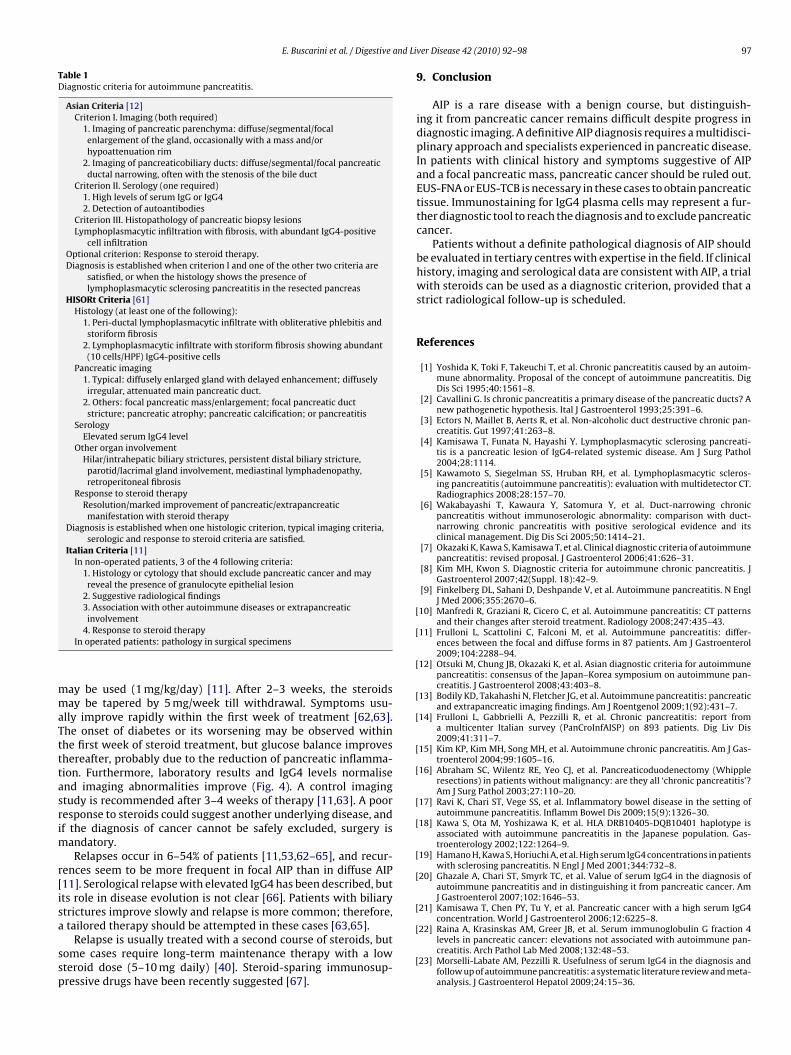
Taken together, these considerations suggest that the onlypathognomonic criteria that can be used to definitively diagnoseAIP are those requiring a surgical specimen. In practice, the diag-nosis usually results either from surgical over-treatment or acombination of different features that together make the diagnosisreliable. Thus far, there is no consensus on the minimal diagnosticcriteria for AIP. Current AIP diagnosis is based on criteria proposedby the Japan Pancreas Society in 2002 and revised in 2006 [7,59].According to these criteria, specific imaging, serological and his-tological criteria must be fulfilled in order to make a diagnosisof AIP. The Korean criteria of response to steroids and extrapan-creatic lesions supplement the Japanese criteria [60]. In 2008, aJapan–Korea Symposium incorporated the previous criteria intothe new Asian diagnostic criteria for AIP [12]. The Mayo Clinic pro-posed criteria for AIP (termed HISORt) in 2006 focus on histologicalfeatures, and these are considered to be the gold standard [61].Italian investigators proposed the use of a combination of histo-logical and cytological findings, including association with otherautoimmune diseases and response to steroid therapy [11]. Thesediagnostic criteria are listed in Table 1.
In summary, the main diagnostic criteria in Asia are based onradiological and serological findings, whilst in the USA diagnosis isbased on disease pathology. In Italy, the primary accepted criterion(if biopsies are not diagnostic for the disease) is the response tosteroids, but this should be used only if clinical, pathological andradiological data are consistent with AIP and pancreatic cancer canbe confidently excluded.
The existence of so many diagnostic criteria underlines the dif-ficulty of preoperative AIP diagnosis, and in particular the difficultyof differentiating it from pancreatic cancer. Appropriate diagnosticcriteria are still under debate and International Consensus Criteriaare awaited. Both the lack of defined guidelines and the risk of mis-diagnosed pancreatic cancer underscore how important it is for AIPpatients to be managed in experienced centres.
8. Therapy
Steroid drugs are standard therapy, although spontaneous res-olutions have also been described [35]. However, a therapeuticschedule has not yet been standardised. Usually therapy startswith 30–40 mg prednisone daily for 1 week, but higher dosages
E. Buscarini et al. / Digestive and Li
Table 1Diagnostic criteria for autoimmune pancreatitis.
Asian Criteria [12]Criterion I. Imaging (both required)
1. Imaging of pancreatic parenchyma: diffuse/segmental/focalenlargement of the gland, occasionally with a mass and/orhypoattenuation rim
2. Imaging of pancreaticobiliary ducts: diffuse/segmental/focal pancreaticductal narrowing, often with the stenosis of the bile duct
Criterion II. Serology (one required)1. High levels of serum IgG or IgG42. Detection of autoantibodies
Criterion III. Histopathology of pancreatic biopsy lesionsLymphoplasmacytic infiltration with fibrosis, with abundant IgG4-positive
cell infiltrationOptional criterion: Response to steroid therapy.Diagnosis is established when criterion I and one of the other two criteria are
satisfied, or when the histology shows the presence oflymphoplasmacytic sclerosing pancreatitis in the resected pancreas
HISORt Criteria [61]Histology (at least one of the following):
1. Peri-ductal lymphoplasmacytic infiltrate with obliterative phlebitis andstoriform fibrosis
2. Lymphoplasmacytic infiltrate with storiform fibrosis showing abundant(10 cells/HPF) IgG4-positive cells
Pancreatic imaging1. Typical: diffusely enlarged gland with delayed enhancement; diffuselyirregular, attenuated main pancreatic duct.
2. Others: focal pancreatic mass/enlargement; focal pancreatic ductstricture; pancreatic atrophy; pancreatic calcification; or pancreatitis
SerologyElevated serum IgG4 level
Other organ involvementHilar/intrahepatic biliary strictures, persistent distal biliary stricture,parotid/lacrimal gland involvement, mediastinal lymphadenopathy,retroperitoneal fibrosis
Response to steroid therapyResolution/marked improvement of pancreatic/extrapancreaticmanifestation with steroid therapy
Diagnosis is established when one histologic criterion, typical imaging criteria,serologic and response to steroid criteria are satisfied.
Italian Criteria [11]In non-operated patients, 3 of the 4 following criteria:
1. Histology or cytology that should exclude pancreatic cancer and mayreveal the presence of granulocyte epithelial lesion
2. Suggestive radiological findings3. Association with other autoimmune diseases or extrapancreaticinvolvement
mmaTtttasrim
r[isa
ssp
[
[
[
[
[
[
[
[
[
[
[
[
4. Response to steroid therapyIn operated patients: pathology in surgical specimens
ay be used (1 mg/kg/day) [11]. After 2–3 weeks, the steroidsay be tapered by 5 mg/week till withdrawal. Symptoms usu-
lly improve rapidly within the first week of treatment [62,63].he onset of diabetes or its worsening may be observed withinhe first week of steroid treatment, but glucose balance improveshereafter, probably due to the reduction of pancreatic inflamma-ion. Furthermore, laboratory results and IgG4 levels normalisend imaging abnormalities improve (Fig. 4). A control imagingtudy is recommended after 3–4 weeks of therapy [11,63]. A pooresponse to steroids could suggest another underlying disease, andf the diagnosis of cancer cannot be safely excluded, surgery is
andatory.Relapses occur in 6–54% of patients [11,53,62–65], and recur-
ences seem to be more frequent in focal AIP than in diffuse AIP11]. Serological relapse with elevated IgG4 has been described, butts role in disease evolution is not clear [66]. Patients with biliarytrictures improve slowly and relapse is more common; therefore,
tailored therapy should be attempted in these cases [63,65].Relapse is usually treated with a second course of steroids, butome cases require long-term maintenance therapy with a lowteroid dose (5–10 mg daily) [40]. Steroid-sparing immunosup-ressive drugs have been recently suggested [67].
[
[
ver Disease 42 (2010) 92–98 97
9. Conclusion
AIP is a rare disease with a benign course, but distinguish-ing it from pancreatic cancer remains difficult despite progress indiagnostic imaging. A definitive AIP diagnosis requires a multidisci-plinary approach and specialists experienced in pancreatic disease.In patients with clinical history and symptoms suggestive of AIPand a focal pancreatic mass, pancreatic cancer should be ruled out.EUS-FNA or EUS-TCB is necessary in these cases to obtain pancreatictissue. Immunostaining for IgG4 plasma cells may represent a fur-ther diagnostic tool to reach the diagnosis and to exclude pancreaticcancer.
Patients without a definite pathological diagnosis of AIP shouldbe evaluated in tertiary centres with expertise in the field. If clinicalhistory, imaging and serological data are consistent with AIP, a trialwith steroids can be used as a diagnostic criterion, provided that astrict radiological follow-up is scheduled.
References
[1] Yoshida K, Toki F, Takeuchi T, et al. Chronic pancreatitis caused by an autoim-mune abnormality. Proposal of the concept of autoimmune pancreatitis. DigDis Sci 1995;40:1561–8.
[2] Cavallini G. Is chronic pancreatitis a primary disease of the pancreatic ducts? Anew pathogenetic hypothesis. Ital J Gastroenterol 1993;25:391–6.
[3] Ectors N, Maillet B, Aerts R, et al. Non-alcoholic duct destructive chronic pan-creatitis. Gut 1997;41:263–8.
[4] Kamisawa T, Funata N, Hayashi Y. Lymphoplasmacytic sclerosing pancreati-tis is a pancreatic lesion of IgG4-related systemic disease. Am J Surg Pathol2004;28:1114.
[5] Kawamoto S, Siegelman SS, Hruban RH, et al. Lymphoplasmacytic scleros-ing pancreatitis (autoimmune pancreatitis): evaluation with multidetector CT.Radiographics 2008;28:157–70.
[6] Wakabayashi T, Kawaura Y, Satomura Y, et al. Duct-narrowing chronicpancreatitis without immunoserologic abnormality: comparison with duct-narrowing chronic pancreatitis with positive serological evidence and itsclinical management. Dig Dis Sci 2005;50:1414–21.
[7] Okazaki K, Kawa S, Kamisawa T, et al. Clinical diagnostic criteria of autoimmunepancreatitis: revised proposal. J Gastroenterol 2006;41:626–31.
[8] Kim MH, Kwon S. Diagnostic criteria for autoimmune chronic pancreatitis. JGastroenterol 2007;42(Suppl. 18):42–9.
[9] Finkelberg DL, Sahani D, Deshpande V, et al. Autoimmune pancreatitis. N EnglJ Med 2006;355:2670–6.
10] Manfredi R, Graziani R, Cicero C, et al. Autoimmune pancreatitis: CT patternsand their changes after steroid treatment. Radiology 2008;247:435–43.
11] Frulloni L, Scattolini C, Falconi M, et al. Autoimmune pancreatitis: differ-ences between the focal and diffuse forms in 87 patients. Am J Gastroenterol2009;104:2288–94.
12] Otsuki M, Chung JB, Okazaki K, et al. Asian diagnostic criteria for autoimmunepancreatitis: consensus of the Japan–Korea symposium on autoimmune pan-creatitis. J Gastroenterol 2008;43:403–8.
13] Bodily KD, Takahashi N, Fletcher JG, et al. Autoimmune pancreatitis: pancreaticand extrapancreatic imaging findings. Am J Roentgenol 2009;1(92):431–7.
14] Frulloni L, Gabbrielli A, Pezzilli R, et al. Chronic pancreatitis: report froma multicenter Italian survey (PanCroInfAISP) on 893 patients. Dig Liv Dis2009;41:311–7.
15] Kim KP, Kim MH, Song MH, et al. Autoimmune chronic pancreatitis. Am J Gas-troenterol 2004;99:1605–16.
16] Abraham SC, Wilentz RE, Yeo CJ, et al. Pancreaticoduodenectomy (Whippleresections) in patients without malignancy: are they all ‘chronic pancreatitis’?Am J Surg Pathol 2003;27:110–20.
17] Ravi K, Chari ST, Vege SS, et al. Inflammatory bowel disease in the setting ofautoimmune pancreatitis. Inflamm Bowel Dis 2009;15(9):1326–30.
18] Kawa S, Ota M, Yoshizawa K, et al. HLA DRB10405-DQB10401 haplotype isassociated with autoimmune pancreatitis in the Japanese population. Gas-troenterology 2002;122:1264–9.
19] Hamano H, Kawa S, Horiuchi A, et al. High serum IgG4 concentrations in patientswith sclerosing pancreatitis. N Engl J Med 2001;344:732–8.
20] Ghazale A, Chari ST, Smyrk TC, et al. Value of serum IgG4 in the diagnosis ofautoimmune pancreatitis and in distinguishing it from pancreatic cancer. AmJ Gastroenterol 2007;102:1646–53.
21] Kamisawa T, Chen PY, Tu Y, et al. Pancreatic cancer with a high serum IgG4concentration. World J Gastroenterol 2006;12:6225–8.
22] Raina A, Krasinskas AM, Greer JB, et al. Serum immunoglobulin G fraction 4levels in pancreatic cancer: elevations not associated with autoimmune pan-creatitis. Arch Pathol Lab Med 2008;132:48–53.
23] Morselli-Labate AM, Pezzilli R. Usefulness of serum IgG4 in the diagnosis andfollow up of autoimmune pancreatitis: a systematic literature review and meta-analysis. J Gastroenterol Hepatol 2009;24:15–36.

9 and Li
[
[
[
[
[
[
[
[
[
[
[
[
[
[
[
[
[
[
[
[
[
[
[
[
[
[
[
[
[
[
[
[
[
[
[
[
[
[
[
[
[
[
8 E. Buscarini et al. / Digestive
24] Taniguchi T, Okazaki K, Okamoto M, et al. High prevalence of autoantibod-ies against carbonic anhydrase II and lactoferrin in type 1 diabetes: conceptof autoimmune exocrinopathy and endocrinopathy of the pancreas. Pancreas2003;27:26–30.
25] Kamisawa T, Egawa N, Nakajima H. Autoimmune pancreatitis is a systemicautoimmune disease. Am J Gastroenterol 2003;98:2811–2.
26] Okazaki K, Autoimmune-related pancreatitis. Curr Treat Options Gastroenterol2001;4:369–75.
27] Okazaki K, Uchida K, Ohana M, et al. Autoimmune-related pancreatitis is asso-ciated with autoantibodies and a Th1/Th2-type cellular immune response.Gastroenterology 2000;118:573–81.
28] Nishino T, Toki F, Oyama H, et al. Biliary tract involvement in autoimmunepancreatitis. Pancreas 2005;30:76–82.
29] Uchiyama-Tanaka Y, Mori Y, Kimura T, et al. Acute tubulointerstitial nephri-tis associated with autoimmune-related pancreatitis. Am J Kidney Dis2004;43:18–25.
30] Hamano H, Kawa S, Ochi Y, et al. Hydronephrosis associated with retroperi-toneal fibrosis and sclerosing pancreatitis. Lancet 2002;359:1403–4.
31] Hirano K, Kawabe T, Komatsu Y, et al. High-rate pulmonary involvement inautoimmune pancreatitis’. Intern Med J 2006;36:58–61.
32] Hirano K, Kawabe T, Yamamoto N, et al. Serum IgG4 concentrations in pancre-atic and biliary diseases. Clin Chim Acta 2006;367:181–4.
33] Kamisawa T, Tu Y, Egawa N, et al. Salivary gland involvement in chronic pan-creatitis of various etiologies. Am J Gastroenterol 2003;98:323–6.
34] Zamboni G, Luttges J, Capelli P, et al. Histopathological features of diagnosticand clinical relevance in autoimmune pancreatitis: a study on 53 resectionspecimens and 9 biopsy specimens. Virchows Arch 2004;445:552–63.
35] Kamisawa T, Yoshiike M, Egawa N, et al. Treating patients with autoim-mune pancreatitis: results from a long-term follow-up study. Pancreatology2005;5:234–8.
36] Notohara K, Burgart LJ, Yadav D, et al. Idiopathic chronic pancreatitis withperiductal lymphoplasmacytic infiltration: clinicopathologic features of 35cases. Am J Surg Pathol 2003;27:1119–27.
37] Deshpande V, Chicano S, Finkelberg D, et al. Autoimmune pancreatitis: a sys-temic immune complex mediated disease. Am J Surg Pathol 2006;30:1537–45.
38] Kojima M, Sipos B, Klapper W, et al. Autoimmune pancreatitis: frequency, IgG4expression, and clonality of T and B cells. Am J Surg Pathol 2007;31:521–8.
39] Kamisawa T. Immunoglobulin G4-positive plasma cells in organs of patientswith autoimmune pancreatitis. Clin Gastroenterol Hepatol 2008;6:715.
40] Levy MJ, Reddy RP, Wiersema MJ, et al. EUS-guided trucut biopsy in establish-ing autoimmune pancreatitis as the cause of obstructive jaundice. GastrointestEndosc 2005;61:467–72.
41] D’Onofrio M, Martone E, Malago R, et al. Contrast-enhanced ultrasonographyof the pancreas. JOP 2007;8:71–6.
42] Morana G, Tapparelli M, Faccioli N, et al. Autoimmune pancreatitis: instrumen-tal diagnosis. JOP 2005;6:102–7.
43] Ohana M, Okazaki K, Hajiro K, et al. Multiple pancreatic masses associated with
autoimmunity. Am J Gastroenterol 1998;93:99–102.44] Koyama R, Imamura T, Okuda C, et al. Ultrasonographic imaging of bile ductlesions in autoimmune pancreatitis. Pancreas 2008;37:259–64.
45] Numata K, Ozawa Y, Kobayashi N, et al. Contrast-enhanced sonography ofautoimmune pancreatitis: comparison with pathologic findings. J UltrasoundMed 2004;23:199–206.
[
[
ver Disease 42 (2010) 92–98
46] Irie H, Honda H, Baba S, et al. Autoimmune pancreatitis: CT and MR character-istics. Am J Roentgenol 1998;170:1323–7.
47] Procacci C, Carbognin G, Biasiutti C, et al. Autoimmune pancreatitis: possibili-ties of CT characterization. Pancreatology 2001;1:246–53.
48] Takahashi N, Fletcher JG, Fidler JL, et al. Dual-phase CT of autoimmune pancre-atitis: a multireader study. Am J Roentgenol 2008;190:280–6.
49] Sahani DV, Kalva SP, Farrell J, et al. Autoimmune pancreatitis: imaging features.Radiology 2004;233:345–52.
50] Takayama M, Hamano H, Ochi Y, et al. Recurrent attacks of autoimmune pan-creatitis result in pancreatic stone formation. Am J Gastroenterol 2004;99:932–7.
51] Kamisawa T, Tu Y, Egawa N, et al. Can MRCP replace ERCP for the diagnosis ofautoimmune pancreatitis? Abdom Imaging 2009;34:381–4.
52] Nakajo M, Jinnouchi S, Noguchi M, et al. FDG PET and PET/CT monitoring ofautoimmune pancreatitis associated with extrapancreatic autoimmune dis-ease. Clin Nucl Med 2007;32:282–5.
53] Nishino T, Oyama H, Hashimoto E, et al. Clinicopathological differentiationbetween sclerosing cholangitis with autoimmune pancreatitis and primarysclerosing cholangitis. J Gastroenterol 2007;42:550–9.
54] Voss M, Hammel P, Molas G, et al. Value of endoscopic ultrasound guidedfine needle aspiration biopsy in the diagnosis of solid pancreatic masses. Gut2000;46:244–9.
55] Buscarini E, De Angelis C, Arcidiacono PG, et al. Multicentre retrospective studyon endoscopic ultrasound complications. Dig Liver Dis 2006;38:762–7.
56] Farrell JJ, Garber J, Sahani D, et al. EUS findings in patients with autoimmunepancreatitis. Gastrointest Endosc 2004;60:927–36.
57] Hyodo N, Hyodo T. Ultrasonographic evaluation in patients with autoimmune-related pancreatitis. J Gastroenterol 2003;38:1155–61.
58] Deshpande V, Mino-Kenudson M, Brugge WR, et al. Endoscopic ultrasoundguided fine needle aspiration biopsy of autoimmune pancreatitis: diagnosticcriteria and pitfalls. Am J Surg Pathol 2005;29:1464–71.
59] Japan Pancreas Society. Diagnostic criteria for autoimmune pancreatitis. J JpnPancreas Soc 2002;17:585–7.
60] Kim KP, Kim MH, Kim JC, et al. Diagnostic criteria for autoimmune chronicpancreatitis revisited. World J Gastroenterol 2006;12:2487–96.
61] Chari ST, Smyrk TC, Levy MJ, et al. Diagnosis of autoimmune pancreatitis: theMayo Clinic experience. Clin Gastroenterol Hepatol 2006;4:1010–6.
62] Wakabayashi T, Kawaura Y, Satomura Y, et al. Long-term prognosis of duct-narrowing chronic pancreatitis: strategy for steroid treatment. Pancreas2005;30:31–9.
63] Kamisawa T, Okamoto A, Wakabayashi T, et al. Appropriate steroid therapy forautoimmune pancreatitis based on long-term outcome. Scand J Gastroenterol2008;43:609–13.
64] Kamisawa T, Okamoto A. Prognosis of autoimmune pancreatitis. J Gastroenterol2007;42(Suppl. 18):59–62.
65] Ghazale AH, Chari ST, Vege SS. Update on the diagnosis and treatment ofautoimmune pancreatitis. Curr Gastroenterol Rep 2008;10:115–21.
66] Ghazale A, Chari ST, Zhang L, et al. Immunoglobulin G4-associated cholan-gitis: clinical profile and response to therapy. Gastroenterology 2008;134:706–15.
67] Church NI, Pereira SP, Deheragoda MG, et al. Autoimmune pancreatitis: clinicaland radiological features and objective response to steroid therapy in a UKseries. Am J Gastroenterol 2007;102:2417–25.