title autoimmune pancreatitis exhibiting multiple...
TRANSCRIPT

Title Autoimmune pancreatitis exhibiting multiple mass lesions.
Author(s)Shiokawa, Masahiro; Kodama, Yuzo; Hiramatsu, Yukiko;Kurita, Akira; Sawai, Yugo; Uza, Norimitsu; Watanabe,Tomohiro; Chiba, Tsutomu
Citation Case reports in gastroenterology (2011), 5(3): 528-533
Issue Date 2011-09
URL http://hdl.handle.net/2433/153021
Right
© 2011 S.Karger AG, Basel; この論文は出版社版でありません。引用の際には出版社版をご確認ご利用ください。This is not the published version. Please cite only the publishedversion.
Type Journal Article
Textversion author
Kyoto University
Titel Autoimmune pancreatitis exhibiting multiple mass lesions
Shiokawa Masahiro1), Kodama Yuzo 1), Hiramatsu Yukiko 1), Kurita Akira 1), Sawai
Yugo 1), Uza Norimitsu1), Watanabe Tomohiro 1), Chiba Tsutomu 1)2)
1)Department of Gastroenterology and Hepatology, Graduate School of medicine, Kyoto
University
2)Corresponding author: Chiba Tsutomu, M.D., Ph.D. Department of
Gastroenterology and Hepatology Graduate School of Medicine, Kyoto University
Short title AIP with multiple mass lesions
54 Shogoinkawara-cho, Sakyo-ku, Kyoto 606-8507, Japan
Phone: +81-75-751-4319; Fax: +81-75-751-4303
E-mail: [email protected]
Key words: Autoimmune pancreatitis, Pancreatic cancer, EUS-FNA, IgG4, Mass
forming

Abstract Our case is a first report of autoimmune pancreatitis (AIP) with multiple
masses within the pancreas which was pathologically diagnosed by endoscopic
US-guided fine-needle aspiration(EUS-FNA) and treated by steroid. The masses
diappeared by steroid therapy. Our case is informative to know that AIP sometimes
exhibits multiple masses within the pancreas and to diagnose it without unnecessary
surgery.
Introduction
Autoimmune pancreatitis (AIP) is a unique form of chronic pancreatitis associated
with an autoimmune inflammatory process [1, 2]. Although diffuse swelling of the
pancreatic parenchyma and diffuse irregular narrowing of the pancreatic duct system
are morphologically characteristic of AIP, a focal type of this clinical entity has been
recently recognized [3]. The focal type of AIP exhibits a localized mass lesion in the
pancreas, similar to pancreatic carcinoma [4]. Consequently, some patients with these
features have been subjected to surgical exploration with a presumed diagnosis of
pancreatic carcinoma. Considering that AIP shows a favorable response to steroid
therapy, the differentiation of these two entities is clinically important to avoid surgery.
There have been only a few cases of AIP with multifocal lesions [5] but no case

pathologically diagnosed by EUS-FNA and treated by steroid. In this report, we describe
the clinical, radiological, and histopathological features of a patient with AIP who
exhibited distinct double masses in the pancreas and was treated by steroid.
Case report
A 63-year-old male patient without any symptoms was admitted because of pancreatic
masses that were picked up on a medical checkup. He had a history of diabetes
mellitus but none of alcohol abuse. On physical examination, the patient showed
bilateral swelling of the submandibular gland. On laboratory examination, his serum
glucose was 179mg/dL (normal range 65–109 mg/dL), γ-GTP was 132IU/L (normal 54.0
IU/L), cancer antigen 19-9 (CA19-9) level was 55.5 ng/mL (normal 37.0 ng/mL) and
serum IgG4 was 773 mg/dL (normal range 4.8-105 mg/dL). Other serological tests,
including pancreatic and hepatobiliary enzymes, γ-globulin, immunoglobulinG, and
tumor markers (CEA and DUPAN-II) were within the normal range. Antinuclear
antibodies (ANA) were also negative at a titer of less than 1 : 20.
On abdominal ultrasonography [6], the patient was found to have low echoic mass in
the pancreatic head uncus and body. Contrast-enhanced computed tomography (CT)
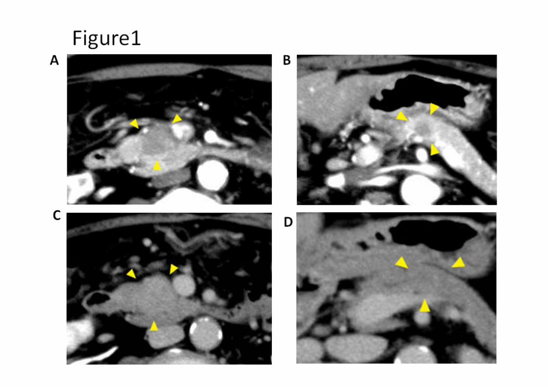
showed two mass lesions in the head and body of the pancreas (Fig. 1A, 1B); the lesions

were 18 mm and 10 mm in diameter, respectively. The tumors showed slight
attenuation in the delayed phase (Fig. 1C, D). On dynamic magnetic resonance imaging
(MRI), they were also hypovascular during the early phase and showed delayed
enhancement during the late phase. Endoscopic retrograde cholangiopancreatography
(ERCP) showed localized narrowing of the main pancreatic duct at the pancreatic head.
The common bile duct was not dilated. Fluorine-18 fluorodeoxyglucose -positron
emission tomography (FDG-PET) showed FDG uptake in the bilateral submandibular
glands, hilar, mediastinal lymph nodes and pancreatic head uncus and no uptake in the
body of pancreas.
EUS-FNA of the two mass and normal pancreas was performed and the specimen were
adequate for cytology and cell block. Cytology in the two mass was rich in inflammatory
cells. The cell block in the two mass revealed a dense lymphoplasmacytic infiltrate and
significant replacement of pancreatic parenchyma by irregular fibrosis (Fig.2A).
Immunostaining for IgG4 revealed diffuse infiltrate of IgG4-positive plasma cells
(Fig.2B). Cytology and the cell block in the normal pancreas had no abnormality
(Fig.2C).
On the basis of the above-mentioned clinical, imaging, and cytological findings, a
diagnosis of autoimmune pancreatitis was made, and treated with prednisone, which

was initiated at a dose of 30 mg per day with a tapering schedule of 5 mg every 2 weeks.
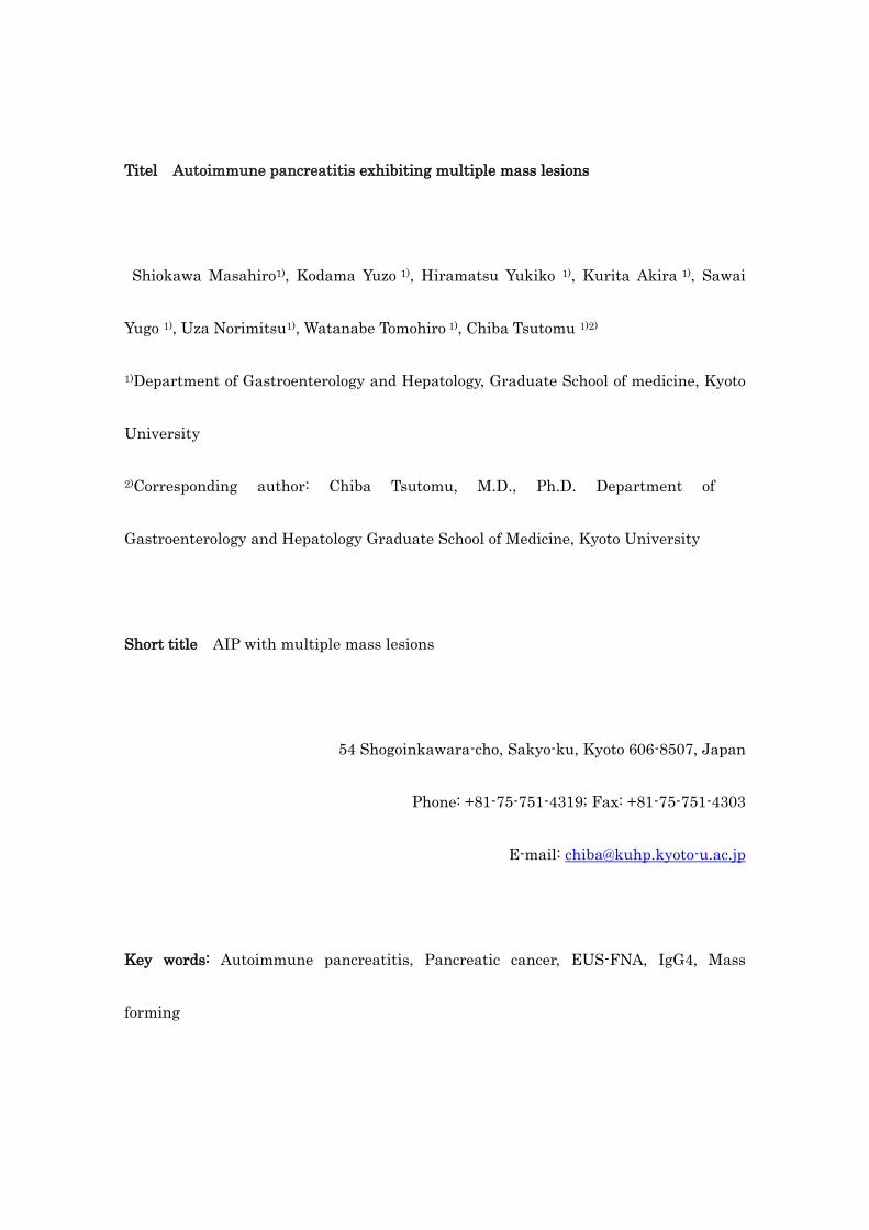
One month after treatment, the elevated IgG level resolved and CT revealed that the
two mass disappeared (Fig.3A, B, C) and FDG uptake in the pancreas, submandibular
glands, hilar, and mediastinal lymph nodes disappeared completely.
Discussion
We showed the clinical, radiological, histopathological and clinical features of a patient
with AIP who had distinct double masses in the pancreas and steroid therapy.
Since Yoshida et al. proposed the term “autoimmune pancreatitis” in 1995, this unique
pancreatic disorder was recognized as a new clinical entity with such characteristics as
increased serum γ-globulin, IgG levels, the presence of autoantibody, diffuse
enlargement of the parenchyma, and irregular narrowing of the pancreatic duct [1, 7].
In 2001, Hamano et al. reported that patients with AIP have a high serum
concentration of IgG4 [6]. The specificity and sensitivity of a high serum IgG4 level in
the diagnosis of AIP are higher than 90% and this is now believed to be the most useful
examination for diagnosing AIP. Hirano et al. [8] recently reported eight cases of AIP
with locally affected lesions. They found that the localized type of AIP is more difficult

to differentiate from pancreatic cancer radiologically because typical radiological
features reflecting diffuse involvement are not observed. They reported that serum IgG4
was elevated in the cases of focal AIP and that serum IgG4 levels might be helpful for
its diagnosis. However, a pancreatic cancer with elevated serum IgG4 was reported [9].
Multiple masses in the pancreas are a rare clinical entity. As previously reported,
multiple pancreatic masses were as follows; metastatic pancreatic tumors such as renal
cell carcinoma[10], pancreatic neuroendocrine tumor (PNET) in multiple endocrine
neoplasia type 1 (MEN1) [11], pancreatic cancer[12], and autoimmune pancreatitis
(AIP) [5, 13]. Our patient had no primary lesion other than pancreas and the imaging
findings in the present case were not suitable for PNET but pancreatic cancer could not
be excluded based on the radiological images in the present case. Radiological findings
were indicative of a malignant pancreatic tumor, especially pancreatic cancer. And also,
few reports on the coexistence of AIP and PDAC have been published posing a new
problem in the management of AIP [14, 15].
Endoscopic US-guided fine-needle aspiration of the pancreas or EUS trucut biopsy
(TCB) are useful for diagnosing AIP and excluding pancreatic cancer [16]. FNA in this
patient was compatible with AIP. However, pancreatic cancer is rarely difficult to
exclude by biopsy specimens alone because of reactive inflammatory cell reactions and

fibrosis surrounding the neoplastic cells.
It is interesting in the present case that multiple mass were formed in the pancreas.
These multiple lesions have been rarely reported for AIP until now. We could not
explain this unusual manifestation, but early phase of AIP could be focal lesions.
The most important thing is that we recognize that AIP can manifest as multiple mass
lesions similar to those in the present case, examine IgG4, perform EUS-FNA or
TCB-FNA and try to deny pancreatic cancer. If IgG4 is elevated and EUS-FNA exclude
malignancy, steroid therapy shoud be carefully challenged, quickly check the response
by CT or PET, and the patient may avoid unnecessary surgery. This case is a first report
of AIP with multifocal masses treated by steroid and we believe this diagnostic process
is informative.
1. Finkelberg, D.L., et al., Autoimmune pancreatitis. N Engl J Med, 2006. 355(25): p.
2670-6.
2. Yoshida, K., et al., Chronic pancreatitis caused by an autoimmune abnormality.
Proposal of the concept of autoimmune pancreatitis. Dig Dis Sci, 1995. 40(7): p.
1561-8.
3. Kamisawa, T., et al., Clinical difficulties in the differentiation of autoimmune
pancreatitis and pancreatic carcinoma. Am J Gastroenterol, 2003. 98(12): p. 2694-9.
4. Matsubara, T., et al., Complete obstruction of the lower common bile duct caused by
autoimmune pancreatitis: is biliary reconstruction really necessary? J Hepatobiliary
Pancreat Surg, 2005. 12(1): p. 76-83.
5. Kajiwara, M., et al., Autoimmune pancreatitis with multifocal lesions. J
Hepatobiliary Pancreat Surg, 2008. 15(4): p. 449-52.
6. Hamano, H., et al., High serum IgG4 concentrations in patients with sclerosing
pancreatitis. N Engl J Med, 2001. 344(10): p. 732-8.
7. Sahani, D.V., et al., Autoimmune pancreatitis: imaging features. Radiology, 2004.
233(2): p. 345-52.
8. Hirano, K., et al., Pancreatic mass lesions associated with raised concentration of
IgG4. Am J Gastroenterol, 2004. 99(10): p. 2038-40.
9. Kamisawa, T., et al., Pancreatic cancer with a high serum IgG4 concentration. World
J Gastroenterol, 2006. 12(38): p. 6225-8.
10. Masetti, M., et al., Analysis of prognostic factors in metastatic tumors of the
pancreas: a single-center experience and review of the literature. Pancreas, 2010.
39(2): p. 135-43.
11. Metz, D.C. and R.T. Jensen, Gastrointestinal neuroendocrine tumors: pancreatic
endocrine tumors. Gastroenterology, 2008. 135(5): p. 1469-92.
12. Fujimori, N., et al., Adenocarcinoma involving the whole pancreas with multiple
pancreatic masses. Intern Med, 2010. 49(15): p. 1527-32.
13. Inoue, D., et al., Autoimmune pancreatitis with multifocal mass lesions. Radiat Med,
2006. 24(8): p. 587-91.
14. Inoue, H., et al., A case of pancreas cancer with autoimmune pancreatitis. Pancreas,
2006. 33(2): p. 208-9.
15. Pezzilli, R., et al., Pancreatic ductal adenocarcinoma associated with autoimmune
pancreatitis. Case Rep Gastroenterol, 2011. 5(2): p. 378-85.
16. Mizuno, N., et al., Histological diagnosis of autoimmune pancreatitis using
EUS-guided trucut biopsy: a comparison study with EUS-FNA. J Gastroenterol,
2009. 44(7): p. 742-50.

Figure legend
Fig. 1A–D.. Computed tomography (CT) revealed mass lesions in the head (A) and body
(B) of the pancreas (arrows); early phase. They showed slight delayed enhancement
during the late phase (arrows in C, D).
960×720pixel
Fig. 2A, B, C Cell block in the two nodule showed dense lymphoplasmacytic infiltrate
and significant replacement of pancreatic parenchyma by irregular fibrosis (H&E×400)
(A). Numerous plasma cells in the two nodule show positive immunoreactivity for IgG4
(H&E×400) (B) Cell block in the normal pancreas showed almost normal acinar
cell ,no lymphoplasmacytic infiltrate and no fibrosis (H&E×400) (C).
960×720pixel
Fig. 3A, B The two nodule in the head(A) and body(B) of CT disappeared.
960×720pixel