digger 5 year old male neutered domestic ferret case #: n07-648 presenter: travis heskett, dvm...
TRANSCRIPT

Digger5 year old male neutered
domestic ferret
Case #: N07-648 Presenter: Travis Heskett, DVM
Presented at SEVPAC 2008 – Permission granted for use on
SEVPAC website only

History
• Presented to referring veterinarian for depression and hind limb weakness– Referring veterinarian had initially been
concerned about insulinoma– Bloodwork WNL– Mass palpated at the base of the skull – Radiographs showed large osteoproliferative
cervical vertebral lesion
Presented at SEVPAC 2008 – Permission granted for use on
SEVPAC website only

CT
Presented at SEVPAC 2008 – Permission granted for use on
SEVPAC website only

Presented at SEVPAC 2008 – Permission granted for use on
SEVPAC website only

Additional diagnostics
• Aspirate cytology– Malignant cells of
epithelial origin
• Neurology consult– Grave prognosis
• Euthanasia
1000x
Presented at SEVPAC 2008 – Permission granted for use on
SEVPAC website only

Gross Findings--Dorsal
Presented at SEVPAC 2008 – Permission granted for use on
SEVPAC website only

Gross Findings--Ventral
Presented at SEVPAC 2008 – Permission granted for use on
SEVPAC website only
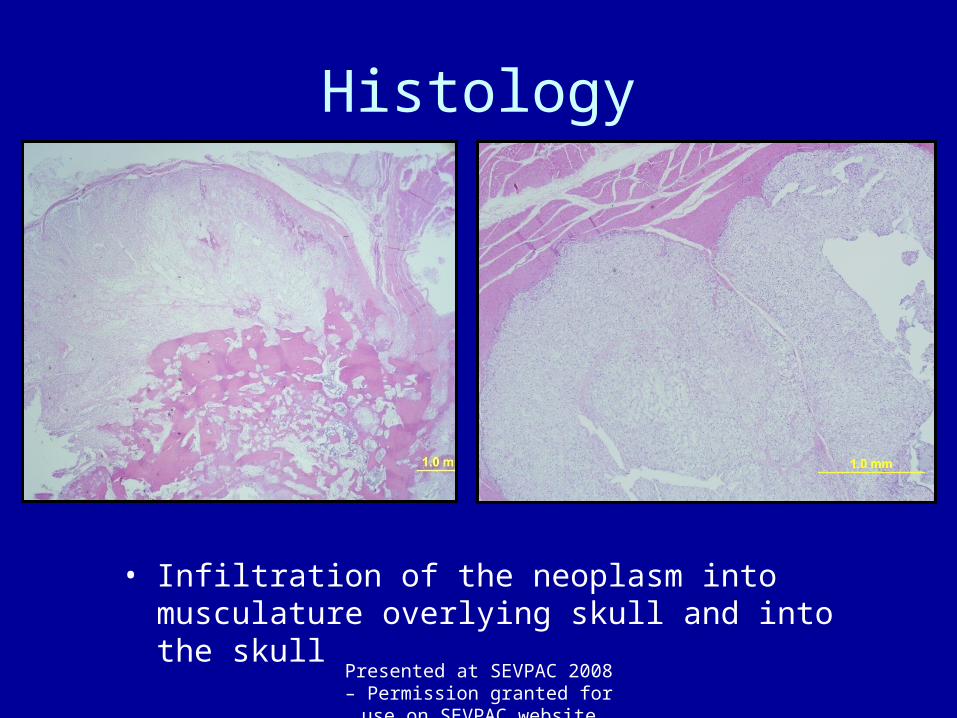
Histology
• Infiltration of the neoplasm into musculature overlying skull and into the skull
Presented at SEVPAC 2008 – Permission granted for use on
SEVPAC website only

Histology
• Neoplasm infiltrating vertebral column with isolated neoplastic nodules within spinal canal
Presented at SEVPAC 2008 – Permission granted for use on
SEVPAC website only

Histology
• Production of new cartilage and bone by the neoplasm and invasion and remodeling of existing bone
Presented at SEVPAC 2008 – Permission granted for use on
SEVPAC website only

Presented at SEVPAC 2008 – Permission granted for use on
SEVPAC website only

Immunohistochemistry
Cytokeratin Vimentin
Presented at SEVPAC 2008 – Permission granted for use on
SEVPAC website only
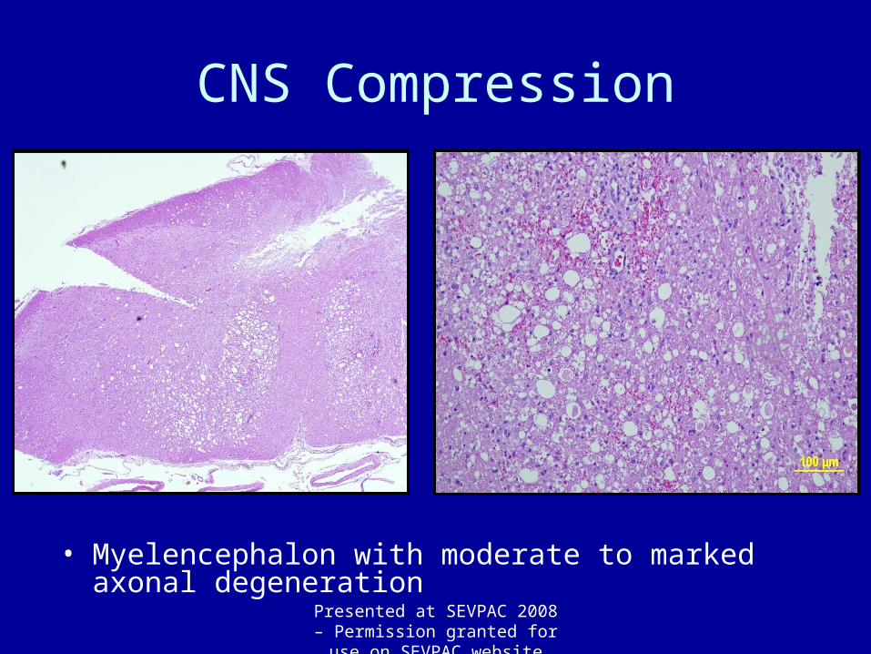
CNS Compression
• Myelencephalon with moderate to marked axonal degeneration
Presented at SEVPAC 2008 – Permission granted for use on
SEVPAC website only

CNS Compression
• Compression of the spinal cord with moderate axonal degeneration
Presented at SEVPAC 2008 – Permission granted for use on
SEVPAC website only

Final Anatomic Diagnosis
• Chordoma—caudodorsal skull in occipital and atlanto-occipital region with invasion into the calavarium, C1&C2, resulting in:– Compression of cerebellum, brainstem, and cervical
spinal cord with moderate to marked axonal degeneration and minimal neuronal degeneration
Presented at SEVPAC 2008 – Permission granted for use on
SEVPAC website only

Ferret Chordomas
• 3-6 years of age
• Typically occur at the tip of the tail beyond the last vertebral segment
• Do not appear attached to preexisting bone
• Slow growing, do not metastasize
Presented at SEVPAC 2008 – Permission granted for use on
SEVPAC website only

AFIP website
Presented at SEVPAC 2008 – Permission granted for use on
SEVPAC website only

Microscopic Appearance
• Classic appearance is that of physaliferous cells surrounding cartilage with a central core of bone
• Immunoreactive to both cytokeratin and vimentin
• The origin of the bone and cartilage is controversial, but may arise from notocord differentiation within the neoplasm
AFIP website
Presented at SEVPAC 2008 – Permission granted for use on
SEVPAC website only

Chordomas
• Arise from the remnants of the fetal notocord
• Anywhere along the axial skeleton
• Chordomas have also been reported in cervical and thoracic vertebrae of ferrets
Presented at SEVPAC 2008 – Permission granted for use on
SEVPAC website only

Chordomas in other species
• Also been reported in rats, mink, dogs, cats, and humans• In humans, they comprise approximately 1-4% of
malignant osseous tumors.• In humans, 50% are present in the sacrococcygeal
region, 25% in the sphenooccipital region, with the remainder arising in the vertebral axis.
• Slow growing and locally invasive• 30% metastasize
Presented at SEVPAC 2008 – Permission granted for use on
SEVPAC website only

Special thanks to:
Dr. Lisa Farina, pathologistDr. David Taylor, advisorHistology service at UF CVMFellow residents
Questions???Presented at SEVPAC 2008 – Permission granted for use on
SEVPAC website only