digital vs -...
TRANSCRIPT

Stip, Republic of Macedonia
28.03-29.03.2018
Multimedia centre, Stip
University ofGoce Delcev- Stip
First International Students
Congress in Dental Medicine
- 2018
DIGITAL VS
ANALOGICAL
IN DENTAL
MEDICINE
Digital VS Analogical in Dental Medicine
Faculty of Medical Sciences at the University “GoceDelcev” in Stip gladly invites you
to participate in First International Students Congress where we will be able
together to invest in the future of modern dentistry.
This Congress is a great opportunity to learn new techniques in dental
treatment, to spend quality time with colleagues, to enjoy in the beautiful scenery of
the Macedonian pearl- our city of Stip!
Enjoy!
Stip, Republic of Macedonia -28.03.-29.03.2018
University of Goce Delcev- Stip
International students congress in dental
medicine

AGENDA
DAY ONE -WEDNESDAY , 28thof march
8:30-10:00 Registration of participants
10:00 OPENING CEREMONY OF THE CONGRESS
WELCOME – HAVE A NICE STAY!
MORNING SESSION
10:30 ORAL PRESENTATIONS- SESSION 1 Chairmen: Ass. Prof. Papakoca K.; Ass. Prof. Zlatanovska K. ; Student -Ilievski S.
10:30-11:00 Invited speaker- Prof. Rubin Gulaboski, dean of the Faculty of medical sciences Fake Science, Fake Journals, Fake Scientists
11:00-11:20 Invited speaker- Ass. Prof. Marija Darkovska- Serafimovska Adverse drug interactions in dental practice
11:20-11:30 Smile design with metal free ceramics
Author: Stefanija Stojanova Co-author: Stefani Joveva
Mentor: Kocovski Darko Co-mentor: Zlatanovska Katerina
11:30-11:40 Modified Direct Composite Resin Bonded Bridge Author: Aleksandar Andreevski, Co-author: Natasha Longurova Mentor: Ivona Kovachevska
11:40-11:50 Nanocomposites – The Future Of Improved Restorative Dentistry Author: Andrej Stojanovski, Co-author: Artion Abdiju Mentor: Vera Stojanovska Faculty of dentistry, Europian University, Skopje
11:50-12:00 Discussion
12:00-13:00- POSTER PRESENTATIONS
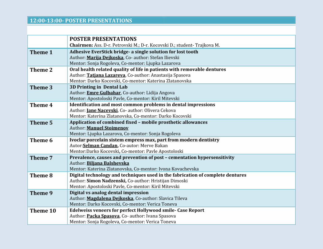
POSTER PRESENTATIONS Chairmen: Ass. D-r. Petrovski M.; D-r. Kocovski D.; student- Trajkova M.
Theme 1 Adhesive EverStick bridge- a single solution for lost tooth Author: Marija Dejkoska, Co- author: Stefan Ilievski Mentor: Sonja Rogoleva, Co-mentor: Ljupka Lazarova
Theme 2 Oral health related quality of life in patients with removable dentures Author: Tatjana Lazareva, Co-author: Anastasija Spasova Mentor: Darko Kocovski, Co-mentor: Katerina Zlatanovska
Theme 3 3D Printing in Dental Lab Author: Emre Gulbahar, Co-author: Lidija Angova Mentor: Apostoloski Pavle, Co-mentor: Kiril Mitevski
Theme 4 Identification and most common problems in dental impressions Author: Jane Nacevski, Co- author: Olivera Cekova Mentor: Katerina Zlatanovska, Co-mentor: Darko Kocovski
Theme 5 Application of combined fixed – mobile prosthetic allowances Author: Manuel Stoimenov Mentor: Ljupka Lazarova, Co-mentor: Sonja Rogoleva
Theme 6 Ivoclar porcelain sistem empress max, part from modern dentistry Autor:Selman Candan, Co-autor: Merve Bakan Mentor:Darko Kocovski, Co-mentor: Pavle Apostoloski
Theme 7 Prevalence, causes and prevention of post – cementation hypersensitivity Author: Biljana Balshevska Mentor: Katerina Zlatanovska, Co-mentor: Ivona Kovachevska
Theme 8 Digital technology and techniques used in the fabrication of complete dentures Author: Simon Nadzenski, Co-author: Hristijan Dimoski Mentor: Apostoloski Pavle, Co-mentor: Kiril Mitevski
Theme 9 Digital vs analog dental impression Author: Magdalena Dejkoska, Co-author: Slavica Tileva Mentor: Darko Kocovski, Co-mentor: Verica Toneva
Theme 10 Edelweiss veneers for perfect Hollywood smile- Case Report Author: Packa Spasova, Co- author: Ivana Spasova Mentor: Sonja Rogoleva, Co-mentor: Verica Toneva

14:00-17:00 HANDS ON COURSES
Theme 11 Hand tracing vs. digital methods of cefalometric analysis Author: Magdalena Koceva, Co-author: Ana Trajkovska Mentor: Sandra Atanasova, Co-mentor: Verica Toneva
Theme 12 Anatomical and morphological variations of the maxillary lateral incisor Author: Teodora Seneva, Co-author: Marija Risteska Mentor: Kiro Papakoca, Co-mentor: Marija Hadji-Vasileva
Theme 13 The manufacture of inlays throughout thedental clinics in the Municipality of Shtip Author: Petar Joleski, Co-author: Stefan Nanev Mentor: Natasa Longurova, Co-mentor: Katerina Zlatanovska
Theme 14 Tooth discolorations after dental interventions Author: Aleksandra Markoska, Co-author: Izabela Brsakoska Mentor: Natasha Longurova, Co-mentor: Sandra Atanasova
Theme 15 Composite laminates for a perfect smile Author: Verica Sajkarova Mentor: Ljupka Lazarova, Co-mentor: Sandra Atanasova
Theme 16 Use of the teeth whitening procedure by dental medicine students Author: Zorka Gjorgieva, Co-author: Sara Talevska Mentor: Natasha Longurova, Co-mentor: Verica Toneva
Theme 17 The most common reasons for toothache Author: Natalija Gorgieva, Co-author:Lidija Angova Mentor: Verica Toneva, Co-mentor: Sandra Atanasova
Theme 18 TMD disorders among dental students Author:Martin Treneski, Co-author: Hristijan Dimovski Mentor: Terzieva-Petroska Olivera, Co-mentor: Petrovski Mihajlo
Theme 19 Mineral trioxide aggregate material use in dental pathology Author: Anita Kirova ,Co-author: Frosina Pandova Mentor: Natasha Longurova, Co-mentor: Ivona Kovachevska
Theme 20 The anatomo-morphological differences between primary and permanent teeth Author:Spase Sulev, Co-author:Andon Stojkov, Mentor: Sanja Naskova, Co-mentor: Verica Toneva
13:00-14.00 LUNCH BREAK

AFTERNOON SESSION
14:00 ORAL PRESENTATIONS– SESSION 2 Chairmen: Ass. Prof. Papakoca K.; D-r. Terzieva Petrovska O.; Student- Sulev S.
14:00-14:30 Invited speaker Ass. Prof. Mirjana Bošković , Prof. Sasa Stankovic, Faculty of medical sciences, Nis, Serbia The importance of the dental profile in forensic dentistry
14:30-14:50 Invited speaker Olivera Terzieva-Petrovska The importance of oral hygiene for the longevity of dental implants
14:50-15:00 Management of post-operative complications in maxillary teeth extraction Author: Aleksandra Miteva,Co-author: Zlatko Maksimov Menthor: Dimova Cena
15:00-15:10 Post-operative complications after extraction of impacted lower third molar Author: Zlatko Maksimov, Co-author: Aleksandra Miteva Menthor: Dimova Cena
15:10-15:20 Invastigation of anesthethic eficacy of modified intraoral conduction mandibular technique by angulated needle Author:Kristina Burić Mentor: Miloš Tijanić Clinic of dentistry, Faculty of Medicine, University of Niš
15:20-15:30 Discussion
15:30-16:00 COFFEE BREAK

16:00 ORAL PRESENTATIONS – SESSION 3 Chairmen: Ass. Prof. Naskova S; Ass. D-r. Petrovski M.; student-Novoselska M.
16:00-16:20 Invited speaker Ass. Prof. Kiro Papakoca Digital Vs. analog x-ray images and their application in dentistry then and now
16:20-16:40 Invited speaker Ass. D-r. Mihajlo Petrovski Implantology vs. Periodontology
16:40-16:50 The prevalence of tooth extraction due to periodontal disease Author: Dzemil Kurtagic, Co-author: Erkin Crnisanin, Lazar Dobric Mentor: Ana Pejcic Clinic of dentistry, Faculty of Medicine, University of Niš
16:50-17:00 The use of soft tissue laser in everyday modern dental practise Author: Stefan Ilievski, Co-author: Spase Sulev Mentor: Papakoca Kiro, Co-mentor: Rogoleva Sonja
17:00-17:10 General plan of treatement in dentistry Author: Ordanka Kostova, Co-author: Sofija Gavrilova Mentor: Petrovski Mihajlo, Co-mentor: Terzieva-Petrovska Olivera
17:10-17:20 Dental Implant Planning using Cone Beam CT imaging Author: Simona Coneva Mentor: Papakoca Kiro
17:20-17:30 Discussion
EVENING SESSION

DAY TWO – THURSDAY, 29thof march
MORNING SESSION
9:00 ORAL PRESENTATIONS-SESSION 4 Chairmen: Prof. Dimova C.;D-r. Terzieva-Petrovska O.; student-Dimovski H.
9:00-9:20 Invited speaker Prof. Lidija Popovska, University “Sv. Kiril I Metodij”- Skopje Cleaning and shaping using rotary endodontic instruments
9:20-9:40 Invited speaker Jetmire Jakupi -Alimani, State University of Tetovo Assessing the caries risk factors among children at the age from 4-5 using the Cariogram program
9:40-9:50 Aesthetic restorations with porcelain venneers Author: Monika Kasuba Mentor: Zlatanovska Katerina, Co-mentor: Longurova Natasa
9:50-10:00 An assesment of some psyhological aspects in children undergoing dental interventions Author: Iva Maslarevska, Co-author:Liljana Petrova, Mentor: Sarakinova Olivera, Co-mentor: Kostadinovska Emilija Faculty of dentistry, European University, Skopje
10:00-10:10 Immediate loading of dental implants with hybrid bridge Author: Danilo Krstevski, Co-author: Katerina Spasovska, Dubravka Angelik Mentor: Velevski Dragoljub, Co-mentor: Dimova Cena
10:10-10:20 Invited speaker Pavle Apostoloski CAD/CAM systems- the basis of modern dental prosthetics
10:20-10:40 Discussion
12:30-13:00 COFFEE BREAK
13:00-14:00 – POSTER PRESENTATIONS
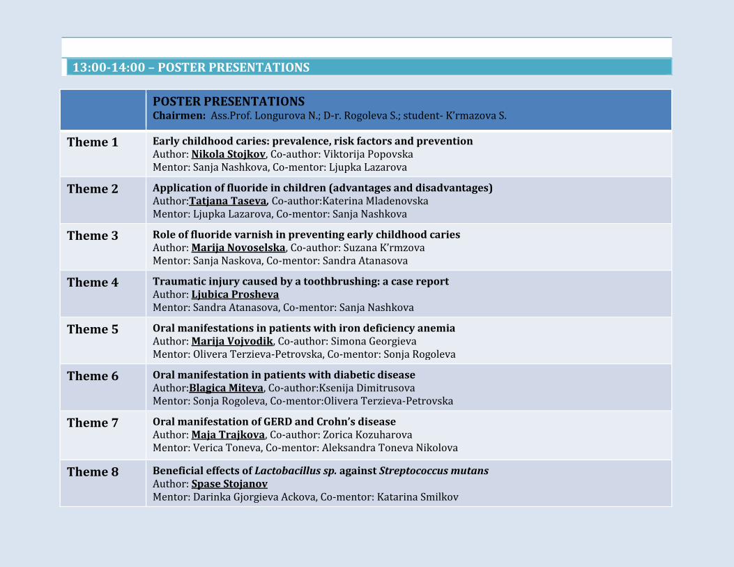
POSTER PRESENTATIONS Chairmen: Ass.Prof. Longurova N.; D-r. Rogoleva S.; student- K’rmazova S.
Theme 1 Early childhood caries: prevalence, risk factors and prevention Author: Nikola Stojkov, Co-author: Viktorija Popovska Mentor: Sanja Nashkova, Co-mentor: Ljupka Lazarova
Theme 2 Application of fluoride in children (advantages and disadvantages) Author:Tatjana Taseva, Co-author:Katerina Mladenovska Mentor: Ljupka Lazarova, Co-mentor: Sanja Nashkova
Theme 3 Role of fluoride varnish in preventing early childhood caries Author: Marija Novoselska, Co-author: Suzana K’rmzova Mentor: Sanja Naskova, Co-mentor: Sandra Atanasova
Theme 4 Traumatic injury caused by a toothbrushing: a case report Author: Ljubica Prosheva Mentor: Sandra Atanasova, Co-mentor: Sanja Nashkova
Theme 5 Oral manifestations in patients with iron deficiency anemia Author: Marija Vojvodik, Co-author: Simona Georgieva Mentor: Olivera Terzieva-Petrovska, Co-mentor: Sonja Rogoleva
Theme 6 Oral manifestation in patients with diabetic disease Author:Blagica Miteva, Co-author:Ksenija Dimitrusova Mentor: Sonja Rogoleva, Co-mentor:Olivera Terzieva-Petrovska
Theme 7
Oral manifestation of GERD and Crohn’s disease Author: Maja Trajkova, Co-author: Zorica Kozuharova Mentor: Verica Toneva, Co-mentor: Aleksandra Toneva Nikolova
Theme 8 Beneficial effects of Lactobacillus sp. against Streptococcus mutans Author: Spase Stojanov Mentor: Darinka Gjorgieva Ackova, Co-mentor: Katarina Smilkov
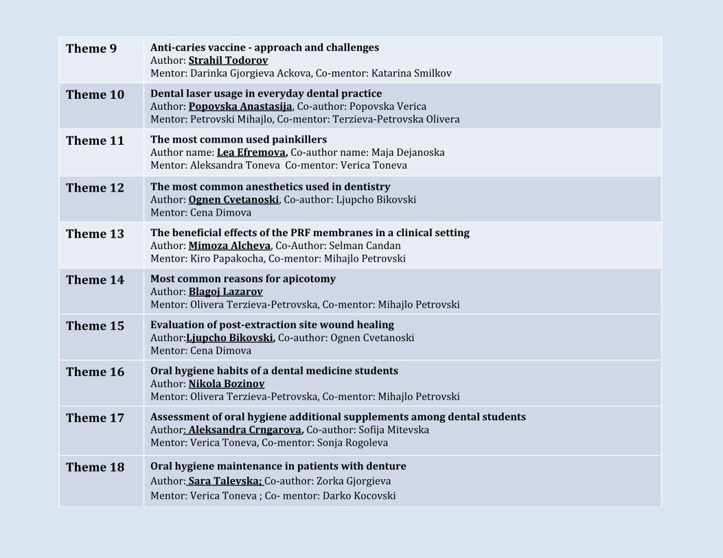
Theme 9 Anti-caries vaccine - approach and challenges Author: Strahil Todorov Mentor: Darinka Gjorgieva Ackova, Co-mentor: Katarina Smilkov
Theme 10 Dental laser usage in everyday dental practice Author: Popovska Anastasija, Co-author: Popovska Verica Mentor: Petrovski Mihajlo, Co-mentor: Terzieva-Petrovska Olivera
Theme 11 The most common used painkillers Author name: Lea Efremova, Co-author name: Maja Dejanoska Mentor: Aleksandra Toneva Co-mentor: Verica Toneva
Theme 12 The most common anesthetics used in dentistry Author: Ognen Cvetanoski, Co-author: Ljupcho Bikovski Mentor: Cena Dimova
Theme 13 The beneficial effects of the PRF membranes in a clinical setting Author: Mimoza Alcheva, Co-Author: Selman Candan Mentor: Kiro Papakocha, Co-mentor: Mihajlo Petrovski
Theme 14 Most common reasons for apicotomy Author: Blagoj Lazarov Mentor: Olivera Terzieva-Petrovska, Co-mentor: Mihajlo Petrovski
Theme 15 Evaluation of post-extraction site wound healing Author:Ljupcho Bikovski, Co-author: Ognen Cvetanoski Mentor: Cena Dimova
Theme 16 Oral hygiene habits of a dental medicine students Author: Nikola Bozinov Mentor: Olivera Terzieva-Petrovska, Co-mentor: Mihajlo Petrovski
Theme 17 Assessment of oral hygiene additional supplements among dental students Author: Aleksandra Crngarova, Co-author: Sofija Mitevska Mentor: Verica Toneva, Co-mentor: Sonja Rogoleva
Theme 18 Oral hygiene maintenance in patients with denture
Author: Sara Talevska; Co-author: Zorka Gjorgieva
Mentor: Verica Toneva ; Co- mentor: Darko Kocovski
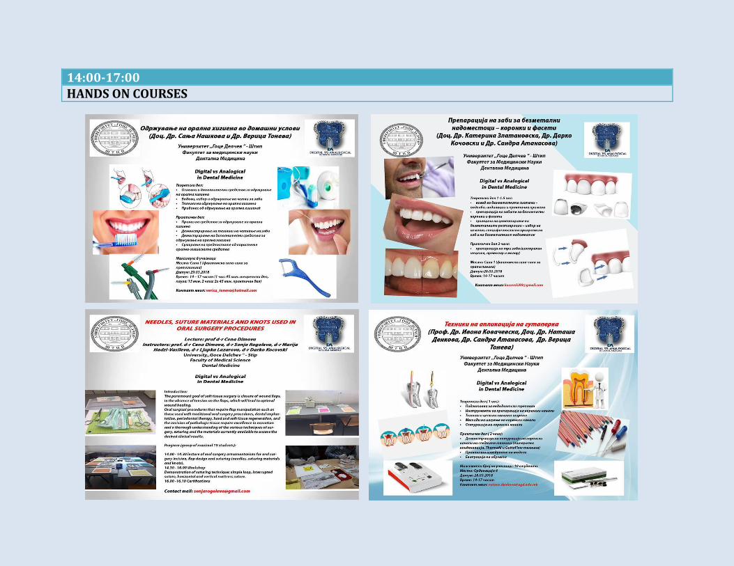
14:00-17:00 HANDS ON COURSES

WE ARE INVITING YOU
TO THE PARTY
LET’S CELEBRATE
FOR THE SUCCESFULL CONGRESS AND FOR THE
WONDERFULL MOMENTS SPEND TOGETHER
WEDNESDAY, 28TH OF MARCH
UNIVERSITY RESTAURANT
THANK YOU FOR YOUR PRESENCE AND
PARTICIPATION!
WE HOPE YOU HAD A WONDERFUL
TIME!

Vice-presidents of the congress Ass. Prof. Papakoca Kiro prof. Dimova Cena
President of the congress Prof. Gulaboski Rubin
Scientific board Organizing board
➢ Prof. Kovacevska Ivona ➢ Prof. Minovska Ana ➢ Prof. Sabanov Erol ➢ Prof. Dimova Cena ➢ Ass. Prof. Carceva-Salja Sofija ➢ Ass. Prof. Papakoca Kiro ➢ Ass. Prof. Zlatanovska Katerina ➢ Ass. Prof. Longurova Natasha ➢ Ass. Prof. Nashkova Sanja ➢ Ass. Petrovski Mihajlo ➢ Ass. Zarkova-Atanasovski Julija ➢ D-r. Terzieva-Petrovska Olivera ➢ Ass. Prof. Kenan Ferati ➢ Ass. Prof Valentina Veselinovic, R.Srpska ➢ Prof. dr. Krasic Dragan, R.Srbija ➢ Prof. dr. Nikola Buric, R.Srbija ➢ Prof. Markovski Velo ➢ Prof. Dr. Milka Zdravkovska
Secretary of the congress Ass. Petrovski Mihajlo
➢ Prof. dr. Vaso Taleski
➢ D-r. Toneva Verica ➢ D-r Atanasova Sandra ➢ D-r. Kocovski Darko ➢ D-r. Lazarova Ljupka ➢ D-r. Rogoleva Sonja ➢ D-r. Hadzivasileva Marija ➢ Apostoloski Pavle ➢ Mitevski Kiril
Students:
➢ Aleksandar Ristovski ➢ Ana Nakova ➢ Anastasija Popovska ➢ Andon Stojkov ➢ Anita Kirova ➢ Filip Gavrilov ➢ Hristijan Dimovski ➢ Ljubica Proseva ➢ Maja Trajkova ➢ Marija Novoselska ➢ Martin Trenevski ➢ Nina Dimevska ➢ Ognen Cvetanovski ➢ Ordanka Kostova ➢ Sofija Gavrilova ➢ Spase Sulev ➢ Stefan Ilievski ➢ Suzana Krmazova ➢ Zlatko Maksimov ➢ Zorica Kozuharova

ABSTRACT BOOK
Digital VS Analogical
First International Students Congress in Dental Medicine
Stip 28-29.03.2018

INVITED SPEAKER LECTURES
Invited speaker lecture
Fake Science, Fake Journals, Fake Scientists
Rubin Gulaboski
Faculty of Medical Sciences, Goce Delcev University, Stip, Macedonia
Abstract
Many scams coming from the “yellow journalism” have not been considered seriously until many “extraordinary findings” and
“real news” did not start meddling in the education, science, politics and every day live worldwide. As we are not still aware
whether Hillary Clinton might have lost the final presidential elections in USA in 2016 mainly because of group of teen-
scammers from Veles (Macedonia) who spread waste number of fake news, it is completely clear that many scientists and
professors all over the world are holding their positions, getting money and projects due to publishing a doubtful science in
predatory journals. The term "predatory journal" was relatively unknown a decade ago, but we now know that the scammy
science journals are making big troubles in many societies. These dishonest publications use a set of obvious “methods” to
convince “scientists” to submit their research in their journals, mainly via spam emails. The publications submitted of the
scientist to such predatory journals doesn't perform any editorial or peer review. However, after the paper is published (or, in
most of the cases, put on a fake internet platform only), the authors will be asked to pay big "publication fees." Having ability
to create official-looking web platforms and sending “scientifically convincing” emails to many scientists, predatory publishers
have gone global. One recent research claims that there are about 8,000 active "predatory journals," publishing roughly
400,000 research articles a year, which brings about 150-250 million US Dollars a year for the scammers! This abnormal
publication explores various resources, and even researches including lab animals used in studies, and patient data can be
easily published in predatory journals, without being asked for any ethical standards. In this talk, we gonna make hints about
the hidden dangers of publishing in predatory journals, and we make notices on how to recognize a potential predatory
journals.
Keywords
Fake journals, fake science, fake scientists, predatory journals
Invited speaker lecture
Adverse drug interactions in dental practice
Marija Darkovska-Serafimovska
Faculty of medical sciences , University “ Goce Delcev”- Stip
Abstract
Introduction: Rapid advancement in dental pharmacotherapy requires clinicians constantly to update their knowledge of
drug-interactions. Adrenergic vasoconstrictors are used by dentists to increase the activity of local anaesthetics and to control
local bleeding. Although commonly considered safe for these applications, vasoconstrictors can participate in drug
interactions that potentially are harmful to patients. Use of antibiotics is an integral part of dental practice. While these agents
are generally considered safe in the dental setting, their use may result in interactions with other drugs that patients are
taking for various medical conditions.The use of local anaesthetics, sedatives or anxiolytic drugs in combination with other
central nervous system depressants or in combination with drugs that inhibit their metabolism can be associated with serious
adverse drug interactions.
Methods: An electronic search of all literature published until December 2017, was made in Medline/Pubmed and specific
web pages devoted to dentistry.
Results: Potentially serious adverse drug interactions involving adrenergic vasoconstrictors and antibioticscan occur in dental
practice. In most circumstances, careful administration of small doses of vasoconstrictors will permit these drugs to be used
with no risk.It is particularly important dentists to be aware of the potentially serious interactions of the antibiotics
erythromycin, clarithromycin and metronidazolewith other drugs whose metabolism is impaired by these antibiotics.The
adverse drug interactions associated with the use of local anaesthetics and oral sedative/anxiolytic agents in general practice
vary in significance.
Conclusion: An understanding of possible adverse drug interactions in dentistry may help practitioners avoid and prevent
complications.
Keywords
Drugs, interactions, complications, dental practice
Invited speaker lecture
The importance of the dental profile in forensic dentistry
Doc.dr Mirjana Bošković, Prof.dr Saša Stanković
Department of Prosthodontics, Faculty of medical sciences, University of Niš, Republic of Serbia
Abstract
The man has 32 permanent teeth, each of these teeth having 5 surfaces or edges, which are 160 surfaces and edges with
general morphological characteristics. In addition to the general morphological characteristics, there are also specific
characteristics, but also anomalies, traumas, dental interventions in terms of treatment and adulthood. The number of
combinations of these factors is enormous. This leads us to conclude that the dental profile of each one of us is unique and
unrepeatable. In everyday life we do not think about this fact, but in extraordinary situations, it is sometimes our only way of
finding someone or unfortunately identifying us.
Based on the dental profile, it is much easier to discover the age of the victim, the sex, the race, and the other parts of the body,
because the teeth are the strongest and most important tissues in the body of a person who remain stable after the effects of
time, water or temperature.
Since the teeth are not exposed, they are already protected by faces and tongue, but also the very structure of the enamel,
cement and dentin are suitable for various tests, especially for DNA testing. Inside the sclerosized dentinal canals, the DNA is
closed and almost hermetically protected from external influences.
Whether we need a very small amount of DNA stored in the tooth to be analyzed by the PCR technique or to help us at least
one remaining tooth in the body to identify the victim, to assess whether some injuries are severe or easy, we come to the
conclusion that the field of forensic dentistry takes more and more attention.
Keywords
forensic dentistry, dental profile, DNA testing, PCR technique
Invited speaker lecture
The importance of oral hygiene for the longevity of dental implants
Olivera Terzieva-Petrovska
Faculty of medical sciences, University “ Goce Delcev”- Stip
Abstract
Starting with the fact that the number of patients treated with dental implants is growing and continues to grow, dentists must
accept the challenges of maintaining these sometimes complex restorations. Proper monitoring and maintenance of the
implants in is essential to ensure the longevity of the dental implant and its suprastructure through a combination of proper
professional care, evaluation of the condition of the dental implants on regular check-ups and effective oral hygiene of the
patient. The use of conventional periodonic parameters for the assessment of peri-implant health is not fully defined and can
not be incorporated into everyday dental practice.Patients must accept responsibility for maintaining the implant, so the
patient selection process should take into account the readiness of the patient to maintain the device and restoration.Dental
plaque not only leads to gingivitis and periodontitis, but can also cause the development of peri-implantitis. Thus, personal
oral hygiene must begin at the time of placement of dental implants and should involve the use of various remedies to remove
the changed morphology of the peri-implant region before, during and after implant placement. During the visit, the dental
professionals should concentrate on the peri-implant tissue margin, the implant body, the prosthetic connection of the implant
and the prosthetic suprastructure. Clinical examination for the presence of signs of inflammation (bleeding on probing),
exudate, mobility and increased sulcus depth, and the radiographic evaluation of the peri-implant area still remain as a
standard approach for evaluating the
status of the implants themselves.
Keywords
Dental implants, oral hygiene, maintenance of dental implants
Invaited speaker lecture
Digital Vs. analog x-ray images and their application in dentistry then and now
Kiro Papakoca
Faculty of medical sciences, University “Goce Delcev” – Shtip
Abstract
Introduction: The purpose of this presentation will be the focus of today's digitalization era, which is increasingly present in
dental medicine in general, as well as in each of its fields individually. Clinical use of X-ray imaging is featured in dental
practice in almost every clinic and is an indispensable part of everyday life.
Material and method: The function and condition of the patient in our study will be able to evaluate and take any therapy if
we have accurate clinical, paraclinical and x-ray analyzes that will be made before any dental treatment is performed. All X-ray
techniques will certainly be discussed, presenting them and all their applications in every area of dentistry. Beginning with
oral surgery and implantology, then orthodontics, periodontics, endodontics, etc.
Results: The next generations of X-ray machines, which are updated every day, will give the detail in every segment that is not
interested in and on which dental therapy should be done. Opportunity to obtain an accurate diagnosis immediately, by
performing a dental treatment with maximum precision, is only part of the benefits of using modern methods of digital X-ray
imaging.
Conclusion: Benefits that our patients nowadays, using computed tomography or 3D X-rays, are huge, because for a very short
time interval we get a very precise and clear picture of the condition of the dentition in each one. With the use of new and
modern appliances, the radiation is reduced to a minimum, both for the patient and the therapistthat controls those X-ray
machines.
Key words
x ray, image, machines, tomography, digital, analogical, benefits, modern
Invited speaker lecture
Implantology vs. Periodontology
Mihajlo Petrovski
Faculty of medical sciences, University “ Goce Delcev”-Stip
Abstract
Dental implantology as one of the most modern dental disciplines possesses numerous “touching zones” with periodontology.
Periodontology is a dental specialty responsable for prevention, diagnosis and treatment of diseases affecting the periodontal
tissues or their substitutes, such as dental implants (i.e. peri-implant tissues) and is also concerned with the maintenance of
the health, function and esthetics of these structures and tissues. For one successful endosseous-type dental implant, in
addition to the osseointegration, is neccecary to achieve perimucosal seals of the various soft tissue layers adjacent to the
implant surface. The presence of an adequate zone of attached keratinized periimplant gingiva, as a part of the periodoncium,
may be essential for maintenance of peri-implant health, prevention of gingival recession, and establishment of stable levels of
the connective tissue and alveolar bony attachments. That is why,a failure to obtain or maintain this perimucosal seal may
result in loss of connective tissue adhesion, and implant loss.
After the successful osseointegration of an endosseous dental implant, the health of the periimplant tissues and subsequent
health and longevity of dental implants are dependent from the ability of the soft-tissue interface of the implantretained
prosthesis to protect against repeated attacks from the bacteria of the dental plaque. It is important for clinicians to
understand that there are both similarities and differences between the soft-tissue attachment of natural teeth and dental
implants.
Also one of the most important factors for the longevity of dental implants is adequate oral hygiene and the existence of
periodontal health. Therefore, an inviolable rule to follow, is first to eliminate any disease in the periodontium before
placement of dental implants.
Keywords
Implantology, periodontology, dental implants, periodontium

Invited speaker lecture
Cleaning and shaping using rotary endodontic instruments
Lidija Popovska
Faculty of dentistry, University “Sv. Kiril and Metodij”-Skopje, Macedonia
Abstract
Since the beginning of modern endodontics, there have been numerous concepts, strategies and techniques for instrumenting
root canals. Successful performance of endodontic procedures traditionally has been achieved by manipulation of hand
instruments made of stainless steel within the root canal space. Innovation of nickel-titanium rotary root canal instruments
lead to dramatically improved root canal instrumentation. These instruments are replacing the conventional hand file systems
to enhance shaping ability of the canal, reduce clinical mishaps like blocks, ledges, transportations and perforations.
It was reported that the NiTi instruments had two to three times the elastic flexibility and greater resistance to torsion
fractures than conventional stainless steel and said to be capable of withstanding 1000% more stress than conventional
stainless steel. Today there are plenty of rotary systems and each system has its own advantages and disantvantages; so the
dentists should be familiar with their own rotary systems. The dentist has an alternative to determine which system best fits
their individual needs and their level of experience to provide the best possible endodontic care for our patients.
The information in these lecture is intended to help students better understand when and how to use NiTi rotary shaping
instruments. Lecture will enhance clinical performance for everyone who intends to perform root canal preparation
procedures regardless of which instruments are chosen and the techniques employed during their utilization. The guidelines
for successful access and the concepts and strategies for canal preparation will be also discussed.
Keywords
NiTi instruments, rotary instruments, root canal cleaning, endodontic therapy
Invited speker lecture
Assessing the caries risk factors among children at the age from 4-5 using the
Cariogram program
Alimani-Jakupi Jetmire
Faculty of Medical Sciences, Dentistry, SUT-Tetovo, Macedonia
Abstract
The Cariogram is a new concept, primarily evolved as an educative model, focused towards simple presentation of the numerous factors
which cause dental caries.For the realization of this research, we defined and accomplished the goal, which was based on the assessment
of the dental caries risk profiles, in examinees with primary teeth, using the Cariogram model.
The research is carried out in a longitudinal study, that lasted 2 years, in which we included 60 examinees at a preschool age, from 4 -5
years (31 male and 29 male). The following was done in every examinee: clinical detection of the dental health; assessment of the diet-
lactobacillus in the saliva; assessment of the oral hygiene index (OHI); assessment of the frequency of meals; assessment of the flow rate
of the 'stimulated' saliva; assessment of the buffering capacity of the saliva; assessment of the clinical evaluation of the examiner. After
finishing the clinical and laboratorial examinations, the results were applied in the Cariogram program, from which we got data about the
caries risk level for every examinee, and then we got recommendations about application of specific preventive measures.
The results of the descriptive statistics and the carried out analysis of the analyzed dental caries risk factors for the dmft and the caries
risk factors, show that the average value of the dmft in the first year of the examination varied in the interval from 2,31±0,62 and the
average value of the dmft index during the second year of the study varied in the interval from 2,88±0,39; the quantity of Lactobacillus in
the saliva varied in the interval from 1,41±0,50 CFU/ml; the average frequency of having meals varied in the interval from 1,94±0,43; the
average value of the plaque index varied in the interval from 1,45±0,50; the average value of Streptococcus Mutans in the saliva varied in
the interval from 2,55±0,50 CFU/ml; the average value of administering fluoride was in the interval from 1,22±0,42; the average value of
the buffering capacity of the saliva varied in the interval from 0,80±0,41; the opinion and the assessment of the examiner varied in the
interval from 1,41±0,57. The results of the Mann-Whitney U Test (Z=0,51) and p>0,05 (p=0,61) for the dental caries risk profiles in the
first year compared to the value of the same test in the second year of the study, showed that there was no statistical significance.
The assessment of the dental caries risk is a very important clinical step, especially when we use the Cariogram model, which in many
ways can lead us to the use of specific preventive measures.
Keywords
caries risk factors; Cariogram; dmft

Invited speaker lecture
CAD/CAM Systems-the basis of modern dental prosthetics
Pavle Apostoloski
Faculty of medical sciences, University “Goce Delcev”-Stip
Abstract
Dental Laboratory CAD/CAM Systems enable a laboratory to produce high-quality fabrications from single crowns to long span bridges.
CAD/CAM systems include digital scanning, digital design software and a production system which is usually a dental mill. Depending on
the CAD/CAM system chosen, various materials may be used including zirconia, titanium, aluminum oxide, gold or other precious metals,
ceramics, resins, waxes and more. A CAD/CAM system can allow faster production of dental restorations, as well as parts for implant
cases, removables and other products the dental lab produces. In order to realize the best return on your technology investment, it is
important to be properly trained and to put the new workflows to use right away so they become an integral part of your lab operations.
Process of production
• The dental impression or the dental cast is placed inside the scanner. The scanner creates the digital impression.
• Dentists can also use intraoral scanners to make a digital impression directly without any impression materials.
• Specific clinical information is entered into the computer. The desired restoration is designed by the main software and the
required data is sent to the milling machine.
• The milling machine carves it out of a solid block of zirconium, ceramic or composite according to the information received.
• If zirconium restorations or frames are manufactured, after milling, the zirconium core is placed inside special furnaces at high
temperatures (1500 degrees Celsius or 2730 Fahrenheit) for 9 hours. This operation aims to increase the tensile strength of zirconium.
• After completion, the structure is sent to the dental office for fitting.
Advantages
• High accuracy.CAD/CAM systems, especially the newly developed, are highly accurate. It is estimated that the system has a margin
of error of less than 20 microns.
• Restorations can be completed in less time.Conventional prosthesis, such as crowns or bridges, have temporaries placed from one
to several weeks while a dental laboratory produces the restoration.
Designed to provide a seamless workflow, in some cases, CAD/CAM systems allow practitioners to provide patients with crowns, inlays,
onlays and veneers in a single appointment.
Keywords
CAD/CAM Systems, options, advantages, modern prosthetics.

ORAL PRESENTATIONS

Smile design with metal free ceramics
Author: Stefanija Stojanova, Co-author: Stefani Joveva
Mentor: Darko Kocovski, Co-mentor: Katerina Zlatanovska
Faculty of Medical Sciences, University “Goce Delcev” – Stip.
Abstract
Introduction: Smiling is the best make up; white smile makes us feel better and the people around us. Metal free ceramics is
one of the solutions for a “Hollywood smile” that we all wish.
Aim: The aim of this case report was to show our management of smile design with metal free ceramics.
Case report: A 32 years old female patient visit our clinic searching for solution for his unsatisfactory smile appearance.
Thought clinical examination tooth discoloration after root canal treatment, caries and discolored tooth were found. We
explain the whole procedure for all the solutions that could solve the problem. Metal free ceramics (zirconium) dental crown
was chose as the best solution at the moment. A preparation on the four frontal teeth was made. Impressions were taken and
sent to the laboratory where four crowns were made. After try-in the crowns were cemented with total etch technique by light
curing cement. After one year follow-up there were no signs of changes in the crowns.
Conclusion: Metal free ceramics systems offer a promising alternative for the aesthetic restoration of anterior teeth, clinical
evaluations showed high success rates.
Keywords
clinical examination, dental crown, metal free ceramics, smile.
Modified Direct Composite Resin Bonded Bridge
Author: Aleksandar Andreevski1, Co-author: Natasha Longurova2
Mentor: Ivona Kovachevska2
1 PZU „AA DENTAL“ - Str. Cane Vasilev N. 3, Bitola, Macedonia
2 University “GoceDelcev” Štip, Faculty of Medical Sciences, Dental Medicine
Abstract
Introduction.Patient came to our clinic for treatment after losing left central lower incisor seeking a solution for this newly
occurred aesthetic problem. After examining the patient, a resin bonded bridge was proposed as treatment option due to the
aesthetics that it provides and the minimally invasive technique performed.
Case Description.A full mouth ultrasonic scaling was conducted prior to the treatment for optimal conditions. The adjacent
teeth were etched with 37 % phosphoric acid for 60 seconds. After etching the teeth were rinsed and kept dry for the entire
treatment. On the etched surface a 7-th generation bond was applied and polymerized following adaptation off the splint and
bonding it to the surface on the adjacent teeth. Tooth like structure was build around the fiber glass tape with a micro-hybrid
composite resin. The treatment ended with finishing and polishing the structure as well as instructions for the patient on how
to maintain the resin bonded bridge.
Discussion.Should the indications be right, a resin bonded bridge enforced with glass fiber tape can be proposed as an
aesthetic treatment option when a tooth is missing in the anterior region. The key roles for the longevity off this structure will
be played by the adhesion that provides the 7-th generation bond and the glass fiber’s strength.
Conclusion/clinical significance.To present the general dental practitioner an alternative option for replacing missing
anterior teeth conducting minimally invasive technique.
Keywords
Composite bridge; Direct bridge; Fiber-glass splint; Maryland bridge; Resin Bonded Bridge (RBB)

Nanocomposites – The Future Of Improved Restorative Dentistry
Author: Andrej Stojanovski, Co-author: Artion Abdiju
Mentor: Vera Stojanovska
Faculty of dentistry, Europian University, Skopje
Abstract
One of the real breakthrough in restorative dentistry has been the development of resin- based composite technology. Today
composite resins have widely dominated the field of aesthetic dentistry for both anterior and posterior restorations. Yet,
polymerization shrinkage and low strength are considered as one of the most challenging problem in the application of dental
composite in restorative techniques and it has been a topic of exploration to develop low shrinkage dental composite resins
over past decades. A major hault in developing low shrinkage dental composite materials is their inferior mechanical
properties to clinical use. The high demand for improved aesthetic restorations has led to the development of several new
restorative materials which exist in today’s market. Recently, nanocomposites materials utilizing nanofillers are being used
extensively to produce restorative materials with improved adhesive, aesthetics and mechanical properties compared to
earlier composites. The goal of this article is to review improved properties and clinical applications of nanocomposites in
restorative dentistry.
Keywords
nanocomposite, nanofiller, nanohybrid, resin-based composite, polymerization shrinkage

Management of post-operative complications during maxillary teeth extraction
Author: Aleksandra Miteva, Co-author: Zlatko Maksimov
Mentor: Cena Dimova
Faculty of medical sciences, Univesity “ Goce Delcev”-Stip
Abstract
Introduction: During maxillary teeth extractions, variety of complications can occur which can significantly affect the post-
operative healing period and patient’s life in general.
Aim: The main aim was to discuss some of the most common complications that clinicians encounter during dental extractions
and proper management of the potential complications associated with the procedure.
Materials and methods: To achieve the aim, variety of materials and methods have been used in order to appropriately
manage the complications. The following materials and methods were used: ligature and resorptive sutures in arterial
bleeding, ice packs and steroids in post-op swelling, Caldwell – Luc procedure in cases of tooth root in maxillary sinus etc.
Results: With the previously noted methods, we have achieved successful control over the complications. For example, use of
steroids decreases swelling in post-op day 3-4 to completely disappear by day 10.
Conclusion: Several common post-operative complications of dental extractions have been discussed here, and their
etiologies and managements explored. It’s our hope that the practitians will be more prepared to manage this kind of
complications which often occur in daily practice.
Keywords
complications, extraction, management, maxillary.
Post-operative complications after extraction of impacted lower third molar
Author: Zlatko Maksimov, Co-author:Aleksandra Miteva
Mentor:Cena Dimova
Faculty of medical sciences, University “Goce Delcev”- Stip
Abstract
Introduction.Complications that can occur after wisdom tooth extraction can significantly impair quality of life and also
prolong the healing period after the extraction.
Aim. The main aim was to determine how the surgical removal of impacted lower third molar affects the postoperative healing
and qualityof life. Determination of the most common complications that impair healing and quality of life was done.
Material and method.To realize the aim, 10 subjects aged were included. In order to observe the most common
complications of the subjects were called for checkup. Among subjects check-ups were performed in the first, second, seventh
and tenth day after the intervention. During the examination by the subjects noted the presence of pain, bleeding, discomfort,
difficulty in opening of the mouth, swelling and redness.
Results. The largest number of respondents aged 18-22 years. The most common complications occurred in the first day were
postoperative pain, followed by trismus, then postoperative appearance of swelling and finally bleeding. In a study observed
that as time passes the number of persons who are present complications decreases.
Conclusion. Based on the collected data and subsequent analysis can be concluded that because of the high incidence of
complications, they affect the daily life and hence a violation of the quality of life of people.
Keywords
Impacted lower third molars, quality of life, oral quality of life, postoperative complications.
Invastigation of anesthethic eficacy of modified intraoral conduction mandibular technique by
angulated needle
Author:Kristina Burić
Mentor: Miloš Tijanić
Clinic of dentistry, Faculty of Medicine, University of Niš
Abstract
Introduction: Dental interventions have become much more acceptable for patients, due to the possibility of eliminating the
pain during the intervention.
The Aim: Examination of the success in performance of modified mandibular conduction anesthesia by angulated needle.
Material and Methods: The study groups consisted of male and female patients, age range from 18-75, without
contraindications for receiving mandibular anesthesia. In the first group of patients (26 patients), standard indirect
conduction mandibular anesthesia (SMA) was applied, whereas in the second group (26 patients), the modified conduction
mandibular anesthesia with the usage of angulated needle (MMA) was applied. The evaluated parameters were: the number of
stings for complete anesthesia, complications during and after application of anesthesia, quantity of anesthetic, appearance of
onset symptoms, duration of anesthesia, duration of analgesia, duration of the surgery. The evaluation of the pain was
measured with the use of combined scale that consisted of numerical and descriptive scale for determination the intensity of
pain.
Results: In this study the appearance of onset symptoms is on average 3 minutes. Duration of anesthesia within SMA
technique is 145 minutes, whereas duration of anesthesia within MMA technique is 217 minutes. Duration of analgesia within
SMA is 147 minutes, whereas duration of analgesia within MMA is 224 minutes. Average rating of intensity of pain within SMA
is 5, whereas the average rating of intensity of pain within MMA is 2. The anesthesia was successful at 62.5% in the first group
(SMA), while it was successful at 90.61% in the second group (MMA).
Conclusion: The modified conduction mandibular anesthesia with the usage of angulated needle was more successful
comparing to the indirect technique.
Keywords
mandibular anesthesia, analgesia, angulated needle

The prevalence of tooth extraction due to periodontal disease
Author: Dzemil Kurtagic, Co-author: Erkin Crnisanin, Lazar Dobric
Mentor: Ana Pejcic
Clinic of dentistry, Faculty of Medicine, University of Niš
Abstract
Introduction: Main etiological factors of teeth loss are tooth decay and periodontal disease. Many research works showed
that tooth decay is the most common cause of teeth loss, but by aging, the main reason of teeth loss is periodontal disease,
regardless of race and sex.
Aim: With this research work we tested the prevalence of tooth extraction due to periodontal disease and their relation to the
factors such as age, sex and reason for the extraction of tooth (periodontal and non-periodontal reason) among the patients
treated at Faculty of Medicine, Clinic of Dentistry, in Nis.
Material and methods: A hundred patients have taken part in this research work (50 male and 50 female), at the age from 20
to 75, in time interval of one year (October 2016 – September 2017). Reasons for the teeth extraction included age, sex and
type of the extracted tooth for periodontal and non-periodontal reasons. The data were processed in SPSS 17.0 program
package and statistical procedure was done by using ANOVA analysis and Hi - square test.
Results: Total of 709 permanent teeth were extracted for different reasons. Most of them (63.2%) were extracted from the
periodontal reasons, 38.5% due to tooth decay, and 47.1% simultaneously due to tooth decay and periodontal disease,
followed by root trauma, vertical fracture of root/crown, orthodontic reason, etc. The difference between extracted teeth due
to periodontal and non – periodontal reasons was statistically significant (p<0.01). Maxillary and mandible molars were more
often extracted for the periodontal reasons than the rest of the side teeth. The front teeth of both jaws were more often
extracted because of periodontal reasons. Besides, the average number of extracted teeth from the periodontal reason was
much higher because of the patients’ age.
Conclusion: Although the aim of WHO, when decreasing of tooth decay is being discussed, is achieved, the periodontal
diseases are still the main reason for teeth extraction, so their percent continue to grow with the age of patients.
Keywords
Periodontal disease, teeth loss, teeth extraction, age of patients

The use of soft tissue laser in everyday modern dental practise
Author:Stefan Ilievski , Co- author: Spase Sulev
Mentor: Kiro Papakoca, Co-mentor: Sonja Rogoleva
Faculty of Medicinal sciences, University “Goce Delcev”- Stip
Abstract
Introduction: Modern technology advances presented through high-tech appliances such as the laser, allows us to ideally
perform a number of procedures, simply, quickly and with great success in the treatment itself. Dental diode soft tissue lasers
allow classical surgery to be replaced by a more up-to-date solution.
Case Desciprition:In this survey we present three cases that are treated with a soft laser.
Case 1 –Gingival Hiperplasion: We made an evaluation of a 35-year-old patient with localized hyperplasia of the gingival tissue
in the upper left quadrant of the premolar region. Conservative treatment did not showany successful results, therefore it was
used laser removal of thehyperplasic tissue with a soft-touch laser with a wavelength of 810 nm, power (cw) of 5.0W using
400 μm fiber type enabled to obtain the desired results.
Case 2 –Dental crown elevation: In our second case we present a female patient to whom was performed elevation of crowns
at the front teeth, followed by the determination and marking of the targeted tissue that is a subject to gingivectomydue to
esthetic reasons. The procedure was performed with the same soft-touch laser.
Case 3 –Generalized chronic gingivitis
In the last case, a patient with generalized chronic gingivitis was evaluated, where conservative treatment combined with
0.3W power laser biostimulation in 15 treatments,for 35 days and exposure time of 20 seconds until the affected sidereached
the desired result.
Conclussion: Diode lasers are modern devices capable of precisely correcting gingival tissue defects, while eliminating the
bleeding in meantime and reducing the lasting of the patient's treatment. They also offer an anti-inflammatory effect, improve
the local circulation and stimulate the overall healing process of the tissue.
Keywords
diode laser, hyperlassia, gingivitis, gingivectomy, crown extension.
General plan of treatment in dentistry
Author: Ordanka Kostova,Co-author: Sofija Gavrilova
Mentor: Mihajlo Petrovski, Co-mentor: Olivera Terzieva- Petrovska
Faculty of Medical Sciences, University “Goce Delcev” - Stip, Republic of Macedonia
Abstract
The master plan incorporates a number of procedures in order to accomplish complete gingival and periodontal health, to
gain a complete health which will be able to accept further procedures.
Preliminary phase, treatment of emergency - dental or periapical, periodontal acute inflammations, extraction of hopeless
teeth and temporary replacement if required.
The first, ethiotropic phase consist: adequate plaque control, elimination of gingival inflammation, correction of prosthetic and
conservative restorations, restoration of caries lesions, antimicrobial therapy, occlusal therapy and orthodontic procedures.
The second, surgical phase consists: periodontal surgery or implant placement, and endodontic treatment.
The third, reconstructive phase consists: final restorations and fixed/mobile prosthesis.
The final, maintenance phase consists: individual instruction for a better oral hygiene, periodontal maintenance and
permanent restorations.
Failure to eliminate periodontal disease results in loss of already affected teeth and shortens the lifespan of all other teeth. But
with appropriate treatment they can serve as the basis for a healthy, functional dentition.
Durability of dental workings can be achieved with proper treatment planning. Jumping some of the steps can influence the
final outcome.
Keywords
gingival health, periodontal health, periodontal treatment.
Dental Implant Planning using Cone Beam CT imaging
Author: Simona Coneva
Mentor: Kiro Papakocha
Faculty of medical sciences, University “Goce Delcev”-Stip
Abstract
Purpose: To understand the indications and advantages for CBCT in dental implant planning.
Materials and methods: We took 20 patients who wanted to get involved in my research, we explained them the protocol for
implant planning using CBCT imaging and what includes the following.
Results: Cone-beam computed tomography provides numerous advantages for general dentistry and, in particular, for
implant therapy. A decisive feature is the detailed and accurate three-dimensional depiction of oral structures with a device
that is located directly in the dental office and also ensures low patient dosage. Diagnostics, planning, making drilling guides
and treating post-implant complications all benefit from this technology.
Conclusion: General radiologists receive little training in oral and maxillofacial imaging. The key aspects for assessing the
dento-maxillary structures for suitability for dental implant placement are: • Assessment of the remaining dentition • Bone
height and width measurement • Bone quality measurement • To determine the long axis of alveolar bone • To identify and
highlight normal anatomical landmarks • To detect any underlying pathology.
Keywords
implant, CBCT, planning, indications, oral, maxillofacial.
Aesthetic restorations with porcelain veneers
Author: Monika Kashuba
Mentor: Katerina Zlatanovska, Co-menthor: NatasaLongurova
Faculty of medical science, University “GoceDelcev” -Stip
Abstract
Aim:Due to high aesthetic qualities, proven biocompatibility and prognosis for long term durability, porcelain veneers have
become a routine restorative procedure for treatment of frontal teeth. The aim of this study was to evaluate their use through
the private dental offices in our country.
Materials and methods:Over 30 dental offices from different parts of Macedonia were included in the study. A specific self-
reported questionnaire to collect information from dental practitioners was also implemented in the study.Statistical analysis
was performed by using paired sample t-test from Statistical software SPSS for Windows version 23. A p-value < 0.05 was
considered as statistically significant.
Results: We registered that a very small percentage of the dental offices use porcelain veneers, slightly greater number are
composite veneers, but most of the offices that were involved in our study did not made this kind of aesthetic restorations at
all.
Conclusion: The study emphasize the poor use of these aesthetic restorations in our patients who focus more on aesthetics
than their dental health, and want to achieve aesthetic for a lower price without caring about the durability of that restoration.
Keywords
Esthetic restorations,frontal teeth, porcelain veneers.

An assement of some psyhologica aspects in children undergoing dental interventions
Author: Iva Maslarevska, Co-author: Liljana Petrova
Mentor: Olivera Sarakinova, Co-mentor: Emilija Kostadinovska
Faculty of dentistry, European University, Skopje
Abstract
Background: Fear of the dentist and dental treatment is a common problem. It can cause treatment difficulties for the
practitioner, as well as severe consequences for the patient.
Aim and methodology: The present study examined the psychological influence of dental interventions on the child as well as
coping patterns used for stress diminution.
We examined two matched groups of patients: a) children with orthodontic problems (anomalies in shape, position and
function of dentomaxillofacial structures) (N=31, mean age 10, 3 ± 2, 02) years; and b) children with ordinary dental problems
(N= 31, mean age 10, 3 ± 2, 4 years)
As psychometric instruments we used: 45 items Sarason’s scale for anxiety, 20 items simple Stress-test adapted for children,
as well as A- cope test for evaluation coping patterns.
Results: Obtained scores confirmed the presence of moderate anxiety in both groups as well as moderate stress level. For
Sarason’s test obtained scores for the group with dental problems are 20, 63 ± 8, 37 (from max 45); and for Stress test 7, 63 ±
3, 45 (from max 20); for the orthodontic group obtained scores are 18, 66 ±6, 85 for Sarason’s test, while for the Stress test
were 7, 76 ±3, 78. One way ANOVA confirmed significant difference in values of obtained scores related to the age and gender.
Conclusion: this study confirmed that moderate stress level and anxiety are present in both groups of patients (orthodontic
and dental). Obtained scores are depending on gender and age.
Keywords
stress, anxiety, coping system, children, dentistry

Immediate loading of dental implants with hybrid bridge
Author: Danilo Krstevski, Co-author: Katerina Spasovska, Dubravka Angelik
Mentor: Velevski Dragoljub, Co-mentor: Dimova Cena
Faculty of medical sciences, University “Goce Delcev”-Stip
Abstract
As a specialists in our field, in the clinical dental practice often we are faced with patients who have insufficient dentition. They
lost their natural teeth in the oral cavity due to various justified or unjustified reasons. The remaining small number of teeth
without proper treatment continues to persist and at the end of the destructive process they are irreversibly lost.
In interest of the patient oral health and the real need of rehabilitation of this functional disability, it is necessary to
reconstruct the lost dentition.
In spite of the numerous choices of combined partial denture and classic complete denture as a treatment of edentulous jaws,
lately there are many modern therapies for practical solution of such a problem. One of them, which is often used in world
trends, is the hybrid prosthesis (multi unit prosthesis) that represents a fixed - mobile denture over implants.
The hybrid prosthesis resembles the classical prosthesis externally, but apart from it, it is suprastructure fixed with nuts. It is
exclusively retested on several osteointegrated implants placed in the alveolar ridge. There is a minimal mild tangential
contact between it and the soft tissues of the alveolar ridge. There is a practical possibility to remove it whenever is needed,
weather it is for hygiene reasons or corrections.
This type of denture as a reasonable choice is firmly fixed on the implants and quickly establishes repair of the defect. It
provides long lasting preventive and functional aesthetic harmony in the mouth of the handicapped patients.
Keywords
Bridge, denture, implant,multi unit, suprastructure.
POSTER PRESENTATIONS

Adhesive EverStick bridge- a single solution for lost tooth
Author: Marija Dejkoska, Co- author: Stefan Ilievski
Mentor: Sonja Rogoleva, Co-mentor: Ljupka Lazarova
Faculty of Medical sciences, University “Goce Delcev”- Stip
Abstract
Introduction: One of the methods for making a temporary dental bridge is using the special glass-fiber EverStick tapes.
Although this is considered as a temporary solution, the practice has shown that its hardness and efficiency offers its using for
a very long period of time. EverStick tapes can be used for upgrading the frontal tooth, also we can use them for teeth
stabilization such as an orthodontic retainer, for temporary upgrade of a side tooth etc.
Case Description: An evaluation of a 27 years old patient due to tooth loss in the frontal region was performed. This loss
refers to an upper left lateral incisor, what affected not just the patient's chewing function, but also his aesthetic as well. The
patient was looking for the fastest possible solution, replacing a tooth for one day, and this was achieved with the help of
EverStick. Setting up the tape itself is quick and simple. After its adaptation with the help of a liquid composite, the tooth was
created using the Gradia® composite restorative materials.
Discussion: The flexibility of the tape, its light adaptation, and its ability to obtain high strength and resistance under the
polymerization light make it ideal solution in other situations where it is certainly a priority to preserve and to strengthen the
teeth. In a large number of cases, this tape offers a quick and a simple solution.
Conclusion: The use of the EverStick tape as a modern solution for creating adhesive bridge construction in cases with tooth
loss, provides an excellent temporary results, but the aesthetic moment that is achieved and also the hardness of the tapes
have proven that this solution can be ideal even for a longer periods of time.
Keywords
EverStick, fiber glass, aesthetics, frontal tooth, upgrades, composites.

Oral health related quality of life in patients with removable dentures
Author:Tatjana Lazareva, Co-author: Anastasija Spasova
Mentor: Darko Kocovski, Co-mentor: Katerina Zlatanovska
Faculty of Medical Sciences, University “Goce Delcev” – Stip
Abstract
Introduction: Removable partial dentures are complex devices that results with difficulty in the patient's functioning. The
purpose of the study was to determinate the quality of life of patients with different types of removable partial dentures.
Material and Methods: This study included 80 respondents from Radovish. They were divided into 3 groups from which
group B was divided into 2 subgroups, group A - patients treated with acrylic (classical) partial dentures, group B1 - patients
treted with cast partial dentures with cast clasps, B2 - patients treated with cast partial dentures retinated with attachments,
group C - patients treated with flexible partial dentures. For the purposes of this study, a questionnaire was made for the
quality of life of patients: anamnestic data, data on physical, social and physiological disability, functional limitations, physical
pain and personal satisfaction of the patient. An intraoral and extraoral examination was also performed. Patients were
examined 1 year after treatment.
Results: All patients show a increase of personal satisfaction. Patients from subgroup B2 and group C show at least the
adverse effects of wearing their dentures. Group A and subgroup B1 show the most of the adverse effects primarily in terms of
functional limitation and physical pain.
Conclusion: In general, the quality of life is improved with removable partial dentures, depending on the type of prosthetic
device at a higher or lower level.
Keywords
attachments, functional limitations, partial dentures, patients.
3D Printing in Dental Lab
Author: Emre Gulbahar, Co-author: Lidija Angova
Mentor: Apostoloski Pavle, Co-mentor: Kiril Mitevski
Faculty of medical sciences, University “Goce Delcev”- Stip
Abstract
Researchers all over the world are very busy developing 3D printers that we will be able to use for many applications and (for
now) in unimaginable ways. With the speed of developing increasing at an exponential rate, those developments are seemingly
around the corner. In the dental technology world, engineers are coming close developing a 3D printer that will be able to
print a complete denture, including both the resin base and the teeth. There is a lot to be excited about in the dental industry.It
is considered a rapid technology because it eliminates several laborious steps used in conventional dental technology
techniques and it takes nearly the same amount of time to produce one object or many. Therefore, its efficiency is enhanced by
printing multiple units and relying upon the economies of scale. The objects the printer can produce for the laboratory include
models (casts), crown and bridge resin burnout patterns for casting or pressing ceramics, temporary crowns, surgical guides,
splints, partial denture framework patterns, custom impression trays, and more. With proper settings, it can consistently
produce resin products of stunning accuracy and detail, especially when compared with subtractive milling technology.
Conventional dental technology is subject to a high degree of inaccuracy, costly labor, and even more expensive materials.
Making these objects not only requires a considerable amount of time, but also a highly skilled technician with a complete
understanding of the process. And, last but not least, researchers at Wake Forest University in North Carolina say they have
created a 3D printer that can produce organs, tissues, and bones that could theoretically be implanted into living humans.
Using some of the same methods we are using to print today these researchers are laying down layers of human cells. They
have printed out an ear-shaped piece of cartilage, a muscle, and a piece of a jawbone. BioPrinting is truly ground breaking. We
may be a few years from printing the final restoration and even farther than that from printing a replacement jaw, but as the
above research suggests we may be there sooner than we think.
Keywords
3D printers, CAD design, digital dental technology, bio print.
Identification and most common problems in dental impressions
Author: Jane Nacevski, Co- author: Olivera Cekova
Mentor: Katerina Zlatanovska, Co-mentor: Darko Kocovski
Faculty of Medical Sciences, University “Goce Delcev” – Stip
Abstract
Introduction: Dental impression is one of the most important steps in manufacture of fixed prosthetics.Clinicians must
carefully identify and correct the potential complications that could affect the final restoration fabricated from the
impressions.
Aim: The aim of this study is to emphasize the most common difficulties, which factors may cause errors and to present
methods to correct and avoid such complications.
Material and Methods: To achieve this research we used the appropriate literature and guides which explaining the
problems with dental impressions.
Results: The most common problems with dental impressions found in the literature were: inadequate margin details,
internal bubbles, drags and pulls, marginal tears, tray selection, separation from the tray, tray distortion, inadequate syringe
material, dual arch trays, surface contamination, inadequate impression material mixing, cast discrepancies and others. There
is a solution for every problem that affects the dental impression.Ten Golden Rules are known for perfect impressions: to
choose appropriate tray/wash material, to ensure adequate retraction, thoroughly to apply tray adhesive, assure a uniform
and homogeneous mix, using gloves, to keep the tip immersed, slowly insert the loaded tray, when removing the tray, check
preparation margins and to disinfect the impression properly.
Conclusion: This article addressed the problems and solutions for correction on the most prevalent impression defects
experienced in clinical practice. By taking the necessary precautions to avoid damaged impressions, clinicians can give their
patients a better dental work.
Keywords
dental impressions, errors, solutions.

Application of combined fixed - mobile prosthetic allowances
Author: Manuel Stoimenov
Mentor: Ljupka Lazarova; Co-mentor: Sonja Rogoleva
Faculty of medical sciences, University “Goce Delcev” – Shtip
Abstract
The aim of this study was to demonstrate the use of a fixed and mobile prosthetic supplement combined, to compensate
retention elements, improve the stability of the prosthesis, better aesthetic appearance, better hygiene, periodontal protection
and better caries protection.
For this purpose, three fixed combinated prosthesis were set up to two different patients and were monitored for a certain
period and compared with other patients who only had a fixed or mobile prosthesis.
The conclusion is that capacities with combined fixed-line prosthetics benefits have a lower strain on natural teeth. Common
accessories connect the bridge and the partial prosthesis. This link is invisible that provides a good aesthetic result. The
mobile prosthetic can be removed and maintained.
Keywords
Benefits,esthetic,connection, fixed, protection, stability.

Ivoclar porcelain system empress max, part from modern dentistry
Autor:Selman Candan, Co-autor: Merve Bakan
Mentor:Darko KocovskiCo-mentor: Pavle Apostoloski
Faculty of medical sciences,University “Goce Delcev” –Stip
Abstract
Purpose: Presentation of modern material for production aesthetic modern fixed prosthetic products. IPS e.max is a comprise
solid and highly aesthetic pressing materials and CAD / CAM technology (IPS e.max Press, IPS e.max Ceram, IPS e.max ZirPress,
IPS e.max ZirCad, IPS e.max Cad).
Material and method: The materials for this research are obtained by requesting the appropriate literature for this subject.
Results: IPS e.max Ceram is a universal fluorapatite ceramic for layering, optimally adapted to the materials of the IPS e.max
system for the production of aesthetic products. IPS e.max Ceram is a nano-fluoride apatite, highly aesthetic ceramic for
faceting non-metallic fixed prosthetic structures. IPS e.max Ceram can be used in the entire IPS e.max system. IPS e.max
ZirPress represents a system of fluorapatous glass- ceramic ignorant, it is fast, easy, efficient and highly aesthetic material.IPS
e.max ZirCAD is a material of choice for cases in which the essence of high mechanical stability, thin restoration walls and
natural aesthetics. The IPS e.max CAD blocks are made of lithium-disilicate ceramics and offer new possibilities in the use of
non-metal ceramics. Glass-ceramic blocks IPS Empress CAD reinforced with leucite, integrates the modern process of
manufacturing with CAD / CAM technology and the exceptional features of non-metal ceramics have a completely natural
translucency and high aesthetics.
Conclusion: IPS e.max is an innovative system for the fabrication of non-metallic structures that covers all indications. The
application of the new forms of indirect restoration in addition to excellent aesthetics also allows for the supra-gingival setting
of the edges of the crown, which is especially suitable for the anthropoietic.
Keywords
CAD / CAM technology, IPS e.max, lithium disilicate ceramics, translucency
Prevalence, causes and prevention of post – cementation hypersensitivity
Author: Biljana Balshevska
Mentor: Katerina Zlatanovska, Co-mentor: Ivona Kovachevska
Faculty of medical science, University “Goce Delcev”- Stip
Abstract
Aim: The aim of this study was to estimate the prevalence and causes of post-cementation hypersensitivity as well the
potential prevention through the experience of dentists in private dental offices in our country.
Materials and methods: 20 dental offices from Eastern Macedonia were included in the study. A specific self-reported
questionnaire to collect information from dental practitioners was also implemented in the study. Statistical analysis was
performed by using paired sample t-test from Statistical software SPSS for Windows version 23. A p-value < 0.05 was
considered as statistically significant.
Results: We registered that the most frequent reason for post-cementation hypersensitivity was the amount of tooth
preparation and consequently the time between preparation and cementation, premature contacts in occlusion and tooth
preparation under low volume water spray.
Conclusion:The use of antimicrobial, desensitizing, resin bonding, varnishes, type of liner material and rotary instruments
was considered to be less effective for prevention of post- cementation sensitivity according to respondents to the
questionnaire. According to the dentists included in this study the best prevention of post-cementation sensitivity is smaller
tooth reduction, preparation under high volume spray and to provide quality of provisional restorations.
Keywords
Preparation, post- cementation, sensitivity, tooth reduction.
Digital technology and techniques used in the fabrication of complete dentures
Author: Simon Nadzenski, Co-author: Hristijan Dimoski
Mentor: Apostoloski Pavle, Co-mentor: Kiril Mitevski
University “Goce Delcev”- Stip, Macedonia
Abstract
Digital denture is a complete manufacturing process for the rapid production of removable full-arch dentures. Exclusive design
software and ideally coordinated materials, combined with well-designed manufacturing strategies and milling equipment
platform, provide predictable and reproducible results.
The material for digitally produced, are tooth colored discs made from acrylic material, which are suitable for the individual
design and production of whole tooth segment. The long-lasting dental restorations are individually customized to integrate
with patients natural antagonist teeth
Base material are PMMA discs for the production of denture bases. The PMMA material is distinguished by its high impact
quality. This enhances the fracture strength and increases the longevity of the restoration. In addition, the industrial
manufacturing process ensures homogeneous material quality.
In the first milling procedure, the dental arch is milled occlusally with oversized dimensions. The basal surfaces, however, are
milled exactly to their final dimension, so that the denture base fits perfectly.
The oversized dental arch is adhesively cemented to the denture base. Cementation is a quick and easy procedure for the
dental technician. During the fine milling process, the dental arch is milled to its final size and the excess bonder is removed.
With digital denture, the new digital manufacturing process for dentures, you save valuable time compared to manual
production methods: less manual working steps, fewer interruptions in production, no complex plaster models and no
articulating.
As a result, porosities and air inclusions in the material can be avoided, which results in a high-quality denture base.
Keywords
Digital denture, CAD design, PMMA discs, prosthetics
Digital vs analog dental impression
Author: Magdalena Dejkoska, Co-author: Slavica Tileva
Mentor: Darko Kocovski, Co-mentor: Verica Toneva
Faculty of Medical Sciences, University “Goce Delcev” – Stip
Abstract
Introduction: For the preparation of one good and functional prosthetic preparation, it is necessary to make an appropriate
and good tissue imprint from the tissues in the patient's mouth. At this moment in dentistry, the convection (analogue)
impressions for registering the patient's tissues is the most used method, but more and more the digital tissue scanners take
on a larger swing.
Aim:The aim of our study is to sublimate the differences between digital and convectional dental printing.
Material and Methods: For the preparation of this study, the materials were obtained by thorough and comprehensive search
of relevant professional and scientific studies.
Results: The digital impressionwas more pleasant to the patient, has no inflatable feeling, the timing of printing is reduced,
there are no impression materials, no temporomandibular joint (TMJ) discomfort, no need for retraction of the gingiva. It can
be transmitted directly to a computer and working digital works, more precise in relation to the analogue impression, has the
possibility of re-scan if the first one is not good. There is less possibility of transmission of infections from the patient to the
dentist and the dental technician.As a negative side, only expensive and large digital printing equipment is emerging.
Conclusion: Digital imaging versus analog in many studies was proving to be better, more precise, and more appropriate for
both, the patient and the dentist.
Keywords
analog imression, dentist, digital impression, precise.

Edelweiss veneers for perfect Hollywood smile- Case Report
Author: Packa Spasova, Co- author: Ivana Spasova
Mentor: Sonja Rogoleva, Co-mentor: Verica Toneva
Faculty of Medical sciences, University “Goce Delcev”-Stip
Abstract
Introduction: The Edelweiss Direct Veneers method is a same day composite veneer system developed using the latest
modern technologies in the world. Edelweiss veneers are thin shells of composite, bonded to the front of the teeth. The
technique is the art for the modern and minimally invasive esthetic dentistry.
Case Desciprition: A female patient in her twenties has short clinical crowns, they are rotated and the lateral incisive is
incorrectly placed. Also the color of the teeth is dark yellow, so the patient wanted a prefect Hollywood smile with white teeth
ideally placed. So, in this case were used Edelweiss veneers, because they can satisfy the requirements of the patient. They
were placed from the right canine to the left canine in the upper jaw, so were used six veneers with minimally invasive
technique. For this case were used small size of Edelweiss veneers, color A1 and the Gradia composite system.
Discussion: The shown case and the materials used through the treatment, also their use in a lot of other studies and cases,
are proving that the Edelweiss veneers are the perfect solution for reaching high esthetic.
Conclusion: The use of the Edelweiss veneers also can improve the appearance of gaps, fractured teeth, discoloration and
misalignment in teeth. The layers of translucent composite allow the light to shine through, reflecting off the opaque of the
tooth dentin below the surface and giving an appearance of brighter, natural-looking teeth.
Keywords
Composite, Discoloration, Edelweiss, Invasive, Misalignment, Veneers.

Hand tracing vs. digital methods of cefalometric analysis
Author: Magdalena Koceva, Co-author: Ana Trajkovska
Mentor: Sandra Atanasova, Co-mentor: Verica Toneva
Faculty of medical sciences, University “Goce Delcev”- Stip
Abstract
Introduction.Cephalometric radiography is an essential tool to orthodontists for studying growth and development of the
facial skeleton, diagnosis, treatment planning, and evaluating pre- and post-treatment changes .Manual cephalometric analysis
has been performed by tracing radiographic landmarks on acetate overlays and measuring linear and angular values. Rapid
technological advances have made it possible to perform cephalometric tracing using computers where the landmarks are
usually digitized first.
Materials and method.We reviewedseveral studies in which comparisonwas made between manual analysis and using
computer software programs for cephalometric analysis.Digital films are transferred to conventional films using a printer.
Printed films are hand-tracked and measured by one observer. Digital films are analyzed twice using computer software
programs by the same observer, using basic and advanced features.
Results.From the reviewed scientific papers it is registered that the basic and the advanced feature procedures took
significantly less time than the total time needed for the hand-tracing procedure. The basic procedure independent of the
software took significantly less time than the advanced procedure. Small discrepancies were also found between hand-tracing
and computerized measurements, but the differences are minimal and clinically acceptable.
Conclusion.Computerized technique can be regarded equally reliable to hand-tracing as far as cephalometric measurements
are concerned. Time-saving characteristics of computerized tracing makes this method preferable to hand tracing for
cephalometric analysis of radiographs used in diagnosis, treatment planning, and the evaluation of treatment outcome.
Keywords
Cephalometric analysis; Cephalometric radiography; Comparison studies Digital methods; Hand-tracing.

Anatomical and morphological variations of the maxillary lateral incisor
Author: Teodora Seneva, Co-author: Marija Risteska
Mentor: Kiro Papakoca, Co-mentor: Marija Hadji-Vasileva
Faculty of medic sciences, University ”Goce Delcev” - Stip
Abstract
The general shape of the lateral incisors is similar to the maxillary central incisors except that they are shorter and narrower.
Next to the third molars, maxillary lateral incisor are the teeth that show most variation in size, shape and form. Besides that,
the upper lateral incisors are among the teeth that are most often congenitally missing with an incidence of ± 2% of the
population.
Goal:Our goal is to present the variations that appears in a representative sample of forty examinees and point out the need
for cooperation between the dentist and the technician in solving this problem in order to satisfy the aesthetic moment, the
phonation and normal function.
Materials and methods:Subject of research were forty students of dental medicine and dental technician, aged between
nineteen and twenty-three years. First, we analyzed and photographed the lateral incisors in each of the examinees. Secondly,
we compared and sorted the obtained photos. The materials we used in the research were medical gloves, dental mirror,
camera and rulerto measure the size.
Results: According to the research the most common is normal anatomy-morphology of the lateral upper incisor, but there
are also variations shown in the pictures and tables.
Conclusion: From this research we came to a conclusion that this issue is quite present in everyday practice so the
collaboration between the dentist and the prosthetic technician should provide an appropriate solution in order to ensure the
perfect smile for our patients.
Keywords
incisor, variations, research, photos, results.

The manufacture of inlays throughout thedental clinics in the Municipality of Shtip
Author: Petar Joleski, Co-author: Stefan Nanev,
Mentor: Natasa Longurova, Co-mentor: Katerina Zlatanovska.
Faculty of medical sciences, University “Goce Delcev” – Shtip
Abstract:
Aim:In 1862, a doctor named Wood introduced the inlays in the everyday dental practice. Up until a few decades ago, the gold
inlays were considered as a proper replacement for the amalgam restorations. Today, the gold inlays are not used anymore, as
the porcelain and composite materials took their swing. Depending on the patient's oral condition as an inlay-making
indicator, how much of the tooth is missing, the type of occlusion, how much space is available and what are the patient's
expectations in relation of the aesthetics and the economic justification, the material for the inlay is chosen. The inlays are
laboratory-produced fillings, based on a previously taken cavity impression. The goal of our research is to find out how much
the Shtip-based dentists make and use the inlays.
Materials and methods: In order to conduct the research, a questionnaire was given to the dentists, where everyone
expressed their personal experience regarding the inlays, the demand and how often do they manufacture it. The clinical
research included ten dental offices.
Results:From our results, we concluded that the dentists use inlays very rarely.
Conclusion:As part of the modern dental practice, inlays themselves are characterized with certain advantages and
disadvantages, such as many more restoration types. Although if needed, they should be suggested as possible functional and
aesthetical solution to the resulting problem in the patients oral region.
Keywords
biocompatibility, cost, esthetic, restoration, strength.

Tooth discolorations after dental interventions
Author: Aleksandra Markoska, Co-author: Izabela Brsakoska
Mentor: Natasha Longurova, Co-mentor: Sandra Atanasova
Faculty of Medical Science, Dental Medicine - University ,,Goce Delcev’’- Stip
Abstract
Background: Tooth discoloration can be the cause of great embarrassment and can make people feel self conscious about
smiling. It is important to remember that no one’s teeth are naturally perfectly white and it’s normal for our teeth to become
duller as we age. Nevertheless, important lifestyle factors also play a large part in tooth discoloration and it is important to
recognize these.
Aim: Тo perform a systematic literature review on materials used in everyday dental practice which cause tooth discoloration.
Materials and Methods: We made an evaluation on the literature, analyzed many studies and we pointed the most common
causes.
Results: Amalgam fillings often give tooth discoloration. This is most noticeable in very old fillings, as pigment slowly leaches
out of the amalgam filling material and its associated corroded surfaces. Tetracycline is a broad spectrum antibiotic, and its
derivative minocycline is common in the treatment of acne. The drug is able to chelate calcium ions and is incorporated into
teeth, cartilage and bone. Later, the tetracycline oxidizes and the staining becomes browner and no longer fluoresces under UV
light. Other drugs derived from tetracycline such as glycylcycline share this side effect.
Conclusion: We can conclude that the many of the patients have tooth discolorations after dental interventions caused by
dental materials especially from amalgam, dental trauma and antibiotic.
Keywords
Discoloration, patient, dental intervention
Composite laminates for a perfect smile
Author: Verica Sajkarova
Mentor: Ljupka Lazarova; Co-mentor: Sandra Atanasova
Faculty of medical sciences, University “Goce Delcev” – Stip
Abstract
Aim: The main aim is to present the composite laminates, such as modern method used in aesthetic dentistry for the
correction of minimal irregularities. Also with this case we want to present the advantages of dental laminates.
Clinical case: It is the patient who was not happy with his smile. We made composite laminates for 12 and 22 in maxilla and to
improve his smile, and we got a very satisfied patient and perfect smile.
Conclusion: The advantages of the laminates are those that they are the latest choice for the patients that are looking for a
perfect smile from a reason that with a proper care they can last a long time, and the need care as natural teeth, making is fast,
and the results- perfect smile.
Keywords
composites, laminates,aesthetics, advantages, smile.
Use of the teeth whitening procedure by dental medicine students
Author name: Zorka Gjorgieva, Co-author name: Sara Talevska
Mentor: Natasha Longurova, Co-mentor: Verica Toneva
Faculty of Medical Sciences, University “Goce Delcev” – Stip
Abstract
Teeth Whitening is one of the most popular procedures in Esthetic dentistry.Teeth can have various colors which are
genetically determined. The color of the teeth is affected by many factors.
With whitening, the initial color is removed from the surface of the teeth. There are two processes approved by the FDA (Food
Drug Administration), Teeth Whitening and Teeth Bleaching.
The goal of our study is to determine how much teeth whitening procedure is present with the students from third, fourth and
fifth year of the studies, how much do they know about the color changing factors of the teeth.
The research was realized with inquiry, which included questions about the procedures, their negative impact and factors that
contribute to change the colors of the teeth.
The results showed that small number of students do teeth whitening procedures because they know the negative
consequences.
It is important to notice that the teeth color is determined by the color of dentine and not by the enamel, so the patients should
not have very big expectations. This should be explained on every patient, which conditions lead to color change and
whitening would not be so effective like would be at the surface coloring, but still can be achieved certain amount of color
correction.
Keywords
Teeth whitening, factors, color, negative consequences.

The most common reasons for toothache
Author: Natalija Gorgieva, Co-author: Lidija Angova
Mentor: Verica Toneva, Co-mentor: Sandra Atanasova
Faculty of medical sciences, University “Goce Delcev”-Stip
Abstract
Aim:Toothaches will make anyone feel uncomfortable and miserable. The drive to figure out what’s causing it one pain is
completely understandable, which is why we made this research to clarify and get closer the reasons to the public and of
course to make a diagnosis.
Materials and methods: We made a questionnaire about five dots: the reason, duration, kind, a cause that intensifies the pain
and а cause that calms the pain. Then we distributed it to the 42 responders on random choice regardless of gender, age,
education etc. They fill up the questionnaire and we analyze the results which we received and sort them in graphics. The
duration of this study was about 2 months.
Results: 78% of the responders answered the reason of toothache is some provoking warm or cold cause and that is the same
cause that intense the pain. The pain lasted 10-15 seconds after the provocation with 45% of the responders. About the cause
that calms the pain, they answered 47% that it passed by itself.
Conclusions: We can concluded that we, as dentists, must know some facts about the tooth pain of the patients, so we can
understand it, make diagnosis and consequently to that, we can threat it.
Keywords
Duration, questionnaire, reasons, responders, toothache
TMD disorders among dental students
Author: Martin Treneski, Co-author: Hristijan Dimovski
Mentor: Terzieva-Petroska Olivera, Co-mentor: Petrovski Mihajlo
Faculty of medical sciences, University “Goce Delcev”- Stip
Abstract
Temporomandibular Disorders (TMD) are a complex and poorly understood set of conditions characterized by pain in the
temporomandibular joint and surrounding tissues and limitation in jaw movements.
Objective: The aims of this study were to assess the incidence of temporomandibular joint (TMJ) pain and dysfunction among
the dental students It consisted of a cross-sectional study at Goce Delcev University.
Material and Methods. The study population comprised 54 dental students, from the last three years of studying. The main
criterion in selection of the surveyed population was the consent of the students to participate in the research. The clinical
examination involved TMJ mobility, TMJ pain, TMJ sounds, morphological and functional dental occlusion.
Results: According to the criteria of TMD, the prevalence of TMD among the study participants was 9.8 %, with no statistically
significant difference betweenthe two genders. Most of the subjects complains on TMJ sounds (10, 6%) and clinically
registered TMJ pain (7.4%). According to the self-reported complaining 7.2% of the subjects responded positively. Only 9 ,4%
from the subjects with some orthodontic anomalies showed TMD signs or symptoms
Conclusion: The prevalence of TMD among dental students is high (near to ten percent), and it is simmular for girls and boys.
Dental occlusion was not rejected as a possible concurrent factor in relation to TMJ pain and/or dysfunction among university
students.
Keywords
dental students, prevalence, temporomandibular disorders, TMD, TMD pain

Mineral trioxide aggregate material use in dental pathology
Author: Anita Kirova, Co-author: Frosina Pandova
Mentor: Natasha Longurova, Co-mentor: Ivona Kovachevska
Faculty of Medical Sciences, University “Goce Delcev”- Stip
Abstract
Goal: The purpose of this paper was to review the composition, properties, biocompatibility, and the clinical results involving
the use of mineral trioxide aggregate (MTA) materials in endodontic treatment, the histomorphological response of human
dental pulps capped with mineral trioxide aggregate (MTA) compounds, the early pulpal cell response and the onset of
reparative dentine formation after capping application of MTA and In root canal therapy, after Apicoectomy.
Materials and methods: A review of the literature was performed by using electronic and hand-searching methods of
scientific papers for the chemical and physical properties and the potential use of this materials in dental pathology.
Results: MTA materials have been shown to have a biocompatible nature and have excellent potential in endodontic use. . The
experiments indicate that MTA is an effective pulp-capping material, able to stimulate reparative dentine formation by the
stereotypic defensive mechanism of early pulpal wound healing. MTA has shown to allow a normal healing response including
the formation of new cementum over the restored root interface and excellent biocompatibility when communicating with
vital tissues. Many of the materials previously used for root repair and root-end filling have shown to be moisture sensitive,
seriously affecting their integrity as good barriers thereby allowing bacterial migration and contamination. In the clinical
environment where complete removal of moisture is not always possible, MTA uses it hydrophilic chemistry to its advantage
where setting to a solid barrier is necessary to be an effective root repair and root end material.
Conclusion: Considering the present literature review, MTA has various exciting clinical applications as it has numerous
qualities mandatory for an ideal dental material: Scientific research has demonstrated the effectiveness of traditional MTA
when used in a range of dental procedures. It is biocompatible, radiopaque and it is harder to infiltrate, compared to classic
materials for root filling.MTA is an excellent material with innumerable qualities required of an ideal material.
Keywords
Biocompatible dental material, Mineral Trioxide Aggregate, MTA
The anatomo-morphological differences between primary and permanent teeth
Author: Spase Sulev, Co-author:Andon Stojkov
Mentor: Sanja Naskova, Co-mentor: Verica Toneva
Faculty of medical scienes, University “Goce Delcev”- Stip
Abstract
Introduction:A human being possesses two types of sets of teeth in a lifetime – Primary and Permanent teeth.Primary teeth
are the first set of teeth possessed by human beings in his lifecycle. Permanent teeth are the second set of teeth possessed by
human beings. Temporary teeth are 20 in number, whereas permanent teeth are usually 32 in number. Primary or milk teeth
retains space for the child’s future permanent teeth. There are many differences between primary and permanent teeth in
context to morphology.
Aim: The aim of this study was to know how to make difference between primary and permanent teeth. This difference we
have noticed of several patients who include children and adults.
Materials and methods:To make sure that there are so many differences between primary and permanent teeth we did
clinical research who includechildren and adults.The method we use was visual inspection.
Results: We collect our results and sort them in some specific groups. The results that we received are:The enamel of the
deciduous teeth is thinner than the enamel on permanent . As a result, the primary teeth usually look a lot whiter than the
permanent teeth. The primary molars are replaced by permanent premolars (also called bicuspids) and the permanent molars
come in behind the primary teeth. Most often, the firsт teeth to emerge are the lower two front teeth (incisors) and the upper
and lower first molars, the molars closest to the front of the mouth. However, sometimes this can be delayed by as much as a
year. The first baby teeth to fall out are typically the two bottom front teeth (lower central incisors) and the two top front teeth
(upper central incisors), followed by the lateral incisors, first molars, canines and second molars.
Conclusions: Differences between primary and permanent teeth include that primary teeth have shorter clinical
crowns,primary teeth have thinner layers of enamel and dentin.,the roots of front primary teeth are narrower.,compared to
their crowns, primary tooth roots are longer and more slender.
Keywords
Permanent teeth, primary teeth, comparation, differences
Early childhood caries: prevalence, risk factors and prevention
Author: Nikola Stojkov, Co-author: Viktorija Popovska
Mentor: Sanja Nashkova, Co-mentor: Ljupka Lazarova
Faculty of medical sciences, University “Goce Delcev”- Stip
Abstract
Goal: Dental caries constitute a serious health problem among the population, in particular the prevalence of caries in early
childhood.
Material and Methods: The prevalence of dental caries and the dental caries index DMF (decay-missing-filled) calculated
according to the WHO standards and recommendations still have high values, as well as a poor state of periodontal health,
according to studies done on children aged 0 -15 years. Multiple variables are associated with an increased risk of early
childhood caries, including Mutants Streptococci (MS) responsible for sugar fermentation and acid production that lead to a
decrease in intraoral pH, de-mineralization of the tooth, and the emergence of caries.
Results: The results are associated with poor nutrition and feeding habits of the child, especially during the night and
prolonged contact of MS with tooth tissue, anomalies in the structure of the teeth provide additional accumulation of plaque
and increase the risk of cavities. Let us also mention the low socioeconomic status as a factor for a greater prevalence of caries
and an increased risk of caries on children.
Conclusion: Prevention of caries in early childhood is best achieved by timely identification of risk factors and appropriate
intervention. Early detection, appropriate with counseling and involvement of health professionals is required.
Keywords
Caries, DMF, prevalence, prevention, risk factors.

Application of fluoride in children (advantages and disadvantages)
Author: Tatjana Taseva, Co-author: Katerina Mladenovska
Mentor: Ljupka Lazarova, Co-mentor: Sanja Nashkova
Faculty of medical sciences, University “Goce Delcev” – Shtip
Abstract
The purpose of this study was to demonstrate the use of fluoride in children’s dentistry, the ways in which they act and which
are pozitive and negative characteristics from treatment with fluorides and the same time to prove that treatment with a
dental fluoride is one of the most effective ways to prevent tooth decay and mentaining excellent dental health in children.
For this study, we compared a group of children that started using fluoride from an early age (during growth and development
of permanent dentition) and a group of children in whom the application of fluoride started later.(in permanent dentition)
The conclusion is that fluoride treatments are effective in decreasing the prevalence of cavities in children, by 95% specifically,
when combined with dental sealants. They are especially active when they start using them since early childhood. However,
fluoride overdoses, that is, the so-called fluorosis, should be avoided, where excessive use of fluoride results in tooth decay.
We conclude that it is necessary to apply fluoride in children at a moderate dose.
Keywords
fluoride; caries; caries prevention;children;treatment; fluorosis
Role of fluoride varnish in preventing early childhood caries
Author: Marija Novoselska, Co-author: Suzana K’rmzova
Mentor: Sanja Naskova, Co-mentor: Sandra Atanasova
Faculty of Medical Sciences, University “Goce Delcev” – Stip
Abstract
Introduction:Dental caries is one of the most common childhood diseases worldwide. Although dental caries can be arrested
and potentially even reversed in its early stages it’s often not self-limiting and progresses without proper care until the tooth
is destroyed, affecting the quality of life. Fluoride varnish which is one of the most important materials to prevent ECC is easy
to apply and well tolerated by children.
Aim: The main outcome of our investigation was prevention of early childhood caries following application of fluoride varnish
and unavoidable fluoride exposure.
Material and Methods: Out of 190 articles originally identified, 17 articles met the inclusion criteria and these studies were
assessed independently for methodology and performance.
Results: Analysis of literature revealed that basically two concentrations of fluoride varnishes have been used: 1% and
5%.Children without any fluoride varnish treatments had a mean dmfs (decay missing filled score) score of 23,6. There was no
decreased in dmfs for children who received only two applications of varnish and only a slight increase for those who receive
three applications. Children who received four or more applications of fluoride varnish over a two-year period had a mean
dmfs of 15, which was 35% lower than children who had no treatments.
Conclusion: The results showed that fluoride varnishes have been used at concentrations of 1% and 5% in the prevention of
ECC. The preventive fraction was influenced by the frequency of application, the duration of study and sample size.
Keywords:
Early childhood caries, fluoride varnish, prevention
Traumatic injury caused by a toothbrushing: a case report
Author: Ljubica Prosheva
Mentor: Sandra Atanasova, Co-mentor: Sanja Nashkova
Faculty of medical sciences, University “Goce Delcev”-Stip
Abstract
The toothbrush has a major role in maintaining proper oral hygiene. Its use has reduced the appearance of dental caries, but
the risk of injuries to the hard and soft tissues in the mouth is increased. Gum injury is one of the most common caused by a
toothbrush with bristles that are too hard, or using improper brushing techniques. Cuts on gums can be quite painful, and may
take up to two weeks or more to heal.During a conversation with the patient, he explained that he began using a new
toothbrush and after several days of use, felt pain in his mouth and spotted gum injuries. Gum injury is observed on the upper
and lower jaws on the right side. The changes are localized to the palate in the area of the upper first molar and lingually at the
lower second premolar. The patient is advised to use a gingival gel, to rinse the mouth with an antiseptic solution, avoid salty
and spicy foods, cigarettes and alcohol. Traumatic injuries can be from superficial to penetrating injuries. Some of these can
cause serious complications. The choice of appropriate treatment will depend on the type of injury. It is necessary to use a
suitable toothbrush with soft fibers and to use the correct brushing technique with dosed force. It is necessary to train patients
for the correct technique and method of brushing and also to help for the correct selection of toothbrushes.
Keywords
Dental examination, Injury, Oral hygiene,Patient ,Toothbrush
Oral manifestations in patients with iron deficiency anemia
Author: Marija Vojvodik, Co-author: Simona Georgieva
Mentor: Olivera Terzieva-Petrovska, Co-mentor: Sonja Rogoleva
Faculty of medical sciences, University “Goce Delcev”-Stip
Abstract
Iron deficiency anemia is a common type of anemia. As the name implies this type of anemia is due to insufficient iron. Without
enough iron the human body can’t produce enough hemoglobin, a substance in red blood cells that enables them to carry
oxygen.
Aim: The main aim of this research is to discover oral manifestations in patients with iron deficiency anemia.
Material and method: In this study were included30 patients with iron deficiency anemia and 30 healthy patients and were
observed oral manifestations in both of them. The age of the patients in both groups was between 20 and 40 years old and
patients were without other systematic diseases.
Results: Patients with iron deficiency anemia had significantly lower hemoglobin and iron level than healthy controls. There is
noticed that patients with iron deficiency anemia had oral manifestations like dry mouth (47%), burning sensation of oral
mucosa (73%), atrophic glossitis (29%), and lingual varicosity (52%). These changes were not observed in the patients from
the control group.
Conclusion: According to the results we can note that patients with iron deficiency anemia had few leading oral
manifestations that can cause an abnormal function of the oral cavitywhich indicates that treatment on this type of anemia in
necessary.
Keywords
Iron deficiency anemia, dry mouth, burning sensation of oral mucosa, atrophic glossitis, and lingual varicosity
Oral manifestation in patients with diabetic disease
Author:Blagica Miteva, Co-author:Ksenija Dimitrusova
Mentor: Sonja Rogoleva, Co-mentor:Olivera Terzieva-Petrovska
University “Goce Delcev” -Stip, Macedonia
Abstract
Diabetes mellitus is one of the major chronic health problems facing the world today and the most common among endocrine
and metabolic disorders and it’s one of the leading causes of death.
The aim of this study is to determine the oral changes caused by diabetes mellitus and alsoto determine the frequency of oral
changes in diabetic patients.
Materials and Methods: The study sample consisted of 55 patients with diabetes between the age of 20 and 50 years, of
which 30 patients were males and 25 were females. The study was made in five dental offices in period of December 2017-
February 2018.
Results: The most common symptoms that we found in diabetic patients were:
In 16.7% was observed burning mouth syndrome. In 14% of the diabetic patients we found association of taste alteration.
Hyposalivation and dry mouth were observed in 12% . In 7.5 % of the diabetic patients were observed changes of the tongue
and candidiasis. The tongue appeared smooth and red.
Conclusion: The frequency of oral manifestations in diabetic patients was significantly high, what shows a correlation
between diabetes mellitus and changes in the oral cavity.
Keywords
Diabetes mellitus, burning mouth syndrome, hyposalivation, candidiasis

Oral manifestation of GERD and Crohn’s disease
Author: Maja Trajkova, Co-author: Zorica Kozuharova
Mentor: Verica Toneva, Co-mentor: Aleksandra Toneva Nikolova
Faculty of Medical Sciences, University “Goce Delcev” – Stip
Abstract
Aim: The aim of this study was to determinate the percentage of oral manifestation of some gastrointestinal diseases (Crohn’s
disease, GERD) because oral alterations can be the first sign of GI disease and may help for early diagnosis and treatment.
Materials and methods: In order to reach our goal, we did clinical oral examination of 30 patientswith GERD and Crohn’s
disease, of both sexes, from 14 to 87 years, in the timeframe of 1 month, in the department of internal medicine, in the clinical
hospital in Stip.
Results: We collect our results and sort them in some specific groups. The results that we received are: dental enamel defects,
dental caries, aphthous ulcers and gingivitis have been demonstrated to occur in 75 % of the patients with gastrointestinal
diseases. Dental erosions are a consequence of 83% GERD patients. Oral manifestations such as geographic tongue, are
common in Crohn’s disease exactly 68%.
Conclusions: Considering the high frequency of oral alterations in patients with gastrointestinal disease, the dentist should be
able to detect those lesions and give advice to maintain better oral health. Some of them are important to diagnose as they can
be clues and may be the primary presenting sign of GI disease.
Keywords
internal medicine,gastrointestinal disease, oral manifestation, percentage.
Beneficial effects of Lactobacillus sp. against Streptococcus mutans
Author: Spase Stojanov
Mentor: Darinka Gjorgieva Ackova, Co-mentor: Katarina Smilkov
Faculty of Medical Sciences, University “Goce Delcev”-Stip
Abstract
Probiotics are known for their beneficial effects to health and their action is often linked to the GI health. Oral health is owed to
the ecological balance in the oral cavity, still considering the pathogen Streptococcus mutans primarily responsible for
initiating dental caries or tooth decay, as well as periodontal disease. Studies have shown that different Lactobacillus species
can exert beneficial effects in oral health as well as GI health.
The main goal of this research is to examine the reported positive effect of Lactobacillus against oral diseases, caused by S.
mutans.
Several studies have shown that co-culture of S. mutans with different Lactobacillus strains significantly reduced the numbers
of S. mutans and total bacteria in the mixed biofilm cultures compared with the control group. Different Lactobacillus strains
have varying inhibition activity against S. mutans. In one study, it has been shown that Lactobacillus acidophilus has a reducing
power to gene expression, and reduced the expression of the genes GtfB and LuxS by 60-80%. Another study showed that the
lipoteichonic acid of the Lactobacillus plantarium is responsible for the antibacterial effect of the probiotics against oral
pathogens. Namely, the biofilm formation from S. mutans in the presence of lipoteichonic acidwas evaluated by using confocal
laser scanning microscopy and scanning electron microscopy. The results of the study showed that S. mutans biofilm
formation and aggregation were inhibited by lipoteichonic acid in a dose-dependent manner.
These results present potential of new concept of prevention/treatment in S. mutans - caused oral diseases.
Keywords
caries, Lactobacillus sp., probiotics, Streptococcus mutans.

Anti-caries vaccine - approach and challenges
Author: Strahil Todorov
Mentor: Darinka Gjorgieva Ackova, Co-mentor: Katarina Smilkov
Faculty of medical sciences, University “Goce Delčev”-Stip
Abstract
Dental caries is the most common bacterial infectious disease in human and has become the basic cause of oral discommode
and tooth damage, seriously touching the quality of life of the patients. It is induced by specific class of acid-producing bac-
terias (e.g., Streptococcus mutans).
S. mutans has been strongly concerned as the prominent pathogen of dental caries in human. Anti-caries DNA vaccination is a
new immunization procedure against dental infectious disease, and has many preferences over traditional vaccines, such as
easy proceeding and contribution, and induction of long-lasting cellular and humoral immune responses. Anti-caries DNA
vaccine may act as a multi-epitope vaccine and elevate strong immune response targeting S. mutans associated antigens. Anti-
caries DNA vaccine can induce S-IgA antibodies against S. mutans, decrease adherence and biofilms disposition, reducing the
frequency of dental caries as a result. As a disadvantage, this vaccine has low immunogenicity because of its low capability for
uptake.
As innovative approach, dental vaccines for prophylactic immunization can be the first non-living vaccines to be applied by
mucosal route during the first three years of life. Further understanding and investigations of the signals that control the
colonization and growth of S. mutans in dental biofilms may help to reduce spread and detrimental effects of cariogenic
bacteria.
Keywords
Dental caries, anti-caries DNA vaccine, immunization, S. mutans
Dental laser usage in everyday dental practice
Author: Popovska Anastasija, Co-author: Popovska Verica
Mentor: Petrovski Mihajlo, Co-mentor: Terzieva-Petrovska Olivera
University “Goce Delcev”- Stip
Abstract
Introduction: In the last ten years, dental lasers become a vital part of many dental practices and a solution to many clinical
problems that are seen daily in private practice. Dental lasers are very useful tool helping general practitioners and dental
specialists for both soft and hard tissues.
The main goal of this our research is to determine the most common dental procedures performed in dental offices using laser
technology.
Material and method: The research included three dental offices, with seven dentists who use lasers in their everyday dental
practice. Each of the doctors responded to a specially designed questionnaire, from which the data were later extracted and
analyzed.
Results: Most commonly performed dental interventions by the clinicians in examined dental practices are interventions
performed on soft oral tissues. The most commonly performed dental soft tissue interventions are laser frenulectomy and
crown lengthening. Also, lasers are often used in periodontal interventions and in dental implantology.
Conclusion: Dental lasers, as a modern tool, can find numerous indications ii everyday dental practice. The numerous
advantages that the laser possesses as a tool should be the reason for its increasing use in various types of dental
interventions. Common use of lasers is reduced to a small number of interventions depending on the knowledge and routine of
the clinician himself. By introducing modern educational programs this situation could be improved.
Keywords
dental lasers, laser dental intervention, laser frenulectomy, crown lengthening.

The most common used painkillers
Author: Lea Efremova, Co-author: Maja Dejanoska
Mentor: Aleksandra Toneva Co-mentor: Verica Toneva
Faculty of medical sciences, University “ Goce Delcev”-Stip
Abstract
Aim: There are many types of pain and different range of pain according to the pain scale. Our goal was to determinate which
are the most common analgesics that are used by the population, to help themselves without visiting the doctor.
Materials and methods: In order to reach our goal, we made a questionnaire about the type of pain and the most common
used and helpful painkiller and the responders have multiple choice questions. We included 40 persons regardless of gender,
age, education, race etc. The duration of the study one month. They answer the questions and we analyze the results.
Results: The results we get, we sum them in percentage. We show the most results in the next three points: Analgin
(metamizole) is used by 86% of the responders; Responders answered that paracetamol (acetaminophen) is used by 74% and
the use of diclofenac and ketoprofen (NSAID) is 72%.
Conclusions: Despite the kind of the pain, the most used painkiller in our study is analgin. Some of them have chronic diseases
and use the analgesics all the time, on the other hand another part of the responders use the painkillers rarely for some
conditions.
Keywords
Analgin, painkillers, paracetamol, questionnaire.

The most common anesthetics used in dentistry
Author: Ognen Cvetanoski, Co-author: Ljupcho Bikovski
Mentor: Cena Dimova
Faculty of medical sciences, University “Goce Delcev”-Stip
Abstract
Introduction:The local anesthesia reduces the pain and leaves the mouth numb for certain time, yet does not leave the patient
unconscious.
Aim: to provide a review and update of essential pharmacology for the various local anesthetics formulations and anesthetic
techniques that are in use will the main aim of this paper.
Material and method: To realize the aim 7 dental offices had been used for the purpose of this research, which were expected
to give answers to the following questions:
1. What is the most used anesthetic by them?
2. What anesthetic techniques are mostly used by them?
Results: Anesthetic techniques that have been mostly used are: Plexus anesthesia, Mandibular anesthesia and
Intraligamentary anesthesia. The most common used anesthetics are: Lidocaine, Ubestesine and Scandonest.
Conclusion: Dentists often rely on the use of local anesthetic agents to relieve patient discomfort, and research continues in an
effort to develop new agents with improved anesthetic qualities. Eventually, a nontoxic, rapidly acting agent may become
available that could provide profound anesthesia of long duration when applied topically to intact skin or wounds.
Keywords
Local anesthetics, anesthesia, anesthetic techniques, dental offices
The beneficial effects of the PRF membranes in a clinical setting
Author: Mimoza Alcheva, Co-Author: Selman Candan
Mentor: Kiro Papakocha, Co-mentor: MihajloPetrovski
Faculty of medical sciences, University “Goce Delcev”-Stip
Abstract
Platelet-rich plasma (PRP) and platelet-rich fibrin (PRF) are autologous platelet concentrates prepared from patient’s own
blood. Contemporary dentistry is focused on the development of alternatives that might result in the local release of growth
factors accelerating tissue healing.However, the plasma content and preservation of platelets depends on the compression
methods used. To accurately evaluate the clinical outcome of PRF, the preparation protocol should be standardized.
Platelet-rich plasma (PRP) is a platelet-rich fraction of plasma and is clinically available as a source of growth factors to
facilitate tissue repair and regeneration. To improve the handling efficiency, the retention at application sites, and the release
of growth factors, bovine thrombin and/or calcium have been preferentially added to PRP to directly or indirectly facilitate the
conversion from fibrinogen to fibrin.
In the field of dental medicine, there are three major applications of PRF preparations. These applications include 1)
biodegradable barrier membranes for guided tissue regeneration, including alveolar ridge augmentation, 2) a reservoir of
growth factors as a gel form of PRP for tissue regeneration, such as bone induction and 3) biodegradable scaffolds for tissue
engineering.
For the standardization of PRF preparations as a grafting material, the use of the region of the PRF membrane with the
greatest platelet enrichment is proposed and not to squeeze out all of the plasma contained in the original PRF clots. In this
regard, the novel compression device would be useful for preparation of biologically active PRF.
Keywords
Platelet-rich plasma (PRP), platelet-rich fibrin (PRF), PRF membranes
Most common reasons for apicotomy
Author: Blagoj Lazarov
Menthor: OliveraTerzieva-Petrovska, Co-menthor: Mihajlo Petrovski
University “Goce Delcev”-Stip, Macedonia
Abstract
An apicotomy is a small surgical procedure designed to cure an infection at the tip of the root or roots of a tooth. It is indicated
when, for a variety of reasons, it has proved impossible for your dentist to cure the infection in the periapical region.
The main goal of this study was to determine the most common causes of apicotomy.
Material and method: A total number of four specialists in oral surgery, working in four dental offices were included in the
research. The research was performed in the period from September to December 2017. The main reasons for surgical
intervention from the dental documentation were noted.
Results: Most common reasonfor apicotomy from the obtained data were teethwithacute periapicalinflammation,
despitethesatisfactoryendodontictherapy in 35.7%. Teeth with periapical inflammation and unsatisfactoryendodontic therapy,
which cannot be retreated because of completely calcified root canal or severely curved root canals were noted in 21.6%.Also
in 9.6% indication for apicotomy is breakage of small instrument in root canal.Teeth with periapical inflammation, where
endodontic therapy is impossible due to foreign bodies into the periapical tissues were reason in 19.7%. Perforation of
inferior wall of pulp chamber or perforation of root were indications in 3.7%.Fracture at apical third of tooth as an indications
were noted in 2.6%.
Conclussion: Most common reasons for apicotomy in our research were teeth with inadequate endodontic therapy, especially
with active periapical inflammation, curved canals or foreign body in the root canals.
Keywords
Apicotomy, unsuccessful endodontic therapy, periapical inflammation, indications
Evaluation of post-extraction site wound healing
Author: Ljupcho Bikovski, Co-author: Ognen Cvetanoski
Mentor: Cena Dimova
Faculty of medical sciences, University “ Goce Delcev”- Stip
Abstract
Introduction: Wound healing comprises a sequence of complex biological processes. All tissues follow an essentially identical
pattern to complete the healing process with minimal scar formation. The process of post-extraction site wound healing is a
long process that can be divided into four time frames : the initial in first 24 hours; Weeks 1 & 2; Weeks 3 & 4; Bone tissue
healing timeline; where different types of changes can be seen.
Aim: The aim of this prospective study is to evaluate the clinical pattern of post-extraction wound healing with a goal to
identify the types, incidence, and pattern of healing complications following non-surgical tooth extraction.
Materials and Methods: To achieve the aim, a patient who is 22 years old have been followed in order to appropriately
manage the process of wound healing after tooth extraction.
Results: With the previously noted methods, we have achieved successful control over the complications. For example, the use
of some medicaments like alvogyl, neocone and Zinc Oxide Eugenol Packing and e.t.c
Conclusion: Wound healing in the mouth occurs in the presence of many challenges, including a high bacterial and viral
infections, and usually proceeds undisturbed and with preserved oral function.
Keywords
wound, healing, extraction.

Oral hygiene habits of a dental medicine students
Author: Nikola Bozinov
Mentor: Olivera Terzieva-Petrovska, Co-mentor: Mihajlo Petrovski
University “GoceDelcev”-Stip, Macedonia
Abstract
Maintainingoralhygienehasanextremelyimportantroleinpreventingthemostcommondiseasesoftheoralcavity.
Themain aim ofthisresearch was todeterminetheoralhygienehabitsofdental medicine students.
Material and method:In order to realize the main aim, adequate research was conducted in the period from 15 January to 15
February 2018. Total number of 108 students were examined.An anonymous questionnaire containing nine questions about
and around the oral hygiene was made.
Results: 97.6% form the students were satisfy with their oral hygiene.Among the students, 16.3% uses a soft toothbrush,
67.3% medium toothbrush and 16.4% hard toothbrush. Most students change their toothbrush in period longer than six
months (48.2%).7.1% from the students do not visit a dentist, except in cases of need, 24.5% visit dentist once a year, 17.3%
visit dentist twice a year.54.1% of the students visits dentist for regular check-up. Among students, 19.4% brush their teeth
once a day, 47.9% brush teeth twice a day and 32.6% brush teeth several times a day.19.6% from the students use a dental
floss to clean their teeth and 49.5% use solution for rinsing their mouths.
Conclussion: According to the results we can note that there is a need for additional efforts to improve the situation by taking
additional preventive and educational programs.
Keywords
Oral hygiene, dental students, tooth brushing, oral health

Assessment of oral hygiene additional supplements among dental students
Author: Aleksandra Crngarova, Co-author: Sofija Mitevska
Mentor: Verica Toneva, Co-mentor:Sonja Rogoleva
Faculty of Medical Sciences,University “Goce Delcev” – Stip
Abstract
Aim:Oral hygiene level is essential part of the hygiene of the body. Good oral hygiene has it benefits on the entire health on the
organism. The purpose of this study is to bring out the importance of the good oral hygiene maintenance.
Materials and methods:Adequate inquiry with following questions were done among all dental students at our university,
type of toothbrush, type of toothpaste, using od dental floss, interdental brush, mouthwashes. The research was done among
20.01-20.02.2018.
Results:According to the analyzed data, 78% of the students use essential oral hygiene agents (toothbrush and
toothpaste).Additional oral hygiene agents were used in 46% of the students. Mouthwash solutions were used in 56%, dental
floss is used by 32% of the students, interdental brushes among 17% of the students.
Conclusions:Oral hygiene has proven that prevent many oral and general diseases, so the population, despite the use of
essential oral hygiene agents, must be educated about the usage of other additional oral hygiene agents, in that way they will
have satisfied oral health level.
Keywords
dental floss, interdental brush, mouthwash, toothbrush, toothpaste.
Oral hygiene maintenance in patients with denture
Author: Sara Talevska, Co-author: Zorka Gjorgieva
Mentor: Verica Toneva, Co-mentor: Darko Kocovski
Faculty of Medical Sciences, University “Goce Delcev” – Stip
Abstract
Aim: The World Health Organisation suggests that populations should retain a minimum of 20 natural teeth for life. The elder
population has partial or total toothless, so they must know how to maintenance the natural teeth health and of course the
hygiene of the denture.
Materials and methods: In order to reach our goal, we did review of the most look over articles and researches from Medline.
We compare the results that they have received from the researches all around the world and sort them in percentage of use.
Results:
• Commercial dental cleaners are used in 78% of the articles.
• Use of soft tooth brush and a mild soup is shown in 86% of the articles.
• Some research shown that the elder population use alcohol in 47%for disinfection during the night, where they place
the denture.
Conclusions: Instructions for use of these cleaners should be specified in the oral care plan of the patience and they should
know about the advantages and disadvantages of the long term use of each other hygiene agents.
Keywords
Denture, disinfection, natural teeth, tooth, toothless.

HANDS ON COURSES


DESIGNED BY
OLIVERA TERZIEVA- PETROVSKA
MIHAJLO PETROVSKI
KIRO PAPAKOCA