discovery of baeyer–villiger monooxygenases from
TRANSCRIPT
University of Groningen
Discovery of Baeyer-Villiger monooxygenases from photosynthetic eukaryotesBeneventi, Elisa; Niero, Mattia; Motterle, Riccardo; Fraaije, Marco; Bergantino, Elisabetta
Published in:Journal of Molecular Catalysis B: Enzymatic
DOI:10.1016/j.molcatb.2013.10.006
IMPORTANT NOTE: You are advised to consult the publisher's version (publisher's PDF) if you wish to cite fromit. Please check the document version below.
Document VersionPublisher's PDF, also known as Version of record
Publication date:2013
Link to publication in University of Groningen/UMCG research database
Citation for published version (APA):Beneventi, E., Niero, M., Motterle, R., Fraaije, M., & Bergantino, E. (2013). Discovery of Baeyer-Villigermonooxygenases from photosynthetic eukaryotes. Journal of Molecular Catalysis B: Enzymatic, 98, 145-154. https://doi.org/10.1016/j.molcatb.2013.10.006
CopyrightOther than for strictly personal use, it is not permitted to download or to forward/distribute the text or part of it without the consent of theauthor(s) and/or copyright holder(s), unless the work is under an open content license (like Creative Commons).
The publication may also be distributed here under the terms of Article 25fa of the Dutch Copyright Act, indicated by the “Taverne” license.More information can be found on the University of Groningen website: https://www.rug.nl/library/open-access/self-archiving-pure/taverne-amendment.
Take-down policyIf you believe that this document breaches copyright please contact us providing details, and we will remove access to the work immediatelyand investigate your claim.
Downloaded from the University of Groningen/UMCG research database (Pure): http://www.rug.nl/research/portal. For technical reasons thenumber of authors shown on this cover page is limited to 10 maximum.
Download date: 12-02-2022

Dp
EMa
b
c
a
ARR2AA
KBPRB
1
iViitaparcft
mm
1h
Journal of Molecular Catalysis B: Enzymatic 98 (2013) 145– 154
Contents lists available at ScienceDirect
Journal of Molecular Catalysis B: Enzymatic
jo ur nal home p age: www.elsev ier .com/ locate /molcatb
iscovery of Baeyer–Villiger monooxygenases fromhotosynthetic eukaryotes
lisa Beneventia, Mattia Nieroa, Riccardo Motterleb,arco Fraaijec, Elisabetta Bergantinoa,∗
Dipartimento di Biologia, Università di Padova, Viale G.Colombo 3, 35131 Padova, ItalyF.I.S.-Fabbrica Italiana Sintetici S.p.A., Viale Milano 26, 36075 Alte di Montecchio Maggiore, Vicenza, ItalyBiomolecular Sciences and Biotechnology Institute, University of Groningen, Nijenborgh 4, 9747 AG Groningen, The Netherlands
r t i c l e i n f o
rticle history:eceived 31 July 2013eceived in revised form3 September 2013ccepted 5 October 2013vailable online 21 October 2013
eywords:aeyer–Villiger monooxygenaseshotosynthetic eukaryotesecombinant enzymesiocatalysis
a b s t r a c t
Baeyer–Villiger monooxygenases are attractive “green” catalysts able to produce chiral esters or lactonesstarting from ketones. They can act as natural equivalents of peroxyacids that are the catalysts classicallyused in the organic synthesis reactions, consisting in the cleavage of C C bonds with the concomitantinsertion of an oxygen atom.
In this study, two type I BVMOs have been identified for the first time in photosynthetic eukaryoticorganisms, the red alga Cyanidioschyzon merolae (Cm) and the moss Physcomitrella patens (Pp). A biocat-alytic characterization of these newly discovered enzymes, expressed in recombinant forms, was carriedout. Both enzymes could be purified as holo enzymes containing a FAD cofactor. Their thermostabilitywas investigated and revealed that the Cm-BVMO is the most thermostable type I BVMO with an appar-ent melting temperature of 56 ◦C. Substrate profiling revealed that both eukaryotic BVMOs accept a widerange of ketones which include aromatic, aliphatic, aryl aliphatic and bicyclic ketones. In particular, linear
aliphatic ketones (C9 and C12), carrying the keto functionality in different positions, resulted to be thebest substrates in steady state kinetic analyses. In order to restore the BVMO-typifying sequence motif inthe Pp-BVMO, a mutant was prepared (Y160H). Intriguingly, this mutation resulted in higher activitieson most tested substrates. The recombinant enzymes displayed kcat values in the 0.1–0.2 s−1 range, whichis relatively low when compared with other known type I BVMOs. This may hint to a role in secondarytosyn
metabolism in these pho. Introduction
The Baeyer–Villiger reaction is an important oxidative reactionn organic synthesis firstly reported by Adolf von Baeyer andictor Villiger in 1899 [1]. In this reaction, ketones are oxidized
nto the corresponding esters or lactones by peroxyacids resultingn oxygen insertion next to the carbonyl group. Reagents ableo carry out the Baeyer–Villiger reaction are peroxyacids suchs meta-chloroperoxybenzoic acid, trifluoroperoxyacetic acid,eroxyacetic acid, hydrogen peroxide and others. Such moleculesre expensive, toxic and hazardous; they are strong oxidants andeact with other functional groups [2]. The reaction mechanism
onsists in the nucleofilic attack of peroxyacid to the carbonylunction of the substrate forming a tetrahedral intermediate calledhe “Criegee intermediate”, which undergoes rearrangement to∗ Corresponding author. Tel.: +39 0498276342; fax: +39 0498276300.E-mail addresses: [email protected] (E. Beneventi),
[email protected] (M. Niero), [email protected] (R. Motterle),[email protected] (M. Fraaije), [email protected] (E. Bergantino).
381-1177/$ – see front matter © 2013 Elsevier B.V. All rights reserved.ttp://dx.doi.org/10.1016/j.molcatb.2013.10.006
thetic organisms, though their exact function remains to be established.© 2013 Elsevier B.V. All rights reserved.
the corresponding ester or lactone; one of the group attached tothe carbonyl carbon migrates onto the electron deficient oxygenatom with the simultaneous dissociation of the O O bond. Theregiochemistry of the reaction depends on the relative migratoryability of the substituents attached to the carbonyl group and onthe stereoelectronic features of the substrate [3]. Moreover, if themigrating carbon is chiral the stereochemistry is retained [4].
Baeyer–Villiger monooxygenases (BVMOs) are flavin-containing monooxygenases able to catalyze a remarkable widevariety of oxidative reactions [5], which are difficult to obtainchemically: the nucleophilic oxidation of carbonyl groups but alsothe electrophilic oxidation of heteroatoms such as sulphur [6,7],nitrogen [8] and boron [9], with good enantioselectivity. Completeenantioselectivity has been reported for the epoxidation of fewselected olefin substrates too [10]. Indeed, the great potential ofthese enzymes is the ability to produce chiral lactones and sulfox-ides by asymmetric synthesis and this makes them attractive for
application in industrial organic synthesis [11,12].BVMOs contain a flavin cofactor, FAD or FMN, not covalentlybut tightly bound to the enzyme. This flavin cofactor has to be acti-vated by electron donors, NADH or NADPH, which give reduction
1 r Cata
ecTdmbaaufl
tsborBe
gtc9fiiBgiIaipw
tapgdgPveBrdp
2
2
ffw5t
t(
vom
46 E. Beneventi et al. / Journal of Molecula
quivalents allowing molecular oxygen binding. Depending onofactor usage, BVMOs can be classified into two types [13].ype I BVMOs are FAD and NADPH dependent and contain two-inucleotide binding domains (���-folds) known as Rossmanotifs, one for the FAD binding and the other one for the NADPH
inding. They are composed of only one polypeptide. Type II BVMOsre FMN and NADH dependent and consist of two distinct subunits,
dehydrogenase using NADH to reduce FMN and a second sub-nit able to perform the Baeyer–Villiger reaction using the reducedavin.
There is another monooxygenase, MtmOIV, able to performhe Baeyer–Villiger reaction, involved in mithramycin biosynthe-is. Mithramycin is a polyketide anticancer antibiotic producedy the soil bacterium Streptomyces argillaceus (ATCC 12956) andther streptomycetes [14]. Sequence analysis and crystal structureevealed that MtmVIO can not be classified as a type I or type IIVMOs but it appears to be an atypical BVMO belonging to a differ-nt flavoprotein monooxygenase family [15].
The best-characterized BVMOs are those of type I, which displayood substrate promiscuity and are therefore attractive for syn-hetic applications [16]. In particular, the most famous enzymes areyclohexanone monooxygenase (CHMO) from Acinetobacter NCIMB871 and phenylacetone monooxygenase (PAMO) from Thermobi-da fusca. The former enzyme was also the first one to be described,
n the middle seventies [17]. Up to ten years ago, however, fewVMOs had been cloned and overexpressed. Thanks to the vastenetic information that has become available by genome sequenc-ng, the number of recombinant BVMOs has increased considerably.n particular, the technique of genome mining has proved to ben efficient approach to discover new biocatalysts. Previous stud-es performed by this approach showed that type I BVMOs wereresent in a vast variety of bacteria and fungi, but no representativeas found in Archea, plants or human genomes [18].
In order to find novel and promising biocatalysts, we usedhe genome mining approach to uncover BVMOs within specificnd unusual organisms. Using the sequence of a prototype BVMO,henylacetone monooxygenase (PAMO), as template and the “fin-erprint” motif for type I BVMOs (FxGxxxHxxxWP/D; [19]) asiscriminant, we have identified two new putative BVMO-encodingenes in the red alga Cyanidioschyzon merolae (Cm) and in the mosshyscomitrella patens (Pp). Photosynthetic eukaryotes appear asery uncommon sources for BVMOs. In fact, only two BVMOs fromukaryotic origin have been cloned and expressed to date. BothVMOs were derived from fungi: the ascomycetes Cylindrocarponadicicola ATCC 11011 [20] and Aspergillus fumigatus Af293 [21]. Theiscovery of BVMOs in photosynthetic eukaryotes is novel and mayrovide new biocatalytic features.
. Experimental
.1. Organisms and culture conditions
Cyanidioschyzon merolae strain 10D (NIES-1332) was obtainedrom the Microbial Culture Collection of the National Instituteor Environmental Studies, Tsukuba, Japan. Cultures of C. merolaeere grown at 30 ◦C in M-Allen medium (http://mcc.nies.go.jp), in
00 ml flasks on a rotatory platform shaker at 70 rpm. Light condi-ions used were 25–27 �mol photons m−2 sec−1.
E. coli strains (XL1-blue and BL21 (DE3)) were routinely cul-ured in LB media and supplemented with the antibiotic kanamycin50 �g/ml).
The starting culture of Physcomitrella patens was kindly pro-ided by Dr. Tomas Morosinotto (Department of Biology, Universityf Padova). It was cultivated by micro propagation in PpNH4 richedium: 0.8 g/l CaNO3 4H2O, 0.25 g/l MgSO4 7H2O, 0.0125 g/l
lysis B: Enzymatic 98 (2013) 145– 154
FeSO4 7H2O, 500 mg/l NH4 tartrate, 5 g/l glucose, 1 ml KH2PO4/KOHbuffer pH 7, and 1 ml trace solution elements per liter. KH2PO4/KOHbuffer contained 25 g KH2PO4 per 100 ml of milliQ water; pH 7 wasobtained by titrating with 4 M KOH. Trace element solution con-tained 55 mg/l CuSO4 5H2O, 55 mg/l ZnSO4·7H2O, 614 mg/l H3BO3,389 mg/l MnCl2·4H2O, 55 mg/l CoCl2·6H2O, 28 mg/l KI, 25 mg/lNa2MoO4·2H2O. Cultures were grown in a growth chambers at24 ◦C, with 16 h light/8 h dark photoperiod and a light intensity of40–50 �E. Nutrition media were autoclaved for 20 min at 121 ◦Cand plant agar (7 g/l) was added in order to prepare solid media.
2.2. Reagents and enzymes
All chemicals were purchased from Fluka and Sigma–Aldrich(Milan, Italy). Restriction enzymes were obtained from NewEngland Biolabs (NEB) or from Promega; PhusionTM High-fidelityDNA polymerase from Finnzymes; TSAP (Thermosensitive AlkalinePhosphatase) from Promega; T4 DNA Ligase from NEB. IMAC-SelectAffinity Gel resin was purchased from Sigma–Aldrich.
2.3. Sequence analysis and cloning
The NCBI resource was used for DNA sequence analysis; searchesand multiple alignments of BVMO sequences were respectivelyproduced by programs BLAST and ClustalW.
Expression vectors were produced by digestion of pET-28a(+)with NcoI/NotI and ligation of the amplified Cm-BVMO and Pp-BVMO sequences, cut by the same enzymes. These sequenceswere obtained by two subsequent PCR reactions, the first oneproducing a preliminary “large” amplimer, which was thenused for the second amplification, by nested mutagenic primers.The following synthetic oligonucleotides were used for PCRamplification of the target gene from C. merolae genomic DNA:external primer pair, forward 5′-AGTGATGCGCGTGGCCGGCA-3′
and reverse 5′-AGGTGTCTGCACCTCGCCAGCG-3′; nested primerpair, forward 5′-TTTGACCGGCCATGGGAGCGGAGCTCAAC-3′
and reverse 5′-GCATCCACCGCGCCGGCGTACAGCGAAG-3′. Thefollowing synthetic oligonucleotides were used for PCR amplifi-cation of the target gene from P. patens genomic DNA: externalprimer pair, forward 5′-ACAGGCCACGGGGGTAGTTCTGTTG-3′ and reverse 5′-CAACCCTGGACAGCATCGGAAGCCT-3′;nested primer pair, forward 5′-AAGTATGTCCAATTCCATGGC-TGAGTTCGATGCTGTTATAGTCGGAG-3′ and reverse 5′-AAACAAT-GCCCGCGGCCGCCAGCTTGAATCCC-3′. The sequence variantY160H was constructed using the QuikChange® II Site-DirectedMutagenesis Kit of Stratagene using plasmid pET28 Pp-BVMOas template with the oligonucleotide 5′-GGCTCATCGTACCA-CACGGGC-3′ and its complementary one.
2.4. Expression, analysis and purification of recombinant proteins
The recombinant enzymes, hereafter called Cm-BVMO, Pp-BVMO and Pp-Y160H-BVMO, were expressed in E. coli BL21 (DE3)or E. coli ARCTIC Express® (Stratagene). Pre-cultures were carriedout in 5 ml LB medium at 37 ◦C containing 50 �g/ml kanamycin.Larger cultures were carried out in 1 L LB medium with riboflavinaddition (100 �M). The cells were grown in a shaking incubatorat 37 ◦C to an optical density at 600 nm (OD600) of 0.4–0.6, theninduced by addition of IPTG to a final concentration of 0.2 mM andcultivated at 18 ◦C overnight. The cells were harvested by centrifu-gation (4 ◦C, 10 min, 4500 × g) and washed with Tris/HCl buffer (Tris
50 mM, pH 8.0). Cell disruption was obtained by French Press andcrude extract was centrifuged (4 ◦C, 20 min, 15,000 × g) to separatesoluble e insoluble fractions. FAD cofactor (at 100 �M final concen-tration) was added to the crude extract before cell disruption.
r Cata
atpSrTvcE3bs[
2
c(T1AoN
wtM
e[wa((wtt
2
pd
2
(3selac
3
3
tdpt
E. Beneventi et al. / Journal of Molecula
Expression of recombinant protein was checked by SDS-PAGEnalysis. The identity of the proteins was verified by immunoblot-ing using anti His-tag antibodies. Overexpressed proteins wereurified by immobilized-metal affinity chromatography (IMAC).oluble fractions obtained from 1 l culture were incubated with theesin for 1 h at 4 ◦C and then loaded on a 5 ml column (Bio-Rad).he column was washed by gravitational flow with three columnolumes of 50 mM Tris/HCl, 300 mM NaCl buffer, then with twoolumn volumes of the same buffer with added 5 mM imidazole.lution was performed by five column volumes of 50 mM Tris/HCl,00 mM imidazole solution. Enzyme concentration was measuredy spectrophotometry, measuring concentration of free flavin in aolution of denatured protein and calculated as previously reported22].
.5. Activity assay, kinetics and ThermoFAD measurements
BVMO activity is usually determined spectrophotometri-ally by monitoring the consumption of NADPH at 340 nmε = 6.22 mM−1 cm−1). Reaction mixtures (1 ml) contained 50 mMris–HCl buffer pH 8.0, 100 �M NADPH, 0.4 �M pure enzyme, and0 �l of 1 mM of rac-bicyclo[3.2.0]hept-2-en-6-one in dioxane.dding the enzyme to the mixture started the reaction. One unitf BVMO is defined as the amount of protein that oxidizes 1 �molADPH per minute.
Steady-state kinetic parameters of the different substratesere determined using purified enzymes and substrate concen-
rations ranging from 0 to 5 mM. Data were fitted using theichaelis–Menten equation by the program SigmaPlot.The apparent unfolding temperatures of the recombinant
nzymes, Tms, were determined using the ThermoFAD method23]. 30 �l of 1 mg/mL protein in Tris-HCl buffer (50 mM, pH 7.5)ere loaded in a Real Time PCR machine (Eppendorf) fitted with
470–543 nm excitation filter and a SYBR Green emission filter523–543 nm). A temperature gradient from 20 to 90 ◦C was applied1 ◦C/min), and fluorescence data were recorded. A sigmoidal curveas obtained after plotting the fluorescence amount against the
emperature. The Tm values were determined as the maximum ofhe derivative of the obtained sigmoidal curve
.6. Substrate screening
The phosphate activity assay was performed according to therotocol reported by Riebel [24]. All substrates were dissolved inioxane (maximum 5%) and tested in 96 wells plates.
.7. Conversions and GC/GC–MS analysis
For GC and GC–MS analysis, samples of 500 �l 50 mM Tris–HClpH 7.5) containing 2 mM substrate, 5% dioxane, 100 �M NADPH,.0 �M PTDH, 10 mM phosphite and 1 �M BVMO were incubatedhaking at 25 ◦C from 1 to 20 h. The reactions were stopped byxtraction with ethyl acetate (3 × 0.5 ml, including 0.1% mesity-ene as an internal standard), dried with magnesium sulfate andnalyzed directly by GC or GC–MS [5] to determine the degree ofonversion.
. Results
.1. Two new BVMOs from photosynthetic eukaryote organisms
By using the protein sequence of PAMO as template for a
BLASTn search and the consensus motif FxGxxxHxxxWP [19] asiscriminant (allowing just single conservative mutation), two newutative type I BVMO enzymes were identified from two pho-osynthetic eukaryotes: Cyanidioschyzon merolae (a red alga) andlysis B: Enzymatic 98 (2013) 145– 154 147
Physcomitrella patens (a moss). Both identified BVMO sequencesalso contained other conserved regions, such as two Rossmannfold motifs (GXGXX(G/A)) that are known to be involved in dinu-cleotide cofactor binding [25] and the recently annotated motif[A/G]GxWxxxx[F/Y]P[G/M]xxxD, which includes residues of theactive site and therefore represents a BVMO-typifying consensussequence [24]. A ClustalW alignment of the two proteins withPAMO and cyclohexanone monooxygenase (CHMO) from Acineto-bacter NCIMB 9871 is shown in Fig. 1.
With the aim of considering the evolutionary relationships ofthe two proteins with other known BVMOs, we selected a set of24 prototype enzymes among those that are well characterizedas recombinant products and representative for substrate speci-ficities. We aligned thus 26 protein sequences and produced thebranching diagram shown in Fig. 2. It resulted that the sequencefrom C. merolae most likely shares a common ancestor with acetonemonooxygenase (ACMO) from the actinobacterium Gordonia sp.TY-5 and methyl ketone monooxygenase (MEKMO) from the pro-teobacterium Pseudomonas veronii, strain MEK700. The protein ofP. patens forms a cluster with two other enzymes of actinobacterialorigin: PAMO and steroid monooxygenase (STMO) from Rhodococ-cus rhodochrous. The observed homologies were considered for thediscussion below.
The intron-less coding gene for the putative BVMO from C. mero-lae is present in chromosome 12 of the organism. The primarystructure of the translated protein, which is annotated in GenBankas a steroid monooxygenase (BAM80902.1), shares 43% identitywith the sequence of PAMO, 37% with cyclohexanone monooxyge-nase (CHMO) from Acinetobacter NCIMB 9871 and 50% with CHMOfrom Rhodococcus Phi1.
The 536 amino acids long putative enzyme from P. patens (pre-dicted protein XP 001758613.1 in the NCBI data bank) is also codedby a continuous open reading frame of the nuclear genome; itshows 54% sequence identity with PAMO and 44% with CHMO fromAcinetobacter NCIMB 9871. One of the two fingerprint motifs fortype I BVMO is, however, not strictly conserved but differs in oneamino acid: FxGxxxYxxxWP instead of FxGxxxHxxxWP (Fig. 1).This is noteworthy, since it is known that the central histidine ofthis consensus sequence (His173 in PAMO) is involved in cataly-sis and FAD binding [26]. For this reason we planned to study thewild-type form of the putative flavoenzyme in comparison witha mutant form in which the consensus motif would be artificiallyrepaired.
The identified coding sequences together with the variantobtained by site-directed mutagenesis were cloned into thepET28a(+) expression vector, permitting their translation in theform of fusion proteins with carboxy-terminal hexahistidine tags.The recombinant enzymes were designated Cm-BVMO (from C.merolae), and Pp-BVMO (from P. patens) and Pp-Y160H-BVMO.
Overexpression tests were then performed in E. coli BL21(DE3)strain and parameters such as temperature and IPTG concentra-tion were optimized. In all cases the proteins were expressed atvery high level and were mostly present in the cell debris after cellhomogenization, indicating insolubility. To overcome this problem,different known strategies were unsuccessfully tried: expressionat low temperature in a bacterial strain containing cold-adaptedchaperones (E.coli ARCTIC Express®) or fusion with partners favor-ing solubility (bacterial MBP and yeast SUMO) (data not shown).Only upon addition to the culture medium of an excess of riboflavin,the precursor of FAD, bright yellow colored recombinant enzymescould be recovered in the soluble fraction of the homogenate. Thisresult underlines the requirement of the flavin cofactor for correct
folding of BVMOs.The proteins were purified to homogeneity by IMAC chromatog-raphy (Fig. 3); the evaluated yields were 45 mg for Cm-BVMO,9.5 mg for Pp-BVMO and 10 mg for Pp-Y160H-BVMO per liter

148 E. Beneventi et al. / Journal of Molecular Catalysis B: Enzymatic 98 (2013) 145– 154
Fig. 1. CLUSTALW Multiple sequence alignment of new BVMOs, PAMO and CHMO. CLUSTALW Multiple sequence alignment of phenylacetone monooxygenase from Thermob-i 1 (CHMm e two
h
obfsnt
3
fiaPrvm[
E7hmar
fida fusca (PAMO), cyclohexanone monooxygenase from Acinetobacter NCIMB 987erolae (Cm-BVMO). The two Rossmann fold domains (GxGxx(G/A), ATG) and thighlighted. Number of omitted terminal residues is in brackets.
f bacterial culture. They were tested in reactions with rac-icyclo[3.2.0]hept-2-en-6-one as substrate, a benchmark moleculeor Baeyer–Villiger monooxygenase activity. Conversion of theubstrate, as observed by consumption of NADPH, confirmed theature of the newly discovered enzymes and prompted their fur-her characterization.
.2. pH and temperature profiles
All recombinant type I BVMOs showed a bell-shaped pH pro-le in reactions with rac-bicyclo[3.2.0]hept-2-en-6-one. Maximumctivity was measured at pH 8.5 for Cm-BVMO; at pH 8.0 forp-BVMO and Pp-Y160H-BVMO. The latter optimum pH value cor-esponds to that found for PAMO, whereas pH 7.5 and 9 are thealues respectively reported for HAPMO (4-hydroxyacetophenoneonooxygenase) and CHMO (cyclohexanone 1,2-monooxygenase;
27]).Temperature optima were measured between 17 ◦C and 70 ◦C.
nzymatic activity of Cm-BVMO increased with temperature up to0 ◦C: at this temperature initial enzymatic activity was five-fold
igher compared to that measured at 30 ◦C. The enzyme from C.erolae also demonstrated considerable thermostability, since itsctivity was fully retained in incubation for 1.5 h in the temperatureange between 20 ◦C and 42 ◦C. Higher incubation temperatures
O) and novel BVMOs from Physcomitrella patens (Pp-BVMO) and CyanidioschyzonBVMO fingerprint motifs (FxGxxxHxxxW [20] and GGxWxxxxYPGxxxD [24]) are
led to inactivation of the enzyme. Considering that the red algatypically grows at 45 ◦C, this result was not unexpected. Unfol-ding experiments using the ThermoFAD method revealed that themelting temperature (Tm) of Cm-BVMO was 56 (±0.5) ◦C, whichis considerably higher than that of most known BVMOs. To ourknowledge, the only other thermostable type I BVMO found sofar is PAMO, i.e. the enzyme discovered by Fraaije et al. [22] inThermobifida fusca, that exhibits an activity half-life of 24 h at 52 ◦Cand a Tm of 61 ◦C.
Optimal temperatures for Pp-BVMO and Pp-Y160H-BVMO wereprobed between 17 ◦C and 40 ◦C: they resulted around 25 ◦C and30 ◦C respectively for Pp-BVMO and Pp-Y160H-BVMO. Concern-ing stability, after 1.5 h incubation between 17 ◦C and 25 ◦C bothenzymes retained their activity. Tm of Pp-BVMO and mutant Pp-Y160H-BVMO were 44.0 (±0.5) ◦C and 43.5 (±0.5) ◦C respectively.Apparently, the restored histidine residue in the fingerprint motif ofthe mutant did not significantly influence the melting temperatureof the enzyme.
3.3. Substrate scope
To explore the biocatalytic potential of the two eukaryoticBVMOs, a broad range of potential substrates was used in a so-calledphosphate assay [24]. In this indirect assay the Baeyer–Villiger

E. Beneventi et al. / Journal of Molecular Catalysis B: Enzymatic 98 (2013) 145– 154 149
EtaA
HAPMO_P.fluorescens
HAPMO_P.putida
STMO
PAMO
Pp-BVMO
CHMO_Brachymonas
CHMO_Arthrobacter_L661
CHMO_Arthrobacter
CHMO1_Rhodococcus
CHMO2_Rhodococcus
CHMO_Rhodococcus_HI-31CHMO_Xanthobacter
CHMO_Acinetobacter
CAMO
BVMO_Af1
CPMO
CHMO2_Brevibacterium
Cm-BVMO
MEKMO
ACMO
OTEMO
CHMO1_BrevibacteriumPtlE
CDMOCPDMO
Fig. 2. Phylogenetic analysis of Cm-BVMO, Pp-BVMO and selected BVMOs. MUSCLE multialignment of 26 type I BVMOs (selected as described in Section 3.1) was used asinput into MEGA program version 5.0 [40] in order to generate a phylogenetic tree using the neighbor-joining method [41] with an associated bootstrap analysis, basedon 1000 replications. The tree was visualized using Dendroscope 3 [42]. Abbreviations and GenBank accession numbers of protein sequences: CHMO Acinetobacter, CHMOAcinetobacter sp. NCIMB 9871 (BAA86293); CHMO1 Brevibacterium, CHMO 1 Brevibacterium sp. HCU (AAG01289); CHMO2 Brevibacterium, CHMO 2 Brevibacterium sp. HCU(AAG01290); CHMO1 Rhodococcus, CHMO Rhodococcus sp. Phi1 (AAN37494); CHMO2 Rhodococcus, CHMO Rhodococcus sp. Phi2 (AAN37491); CHMO Arthrobacter, CHMOArthrobacter sp. BP2 (AAN37479); CHMO Brachymonas, CHMO Brachymonas petroleovorans (AAR99068); CHMO Xanthobacter, CHMO Xanthobacter sp. ZL5 (CAD10801);CPMO, cyclopentanone monooxygenase Comamonas sp. NCIMB 9872 (BAC22652); PAMO, phenylacetone monooxygenase Thermobifida fusca (PDB: 1W4X A); HAPMO P.fluorescens, 4-hydroxyacetophenone monooxygenase Pseudomonas fluorescens (AAK54073); STMO, steroid monooxygenase Rhodococcus rhodochrous (BAA24454); CDMO,cyclododecanone monooxygenase Rhodococcus rubber (AAL14233); EtaA, EtaA Mycobacterium tuberculosis H37Rv (CAB06212); CPDMO, cyclopentadecanone monooxygenasePseudomonas sp. HI-70 (BAE93346); ACMO, acetone monooxygenase Gordania sp. strain TY-5 (BAF43791); MEKMO, methylethylketone monooxygenases Pseudomonas veroniiMEK700 (ABI15711); OTEMO, �(3)-4,5,5-trimethylcyclopentenylacetyl-coenzyme A monooxygenase Pseudomonas putida ATCC 17453 (H3JQW0); CAMO, cycloalkanonemonooxygenase Cylindrocarpon radicicola ATCC 11011 (AET80001); BVMO AF1, BVMO Aspergillus fumigatus Af293 (NCBI: XM 742067); PtlE, BVMO Streptomyces avermitilisMA-4680 (NP 824170); CHMO Arthrobacter L661, CHMO Arthrobacter sp. L661 (ABQ10653); CHMO Rhodococcus HI-31, CHMO Rhodococcus sp. HI-31(BAH56677); HAPMO P.p
opmwftc
FFp
utida, HAPMO Pseudomonas putida (ACJ37423).
xidation is coupled with a regeneration reaction catalyzed byhosphite dehydrogenase (PTDH). PTDH is able to generate oneolecule of phosphate from a molecule of phosphite in water,
ith simultaneous regeneration of one molecule of NADPH. Theormed phosphate can be quantified by using a chromogenic reac-ion that allows spectrophotometric detection of the degree ofonversion.
ig. 3. Expression and purification of His-tagged Pp- and Cm-BVMOs. 12% SDS-PAGE anaractions from progressive purification fractions were loaded: protein standard marker (ellet fraction (p) and soluble protein fraction (sur), flow trough (Ft) and elution fractions
By using this assay, a collection of 46 potential substrates forBVMOs was tested. They were chosen among different classes ofmolecule, e.g. linear and cyclic aliphatic ketones, aryl and aromatic
ketones, steroids, aromatic amines, and aromatic sulphides. Thesubstrate scope obtained for each enzyme is summarized in Table 1.In general, all the three BVMOs seem to have a broad sub-strate scope. As expected, they exhibited significant activity against
lysis of cell extracts from E. coli cells expressing Pp-BVMO (A) and Cm- BVMO (B).M), total cell extracts from non induced cells (NI) or over night induced cells (ON),
(E).

150 E. Beneventi et al. / Journal of Molecular Cata
Table 1Substrate scope.
Substrate Cm Pp PpY160H
Acetone +Methylketone + +Methyl vinyl ketone + + +2-Octanone ++ ++ +++3-Octanone + ++ +4-Octanone +2-Dodecanone +++ ++ +++3-Methyl-2,4-pentanedione + +Cyclobutanone + + +Cyclopentanone ++ + ++Cyclohexanone + +Cyclopentadecanone +2-Oxocyclohexanecarbonitrile ++ +++4-Methylcyclohexanone + + +2-Propylcyclohexanone + +Dihydrocarvone +Cyclopropylmethylketone + ++Norcamphor + +++Bicycloheptenone ++ ++ +Progesterone +Androstenedione +++4-Dimethylaminobenzaldehyde + +Nicotin ++Thioanisole +Benzylethyl sulfide ++ +++Benzylphenyl sulfide +++Ethionamide +Diphenylmethylthioacetamide +Thiacetazone +Indole + +++3-Acetylindole ++5-Mathylfurfural + ++ +Benzaldehyde + +Acetophenone + ++4-Hydroxyacetophenone + +2,6-Dihydroacetophenone +++3-Phenylpentane-2,4-dione ++ ++ +++Phenylacetone +++ ++ +++4-(4-Hydroxyphenyl)-2-butanone +++ +++ +2-Phenylcyclohexanone +BenzoinPhenindione2-Indanone + +1-Indanone + +6-Hydroxy-1-indanone +
Observed activities for Cm-BVMO, Pp-BVMO and Pp-Y160H-BVMO, measured byphosphate formation. Activities are indicated as +, ++ or +++ and reflect a 1.2-, 2- or 5-fold increase in phosphate formation, respectively, when compared with uncouplingreactions that lack the tested compound.
rsftsiPawBdPmio
as
blocks for the chemical synthesis of prostaglandins; their pro-duction has been therefore studied in conversion experiments by
ac-bicyclo[3.2.0]hept-2-en-6-one, the above-mentioned typicalubstrate for type I BVMOs. Good activities were also observedor linear ketones, in particular long-chain alkanones. Phenylace-one and some derivatives also seemed to be easily accepted asubstrates, as well as some cyclic ketones like cyclopentanone. Its worth noting that, by directly comparing results collected forp-BVMO and for Pp-Y160H-BVMO, the mutant displays higherctivity and larger substrate profile. This result is in agreementith the fundamental role reported for the central histidine of theVMO consensus motif in catalysis and FAD binding. Indeed, in site-irected mutants of 4-hydroxyacetophenone monooxygenase fromseudomonas fluorescens (His296Ala; [19]) and of cyclohexanoneonooxygenase from Acinetobacter NCIB 9871 (His163Gln; [28]),
t was shown that substitution of this residue drastically reducesr nearly abolishes activity.
Other potential substrates tested with the new BVMOs, such as
cetone, indanone, phenindione, cyclopentadecanone, benzoin andteroids, were found to be poor or not accepted substrates.lysis B: Enzymatic 98 (2013) 145– 154
3.4. Steady-state kinetics
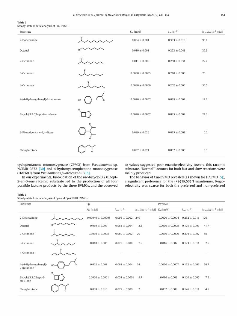
Steady-state kinetic parameters of the three recombinantenzymes were determined in 50 mM Tris/HCl using the optimalconditions defined for each enzyme, that are pH 8.0, at 25 ◦C for Pp-and Pp-H160Y BVMOs and pH 8.5, at 35 ◦C for Cm-BVMO. All thethree enzymes showed a typical Michaelis–Menten behavior. Thekinetic parameters kcat and KM were calculated for a set of iden-tified substrates besides rac-bicyclo[3.2.0]hept-2-en-6-one, usedin the preliminary assay. Selected substrates were linear aliphaticketones, cyclic aliphatic ketones and aromatic ketones. In particu-lar, for the linear ketone containing eight carbon atoms, the positionof the keto group in the molecule was investigated. Tables 2 and 3summarize the results obtained for Cm-BVMO, and Pp- and Pp-Y160H BVMOs, respectively.
Cm-BVMO exhibited the best catalytic efficiency against 2-dodecanone (kcat/KM 90.8 s−1 mM−1). Octanones also appeared tobe good substrates. In particular, by comparing KM and kcat/KM val-ues reported in Table 2, the position of the keto group at C3 seemedto be preferred. By limiting the observation to the positions of thecarbonyl function in octanone, it can be noticed that the Michaelisconstant is the main parameter influencing the catalytic efficiency,since the kcat values were similar for all investigated positions ofthe keto groups.
Also for Pp- and Pp-Y160H BVMOs, linear ketones seemed to bevery well accepted substrates (Table 3). 2-dodecanone was the onefor which the enzymes display the highest catalytic efficiency. Dif-ferently from the enzyme from C. merolae, the one from P. patenspreferred the keto group at position 2 of octanone. 4-octanone wasnot a good substrate for either wt or mutant enzymes, as alreadyobserved in the previous substrate-screening assay (Table 1). kcat
values, for all the four linear C8-ketones tested, were approximatelyin the same range of those measured for the algal enzyme. Asdiscussed above in relation to the activity and substrate profile,restoration of the consensus histidine makes the mutant variantof Pp-BVMO more competent for catalysis and faster than the wtform, showing higher turnover number.
All the recombinant enzymes were able to oxidize some substi-tuted aryl ketones, like phenylacetone and 4-(4-hydroxyphenyl)-2-butanone. The latter compound was a good substrate, particularlyfor Pp- and Pp-Y160H BVMOs. Due to the limited solubility,other substrates such as 4-dimethylaminobenzaldehyde and 3-acetylindole could not be investigated by kinetic analysis. Finally,the affinity for coenzyme NADPH was found to be very high, as aKM values <5 �M were determined.
3.5. Conversions
Biotransformations using purified enzymes and different typesof substrates were investigated using GC and GC-MS analysis inorder to detect both substrates and products. PTDH and phosphitewere added for NADPH regeneration.
As already mentioned, rac-bicyclo[3.2.0]hept-2-en-6-one is astandard probe for testing the biocatalytic potential of type IBVMOs. This conversion is a good example of regiodivergentparallel kinetic resolution (PKR) because the racemic substrateleads to two regioisomeric compounds (−)-(1S,5R) 2 and (−)-(1R,5S) 3 (Fig. 4A). One compound originates from the favoredBaeyer–Villiger-type oxygen insertion between the more substi-tuted carbon atom and the carbonyl group in a fast reaction; thesecond lactone is more slowly produced in the chemically disfa-vored regiochemistry. The two chiral lactones are relevant building
using different strains, i.e. different BVMOs. Some examples ofsuch studies are those regarding CHMO from Acinetobacter [29],
E. Beneventi et al. / Journal of Molecular Catalysis B: Enzymatic 98 (2013) 145– 154 151
Table 2Steady-state kinetic analysis of Cm-BVMO.
Substrate KM [mM] kcat [s−1] kcat/KM [s−1 mM]
2-DodecanoneO
0.004 ± 0.001 0.383 ± 0.018 90.8
Octanal O 0.010 ± 0.008 0.252 ± 0.043 25.3
2-OctanoneO
0.011 ± 0.006 0.250 ± 0.031 22.7
3-OctanoneO
0.0030 ± 0.0005 0.210 ± 0.006 70
4-OctanoneO
0.0040 ± 0.0009 0.202 ± 0.006 50.5
4-(4-Hydroxyphenyl)-2-butanone HO O 0.0070 ± 0.0007 0.079 ± 0.002 11.2
Bicyclo[3.2.0]hept-2-en-6-one
O
0.0040 ± 0.0007 0.085 ± 0.002 21.3
3-Phenylpentane-2,4-dione
O
O
0.099 ± 0.026 0.015 ± 0.001 0.2
cN(
2p
TS
Phenylacetone O
yclopentanone monooxygenase (CPMO) from Pseudomonas sp.CIMB 9872 [30] and 4-hydroxyacetophenone monooxygenase
HAPMO) from Pseudomonas fluorescens ACB [5].In our experiments, biooxidation of the rac-bicyclo[3.2.0]hept-
-en-6-one racemic substrate led to the production of all fourossible lactone products by the three BVMOs, and the observed
able 3teady-state kinetic analysis of Pp- and Pp-Y160H BVMOs.
Substrate Pp
KM [mM] kcat [s−1]
2-DodecanoneO
0.00040 ± 0.00008 0.096 ±
Octanal O 0.019 ± 0.009 0.061 ±
2-OctanoneO
0.0030 ± 0.0008 0.060 ±
3-OctanoneO
0.010 ± 0.005 0.075 ±
4-OctanoneO
– –
4-(4-Hydroxyphenyl)-2-butanone
HO O 0.002 ± 0.001 0.068 ±
Bicyclo[3.2.0]hept-2-en-6-one
O
0.0060 ± 0.0001 0.058 ±
Phenylacetone O 0.038 ± 0.016 0.077 ±
0.097 ± 0.071 0.032 ± 0.006 0.3
ee values suggested poor enantioselectivity toward this racemicsubstrate. “Normal” lactones for both fast and slow reactions were
mainly produced.The behavior of Cm-BVMO revealed (as shown for HAPMO [5]),a significant preference for the (+)-(1R,5S) 1 enantiomer. Regio-selectivity was scarce for both the preferred and non-preferred
PpY160H
kcat/KM [s−1 mM] KM [mM] kcat [s−1] kcat/KM [s−1 mM]
0.002 240 0.0020 ± 0.0004 0.252 ± 0.011 126
0.004 3.2 0.0030 ± 0.0008 0.125 ± 0.006 41.7
0.002 20 0.0030 ± 0.0006 0.204 ± 0.007 68
0.008 7.5 0.016 ± 0.007 0.123 ± 0.011 7.6
– – – –
0.004 34 0.0030 ± 0.0007 0.152 ± 0.006 50.7
0.0001 9.7 0.016 ± 0.002 0.120 ± 0.005 7.5
0.009 2 0.032 ± 0.009 0.146 ± 0.011 4.6

152 E. Beneventi et al. / Journal of Molecular Catalysis B: Enzymatic 98 (2013) 145– 154
Table 4Regiodivergent biooxidation by Cm-, Pp- and Pp-Y160H BVMOs.
Substrate Cm Pp PpY160H
% Conv ratio % ee % Conv ratio % ee % Conv ratio % ee
OH
H
Fast reaction 100 73:27 100 76:24 100 72:28
12 (+)-(1R,5S)237 (+)-(1S,5R)3
7 (+)-(1R,5S)269 (+)-(1S,5R)3
14 (+)-(1R,5S)268 (+)-(1S,5R)3
OH
62:4
% bnorm
s23ao1(c
b(d2wcetuafle
uikuwubfmtprcw
4
fpot
HSlow reaction 100 88:12 66
Conv, percentage yield of conversion; ratio, ratio between produced normal and a
ubstrate enantiomers, that respectively produced 73% (−)-(1S,5R) versus 27% (+)-(1S,5R) 3, and 88% (+)-(1R,5S) 2 and 12% (+)-(1R,5S), enantiomers. All four possible lactone products, in differentmounts, were therefore finally obtained (Fig. 4B). The ee valuesf the product mixture resulting from complete conversion were2% for the “normal” (+)-(1R,5S) 2 and 37% for the “abnormal”+)-(1S,5R) 3. (Table 4). After 5 h, the racemic substrate was fullyonverted.
Pp- and Pp-Y160H BVMOs approximately showed the sameehavior (Fig. 4C and D) (Table 4). The favored substrate was the+)-(1R,5S) 1 enantiomer for both enzymes; they respectively pro-uced 76% and 72% of the expected (−)-(1S,5R) 2 versus 24% and8% of the (+)-(1S,5R) 3. The mutant enzyme, compared to theild-type one, exhibited much higher regioselectivity in the slow
onversion of the (−)-(1S,5R) 1 substrate, yielding 95% of thexpected lactone and 5% of the unexpected one. In such slow reac-ion, the WT enzyme yielded only 62% of the expected and 4%nexpected lactone and the conversion was not complete (66%)fter 16 h. As for the case of the conversion by Cm-BVMO, ee valuesor the “normal” and “abnormal” enantiomeric products were tooow to be considered for a direct biocatalytic application of the newnzymes.
Conversions by Cm-, Pp- and Pp-Y160H BVMOs were also eval-ated with other compounds, chosen among those previously
dentified as substrates. As shown in Table 5, conversion of theseetones resulted in the formation of the expected ester prod-ct, obeying the stereo-chemical rule of Baeyer–Villiger reactionshere the migrating group is the most substituted one. In partic-lar, 2-octanone, 3-octanone and 4-octanone were fully convertedy Cm-BVMO. The mutated BVMO from Physcomitrella performedull conversion of 2-octanone and 3-octanone and poor transfor-
ation of 4-octanone, whereas the unmodified enzyme was ableo oxidize in good amount only the first two linear ketones. Finallyhenylacetone was fully converted by all the three enzymes. Theecombinant enzymes gave poor or almost no conversion of theyclic ketones cyclohexanone and cyclopentanone; only Cm-BVMOas able to transform the former molecule.
. Discussion
BVMOs are currently in the spotlight of industrial biocatalysis
or their capability to perform challenging steps in organic chemicalrocesses, such as oxidations of ketones to esters (Baeyer–Villigerxidations), but also sulfoxidations and other oxidative reac-ions. Up to 2012, all reported type I BVMOs originated from100 95:5
al lactones; % ee, percentage enantiomeric excess values.
microbial organisms, particularly actinobacteria and filamentousfungi. Only two BVMOs of eukaryotic origin have been identifiedand expressed in recombinant form very recently: the BVMOs fromthe ascomycetes Cylindrocarpon radicicola ATCC 11011 [20] andAspergillus fumigatus Af293 [21]. We have identified and selectedtwo new putative BVMOs from the photosynthetic eukaryotesCyanidioschizon merolae and Physcomitrella patens.
The two organisms can be certainly considered very uncom-mon sources for BVMOs, being eukaryotic and photosynthetic. Theprimitive red alga C. merolae 10D is a unicellular organism livingin acidic environments (like hot springs), even at pH < 2 and tem-perature of 45 ◦C. It is one of the photosynthetic eukaryotes withthe most simple cell architecture: its cell does not present a rigidcell wall and contains a single nucleus, a single mitochondrion anda single chloroplast. This clearly offers advantages for studies onthese organelles [29,30]. The moss P. patens is classified as a novascular plant, since it differs from higher plants in not having inter-nal vessels [31], and stands in an important phylogenetic positionfor studying the evolutionary transition from the aquatic environ-ment of algae to the terrestrial one of higher plants. Noticeably,both organisms undergo homologous recombination with a fre-quency that allows easy targeting of genes for replacement andelimination [32,33]. This allows handy studying of gene func-tion and helps in predicting the physiological role of genes inhigher organisms. P. patens and C. merolae are therefore consideredmodel systems for studying origin and fundamental mechanismsof eukaryotic cells and plant evolution.
We succeeded in producing the identified putative BVMOsin recombinant form, together with a mutant variant of theprotein from Physcomitrella completely restored in the consen-sus sequence firstly described. The second BVMO-typifying motif([A/G]GxWxxxx[F/Y]P[G/M]xxxD), recognized and annotated afterthe beginning of our work [24], is fully conserved in both proteins.The biocatalytic characterization of the recombinant productscould demonstrate their BVMO activity on a broad range of sub-strates as linear and cyclic aliphatic ketones, aryl and aromaticketones (Table 1). Among linear ketones (C8 and C12), proven tobe the best substrates, 2-dodecanone was the preferred one.
In an overview of the data from the kinetic analysis, thethree enzymes displayed different rates of catalysis, dependingon the substrate examined (Tables 2 and 3), but in all cases mea-
sured kcat values were not higher than 0.4 s−1. Since most naturalmonooxygenase enzymes display kcat values in the range of1–100 s−1 when tested on ketones among those selected for ouranalysis [34], we conclude that the new BVMOs (at least as such)
E. Beneventi et al. / Journal of Molecular Catalysis B: Enzymatic 98 (2013) 145– 154 153
Fig. 4. Biooxidation of racemic bicyclo[3.2.0]hept-2-en-6-one. Possible structures of bicyclo[3.2.0]hept-2-en-6-one isomers and lactones formed by type I BVMO activity (A).Time course of Cm- (B), Pp- (C) and Pp-Y160H- (D) BVMOs catalyzed oxidation of racemic bicyclo[3.2.0]hept-2-en-6-one to 2-oxabicyclo[3.3.0]oct-6-en-3-one (+)-(1S,5R)2/(−)-(1R,5S) 2 and 3-oxabicyclo[3.3.0]oct-6-en-2-one (−)-(1S,5R) 3/(+)-(1R,5S) 3.
Table 5Conversion of some identified substrates and corresponding products.
Substrate Product Conversion (%)
Cm Pp PpY160H
O
O
O
– 17 27
O
O
O
80 1 3
OO
O
100 100 100
O
O
O
100 90 100
O
O
O
100 67 100
O
O
O
100 – 17

1 r Cata
sop6
fdaddlM[ote
tcpw[wf
dmwtopip
tvtHmpsiimCuo
A
opncDP
[
[
[[[
[[[[
[
[
[
[
[
[
[[[
[
[[
[[[
[
[
[
[
[
[
54 E. Beneventi et al. / Journal of Molecula
eem rather inefficient bio-catalysts. This is also true in termsf enantioselectivity, which was shown to be very poor whenrobed on the typical substrate racemic bicyclo[3.2.0]hept-2-en--one (Table 4), transformed in the four possible lactone.
Nevertheless, some results deserve to be highlighted. As knownrom the literature, representative BVMOs can be clustered in well-efined groups with different substrate profile. The phylogeneticnalysis that we performed (Fig. 2) places the new BVMOs in twoistinct positions. The protein from P. patens is localized in the cladeefined by PAMO and STMO, which transform aromatic (but also
inear) ketones; the one from C. merolae is clustered together withEKMO and ACMO, enzymes known for converting linear ketones
35,36]. This grouping is in agreement with the substrate scopebtained for the recombinant enzymes even though, because ofheir eukaryotic origin, a different biocatalytic behavior could bexpected.
The second noteworthy result concerns the central histidine ofhe motif FxGxxxHxxxWP: this residue, although dispensable foratalysis and not always conserved (see [21] and [24]), has beenroposed to be involved in conformational changes occurring alongith catalysis [26]. Its fundamental role has been already shown
19,28]. By comparing activities of wild type and mutant Pp-BVMOs,e further confirm here that this histidine is certainly important
or enzymatic performance.Finally, we could show that the enzyme from Cyanidioschizon
isplays a relatively high thermostability by having an apparentelting temperature of 56 ◦C. This compares favorably with otherell-studied BVMOs: CHMO has a Tm of 39 ◦C (near to the value
hat we measured for Pp-BVMO, i.e. 44 ◦C), while PAMO has a Tmnly five degrees higher than that measured for Cm-BVMO [37]. Ifrotein engineering will be considered to boost the enzyme activ-
ties in view of a possible re-consideration of their utility, therimary sequence of Cm-BVMO could be a favorable starting point.
In the same perspective, a better characterization of the reper-oire of accepted molecules could also be useful. The displayedery low catalytic efficiency suggests that during evolution thewo new BVMOs had been subjected to a weak selective pressure.ence, according to the current view, a potential role as secondaryetabolism enzymes can be envisaged [38,39]. Their substrate
references point toward linear alkanones, structurally similar toide chains of chlorophylls and pheophytins, and to their possiblenvolvement in modifications of photosynthetic pigments. Work isn progress to produce knockout mutants in both the alga and the
oss, by exploiting their endogenous homologous recombination.omparison of null mutants with wild-type strains would help inncovering the natural substrates of Cm- and Pp-BVMOs in theirriginal biological context.
cknowledgements
We thank Dr. T. Morosinotto (Department of Biology, Universityf Padova, Italy) for providing a starting culture of Physcomitrellaatens. We are grateful to Dr. P. Polverino de Laureto (CRIBI Biotech-
ology Center, University of Padova, Italy) for help in analyticalharacterization of the recombinant flavoprotein Pp-BVMO andr. Enrico Negrisolo (Department of Public Health, Comparativeathology and Veterinary Hygiene, University of Padova) for useful[[[
lysis B: Enzymatic 98 (2013) 145– 154
suggestions on use of programs for dendrogram production. Thiswork has been entirely financed by F.I.S.-Fabbrica Italiana Sintetici(Alte di Montecchio Maggiore, Italy).
References
[1] A. Baeyer, V. Villiger, Ber. Dtsch. Chem. Ges. 32 (1899) 3625–3633.[2] G. Ten Brink, I. Arends, R. Sheldon, Chem. Rev. 104 (2004) 4105–4124.[3] S. Chandrasekhar, C.D.J. Roy, Chem. Soc. Perkin Trans. 2 (1994) 2141–2143.[4] R.B. Turner, J. Am. Chem. Soc. 72 (1950) 878–882.[5] N.M. Kamerbeek, A.J.J. Olsthoorn, M.W. Fraaije, D.B. Janssen, Appl. Environ.
Microbiol. 69 (2003) 419–426.[6] S. Colonna, N. Gaggero, P. Pasta, G. Ottolina, Chem. Commun. (Camb.) 20 (1996)
2303–2307.[7] G. Ottolina, P. Pasta, G. Carrea, S. Colonna, S. Dallavalle, H.L. Holland, Tetrahe-
dron Asymmetry 6 (1995) 1375–1386.[8] G. Ottolina, S. Bianchi, B. Belloni, G. Carrea, B. Danieli, Tetrahedron Lett. 40
(1999) 8483–8486.[9] P.B. Brondani, G. de Gonzalo, M.W. Fraaije, L.H. Andrade, Adv. Synth. Catal. 353
(2011) 2169–2173.10] S. Colonna, N. Gaggero, G. Carrea, G. Ottolina, P. Pasta, F. Zambianchi, Tetrahe-
dron Lett. 43 (2002) 1797–1799.11] M.W. Fraaije, D.B. Janssen, in: R.D. Schmid, V.B. Urlacher (Eds.), Modern Bioox-
idation, Wiley-VCH, Weinheim, 2007.12] M.D. Mihovilovic, Curr. Org. Chem. 10 (2006) 1265–1287.13] A. Willetts, Trends Biotechnol. 15 (1997) 55–62.14] M.P. Beam, M.A. Bosserman, N. Noinaj, M. Wehenkel, J. Rohr, Biochemistry 48
(2009) 4476.15] W. Van Berkel, N. Kamerbeek, M. Fraaije, J. Biotechnol. 124 (2006) 670–689.16] M.D. Mihovilovic, B. Müller, P. Stanetty, Eur. J. Org. Chem. 22 (2002) 3711–3730.17] N.A. Donoghue, D.B. Norris, P.W. Trudgill, Eur. J. Biochem. 63 (1976) 175–192.18] N.M. Kamerbeek, D.B. Janssen, W.J.H. van Berkel, M.W. Fraaije, Adv. Synth. Catal.
345 (2003) 667–678.19] M.W. Fraaije, N.M. Kamerbeek, W.J.H. van Berkel, D.B. Janssen, FEBS Lett. 518
(2002) 43–47.20] F. Leipold, R. Wardenga, U.T. Bornscheuer, Appl. Microbiol. Biotechnol. 94
(2012) 705–717.21] M.L. Mascotti, M.J. Ayub, H. Dudek, M.K. Sanz, M.W. Fraaije, AMB Expr. 3 (2013)
33.22] M.W. Fraaije, J. Wu, D.P.H.M. Heuts, E.W. van Hellemond, J.H.L. Spelberg, D.B.
Janssen, Appl. Microbiol. Biotechnol. 66 (2005) 393–400.23] F. Forneris, R. Orru, D. Bonivento, L.R. Chiarelli, A. Mattevi, FEBS J. 276 (2009)
2833–2840.24] A. Riebel, H. Dudek, G. de Gonzalo, P. Stepniak, L. Rychlewski, M. Fraaije, Appl.
Microbiol. Biotechnol. 95 (2012) 1479–1489.25] O. Vallon, Proteins 38 (2000) 95–114.26] E. Malito, A. Alfieri, M. Fraaije, A. Mattevi, PNAS 101 (2004) 13157–13162.27] J. Rehdorf, C.L. Zimmer, U.T. Bornscheuer, Appl. Environ. Microbiol. 75 (2009)
3106.28] M.J. Cheesman, M.B. Kneller, A.E. Rettie, Chem. Biol. Interact. 146 (2003)
157–164.29] V. Alphand, R. Furstoss, J. Org. Chem. 57 (1992) 1306–1309.30] M.D. Mihovilovic, P. Kapitán, P. Kapitánová, ChemSusChem 1 (2008)
143–148.31] D. Lang, A.D. Zimmer, S.A. Rensing, R. Reski, Trends Plant Sci. 13 (2008) 542–549.32] D.G. Schaefer, Annu. Rev. Plant Biol. 53 (2002) 477–501.33] A. Minoda, R. Sakagami, F. Yagisawa, T. Kuroiwa, K. Tanaka, Plant Cell Physiol.
45 (2004) 667–671.34] A. Bar-Even, E. Noor, Y. Savir, W. Liebermeister, D. Davidi, D.S. Tawfik, R. Milo,
Biochemistry 50 (2011) 4402–4410.35] D.V. Rial, P. Cernuchova, J.B. van Beilen, M.D. Mihovilovic, J. Mol. Catal., B
Enzyme 50 (2008) 61–68.36] M.D. Mihovilovic, F. Rudroff, B. Grötzl, P. Kapitan, R. Snajdrova, J. Rydz, R. Mach,
Angew. Chem. Int. Ed. Engl. 44 (2005) 3609–3613.37] H.L. van Beek, G. de Gonzalo, M.W. Fraaije, Chem. Commun. (Camb.) 48 (2012)
3288–3290.38] S. Picaud, L. Olofsson, M. Brodelius, P.E. Brodelius, Arch. Biochem. Biophys. 436
(2005) 215–226.39] B.T. Greenhagen, P.E. O’Maille, J.P. Noel, J. Chappell, Proc. Natl. Acad. Sci. U.S.A.
103 (2006) 9826–9831.40] K. Tamura, J. Dudley, M. Nei, S. Kumar, Mol. Biol. Evol. 24 (2007) 1596–1599.41] N. Saitou, M. Nei, Mol. Biol. Evol. 4 (1987) 406–425.42] D.H. Huson, D.C. Richter, C. Rausch, T. Dezulian, M. Franz, R. Rupp, BMC Bioin-
formatics 8 (2007) 460.