dna aptamer raised against ages blocks the progression of
TRANSCRIPT

Kaida et al.
1
DNA aptamer raised against AGEs blocks the progression of experimental
diabetic nephropathy
Yusuke Kaida1,*
, Kei Fukami1,*
, Takanori Matsui2, Yuichiro Higashimoto
3, Yuri Nishino
2,
Nana Obara1, Yosuke Nakayama
1, Ryotaro Ando
1, Maki Toyonaga
1, Seiji Ueda
1,
Masayoshi Takeuchi4, Hiroyoshi Inoue
5, Seiya Okuda
1 and Sho-ichi Yamagishi
2,*
1Division of Nephrology, Department of Medicine,
2Department of Pathophysiology
and Therapeutics of Diabetic Complications, 3Department of Medical Biochemistry,
Kurume University School of Medicine, Kurume, Japan, 4Department of Advanced
Medicine Medical Research Institute, Kanazawa Medical University, Ishikawa, Japan, 5Department of Chemistry, Keio University School of Medicine, Tokyo, Japan
Word content; 4,288
Running title; AGEs-aptamer against diabetic nephropathy
*These authors equally contributed to the present study.
Corresponding authors:
Kei Fukami, MD, PhD, Division of Nephrology, Department of Medicine, Kurume
University School of Medicine, 67 Asahi-machi, Kurume 830-0011, Japan. Tel:
+81-942-31-7002, Fax; +81-942-31-7763, Email; [email protected]
Or
Sho-ichi Yamagishi, MD, PhD, Department of Pathophysiology and Therapeutics of
Diabetic Vascular Complications, Kurume University School of Medicine, 67
Asahi-machi, Kurume 830-0011, Japan. Tel: +81-942-31-7873, Fax: +81-942-31-7895,
Email; [email protected]
Page 2 of 42Diabetes
Diabetes Publish Ahead of Print, published online April 29, 2013

Kaida et al.
2
ABSTRACT
Advanced glycation end products (AGEs) and their receptor (RAGE) play a role in
diabetic nephropathy. We screened DNA aptamer directed against AGEs
(AGEs-aptamer) in vitro, and examined its effects on renal injury in KKAy/Ta mice, an
animal model of type 2 diabetes. Eight week-old male KKAy/Ta or C57BL/6J mice
received continuous intraperitoneal infusion of AGEs- or control-aptamer for 8 weeks.
AGEs-aptamer was detected and its level was increased in the kidney for at least 7 days.
The elimination half-lives of AGEs-aptamer in the kidney were about 7 days. Compared
with C57BL/6J mice, glomerular AGEs levels were significantly increased in KKAy/Ta
mice, which were blocked by AGEs-aptamer. Urinary albumin and
8-hydroxy-2’-deoxy-guanosine levels were increased, and glomerular hypertrophy and
enhanced extracellular matrix accumulation were observed in KKAy/Ta mice, all of
which were prevented by AGEs-aptamer. Moreover, AGEs-aptamer significantly
reduced gene expression of RAGE, monocyte chemoattractant protein-1, connective
tissue growth factor or type-IV collagen both in the kidney of KKAy/Ta mice and in
AGEs-exposed human cultured mesangial cells. Our present data suggest that
continuous administration of AGEs-aptamer could protect against experimental diabetic
nephropathy by blocking the AGEs-RAGE axis and may be a feasible and promising
therapeutic strategy for the treatment of diabetic nephropathy.
KEY WORDS; aptamer, AGEs, diabetic nephropathy, RAGE
Page 3 of 42 Diabetes

Kaida et al.
3
INTRODUCTION
Diabetic nephropathy is a leading cause of end-stage renal disease, which
accounts for disability and high mortality rate in patients with diabetes (1, 2). The
development of diabetic nephropathy is characterized by glomerular hypertrophy and
inflammatory cell infiltration, followed by extracellular matrix (ECM) accumulation in
mesangial area and an increased urinary albumin excretion (UAE) rate (3). Diabetic
nephropathy ultimately progresses glomerular sclerosis associated with renal
dysfunction (4). Large-scale clinical studies have shown that intensive glycemic and
blood pressure control reduce the risk of diabetic nephropathy (5). However,
management of diabetic nephropathy is far from satisfactory, because strict control of
blood glucose and/or pressure is often difficult to maintain and may increase the risk of
hypoglycemia or hypotension. Further, despite intensive management of classical
cardiovascular risk factors using blood pressure-lowering and lipid-lowering agents, a
substantial number of diabetic patients still have experienced end-stage renal disease.
Therefore, to develop novel therapeutic strategies that specifically target diabetic
nephropathy is urgently needed.
Reducing sugars can react non-enzymatically with the amino groups of
proteins to initiate a complex series of rearrangements and dehydrations, and then to
Page 4 of 42Diabetes

Kaida et al.
4
produce a class of irreversibly cross-linked moieties termed advanced glycation end
products (AGEs) (6-8). The formation and accumulation of AGEs in various tissues
have been shown to progress at an accelerated rate under hyperglycemic conditions
(9-11). There is accumulating evidence that AGEs and RAGE (receptor for AGEs)
interaction stimulates oxidative stress generation and subsequently evokes inflammatory
reactions, thereby causing progressive alteration in renal architecture and loss of renal
function in diabetes (12-14). Indeed, inhibitors of AGEs formation have been shown to
attenuate the increase in albuminuria and prevent the development and progression of
experimental diabetic nephropathy (15, 16). Further, RAGE-overexpressing diabetic
mice showed progressive glomerulosclerosis with renal dysfunction, while diabetic
homozygous RAGE null mice failed to develop significantly increased mesangial
matrix expansion or thickening of the glomerular basement membrane (14, 17). These
observations suggest that the inhibition of the AGEs-RAGE axis could be a novel
therapeutic target for diabetic nephropathy.
Aptamers are short, single-stranded DNA or RNA molecules that can bind with
high affinity and specificity to a wide range of target proteins (18). Recently, numerous
aptamers have been developed and used in the clinical fields as a tool for modulating
various protein function (19-21). Pegaptanib, an RNA aptamer directed against vascular
Page 5 of 42 Diabetes

Kaida et al.
5
endothelial growth factor165 (VEGF165) isoform, has been shown to be effective in
treating choroidal neovascularization in patients with age-related macular degeneration
(19). In addition, ARC1779, a DNA-aptamer raised against the A1 domain of von
Willebrand factor, has also been reported to inhibit its pro-thrombotic function in vivo,
and phase II clinical trials of ARC1779 are currently underway for the treatment of
thrombotic thrombocytopenic purpura and cerebral embolism after carotid
endarterectomy (20, 21).
Therefore, in this study, we screened a high-affinity DNA aptamer directed
against AGEs (AGEs-aptamer) using a combinatorial chemistry in vitro, and examined
its effects on experimental diabetic nephropathy. For this, we chose KKAy/Ta mice
because they are an animal model of obese and type 2 diabetes associated with ECM
accumulation, inflammatory cell infiltration and sclerotic changes within the glomerular
areas, whose characteristics closely resemble those in human diabetic nephropathy (22).
Furthermore, we investigated the effects of AGEs-aptamer on mesangial cell damage in
vitro.
RESEARCH DESIGN AND METHODS
Animals. Male 8-week old KKAy/Ta (DM) and 8-week old C57BL/6J (Ctr) mice were
Page 6 of 42Diabetes

Kaida et al.
6
purchased from CLEA Japan (Tokyo, Japan). These mice were divided into two groups
respectively, and received continuous intraperitoneal infusion of either AGEs-aptamer
or control-aptamer (Ctr-aptamer) (0.136 µg/day) by an osmotic mini pump (Alzet
osmotic pumps, model 1004, Cupertino, CA) [Ctr-aptamer-treated Ctr mice
(Ctr-Ctr-aptamer) (n=8), AGEs-aptamer-treated Ctr mice (Ctr-AGEs-aptamer) (n=9),
Ctr-aptamer-treated DM mice (DM-Ctr-aptamer) (n=11) and AGEs-aptamer-treated DM
mice (DM-AGEs-aptamer) (n=10) group]. Eight weeks after the continuous
intraperitoneal infusion, blood pressure was measured by a tail-cuff sphygmomanometer
using an automated system with a photoelectric sensor (BP-98A; Softron, Tokyo, Japan),
and mice were transferred to metabolic cages for 24 hr for urinalysis. Then, mice were
killed and blood and kidney samples were obtained. All experimental procedures were
conducted in accordance with the National Institutes Health Guide for Care and Use of
Laboratory Animals and were approved by the ethnical committee of Kurume
University School of Medicine.
Measurement of clinical variables. Albuminuria was determined with commercially
available enzyme-linked immunosorbent assay (ELISA) kit (Exocell, Philadelphia, PA,
USA). Blood was collected, centrifuged and plasma and serum were obtained, and
Page 7 of 42 Diabetes

Kaida et al.
7
stored at –40°C. Total cholesterol and transaminase levels were determined by
commercially available kits (Wako, Osaka, Japan). Plasma creatinine levels were
measured by an enzymatic method (SRL Inc., Tokyo, Japan). Blood urea nitrogen levels
were measured by an auto-analyzer (Nihondenshi Co., Tokyo, Japan). Plasma glucose
was measured by a glucose oxidase method (Shionotest Co., Tokyo, Japan). HbA1c was
determined by latex coagulation method (TFB Co., Tokyo, Japan). Urinary
8-hydroxy-2’-deoxy-guanosine (8-OHdG) levels were measured with ELISA system
(Japan Institute for the Control of Aging, Fukuroi, Shizuoka, Japan). Urinary
N-acetyl-β-D-glucosaminidase (NAG) was measured with a fractional colorimetric
determination method (Shionogi Pharma, Inc., Osaka, Japan). Intra- and interassay
coefficients of variation were 6.2 and 8.8%, respectively.
Preparation of AGEs-human serum albumin (AGEs-HSA) and AGEs-bovine
serum albumin (AGEs-BSA). AGEs-modified proteins were prepared as described
previously (23).
Immobilizing AGEs-HSA on agarose beads. AGEs-HSA was covalently coupled to
indoacetyl groups on SulfoLink Coupling Gel (Pierce, Rockford, IL, USA) as described
Page 8 of 42Diabetes

Kaida et al.
8
previously (24).
Screening and modification of AGEs-aptamer (Systematic Evolution of Ligands by
EXponential enrichment (SELEX)). Preparation and selection of DNA aptamers were
performed as described previously (24). Sequences of AGEs- and Ctr-DNA aptamers
are below. AGEs-aptamer;
5’-CCGAAACCAGACCACCCCACCAAGGCCACTCGGTCGAACCGCCAACACT
CACCCCA-3’, Ctr-aptamer;
5’-GTTATCTGTCATAGGAACAGTCAGACTCAGCGTCGCAGTTCAGGGCACTTT
AGCAC-3’. DNA aptamers are susceptible to degradation by nucleases. This will limit
their applications for real samples, such as blood and tissues. To solve this issue, we
modified aptamers with phosphorothioate as described previously (25).
Binding affinity of AGEs-aptamer to AGEs-HSA or AGEs-HSA to RAGE. The
binding affinity of the selected AGEs-aptamer to AGEs-HSA or AGEs-HSA to
extracellular AGEs-binding V-domain of RAGE (vRAGE) was measured using
sensitive 27-MHz Quarts crystal microbalance (QCM) (Affinix Q; Initium, Tokyo,
Japan) according to the method of Okahata et al (26). In brief, AGEs-HSA or vRAGE
Page 9 of 42 Diabetes
Kaida et al.
9
was immobilized on an avidin-bound QCM surface. After adding Ctr-aptamer or
AGEs-aptamer to a reaction vessel in the presence or absence of 70 ng/ml AGEs-HSA,
the time course of the frequency decrease of bound AGEs-HSA or bound vRAGE on the
QCM was monitored. The binding affinity of AGEs-aptamer to AGEs-HSA or
AGEs-HSA to vRAGE was calculated from curve fitting to the QCM frequency
decrease. Human vRAGE (residues 23–121) was prepared as described previously (27,
28). SDS-PAGE analysis of purified vRAGE proteins revealed a single band, which
showed positive reactivity with polyclonal antibody raised against RAGE (data not
shown).
Distribution and kinetics of [γ-32
P]ATP-labeled AGEs-aptamer. Aptamer was
radioactively labeled by a 5'-end-labeling technique using T4 polynucleotide kinase (T4
PNK; Promega Corporation, WI, USA) and [γ-32
P]ATP (PerkinElmer Japan Co., Ltd.,
Kanagawa, Japan) according to the manufacture's protocol. Male 8-week old Ctr mice
received continuous intraperitoneal infusion of [γ-32
P]ATP-labeled AGEs-aptamer
(0.136 µg/day) by an osmotic mini pump, and killed at 0 hr, 1 hr, 6 hr, 1 day, 2 days and
7 days after the infusion. At 0, 3, 7 and 14 days after stopping the infusion, mice were
killed, and blood and organs were obtained. The radioactivity of AGEs-aptamer was
Page 10 of 42Diabetes

Kaida et al.
10
measured by Cherenkov counting using LS-6500 scintillation counter (Beckman Coulter,
San Francisco, CA, USA).
Turnover rate of aptamer-bound AGEs by macrophages. Human THP-1 monocytic
leukemia cells were differentiated to macrophages as described previously (29). One
hundred µg/ml AGEs-BSA was added to the culture medium in the presence or absence
of 2 µM AGEs-aptamer where differentiated THP-1 macrophages were grown or
ungrown. After 4 hr, the supernatant was collected, concentrated with Amicon Urtra
centrifugal filter (Millipore Co., Billerica, MA), separated by SDS-PAGE and
transferred to polyvinylidene fluoride membrane as described previously (30).
Membranes were probed with rabbit polyclonal antibodies raised against AGEs, and
then immune complexes were visualized with an enhanced chemiluminescence
detection system (ECL; Amersham Bioscience, Buckinghamshire, UK).
Morphological analysis. Three-micrometer paraffin sections were stained with periodic
acid-schiff and Masson’s trichrome for light microscopic analysis as described
previously (31).
Page 11 of 42 Diabetes

Kaida et al.
11
Measurement of serum AGEs. Measurement of serum AGEs levels were performed
with a competitive enzyme-linked immunosorbent assay (ELISA) as described
previously (32).
Immunostaining. Specimens of kidney cortex were fixed with 4% paraformaldehyde,
embedded in paraffin, sectioned at 4-µm intervals and mounted on glass slides. The
sections were incubated in 0.3% hydrogen peroxide methanol for 30 min, and incubated
overnight at 4°C with rabbit polyclonal antibodies raised against AGEs and
synaptopodin (PROGEN Biotechnik GmbH, Heidelberg, Germany) as described
previously (32). Immunoreactivity in 10 different fields (x 600) in each sample was
measured by an image analysis software (Optimas version 6.57; Media Cybernetics,
Silver Spring, MD, USA).
Cells. Human mesangial cells were maintained in basal medium supplemented with 5 %
fetal bovine serum according to the supplier’s instructions (Clonetics Corporation, San
Diego, California, USA). AGEs treatment was carried out in a medium containing 0.5 %
fetal bovine serum. Mesangial cells less than 6 passages were used for the experiments.
Mesangial cells were treated with 100 µg/ml AGEs-BSA or non-glycated BSA for 4 and
Page 12 of 42Diabetes

Kaida et al.
12
24 hr in the presence or absence of 0.2 or 2 µM AGEs-aptamer.
Real-time reverse-transcription PCR (RT-PCR). Three to six µg of total RNA
extracted from each kidney cortex were used to synthesize cDNA with the Superscript
First Strand synthesis system for RT-PCR (Invitrogen Co., Carlsbad, CA, USA).
Quantitative real-time RT-PCR was performed using Assay-on-Demand and TaqMan 5
fluorogenic nuclease chemistry (Applied Biosystems, Foster city, CA, USA) according
to the supplier’s recommendation. IDs of primers and probe for mice monocyte
chemoattractant protein-1 (MCP-1), tumor necrosis factor-alpha (TNF-α), connective
tissue growth factor (CTGF), type-IV collagen, vascular endothelial growth factor
(VEGF), AGE-receptor 3 (AGE-R3) and RAGE gene were Mm00441242_ml,
Mn00443258_m1, Mm01192933_g1, Mm01210125_m1, Mm01281449_m1,
PN4331348 and Mm00545815_m1, respectively (Applied Biosystems, Foster city, CA,
USA). TaqMan Ribosomal RNA Control Reagents (18S) was used as an endogenous
control (Applied Biosystems, Foster city, CA, USA). Total RNAs were extracted from
cultured human mesangial cells and THP-1 cells with RNA queous-4 PCR kit (Ambion
Inc., Austin, TX, USA), and quantitative real-time RT-PCR was performed. IDs of
primers and probe for human MCP-1, TNF-α, CTGF, RAGE and
Page 13 of 42 Diabetes

Kaida et al.
13
glyceraldehyde-3-phosphate dehydrogenase (GAPDH) gene were Hs00234140_m1,
Hs00170014_m1, Hs00174128_m1, Hs00153957_m1 and Hs99999905_m1,
respectively (Applied Biosystems, Foster city, CA, USA).
Measurement of reactive oxygen species (ROS) generation. The intracellular
formation of ROS was detected using the fluorescent probe CM-H2DCFDA (Molecular
Probes, Inc., Eugene, OR, USA) as described previously (33).
Statistical analysis. All data are presented as means ± standard error of mean. Analysis
of variance (ANOVA) followed by the Turkey’s or Games-Howell post-hoc test was
performed for all studied parameters for statistical comparisons; p<0.05 was considered
significant. All statistical analyses were performed with SPSS 19 system.
RESULTS
Isolation and characterization of DNA aptamers directed against AGEs-modified
proteins. DNA aptamers specific for AGEs-HSA were isolated by an in vitro selection
process, SELEX, from a pool of ~1015
different nucleic acid sequences as described
previously (24). In this study, 35 clones were sequenced from the pool of selected
Page 14 of 42Diabetes

Kaida et al.
14
single-stranded DNAs to obtain 15 unique sequences, indicating that some of the
sequences among the 35 clones were identified and that multiple selection of the same
clone occurred. Structural analysis revealed that all of the aptamers had bulge-loop
structure with cytosine-rich sequences. We have previously shown that although all the
clones significantly inhibit the AGEs-induced decrease in DNA synthesis in cultured
pericytes, a counterpart of mesangial cells in the kidney, clone 1 had the strongest effect
(24). So, we modified clone 1 with phosphorothioate for generating nuclease-resistant
aptamer and used it for the following experiments. The structure of phosphorothioate
AGEs-aptamer used here is shown in Fig. 1A.
Binding affinity of phosphorothioate AGEs-aptamer to AGEs-HSA or AGEs-HSA
to vRAGE. We first examined the binding affinity of phosphorothioate AGEs-aptamer
to AGEs-HSA in vitro. For this, AGEs-HSA was immobilized on an avidin-bound
QCM surface, and then Ctr- or AGEs-aptamer was added to a reaction vessel.
AGEs-aptamer bound to AGEs-HSA with a dissociation constant of 1.38 x 10⁻⁶
M,
whereas Ctr-aptamer did not bind to AGEs-HSA at all (Fig. 1B). Further, as shown in
Fig. 1C, the binding of AGEs-HSA to vRAGE was dose-dependently inhibited by the
co-treatment with AGEs-aptamer.
Page 15 of 42 Diabetes

Kaida et al.
15
Turnover rate of aptamer-bound AGEs-BSA. Compared with unbound AGEs-BSA,
turnover rate of aptamer-bound AGEs by differentiated THP-1 macrophages were more
increased (Fig. 1D). On the contrary, under the conditions without THP-1 cells,
presence of AGEs-aptamer did not affect the turnover rate of AGEs-BSA (Fig. 1E).
Distribution and kinetics of infused AGEs-aptamer. To examine the kinetics of
injected AGEs-aptamer, we labeled the aptamer with [γ-32
P]ATP and infused it into the
peritoneal cavity of 8-week old C57BL/6J (Ctr) mice by an osmotic pump. As shown in
Fig. 2A, when AGEs-aptamer was continuously administrated up to 7 days, it was
distributed mainly in the kidney, aorta and muscle. The levels of AGEs-aptamer were
increased in the kidney, liver and blood of 8-week old Ctr mice for at least 7 days
following continuous 0.136 µg/day-intraperitoneal infusion (Fig. 2B). After stopping the
injection, AGEs-aptamer levels were gradually decreased. But the aptamer was still
detected in the kidney, liver and blood at day 14 after the removal of an osmotic pump
(Fig. 2B). The elimination half-lives of AGEs-aptamer in the kidney were about 7 days.
Characteristics of animals. Clinical characteristics of each group are shown in Table 1.
Page 16 of 42Diabetes

Kaida et al.
16
Compared with Ctr-Ctr-aptamer mice, body weight, plasma glucose, HbA1c and total
cholesterol levels were significantly increased in DM-Ctr-aptamer mice (p<0.05).
Treatment with AGEs-aptamer did not affect these parameters in mice. Plasma levels of
blood urea nitrogen and creatinine were tended to be higher and kidney-to-body weight
ratio was significantly increased in DM-Ctr-aptamer mice, which were ameliorated by
the treatment of AGEs-aptamer (p<0.05). There were no significant differences of
clinical variables between Ctr-Ctr-aptamer and Ctr-AGEs-aptamer mice.
Effect of AGEs-aptamer on serum and renal levels of AGEs in mice. We
investigated whether treatment with AGEs-aptamer could reduce serum and renal levels
of AGEs in KKAy/Ta mice. As shown in Fig. 3A, compared with Ctr-Ctr-aptamer mice,
serum levels of AGEs were significantly increased to about 3-fold in DM-Ctr-aptamer
mice (p<0.05), which were not affected by the treatment with AGEs-aptamer. However,
immunohistochemical analysis revealed that AGEs levels in the glomeruli of
DM-Ctr-aptamer mice were significantly higher than those of Ctr-Ctr-aptamer mice,
which was prevented by the treatment with AGEs-aptamer (Fig. 3B-F). AGEs-aptamer
itself did not affect serum or renal levels of AGEs in Ctr mice.
Page 17 of 42 Diabetes

Kaida et al.
17
Treatment with AGEs-aptamer decreased UAE levels in DM mice. We examined the
effect of AGEs-aptamer on UAE levels in mice. As shown in Fig. 4, UAE levels were
gradually and significantly elevated in DM-Ctr-aptamer mice compared with
Ctr-Ctr-aptamer mice (p<0.01). Administration of AGEs-aptamer for 8 weeks
significantly decreased UAE levels in DM mice (p<0.01). AGEs-aptamer itself did not
affect UAE levels in Ctr mice.
Treatment with AGEs-aptamer prevented glomerular hypertrophy and ECM
protein accumulation in the kidney of DM mice. Glomerular hypertrophy and ECM
accumulation are one of the characteristic features of diabetic nephropathy (3). So, we
investigated whether AGEs-aptamer treatment could prevent the structural changes in
the kidney of DM mice. As shown in Fig. 5A-E, periodic acid-schiff staining revealed
that diabetes was associated with glomerular hypertrophy, which was significantly
ameliorated by the administration of AGEs-aptamer. Further, ECM accumulation
assessed by Masson’s trichrom staining was significantly increased in DM-Ctr-aptamer
mice, which was also prevented by the treatment with AGEs-aptamer (Fig. 5F-J).
AGEs-aptamer itself did not affect glomerular hypertrophy or ECM accumulation in Ctr
mice. Further, immunohistochemical analysis revealed that synaptopodin levels, a
Page 18 of 42Diabetes

Kaida et al.
18
marker of podocytes were reduced in DM-Crl-aptamer mice, which were significantly
restored by the treatment with AGEs-aptamer (Fig. 5K-O). There was no significant
difference of renal VEGF expression, tubulointerstitial fibrosis (data not shown) or
NAG levels (Table 1) among the groups.
Treatment with AGEs-aptamer decreased urinary excretion levels of 8-OHdG in
DM mice. We next examined whether AGEs-aptamer treatment could decrease urinary
excretion levels of 8-OHdG, a marker of oxidative stress, in DM mice. Compared with
Ctr-Ctr-aptamer mice, urinary 8-OHdG levels were significantly increased in
DM-Ctr-aptamer mice to about 2.5-fold (p<0.05). Administration of AGEs-aptamer
significantly reduced urinary excretion levels of 8-OHdG in DM mice (p<0.05) (Fig.
5P).
Treatment with AGEs-aptamer decreased inflammatory and fibrotic gene
expression in the kidney of KKAy/Ta mice. AGEs induce a variety of inflammatory
and fibrotic gene expression, thus being involved in diabetic nephropathy (16, 33, 34).
So, we studied the effects of AGEs-aptamer on MCP-1, TNF-α, CTGF, type-IV
collagen and RAGE gene expression in KKAy/Ta mice. As shown in Fig. 6A-D, renal
MCP-1, TNF-α, CTGF and type-IV collagen gene expression was significantly
Page 19 of 42 Diabetes

Kaida et al.
19
increased in DM-Ctr-aptamer mice, which were prevented by the administration of
AGEs-aptamer. Moreover, although there were no significant differences in gene
expression levels of RAGE and other AGEs scavenging receptor, AGE-R3 between
Ctr-Ctr-aptamer and DM-Ctr-aptamer mice, treatment with AGEs-aptamer significantly
reduced RAGE and AGE-R3 mRNA levels in the kidney of DM mice (Fig. 6E and 6F).
AGEs-aptamer decreased ROS generation and inflammatory and fibrotic gene
expression in AGEs-exposed human mesangial cells. We next examined whether
AGEs-aptamer could inhibit AGEs-elicited ROS generation and RAGE, MCP-1, and
CTGF gene expression in human mesangial cells. One hundred µg/ml AGEs-BSA for 4
hr significantly increased ROS generation (Fig. 7A). RAGE gene expression was
increased at 4 and 24 hr after the treatment with AGEs-BSA, whereas MCP-1 and
CTGF mRNA levels were elevated at 24 hr only (Fig. 7B-D). AGEs-aptamer
significantly prevented these harmful effects of AGEs although they did not have any
toxic effects on BSA-treated cells. Furthermore, AGEs-BSA for 4 hr increased gene
expression of TNF-α, but not RAGE, MCP-1 or CTGF in THP-1 cells, which was also
blocked by AGEs-aptamer (data not shown).
Page 20 of 42Diabetes

Kaida et al.
20
DISCUSSION
In the present study, we demonstrated for the first time that although infusion
of in vitro-selected DNA-aptamer raised against AGEs-HSA did not affect glucose,
HbA1c or blood pressure levels, it not only inhibited glomerular hypertrophy and ECM
protein accumulation, but also decreased urinary excretion levels of albumin and
8-OHdG in DM mice. In addition, administration of AGEs-aptamer significantly
decreased plasma levels of blood urea nitrogen and creatinine, thus preventing renal
dysfunction in DM mice. Moreover, treatment with AGEs-aptamer significantly
suppressed MCP-1, TNF-α, CTGF, type-IV collagen and RAGE gene expression in the
kidney of DM mice, whereas AGEs-aptamer completely blocked the AGEs-induced
up-regulation of RAGE, MCP-1, and CTGF mRNA levels in mesangial cells. In this
study, we found that levels of AGEs-aptamer were increased in the kidney of 8-week
old Ctr mice for at least 7 days following the continuous intraperitoneal infusion.
Further, at day 14 after stopping the injection, AGEs-aptamer was still detected in the
kidney. These findings suggest that phosphorothioate AGEs-aptamer used in these
experiments may be resistant to nuclease attack and quite stable, therefore suitable as a
therapeutic agent. Given the fact that no toxicities related to AGEs-aptamer were
observed following the intraperitoneal injection, our present observations indicate that
Page 21 of 42 Diabetes

Kaida et al.
21
continuous infusion of AGEs-aptamer may be a safe and effective therapeutic strategy
for preventing the development and progression of diabetic nephropathy. However, this
will have to be affirmed before widespread clinical use of the AGEs-aptamer is
entertained. To compare the efficacy of AGEs-aptamer with other means to reduce the
load of AGEs would also be interesting because the latter is mostly delivered orally.
Aptamers have the following advantages over antibodies for blocking the
function of targeted proteins; [1] production of aptamers does not rely on biological
systems, and can be easily selected and constructed from oligonucleotide library with
low-cost and time-saving in vitro; [2] aptamers are quite thermally stable and can be
denatured and renatured multiple times without loss of activity and specificity; [3]
aptamers do not have immunogenicity over antibodies; [4] small size allows more
efficient entry into biological compartments (35, 36). There is accumulating evidence
that AGEs play a role in various disorders such as Alzheimer’s disease, cancers and
cardiovascular disease (37-39). Increased formation and accumulation of AGEs could
link the increased risks for these disorders to diabetes (37-39). Since feasibility and
efficacy of insulin pump therapy has been already established in diabetic patients (40)
and that various clinical trials with AGEs inhibitors have been terminated because of the
safety concern (41), continuous pump infusion of insulin plus AGEs-aptamer may be
Page 22 of 42Diabetes

Kaida et al.
22
promising for preventing various life-threatening AGEs-related disorders in diabetes.
In the present study, we demonstrated that continuous infusion of
AGEs-aptamer for 8 weeks dramatically decreased AGEs levels in the glomeruli of DM
mice to basal levels. Engagement of RAGE with AGEs stimulates ROS generation in a
variety of cells, which could in turn promote the formation and accumulation of AGEs,
thus forming a positive feedback loop between RAGE-downstream signaling and AGEs
generation (16, 42). Indeed, aortic AGEs accumulation has been suppressed in
RAGE-deficient diabetic apoE knockout mice (42). In the present study, AGEs-aptamer
directly bound to AGEs and resultantly blocked the binding of AGEs to RAGE (Fig. 1B
and 1C). These observations suggest that AGEs-aptamer could decrease the glomerular
accumulation of AGEs via the blockade of AGEs binding to RAGE in the kidney.
Further, we found here that turnover rate of aptamer-bound AGEs by THP-1
macrophages was increased compared with that of unbound AGEs (Fig. 1D). This is
another possible mechanism by which AGEs-aptamer decreased the glomerular
accumulation of AGEs. Since treatment with AGEs-aptamer did not affect serum AGEs
levels in DM mice, AGEs-aptamer may be preferentially distributed and accumulated in
the kidney and vessels, locally suppress the actions of AGEs, and finally eliminated
from the body via the increased turnover by macrophages. Under non-diabetic normal
Page 23 of 42 Diabetes

Kaida et al.
23
conditions, oxidative stress, RAGE expression and AGEs accumulation are largely
suppressed. This is a possible reason why AGEs-aptamer did not reduce renal AGEs
levels of Ctr mice.
AGEs-RAGE interaction elicits inflammatory and fibrotic reactions in the
kidney cells via oxidative stress generation (13). In this study, we found that treatment
with AGEs-aptamer decreased urinary 8-OHdG and albumin levels and reduced
inflammatory and fibrotic gene expression (MCP-1, TNF-α, CTGF and type-IV
collagen) in the kidney of DM mice. MCP-1 is a specific chemokine that recruits and
activates monocytes from circulation to inflammatory sites (43). Increased MCP-1
expression associated with monocyte infiltration in mesangial areas has been observed
in early phase of diabetic nephropathy (44). Plasma MCP-1 level was positively
correlated with UAE in type 1 diabetic patients (44). Further, administration of AGEs
has been shown to cause glomerular hypertrophy and ECM accumulation in the rat
kidney via induction of CTGF and type-IV collagen expression (45). Since
AGEs-aptamer completely blocked the AGEs-induced up-regulation of RAGE, MCP-1
and CTGF mRNA levels in human mesangial cells, the present findings suggest that
blockade of the AGEs-RAGE axis by AGEs-aptamer could decrease albuminuria and
fibrotic reactions in the diabetic kidney via suppression of ROS. It would be interesting
Page 24 of 42Diabetes

Kaida et al.
24
to examine the effects of AGEs-aptamer on nitrotyrosine levels, another marker of
oxidative stress in the diabetic kidney. Further, although decreased synaptopodin levels
were restored by AGEs-aptamer, to examine the effects of aptamer on podocyte
apoptosis and injury using cell culture system may be more helpful.
If we assume that volume of distribution for the aptamer is extracellular fluid
and that total body water is about 60 % of body weight, the concentration of aptamer in
mice is estimated to be 1.59 x 10⁻⁹
M. Therefore, the estimated delivered dose of
aptamer seems to be 880 times less than the measured KD of AGEs-aptamer (1.38 x
10-6
M) at a glance. We may not be able to reconcile the large discrepancy in the
delivered dose of AGEs-aptamer and the KD. However, in this study, AGEs-aptamer
was more accumulated in the kidney compared with the circulating blood; after 7
day-continuous injection, levels of AGEs-aptamer in the kidney were about 8-fold
higher than those in the blood (Fig. 2A and 2B). Furthermore, AGEs-aptamer was
continuously administrated for 8 weeks. So, although the cumulative effect of dosing
may plateau or increase in a non-linear fashion after several weeks of dosing and that
we did not measure the kidney concentration of the AGEs-aptamer after 8 weeks of
treatment, it could be estimated that there is a 64-fold (8x8) preferential distribution of
AGEs-aptamer in the glomeruli compared with the blood. In addition, AGEs-aptamer
Page 25 of 42 Diabetes

Kaida et al.
25
dramatically suppressed the glomerular accumulation of AGEs; the levels in the kidney
were decreased to about 1/20 of Ctr-aptamer-treated DM mice (Fig. 3F). These findings
suggest that the delivered dose of AGEs-aptamer used here (0.136 µg/day) may not be
necessarily insufficient for suppressing the actions of AGEs. We have found in the
preliminary animal experiments that intravenous administration of about 70 times
higher dose of AGEs-aptamer every other day up to two months causes severe diarrhea.
This is a rationale why we chose the original relatively lower dose of AGEs-aptamer in
the present experiments.
In the present study, AGEs-aptamer not only inhibited the inflammatory and
fibrotic gene expression in both kidney and mesangial cells, but also prevented the
development of histologic and physiologic parameters associated with experimental
diabetic nephropathy. Further, Ctr-aptamer alone did not have any specific actions in
either the kidney or mesangial cells. These observations suggest the biological relevance
of changes in gene expression although the differences among the groups are modest. In
this study, there were no significant differences in gene expression levels of RAGE and
AGE-R3 between Ctr-Ctr-aptamer and DM-Ctr-aptamer mice. However, we performed
RT-PCR analysis for RAGE and AGE-R3 gene using whole kidney. So, the gene
expression in response to AGEs may be differently regulated in mesangial cells and
Page 26 of 42Diabetes

Kaida et al.
26
whole kidney, because the latter is mainly composed of tubular cells.
Moreover, it would be helpful to determine whether a long-term administration
of AGEs-aptamer could stabilize the excretion of albuminuria and resultantly prevent
the progression of diabetic nephropathy.
ACKNOWLEDGMENTS
This work was supported in part by a Grant-in-Aid for Diabetic Nephropathy Research,
from the Ministry of Health, Labour and Welfare, and Scientific Research (C) (no.
22590904) from the Ministry of Education, Culture, Sports, Science and Technology of
Japan (K.F), by Grants of Collaboration with Venture Companies Project from the
Ministry of Education, Culture, Sports, Science and Technology, Japan (S.Y). No
potential conflicts of interest relevant to this article were reported. Y.K., K.F and S.Y.
equally contributed to the present study, conceptualized and designed the study,
acquired, analyzed, and interpreted data, and drafted the manuscript, and took
responsibility for the integrity of the data and the accuracy of the data analysis. T.M.,
Y.H., Y.N., N.O., Y.N., R.A., M.T., S.U., M.T., and H.I. acquired, analyzed, and
interpreted data. S.O. conceptualized and designed the study, acquired, analyzed, and
interpreted data, and critically revised the manuscript for important intellectual content.
Page 27 of 42 Diabetes

Kaida et al.
27
Dr. Kei Fukami is the guarantor of this work and, as such, had full access to all the data
in the study and takes responsibility for the integrity of the data and the accuracy of the
data analysis. The authors thank Mai Tsukaguchi, Department of Medical Biochemistry,
Kurume University School of Medicine, Japan and Miyuki Yokoro, Division of
Nephrology, Department of Medicine, Kurume University School of Medicine, Japan
for their excellent technical assistance.
Page 28 of 42Diabetes

Kaida et al.
28
REFERENCES
1. Krolewski AS, Warram JH, Valsania P, Martin BC, Laffel LM, Christlieb AR:
Evolving natural history of coronary artery disease in diabetes mellitus. Am J Med
1991;90:56S-61S
2. Maisonneuve P, Agodoa L, Gellert R, Stewart JH, Buccianti G, Lowenfels AB, Wolfe
RA, Jones E, Disney AP, Briggs D, McCredie M, Boyle P: Distribution of primary renal
diseases leading to end-stage renal failure in the United States, Europe, and
Australia/New Zealand: results from an international comparative study. Am J Kidney
Dis 2000;35:157-165
3. Mauer SM, Lane P, Hattori M, Fioretto P, Steffes MW: Renal structure and function
in insulin-dependent diabetes mellitus and type I membranoproliferative
glomerulonephritis in humans. J Am Soc Nephrol 1992;2:S181-184
4. Sharma K, Ziyadeh FN: Hyperglycemia and diabetic kidney disease. The case for
transforming growth factor-beta as a key mediator. Diabetes 1995;44:1139-1146
5. Skyler JS, Bergenstal R, Bonow RO, Buse J, Deedwania P, Gale EA, Howard BV,
Kirkman MS, Kosiborod M, Reaven P, Sherwin RS, Association AD, Foundation ACoC,
Association AH: Intensive glycemic control and the prevention of cardiovascular
events: implications of the ACCORD, ADVANCE, and VA Diabetes Trials: a position
statement of the American Diabetes Association and a Scientific Statement of the
American College of Cardiology Foundation and the American Heart Association. J Am
Coll Cardiol 2009;53:298-304
6. Brownlee M, Cerami A, Vlassara H: Advanced glycosylation end products in tissue
and the biochemical basis of diabetic complications. N Engl J Med 1988;318:1315-1321
7. Grandhee SK, Monnier VM: Mechanism of formation of the Maillard protein
cross-link pentosidine. Glucose, fructose, and ascorbate as pentosidine precursors. J
Biol Chem 1991;266:11649-11653
8. Dyer DG, Blackledge JA, Thorpe SR, Baynes JW: Formation of pentosidine during
nonenzymatic browning of proteins by glucose. Identification of glucose and other
carbohydrates as possible precursors of pentosidine in vivo. J Biol Chem
1991;266:11654-11660
9. Genuth S, Sun W, Cleary P, Sell DR, Dahms W, Malone J, Sivitz W, Monnier VM,
Group DSCAS: Glycation and carboxymethyllysine levels in skin collagen predict the
risk of future 10-year progression of diabetic retinopathy and nephropathy in the
diabetes control and complications trial and epidemiology of diabetes interventions and
complications participants with type 1 diabetes. Diabetes 2005;54:3103-3111
Page 29 of 42 Diabetes

Kaida et al.
29
10. Thomas MC, Forbes JM, Cooper ME: Advanced glycation end products and
diabetic nephropathy. Am J Ther 2005;12:562-572
11. NoŜyński J, Zakliczyński M, Konecka-Mrowka D, Zielinska T, Zakliczynska H,
Nikiel B, Mlynarczyk-Liszka J, Mrowka A, Zembala-Nozynska E, Pijet M,
Rdzanowska K, Lange D, Przybylski R, Zembala M: Advanced glycation end product
accumulation in the cardiomyocytes of heart failure patients with and without diabetes.
Ann Transplant 2012;17:53-61
12. Beisswenger PJ, Drummond KS, Nelson RG, Howell SK, Szwergold BS, Mauer M:
Susceptibility to diabetic nephropathy is related to dicarbonyl and oxidative stress.
Diabetes 2005;54:3274-3281
13. Fukami K, Yamagishi S, Ueda S, Okuda S: Role of AGEs in diabetic nephropathy.
Curr Pharm Des 2008;14:946-952
14. Coughlan MT, Thorburn DR, Penfold SA, Laskowski A, Harcourt BE, Sourris KC,
Tan AL, Fukami K, Thallas-Bonke V, Nawroth PP, Brownlee M, Bierhaus A, Cooper
ME, Forbes JM: RAGE-induced cytosolic ROS promote mitochondrial superoxide
generation in diabetes. J Am Soc Nephrol 2009;20:742-752
15. Tsuchida K, Makita Z, Yamagishi S, Atsumi T, Miyoshi H, Obara S, Ishida M,
Ishikawa S, Yasumura K, Koike T: Suppression of transforming growth factor beta and
vascular endothelial growth factor in diabetic nephropathy in rats by a novel advanced
glycation end product inhibitor, OPB-9195. Diabetologia 1999;42:579-588
16. Yamagishi S, Imaizumi T: Diabetic vascular complications: pathophysiology,
biochemical basis and potential therapeutic strategy. Curr Pharm Des
2005;11:2279-2299
17. Yamamoto Y, Kato I, Doi T, Yonekura H, Ohashi S, Takeuchi M, Watanabe T,
Yamagishi S, Sakurai S, Takasawa S, Okamoto H, Yamamoto H: Development and
prevention of advanced diabetic nephropathy in RAGE-overexpressing mice. J Clin
Invest 2001;108:261-268
18. Bock LC, Griffin LC, Latham JA, Vermaas EH, Toole JJ: Selection of
single-stranded DNA molecules that bind and inhibit human thrombin. Nature
1992;355:564-566
19. Gragoudas ES, Adamis AP, Cunningham ET, Feinsod M, Guyer DR, Group
VISiONCT: Pegaptanib for neovascular age-related macular degeneration. N Engl J
Med 2004;351:2805-2816
20. Jilma-Stohlawetz P, Gilbert JC, Gorczyca ME, Knöbl P, Jilma B: A dose ranging
phase I/II trial of the von Willebrand factor inhibiting aptamer ARC1779 in patients
with congenital thrombotic thrombocytopenic purpura. Thromb Haemost
Page 30 of 42Diabetes
Kaida et al.
30
2011;106:539-547
21. Markus HS, McCollum C, Imray C, Goulder MA, Gilbert J, King A: The von
Willebrand inhibitor ARC1779 reduces cerebral embolization after carotid
endarterectomy: a randomized trial. Stroke 2011;42:2149-2153
22. Hagiwara S, Makita Y, Gu L, Tanimoto M, Zhang M, Nakamura S, Kaneko S, Itoh T,
Gohda T, Horikoshi S, Tomino Y: Eicosapentaenoic acid ameliorates diabetic
nephropathy of type 2 diabetic KKAy/Ta mice: involvement of MCP-1 suppression and
decreased ERK1/2 and p38 phosphorylation. Nephrol Dial Transplant 2006;21:605-615
23. Takeuchi M, Makita Z, Bucala R, Suzuki T, Koike T, Kameda Y: Immunological
evidence that non-carboxymethyllysine advanced glycation end-products are produced
from short chain sugars and dicarbonyl compounds in vivo. Mol Med 2000;6:114-125
24. Higashimoto Y, Yamagishi S, Nakamura K, Matsui T, Takeuchi M, Noguchi M,
Inoue H: In vitro selection of DNA aptamers that block toxic effects of AGE on cultured
retinal pericytes. Microvasc Res 2007;74:65-69
25. King DJ, Ventura DA, Brasier AR, Gorenstein DG: Novel combinatorial selection of
phosphorothioate oligonucleotide aptamers. Biochemistry 1998;37:16489-16493
26. Okahata Y, Niikura K, Sugiura Y, Sawada M, Morii T: Kinetic studies of
sequence-specific binding of GCN4-bZIP peptides to DNA strands immobilized on a
27-MHz quartz-crystal microbalance. Biochemistry 1998;37:5666-5672
27. Yamagishi S, Inagaki Y, Amano S, Okamoto T, Takeuchi M, Makita Z: Pigment
epithelium-derived factor protects cultured retinal pericytes from advanced glycation
end product-induced injury through its antioxidative properties. Biochem Biophys Res
Commun 2002;296:877-882
28. Suga T, Iso T, Shimizu T, Tanaka T, Yamagishi S, Takeuchi M, Imaizumi T,
Kurabayashi M: Activation of receptor for advanced glycation end products induces
osteogenic differentiation of vascular smooth muscle cells. J Atheroscler Thromb
2011;18:670-683
29. Inagaki Y, Yamagishi S, Amano S, Okamoto T, Koga K, Makita Z:
Interferon-gamma-induced apoptosis and activation of THP-1 macrophages. Life Sci
2002;71:2499-2508
30. Ojima A, Ishibashi Y, Matsui T, Maeda S, Nishino Y, Takeuchi M, Fukami K,
Yamagishi S: Glucagon-like peptide-1 receptor agonist inhibits asymmetric
dimethylarginine generation in the kidney of streptozotocin-induced diabetic rats by
blocking advanced glycation end product-induced protein arginine methyltranferase-1
expression. Am J Pathol 2013;182:132-141
31. Takamiya Y, Fukami K, Yamagishi SI, Kaida Y, Nakayama Y, Obara N, Iwatani R,
Page 31 of 42 Diabetes

Kaida et al.
31
Ando R, Koike K, Matsui T, Nishino Y, Ueda S, Cooper ME, Okuda S: Experimental
diabetic nephropathy is accelerated in matrix metalloproteinase-2 knockout mice.
Nephrol Dial Transplant 2013;28:55-62
32. Matsui T, Nishino Y, Takeuchi M, Yamagishi S: Vildagliptin blocks vascular injury
in thoracic aorta of diabetic rats by suppressing advanced glycation end
product-receptor axis. Pharmacol Res 2011;63:383-388
33. Fukami K, Ueda S, Yamagishi S, Kato S, Inagaki Y, Takeuchi M, Motomiya Y,
Bucala R, Iida S, Tamaki K, Imaizumi T, Cooper ME, Okuda S: AGEs activate
mesangial TGF-beta-Smad signaling via an angiotensin II type I receptor interaction.
Kidney Int 2004;66:2137-2147
34. Matsui T, Yamagishi S, Takeuchi M, Ueda S, Fukami K, Okuda S: Irbesartan
inhibits advanced glycation end product (AGE)-induced proximal tubular cell injury in
vitro by suppressing receptor for AGEs (RAGE) expression. Pharmacol Res
2010;61:34-39
35. Osborne SE, Matsumura I, Ellington AD: Aptamers as therapeutic and diagnostic
reagents: problems and prospects. Curr Opin Chem Biol 1997;1:5-9
36. Famulok M, Hartig JS, Mayer G: Functional aptamers and aptazymes in
biotechnology, diagnostics, and therapy. Chem Rev 2007;107:3715-3743
37. Takeuchi M, Kikuchi S, Sasaki N, Suzuki T, Watai T, Iwaki M, Bucala R, Yamagishi
S: Involvement of advanced glycation end-products (AGEs) in Alzheimer's disease.
Curr Alzheimer Res 2004;1:39-46
38. Abe R, Yamagishi S: AGE-RAGE system and carcinogenesis. Curr Pharm Des
2008;14:940-945
39. Yamagishi S, Nakamura K, Matsui T, Ueda S, Noda Y, Imaizumi T: Inhibitors of
advanced glycation end products (AGEs): potential utility for the treatment of
cardiovascular disease. Cardiovasc Ther 2008;26:50-58
40. Bergenstal RM, Tamborlane WV, Ahmann A, Buse JB, Dailey G, Davis SN, Joyce C,
Peoples T, Perkins BA, Welsh JB, Willi SM, Wood MA, Group SS: Effectiveness of
sensor-augmented insulin-pump therapy in type 1 diabetes. N Engl J Med
2010;363:311-320
41. Fukami K, Yamagishi S, Ueda S, Okuda S: Novel therapeutic targets for diabetic
nephropathy. Endocr Metab Immune Disord Drug Targets 2007;7:83-92
42. Soro-Paavonen A, Watson AM, Li J, Paavonen K, Koitka A, Calkin AC, Barit D,
Coughlan MT, Drew BG, Lancaster GI, Thomas M, Forbes JM, Nawroth PP, Bierhaus A,
Cooper ME, Jandeleit-Dahm KA: Receptor for advanced glycation end products
(RAGE) deficiency attenuates the development of atherosclerosis in diabetes. Diabetes
Page 32 of 42Diabetes

Kaida et al.
32
2008;57:2461-2469
43. Wenzel UO, Abboud HE: Chemokines and renal disease. Am J Kidney Dis
1995;26:982-994
44. Banba N, Nakamura T, Matsumura M, Kuroda H, Hattori Y, Kasai K: Possible
relationship of monocyte chemoattractant protein-1 with diabetic nephropathy. Kidney
Int 2000;58:684-690
45. Zhou G, Li C, Cai L: Advanced glycation end-products induce connective tissue
growth factor-mediated renal fibrosis predominantly through transforming growth factor
beta-independent pathway. Am J Pathol 2004;165:2033-2043
Page 33 of 42 Diabetes

Kaida et al.
33
FIGURE REGENDS
Fig. 1
(A) Predicted secondary structure of AGEs-DNA aptamer. AGEs-aptamer was modified
with phosphorothioate. S; phosphorothioate. C; cytosine, G; guanine, A; adenine, T;
thymine. (B) Binding affinity of Ctr- or AGEs-aptamer to AGEs-HSA. Dashed line;
Ctr-apmtamer, solid line; AGEs-aptamer. n=4. (C) Binding affinity of AGEs-HSA to
vRAGE. Solid line; AGEs-aptamer 0 nM, dashed line; AGEs-apmtamer 20 nM, dotted
line; AGEs-aptamer 100 nM. n=3. (D) and (E) One hundred µg/ml AGEs-BSA was
added to the culture medium in the presence or absence of 2 µM AGEs-aptamer where
differentiated THP-1 macrophages were grown (D) or ungrown (E). After 4 hr, AGEs
levels in the supernatant were determined with western blots.
Fig. 2
(A) Biodistribution of [γ-32
P]ATP-labeled AGEs-DNA aptamer. C57BL/6J mice
received continuous intraperitoneal infusion of [γ-32
P]ATP-labeled AGEs-aptamer for 7
days. Then blood, urine and several organs were obtained. [γ-32
P]ATP-labeled
AGEs-aptamer was detected by Cherenkov counting. Results were presented as mole
per gram of tissue. n=3. (B) Time course kinetics of [γ-32
P]ATP-labeled AGEs-aptamer
(0.136 µg/day). C57BL/6J mice received continuous intraperitoneal infusion of
[γ-32
P]ATP-labeled AGEs-aptamer for 7 days. Then blood, liver and kidney were
obtained. Results are presented as mole per gram of tissue. n=2.
Fig. 3
Serum and renal levels of AGEs in each animal. (A) Serum levels of AGEs were
measured with ELISA. n=4-5 per group. Representative photographs of AGEs
immunostaining in the kidney. (B) Ctr-Ctr-aptamer mice, (C) Ctr-AGEs-aptamer mice,
(D) DM-Ctr-aptamer mice, (E) DM-AGEs-aptamer mice. (F) Quantitative data of
glomerular staining for AGEs. n=4-5 per group. Magnification, x600.
Fig.4
Effect of AGEs-aptamer on UAE levels (µg/g creatinine) in each animal. n=8-10 per
group. ** p<0.01 vs Ctr-Ctr-aptamer mice. ##
p<0.01 vs DM-Ctr-aptamer mice. Open
triangle; Ctr-Ctr-aptamer, open square; Ctr-AGEs-aptamer, open circle;
DM-Ctr-aptamer, closed circle; DM-AGEs-aptamer.
Page 34 of 42Diabetes

Kaida et al.
34
Fig. 5
Representative photographs of glomerular hypertrophy. Glomerular hypertrophy was
evaluated by measuring glomerular area of cross section in the distal cortex. (A)
Ctr-Ctr-aptamer mice, (B) Ctr-AGEs-aptamer mice, (C) DM-Ctr-aptamer mice, (D)
DM-AGEs-aptamer mice. (E) Quantitative data of glomerular area. n=4-5 per group.
Magnification, x600. Effect of AGEs-aptamer on glomerular ECM accumulation in each
animal. Glomerular ECM accumulation was evaluated by the intensity of Masson’s
trichrome staining in the glomeruli. Representative photographs of the kidney in (F)
Ctr-Ctr-aptamer mice, (G) Ctr-AGEs-aptamer mice, (H) DM-Ctr-aptamer mice, (I)
DM-AGEs-aptamer mice. (J) Quantitative data of ECM accumulation. n=3-5 per group.
Synaptopodin levels in the glomeruli. Representative photographs of the kidney in (K)
Ctr-Ctr-aptamer mice, (L) Ctr-AGEs-aptamer mice, (M) DM-Ctr-aptamer mice, (N)
DM-AGEs-aptamer mice. (O) Quantitative data of synaptopodin expression. n=3-5 per
group. Magnification, x600. (P) Effect of AGEs-aptamer on urinary 8-OHdG levels
(ng/day) in each animal group. Urinary 8-OHdG levels were measured by ELISA.
n=8-11 per group.
Fig. 6
Effect of AGEs-aptamer on cortical (A) MCP-1, (B) TNF-α, (C) CTGF, (D) type-IV
collagen, (E) RAGE and (F) AGE-R3 gene expression in each animal group. Total
RNAs were transcribed and amplified by real-time PCR. Data were normalized by the
intensity of 18S rRNA-derived signals and then related to the value obtained with
Ctr-Ctr-aptamer mice. n=10-16 per group.
Fig. 7
Effect of AGEs-atamer on (A) ROS generation and (B) RAGE, (C) MCP-1 and (D)
CTGF gene expression in human mesangial cells. Mesangial cells were treated with 100
µg/ml AGEs-BSA or non-glycatd BSA for 4 and 24 hr in the presence or absence of 0.2
or 2 µM AGEs-aptamer. Total RNAs were transcribed and amplified by real-time PCR.
Data were normalized by the intensity of GAPDH mRNA-derived signals and then
related to the value obtained with non-glycated BSA. n=3 per group.
Page 35 of 42 Diabetes

Kaida et al.
1
Table 1. Characteristics of animals
Group Ctr-
Ctr-aptamer
Ctr-
AGEs-aptamer
DM-
Ctr-aptamer
DM-
AGEs-aptamer
Number 8 9 11 10
Body weight (g) 24.9±1.5 25.1±1.1 45.5±1.2* 43.2±1.2*
Systolic blood pressure
(mmHg)
99.7±3.9 107.0±2.7 104.5±2.5 102.6±2.5
Plasma glucose (mg/dl) 177.0±9.9 185.4±9.8 297.9±42.4* 328.2±53.3*
HbA1c (%) 4.0±0.05 4.0±0.08 6.0±0.4* 6.3±0.3*
Total cholesterol (mg/dl) 70.6±2.8 66.1±3.9 103.5±13.9* 88.4±5.3*
Blood urea nitrogen (mg/dl) 34.3±2.1 31.4±2.4 33.3±1.6 28.0±1.2#
Creatinine (mg/dl) 0.14±0.01 0.12±0.01 0.15±0.01 0.10±0.02#
Aspartate aminotransferase
(IU/L)
158.4±60.6 149.8±39.9 273.0±105.7 99.5±15.4
Alanine aminotransferase
(IU/L)
6.8±3.0 6.5±1.6 29.4±8.0 24.5±11.9
Kidney/body weight ratio 13.6±0.7 13.7±0.4 20.3±2.5* 14.8±0.7#
Urinary
N-acetyl-β-D-glucosaminidase
(U/day)
0.22±0.06 0.10±0.01 0.34±0.06 0.26±0.02
Values are shown as mean ± SEM, * p<0.05 vs Ctr-Ctr-aptamer, # p<0.05 vs DM-Ctr-aptamer.
Page 36 of 42Diabetes

Fig. 1 (A) Predicted secondary structure of AGEs-DNA aptamer. AGEs-aptamer was modified with phosphorothioate. S; phosphorothioate. C; cytosine, G; guanine, A; adenine, T; thymine. (B) Binding affinity
of Ctr- or AGEs-aptamer to AGEs-HSA. Dashed line; Ctr-apmtamer, solid line; AGEs-aptamer. n=4. (C) Binding affinity of AGEs-HSA to vRAGE. Solid line; AGEs-aptamer 0 nM, dashed line; AGEs-apmtamer 20 nM,
dotted line; AGEs-aptamer 100 nM. n=3. (D) and (E) One hundred µg/ml AGEs-BSA was added to the culture medium in the presence or absence of 2 µM AGEs-aptamer where differentiated THP-1 macrophages were grown (D) or ungrown (E). After 4 hr, AGEs levels in the supernatant were determined with western
blots.
119x79mm (600 x 600 DPI)
Page 37 of 42 Diabetes

Fig.2 (A) Biodistribution of [γ-32P]ATP-labeled AGEs-DNA aptamer. C57BL/6J mice received continuous intraperitoneal infusion of [γ-32P]ATP-labeled AGEs-aptamer for 7 days. Then blood, urine and several organs were obtained. [γ-32P]ATP-labeled AGEs-aptamer was detected by Cherenkov counting. Results
were presented as mole per gram of tissue. n=3. (B) Time course kinetics of [γ-32P]ATP-labeled AGEs-aptamer (0.136 µg/day). C57BL/6J mice received continuous intraperitoneal infusion of [γ-32P]ATP-labeled AGEs-aptamer for 7 days. Then blood, liver and kidney were obtained. Results are presented as mole per
gram of tissue. n=2. 88x43mm (600 x 600 DPI)
Page 38 of 42Diabetes
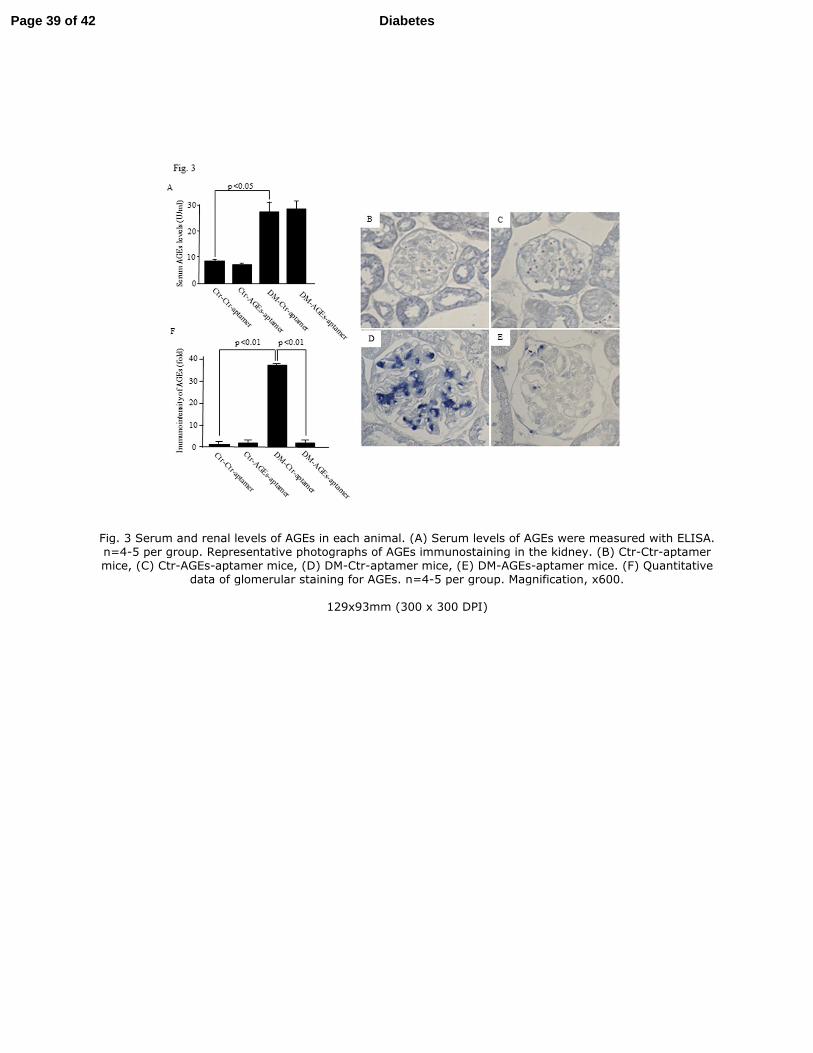
Fig. 3 Serum and renal levels of AGEs in each animal. (A) Serum levels of AGEs were measured with ELISA. n=4-5 per group. Representative photographs of AGEs immunostaining in the kidney. (B) Ctr-Ctr-aptamer mice, (C) Ctr-AGEs-aptamer mice, (D) DM-Ctr-aptamer mice, (E) DM-AGEs-aptamer mice. (F) Quantitative
data of glomerular staining for AGEs. n=4-5 per group. Magnification, x600.
129x93mm (300 x 300 DPI)
Page 39 of 42 Diabetes

Fig.4 Effect of AGEs-aptamer on UAE levels (µg/g creatinine) in each animal. n=8-10 per group. ** p<0.01 vs Ctr-Ctr-aptamer mice. ## p<0.01 vs DM-Ctr-aptamer mice. Open triangle; Ctr-Ctr-aptamer, open
square; Ctr-AGEs-aptamer, open circle; DM-Ctr-aptamer, closed circle; DM-AGEs-aptamer.
88x88mm (600 x 600 DPI)
Page 40 of 42Diabetes

For Peer Review O
nly
Fig. 5 Representative photographs of glomerular hypertrophy. Glomerular hypertrophy was evaluated by measuring glomerular area of cross section in the distal cortex. (A) Ctr-Ctr-aptamer mice, (B) Ctr-AGEs-aptamer mice, (C) DM-Ctr-aptamer mice, (D) DM-AGEs-aptamer mice. (E) Quantitative data of glomerular
area. n=4-5 per group. Magnification, x600. Effect of AGEs-aptamer on glomerular ECM accumulation in each animal. Glomerular ECM accumulation was evaluated by the intensity of Masson’s trichrome staining in the glomeruli. Representative photographs of the kidney in (F) Ctr-Ctr-aptamer mice, (G) Ctr-AGEs-aptamer mice, (H) DM-Ctr-aptamer mice, (I) DM-AGEs-aptamer mice. (J) Quantitative data of ECM accumulation.
n=3-5 per group. Synaptopodin levels in the glomeruli. Representative photographs of the kidney in (K) Ctr-Ctr-aptamer mice, (L) Ctr-AGEs-aptamer mice, (M) DM-Ctr-aptamer mice, (N) DM-AGEs-aptamer mice. (O) Quantitative data of synaptopodin expression. n=3-5 per group. Magnification, x600. (P) Effect of AGEs-
aptamer on urinary 8-OHdG levels (ng/day) in each animal group. Urinary 8-OHdG levels were measured by ELISA. n=8-11 per group.
Page 41 of 42 Diabetes

250x347mm (300 x 300 DPI)
Page 42 of 42Diabetes

Fig. 6 Effect of AGEs-aptamer on cortical (A) MCP-1, (B) TNF-α, (C) CTGF, (D) type-IV collagen, (E) RAGE and (F) AGE-R3 gene expression in each animal group. Total RNAs were transcribed and amplified by real-time PCR. Data were normalized by the intensity of 18S rRNA-derived signals and then related to the value
obtained with Ctr-Ctr-aptamer mice. n=10-16 per group.
99x55mm (600 x 600 DPI)
Page 43 of 42 Diabetes

Fig. 7 Effect of AGEs-atamer on (A) ROS generation and (B) RAGE, (C) MCP-1 and (D) CTGF gene expression in human mesangial cells. Mesangial cells were treated with 100 µg/ml AGEs-BSA or non-glycatd BSA for 4 and 24 hr in the presence or absence of 0.2 or 2 µM AGEs-aptamer. Total RNAs were transcribed
and amplified by real-time PCR. Data were normalized by the intensity of GAPDH mRNA-derived signals and then related to the value obtained with non-glycated BSA. n=3 per group.
179x179mm (600 x 600 DPI)
Page 44 of 42Diabetes