dna damage and repair in oncology
DESCRIPTION
DNA is the only macromolecule in the cell that is repaired when damaged. This demonstrates the vital importance of genome integrity to cell function and viability. Mammals have five major DNA repair mechanisms required for genome maintenance: nucleotide excision repair of bulky monoadducts, double-strand break repair, DNA interstrand crosslink repair, mismatch repair, base excision repair for endogenous oxidative, alkylation lesions and single-strand breaks. Defects in any of these pathways leads to increased DNA damage accumulation and increased risk of mutation and/or chromosomal aberrations. Inherited mutations in genes whose products are required for many of these DNA repair mechanisms cause cancer predisposition syndromes, for example xeroderma pigmentosum and hereditary nonpolyposis colorectal carcinoma. In addition, many chemotherapeutic agents used to treat cancer are genotoxic agents. This leads to the hypothesis that DNA repair genes/proteins could be used as biomarkers to predict cancer risk and tumor resistance to chemotherapy.TRANSCRIPT

DNA damage and repair in oncology
Laura J. Niedernhofer, M.D., Ph.D. Associate Professor
Department of Metabolism & Aging The Scripps Research Institute, Florida
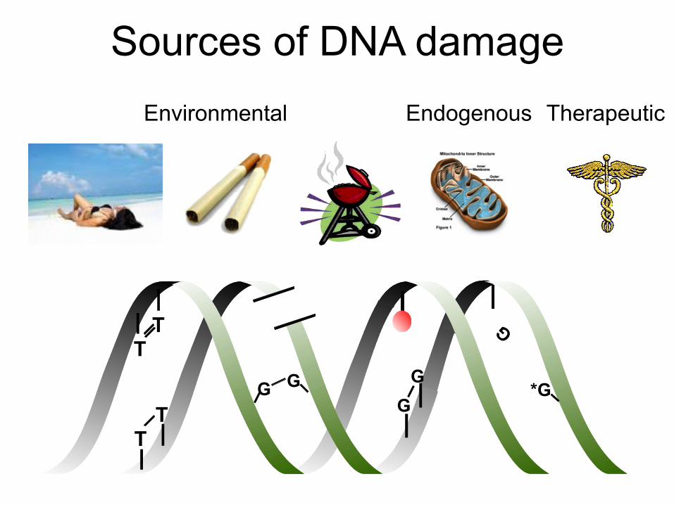
Sources of DNA damage
G G T
T
T T
G *G
G
Environmental Endogenous Therapeutic

T
A repair mechanism for each type of DNA damage
Bulky base Interstrand Small base Double-strand changes crosslinks changes breaks
T -OH!
-CH3!
Nucleotide Excision Repair
Interstrand Crosslink
Repair
Base Excision Repair
Homologous Recombination
& NHEJ

Rationale for measuring DNA repair capacity in the oncology
setting
Less DNA repair: - greater risk of cancer - respond to therapy
More DNA repair: - less risk of cancer - resistant to therapy
Look for SNPs in DNA repair genes that correlate with cancer risk
Measure DNA repair protein expression in tumors
Limitations: • Don’t know rate limiting step of repair • Expression typically regulated by PTM
• Function of SNPs not evaluated

Repair
Pathway:
Nucleotide excision
repair
Double-strand break
repair
Single-strand break repair
Base excision
repair
Interstrand crosslink
repair
Linked to cancer?
Evidence Humans Mice
Humans Mice SNPs
SNPs
Humans Mice
Lesions driving cancer
UV Induced DSBs in immune cells
ionizing radiation; topoisomerase-induced lesions;
BER repair intermediates
Hydrolyzed bases
? ICLs from LPO
or AP sites
Evidence to support this rationale

Rationale for targeting DNA repair mechanisms in oncology
• Synthetic lethality
Tumor Genome instability → evolu7on of selec7ve growth advantage = defec7ve in DNA repair
Inhibit 2nd DNA repair pathway
Selec7ve killing of tumor cells
Priori)es: measure DNA repair factors develop inhibitors of DNA repair

Model for exploiting tumor vulnerabilities
G0-G1 S G2 M
NHEJ, NER, BER
Tumor cell
Cel
l cyc
le
Somatic cell
Check points
Inhibitor Genotoxin Inhibitor Inhibitor or Genotoxin
X HR TOP, ICL-R

DNA lesions #1 pathway
#2 pathway
#3 pathway
How to induce more lesions
Small base lesions or single-strand breaks BER HR Temozolamide
Bulky base lesion or intrastrand crosslink NER HR Cisplatin
Nitrogen mustards Top1-induced single-strand breaks TDP1 ERCC1-
XPF HR Camptothecin
Top2-induced single-strand breaks TDP2 (FEN1) HR Etoposide
Replication stress HR Hydroxyurea 5-FU
Interstrand crosslinks ICL-R + HR NER Melphalan
Cisplatin Double-strand break NHEJ HR Radiation
Genotoxic cancer therapies and DNA repair pathways that cope
X
Inhibit = PARPi Add genotoxin
X
X Inhibit Add genotoxin
X X

Common steps of DNA repair 1) Recognize the DNA damage 2) Remove the damage (nuclease)
3) Replace coding information (polymerase) 4) Restore the integrity of the phosphate backbone (ligase)
Enzymes more “druggable” Often essential

Nucleotide Excision Repair
TFIIH XPB XPD
XPG
23B
XPB XPD TFIIH
XPG
TFIIH XPB XPD RPA
XPG
RNA polII CSB
CSA
23B XPC DDB1 XPE
Recognize
RFC pol d, e
PCNA LIG I LIG III
XRCC3 or
Replace &
Restore
RPA TFIIH
XPB XPD
XPG XPF ERCC1
Remove
Good drug targets to block NER
Good biomarkers
Have inhibitors
X
X
X

MUS81 EME1
FANCQ/XPF
ERCC1
FANCP/SLX4
SLX1
ATR
FANCD2
P FANCI
P
Ub
Ub
FANCM
FAAP24
FANCL
E C G
A F FANCB
MHF
FA CORE
FAAP20
NBS1 MRE11
RAD50
P
CtIP
FANCM
FAAP24
CORE
FANCI
MHF FANCD2
BLM
TOPOIIIa Blap75
FAN1
FANCM
FAAP24
MHF
P A
R
FANCJ/ BRIP1
BRCA1
FANCD1/ BRCA2
RAD51 FANCO/ RAD51C
FANCN/ PALB2
HR Machinery
η Rev1 ζ
Interstrand CrossLink Repair
Good drug targets to block ICL-R
Recognize
Remove
Replace & Restore Have inhibitors
gH2AX Good biomarkers

Base excision &
single-strand repair
*Essential, but partial rescue in p53-null
Recognize Remove
Replace
Restore
Have inhibitors
Good drug targets to block BER
Good biomarkers

Double- strand break repair Homologous Recombination (HR)
Non-homologous end-joining (NHEJ)
Have inhibitors
Good drug targets to block HR
Good biomarkers

Topoisomerases
Good drug targets
Good biomarkers
Have inhibitors

What you would like to have before embarking on a clinical trial:
• Knowledge about DNA repair mechanisms: pathways, redundancy, compensation
(Call me maybe?) • Validated tools: tested with positive and
negative controls. • More information about your target: normal
levels of expression, regulation, rate-limiting, inducible, redundancy, compensation.

ERCC1-XPF is a structure-specific endonuclease
5’ 3’
Nucleotide excision repair
5’ 3’
Homologous recombination
5’ 3’
End-joining of DSBs
5’ 3’
ICL repair Liver

The best chemotherapeutic agents crosslink DNA
Fig. 2.29 DNA Repair and Mutagenesis, 2nd ed.

All crosslinking agents cause a mixture of DNA lesions
1%
NER NER
NER ICL-R
Fig. 2.32 DNA Repair and Mutatgenesis, 2nd ed.

ERCC1-XPF is the only enzyme required for both NER & ICL repair
and therefore it is the only protein complex required to remove
all DNA damage cause by crosslinking agents

Significant correlation between ERCC1 and XPF expression
=> either ERCC1 or XPF can be measured and predict DNA repair
Cor
rect
ed p
rote
in le
vel f
rom
imm
unob
lot
Ovarian tumors
XPF
ERCC1

ERCC1 expression is not transcriptionally regulated
0
2
4
6
8
10
12
TP05
-939
TP06
-602
TP06
-392
TP04
-961
TP07
-001
TP07
-135
TP06
-083
TP03
-426
TP06
-252
TP06
-106
1
TP03
-376
TP99
-045
TP05
-822
TP05
-244
TP07
-363
TP07
-470
TP06
-938
TP07
-397
TP04
-115
TP05
-322
TP06
-096
TP06
-477
TP05
-022
TP06
-674
TP04
-195
ovarian tumors
prot
ein
ban
d in
ten
sity
0.0
1.0
2.0
3.0
4.0
5.0
6.0
7.0
8.0
mR
NA
leve
ls
ERCC1/Actin ERCC1 mRNA

Rate limiting?
DNA Repair (Amst). 2006 May 10;5(5):641-8.
XPA protein as a limiting factor for nucleotide excision repair and UV sensitivity in human cells.
Koberle B, Roginskaya V, Wood RD.
UPCI and Department of Pharmacology, University of Pittsburgh, Hillman Cancer Center, Research Pavilion, Suite 2.6, 5117 Centre Avenue, Pittsburgh, PA 15213-1863, USA.
“These data indicate that XPA levels must be reduced to <10% of that present in a normal cell to render XPA a limiting factor for NER and consequent cellular sensitivity. To inhibit NER, it may be more effective to interfere with XPA protein function, rather than reducing XPA protein levels.”

Immunoblots to check specificity of antibody 8F1 for ERCC1
XP
F nu
ll
ER
CC
1 nu
ll
Nor
mal
XP
F nu
ll
ER
CC
1 nu
ll
Nor
mal
XP
F nu
ll
Nor
mal

4H4
8F1
Normal ERCC1-XPF deficient
8F1 cannot distinguish between normal and ERCC1-deficient cells by IHC

Phosphocholine cytidylyltransferase-α (PCTY1 or CCTa)

CCTa (PCYT1a)
• Ubiquitously expressed in all nucleated mammalian cells.
• Catalyzes the rate-limiting step in phosphatidylcholine (PC) biosynthesis.
• PC mass doubled in late S phase. • Regulated by hRAS, TNF and SP1. • Expression induced by growth factors,
proliferation stimuli, and S phase.
Banehoi et al 2003 J Biol Chem 278(34): 32457

Cheryl Clauson U Pittsburgh
Chelsea Feldman Duke
Siobhan Gregg Rockefeller
Advaitha Madireddy Albert Einstein
Vaishali Patil Luigi Aurelio Nasto Catholic University Rome
Nikhil Bhagwat UC Davis
Andria Robinson U Pittsburgh
Tania Rozgaja Scripps
Diana Navarro Scripps
Sara McGowan Scripps
Acknowledgements
Alec Vaezi U Mass