do 14-3-3 proteins interact with the f-box stress induced
TRANSCRIPT

Do 14-3-3 Proteins Interact with the F-box Stress Induced Protein
Family in Arabidopsis thaliana?
by
Ryan Apathy
This thesis is submitted to the faculty of the Biology Department
of the University of Puget Sound in partial fulfillment of the requirements
for the Coolidge Otis Chapman Honors Program
April 16, 2018
Approved by:
Dr. Bryan Thines
Thesis Advisor
Dr. Leslie Saucedo
Reader
Dr. Greg Johnson
Reader

2
Table of Contents
Abstract ......................................................................................................................................................... 3
Introduction ................................................................................................................................................... 4
The Ubiquitin 26S Proteasome System .................................................................................................... 4
Figure 1. Ubiquitin 26S Proteasome System ........................................................................................ 6
Figure 2. Structure of the SCF complex ............................................................................................... 7
Figure 3. FBX protein domain architecture .......................................................................................... 9
14-3-3 Proteins ........................................................................................................................................ 11
Figure 4. Functional roles of 14-3-3 proteins...................................................................................... 11
Figure 5. Phylogenic classification of 14-3-3 proteins ....................................................................... 12
14-3-3 Proteins and the UPS ................................................................................................................... 12
Figure 6. FBS1 and 14-3-3 proteins interact via BiFC in N. benthamiana ......................................... 13
Methods ...................................................................................................................................................... 15
Gene amplification .................................................................................................................................. 15
Cloning .................................................................................................................................................... 16
Subcloning and Y2H binary vectors ....................................................................................................... 16
Yeast strains and transformations ........................................................................................................... 17
Results ......................................................................................................................................................... 18
Figure 7. FBS1-4 multiple sequence alignment .................................................................................. 19
Figure 8. Schematic representation of Yeast 2-Hybrid constructs ...................................................... 19
Table 1. FBS Gal4 BD construct status .............................................................................................. 20
Table 2. 14-3-3 Gal4 AD construct status ........................................................................................... 20
Figure 9. Yeast 2-Hybrid results. ........................................................................................................ 21
Figure 10. FBS1 Gal4 BD yeast colony PCR ..................................................................................... 22
Discussion ................................................................................................................................................... 22
Figure 11. Multiple sequence alignment of No-0 and Col-0 FBS1 .................................................... 24
Acknowledgements ..................................................................................................................................... 26
References ................................................................................................................................................... 28
Supplemental Information .......................................................................................................................... 34
SI A. Primer sequences and restriction sites ....................................................................................... 34
SI B. Cloning and binary Y2H vector maps ....................................................................................... 35

3
Abstract
One of the most important abilities that eukaryotic cells possess is the capacity to
undergo intracellular change. Two families of regulatory protein that function to facilitate this
change in the model plant Arabidopsis thaliana are F-box (FBX) and 14-3-3 proteins. FBX
proteins serve as variable substrate adaptors for the Ubiquitin 26S Proteasome System (UPS),
which selectively tags proteins with ubiquitin (Ub) for degradation by the 26S proteasome. FBX
proteins, of which >700 have been identified and classified in Arabidopsis, provide the SCF
complex (SKP, Cullin, F-box) with extreme specificity in its protein degradation capabilities. 14-
3-3s subunits bind to each other to form homo- or heterodimers to bind phosphorylated residues
in targets to regulate subcellular localization, inhibit protein interactions, or act as a scaffold to
facilitate protein interactions. 14-3-3 proteins interact with the Ubiquitin 26S Proteasome System
in only a select few instances, including with F-box Stress Induced 1 (FBS1). However, both the
extent and biological consequences of this interaction are unknown. We hypothesize that FBS1
and closely related proteins FBS2-4 interact with non-epsilon 14-3-3 proteins, a plant-specific
subset of 14-3-3 isoforms. A Yeast 2-Hybrid assay is developed to assess the interactions
between FBS1 and non-ε 14-3-3 φ, ψ, λ, κ, and ω protein constructs. Full length and amino-
termini deletions (ΔNT) protein constructs are being developed to probe the degree of interaction
between these two protein families. This ongoing research examines the extent of this 14-3-3-
FBS1 interaction as well as the broader regulatory mechanisms and implications.

4
Introduction
As sessile organisms, plants have evolved to possess powerful mechanisms for responding to
biotic and abiotic stress conditions (1–3). Each form of stress, from pathogenic invasions (4) to
drought and cold (5) initiates a large-scale transcriptome response necessary for cellular survival.
For eukaryotic species, stress responses are predominately regulated on the molecular level, as
environmental conditions trigger an extensive array of stress-induced pathways (6). Often these
induced responses regulate proteins that are critical to the maintenance of cellular homeostasis.
One such example is the regulation of proteins called transcription factors (TFs), which bind to a
DNA sequence upstream of tens to thousands of genes to control genetic transcription rates (7).
The regulation of TFs can have a large impact on the activity and abundance of many
downstream genetic processes (8). Stress-induced molecular responses can also regulate rate-
limiting enzymes in critical cellular processes (e.g. cell division), resulting in the decreased
function of those respective pathways and overall metabolic processes (9). This system of
intracellular protein regulation is largely controlled by the ubiquitin 26S proteasome system
(UPS), a complex and highly organized system of protein degradation (6, 10–12).
The Ubiquitin 26S Proteasome System
The UPS facilitates selective protein degradation, or proteolysis. This system participates in
regulating a diverse array of essential cellular functions, including cell division and development
(11, 12), DNA replication (13), hormone signaling (14), circadian clock regulation (15, 16), and
disease response (17–19), as well as general cellular homeostasis (11, 12, 20). So critical is
proper UPS function that malfunctions in the system have been associated with numerous human
diseases, including neurodegenerative and age-related disorders such as Alzheimer's,

5
Parkinson’s, and Huntington's disease (21), stroke (22), diabetic nephropathy (23), multiple
myeloma (24), and progeroid syndromes (25). The UPS has also been implicated in cancer, with
roles in tumor suppression (26), carcinogenesis (27), as well as in the diagnosis and treatment of
multiple forms of cancer (28–33). The role of the UPS is critical to cellular survival and the
coordination of each component of the system is essential for its proper function. In Arabidopsis
thaliana, a model plant, this system involves the concerted function and regulation of nearly
1,400 proteins, or more than 5% of its entire genome (10, 12).
Selective proteolysis is accomplished by tagging target proteins with ubiquitin (Ub), a 76-
amino acid peptide that is absolutely conserved among higher order plants and vertebrates (34).
Even among animals, plants, and fungi, Ub sequences only differ by two or three residues (34).
Ub tagging and subsequent proteolysis is accomplished through a sequential enzyme cascade via
E1, E2, and E3 enzymes, respectively, followed by degradation by the 26S proteasome (Fig. 1)
(10, 35). This cascade begins with the activation of Ub by the E1 Ub-activating enzyme (34) and
the transfer of this activated Ub to the E2 Ub-conjugating enzyme (34). Ub is then delivered to
the E3 Ub ligase enzyme complex which is bound to the protein targeted for degradation (39). In
the final step, the E3 Ub ligase acts as a bridge between the E2 conjugating enzyme and the
substrate and covalently binds the activated Ub to the target protein (34, 36). This ubiquitination
cascade occurs many times, resulting in the construction of a polyUb chain on the target protein
(35). A chain length of four Ubs through Lys48 is considered the minimum chain length for the
protein to be degraded by the 26S proteasome (35). The 26S proteasome, a ~65 subunit, 2.1 MDa
compex, degrades proteins by disrupting peptide bonds and recycles the digested amino acids
and Ub for further use (34–36).

6
Figure 1. Ubiquitin 26S Proteasome System. Ubiquitin (Ub) molecules are activated, conjugated
and covalently linked to the target substrate by E1, E2 and E3 enzymes, respectively. Proteins are
targeted for degradation through polyubiquitylation via the E3 Ub-ligase enzyme component.
Following polyubiquitylation, proteins are degraded by the 26S proteasome. Ub and substrate
amino acids are recycled for further use in the cell (Figure adapted from Rahimi, 2012).
Protein target selectivity for the UPS to conferred by the E3 Ub ligases enzyme complex.
There exist three primary E3 ligase families, each defined by their protein components and by
their mode of Ub transfer (37). These families include complexes with the motifs Really
Interesting New Gene (RING) (38), RING-between-RING (RBR) (39), and homologous to the
E6AP carboxyl terminus (HECT) (40). RING E3s are the largest family of Ub ligases and are
characterized by the presence of either a zinc-binding RING motif or a RING-like U-box motif
(41). Several RING E3s exist, including mono-, homo-, and heterodimeric RINGs, mono- and
homodimeric U-boxes, and Cullin-RING Ligases (CRLs) (38, 41). Regardless of their E3 family
classification, all E3 complexes are modular structures composed of several more specific
protein members. The modular character of E3 ligases, combined with the size of each
Protein
peptides

7
component protein family suggests that a vast number of putative E3s that can be synthesized in
vivo, demonstrating the necessity for these complexes to perform highly regulated and specific
tasks (46).
The SKP-Cullin-F-box (SCF) complex is the
largest E3 ligase subfamily within CRLs (20). Target
specificity is provided to SCFs via variable F-box
(FBX) proteins, which are defined by the presence of
conserved F-box domains (Fig. 2) (20, 43). The F-
box domains connect the FBX protein to the SCF
complex by binding one of 21 different Arabidopsis
SKP-like proteins in the core complex (ASKs) (44).
Unique domains in the FBX amino-terminus (NT)
bind to target substrates and initiate the
ubiquitination process (42). In Arabidopsis, nearly
700 putative FBX proteins have been identified and
classified based on their unique domain architecture
(42). This large number of FBX proteins is common
among other plants and is an order of magnitude
larger than the number of FBX proteins found in
humans, yeast, and Drosophila, to name a few (42).
The fact that so many SCFFBX combinations can be
constructed in Arabidopsis suggests that SCFs
provide the UPS with extraordinary specificity in their regulatory capabilities.
Figure 2. Structure of the SCF
complex. The SCF-type E3 Ubiquitin
ligase is comprised of SKP, Cullin and
F-box proteins. R-box (RBX) proteins
bind the Ub-loaded E2 ligase and
transfer Ub to the target protein.
Generally, FBX proteins interact with
SKP proteins via an F-Box domain in
the amino-terminus and with target
substrates via interaction domains in
the carboxy-terminus. The ribbon
model (A) and 3-dimentional depiction
(B) of the SCF complex demonstrate
its modular structure (Figure adapted
from Hua and Vierstra, 2011).

8
The FBX protein superfamily is tremendously diverse in function and domain composition.
Most FBX proteins contain a ~60-aa amino-terminal F-box motif that is responsible for
interacting with the core SCF complex by binding to a SKP subunit (42). Several different
carboxy-terminal domains have been identified in Arabidopsis FBX proteins that interact with
variable targets (Fig. 3A). FBX proteins most commonly contain either leucine-rich repeats
(LRRs) and Kelch domains (42). LRRs are small (~20-aa) motifs that contain positionally
conserved leucine residues (45). Multiple LRRs associate with one another to create a curved
docking structure for target substrates (45). Canonical Arabidopsis FBX proteins Transport
Inhibitor Response-1 (TIR1) and Coronatine Insensitive-1 (COI1) both contain LRRs in their
substrate recognition C-termini. Of the ~700 identified F-box proteins, 202 contain LRRs or a
plant-specific LRR derivative (42). Another 100 FBX proteins contain Kelch domain repeats, in
which β-sheets assemble to create a substrate-binding β-propeller tertiary structure (42, 46).
Additional substrate recognition F-box domains include WD-40, Armadillo, and tetratricopeptide
repeats, as well as Tub, actin, DEAD-like helicase and jumonji C domains (42). Such variability
in target recognition capabilities allow single FBX protein targets to range from single other
proteins (15) to entire protein families (14, 47).
This characteristic domain architecture is not found in all FBX proteins. One such example
of proteins with noncanonical architectures is the F-box Stress Induced family (FBS1-4). Much
about this protein family is unknown and their domain structure differs from that of other well-
classified FBX proteins, including COI1 and TIR1 (Fig. 3A, B). Like other FBX proteins, the
FBS family interacts with targets in via carboxy-terminus. Unlike other FBX proteins, however,
the F-box domain in appears in the center of the gene, rather than the amino-terminus (48). This
F-box domain architecture is not the only of its kind; three additional F-box proteins exist with

9
N-terminal domains, including LKP2, ZTL, and FKF1 (15, 16, 49). The N-terminal domain
(PAS/LOV) in these proteins is hypothesized to function as flavin-containing photoreceptors, as
all three proteins are required for regulating flowering and circadian rhythm (42). The
significance of this N-terminal domain in FBS1-4 domain architecture has yet to be elucidated.
Figure 3. FBX protein domain architecture. (A) The domain architecture of all 694 FBX
proteins, listed in order from most common to least. In general, the F-box domain occurs in the
protein N-term. and the substrate interaction domain in the C-term. (B) The domain architecture
of FBS1 differs from others in the F-box superfamily as the F-box domain is found in the protein
center (Figure adapted from Gagne, 2012).
As its name indicates, FBS1 is implicated in Arabidopsis abiotic stress response (44). FBS1
mRNA accumulates in response to several stress factors, including general wounding and NaCl
(50). Additionally, FBS1 mRNA accumulates in the presence of growth regulators such as
10
jasmonic acid (JA), ethylene biosynthesis precursor ACC, salicyclic acid (SA), and absiscic acid
(ABA) (50). FBS1 expression levels were shown to increase in response to cold and drought
stress (51). Additionally, fbs1-1 knock-out lines reveal that FBS1 is required for elevation of JA
genes, and a more complicated expression profile was observed for ABA genes (51). This
correlation between abiotic stress and FBS1 mRNA accumulation suggests that FBS1
participates in plant stress response. Much less is known about the biological function of the rest
of the FBS family, FBS2-4. These proteins are similar in their domain architecture to FBS1 and
share a high degree of homology in their N-, F-Box and C-terminal domains. Little is known
about the biological function of FBS2-4 as their specific roles have not been the subject of much
research.
Since little is known of the FBS family regarding their biological function, these proteins can
be used as a unique tool for investigating modes of UPS regulation. Similar investigations of
UPS regulation and interactions with additional regulatory protein families in humans has led to
the increased understanding and treatment of a variety of diseases (52–54) and additional
research can provide novel information regarding native UPS function. One clue for
understanding the biological function of the FBS family is provided by the observation of an
unique interaction between FBS1 and 14-3-3 proteins, another well-studied family of regulatory
proteins in Arabidopsis (48, 50). 14-3-3 proteins are highly conserved among eukaryotes and
perform extremely diverse regulatory roles, though very little is known about the significance of
this novel interaction.

11
14-3-3 Proteins
14-3-3 proteins facilitate and promote intracellular change by forming homo- or heterodimers
and binding client substrates to regulate post-translational activity (55–57). Such dimers function
in four primary categories: they directly interact with clients to alter their structure and modify
that client’s activity (58); they act as a scaffold to facilitate protein-protein interactions (59); they
physically occlude clients, preventing interactions from occurring (60); and promoting the
subcellular localization of clients (Fig. 4) (61–63). Examples of biochemical regulation include
14-3-3 protein association with the Ras-Raf signaling pathway, cell death agonist BAD, and
hypothesized localization of cell cycle mediator cdc25 (60).
Figure 4. Functional roles of 14-
3-3 proteins. 14-3-3 monomers
dimerize and modify a broad range
of targets. Their function ranges
from modulating the structure of
proteins to increase or decrease
their efficiency, inhibit of promote
the interaction of two proteins, and
increase or decrease proteins
stability (Figure adapted from
Obsil and Obsilova, 2011).
In Arabidopsis, 14-3-3 proteins isoforms are divided into two phylogenic groups: epsilon,
which contains epsilon (ε), iota (ι), mu (μ), omicron (ο), and pi (π) isoforms; and non-epsilon,
which contains lambda (λ), upsilon (υ), omega (ω), kappa (κ), phi (φ), psi (ψ), chi (χ), and nu (ν)

12
isoforms (Fig. 5) (64). Epsilon 14-3-3s are located across humans and other eukaryotes, while
non-epsilon 14-3-3s are absent from the human genome (64). Further phylogenic classification
within non-epsilon and epsilon families is accomplished through intron and exon composition
(64). Additionally, the subcellular localization of each 14-3-3 protein can be used to provide
further classification: χ, ν, κ, and λ are active within the chloroplast; λ, ω, κ, and φ in the nucleus;
and λ in the vacuolar membrane, among other distinct locations (50). Regardless of epsilon or
non-epsilon designation, isoforms all share a high degree of homology, particularly within client
interaction domains.
Figure 5. Phylogenic
classification of 14-3-3 proteins.
Thirteen 14-3-3 isoforms are
expressed in Arabidopsis
thaliana. Epsilon and non-epsilon
classification is determined by the
number of exons and introns
within a given protein. Further
classification is provided by the
subcellular localization of
individual proteins (Figure
adapted from DeLille et al., 2001).
14-3-3 Proteins and the UPS
14-3-3 proteins interact with and regulate the UPS in a variety of ways. Recently, F-box
proteins AtSKIP18 and AtSKIP31 were shown to degrade 14-3-3 proteins in Arabidopsis to
regulate primary root growth in nitrogen deficiency (65). 14-3-3s are also degraded by ubiquitin

13
ligases ATL31 and ATL6 to control signaling for carbon and nitrogen metabolisms and C/N
balance response (66). In humans, the treatment of cells with the proteasome inhibitor MG132
caused a downregulation of 14-3-3 isoforms ε and ϴ/τ, with implications in cancer development
and treatment (67). More relevantly, FBS1 directly interacts with non-epsilon 14-3-3 proteins λ,
κ, ω, φ and ψ, though the exact biological significance of this interaction is unknown (48).
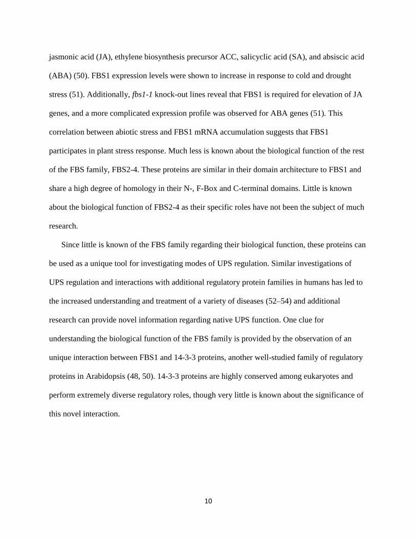
Bimolecular Fluorescence Complementation results verify an interaction between FBS1 and 14-
3-3 κ and ψ (Fig. 6). This research serves to probe the extent and relevance of the interaction
between FBS1 and 14-3-3 non-epsilon isoforms.
Figure 6. FBS1 and 14-3-3 proteins interact via BiFC in N. benthamiana. Confocal imaging of
(A) YFPn-FBS1 and YFPc-14-3-3 and (B) YFPn-FBS1 and YFPc-14-3-3 . Images were taken
with a 10x objective and 512x512 pixel resolution (Figure adapted from M. Bacher, unpublished).
14-3-3s interact with phosphorylated residues contained in canonical recognition motifs in
target proteins. One such recognition motif is the amphipathic grooves that result from conserved
A B

14
amino acid sequences in protein targets (62). Such conserved sites include RxRSxpSx, Rx1-2Sx2-
3S, and related derivatives (68, 69). FBS1 was not found to contain any such derivatives (48).
However, 14-3-3 proteins ζ and ε interact with the KATP channel α subunit, Kir6.2, in way that is
differs significantly from the amphipathic groves described above. 14-3-3 proteins ζ and ε
interact with Kir6.2 via a unique RKR arginine-based endoplasmic reticulum (ER) localization
signal in the protein C-terminal tail (70). An identical tri-basic motif exists in the N-terminal tails
of FBS1-4. This motif thus provides a putative means in which non-epsilon F-box proteins
interact with 14-3-3s (48).
One function of this observed interaction could be the FBS1-medicated degradation of 14-3-3
proteins. However, two primary pieces of evidence suggest that 14-3-3s are not targets of FBS1.
The first piece of evidence is that specific target recognition capabilities for the SCF complex are
conferred via interaction domains contained in the FBS1 carboxy-terminus (48). FBS1 and 14-3-
3s, however, interact via the FBS1 amino-terminus (48). This N-terminal interaction suggests
that FBS1 does not interact with 14-3-3s in the same manner as it does with its target(s).
Previous studies indicate that proteasome inhibitor MG132 was not necessary to visualize the 14-
3-3/FBS1 interaction, demonstrating that 14-3-3s are stable upon interaction with FBS1 (48).
Taken together, these results suggest that 14-3-3s are unlikely to be targeted for ubiquitination by
FBS1.
What is the biological function of the interaction between 14-3-3s and FBS1? Currently,
both the extent of this protein family interaction, as well as the specific interaction mechanism(s)
are unknown. It has previously been hypothesized that 14-3-3s help promote the dimerization of
SCFFBS1 (50). This hypothesis is supported by evidence that demonstrates the 14-3-3ε-mediated
dimerization of SCFFBS4 in various forms of human cancer (71). Additionally, it is possible that

15
14-3-3 proteins facilitate proper subcellular localization of F-box proteins. As mentioned above,
the RKR arginine-based endoplasmic reticulum (ER) localization signal and closely related basic
residue motifs in the C-terminal tail of FBS1-4 could prove necessary for the subcellular
localization of the FBS protein family.
This thesis serves to establish a protocol to probe the interaction between 14-3-3 proteins
and FBS1-4 in Arabidopsis. As such, a Yeast 2-Hybrid (Y2H) test was developed to assess the
extent of this 14-3-3/FBS interaction. If such an interaction is observed, then there might exists a
greater, unidentified regulatory function provided to the UPS via 14-3-3 proteins. The current
gap in knowledge about this interaction prevents us from understanding the role played by 14-3-
3s within the UPS. A broad interaction could suggest that 14-3-3 proteins play an important and
previously unrecognized regulatory role within the UPS. I hypothesize that the F-box Stress
Induced protein family interacts with non-epsilon 14-3-3 proteins in Arabidopsis thaliana.
Methods
Gene amplification
F-box and 14-3-3 genes were amplified from pooled Arabidopsis thaliana ecotype
Nossen (No-0) cDNA with gene-specific primers. Primers were designed using Primer3
(http://bioinfo.ut.ee/ primer3) to include restriction sites to facilitate sub-cloning into Yeast 2-
Hybrid (Y2H) binary vectors. Gene constructs were amplified via polymerase chain reaction
(PCR) with Phusion High Fidelity Polymerase (ThermoFisher) and were separated on an 1.5%
weight/volume (w/v) agarose gel. DNA fragments were excised and purified using QIAquick
Gel Extraction Kit for subsequent use according to manufacturer’s guidelines (Qiagen). Detailed
primer and restriction site information can be found in Supplemental Information A.

16
Cloning
Amplified gene constructs were A-tailed with GoTaq DNA Polymerase (Promega) at 72
°C for 30 min. and ligated into cloning vector pGEM-T with T4 DNA Ligase overnight at 4 °C,
according to manufacturer protocol (Promega). Following ligation, plasmids were transformed
into One Shot™ Top10 chemically competent E. coli cells according to manufacturer guidelines
(ThermoFisher) and were then plated on Lysogeny broth (LB) media containing carbenicillin
(100 μg/mL), X-Gal (50 μg/mL), and IPTG (1mM) and incubated overnight at 37 °C. White
colonies indicating insertion of product were selected and cultured in LB overnight at 37 °C for
plasmid isolation. Plasmids were prepared using QIAprep Spin Miniprep Kit according to
manufacture guidelines (Qiagen) and sequenced using T4 and SP6 forward and reverse primers
by EuroFins Genomics and mapped to reference sequences with Geneious software. CDS
reference sequences were obtained from the Arabidopsis Information Resource (TAIR). Detailed
plasmid maps can be found in Supplemental Information B.
Subcloning and Y2H binary vectors
Yeast 2-Hybrid vectors pGADT7-Rec and pGBKT7 were linearized with appropriate
restriction enzymes at 37 °C, 1hr. Restriction digest products were separated on 1.5% w/v
agarose gel. Gene constructs and linearized Y2H plasmids were extracted via QIAquick Gel
Extraction Kit according to manufacturer’s protocol (Qiagen). Digested products and plasmids
were mixed at a 3:1 molar ratio with T4 DNA Ligase at 16 °C, 24 hrs. Ligation reactions were
inactivated at 65 °C for 20 min. DNA (2 μL) was transformed into One Shot™ Top10
chemically competent E. coli cells. Cells were plated on LB media and grown at 37 °C, 12 hrs.

17
Vector-specific antibiotics (kanamycin or carbenicillin) were used to select transformed DNA.
Colonies were cultured in LB at 37 °C, 12 hrs for plasmid isolation. Detailed information
regarding vector sequences and restriction enzymes can be found in Supplementary Information
A and B.
Yeast strains and transformations
Saccharomyces cerevisiae yeast strains Y187 (genotype MATα, ura3-52, his3-200, ade2-
101, trp1-901, leu2-3, 112, gal4Δ, met–, gal80Δ, MEL1, URA3::GAL1UAS -GAL1TATA-lacZ) and
Y2HGold (genotype MATa, trp1-901, leu2-3, 112, ura3-52, his3-200, gal4Δ, gal80Δ,
LYS2::GAL1UAS–Gal1TATA–His3, GAL2UAS–Gal2TATA–Ade2 URA3::MEL1UAS–Mel1TATA AUR1-C
MEL1) were utilized as complementary mating pairs (Clontech). DNA (1 μg) was transformed
into yeast cells (50 μL) with 50% w/v polyethylene glycol (PEG), 1.0 M LiAc, and 2.0 mg/mL
single-stranded carrier DNA (salmon sperm) and incubated in a 42 °C water bath, 20-180 min.
Cells were resuspended in 400 μL H2O and plated on appropriate yeast extract, peptone, adenine,
and glucose (YPAD) media containing 1% w/v yeast extract, 2% w/v polypeptone, and 2% w/v
glucose. Diploid strains were developed by combining opposite yeast mating types in 500 μL
and incubated at 30 °C, 12-16 hrs. Serial dilutions (1/10, 1/100, 1/1000) from OD600 = 1.0 were
spotted on synthetic drop-out (SD) nutrient-minus media at three levels of stringency: double
drop-out (DDO; SD/–Leu/–Trp); triple drop-out (TDO; SD/–Leu/-Trp/–His/); and quadruple
drop-out (QDO; SD/–Leu/–Trp/-His/-Ala). Reporter genes AUR1-C, HIS3, ADE2, and MEL1
were used to assess interaction results.

18
Results
To test the hypothesis that FBS1-4 interact with non-epsilon 14-3-3 proteins in
Arabidopsis, a Yeast 2-Hybrid assay was developed. This hypothesis is informed by the
relatively high degree of homology shared between FBS1 and the remaining three members of
the FBS family (Fig. 7A, B) (72, 73). A multiple sequence alignment reveals a highly conserved
domain in the amino-termini of FBS1-4, Mystery Domain 1 (MD1) (Fig. 7A). We hypothesize
that MD1 interacts with 14-3-3 proteins. Within MD1 exists a tri-basic motif that is conserved
among all four FBS proteins (Fig. 7B). This RKR motif, or KKR in FBS4, serves as a putative
14-3-3 interaction domain. An interaction between FBS1 and non-epsilon 14-3-3 proteins exists
in Arabidopsis (48), but it is not known if the same 14-3-3 proteins interact with FBS2-4.
A Yeast 2-Hybrid assay was developed to probe the FBS/14-3-3 interaction using full-
length (FL) FBS proteins in combination with FL and amino-terminus deletion (ΔNT) 14-3-3
protein constructs (Fig. 8A, B). To comprehensively assess the extent of this hypothesized
interaction, all FL and ΔNT 14-3-3 constructs are to be tested against FBS1-4. Progress thus far
has been to develop a library of experimental tools and to optimize the Y2H assay for future
work. FBS and 14-3-3 constructs exist at various stages of development (Table 1, 2). The FBS1
Gal4 Binding Domain (BD) construct is confirmed in yeast strain Y2HGold and is thus ready for
Y2H experimentation (Table 1). 14-3-3 Gal4 Activation Domain (AD) ω, ψ, ω ΔNT, and ψ ΔNT
constructs have also been successfully transformed into yeast strain Y187 (Table 2).

19
Figure 7. FBS1-4 multiple sequence alignment. F-box proteins FBS1-4 were aligned. (A) FBS1-
4 amino-termini were aligned. Sequences were truncated at the F-box domain (72). (B) WeboLogo
alignment reveals that a tri-basic motif (RKR or KKR) within MD1 is functionally conserved
across all four FBS proteins. Black = non-polar residue; Red = acidic residue; Blue = basic residue;
letter size corresponds with relative residue frequency (73).
Figure 8. Schematic representation of Yeast 2-Hybrid constructs. (A) FBS and (B) 14-3-3
constructs. Nt = Amino-terminus; F-Box = F-Box domain; Ct = Carboxy-terminus; Gal4 BD =
Gal4 DNA Binding Domain; and Gal4 AD = Gal4 Activation Domain.

20
Table 1. FBS Gal4 BD construct status. FBS Y2H constructs exist at various stages of
development. FBS1 is confirmed in the appropriate yeast strain Y2HGold and is thus ready for
Y2H experimentation. FBS2-4 constructs have yet to be successfully transformed into Y2HGold.
Table 2. 14-3-3 Gal4 AD construct status. 14-3-3 Y2H constructs exist at various stages of
development. 14-3-3 ω, ψ, ω ΔNT, and ψ ΔNT are confirmed in the appropriate yeast strain Y187
and are thus ready for Y2H experimentation. All other 14-3-3 constructs have yet to be successfully
transformed into Y187.

21
Cell growth was observed for all diploid yeast cells on nutrient-minus DDO media,
indicating that all diploid strains contained appropriate reporter genes (Fig. 9, Col. 1). Growth
was observed for diploid yeast cells containing protein interaction positive control pGADT7-T
(SV40 large T-antigen) and pGBKT7-53 (Murine p53) across all nutrient-minus media (DDO,
TDO, and QDO) (Fig. 9, Row A). Negative controls consist of AD or BD experimental
constructs mated with an empty Y2H binary vector containing the opposite Gal4 domain (Fig. 9,
Col. 2 and 3, Rows B and C). Yeast cell growth was not observed for experimental protein
interaction diploid yeast strains containing FBS4 and 14-3-3 κ FL Y2H constructs on nutrient-
minus media (TDO and QDO) (Fig. 9, Col. 2 and 3, Row D).
Figure 9. Yeast 2-Hybrid results. Serial dilutions (1/10, 1/100, 1/1000) were spotted on synthetic
drop-out (SD) media minus Leu (L) and Trp (W), and His (H), or minus L, W, H, and Ala (A).
pGADT7-T (SV40 large T-antigen) and pGBKT7-53 (Murine p53) are used as a positive protein
interaction control (Row A). Negative controls consist of AD or BD experimental constructs mated
with an empty vector containing the opposite Gal4 domain (Rows B and C). Cell growth is not
observed for the FBS4 + 14-3-3 κ FL diploid strains on nutrient-minus media (Col. 2 and 3, Row
D).
To assess the validity of all Y2H experimental results, a yeast colony PCR protocol was
developed. Yeast strains Y2HGold and Y187 were probed for the presence of appropriate

22
constructs both before and after mating (Fig. 10). To assess whether the FBS1 Gal4 BD Y2H
construct was present in both haploid Y2HGold and diploid cells post-mating, yeast plasmids
were used as DNA templates for a PCR amplification reaction using FBS1 BD gene-specific
primers (SI A). Yeast colony PCR of FBS1 BD reveals the presence of the expected construct at
558 base pairs (Bps) in haploid Y2HGold cells (Fig. 10, Lane 1), but not in diploid cells (Fig.
10, Lane 2). This result indicates that the yeast strain did not confer the contained construct to
diploid cells.
Figure 10. FBS1 Gal4 BD yeast colony PCR. PCR amplification using gene-specific primers
suggests that the FBS1 Y2H construct is present in haploid yeast strains (Lane 1) but not in diploid
strains (Lane 2). Expected band size is 558 Bp.
Discussion
The UPS is extremely important in regulating cellular survival, both in response to
various biotic and abiotic stresses and in the maintenance of intracellular homeostasis (6, 11). In
particular, the regulation of the E3 ubiquitin ligase component is critical, as it provides
specificity to the entire UPS (38). As a diverse form of E3 ligases, SCFs receive their specificity
through FBX proteins. As only ~15% of the known Arabidopsis FBX proteins have been
biologically characterized, investigating unknown FBX proteins can help elucidate new forms of
A B A B

23
UPS regulation. Here, I develop methods to investigate the interaction of the F-box Stress
Induced proteins family FBS1-4 and 14-3-3s proteins in Arabidopsis.
FBS1 interacts with 14-3-3 λ, ψ, κ, φ, and ω (48). These results were first observed in
yeast (48) and confirmed in N. benthamiana (M. Bacher, unpublished) (Fig. 6). Curiously, only
the results of the interaction between 14-3-3 λ and FBS1 were described in detail (48). As such,
it is especially important to validate the results reported previously in a similar Y2H system.
However, difficulties in the Y2H experiment have prevented the substantive analysis of the
protein interactions described above. The inability to observe an FBS/14-3-3 interaction thus far
is indicative of problems within our Y2H experimental protocol. This is indicated by the lack of
the FBS1 construct in diploid yeast strains after mating (Fig. 9, Lane 2). Mating experiment will
be repeated under optimized conditions and the presence of desired constructs will be further
assessed. Furthermore, both Gal4 bait and prey hybrid constructs will be swapped to increase the
confidence of any observed interaction. As the interaction was first identified in a Y2H system, it
is expected that an optimized experimental set-up will result in a visualization of the published
protein interactions.
There are several additional reasons that Y2H experimentation has not revealed expected
protein-protein interactions. First, the cDNA pooled for initial PCR amplification of all
constructs originated from the Arabidopsis thaliana Nossen ecotype (No-0). The interaction
previously observed utilized FBS1 from a different Arabidopsis thaliana ecotype, Columbia
(Col-0) cDNA (48). While it is unlikely that the difference between No-0 and Col-0 is enough to
yield a difference in protein interaction in vivo, it is possible that No-0 FBS1 does not interact
with 14-3-3s in the same manner as Col-0 FBS1. A multiple sequence alignment of FBS1 from
No-0 and Col-0 reveals slight difference in residue composition (Fig. 11). In the coding DNA

24
region corresponding to MD1 a single base pair mutation is observed. This change, adenine to
thymine, occurs in the third position of the codon and does not result in a change in the final
amino acid residue at that position. Both ATA in No-0 and ATT in Col-0 code for the amino acid
isoleucine.
Figure 11. Multiple sequence alignment of No-0 and Col-0 FBS1. The alignment of the FBS1
coding DNA sequence from two Arabidopsis thaliana ecotypes, Col-0 and No-0. No-0 FBS1
template cDNA was used for construct amplification for Y2H experimentation. The underlined
region corresponds with MD1. A single base pair mutation occurs in this domain (A to T) but does
not confer a change in final protein residue. Maroon = conserved residues; Red = SNPs (72).

25
It is possible that protein interactions cannot be observed in a Y2H assay. This result
could be due to either improper protein folding as a result of the Gal4 construct hybrid or the
absence of an additional factor required for the protein interaction in vivo. Previous interactions
were reported in yeast between full-length FBS1 and residues 32-248 of 14-3-3 λ (48). It is not
clear why the deletion of the amino-terminus of 14-3-3 λ is required for an interaction to be
observed in yeast (48). Explanations include the possibility that residues 1-32 physically occlude
interaction domain(s), prevent proper 14-3-3 protein folding, or several additional possibilities.
We are developing both full length and amino-termini deletions (ΔNTs) of all non-epsilon 14-3-
3s to understand the role 14-3-3 NTs play in the interaction with FBS1. Additionally, Gal4
construct hybrids can prevent native protein folding and, as such, Y2H assays often require
constructing multiple derivations of test proteins to observe a native interaction. This could
explain why ΔNT constructs were required to visualize the interaction. Furthermore, Y2H
hybrids differ from constructs used to visualize interactions via BiFC, as BiFC utilizes yellow
fluorescent protein rather than the Gal4 transcriptional activation protein (Fig. 6). These
alternative constructs could result in alternative folding and therefore conflicting observations
across systems.
Additionally, a Y2H assay could not contain all the necessary factors required for a
protein interaction to occur. Techniques such as Yeast 3-Hybrid assays function in the same way
as the Y2H assay with the addition of a third protein component required for interaction. A Y3H
assay with a third, unknown protein could be necessary to observe 14-3-3/FBS interactions, but
this is unlikely as evidence suggests that only the two protein families in question are required
for the interaction to occur (48). Protein interactions that require native biological conditions

26
may not be visualized in a Y2H assay but can be observed via BiFC. If specific biological
conditions and/or factors are required to facilitate a protein interaction, those factors might be
absent in the in vitro system of yeast but are present in heterologous systems such as N.
benthamiana. We anticipate the Y2H assay to yield meaningful results, however, as this system
was used to demonstrate the existence of the interaction in the first place (48).
We hypothesize that the same 14-3-3 proteins demonstrated to interact with FBS1 – λ, ψ,
κ, φ, and ω – will also interact with FBS2-4. The high degree of sequence similarity among the
FBS family suggests that functional similarities among FBS1-4 can be expected in vivo. The
function of the conserved tri-basic motif observed in MD1 (Fig. 7B) is unknown; FBS RKR
derivatives (ΔRKR) are in development to prove the relevance of this motif in the FBS/14-3-3
interaction. Since little is known regarding the function of FBS1 in Arabidopsis, future research
is required to characterize the native function of this protein. In particular, the identity of the
protein targets needs to be elucidated, as well as the subcellular localization of FBS1 protein-
protein interactions. These questions are the subject of further research in the Thines lab, and the
answers obtained will help clarify the role of FBS1 in Arabidopsis. By illuminating the role of
the FBS family in vivo, we can further elucidate the function of their unique interaction with 14-
3-3 proteins.
Acknowledgements
First and foremost, I want to thank my mentor and advisor Dr. Bryan Thines for his
exceptional guidance and patience. I also want to thank Dr. Leslie Saucedo and Dr. Greg
Johnson for their invaluable feedback and support. This research would not be possible without
the financial support provided by the University of Puget Sound University Enrichment

27
Committee (UEC) and the M.J. Murdock Charitable Trust. Many thanks to the Coolridge Otis
Chapman Honors Program, especially George Erving and Ti Allshouse. Additionally, I want to
thank the Biology Department at UPS, especially Michal Morrison-Kerr and Laura Strong, as
well as the entire Thines lab, including Tina Chapman, Lily O’Connor, Megan Tegman, Elena
Fulton, Anneke Flemming, and Megan Bacher; I am constantly inspired by their intelligence,
hard work, and creativity.

28
References
1. Chinnusamy V, Schumaker K, Zhu J (2004) Molecular genetic perspectives on cross-talk
and specificity in abiotic stress signalling in plants. J Exp Bot 55(395):225–236.
2. Mahalingam R, Gomez-Buitrago A, Eckardt N, Shah N, Guevara-Garcia A, Day P, Raina
R, Fedoroff N (2003) Characterizing the stress/defense transcriptome of Arabidopsis.
Genome Biol 4(3):R20.
3. Singh K, Foley R, Oñate-Sánchez L (2002) Transcription factors in plant defense and stress
responses. Curr Opin Plant Biol 5(5):430–436.
4. Baker B, Zambryski P, Staskawicz B, Dinesh-Kumar SP (1997) Signaling in Plant-Microbe
Interactions. Science 276(5313):726–733.
5. Seki M, Narusaka M, Abe H, Kasuga M, Yamaguchi-Shinozaki K, Carninci P, Hayashizaki
Y, Shinozaki K (2001) Monitoring the Expression Pattern of 1300 Arabidopsis Genes under
Drought and Cold Stresses by Using a Full-Length cDNA Microarray. Plant Cell 13(1):61–
72.
6. Flick K, Kaiser P (2012) Protein Degradation and the Stress Response. Semin Cell Dev Biol
23(5):515–522.
7. Marino D, Froidure S, Canonne J, Khaled S, Khafif M, Pouzet C, Jauneau A, Roby D,
Rivas S. (2013) Arabidopsis ubiquitin ligase MIEL1 mediates degradation of the
transcription factor MYB30 weakening plant defence. Nat Commun 4:1476.
8. Zhai Q, Yan L, Tan D, Chen R, Sun J, Gau L, Dong M, Wang Y, Li C (2013)
Phosphorylation-Coupled Proteolysis of the Transcription Factor MYC2 Is Important for
Jasmonate-Signaled Plant Immunity. PLoS Genet 9(4). doi:10.1371/journal.pgen.1003422.
9. Planchais S, Samland A, Murray J (2004) Differential stability of Arabidopsis D-type
cyclins: CYCD3;1 is a highly unstable protein degraded by a proteasome-dependent
mechanism. Plant J 38(4):616–625.
10. Vierstra R (2003) The ubiquitin/26S proteasome pathway, the complex last chapter in the
life of many plant proteins. Trends Plant Sci 8(3):135–142.
11. Hellmann H, Estelle M (2002) Plant Development: Regulation by Protein Degradation.
Science 297(5582):793–797.
12. Smalle J, Vierstra R (2004) The Ubiquitin 26s Proteasome Proteolytic Pathway. Annu Rev
Plant Biol 55(1):555–590.
13. Abbas T, Dutta A (2017) Regulation of Mammalian DNA Replication via the Ubiquitin-
Proteasome System. Adv Exp Med Biol 1042:421–454.

29
14. Gray WM, Kepinski S, Rouse D, Leyser O, Estelle M (2001) Auxin regulates SCFTIR1-
dependent degradation of AUX/IAA proteins. Nature 414(6861):271–276.
15. Somers D, Schultz T, Milnamow M, Kay S (2000) ZEITLUPE Encodes a Novel Clock-
Associated PAS Protein from Arabidopsis. Cell 101(3):319–329.
16. Nelson D, Lasswell J, Rogg L, Cohen M, Bartel B (2000) FKF1, a Clock-Controlled Gene
that Regulates the Transition to Flowering in Arabidopsis. Cell 101(3):331–340.
17. Austin M, Muskett P, Kahn K, Feys B, Jones J, Parker J (2002) Regulatory Role of SGT1
in Early R Gene-Mediated Plant Defenses. Science 295(5562):2077–2080.
18. Azevedo C, Sadanandom A, Kitagawa K, Freialdenhoven A, Shirasu K, Schulze-Lefert P
(2002) The RAR1 Interactor SGT1, an Essential Component of R Gene-Triggered Disease
Resistance. Science 295(5562):2073–2076.
19. Becker F, Buschfeld E, Schell J, Bachmair A (1993) Altered response to viral infection by
tobacco plants perturbed in ubiquitin system. Plant J 3(6):875–881.
20. Hua Z, Vierstra R (2011) The Cullin-RING Ubiquitin-Protein Ligases. Annu Rev Plant Biol
62(1):299–334.
21. Zheng Q, Huang T, Zhang L, Zhou Y, Luo H, Xu H, Wang X (2016) Dysregulation of
Ubiquitin-Proteasome System in Neurodegenerative Diseases. Front Aging Neurosci 8:303.
22. Graham S, Liu H (2017) Life and death in the trash heap: The ubiquitin proteasome
pathway and UCHL1 in brain aging, neurodegenerative disease and cerebral Ischemia.
Ageing Res Rev 34:30–38.
23. Goru S, Kadakol A, Gaikwad A (2017) Hidden targets of ubiquitin proteasome system: To
prevent diabetic nephropathy. Pharmacol Res 120:170–179.
24. Yun Z, Zhichao J, Hao Y, Ou J, Ran Y, Wen D, Qun S (2017) Targeting autophagy in
multiple myeloma. Leuk Res 59:97–104.
25. Chondrogianni N, Voutetakis K, Kapetanou M, Delitsikou V, Papaevgeniou N, Sakellari
M, Lefaki M, Filippopoulou K, Gonos E (2015) Proteasome activation: An innovative
promising approach for delaying aging and retarding age-related diseases. Ageing Res Rev
23(Pt A):37–55.
26. Armstrong S, Wu H, Wang B, Abuetabh Y, Sergi C, Leng R (2016) The Regulation of
Tumor Suppressor p63 by the Ubiquitin-Proteasome System. Int J Mol Sci 17(12).
doi:10.3390/ijms17122041.
27. Abramova E, Sharova N, Karpov V (2002) The proteasome: destroy to live. Mol Biol
(Mosk) 36(5):761–776.

30
28. Huang J, Xie Y, Sun X, Zeh H, Kang R, Totze M, Tang D (2015) DAMPs, ageing, and
cancer: The “DAMP Hypothesis.” Ageing Res Rev 24(Pt A):3–16.
29. Yang J (2007) Emerging roles of deubiquitinating enzymes in human cancer. Acta
Pharmacol Sin 28(9):1325–1330.
30. Driscoll J, Minter A, Driscoll D, Burris J (2011) The ubiquitin+proteasome protein
degradation pathway as a therapeutic strategy in the treatment of solid tumor malignancies.
Anticancer Agents Med Chem 11(2):242–246.
31. Voutsadakis I (2013) Ubiquitination and the ubiquitin - proteasome system in the
pathogenesis and treatment of squamous head and neck carcinoma. Anticancer Res
33(9):3527–3541.
32. Salomè M, Campos J, Keeshan K (2015) TRIB2 and the ubiquitin proteasome system in
cancer. Biochem Soc Trans 43(5):1089–1094.
33. Leithe E (2016) Regulation of connexins by the ubiquitin system: Implications for
intercellular communication and cancer. Biochim Biophys Acta 1865(2):133–146.
34. Callis J (2014) The Ubiquitination Machinery of the Ubiquitin System. Arab Book Am Soc
Plant Biol 12. doi:10.1199/tab.0174.
35. Thrower J, Hoffman L, Rechsteiner M, Pickart C (2000) Recognition of the polyubiquitin
proteolytic signal. EMBO J 19(1):94–102.
36. Ravid T, Hochstrasser M (2008) Degradation signal diversity in the ubiquitin-proteasome
system. Nat Rev Mol Cell Biol 9(9):679–690.
37. Berndsen C, Wolberger C (2014) New insights into ubiquitin E3 ligase mechanism. Nat
Struct Mol Biol 21(4):301–307.
38. Lorick K, Jensen J, Fang S, Ong A, Hatakeyama S, Weissman A (1999) RING fingers
mediate ubiquitin-conjugating enzyme (E2)-dependent ubiquitination. Proc Natl Acad Sci
96(20):11364–11369.
39. Lechtenberg B, Rajput A, Sanishvili R, Dobaczewske M, Ware C, Mace P, Riedl S (2016)
Structure of a HOIP/E2~ubiquitin complex reveals RBR E3 ligase mechanism and
regulation. Nature 529(7587):546–550.
40. Krist D, Park S, Boneh G, Rice S, Statsyuk A (2016) UbFluor: A Mechanism-Based Probe
for HECT E3 Ligases. Chem Sci 7(8):5587–5595.
41. Stone S, Hauksdottir H, Troy A, Herschleb J, Kraft E, Callis J (2005) Functional Analysis
of the RING-Type Ubiquitin Ligase Family of Arabidopsis. Plant Physiol 137(1):13–30.

31
42. Gagne J, Downes B, Shiu S, Durski A, Vierstra R (2002) The F-box subunit of the SCF E3
complex is encoded by a diverse superfamily of genes in Arabidopsis. Proc Natl Acad Sci
99(17):11519–11524.
43. Metzger M, Pruneda J, Klevit R, Weissman A (2014) RING-type E3 ligases: Master
manipulators of E2 ubiquitin-conjugating enzymes and ubiquitination. Biochim Biophys
Acta 1843(1):47.
44. Liu F, Ni W, Griffith M, Huang Z, Chang C, Peng W, Ma H, Xie D (2004) The ASK1 and
ASK2 Genes Are Essential for Arabidopsis Early Development. Plant Cell 16(1):5–20.
45. Kobe B, Deisenhofer J (1994) The leucine-rich repeat: a versatile binding motif. Trends
Biochem Sci 19(10):415–421.
46. Andrade M, Perez-Iratxeta C, Ponting C (2001) Protein Repeats: Structures, Functions, and
Evolution. J Struct Biol 134(2):117–131.
47. Villalobos L, Lee S, Oliveira C, Ivetac A, Brandt W, Armitage L, Sheard L, Tan X, Parry
G, Mao H, Zheng N, Napier R, Kepinski S, Estelle M (2012) A combinatorial TIR1/AFB-
Aux/IAA co-receptor system for differential sensing of auxin. Nat Chem Biol 8(5):477–
485.
48. Sepúlveda-García E, Rocha-Sosa M (2012) The Arabidopsis F-box protein AtFBS1
interacts with 14-3-3 proteins. Plant Sci Int J Exp Plant Biol 195:36–47.
49. Schultz T, Kiyosue T, Yanovsky M, Wada M, Kay SA (2001) A Role for LKP2 in the
Circadian Clock of Arabidopsis. Plant Cell 13(12):2659–2670.
50. Maldonado-Calderón M, Sepúlveda-García E, Rocha-Sosa M (2012) Characterization of
novel F-box proteins in plants induced by biotic and abiotic stress. Plant Sci Int J Exp Plant
Biol 185–186:208–217.
51. Gonzalez L, Keller K, Chan K, Gessel M, Thines B (2017) Transcriptome analysis
uncovers Arabidopsis F-BOX STRESS INDUCED 1 as a regulator of jasmonic acid and
abscisic acid stress gene expression. BMC Genomics 18. doi:10.1186/s12864-017-3864-6.
52. Gumeni S, Trougakos I (2016) Cross Talk of Proteostasis and Mitostasis in Cellular
Homeodynamics, Ageing, and Disease. Oxid Med Cell Longev 2016:4587691.
53. Wojcik S (2013) Crosstalk between autophagy and proteasome protein degradation
systems: possible implications for cancer therapy. Folia Histochem Cytobiol 51(4):249–
264.
54. Liebl M, Hoppe T (2016) It’s all about talking: two-way communication between
proteasomal and lysosomal degradation pathways via ubiquitin. Am J Physiol Cell Physiol
311(2):C166-178.

32
55. Keller C, Radwan O (2015) The functional role of 14-3-3 proteins in plant-stress
interactions. -ACES 1(2):100–110.
56. Sun G, Xie F, Zhang B (2011) Transcriptome-wide identification and stress properties of
the 14-3-3 gene family in cotton (Gossypium hirsutum L.). Funct Integr Genomics
11(4):627–636.
57. Lozano-Durán R, Robatzek S (2015) 14-3-3 proteins in plant-pathogen interactions. Mol
Plant-Microbe Interact MPMI 28(5):511–518.
58. Zhou H, Lin H, Chen S, Becker K, Yang Y, Zhao J, Kudla J, Schumaker K, Guo Y (2014)
Inhibition of the Arabidopsis Salt Overly Sensitive Pathway by 14-3-3 Proteins[C][W].
Plant Cell 26(3):1166–1182.
59. Braselmann S, McCormick F (1995) Bcr and Raf form a complex in vivo via 14‐3‐3
proteins. EMBO J 14(19):4839–4848.
60. Peng C, Graves P, Thoma R, Wu T, Shaw A, Piwnica-Works H (1997) Mitotic and G2
Checkpoint Control: Regulation of 14-3-3 Protein Binding by Phosphorylation of Cdc25C
on Serine-216. Science 277(5331):1501–1505.
61. Xu W, Shi W (2006) Expression Profiling of the 14-3-3 Gene Family in Response to Salt
Stress and Potassium and Iron Deficiencies in Young Tomato (Solanum lycopersicum)
Roots: Analysis by Real-time RT–PCR. Ann Bot 98(5):965–974.
62. Obsil T, Obsilova V (2011) Structural basis of 14-3-3 protein functions. Semin Cell Dev
Biol 22(7):663–672.
63. Takeo K, Ito T (2017) Subcellular localization of VIP1 is regulated by phosphorylation and
14-3-3 proteins. FEBS Lett 591(13):1972–1981.
64. DeLille J, Sehnke P, Ferl R (2001) The Arabidopsis 14-3-3 Family of Signaling Regulators.
Plant Physiol 126(1):35–38.
65. Hong J, Adams E, Yanagawa Y, Matsui M, Shin R (2017) AtSKIP18 and AtSKIP31, F-box
subunits of the SCF E3 ubiquitin ligase complex, mediate the degradation of 14-3-3
proteins in Arabidopsis. Biochem Biophys Res Commun 485(1):174–180.
66. Sato T, Maekawa S, Yasuda S, Yamaguchi J (2011) Carbon and nitrogen metabolism
regulated by the ubiquitin-proteasome system. Plant Signal Behav 6(10):1465–1468.
67. Yan Y, Xu Y, Gao Y, Zong Z, Zhang Q, Li C, Wang H (2013) Implication of 14-3-3ε and
14-3-3θ/τ in proteasome inhibition-induced apoptosis of glioma cells. Cancer Sci
104(1):55–61.
68. Muslin A, Tanner J, Allen P, Shaw A (1996) Interaction of 14-3-3 with signaling proteins is
mediated by the recognition of phosphoserine. Cell 84(6):889–897.

33
69. Fu H, Subramanian R, Masters S (2000) 14-3-3 Proteins: Structure, Function, and
Regulation. Annu Rev Pharmacol Toxicol 40(1):617–647.
70. Yuan H, Michelsen K, Schwappach B (2003) 14-3-3 Dimers Probe the Assembly Status of
Multimeric Membrane Proteins. Curr Biol 13(8):638–646.
71. Barbash O, Lee E, Diehl J (2011) Phosphorylation-dependent regulation of SCFFbx4
dimerization and activity involves a novel component, 14-3-3ε. Oncogene 30(17):1995–
2002.
72. Notredame C, Higgins D, Heringa J (2000) T-coffee: a novel method for fast and accurate
multiple sequence alignment. J Mol Biol 302(1):205–217.
73. Crooks G, Hon G, Chandonia J, Brenner S (2004) WebLogo: A Sequence Logo Generator.
Genome Res 14(6):1188–1190.
74. pGEM®-T Vector Systems Available at: http://www.promega.com/products/pcr/pcr-
cloning/pgem-t-vector-systems/ [Accessed April 13, 2018].
75. pGADT7-Rec Sequence and Map Available at:
http://www.snapgene.com/resources/plasmid_files/yeast_plasmids/pGADT7-Rec/
[Accessed April 13, 2018].
76. pGBKT7 Sequence and Map Available at:
http://www.snapgene.com/resources/plasmid_files/yeast_plasmids/pGBKT7/ [Accessed
April 13, 2018].
77. Rahimi N (2012) The Ubiquitin-Proteasome System Meets Angiogenesis. Mol Cancer Ther
11(3):538–548.

34
Supplemental Information
SI A. Primer sequences and restriction sites. Optimal primers were designed amplify target
genes and add unique restriction sites to each construct. Restriction sites were added to the 5’
end of each forward and reverse primer*.
FBX Protein Activation Domain Forward Primer Activation Domain Reverse Primer
FBS1 CATATGATGGCATTGGGGAAGAAAAGA GAATTCTCAGTGGAATAGAGCCACTGA
FBS2 CATATGATGATCCATTATCTCCATTTCATCGG GAATTCTCATGTAAACAAAGCCGCAGAG
FBS3 CATATGATGGCGTATTTGAGTGATGGATTTC GAATTCTCATTTAAACAATACCATAGAGATCTTCG
FBS4 CATATGATGGGGAAGGTATCTCCAAAGGAT GAATTCTCAGGTGAGGTTGTTTTGAGCAA
FBX protein DNA Binding Domain Forward Primer DNA Binding Domain Reverse Primer
FBS1 CATATGATGGCATTGGGGAAGAAAAGA GGATTCTCAGTGGAATAGAGCCACTGA
FBS2 CATATGATGATCCATTATCTCCATTTCATCGG GGATTCTCATGTAAACAAAGCCGCAGAG
FBS3 CATATGATGGCGTATTTGAGTGATGGATTTC GGATTCTCATTTAAACAATACCATAGAGATCTTCG
FBS4 CATATGATGGGGAAGGTATCTCCAAAGGAT GGATTCTCAGGTGAGGTTGTTTTGAGCAA
14-3-3 protein Activation Domain Forward Primer Activation Domain Reverse Primer
κ FL CATATGATGGCGACGACCTTAAGCAGAGAT GAATTCTCAATATGCGAGTTTCTGATGATG
ω FL CATATGATGGCGTCTGGGCGTGAAGAGTTC GAATTCTCACTGCTGTTCCTCGGTC
λ FL CATATGATGGCGGCGACATTAGGC GAATTCTTACATAGAGTAGTAATAACTCAGCACAC
χ FL CATATGATGGCGACACCAGGAGCTTC CCATGGTTAGGATTGTTGCTCGTCAGC
φ FL CATATGATGGCGGCACCACCAGCATCATC GGATTCTTAGATCTCCTTCTGTTCTTCAGC
ν FL CATATGATGTCGTCTTCTCGGGAAGA GAATTCTCACTGCCCTGTCTCAGC
ψ FL CATATGATGTCGACAAGGGAAGAGAATGTT GAATTCTTACTCGGCACCATCGGG
υ FL CATATGATGTCTTCTGATTCGTCCCG CCATGGTCACTGCGAAGGTGGTGG
14-3-3 Protein DNA Binding Domain Forward Primer DNA Binding Domain Reverse Primer
κ FL CATATGATGGCGACGACCTTAAGCAGAGAT GGATTCTCAATATGCGAGTTTCTGATGATG
ω FL CATATGATGGCGTCTGGGCGTGAAGAGTTC GGATTCTCACTGCTGTTCCTCGGTC
λ FL CATATGATGGCGGCGACATTAGGC GGATTCTTACATAGAGTAGTAATAACTCAGCACAC
χ FL CATATGATGGCGACACCAGGAGCTTC GGATTCTTAGGATTGTTGCTCGTCAGC
φ FL CATATGATGGCGGCACCACCAGCATCATC GGATTCTTAGATCTCCTTCTGTTCTTCAGC
ν FL CATATGATGTCGTCTTCTCGGGAAGA GGATTCTCACTGCCCTGTCTCAGC
ψ FL CATATGATGTCGACAAGGGAAGAGAATGTT GGATTCTTACTCGGCACCATCGGG
υ FL CATATGATGTCTTCTGATTCGTCCCG CCATGGTCACTGCGAAGGTGGTGG
14-3-3 protein Activation Domain Forward Primer Activation Domain Reverse Primer
κ ΔNT CATATGCAGCTCGTAAGTGGAGC GAATTCTCAATATGCGAGTTTCTGATGATG
ω ΔNT CATATGAAAGTCTCCGCCGCC GAATTCTCACTGCTGTTCCTCGGTC
λ ΔNT CATATGCAGCTCGTTACAGGCG GAATTCTTACATAGAGTAGTAATAACTCAGCACAC
ψ ΔNT CATATGAAAGTTGCGAAAACTGTTGATGTTGAGG CCATGGTTAGGATTGTTGCTCGTCAGC
φ ΔNT CATATGAAAGTCGCTGAAGCCGTTG GGATTCTTAGATCTCCTTCTGTTCTTCAGC
*Restriction site key
CATATG = NdeI
GAATTC = EcoRI CCATGG = NcoI GGATTC = BamHI

35
SI B. Cloning and binary Y2H vector maps. (A) Cloning vector pGEM-T confers ampicillin
resistance and lacZ activity. (B) Y2H subcloning vector pGADT7-Rec confers ampicillin
resistance and contains the GAL4 activation domain. (C) Y2H subcloning vector pGBKT7 confers
kanamycin resistance and contains the GAL4 DNA binding domain (74–76).