doktorarbeit 8 upload - startseite ǀ stabi...
TRANSCRIPT

! 1!
UNIVERSITÄTSKLINIKUM HAMBURG-EPPENDORF
Transplantation und Stammzell Immunbiologie Labor (TSI)
Klinik und Poliklinik für Herz- und Gefäßchirurgie
Universitäres Herzzentrum Hamburg (UHZ)
Prof. Dr. med. Sonja Schrepfer
Individualized Cardiovascular Medicine: Identifying New Mechanisms to Inhibit the
Development of Myointimal Hyperplasia
Dissertation
zur Erlangung des Grades eines Doktors der Medizin an der Medizinischen Fakultät
der Universität Hamburg
vorgelegt von: Dong Wang aus Beijing, China
Hamburg 2015
! 2!
Angenommen von der
Medizinischen Fakultät der Universität Hamburg am:
Veröffentlicht mit Genehmigung der
Medizinischen Fakultät der Universität Hamburg.
Prüfungsausschuss, der/die Vorsitzende:
Prüfungsausschuss, zweite/r Gutachter/in:
Prüfungsausschuss, dritte/r Gutachter/in:

! 3!
Inhaltsangabe
1. Hintergrund ................................................................................................. 4!
2. Zielsetzung ................................................................................................. 6!
3. Myointimale Hyperplasie ........................................................................... 7!3.1. microRNA basierte Therapiestrategie zur Verhinderung der
myointimalen Hyperplasie und In-Stent- Restenose .......................................... 7!3.2. Identifizierung des PDK2-Pathways als neues therapeutisches Ziel zur
Verhinderung der Entwicklung einer myointimalen Hyperplasie .................... 11!
4. Stammzell-Immunbiologie ....................................................................... 14!
5. Zusammenfassung .................................................................................. 17!
6. Literaturangabe ........................................................................................ 19!
7. Appendix ................................................................................................... 27!7.1. Abkürzungen ................................................................................................. 27!7.2. Publikationen ................................................................................................. 28!7.3. Lebenslauf ..................................................................................................... 29!
8. Danksagung .............................................................................................. 32!
9. Eidesstattliche Erklärung ........................................................................ 33!

! 4!
1. Hintergrund
Jährlich versterben ca. 17 Millionen Menschen an kardiovaskulären Erkrankungen,
der führenden Todesursache weltweit 1,2.
Ein großer Teil dieser Patienten erliegt den Folgen von vaskuloproliferierenden
Erkrankungen, wie der Atherosklerose oder der Myointimalen Hyperplasie. Neben
den koronaren Herzerkrankungen (KHK) und Myokardinfarkten gelten Schlaganfälle
als die bedrohlichste Folge von vaskuloproliferierenden Erkrankungen. Nach
Schätzung der WHO wird im Jahr 2030 die Zahl der durch KHK bedingten Todesfälle
auf 25 Millionen steigen1. Wenn Patienten einen Myokardinfarkt überleben, führt die
durch Ischämie bedingte Infarktnarbe oft zu einer Herzinsuffizienz verbunden mit
einer verkürzten Lebenserwartung 3-5.
Die pathophysiologische Ursache vaskuloproliferativer Erkrankungen ist eine De-
Differenzierung der glatten Gefäßmuskelzellen (VSMC), einhergehend mit
gesteigerter Proliferation und Migration der Zellen, die zu einer Einengung des
Gefäßlumens führt6.
Aktuelle Therapiestrategien fokussieren auf eine Wiederherstellung des Blutflusses
durch Wiedereröffnen des Gefäßes oder der Anlage eines Bypasses. Bei der
interventionellen Angioplastie wird ein Draht über das Gefäßsystem in die verengte
Gefäßstelle vorgeschoben und die Verengung durch einen Ballon aufgedehnt. Die
erste perkutane koronare Angioplastie (PTCA) wurde im Jahr 1977 von Andreas
Grüntzig durchgeführt 7. Trotz der initial hohen Erfolgsraten zeigte sich bei vielen
Patienten eine Gefäßrestenose. Unterschiedliche Studien beziffern die
Restenoseraten zwischen 20-48% nach PTCA 8-11. Eine deutliche Reduktion der
Komplikationsrate wurde durch den Einsatz von Stents durch Ulrich Sigward im Jahr
1987 erzielt12-14. Nach erfolgreicher Ballondilatation wird im dilatierten Gefäßabschnitt
ein expandierbares Metallgitter (Stent) implantiert und schützt das Gefäß vor einem
akuten elastischen Rückzug15. Dennoch zeigte sich in Langzeitstudien, dass
insgesamt die Restenoserate nicht unter 30% fiel 16,17. Die Hauptursache hierfür war
eine andauernde Irritation des Gefäßes durch den Stent, was einen Proliferations-
und De-Differenzierungsreiz für die VSMCs darstellt 18-21. Mit der erhöhten
Proliferation und Matrixsynthese der VSMCs verkleinert sich das Gefäßvolumen
kontinuierlich bis zur Entwicklung der In-Stent-Restenose.
Um die erhöhte Proliferation der VSMCs zu verhindern, wurden um die
Jahrtausendwende neuartige Stents eingeführt, die mit antiproliferativen-
Medikamenten beschichtet sind. Diese sogenannten Drug-Eluting-Stents (DES)
hemmten die Proliferation von Zellen und konnten die Restenoserate auf unter 10%

! 5!
deutlich senken22-25. Die antiproliferative Wirkung der Medikamente ist jedoch
unspezifisch und beeinflusst deshalb auch die Proliferation von Endothelzellen.
Endothelzellen kleiden als innerste Schicht die Gefäße aus und sind für die
Gefäßbiologie und -physiologie von großer Bedeutung26,27. Während einer
Ballondilatation oder Stentimplantation wird diese Schicht iatrogen verletzt, und es
kommt zu einer lokalen De-Endothelialisierung des Gefäßes. Das damit verbundene
Ungleichgewicht zwischen relaxierenden und kontraktilen Faktoren, zwischen
wachstumshemmenden und –fördernden Faktoren sowie zwischen anti- und pro-
koagulierenden Faktoren verursacht eine Reihe von schwerwiegenden
Konsequenzen wie Gefäßspasmus, myointimale Hyperplasie oder
Thrombusformation 28. Besonders die Komplikation der In-Stent-Thrombose tritt
vermehrt bei DES auf und limitiert dessen klinischen Einsatz29. Deshalb ist die
Erforschung von neuen spezifischeren Medikamenten zur Verhinderung der
Entwicklung einer myointimalen Hyperplasie und In-Stent-Restenose von großer
Bedeutung.

! 6!
2. Zielsetzung !Trotz großer Fortschritte in der Behandlung der myointimalen Hyperplasie in den
letzten Jahrzehnten erzeugen die aktuellen Therapieoptionen Nebenwirkungen, wie
Restenose, Gefäßthrombose oder lokale Überempfindlichkeitsreaktion. Die
Erforschung von neueren, spezifischen und nebenwirkungsärmeren
Behandlungsmöglichkeiten stellt eine wichtige Herausforderung in der Herz-
Kreislaufforschung dar. Vielversprechende neue Zielgruppen für die Entwicklung
neuer pharmakologischer Medikamente sind die sogenannten microRNAs, die als
Genregulatoren eine Vielzahl von zellulären Prozessen steuern. Einen weiteren
neuen therapeutischen Ansatz bieten niedermolekulare Moleküle, die aufgrund ihres
geringen Molekulargewichtes die Zellmembran passieren und oral absorbiert
werden.
Ziel dieser Arbeit ist es neue molekulare Ziele und Therapiestrategien zu
untersuchen, um die Entwicklung der myointimalen Hyperplasie und In-Stent-
Restenose zu verhindern. Dieses Ziel ist umso bedeutender, da andere aktuelle
Therapieoptionen wie z.B. die Stammzelltransplantation zur Myokardregeneration,
eine myointimale Hyperplasie nicht verhindern sondern das bereits abgestorbene
Gewebe regenerieren sollen. Zur Myokardregeneration stellen die pluripotenten
Stammzellen eine vielversprechende Zellpopulation dar. Ziel meiner Arbeit in diesem
Gebiet war es ihre klinische Einsetzbarkeit aus immunbiologischer Sicht zu
untersuchen.

! 7!
3. Myointimale Hyperplasie
3.1. microRNA basierte Therapiestrategie zur Verhinderung der myointimalen Hyperplasie und In-Stent- Restenose
Am Anfang des Jahrhunderts führte die Entdeckung von microRNAs (miRs) als
entscheidende Genexpressionsregulatoren, zu zuvor unvorstellbaren Möglichkeiten,
den Zustand von diversen „Krankheits-Phänotypen“ zu verändern. MiRs sind kleine,
endogene, antisense RNA Moleküle, die die Genexpression post-transkriptional
durch mRNA-Degradation oder Translationsrepression regulieren30-32. Das
Besondere hierbei ist, dass eine miR mehrere unterschiedliche mRNAs bindet und
somit Regulationspathways auf unterschiedlichen Ebenen gleichzeitig koordinieren
kann 33. Die biologische Funktion von einer miR ist zell- und gewebs-spezifisch34,35.
So besitzt miR-21 einen pro-apoptotischen Effekt in Hela-Zellen, hemmt aber die
Apoptose in Glioblastom Zellen36,37. Ferner stehen abnormale miR- Level mit
unterschiedlichen Erkrankungen in Verbindung, wie Tumorerkrankungen, viralen
Infektionen oder kardiovaskuläre Erkrankungen38-40. Diese Eigenschaften machen
miRs zu vielsprechenden diagnostischen und therapeutischen Zielstrukturen41-43. So
hat Miravirsen (ein miR-122 Inhibitor gegen Hepatitis C) bereits die Phase 2a Studie
erfolgreich bestanden44 .!Vorausgegangene Studien deuten darauf hin, dass miRs maßgeblich verantwortlich
sind für die Entwicklung der myointimalen Hyperplasie und Restenose 45. Durch die
Hemmung oder Wiederherstellung spezifischer miRs kann die myointimale
Formation inhibiert werden39,46,47. Damit stellt sie ein erfolgsverheißendes
Therapieziel dar. Trotz des zunehmenden Wissens über die biologische Funktion
der miRs, besteht die Limitation, dass dieses Wissen überwiegend aus Versuchen
mit Nagetieren stammt. Ferner wurde in vielen Studien eine systemische Gabe von
einem miR-Modulator verwendet, was potenzielle Nebenwirkungen mit sich bringt 48.
Diese Nebenwirkungen sind im Gegensatz zu anderen Medikamenten
multidimensional, da miRs nicht nur ein Target, sondern ganze Genfamilien
regulieren 43.
In der folgende Studie untersuchten wir die miR Expression in Patientenproben und
in einem neuen humanisierten Tiermodell. Dieses Modell besitzt zum einen ein
besseres translationales Potenzial als die bisher gängigen Tiermodelle, zum anderen
erlaubt es die lokale Applikation eines miR-Modulators.

! 8!
Zusammenfassend wird ein Stück humane Arteria mammaria interna (IMA) mit
einem Ballon dilatiert um eine Gefäßverletzung zu induzieren. Die IMA wird in die
abdominelle Aortenposition einer immundefizienten RNU-Ratte durch End-zu-End-
Anastomosen transplantiert (hMa)49. Für eine lokale Applikation wurde vor
Implantation zusätzlich ein Stent in die IMA gesetzt50. Innerhalb von 28 Tagen
entwickelt sich eine myointimale Hyperplasie deren VSMC menschlichen Ursprungs
sind.
Wang D, Deuse T, Stubbendorff M, Chernogubova E, Erben RG, Eken SM, Jin H,
Behnisch B, Reichenspurner H, Robbins RC, Tsao PS, Maegdefessel L, Schrepfer
S. Local microRNA modulation is feasible: A novel anti-miR-21-eluting stent
prevents in-stent restenosis. Arteriosclerosis, Thrombosis, and Vascular
Biology 2015 (in Revision).
Von sieben miRs, die eine potenziell bedeutende Rolle bei der myointimalen
Hyperplasie besitzen, stellten wir in qRT-PCR Versuchen bei 6 miRs signifikante
Veränderungen in unserem humanisierten Tiermodel fest, wobei nur die miR-21
erhöht war, während die Expression der anderen miRs durch Gefäßverletzung
signifikant vermindert war. Interessanterweise gab es keine Unterschiede zwischen
den ballondilatierten Gefäßen und denen mit einem zusätzlichen Stent. Ein Vergleich
zwischen gesunden humanen Koronararterien und kranken gestenteten
Koronargefäßen zeigte ähnliche Relationen der miR-21 Level. Das Vorhandensein
von miR-21 in den myointimalen Läsionen der transplantierten Gefäße konnte mit
Hilfe von In-situ Hybridisierungen dargestellt werden.
Um die funktionelle Bedeutung von miR-21 bei der myointimalen Hyperplasie zu
untersuchen, verabreichten wir den Tieren einen Tag nach der Transplantation
systemisch fluoreszenz-markierte anti-miR-21-LNA-Inhibitoren (anti-21) und werteten
die Gefäße nach 28 Tagen aus. Nicht behandelte Tiere wurden mit einer Low-Dose
Gruppe (1mg/kg Tiergewicht)(LD-Gruppe) und einer High-Dose Gruppe (5mg/kg
Tiergewicht) (HD-Gruppe) verglichen. Es zeigten sich deutliche myointimale
Läsionen in der unbehandelten und der LD-Gruppe. In der HD-Gruppe war die
Läsion signifikant niedriger.
VSMCs besitzen die Fähigkeit, ihren Phänotyp zu regulieren als Reaktion auf eine
Veränderung ihrer Umgebung 51. Wachstumsfaktoren, Zytokine, Zell-Zell-Kontakte,
Lipide und extrazelluläre Matrixproteine sind alles Einflussmöglichkeiten, die zu einer
Phänotyp-Veränderung führen. Die Expression von Proteinen des
Kontraktionssystems wie!SM α-actin, SM myosin heavy chain, SM22α, Calponin und

! 9!
Smoothelin findet nur in differenzierten kontraktilen VSMCs statt, während in de-
differenzierten synthetisierenden VSMCs die embryonale Form des SM myosin
heavy chain (SMemb) primär exprimiert wird. Diese Alternation der
Proteinexpression und deren Regulierung stellt einen wichtigen Teil des Phänotyp-
Switches dar und macht sie zu einem vielversprechenden Angriffspunkt für die
Therapie gegen Myointimale Hyperplasie6,52,53. Immunfluoreszenz-Färbungen an
hMa-Gefäßen demonstrieren, dass im myointimalen Läsion vermehrt SMemb
positive Zellen lumennah zu finden sind, während VSMCs mit SM myosin heavy
chain in der Nähe der Media lokalisiert sind.
Da das verwendete anti-21 mit einem Farbstoff markiert war, konnte die Verteilung in
unterschiedlichen Organen von anti-21 mittels dem Xenogen IVIS Imaging System
verfolgt werden. Hierbei beobachteten wir ein deutlich höheres Fluoreszenzsignal in
den Nieren der HD-Gruppe als in den anderen beiden Gruppen und erniedrigte miR-
21 Expression. Gleichzeitig zeigten sich bei den Tieren, die mit der hohen anti-21-
Dosis behandelt wurden, signifikant höhere Serumkreatinin- Werte. Diese
Ergebnisse deuten darauf hin, dass die nierenschädliche Wirkung von hochdosierten
anti-21 bei systemischer Gabe für klinische Studien berücksichtigt werden sollte.
Um die unspezifischen Nebenwirkungen einer systemischen miR-21 Inhibition zu
vermeiden, wurde in einer weiteren Studie die lokale Anwendung eines mit anti-21-
beschichteten Stents untersucht. Die Menge des anti-21 auf dem Stent, entsprach
dem der systemisch hoch dosierten Gruppe (5mg/kg Tiergewicht). Als
Vergleichsgruppe dienten Tiere, die eine IMA mit einem unbeschichteten Stent
(BMS-Gruppe) erhielten. Nach 28 Tagen zeigte sich in der anti-21-Stent Gruppe
sowohl in den optischen Kohärenztomografie- Aufnahmen (OCT) als auch in den
histopathologischen Schnitten eine deutlich geringer ausgeprägte myointimale
Hyperplasie als in der BMS-Gruppe. Die lokale Applikation von anti-21 zeigte keine
Nebenwirkungen. Die Serum-Kreatinin- Level waren bei lokaler Gabe nicht erhöht
und es gab keine Akkumulation von anti-21 in den Organen.
Ein wesentlicher Nachteil von gegenwärtigen DES ist die verzögerte Re-
Endothelialisierung aufgrund der unspezifischen Proliferationshemmung des
beschichteten Medikaments. Um den Einfluss von anti-21 auf die Re-
Endothelialisierung zu untersuchen, wurde in einer weiteren Studie anti-21
systemisch in Lewis Ratten appliziert, deren Aorta zuvor mit einem Ballonkatheter
de-endothelialisiert wurde. Sowohl in immunhistologischen Färbungen als auch im
Organbadversuch, welches die physiologische Funktion der Endothelzellen in der
Gefäßwand untersucht, zeigten sich keine negativen Einflüsse durch die anti-21-

! 10!
Gabe. Proliferationsversuche mit humanen Koronarendothelzellen bestätigten das
Ergebnis, dass anti-21 die Endothelproliferation nicht beeinflusst.
Zusammenfassend haben diese experimentellen Studien gezeigt, dass miR-21 eine
wichtige Rolle bei der Entstehung der myointimalen Hyperplasie spielt. Systemische
miR-21 Hemmung reduziert zwar die Hyperplasie, geht aber mit Nebenwirkungen
einher. Eine translationale Lösung bietet eine lokale anti-21 Applikation mittels eines
beschichteten Stents ohne nachweisbare Nebenwirkungen.

! 11!
3.2. Identifizierung des PDK2-Pathways als neues therapeutisches Ziel zur Verhinderung der Entwicklung einer myointimalen Hyperplasie
Als weiteres Therapietarget gegen die myointimale Hyperplasie haben wir erstmals
ein Protein aus der Gruppe der Zellatmungsregulatoren identifiziert. Pyruvat-
Dehydrogenase- Kinase (PDK) ist ein Kinase- Enzym und reguliert über die
Inaktivierung von Pyruvat-Dehydrogenase (PDH) die oxidative Decarboxylierung in
Zellen54. Es nimmt eine zentrale Rolle in der Zellatmung ein, da die oxidative
Decarboxylierung die Voraussetzung für aerobe Energiegewinnung im
Mitochondrium ist. Durch Regulation dieser mitochondrialen Vorgänge nimmt es
auch Einfluss auf die mitochondrialen Apoptosepathways55. In unserer Studie stellten
wir fest, dass PDK-Isotyp 2 (PDK2) eine essentielle Rolle bei der myointimalen
Formation spielt und deren pharmakologische oder genetische Hemmung die
myointimale Hyperplasie vermindert.
Deuse T, Hua XQ, Wang D, Maegdefessel L, Heeren J, Scheja L, Bolanos JP,
Rakovic, SpinJM, Stubbendorff M, Ikeno F, Länger F, Zeller T, Schulte-Uentrop L,
Stöhr A, Itagaki R, Haddad F, Eschenhagen T, Blankenberg S, Kiefmann R,
Reichenspurner H, Velden J, Klein C, Yeung A, Robbins RC, Tsao PS, Schrepfer
S.
Dichloroacetate prevents restenosis in preclinical animal models of vessel injury.
Nature. 2014 May 29;509(7502):641-4.
Um ein genaues pathophysiologisches Verständnis der myointimalen Entstehung zu
erlangen, wurde die myointimale Entwicklung über 28 Tagen hinweg
histopathologisch und molekularbiologisch sowohl in einem Rattenmodel als auch in
dem oben beschriebenen, humanen hMa Model verfolgt. Während der Entstehung
der myointimalen Hyperplasie weisen VSMCs temporär eine Hyperpolarisation des
mitochondrialen Membranpotentials (Δψ) auf. Damit einhergehend ist eine erhöhte
Proliferationsrate mit gleichzeitiger Resistenz gegen mitochondriale
Apoptoseinduktion. Als wichtigen Auslöser der myointimalen Hyperplasie wurde der
Wachstumsfaktor PDGF identifiziert, da dieser vermehrt in den ersten Tagen nach
Gefäßverletzung freigesetzt wird und eine Blockade vom PDGF-Rezeptor die
Krankheitsentstehung verhindert. Diese Ergebnisse konnten auch auf zellulärer
Ebene reproduziert werden. PDGF-stimulierte VSMC-Kulturen zeigen eine
Hyperpolarisation von Δψ und eine De-Differenzierung der Zellen mit verminderter

! 12!
Apoptose. Hierbei kommt der Hyperpolarisation von Δψ eine besondere Bedeutung
zu, da Δψ eng mit der Freisetzung von mitochondrialen Apoptosemolekülen
verbunden ist56-58.
Eine Möglichkeit, Δψ zu reduzieren ist das niedermolekulare Molekül Dichloracetat
(DCA). Die Gabe von DCA führte bei PDGF-stimulierten VSMC Zellen zur
Wiederherstellung von Δψ und Apoptoserate. Auch in vivo zeigte sich eine
signifikante Reduktion von Δψ in den mit DCA behandelten Tieren. Gleichzeitig
zeigten sich erhöhte Apoptoseraten und signifikant weniger myointimale Läsionen.
Um die gefäßprotektive Wirkung von DCA genauer zu untersuchen, wurde der Effekt
in fünf unterschiedlichen Tiermodellen studiert: dem Rattenaortenmodell, dem
Kaninchenilicamodell, dem humanisierten IMA Model, dem humanisierten
Koronararterienmodell und dem Schweineangioplastiemodell. Dabei war die
Entwicklung der myointimalen Hyperplasie in allen Modellen in den DCA-
behandelten Gruppen signifikant geringer ausgeprägt als in den unbehandelten
Kontrollgruppen. Besonders vielversprechend für einen zukünftigen klinischen
Einsatz von DCA ist die Beobachtung, dass DCA keine hemmende Wirkung auf die
Re- Endothelialisierung oder Endothelzellenmigration hat.
Die molekulare Wirkung von DCA basiert auf einer Hemmung von PDK, da dieser
die einzig bekannte Zielstruktur von DCA ist 59. Dabei besitzt PDK2 die höchste
Affinität zu DCA 60. Lentiviraler Knockdown von PDK 2 führte in VSMCs zu PDGF-
resistenten Δψ-Werten. Gleichzeitig beobachteten wir konstant hohe Apoptoseraten,
die durch PDGF nicht beeinflussbar waren. Um die Bedeutung von PDK2 in vivo zu
untersuchen, führten wir einen Knockdown von PDK2 in humanen IMA Gefäßen ex
vivo durch und implantierten diese wie im hMA Modell in die Rattenaorta.
Histologische Untersuchungen zeigten in PDK2-Knockdown Gefäßen einen
ähnlichen Verlauf wie bei hMA-Tieren die mit DCA behandelt wurden: Niedrige Δψ
Werte, konstant hohe Apoptoseraten und verminderte myointimale Läsionen sind
dabei charakteristisch für eine PDK2 Hemmung, sowohl medikamentös mit DCA als
auch Lentiviral mit short hairpin RNA (shRNA).
Zusammenfassend haben diese Untersuchungen gezeigt, dass eine
Hyperpolarisation von Δψ eine wichtige Rolle bei der Entstehung der myointimalen
Hyperplasie spielt. Die medikamentöse oder lentivirale Hemmung von PDK 2 senkt
Δψ und inhibiert die Ausbildung von myointimalen Läsionen. Aufgrund der guten
Bioverfügbarkeit und Verträglichkeit stellt die Hemmung des PDK2-Pathways durch

! 13!
DCA eine vielversprechende klinische Option dar die Entwicklung einer myointimalen
Hyperplasie nach Gefäßverletzung zu verhindern.

! 14!
4. Stammzell-Immunbiologie
Werden koronarkranke Patienten nicht oder zu spät behandelt, können durch die
Minderversorgung des Myokards irreversible Schäden am Herzen entstehen61.
Verminderte Kontraktilität und Dilatation des Ventrikels sind Charakteristika der
ischämischen Kardiomyopathie und führen zur Herzinsuffizienz62. Durch den
Fortschritt in der Behandlung von KHK und Myokardinfarkten konnte zwar die
Mortalität gesenkt werden, aber die Inzidenz der ischämischen Kardiomyopathien
und die damit verbundene Herzinsuffizienz nahmen deutlich zu 63,64. Die Therapie
solcher Patienten mit terminaler Herzinsuffizienz ist limitiert und basiert auf dem
Prinzip, das geschädigte Herz durch eine mechanische Pumpe (Assist-Device) oder
ein fremdes Herz zu ersetzen65. Der Einsatz von Assist-Devices ist jedoch mit
Nebenwirkungen und Einschränkungen verbunden66-69, und die Herztransplantation
ist durch den deutlichen Mangel an Organspenden limitiert70,71. Umso wichtiger ist
es, neue Therapiestrategien für Patienten mit terminaler Herzinsuffizienz zu finden.
Eine große Hoffnung wird in die regenerative Medizin gesetzt mit dem Wunsch, das
geschädigte Gewebe durch Stammzellen zu regenerieren. Es konnte gezeigt
werden, dass die Injektion von Knochenmark-Stammzellen die Herzfunktion
verbessert 72,73, heute wird jedoch davon ausgegangen, dass dieser Effekt auf die
parakrine Wirkung der transplantierten Zellen zurückzuführen ist74,75. Eine
Transdifferenzierung von hämatopoetischen Stammzellen zu Kardiomyozyten
scheint nicht möglich zu sein 76. Stattdessen müssen pluripotente Stammzellen für
eine effektive Regeneration von Kardiomyozyten verwendet werden. Humane
embryonale pluripotente Stammzellen scheiden aufgrund ethischer Bedenken aus.
Induzierte pluripotente Stammzellen (iPS) können zwar zu Kardiomyozyten
differenziert werden 77, neigen aber zu Genomabberationen 78. Eine neue
vielversprechende Möglichkeit ist der Einsatz von zellkerntransferierten embryonalen
Stammzellen (Nuclear Transfer Embryonic Stem Cell (NT-ESC)). Dabei entnimmt
man einer Eizelle den Zellkern, und ersetzt ihn durch einen fremden somatischen
Zellkern (Somatic Cell Nucleus Transfer (SCNT)). Anschließend wird die Zelle durch
eine elektrische Stimulation zum Teilen angeregt und entwickelt sich zu einer
Blastozyste, aus der pluripotente Stammzellen gewonnen werden können. Zwar
wurde diese Methode bereits in den 50ger Jahren beschrieben 79, es gelang jedoch
erst in 2013, menschliche pluripotente Zellen durch SCNT zu gewinnen 80. Ein
Merkmal dieser Methode ist, dass NT-ESCs Hybridzellen sind. Der Zellkern
(Zellkernspender) und die Mitochondrien (Eizellspender) sind dabei
unterschiedlichen Ursprungs.

! 15!
Aus diesem Grund stellten wir uns die Frage, in wie weit diese Inkongruenz von
Zellkern und Mitochondrien zu immunologischen Problemen führt und den klinischen
Einsatz dieser neuen Technologie eingrenzt.
Deuse T, Wang D, Stubbendorff M, Itagaki R, Grabosch A, Greaves LC, Alawi M,
Grünewald A, Hu X, Hua X, Velden J, Reichenspurner H, Robbins RC, Jaenisch
R, Weissman IL, Schrepfer S.
SCNT-Derived ESCs with Mismatched Mitochondria Trigger an Immune Response in
Allogeneic Hosts. Cell Stem Cell. 2015 Jan 8;16(1):33-8.
NT-ESCs wurden aus einer BDF1 Eizelle (Mausstamm) und dem
Fibroblastenzellkern einer BALB/c (Mausstamm) generiert 81. Um die mitochondrien-
spezifische Antigenität zu untersuchen, wurden unterschiedliche
Transplantationsversuche mit NT-ESCs im Mausmodell durchgeführt und die
Immunantwort untersucht.
Die folgenden fünf Mausstämme dienten als Empfänger:
• BALB/c (Unterschied in Mitochondrien)
• BDF1 (Unterschied in minor histocompatibility antigen (MiHA))
• C57BL/6 (Unterschied in major histocompatibility antigen (MHC))
• NOD SCID (Immundefizient)
• CBA (Unterschied in MHC und Mitochondrien)
Die größte Immunantwort (gemessen anhand der TH1 Aktivität mittels ELISPOT)
entwickelte sich wie erwartet in den CBA Mäusen gefolgt von C57BL/6 Mäusen.
Etwas niedrigere Aktivitäten fanden wir zwischen BDF1 und BALB/c vor, die jeweils
Unterschiede in MiHA oder Mitochondrien aufweisen. Die immundefizienten NOD-
SCID Mäusen zeigten keine Reaktivität.
Ähnliche Relationen zwischen den Mäusestämmen wurden auch im Interleukin-4
(IL-4) ELISPOT Versuch beobachtet, durch den die TH2 Aktivität gemessen wird.
Die IgM Produktion war in BALB/c und BDF1 Empfänger vorhanden, aber signifikant
niedriger als im C57BL/6 Maus. Diese Ergebnisse demonstrieren, dass ein
Mismatch von Mitochondrien ausreichend für eine Th1 und 2 Immunantwort und
Immunglobulinproduktion ist. Die Intensität dieser Immunreaktion entspricht ungefähr
dem des MiHA-Mismatches und ist schwächer als beim MHC Mismatch.
Das Überleben eines Transplantats bildet einen wichtigen Wegweiser für die
Intensität der Abstoßungsreaktion. Während in den immundefizienten NOD SCID

! 16!
Mäusen alle NT-ESC Transplantate überlebten und Teratome formten, wurden alle
Transplantate in den CBAs abgestoßen. Auch in den C57BL/6 Empfängern, die sich
im MHC vom NT-ESC unterscheidet, wurden viele Transplantate abgestoßen.
Kongruent mit unseren ELISPOT Ergebnissen fanden wir in BDF1 und BALB/c
Tieren eine schwache Abstoßung vor. Von den mitochondrien-inkongruenten BALB/c
Tieren formten 60% ein Teratom. Das Tumorwachstum war jedoch stark
verlangsamt.
Um den Fehler auszuschließen, dass die Immunantwort durch die Pluripotenz der
Zellen bedingt ist, wiederholten wir die Versuche mit isogenen BALB/c ESCs
(isoESCs). Die Transplantation in isogene BALB/c Mäuse zeigte im ELISPOT
vernachlässigbare TH1 und TH2 Reaktionen. Wurden isoESCs in BDF1 Mäuse
injiziert, die sich in Mitochondrien und MiHA unterscheiden, wurde eine schwache
Immunreaktion induziert. Die stärkste Immunantwort zeigte sich in den C57BL/6
Tieren, die sich in Mitochondrien und MHC von den isoESC unterscheiden. Die
angeborene Immunabwehr schien eine untergeordnete Rolle bei den
Immunreaktionen gegen Mitochondrien zu spielen.
Die besondere Bedeutung der adaptiven Immunantwort wurde in einem modifizierten
Medawar Experiment bestätigt82. Neonatale BALB/c Mäuse wurden mit NT-ESCs
immunisiert und im ausgewachsenen Zustand ein zweites Mal mit NT-ESCs injiziert.
Im Vergleich zu BALB/c Tieren, die nicht immunisiert wurden, zeigten immunisierte
Tiere keine Immunreaktion in ELISPOT Versuchen und stießen das Transplantat
nicht ab. Somit hatte das adaptive Immunsystem im neonatalen Alter gelernt die
allogenen Zellen zu akzeptieren.
Zusammenfassend haben diese Versuche gezeigt, dass inkongruente Mitochondrien
nicht zu vernachlässigende Antigene darstellen, die eine Abstoßungsreaktion trotz
Zellkern-Kongruenz auslösen. Da die mitochondriale DNA beim Menschen eine
deutlich höhere Variabilität als bei Mäusen hat83,84, sind beim Menschen auch
Immunreaktionen zu erwarten. Dabei erfolgt die Immunreaktion vor allem auf der
adaptiven Immunzellebene mit Aktivierung von Lymphozyten. Diese Immunogenität
von Mitochondrien sollte bei der Entwicklung von SCNT-basierten Therapiestrategien
beachtet werden.

! 17!
5. Zusammenfassung
Ziel meiner Doktorarbeit war die Identifizierung neuer therapeutischer Zielstrukturen
zur Hemmung der myointimalen Hyperplasie nach Gefäßverletzung. Die myointimale
Hyperplasie ist charakterisiert durch eine verminderte Apoptose, vermehrte
Proliferation und Migration von VSMCs und die dadurch resultierende Zunahme der
Gefäßwand und Verengung des Gefäßlumens. Vielversprechende therapeutische
Targets sind miRs, die die Genexpression auf post-transkriptionaler Ebene
beeinflussen. Die miR 21 ist ein wichtiger Akteur bei der Entstehung der
myointimalen Hyperplasie. Die systemische Hemmung durch anti-21 verhindert zwar
dosisabhängig die Hyperplasie, führt aber zu Nebenwirkungen in anderen Organen.
In meiner Arbeit zeige ich, dass ein vielversprechender therapeutischer Ansatz der
Einsatz eines mit anti-21 beschichteten Stents wäre. Hierdurch wird miR 21 lokal
gehemmt ohne systemische Nebenwirkungen zu verursachen.
Neben der vermehrten Proliferation und verminderten Apoptose ist die Erhöhung des
mitochondrialen Membranpotentials (Δψ) ein pathophysiologisches Merkmal der
myointimalen Hyperplasie. Hyperpolarisation von Δψ hemmt die Freisetzung von
mitochondrialen Apoptosemolekülen und verleiht der Zelle eine Apoptoseresistenz.
Dichloracetat (DCA) inhibiert PDK-2 und verhindert eine vermehrte Hyperpolarisation
von Δψ in VSMCs. Die Wiederherstellung von Δψ durch DCA beseitigt die
Apoptoseresistenz und hemmt eine Entwicklung der myointimalen Hyperplasie. In
fünf unterschiedlichen Tiermodellen (unter anderem dem translationalen
Schweineangioplastie-Modell) und in der Zellkultur zeigte sich der Einsatz von DCA
als nebenwirkungsfrei und vaskuloprotektiv und stellt damit eine aussichtsreiche
zukünftige Therapieoption dar.
Neben der Identifikation von neuen Behandlungsmöglichkeiten gegen die
myointimale Hyperplasie, sollte die Therapie von Patienten, die durch die
Gefäßverengung eine ischämische Kardiomyopathie und Herzinsuffizienz erleiden,
nicht außer Acht gelassen werden. Den meisten Patienten mit terminaler
Herzinsuffizienz kann nur durch eine Herztransplantation geholfen werden, jedoch ist
diese Behandlungsoption stark durch den Organspendermangel eingeschränkt. Die
regenerative Zelltherapie stellt eine vielversprechende neue Therapieoption dar.
Embryonale Stammzellen, die durch den Transfer eines somatischen Zellkerns in
eine fremde zellkernlose Eizelle (NT-ESC) entstehen, können zu Kardiomyozyten
differenziert werden und sind damit für eine Zelltransplantationstherapie geeignet. In
meiner Arbeit untersuchte ich die Immunogenität von NT-ESCs und stellte fest, dass

! 18!
die allogenen Mitochondrien eine Immunreaktion auslösen, die zu einer Abstoßung
des Zelltransplantates führt. Die Immunreaktion ist adaptiv, gegen mitochondriale
Proteine gerichtet und empfänglich für Toleranzinduktion. Durch Verwendung von
eigenen Eizellen oder die von nahen Verwandten (z.B. Mutter oder Schwester) kann
die Abstoßungsreaktion umgangen und ein therapeutischer Einsatz ermöglicht
werden.
Zusammenfassend eröffnen meine dargestellten Arbeiten neue konzeptionelle und
methodische Ansätze zur Pharmakotherapie von Arterienverengung und zu
zellbasierten Therapien.

! 19!
6. Literaturangabe
1 MacKay J, M. G. The Atlas of Heart Disease and Stroke. (World Health
Organization (WHO), 2004).
2 Pagidipati, N. J. & Gaziano, T. A. Estimating deaths from cardiovascular
disease: a review of global methodologies of mortality measurement.
Circulation 127, 749-756, doi:10.1161/CIRCULATIONAHA.112.128413
(2013).
3 Vilahur, G. et al. Molecular and cellular mechanisms involved in cardiac
remodeling after acute myocardial infarction. Journal of molecular and cellular
cardiology 50, 522-533, doi:10.1016/j.yjmcc.2010.12.021 (2011).
4 Pfeffer, M. A. & Braunwald, E. Ventricular remodeling after myocardial
infarction. Experimental observations and clinical implications. Circulation 81,
1161-1172 (1990).
5 Lewis, E. F. et al. Predictors of late development of heart failure in stable
survivors of myocardial infarction: the CARE study. Journal of the American
College of Cardiology 42, 1446-1453 (2003).
6 Owens, G. K., Kumar, M. S. & Wamhoff, B. R. Molecular regulation of
vascular smooth muscle cell differentiation in development and disease.
Physiological reviews 84, 767-801, doi:10.1152/physrev.00041.2003 (2004).
7 Gruntzig, A. Transluminal dilatation of coronary-artery stenosis. Lancet 1, 263
(1978).
8 Serruys, P. W. et al. Incidence of restenosis after successful coronary
angioplasty: a time-related phenomenon. A quantitative angiographic study in
342 consecutive patients at 1, 2, 3, and 4 months. Circulation 77, 361-371
(1988).
9 Poon, M., Badimon, J. J. & Fuster, V. Overcoming restenosis with sirolimus:
from alphabet soup to clinical reality. Lancet 359, 619-622 (2002).
10 Nobuyoshi, M. et al. Restenosis after successful percutaneous transluminal
coronary angioplasty: serial angiographic follow-up of 229 patients. Journal of
the American College of Cardiology 12, 616-623 (1988).
11 Sriram, V. & Patterson, C. Cell cycle in vasculoproliferative diseases:
potential interventions and routes of delivery. Circulation 103, 2414-2419
(2001).
12 Roubin, G. S. et al. Intracoronary stenting for acute and threatened closure
complicating percutaneous transluminal coronary angioplasty. Circulation 85,
916-927 (1992).

! 20!
13 Serruys, P. W. et al. A comparison of balloon-expandable-stent implantation
with balloon angioplasty in patients with coronary artery disease. Benestent
Study Group. The New England journal of medicine 331, 489-495,
doi:10.1056/NEJM199408253310801 (1994).
14 Serruys, P. W. et al. Randomized comparison of primary stenting and
provisional balloon angioplasty guided by flow velocity measurement. Doppler
Endpoints Balloon Angioplasty Trial Europe (DEBATE) II Study Group.
Circulation 102, 2930-2937 (2000).
15 Sigwart, U., Puel, J., Mirkovitch, V., Joffre, F. & Kappenberger, L.
Intravascular stents to prevent occlusion and restenosis after transluminal
angioplasty. The New England journal of medicine 316, 701-706,
doi:10.1056/NEJM198703193161201 (1987).
16 Fischman, D. L. et al. A randomized comparison of coronary-stent placement
and balloon angioplasty in the treatment of coronary artery disease. Stent
Restenosis Study Investigators. The New England journal of medicine 331,
496-501, doi:10.1056/NEJM199408253310802 (1994).
17 Elezi, S. et al. Vessel size and long-term outcome after coronary stent
placement. Circulation 98, 1875-1880 (1998).
18 Hoffmann, R. et al. Patterns and mechanisms of in-stent restenosis. A serial
intravascular ultrasound study. Circulation 94, 1247-1254 (1996).
19 Dzau, V. J., Braun-Dullaeus, R. C. & Sedding, D. G. Vascular proliferation
and atherosclerosis: new perspectives and therapeutic strategies. Nature
medicine 8, 1249-1256, doi:10.1038/nm1102-1249 (2002).
20 Bennett, M. R. In-stent stenosis: pathology and implications for the
development of drug eluting stents. Heart 89, 218-224 (2003).
21 Aikawa, M. et al. Redifferentiation of smooth muscle cells after coronary
angioplasty determined via myosin heavy chain expression. Circulation 96,
82-90 (1997).
22 Morice, M. C. et al. A randomized comparison of a sirolimus-eluting stent with
a standard stent for coronary revascularization. The New England journal of
medicine 346, 1773-1780, doi:10.1056/NEJMoa012843 (2002).
23 Coolong, A. & Kuntz, R. E. Understanding the drug-eluting stent trials. The
American journal of cardiology 100, 17K-24K,
doi:10.1016/j.amjcard.2007.06.004 (2007).
24 Yoshida, T. et al. Short- and long-term benefits of drug-eluting stents
compared to bare metal stents even in treatment for large coronary arteries.
Heart and vessels, doi:10.1007/s00380-015-0655-3 (2015).

! 21!
25 Alfonso, F., Byrne, R. A., Rivero, F. & Kastrati, A. Current treatment of in-
stent restenosis. Journal of the American College of Cardiology 63, 2659-
2673, doi:10.1016/j.jacc.2014.02.545 (2014).
26 Mayr, U. et al. Accelerated arteriosclerosis of vein grafts in inducible NO
synthase(-/-) mice is related to decreased endothelial progenitor cell repair.
Circulation research 98, 412-420, doi:10.1161/01.RES.0000201957.09227.6d
(2006).
27 Bai, X. et al. Protein kinase C{delta} deficiency accelerates neointimal lesions
of mouse injured artery involving delayed reendothelialization and vasohibin-1
accumulation. Arteriosclerosis, thrombosis, and vascular biology 30, 2467-
2474, doi:10.1161/ATVBAHA.110.215723 (2010).
28 Van Belle, E., Bauters, C., Asahara, T. & Isner, J. M. Endothelial regrowth
after arterial injury: from vascular repair to therapeutics. Cardiovascular
research 38, 54-68 (1998).
29 Pfisterer, M. et al. Late clinical events after clopidogrel discontinuation may
limit the benefit of drug-eluting stents: an observational study of drug-eluting
versus bare-metal stents. Journal of the American College of Cardiology 48,
2584-2591, doi:10.1016/j.jacc.2006.10.026 (2006).
30 Bartel, D. P. MicroRNAs: genomics, biogenesis, mechanism, and function.
Cell 116, 281-297 (2004).
31 Guo, H., Ingolia, N. T., Weissman, J. S. & Bartel, D. P. Mammalian
microRNAs predominantly act to decrease target mRNA levels. Nature 466,
835-840, doi:10.1038/nature09267 (2010).
32 Huntzinger, E. & Izaurralde, E. Gene silencing by microRNAs: contributions of
translational repression and mRNA decay. Nature reviews. Genetics 12, 99-
110, doi:10.1038/nrg2936 (2011).
33 Bartel, D. P. MicroRNAs: target recognition and regulatory functions. Cell
136, 215-233, doi:10.1016/j.cell.2009.01.002 (2009).
34 Torella, D. et al. MicroRNA-133 controls vascular smooth muscle cell
phenotypic switch in vitro and vascular remodeling in vivo. Circulation
research 109, 880-893, doi:10.1161/CIRCRESAHA.111.240150 (2011).
35 Liu, X., Cheng, Y., Yang, J., Xu, L. & Zhang, C. Cell-specific effects of miR-
221/222 in vessels: molecular mechanism and therapeutic application.
Journal of molecular and cellular cardiology 52, 245-255,
doi:10.1016/j.yjmcc.2011.11.008 (2012).

! 22!
36 Chan, J. A., Krichevsky, A. M. & Kosik, K. S. MicroRNA-21 is an antiapoptotic
factor in human glioblastoma cells. Cancer research 65, 6029-6033,
doi:10.1158/0008-5472.CAN-05-0137 (2005).
37 Cheng, A. M., Byrom, M. W., Shelton, J. & Ford, L. P. Antisense inhibition of
human miRNAs and indications for an involvement of miRNA in cell growth
and apoptosis. Nucleic acids research 33, 1290-1297, doi:10.1093/nar/gki200
(2005).
38 Calin, G. A. & Croce, C. M. MicroRNA signatures in human cancers. Nature
reviews. Cancer 6, 857-866, doi:10.1038/nrc1997 (2006).
39 Small, E. M., Frost, R. J. & Olson, E. N. MicroRNAs add a new dimension to
cardiovascular disease. Circulation 121, 1022-1032,
doi:10.1161/CIRCULATIONAHA.109.889048 (2010).
40 van Rooij, E. & Olson, E. N. MicroRNA therapeutics for cardiovascular
disease: opportunities and obstacles. Nature reviews. Drug discovery 11,
860-872, doi:10.1038/nrd3864 (2012).
41 Mishra, P. K., Tyagi, N., Kumar, M. & Tyagi, S. C. MicroRNAs as a
therapeutic target for cardiovascular diseases. Journal of cellular and
molecular medicine 13, 778-789, doi:10.1111/j.1582-4934.2009.00744.x
(2009).
42 Li, Z. & Rana, T. M. Therapeutic targeting of microRNAs: current status and
future challenges. Nature reviews. Drug discovery 13, 622-638,
doi:10.1038/nrd4359 (2014).
43 van Rooij, E., Purcell, A. L. & Levin, A. A. Developing microRNA therapeutics.
Circulation research 110, 496-507, doi:10.1161/CIRCRESAHA.111.247916
(2012).
44 Janssen, H. L. et al. Treatment of HCV infection by targeting microRNA. The
New England journal of medicine 368, 1685-1694,
doi:10.1056/NEJMoa1209026 (2013).
45 Ji, R. et al. MicroRNA expression signature and antisense-mediated depletion
reveal an essential role of MicroRNA in vascular neointimal lesion formation.
Circulation research 100, 1579-1588, doi:10.1161/CIRCRESAHA.106.141986
(2007).
46 Wei, Y., Schober, A. & Weber, C. Pathogenic arterial remodeling: the good
and bad of microRNAs. American journal of physiology. Heart and circulatory
physiology 304, H1050-1059, doi:10.1152/ajpheart.00267.2012 (2013).
47 McDonald, R. A., Hata, A., MacLean, M. R., Morrell, N. W. & Baker, A. H.
MicroRNA and vascular remodelling in acute vascular injury and pulmonary

! 23!
vascular remodelling. Cardiovascular research 93, 594-604,
doi:10.1093/cvr/cvr299 (2012).
48 Maegdefessel, L. et al. Micromanaging abdominal aortic aneurysms.
International journal of molecular sciences 14, 14374-14394,
doi:10.3390/ijms140714374 (2013).
49 Deuse, T. et al. Dichloroacetate prevents restenosis in preclinical animal
models of vessel injury. Nature 509, 641-644, doi:10.1038/nature13232
(2014).
50 Hua, X. et al. Human internal mammary artery (IMA) transplantation and
stenting: a human model to study the development of in-stent restenosis.
Journal of visualized experiments : JoVE, e3663, doi:10.3791/3663 (2012).
51 Owens, G. K. Regulation of differentiation of vascular smooth muscle cells.
Physiological reviews 75, 487-517 (1995).
52 Rzucidlo, E. M., Martin, K. A. & Powell, R. J. Regulation of vascular smooth
muscle cell differentiation. Journal of vascular surgery 45 Suppl A, A25-32,
doi:10.1016/j.jvs.2007.03.001 (2007).
53 Gomez, D. & Owens, G. K. Smooth muscle cell phenotypic switching in
atherosclerosis. Cardiovascular research 95, 156-164,
doi:10.1093/cvr/cvs115 (2012).
54 Linn, T. C., Pettit, F. H. & Reed, L. J. Alpha-keto acid dehydrogenase
complexes. X. Regulation of the activity of the pyruvate dehydrogenase
complex from beef kidney mitochondria by phosphorylation and
dephosphorylation. Proceedings of the National Academy of Sciences of the
United States of America 62, 234-241 (1969).
55 Sutendra, G. & Michelakis, E. D. Pyruvate dehydrogenase kinase as a novel
therapeutic target in oncology. Frontiers in oncology 3, 38,
doi:10.3389/fonc.2013.00038 (2013).
56 Zamzami, N. & Kroemer, G. The mitochondrion in apoptosis: how Pandora's
box opens. Nature reviews. Molecular cell biology 2, 67-71,
doi:10.1038/35048073 (2001).
57 Halestrap, A. P. What is the mitochondrial permeability transition pore?
Journal of molecular and cellular cardiology 46, 821-831,
doi:10.1016/j.yjmcc.2009.02.021 (2009).
58 Bernardi, P. Modulation of the mitochondrial cyclosporin A-sensitive
permeability transition pore by the proton electrochemical gradient. Evidence
that the pore can be opened by membrane depolarization. The Journal of
biological chemistry 267, 8834-8839 (1992).

! 24!
59 Whitehouse, S., Cooper, R. H. & Randle, P. J. Mechanism of activation of
pyruvate dehydrogenase by dichloroacetate and other halogenated carboxylic
acids. The Biochemical journal 141, 761-774 (1974).
60 Roche, T. E. & Hiromasa, Y. Pyruvate dehydrogenase kinase regulatory
mechanisms and inhibition in treating diabetes, heart ischemia, and cancer.
Cellular and molecular life sciences : CMLS 64, 830-849,
doi:10.1007/s00018-007-6380-z (2007).
61 Elsasser, A. et al. Hibernating myocardium: an incomplete adaptation to
ischemia. Circulation 96, 2920-2931 (1997).
62 Anversa, P., Li, P., Zhang, X., Olivetti, G. & Capasso, J. M. Ischaemic
myocardial injury and ventricular remodelling. Cardiovascular research 27,
145-157 (1993).
63 Velagaleti, R. S. et al. Long-term trends in the incidence of heart failure after
myocardial infarction. Circulation 118, 2057-2062,
doi:10.1161/CIRCULATIONAHA.108.784215 (2008).
64 Bourassa, M. G. et al. Natural history and patterns of current practice in heart
failure. The Studies of Left Ventricular Dysfunction (SOLVD) Investigators.
Journal of the American College of Cardiology 22, 14A-19A (1993).
65 Friedrich, E. B. & Bohm, M. Management of end stage heart failure. Heart 93,
626-631, doi:10.1136/hrt.2006.098814 (2007).
66 Rose, E. A. et al. Long-term use of a left ventricular assist device for end-
stage heart failure. The New England journal of medicine 345, 1435-1443,
doi:10.1056/NEJMoa012175 (2001).
67 Marcuccilli, L. & Casida, J. J. Overcoming alterations in body image imposed
by the left ventricular assist device: a case report. Progress in transplantation
22, 212-216, doi:10.7182/pit2012579 (2012).
68 Schaffer, J. M. et al. Bleeding complications and blood product utilization with
left ventricular assist device implantation. The Annals of thoracic surgery 91,
740-747; discussion 747-749, doi:10.1016/j.athoracsur.2010.11.007 (2011).
69 Goldstein, D. J. & Beauford, R. B. Left ventricular assist devices and
bleeding: adding insult to injury. The Annals of thoracic surgery 75, S42-47
(2003).
70 Livi, U. et al. Donor shortage in heart transplantation. Is extension of donor
age limits justified? The Journal of thoracic and cardiovascular surgery 107,
1346-1354; discussion 1354-1345 (1994).

! 25!
71 Struck, E., Hagl, S., Meisner, H. & Sebening, F. Heart transplantation:
limitations and perspectives. Zeitschrift fur Kardiologie 74 Suppl 6, 59-63
(1985).
72 Orlic, D. et al. Bone marrow cells regenerate infarcted myocardium. Nature
410, 701-705, doi:10.1038/35070587 (2001).
73 Jackson, K. A. et al. Regeneration of ischemic cardiac muscle and vascular
endothelium by adult stem cells. The Journal of clinical investigation 107,
1395-1402, doi:10.1172/JCI12150 (2001).
74 Gnecchi, M. et al. Paracrine action accounts for marked protection of
ischemic heart by Akt-modified mesenchymal stem cells. Nature medicine 11,
367-368, doi:10.1038/nm0405-367 (2005).
75 Perez-Ilzarbe, M. et al. Characterization of the paracrine effects of human
skeletal myoblasts transplanted in infarcted myocardium. European journal of
heart failure 10, 1065-1072, doi:10.1016/j.ejheart.2008.08.002 (2008).
76 Murry, C. E. et al. Haematopoietic stem cells do not transdifferentiate into
cardiac myocytes in myocardial infarcts. Nature 428, 664-668,
doi:10.1038/nature02446 (2004).
77 Zhang, J. et al. Functional cardiomyocytes derived from human induced
pluripotent stem cells. Circulation research 104, e30-41,
doi:10.1161/CIRCRESAHA.108.192237 (2009).
78 Laurent, L. C. et al. Dynamic changes in the copy number of pluripotency and
cell proliferation genes in human ESCs and iPSCs during reprogramming and
time in culture. Cell stem cell 8, 106-118, doi:10.1016/j.stem.2010.12.003
(2011).
79 Gurdon, J. Nuclear reprogramming in eggs. Nature medicine 15, 1141-1144,
doi:10.1038/nm1009-1141 (2009).
80 Tachibana, M. et al. Human embryonic stem cells derived by somatic cell
nuclear transfer. Cell 153, 1228-1238, doi:10.1016/j.cell.2013.05.006 (2013).
81 Kirak, O. et al. Transnuclear mice with predefined T cell receptor specificities
against Toxoplasma gondii obtained via SCNT. Science 328, 243-248,
doi:10.1126/science.1178590 (2010).
82 Billingham, R. E., Brent, L. & Medawar, P. B. Actively acquired tolerance of
foreign cells. Nature 172, 603-606 (1953).
83 Goios, A., Pereira, L., Bogue, M., Macaulay, V. & Amorim, A. mtDNA
phylogeny and evolution of laboratory mouse strains. Genome research 17,
293-298, doi:10.1101/gr.5941007 (2007).

! 26!
84 Ridge, P. G. et al. Mitochondrial genomic variation associated with higher
mitochondrial copy number: the Cache County Study on Memory Health and
Aging. BMC bioinformatics 15 Suppl 7, S6, doi:10.1186/1471-2105-15-S7-S6
(2014).

! 27!
7. Appendix !7.1. Abkürzungen !Anti-21 anti-microRNA-21-Locked-Nucleic-Acid
BMS Bare Metal Stent
DCA Dichloracetat
DES Drug Eluting Stent
ELISPOT Enzyme Linked Immuno Spot Assay
hMa Ballondilatierte humane Arterie mammaria interna
IMA Arteria mammaria interna
iPS induzierte pluripotente Stammzelle
IFN-γ Interferon Gamma
IL-4 Interleukin 4
KHK koronare Herzerkrankung
MHC Major Histocompatibility Complex
MiHA Minor Histocompatibility Antigen
miR microRNA
NT-ESC Zellkerntransferierte embryonale Stammzelle
OCT Optische Kohärenztomografie
PDGF Platelet Derived Growth Factor
PDK Pyruvat-Dehydrogenase-Kinase
PDH Pyruvat-Dehydrogenase
PTCA perkutane transluminale Koronarangioplastie
qRT-PCR quantitative real-time PCR
SCNT Somatischer Zellkerntransfer
RNU T-cell-defiziente Rowett nude Ratten
shRNA short hairpin RNA
SMemb Embryonic smooth muscle myosin heavy chain
VSMC Gefäßmuskelzellen (Vascular Smooth Muscle Cell)
WHO Weltgesundheitsorganisation
Δψ Mitochondriale Membranpotential
! !

! 28!
7.2. Publikationen !!1. Wang D, Deuse T, Stubbendorff M, Chernogubova E, Erben RG, Eken SM,
Jin H, Behnisch B, Reichenspurner H, Robbins RC, Tsao PS, Maegdefessel L,
Schrepfer S. Local microRNA modulation is feasible: A novel anti-miR- 21-
eluting stent prevents in-stent restenosis. Arteriosclerosis, Thrombosis, and
Vascular Biology 2015 (in Revision).
2. Schmidt-Lucke C, Zobel T, Kühl U, Schrepfer S, Wang D, Klingel K, Becher
PM, Fechner H, Pozzuto T, Van Linthout S, Lassner D, Spillmann F, Escher F,
Holinski S, Volk HD, Schultheiss HP, Tschöpe C. Impaired endothelial
regeneration through human parvovirus B19-infected circulating angiogenic
cells in cardiomyopathy. Journal of Infectious Diseases 2015. Mar 24. pii:
jiv178.
3. Deuse T, Wang D, Stubbendorff M, Itagaki R, Grabosch A, Greaves LC, Alawi
M, Grünewald A, Hu X, Hua X, Velden J, Reichenspurner H, Robbins RC,
Jaenisch R, Weissman IL, Schrepfer S. SCNT-Derived ESCs with Mismatched
Mitochondria Trigger an Immune Response in Allogeneic Hosts. Cell Stem
Cell. 2015 Jan 8;16(1):33-8.
4. Deuse T, Hua XQ, Wang D, Maegdefessel L, Heeren J, Scheja L, Bolanos JP,
Rakovic, SpinJM, Stubbendorff M, Ikeno F, Länger F, Zeller T, Schulte-
Uentrop L, Stöhr A, Itagaki R, Haddad F, Eschenhagen T, Blankenberg S,
Kiefmann R, Reichenspurner H, Velden J, Klein C, Yeung A, Robbins RC,
Tsao PS, Schrepfer S. Dichloroacetate prevents restenosis in preclinical
animal models of vessel injury. Nature. 2014 May 29; 509(7502): 641-4.
! !

! 32!
8. Danksagung !!Mein herzlichster Dank gilt Frau Prof. Dr. Sonja Schrepfer für die Überlassung des
spannenden Themas, der herausragenden kontinuierlichen und immer wieder
motivierenden Betreuung und der persönlichen Förderung.
Gleichzeitig möchte ich Herrn Prof. Dr. Tobias Deuse meinen Dank für seine
Unterstützung während meiner Arbeit, seiner Diskussionsbereitschaft und seinen
wissenschaftlichen Rat aussprechen.
Mein Dank gilt ebenso Frau Christiane Pahrmann, Dr. Xiaoqin Hua und Dr. Mandy
Stubbendorff und allen Mitarbeitern des Transplantation und Stammzell
Immunbiologie Labors für die freundliche Anleitung, Unterstützung und Hilfeleistung
bei meiner Arbeit.
Des Weiteren möchte ich mich für die Unterstützung durch den Cardiovascular
Research Center (CVRC) Hamburg und dem Deutschen Zentrum für Herz-Kreislauf-
Forschung (DZHK) bedanken.
Meinen Eltern danke ich von ganzem Herzen für all ihre Liebe, Unterstützung und
Förderung, ohne die ich nicht derjenige wäre, der ich heute bin. Auch möchte ich
mich bei meinem Bruder bedanken für seine Anregungen, Kritik und Geduld.
!!!!!!!!! !

! 33!
9. Eidesstattliche Erklärung !!Hiermit versichere ich, dass ich die Arbeit selbständig und ohne fremde Hilfe
verfasst, andere als die von mir angegebenen Quellen und Hilfsmittel nicht benutzt
und die aus den benutzten Werken wörtlich oder inhaltlich entnommenen Stellen
einzeln nach Ausgabe (Auflage und Jahr des Erscheinens), Band und Seite des
benutzten Werkes kenntlich gemacht habe.
Ferner versichere ich, dass ich die Dissertation bisher nicht einem Fachvertreter an
einer anderen Hochschule zur Überprüfung vorgelegt oder mich anderweitig um
Zulassung zur Promotion beworben habe.
Ich erkläre mich einverstanden, dass meine Dissertation vom Dekanat der
Medizinischen Fakultät mit einer gängigen Software zur Erkennung von Plagiaten
überprüft werden kann.
Hamburg, den 01.04.2015
Dong Wang

!
1 !
Local microRNA modulation using a novel anti-miR-21-eluting stent effectively prevents in-stent restenosis Dong Wang1,2, Tobias Deuse1,2,3, Mandy Stubbendorff1,2, Ekaterina Chernogubova4, Reinhold G. Erben5, Suzanne M. Eken4, Hong Jin4, Yuhuang Li4, Christian H. Heeger6, Boris Behnisch7, Hermann Reichenspurner3, Robert C. Robbins8, Joshua M. Spin9, Philip S. Tsao9, Sonja Schrepfer1,2,8* and Lars Maegdefessel4*
1TSI-laboratory, University Heart Center Hamburg, Martinistraße 52, 20246 Hamburg, Germany. 2Cardiovascular Research Center Hamburg (CVRC) and DZHK (German Center for Cardiovascular Research), partner site Hamburg/Kiel/Luebeck, University Medical Center Hamburg-Eppendorf, Martinistraße 52, 20246 Hamburg, Germany. 3Cardiovascular Surgery, University Heart Center Hamburg, Martinistraße 52, 20246 Hamburg, Germany. 4Department of Medicine, Atherosclerosis Research Unit, Karolinska Institute, CMM L8:03, 17176 Stockholm, Sweden. 5University of Veterinary Medicine, Vienna, Austria. 6Department of Cardiology Asklepios Clinic St. Georg, Hamburg, Germany. 7Translumina GmbH, Hechingen, Germany. 8Department of Cardiothoracic Surgery and Stanford Cardiovascular Institute, Stanford University, 300 Pasteur Drive, 94305 Stanford, California, USA. 9Veterans Affairs Palo Alto Health Care System, 3801 Miranda Avenue, 94304 Palo Alto, California, USA and Stanford Cardiovascular Institute, Stanford University, 300 Pasteur Drive, 94305 Stanford, California, USA. * authors share senior authorship Running title: Local mir-21 inhibition prevents restenosis Corresponding author: Prof. Dr. med. Sonja Schrepfer University Heart Center Hamburg Transplant and Stem Cell Immunobiology Lab (TSI) Campus Forschung (N27), Martinistr. 52 20246 Hamburg Germany phone: +49-40-7410-59982 fax: +49-40-7410-59663 e-mail: [email protected] Key words: Myointimal hyperplasia, restenosis, microRNA, local drug delivery, rat, humanized model Subject Codes: [116] Restenosis [130] Animal models of human disease [162] Vascular Biology: Smooth muscle proliferation and differentiation Word count: 3958 words; 4 figures in main manuscript TOC category: Basic studies TOC subcategory: Vascular Biology
in revision ATVB

!
2 !
Abstract Despite advances in stent technology for vascular interventions, in-stent restenosis (ISR) due to myointimal hyperplasia (MH) remains a major complication. We investigated the regulatory role of microRNAs (miRNAs) in MH/ISR utilizing a humanized animal model, in which balloon-injured human internal mammary arteries (IMAs) with or without stenting were transplanted into RNU rats, followed by miRNA profiling. miR-21 was the only significantly up-regulated candidate known to be associated with vascular pathology. We systemically repressed miR-21 via intravenous FAM-tagged-LNA-anti-miR-21 (anti-21) in our humanized MH-model. Suppression of vascular miR-21 correlated dose-dependently with reduced luminal obliteration. Further, anti-21 did not impede re-endothelialization. However, systemic anti-miR-21 had substantial off-target effects, lowering miR-21 levels in liver, heart, lung, and kidney while increasing serum creatinine. We therefore assessed the feasibility of local miR-21 suppression using anti-21-coated stents. When compared to bare metal stents, anti-21-coated stents effectively reduced ISR, and we observed no significant off-target effects. This is the first study to demonstrate efficacy of an anti-miR-coated stent for the reduction of ISR. Abbreviations BMS: bare metal stent DES: drug eluting stent ECM: extracellular matrix FAM: fluorescein hCA: human coronary artery HD: high dose HLA I: human leukocyte antigen I hMA: humanized mammary artery model IMA: internal mammary artery ISH: in situ hybridization ISR: in-stent-restenosis LD: low dose LNA: locked nucleic acid MH: myointimal hyperplasia MHC I: major histocompatibility complex I miR: microRNA miRNA: microRNA mRNA: messenger RNA OCT: optical coherence tomography PDGF: platelet derived growth factor qRT-PCR: quantitative real time polymerase chain reaction RNU: Rowett nude (rats) SMA: smooth muscle cell α-actin VSMC: vascular smooth muscle cell
in revision ATVB

!
3 !
Materials and Methods Materials and Methods are available in the online-only Supplement. Introduction Cardiovascular disease is often characterized by myointimal hyperplasia (MH), with vascular smooth muscle cell (VSMC) de-differentation, augmented proliferation and migration, and increased synthesis of extracellular matrix (ECM), with progressively decreasing vessel lumen1. VSMC phenotypic switching occurs in response to various stimuli, including growth factors, cytokines, oxidized lipids, and changes in local environment, and is accompanied by gene expression changes, hyperpolarized mitochondria, and an imbalanced replication-apoptosis ratio2,3. Mitigation of VSMC phenotypic switching may alleviate myointimal hyperplasia, and resultant obliterative vascular processes, like in-stent restenosis (ISR). MicroRNAs (miRNAs) are small, endogenous antisense RNAs that regulate gene expression typically via mRNA degradation or translational repression4. miRNA manipulation can lead to broad alterations in regulatory pathways at multiple levels, as a single miRNA is capable of binding multiple mRNAs. These effects can be cell type- or tissue-specific. Previous studies suggest that miRNAs are critically involved in MH and ISR5. Modulation of various miRNAs can inhibit myointimal growth, suggesting a therapeutic approach for vascular disease5,6. Most in vivo studies examining MH have utilized rodent models, and systemic miRNA modulation which carries risks of off-target effects7. In order to increase translational potential, we decided to utilize our previously described orthotopic transplant model3,
8, and now report a novel strategy for local anti-miRNA (-21) delivery engaging coatable drug eluting stents (DES). Results miR-21 is up-regulated during myointima formation Balloon-injured human IMAs were implanted into the abdominal aortic position of athymic RNU rats (hMA model) to study MH. In a second cohort, each IMA had a single bare metal stent (BM-stent model) deployed prior to implantation. Ten days post-implantation we retrieved the IMAs from both groups (denoted hMA10 and BM-stent10, respectively) and measured tissue expression of eight miRNAs previously shown to be associated with vascular (patho-)physiology and VSMC plasticity (miR-1, -21, -29b, -133a, -143, -145, -221, and -222)6, 9. Non-denuded native hMAs served as controls (“native”). Significant changes in miRNA expression were detected for all except one (miR-221) (Fig.1A and Fig.S1). All other miRNAs were down-regulated except for miR-21 (significantly up-regulated). miRNA expression levels were similar in the hMA10 and BM-stent10 groups for all miRNAs measured. miR-21 expression was low in native hMA, and healthy human coronary arteries (hCAhealthy), while previously-stented, diseased human coronary arteries (hCAstent) had similar miR-21 levels to the hMA10/BM-stent10 groups (Fig. 1A), suggesting that our humanized in vivo models closely mimic human ISR. As miR-21 was up-regulated, it appeared distinctly suitable for anti-miR-based therapeutic modulation. SMA-positive myointimal cells in the hMA model co-localized with HLA I and did not express rat MHC I (Fig. 1B), confirming human myointimal origin. SMemb (VSMC de-differentiation marker), and SM heavy chain (VSMC-specific marker) were found in the myointima of hMA28 vessels (immunofluorescence; Fig. 1C), as was miR-21 in
in revision ATVB

!
4 !
comparison to untreated control human coronary arteries when analyzed by in situ hybridization (ISH; Fig. 1D). miR-21 was approximately 4-fold up-regulated in injured human arteries, and abundantly expressed in the VSMC-rich human myointima. Systemic miR-21 repression We sought to inhibit miR-21 in the hMA model through systemic administration. RNU rats received one intravenous dose of either 1mg/kg anti-21 in the low-dose (LD) or 5mg/kg anti-21 in the high-dose (HD) group one day after hMA implantation. Vessels were retrieved after 28 days for histologic evaluation (Fig. 2A). Untreated hMA28 arteries showed a circular, cell-rich myointima with increased extracellular matrix deposition. Myointimal area was reduced in both anti-21 treatment groups, an effect that was markedly more pronounced with HD treatment. Confocal immunofluorescence staining revealed similar composition of the myointimas in all of the above groups (Fig. 2B). Double immunofluorescence staining against SMA and FAP showed high myointimal SMC content with fibroblastic cells indicating an area of active tissue remodeling. Quantification of luminal obliteration confirmed significantly more luminal preservation in the anti-21 HD group compared to both untreated controls and the anti-21 LD group (Fig. 2C). Effective knockdown of miR-21 in the hMA vessels with anti-21 HD treatment was confirmed by qRT-PCR (Fig. 2D). We next evaluated miR-21 off-target knockdown in other organs. As expected, we observed significant reduction of miR-21 expression in the kidneys (Fig. 2E), as well as liver, heart, and lungs (Fig. S2A). While other measured serum markers did not change significantly, creatinine elevation coincided with kidney miR-21 repression, indicating that systemic anti-21 HD administration can negatively affect kidney function (Fig. 2F). As the LNA-antisense was FAM-tagged, we were able to demonstrate (by fluorescence imaging) heavy renal accumulation of systemically-delivered anti-21 (Fig. 2G). This potentially kidney-specific toxic side effect of anti-21 should be taken into account when considering translational systemic miR-21 inhibition. Local miR-21 repression using anti-21-eluting stents To minimize the above-noted off-target effects, we coated stents with anti-21 to permit local delivery to the injured hMA vessels. Successful stent coating with anti-21 was confirmed by FAM fluorescence and scanning electron microscopy (Fig. S3). The total anti-21 amount coated onto each stent was similar to the systemic HD dosage (5mg/kg). Anti-21-stents were compared with otherwise-identical BM-stents in the humanized model, and ISR was assessed after 28 days. Trichrome staining revealed marked myointima formation within the BM-stent lumen, and much reduced myointima in anti-21-stented hMA (Fig. 3A). Fixation and cutting protocols for stented vessels necessitated stent strut removal, leaving gaps adjacent to the internal elastic lamina. Despite this, ISR was clearly decreased in the anti-21-stent group by histological quantification (Fig. 3B). Optical coherence tomography (OCT) images obtained immediately after vessel retrieval and prior to fixation also showed markedly smaller myointimal growth within the anti-21-stent28 lumen (Movie S1, Fig. 3C). Quantification of miR-21 expression in stented vessels was not technically feasible. Screening for off-target regulation did not reveal miR-21 expression changes in kidney (Fig. 3D), liver, heart, or lung (Fig. S2B). Further, serum creatinine was unaffected by anti-21-stent placement (Fig. 3E) and fluorescence confirmed no accumulation of anti-21 in the kidneys (Fig. 3F).
in revision ATVB

!
5 !
Anti-21 does not affect re-endothelialization We investigated whether anti-21 affects re-endothelialization of balloon-injured arteries. Lewis rats underwent balloon injury of their abdominal aortas and received a single dose of anti-21 HD intravenously or remained untreated. After 28 days, endothelial cell (EC)-specific RECA-1 staining revealed near complete re-endothelialization in both groups (Fig. 4A-B). We also performed organ chamber experiments to assess the functional capacity of the regenerated endothelium to regulate vasodilatation (Fig. 4C). Both control and anti-21-treated aortas showed similar endothelial-dependent and -independent relaxation characteristics. Together, these results indicate that re-growth of functionally intact endothelium in denuded arteries occurs despite anti-21 treatment. Of importance, in vitro proliferation of human coronary artery ECs (hCAECs) remained unaffected by anti-21 after applying either serum starvation or PDGF treatment (Fig. 4D-E). Interestingly, forced overexpression of miR-21 (pre-21 treatment) significantly increased MTT assay-detected EC proliferation (in comparison to scr-miR and anti-21; Fig. 4D-E), indicating a less prominent regulatory effect of miR-21 inhibition in vascular ECs, an effect that was previously reported by others as well5, 10. Anti-21 inhibits proliferation of smooth muscle cells ex vivo and in vitro To confirm the observed anti-proliferative effect of miR-21 repression on SMCs in vivo, we stimulated fresh pieces of IMA with platelet derived growth factor (PDGF), an important driving factor for MH11 and miR-21 up-regulation in SMCs12 and fibroblasts13. PDGF not only increased miR-21 expression (Fig. 4F), but also decreased PTEN (Fig. 4G), a well-established target gene of miR-21 in vascular remodeling5. Simultaneous anti-21 treatment diminished PDGF-mediated PTEN suppression, confirming the intermediary role of miR-21 (Fig. 4G). Inhibition of miR-21 abolished the pro-proliferative effect of PDGF and serum starvation on human coronary artery SMCs, whereas miR-21 overexpression substantially induced the pro-proliferative response (Fig. 4H-I). Discussion Despite advances in device technology, the prevention of ISR remains challenging. BM stents are still widely used during percutaneous coronary interventions, particularly if extended-duration dual anti-platelet therapy is high-risk. This despite the increased rate of ISR, especially when utilized for complex pathologic lesions14. Anti-proliferative drug-eluting stents (DES) substantially decrease ISR. However, ISR occurring late after DES placement remains a significant problem. Further, DES are associated with delayed re-endothelialization and prolonged stent thrombogenicity. New agents that permit local delivery but avoid the above drawbacks would be appealing14. Numerous miRNAs play a role in the proliferative mechanism of vascular disease. Of the miRNA candidates assessed in this study, all except for one miRNA (miR-221) were substantially altered in both injured (hMA group) and stented hMAs (BM-stent group). Of importance for treatment consideration, miR-21 was the only miRNA that was significantly up-regulated in response to injury. Such miRNAs are often amenable to antisense oligonucleotide (anti-miR) inhibition. While LNA-based miRNA-mimics are becoming available, traditional delivery has involved lenti- or adenoviral approaches, presenting risks common to gene therapy15. Some studies
in revision ATVB

!
6 !
from the cancer field have explored packaging synthetic miRNA duplexes within lipid nanoparticles in murine models16. A major difference between the classical pharmacologic approach, and the novel field of miRNA-modulation is that the latter may regulate entire gene or protein networks6. Recent animal and human efficacy data are promising, suggesting that anti-miRs have the potential to become a new class of drugs17,18. Anti-miRs have several advantages for drug development, including their small molecular size and the frequent conservation of their target miRNAs across species4. Using lessons learned from siRNA technologies, potent oligonucleotide anti-miR agents are currently being developed19. Regarding the use of anti-miRs in humans, there have been no immunogenic or toxicological safety issues reported to date20. miR-21 is considered to be the among the most readily inducible vascular miRNAs, associated with increased cell proliferation and decreased apoptosis in the vessel wall21,10. In a previous study, antisense-mediated inhibition of miR-21 using pluronic gel reduced myointima in balloon-injured rat carotid arteries5. The authors showed that PTEN and Bcl-2 up-regulation were involved in anti-miR-21-mediated cellular effects5. Similarly, in vein graft failure studies, miR-21 was significantly up-regulated following engraftment in mouse, pig, and human models. Genetic deletion of miR-21 in mice and grafted veins limited the proliferative response and reduced myointima formation22. Our anti-21-eluting stent was highly effective in reducing VSMC proliferation and alleviating myointimal hyperplasia and ISR. Importantly, we did not observe miR-21 suppression in other organs, nor did we observe off-target side effects with this approach. Anti-21, in contrast to the currently employed DES medications such as rapamycin23, did not delay vessel re-endothelization, a side effect believed to be a major cause of stent thrombosis and late ISR14. In our study, systemic anti-21 allowed re-coverage of balloon-injured rat aortas with functionally intact endothelium. Further, anti-21 did not inhibit EC proliferation in vitro. This was most likely due to the very low expression of miR-21 in the endothelium compared to the VSMC-enriched myointima and vessel media10. We believe this proof-of-concept study for the efficacy and safety of anti-21-eluting stents demonstrates the feasibility of this approach. The method could potentially be optimized by modulating the release kinetics and dosing of the anti-miR, or potentially by combining multiple anti-miRs on a single stent. Future trials should address these issues, as well as its long-term efficacy in preventing the occurrence of late ISR.
in revision ATVB

!
7 !
Acknowledgements We thank Christiane Pahrmann, Claudia Bergow and Hartwig Wieboldt for their technical assistance. We also thank Martin W. Bergmann for his support with the OCT videos. Special thanks to Ethicon for providing the suture material. The confocal images were taken at the umif, University Medical Center Hamburg-Eppendorf (Bernd Zobiak). Sources of funding D.W. received a graduate student stipend “Individualized Cardiovascular Medicine” from the Cardiovascular Research Center (CVRC) at the University Medical Center Hamburg-Eppendorf and a student stipend form the German Center for Cardiovascular Research (DZHK), partner site Hamburg/Kiel/Luebeck. T.D. received the Else Kröner Excellence Stipend from the Else-Kröner-Fresenius-Stiftung (2012_EKES.04). S.S. received research grants from the Fondation Leducq (CDA 2013-2015; S.S.) and the Deutsche Forschungsgemeinschaft (DFG) (SCHR992/3-1, SCHR992/4-1). L.M. is a Ragnar Söderberg fellow in Medicine (M-14/55), and received funding from the Karolinska Institute Cardiovascular Program Career Development Grant and the Swedish Heart-Lung-Foundation (20120615, 20130664, 20140186). Disclosures None.
in revision ATVB

!
8 !
References
1. Powell JS, Rouge M, Muller RK, Baumgartner HR. Cilazapril suppresses myointimal proliferation after vascular injury: Effects on growth factor induction in vascular smooth muscle cells. Basic research in cardiology. 1991;86 Suppl 1:65-74
2. Aikawa M, Sakomura Y, Ueda M, Kimura K, Manabe I, Ishiwata S, et al. Redifferentiation of smooth muscle cells after coronary angioplasty determined via myosin heavy chain expression. Circulation. 1997;96:82-90
3. Deuse T, Hua X, Wang D, Maegdefessel L, Heeren J, Scheja L, et al. Dichloroacetate prevents restenosis in preclinical animal models of vessel injury. Nature. 2014;509:641-644
4. Bartel DP. Micrornas: Genomics, biogenesis, mechanism, and function. Cell. 2004;116:281-297
5. Ji R, Cheng Y, Yue J, Yang J, Liu X, Chen H, et al. Microrna expression signature and antisense-mediated depletion reveal an essential role of microrna in vascular neointimal lesion formation. Circulation research. 2007;100:1579-1588
6. Small EM, Frost RJ, Olson EN. Micrornas add a new dimension to cardiovascular disease. Circulation. 2010;121:1022-1032
7. Maegdefessel L, Spin JM, Adam M, Raaz U, Toh R, Nakagami F, et al. Micromanaging abdominal aortic aneurysms. International journal of molecular sciences. 2013;14:14374-14394
8. Hua X, Deuse T, Michelakis ED, Haromy A, Tsao PS, Maegdefessel L, et al. Human internal mammary artery (ima) transplantation and stenting: A human model to study the development of in-stent restenosis. Journal of visualized experiments : JoVE. 2012:e3663
9. Wei Y, Schober A, Weber C. Pathogenic arterial remodeling: The good and bad of micrornas. American journal of physiology. Heart and circulatory physiology. 2013;304:H1050-1059
10. Maegdefessel L, Azuma J, Toh R, Deng A, Merk DR, Raiesdana A, et al. Microrna-21 blocks abdominal aortic aneurysm development and nicotine-augmented expansion. Science translational medicine. 2012;4:122ra122
11. Sirois MG, Simons M, Edelman ER. Antisense oligonucleotide inhibition of pdgfr-beta receptor subunit expression directs suppression of intimal thickening. Circulation. 1997;95:669-676
12. Alshanwani A, Riches K, Wood I, Turner N, Porter K. 168 exploring the role of microrna-21 on human saphenous vein smooth muscle cell function. Heart. 2014;100 Suppl 3:A96
13. Polajeva J, Swartling FJ, Jiang Y, Singh U, Pietras K, Uhrbom L, et al. Mirna-21 is developmentally regulated in mouse brain and is co-expressed with sox2 in glioma. BMC cancer. 2012;12:378
14. Alfonso F, Byrne RA, Rivero F, Kastrati A. Current treatment of in-stent restenosis. Journal of the American College of Cardiology. 2014
15. Mishra PK, Tyagi N, Kumar M, Tyagi SC. Micrornas as a therapeutic target for cardiovascular diseases. Journal of cellular and molecular medicine. 2009;13:778-789
16. Trang P, Wiggins JF, Daige CL, Cho C, Omotola M, Brown D, et al. Systemic delivery of tumor suppressor microrna mimics using a neutral lipid emulsion inhibits lung tumors in mice. Molecular therapy : the journal of the American Society of Gene Therapy. 2011;19:1116-1122
in revision ATVB

!
9 !
17. van Rooij E, Olson EN. Microrna therapeutics for cardiovascular disease: Opportunities and obstacles. Nature reviews. Drug discovery. 2012;11:860-872
18. Janssen HL, Reesink HW, Lawitz EJ, Zeuzem S, Rodriguez-Torres M, Patel K, et al. Treatment of hcv infection by targeting microrna. The New England journal of medicine. 2013;368:1685-1694
19. van Rooij E, Purcell AL, Levin AA. Developing microrna therapeutics. Circulation research. 2012;110:496-507
20. Li Z, Rana TM. Therapeutic targeting of micrornas: Current status and future challenges. Nature reviews. Drug discovery. 2014
21. Wang M, Li W, Chang GQ, Ye CS, Ou JS, Li XX, et al. Microrna-21 regulates vascular smooth muscle cell function via targeting tropomyosin 1 in arteriosclerosis obliterans of lower extremities. Arteriosclerosis, thrombosis, and vascular biology. 2011;31:2044-2053
22. McDonald RA, White KM, Wu J, Cooley BC, Robertson KE, Halliday CA, et al. Mirna-21 is dysregulated in response to vein grafting in multiple models and genetic ablation in mice attenuates neointima formation. European heart journal. 2013;34:1636-1643
23. Chen BX, Ma FY, Luo W, Ruan JH, Xie WL, Zhao XZ, et al. Neointimal coverage of bare-metal and sirolimus-eluting stents evaluated with optical coherence tomography. Heart. 2008;94:566-570
Significance microRNA-based therapeutics have the potential of becoming a whole new class of drugs. However, the systemic form of their administration carries a substantial risk for off-target effects on organ systems, in which these modulators assimilate to a much higher extent than the targeted tissue, such as the vasculature in our case. The utilization of local forms of delivery, such as drug eluting stents could significantly elevate the therapeutic potential of microRNA modulation in cardiovascular pathologies.
in revision ATVB

!
10 !
Figure legends Figure 1. MiR-21 is involved in human myointima hyperplasia. (A) Human IMAs underwent either balloon denudation (hMA), or denudation and stenting (BM-stent) and were implanted into the abdominal aortic position of RNU rats to await myointima development. After 10 days, qRT-PCR measurement of miR-21 revealed significant overexpression in both hMA and BM-stent groups as compared to native IMA (n=10 (native), n=6 (hMA), n=6 (BM-stent), ANOVA with Bonferroni’s post-hoc test). Likewise, miR-21 levels in diseased, stented human coronary artery (hCAstent n=5;) were similarly altered vs. normal vessels (hCAhealthy n=4; unpaired t-test). (B) Human origin of myointimal cells in the hMA model was confirmed by double-positive immunofluorescence staining for SMA and human leukocyte antigen class I (HLA I) and the lack of rat major histocompatibility complex class I (rat MHC I). (C) Smooth muscle cells (SMC) within the myointima of hMA28 showed expression of SMemb and/or SM heavy chain. Synthetic, SMemb positive cells were distributed more in neo-myointimal regions and SM-heavy chain positive, contractile SMCs were found near the elastic laminae. (D) Presence of miR-21 in myointimal lesions of hMA10 confirmed by in situ hybridization in comparison to uninjured healthy human coronary arteries (hCAhealthy; purple chromagen). † p<0.01. Figure 2. Systemic modulation of miR-21 inhibits myointima development, but causes off-target effects. (A) To assess the effect of miR-21 modulation on myointimal hyperplasia, 1mg/kg (LD), 5mg/kg (HD) anti-miR-21 (anti-21) or vehicle were injected intravenously one day post-IMA-implantation. After 28 days, hMA-vessels were excised and stained with Masson’s Trichrome. (B) Double immunofluorescence staining against SMA and FAP-alpha demonstrates similar myointimal composition in all groups. (C) Analysis of hMA vessels revealed marked luminal obliteration in the non-treated group, while anti-21-treated vessels showed less obliteration in a dose dependent manner (mean ± s.d., n=7 animals per group, ANOVA with Bonferroni’s post-hoc test). (D) Sufficient inhibition of miR-21 in hMA vessel through anti-21 was confirmed by qRT-PCR. (mean ± s.d., n=7 (hMA), n=6 (anti-21 LD), n=6 (anti-21 HD), ANOVA with Bonferroni’s post-hoc test). (E) Systemic miR-21 inhibition caused marked reduction of miR-21 expression in kidneys (mean ± s.d., n=6 (hMA), n=7 (anti-21 LD), n=7 (anti-21 HD), ANOVA with Bonferroni’s post-hoc test). (F) This correlated with an increase in creatinine (mean ± s.d., n=7 animals per group, ANOVA with Bonferroni’s post-hoc test). (G) Injected anti-21 was tracked via fluorescence imaging. High fluorescence signal was detected in HD-treated rat kidneys, indicating an accumulation of anti-21 (left: photo picture; right: overlay picture with fluorescence signal). * p<0.05, † p<0.01. Figure 3. Local inhibition of miR-21 prevents myointima development without exerting off-target effects. (A) For local miR-21 inhibition, anti-21-coated stents were expanded into denuded IMAs, which were then implanted into RNU rats. Histological sections of stented vessel stained with Masson’s Trichrome are shown 28 days after implantation. (B) Anti-21-stent coating resulted in significantly less luminal obliteration compared to the BM-stent group (mean± s.d., n=8 (BM-stent), n=6 (anti-21-stent), unpaired t-test). (C) OCT images revealed considerably less myointima formation in the anti-21-stent group (white arrow marks a stent strut, luminal side is to the right). (D) miR-21 levels in kidneys were not affected by anti-21-coated stent delivery (mean± s.d., n=6 per group, unpaired t-test) and (E) serum
in revision ATVB

!
11 !
creatinine showed no significant difference between the BM-stent and anti-21-stent groups (mean± s.d., n=8 (BM-stent), n=6 (anti-21-stent), unpaired t-test). (F) In fluorescence imaging, similar signals were detected in both groups (left: photo picture; right: overlay picture with fluorescence signal). † p<0.01. Figure 4. Anti-21 treatment does not impair re-endothelialization and abolishes stimulatory proliferative effects on SMCs in vitro. (A) To assess the effect of anti-21 on vessel re-endothelialization, rat aortas underwent endothelial denudation via balloon injury. Animals in the treatment group received 5mg/kg anti-21 intravenously one day post-denudation. After 28 days, vessels were recovered and stained for endothelial cells. (B) There were no relevant differences in endothelial coverage between control28 and anti-2128 (mean± s.d., n=5 per group, unpaired t-test). (C) Endothelial function was assessed in organ chamber experiments (mean± SEM, n=6 (control28), n=6 (anti-21 HD28), n=3 (control3)). All three groups showed similar endothelial-independent relaxation characteristics, when stimulated with nitroglycerine. In contrast, stimulation with acetylcholine had no effect 3 days after vessel injury, while both control28 and anti-21 HD28 groups showed similar physiologic endothelial-dependent relaxation. (D) Proliferation assays with hCAECs after PDGF or 48h serum starvation (E) treatment revealed a negligible effect of miR-21 inhibition on EC proliferation (mean± s.d., quadruplicates of 3 independent experiments, unpaired t-test). (F) To determine the effect of PDGF on miR-21 expression in vessels, fresh pieces of IMA were stimulated with PDGF. 24h post-stimulation, miR-21 expression was significantly elevated (mean± s.d., n=6 (control), n=10 (PDGF), unpaired t-test) (G), while PTEN expression was suppressed. Concurrent transfection with anti-21 abolished this effect (mean± s.d., n=5 (control), n= 5 (PDGF), n=4 (PDGF+anti-21), ANOVA with LSD post-hoc test). (H) Inhibition of miR-21 with anti-21 caused diminished proliferation in PDGF-stimulated hCASMCs and (I) after 48h of serum starvation (mean± s.d., quadruplicates of 3 independent experiments, unpaired t-test). * p<0.05, † p<0.01.
in revision ATVB

!
12 !
Figures Figure 1.
in revision ATVB

!
13 !
Figure 2.
in revision ATVB

!
14 !
Figure 3.
in revision ATVB

!
15 !
Figure 4.
in revision ATVB

Materials and Methods (Supplement Only) Human sample acquisition Remnant segments of human internal mammary arteries (IMAs) from routine coronary artery bypass surgeries, and stented coronary vessels retrieved from explanted native hearts of heart transplant recipients were obtained. Their use for research was approved by the Institutional Review Board (IRB), and patients gave written informed consent. Rats Male athymic RNU-rats (Crl:NIH-Foxn1rnu) and Lewis rats were purchased from Charles River (Sulzfeld, Germany). Animals were housed in the animal care facilities of the University Hospital Hamburg-Eppendorf and received humane care in compliance with the Guide for the Principles of Laboratory Animals, prepared by the Institute of Laboratory Animal Resources, and published by the National Institutes of Health. All animal protocols were approved by the local authority (‘‘Amt für Gesundheit und Verbraucherschutz, Hansestadt Hamburg’’). Humanized mammary artery denudation (hMA) and stent implantation model IMA endothelium was removed by balloon injury using a 2-French (Fr) Fogarty catheter (Suppl. Fig. 3B) as described before1, 2. Briefly, a segment of the abdominal aorta of RNU-rats is removed and replaced by the denuded human IMA via end-to-end anastomosis using 8-0 sutures (Ethicon, Norderstedt, Germany). In this model we do not detect any signs of advanced cell death and/or proliferation, as well as altered inflammatory activation in the IMA-recipient RNU rats. Stent implantation was performed as indicated in Suppl. Fig. 3B, using either bare metal stents (BM-stent) or anti-miR-21-eluting stents (anti-21-stent). 2.75mmx8mm stents (Translumina, Hechingen, Germany) were deployed into balloon-injured IMAs, which were then implanted into the abdominal position of RNU rats as previously described by our group1, 2. Rat aortic denudation model To study vessel re-endothelialization after balloon injury with and without systemic anti-21 treatment, endothelial denudation in male Lewis rats was performed via balloon inflation as previously described1. Vessels from all models described above were retrieved at the postoperative day indicated in the subscript number after the group name. Systemic anti-21 treatment in vivo FAM-tagged locked nucleic acid (LNA) anti-miR-21 (anti-21) (Exiqon, Vedbaek, Denmark; 5’-3’: 56-FAM-/TCAGTCTGATAAGCT) was dissolved in PBS. Animals were treated with one dose of anti-21 via intravenous femoral vein injection, one day after vessel implantation. The concentration of anti-21 was 1mg/kg in the low-dose (LD) group and 5mg/kg in the high-dose (HD) group. Anti-21 stent coating The Translumina YUKON DES System with PEARL microporous surface was used as previously described3. Stents were coated with FAM-tagged-LNA-anti-miR-21 (Exiqon) in 70% isopropyl alcohol to a final drug amount of 5mg/kg animal weight. The coated stent surface was visualized with a fluorescence microscope (Leica, Wetzlar, Germany) and a Hitachi S-4800 scanning electron microscope (Hitachi, Düsseldorf, Germany).
in revision ATVB

Histological analysis All arteries were perfusion fixed with 4% paraformaldehyde to ensure full distension before performing quantification if applicable. Tissues were then dehydrated, and embedded in paraffin. Stented grafts were fixed and dehydrated alike and infiltrated in a mixture (MMA I) of 59.3% methyl methacrylate, 34.6% butyl methacrylate, 4.9% methyl benzoate and 1.2% polyethylene glycol for 2 days, MMA I with 0.4% dry benzoyl peroxide for 2 days, and MMA I with 0.8% dry benzoyl peroxide (MMA III) for 2 days at 4°C. Subsequently, grafts were embedded on a pre-polymerized base in a solution of MMA III with N,N-dimethyl-p-toluidine. Continuous polymerization was conducted in a climate chamber (Binder, Tuttlingen, Germany). Slides of 5µm thickness were cut and stained with Masson’s trichrome for morphometric analysis of myointimal lesion. Luminal obliteration was defined as the percentage of the cross-sectional area within the internal elastic lamina taken up by the intima. The endothelium was stained using antibodies against RECA-1 (HIS52; Serotec, Raleigh, NC) and the ZytoChem-Plus AP Polymer-Kit (Zytomed Systems, Berlin, Germany). Immunofluorescence staining Rehydrated tissue slides underwent heat-induced antigen retrieval with Dako antigen retrieval solution (Dako, Glostrup, Denmark) in a steamer, followed by antigen blocking with Image-iT® FX signal enhancer (Invitrogen, Carlsbad, CA). Antibodies against smooth-muscle-actin (SMA, ab5694; Abcam, Cambridge, UK), smooth-muscle-myosin-heavy-chain (EPR5335; Abcam), SMemb (3H2; Yamasa, Tokyo, Japan), fibroblast-activation-protein (FAP, ab28246; Abcam), human leukocyte antigen I (HLA I, 3F10; Santa Cruz, Santa Cruz, CA), and rat FITC-conjugated major histocompatibility complex I (MHC I, B5; BD Pharmingen, Franklin Lakes, NJ) were incubated for 1h at 37°C. After washing with PBS, sections were incubated for 1h at 37°C with a corresponding secondary antibody conjugated to Alexa Fluor 488 or Alexa Fluor 555 (Invitrogen). Cell nuclei were counterstained with DAPI and imaging was performed using a Nikon Eclipse TiE microscope (Nikon, Tokyo, Japan) equipped with the Perkin Elmer UltraVIEW VoX confocal imaging system (Perkin Elmer, Waltham, MA) Fluorescence imaging To demonstrate overall anti-21 distribution, freshly recovered organs (heart, lung, liver, kidney, stented or un-stented vessels) were washed in PBS and imaged using a Xenogen IVIS200 imaging system (Caliper Lifesciences, Hopkinton, MA). Fluorescent signal of FAM-tagged anti-21 was visualized at 488nm in relative light units (photons/s/cm2). In situ hybridization The miRCURY LNA microRNA ISH Optimization Kit (Exiqon) was used for in situ hybridization (ISH) of miR-21. Staining was performed according to manufacturer’s instructions and as previously described4. In brief, tissue sections were treated with Proteinase K followed by a two-hour hybridization at the appropriate temperature. After washing in saline-sodium citrate buffers, we applied standard DIG blocking and staining procedures. Nuclear counterstaining was done with Nuclear Fast Red (Sigma Aldrich, St. Louis, MO). Off-target effect screening Rat blood was collected during vessel retrieval, and the following parameters were measured at University Hospital Hamburg-Eppendorf using routine techniques: aspartate aminotransferase (AST), alanine aminotransferase (ALT), blood urea nitrogen (BUN), creatinine, total cholesterol, low density lipoprotein (LDL), high density lipoprotein (HDL), and triglycerides. At sacrifice, rat hearts, lungs, livers, and kidneys were harvested for fluorescent microscopy, RNA extraction, and qRT-PCR.
in revision ATVB

Organ chamber and ex vivo IMA ring assays Freshly harvested aortae were cut into rings of 4mm size, and endothelium-dependent and -independent relaxation was measured as previously described1. Fresh pieces of IMA were denuded using 2-Fr Fogarty catheter, cut into rings of 2mm size, and kept in RPMI medium 1640 (Gibco, Carlsbad, CA) containing 10% fetal bovine serum. PDGF-BB (Sigma) at 200 ng/ml and/or anti-21 at 10µg/ml were added to the IMA-pieces and incubated for 24h at 4°C. Optical coherence tomography (OCT) imaging OCT images were recorded using the M2 OCT imaging system (LightLab Imaging, Westford, MA) as described previously5. RNA extraction and quantitative real-time PCR Total RNA was isolated using the miReasy mini kit (Qiagen, Hilden, Germany). Tissues were disrupted using a tissue homogenizer (ProScientific, Oxford, CT), or stainless steel beads (5mm) in combination with TissueLyser (Qiagen). RNA quantity and quality was assessed using the Nanodrop 1000 spectrophotometer (Thermo Scientific, Wilmington, DE; 260/280 ratio 1.9-2.1). RNA was reverse-transcribed using the TaqMan microRNA Reverse Transcription Kit (Life Technologies, Carlsbad, CA), according to the manufacturer’s instructions. MicroRNA Assay Kits (Life Technologies) for hsa-miR-1 (UGGAAUGUAAAGAAGUAUGU AU), hsa-miR-21 (UAGCUUAUCAGACUGAUGUUGA), hsa-miR-29b (UAGCACCAUUUGAAAUCAGUGUU), hsa-miR-133a (UUUGGUCCCCU UCAACCAGCUG), hsa-miR-143 (UGAGAUGAAGCACUGUAGCUC), hsa-miR-145 (GUCCAGUUUUCCCAGGAAUCCCU), hsa-miR-221 AGCUACAUUG UCUGCUGGGUUUC, hsa-miR-222 (AGCUACAUCUGGCUACUGGGU) and U6, RNU44, RNU48 (endogenous controls for normalization in human) and U6 and U87 (endogenous controls for normalization in rat samples) were used. For mRNA, the High Capacity RT kit with Rnase out (Life Technologies) was used to synthetize first-strand cDNA according to the manufacturer’s protocol. qRT-PCR was performed using rat and human specific primers for PTEN (Life Technologies). Probes were normalized to RPLP0 (human) or HPRT1 (rat) as internal controls. Amplification took place on an ABI PRISM 7900HT (Applied Biosystems, Carlsbad, CA). All data were calculated using the method of ΔΔCt values and are expressed as mean ± SD. Proliferation assay The MTT (3-[4,5-dimethyl-thiazol-2-yl]-2,5-diphenyltetrazolium bromide) assay (Vybrant, Life Technologies) was performed following the manufacturer’s instructions. Anti-21 treatment group was compared to the scrambled miRNA control group. After cell transfection, cells were incubated for 4 h in the presence of 10 ml of MTT AB solution (Millipore, Billerica, MA). The formazan product was dissolved through adding 100 ml acidic isopropanol (0.04N HCl) and absorbance was measured at 570 nm (reference wavelength 630 nm) on an ELISA plate reader. In vitro studies Human coronary artery smooth muscle cells (hCASMCs) and human coronary artery endothelial cells (hCAECs) were propagated in appropriate growth media in standard culture conditions with 5% FBS (Lonza; passage 5 to 6). Cells were either serum-starved or treated with PDGF for 48 h prior to transfection. Cells were transfected at 70-80% confluency with anti-21, or mismatch (scrambled) miRNAs (Life Technologies) using Lipofectamine RNAiMAX (Life Technologies). Transfection followed manufacturer’s protocol, allowing a 12h transfection time. Experiments were carried out in triplicates. Proliferation assays were performed as described in the Supplement.
in revision ATVB

Statistical analysis Data are presented as mean ± standard deviation or mean ± standard error of the mean as indicated. Analysis was carried out using SPSS statistical software package 22.0 (SPSS, Chicago, IL). Groups were compared with the unpaired Student’s t-test, or analysis of variance (ANOVA) with Bonferroni’s or LSD post-hoc test as indicated. Significance is displayed as * p<0.05 and † p<0.01. References 1. Deuse T, Hua X, Wang D, Maegdefessel L, Heeren J, Scheja L, et al.
Dichloroacetate prevents restenosis in preclinical animal models of vessel injury. Nature. 2014;509:641-644
2. Hua X, Deuse T, Michelakis ED, Haromy A, Tsao PS, Maegdefessel L, et al. Human internal mammary artery (ima) transplantation and stenting: A human model to study the development of in-stent restenosis. Journal of visualized experiments : JoVE. 2012:e3663
3. Deuse T, Erben RG, Ikeno F, Behnisch B, Boeger R, Connolly AJ, et al. Introducing the first polymer-free leflunomide eluting stent. Atherosclerosis. 2008;200:126-134
4. Maegdefessel L, Azuma J, Toh R, Deng A, Merk DR, Raiesdana A, et al. Microrna-21 blocks abdominal aortic aneurysm development and nicotine-augmented expansion. Science translational medicine. 2012;4:122ra122
5. Deuse T, Koyanagi T, Erben RG, Hua X, Velden J, Ikeno F, et al. Sustained inhibition of epsilon protein kinase c inhibits vascular restenosis after balloon injury and stenting. Circulation. 2010;122:S170-178
in revision ATVB

1 !
Supplementary Results Supplemental Figure 1: miR qRT-PCR expression in the humanized balloon injury and stent-model.
Human IMA were denuded with a 2 Fr. Catheter (hMA) or had a stent deployed (BM-stent), followed by implantation into the abdominal aorta of RNU rats. microRNA expression was measured using qRT-PCR 10 days after implantation. Six of the evaluated miRNAs were differentially expressed compared to native IMA vessels. Significantly down-regulated miRNAs included: miR -1, miR-29b, miR-133a, miR-143, miR-145 and miR-222. Notably, stented hMA-vessels had similar expression profiles to balloon-denuded vessels. Thus, vessel injury in the humanized model resulted in an similar dysregulation of miRNA expression, regardless of additional stenting (mean± s.d., n=6 animals per group, ANOVA with Bonferroni’s post-hoc test). * p<0.05, † p<0.01.
in revision ATVB

2 !
Supplemental Figure 2: Off-target effects of systemic (A) and local (B) anti-21 treatment.
To evaluate potential off-target effects of anti-21 treatment, miR-21 expression in organs from systemically treated (A) and locally treated animals (B) were determined by qRT-PCR. Systemically treated animals showed considerably reduced miR-21 expression in the liver (mean± s.d., n=7 per group, ANOVA with Bonferroni’s post-hoc test), heart (mean± s.d., n=6 (hMA), n=7 (anti-21 LD), n=6 (anti-21 HD), ANOVA with Bonferroni’s post-hoc test) and lung (mean± s.d., n=7 per group, ANOVA with Bonferroni’s post-hoc test). In contrast, no relevant differences were detected in the liver, heart and lung (mean± s.d., n=8 (BM-stent), n=6 (anti-21-stent), unpaired t-test) between the BM-stent group and the local anti-21-stent group, indicating no relevant off-target effects by local anti-21 delivery. *p<0.05, † p<0.01.
in revision ATVB

3 !
Supplemental Figure 3: Confirmation of successful stent coating and humanized balloon injury and stent model A
B
(A) FAM-tagged-LNA-anti-miR-21 (anti-21) was dissolved in 70% isopropyl alcohol and coated onto stents. Fluorescence (left) and scanning electron (right) microscope images, confirm successful anti-21-coating onto the stent surface. (B) Surgical technique of the hMA model: Human internal mammary arteries (IMA) were balloon injured with a 2 French catheter and implanted into the abdominal aortic position of RNU rats via end-to-end anastomosis. (B) Surgical technique of BM-stent model: A stent was deployed into balloon-injured IMA, followed by implantation into the abdominal aorta of RNU rats. Movie S1: Optical coherence tomography (OCT) imaging video showing in-stent restenosis in BM-stent versus anti-21-stent. Motorized OCT pullbacks were performed at a rate of 1 mm/s and images were acquired at 15 frames/s. Images revealed considerably less myointima formation in the anti-21-stent group (right) compared to BM-stent (left).
in revision ATVB

LETTERdoi:10.1038/nature13232
Dichloroacetate prevents restenosis in preclinicalanimal models of vessel injuryTobias Deuse1,2,3, Xiaoqin Hua1,2, Dong Wang1,2, Lars Maegdefessel4, Joerg Heeren5, Ludger Scheja5, Juan P. Bolanos6,Aleksandar Rakovic7, Joshua M. Spin8, Mandy Stubbendorff1,2, Fumiaki Ikeno8, Florian Langer9, Tanja Zeller2,10,Leonie Schulte-Uentrop2,11, Andrea Stoehr2,12, Ryo Itagaki1,2, Francois Haddad8, Thomas Eschenhagen2,12, Stefan Blankenberg2,10,Rainer Kiefmann2,11, Hermann Reichenspurner2,3, Joachim Velden13, Christine Klein7, Alan Yeung8, Robert C. Robbins14,Philip S. Tsao8,15 & Sonja Schrepfer1,2,3,14
Despite the introduction of antiproliferative drug-eluting stents, cor-onary heart disease remains the leading cause of death in the UnitedStates1. In-stent restenosis and bypass graft failure are character-ized by excessive smooth muscle cell (SMC) proliferation2,3 and con-comitant myointima formation with luminal obliteration. Here weshow that during the development of myointimal hyperplasia inhuman arteries, SMCs show hyperpolarization of their mitochon-drial membrane potential (DYm) and acquire a temporary state witha high proliferative rate and resistance to apoptosis. Pyruvate dehy-drogenase kinase isoform 2 (PDK2) was identified as a key regula-tory protein, and its activation proved necessary for relevant myointimaformation. Pharmacologic PDK2 blockade with dichloroacetate orlentiviral PDK2 knockdown prevented DYm hyperpolarization, facil-itated apoptosis and reduced myointima formation in injured humanmammary and coronary arteries, rat aortas, rabbit iliac arteries andswine (pig) coronary arteries. In contrast to several commonly usedantiproliferative drugs, dichloroacetate did not prevent vessel re-endothelialization. Targeting myointimal DYm and alleviating apo-ptosis resistance is a novel strategy for the prevention of proliferativevascular diseases.
Balloon injury of Lewis rat aortas triggered an inflammatory responseand caused leukocyte infiltration in the SMC-rich media after 48 h,consisting mainly of CD68-positive (CD681) macrophages and somemyeloperoxidase-positive (MPO1) neutrophils; CD31 lymphocytes werenot observed (Extended Data Fig. 1a–c). Compared to healthy, non-injured aortas, we observed increased phosphorylation of AKT (pAKT),and ERK1, ERK2 (pERK1/2) andDYm hyperpolarization in media cellsof injured vessels (Extended Data Fig. 1d, e). A myointima subsequentlydeveloped luminal to the internal elastic lamina, which caused progres-sive luminal obliteration over 28 days (Extended Data Fig. 1f, g). Thisprocess was accompanied by leukocyte infiltration and inflammatorycytokine release, which was strong after 7 days and markedly reducedat 28 days (Extended Data Fig. 1h, i).
A humanized model was subsequently developed to study myointimaformation longitudinally in human arteries. Balloon-injured human inter-nal mammary arteries (HMAs) were implanted into the abdominal aorticposition of T-cell-deficient Rowett nude (RNU) rats (SupplementaryVideo 1). Myointimal hyperplasia rapidly developed over 4 weeks (Fig. 1a)causing progressive luminal obliteration (Fig. 1c). By histopathology(Fig. 1b) and confocal immunofluorescence (Extended Data Fig. 2a, b),
the myointima in the HMA model after 28 days or later closely resembledlesions of diseased human coronary arteries retrieved from autopsy sam-ples. Using human leukocyte antigen class I (HLA I) and rat MHC Iantibodies, the human origin of the SMCs within the myointima was con-firmed (Extended Data Fig. 2c). Only the mechanical vessel injury wascausally related to myointima formation and no relevant xenoantigen-triggered immune activation was observed (Extended Data Fig. 3a, b).Similar to the immunocompetent Lewis rat aortic injury model, weobserved accumulation of CD681 macrophages and MPO1 neutrophilsin HMA vessels after 7 days, which was markedly attenuated by day 28(Extended Data Fig. 3c). Immune cell infiltration was again accom-panied by the elevation of inflammatory cytokines (Extended DataFig. 3d).
Analysis of cell growth dynamics in HMAs showed a transient butstrong increase in proliferative activity within the myointima between7 and 21 days after injury, accompanied by a persistently low rate of apo-ptosis (Fig. 1d, e). Proliferation and apoptosis leveled off after 28 dayswhen there was also no further progression of myointimal disease(Fig. 1c, e). Only during the time period of highly positive net prolifer-ation did myointimal cells demonstrateDYm hyperpolarization (Fig. 1f).Within the myointima, cells in the luminal region showed higher pro-liferative activities and higher DYm than cells closer to the media(Extended Data Fig. 3e).
Platelet-derived growth factor (PDGF) was suspected to be the majordriving factor promoting myointimal hyperplasia as it was temporarilyincreased in injured HMA vessels and PDGF receptor blockade pre-vented the development of relevant disease (Extended Data Fig. 3f, g).Human vascular SMCs were isolated from fresh HMAs and character-ized (Extended Data Fig. 4a, b). PDGF was then shown to induceDYmhyperpolarization in cultured SMCs (Extended Data Fig. 4c), similar totheDYm hyperpolarization previous observed in injured HMAs (Fig. 1f).Thus, mitochondrial DYm hyperpolarization in myointimal SMCsand cultured SMCs coincided with the availability of PDGF. PDGF alsocaused a phenotype switch in SMCs from a contractile to a dediffer-entiated state (Extended Data Fig. 4d).
Mitochondria have been shown to regulate apoptosis via their mito-chondrial apoptotic pathway4. This involves mitochondrial permeabil-ity transition and the release of toxic components such as cytochromeC and caspases5. In this context, DYm has an important role in thecontrol of the mitochondrial permeability transition pore, as DYm
1TSI-laboratory, University Heart Center Hamburg, Martinistraße 52, 20246 Hamburg, Germany. 2Cardiovascular Research Center Hamburg (CVRC) and DZHK (German Center for CardiovascularResearch), partner site Hamburg/Kiel/Luebeck, University Medical Center Hamburg-Eppendorf, Martinistraße 52, 20246 Hamburg, Germany. 3Cardiovascular Surgery, University Heart Center Hamburg,Martinistraße 52, 20246 Hamburg, Germany. 4Department of Medicine, Atherosclerosis Research Unit, Karolinska Institute, CMM L8:03, 17176 Stockholm, Sweden. 5Department of Biochemistry andMolecularCell Biology,University MedicalCenterHamburg-Eppendorf, Martinistraße52, 20246Hamburg, Germany. 6Institute of FunctionalBiology and Genomics,University of Salamanca-CSIC, ZacariasGonzalez 2, 37007 Salamanca, Spain. 7Institute of Neurogenetics, University of Lubeck, Maria-Goeppert-Straße 1, 23562 Lubeck, Germany. 8Cardiovascular Medicine and Stanford CardiovascularInstitute, Stanford University, 300 Pasteur Drive, Stanford, California 94305, USA. 9Institute of Pathology, Hannover Medical School, Carl-Neuberg-Straße 1, 30625 Hannover, Germany. 10Department ofGeneral and Interventional Cardiology, University Heart Center Hamburg, Martinistraße 52, 20246 Hamburg, Germany. 11Department of Anaesthesiology, University Medical Center Hamburg-Eppendorf,Martinistraße 52, 20246 Hamburg, Germany. 12Institute of Experimental Pharmacology and Toxicology, University Medical Center Hamburg-Eppendorf, Martinistraße 52, 20246 Hamburg, Germany.13Department of Nephropathology, Institute of Pathology, University Hospital Erlangen, Krankenhausstraße 8-10, 91054 Erlangen, Germany. 14Department of Cardiothoracic Surgery and StanfordCardiovascular Institute, Stanford University, 300 Pasteur Drive, Stanford, California 94305, USA. 15Veterans Affairs Palo Alto Health Care System, 3801 Miranda Avenue, Palo Alto, California 94304, USA.
2 9 M A Y 2 0 1 4 | V O L 5 0 9 | N A T U R E | 6 4 1
Macmillan Publishers Limited. All rights reserved©2014

hyperpolarization has been suspected to impede pore opening6,7. Dich-loroacetate (DCA), a rapid-acting small molecule targeting mitochon-drial PDKs, has previously been demonstrated to reduceDYm in A549cells8. As elevated DYm and suppressed apoptosis have been observedafter injury in our HMA model, we reasoned that DCA would preventpost-injury DYm hyperpolarization, facilitate apoptosis and reducemyointimal growth.
DCA effectively preventedDYm hyperpolarization in PDGF-treatedSMCs isolated from either healthy or atherosclerotic vessels (ExtendedData Fig. 5a, b). Previously, mitochondrial cytochrome C release andapoptosis induction were shown to be suppressed by hyperpolarizedDYm7,9. Consistent with this observation, PDGF reduced staurosporine-induced mitochondrial cytochrome C leakage (Extended Data Fig. 5c, d)and rendered SMCs resistant to apoptosis (Extended Data Fig. 5e). DCAboth increased cytochrome C release and restored the ability to enterapoptosis (Extended Data Fig. 5c–e).
In vivo, oral DCA did not affect leukocyte infiltration in the media ofballoon-injured aortas in immunocompetent Lewis rats, but effectivelyloweredDYm in the media at 48 h (Extended Data Fig. 6a–d). The accu-mulation of CD681 macrophages and MPO1 neutrophils in the devel-oping myointima was also not affected by DCA (Extended Data Fig. 6e).However, at 7 days, DCA markedly reduced myointimal DYm andpermitted apoptosis (Extended Data Fig. 6f, h). At 28 days,DYm in thedeveloped control myointima had already lowered and DCA showedlittle effect (Extended Data Fig. 6g). Also, proliferation and apoptosiswere low at that time point (Extended Data Fig. 6i).
The potential vasculoprotective effect of DCA was then tested in theHMA and human coronary artery (HCA) models (Fig. 2a–d). For thelatter, HCAs with minor pre-existing disease, which underwent the sameballoon injury and implantation procedure as described for HMA, wereused to better reflect the coronary pathophysiology. Oral DCA admin-istration strongly reduced the development of myointima and luminalnarrowing in both HMAs (Fig. 2a) and HCAs (Fig. 2d). Similar to un-treated HMA controls, the proliferative response to injury in DCA-treatedvessels was strongest within the first 14 days and weakened thereafter(Fig. 2b). However, the apoptotic activity was also enhanced and mir-rored the proliferative activity at each time point, resulting in a muchlower net proliferation. In accordance with the in vitro data, we observedsignificantly lower DYm in the 21-day HMA specimens of the DCAgroup (Fig. 2c). To exclude the possibility that ischaemia-reperfusioninjury or xenogeneic immune interactions might have affected our results,DCA was tested further in the rat aortic (Fig. 2e) and the rabbit iliac arteryballoon injury models (Fig. 2f). Again, myointima formation after 28 dayswas remarkably reduced by DCA in both models. Interestingly, DCAneither demonstrated anti-migratory effects on endothelial cells in vitronor inhibited vessel re-endothelialization in vivo (Extended Data Fig. 7and Supplementary Video 2).
PDKs are the only known targets of DCA and PDKs exclusively phos-phorylate and thus inactivate pyruvate dehydrogenase (PDH). It wastherefore suspected that PDK knockdown would generate similar bio-logical effects as PDK inhibition by DCA. Although SMCs express PDK1,PDK2 and PDK3 (Extended Data Fig. 8a), PDK2 has the highest affinity
BF/
Ki6
7D
AP
IK
i67
TUN
EL
Control HMA (days after injury) Diseased HCA
100
80
60
40
20
07 14 21 28 42 60
Lum
inal
obl
itera
tion
(%)
Days after injury
Net proliferationTUNELKi6760
5040302010
0–10–20–30
Pos
itive
cel
ls (%
)
7 14 21 28 42 600
e
f
d
a b c
100 μm
1 mm
0 7 14 21 28 42
100 μm
0
Net prolifTUNELKi67
7 14 21 28 420
175
150
125
100
75
50
25
014 21 28 42
TMR
M (F
U)
Days after injury
ΔΨm
100 μm
100 μm
100 μm
****
**
*
**
Days after injury
Figure 1 | Chronology and growth dynamics of myointima formation in theHMA model. a, Development of myointimal hyperplasia (trichrome; blackarrows, internal elastic lamina). b, Non-calcified lesions in naturally diseasedhuman coronary artery (HCA). c, Luminal obliteration over time (mean 6 s.d.,n 5 5 animals (days 0–28), 4 animals (days 42–60), ANOVA with Bonferroni’spost-hoc test). d, e, The timeline of proliferative (Ki67) and apoptotic activity(TUNEL, TdT-mediated dUTP nick end labelling) within the myointima was
monitored by immunofluorescence (d) and quantified (e, mean 6 s.e.m., n 5 7animals (day 7), 5 animals (day 14), 6 animals (days 21–60)). Net proliferationwas calculated as the percentage of Ki67-positive minus TUNEL-positive cells.BF, brightfield. f, DYm in HMA myointima (mean 6 s.d., n 5 5 animals (day14), 9 animals (day 21), 6 animals (days 28–42), ANOVA with Bonferroni’spost-hoc test). FU, arbitrary fluorescence units; TMRM, tetramethylrhodaminemethyl ester perchlorate assay. *P , 0.05, **P , 0.01.
RESEARCH LETTER
6 4 2 | N A T U R E | V O L 5 0 9 | 2 9 M A Y 2 0 1 4
Macmillan Publishers Limited. All rights reserved©2014

to DCA10 and is likely to mediate the vast majority of the DCA effect.Therefore, PDK2-knockdown SMCs were generated using PDK2 lentiviralshort hairpin RNA (shRNA) (Extended Data Fig. 8b, c) to verify themechanistic involvement of PDK2 in the DCA effect. Indeed PDK2-knockdown SMCs maintained steady lowDYm (Extended Data Fig. 9a)under control conditions and with PDGF stimulation. Also, PDK2-knockdown SMCs maintained increased cytochrome C release and ele-vated apoptotic rates even during PDGF incubation (Extended DataFig. 9b–d). In addition, DCA lost its ability to affect DYm in PDK2-knockdown SMCs (Extended Data Fig. 9a). As expected, when PDH(the primary inhibitory target of PDK2) was knocked down (ExtendedData Fig. 9e) instead of PDK2, the opposite effect was observed: DYmwas permanently elevated, even under control conditions (Extended DataFig. 9f). DCA again lost its ability to depolarize DYm, indicating thatthe DCA effect on DYm depended on both PDK2 and PDH.
To assess whether arteries with ex vivo PDK2 knockdown would developsimilarly reduced myointima as arteries under DCA treatment, PDK2knockdown was induced in HMAs and HCAs before implantation(Extended Data Fig. 8d–f). Comparable to the DCA group, PDK2-knockdown HMAs showed low net proliferation throughout the28-day study period (Fig. 3a). DYm hyperpolarization was also effec-tively prevented and the lowDYm of PDK2 knockdown HMAs at day21 (Fig. 3b) closely resembled the DYm values of DCA-treated HMAs
(Fig. 2c). Reduced luminal obliteration was observed in both PDK2-knockdown HMAs (Fig. 3c) and HCAs (Fig. 3d) at day 28.
DCA was ultimately evaluated in a translationally relevant swine modelof coronary artery restenosis. Yorkshire swine underwent standardizedcoronary artery balloon injury under fluoroscopic guidance. After 28 days,DCA-treated animals showed significantly reduced luminal oblitera-tion, proliferation area, and max. proliferation thickness (Fig. 4a–d).In summary, DCA reduced myointima formation in five different pre-clinical in vivo models.
Upon PDGF stimulation of cultured SMCs, we observed an activa-tion of the downstream PI(3)K (phosphatidylinositol-3-OH kinase) andMEK (MAPK kinase) pathways with AKT phosphorylation, ERK1 andERK2 phosphorylation, hexokinase 2 (HK2) upregulation, and increasedHK2–mitochondrial association (Extended Data Fig. 10a–d). HK2 haspreviously been shown to have a binding site close to VDAC (voltage-dependent anion-selective channel), the most abundant protein of theouter mitochondrial membrane, and HK2 binding was reported to reducechannel conductance11,12. Consistent with this previous work, we showthat HK2–mitochondrial association coincides with DYm hyperpolar-ization and apoptosis resistance in our study. Furthermore, displacementof HK2 from its mitochondrial VDAC binding site both reducedDYm andrestored the susceptibility to apoptosis (Extended Data Fig. 10e). VDACclosure using the inhibitor DIDS also increased SMCDYm and reduced
HMA DCA
HCA Rat aorta Rabbit iliac artery
1 mm
100 μm
1 mm
8070605040302010
0Lum
inal
obl
itera
tion
(%)
Con
trol
(day
28)
DC
A(d
ay 2
8)
DC
A(d
ay 6
0)
Pos
itive
cel
ls (%
)
6050403020100
–10–20–30
Net proliferationTUNELKi67
150
100
50
0
TMR
M (F
U)
Con
trol
DC
A
DCA (day 28)
Control
DCA
d e f
a b c
**
0 7 14 2821
Control
DCA
Days after injury
**
Lum
inal
obl
itera
tion
(%) 70
605040302010
0
Con
trol
DC
A
**
1 mm
Lum
inal
obl
itera
tion
(%)
Con
trol
DC
A
50
40
30
20
10
0
**Control
DCA1 mm
Lum
inal
obl
itera
tion
(%)
70605040302010
0
8090
Con
trol
DC
A
**
ΔΨmFigure 2 | DCA alleviates myointima formationin vivo. a, After 28 days, luminal obliteration wassignificantly less in the DCA group of the HMAmodel (mean 6 s.d., n 5 5 animals per group,Student’s t-test). In animals treated for 60 days(n 5 4), there was no further disease progression.b, Myointimal growth dynamics in the HMA DCAgroup (mean 6 s.e.m., n 5 4 animals (day 7),5 animals (day 14), 3 animals (day 21), 6 animals(day 28)). c, At day 21, DYm was significantlylower in HMA DCA vessels than in control arteries(mean 6 s.d., n 5 9 control animals, n 5 4 DCAanimals, Student’s t-test). d, At day 28, DCAreduced luminal obliteration in the HCA model(mean 6 s.d., n 5 8 control animals, n 5 14 DCAanimals, Student’s t-test). e, f, At day 28, DCAeffectively alleviated myointima formation inballoon-injured rat aortas (e, mean 6 s.d., n 5 6animals per group, Student’s t-test) and rabbit iliacarteries (f, mean 6 s.d., n 5 12 control animals,n 5 15 DCA animals, Student’s t-test). **P , 0.01.
PDK2-knockdown HMA
60
50
40
30
20
10
0
–10
–20
–30
Pos
itive
cel
ls (%
)
0 7 14 21 28
Net proliferationTUNEL
Ki67
HMA HCA140
120
100
80
60
40
20
0
PD
K2-
knoc
kdow
n
TMR
M (F
U)
PDK2-knockdown
Scramble
1 mm
70
60
50
40
30
20
10
0
Lum
inal
obl
itera
tion
(%)
Scr
ambl
e
a b c d
Days after injury
ΔΨm
PD
K2-
knoc
kdow
n
**PDK2-knockdown
1 mm
PD
K2-
knoc
kdow
n
70
60
50
40
30
20
10
0
Lum
inal
obl
itera
tion
(%)
Figure 3 | PDK2 knockdown alleviates myointima formation in vivo.a, PDK2-knockdown HMAs showed elevated apoptosis rates during theearly time points of increased proliferative activity resulting in low myointimalnet proliferation (mean 6 s.e.m., n 5 3 (day 7), 4 (day 14), 3 (day 21), 8 (day 28)animals). b, At day 21, DYm in PDK2-knockdown HMA vessels wasapproximately as low as in DCA HMA vessels (mean 6 s.d., n 5 4 animals;
see Fig. 2c). c, At day 28, luminal obliteration was significantly reduced inPDK2-knockdown HMA compared to scrambled shRNA controls(mean 6 s.d., n 5 4 animals (scramble) and n 5 9 animals (PDK2-knockdown,Student’s t-test). d, Luminal obliteration was low in PDK2-knockdown HCAvessels at day 28 (mean 6 s.d., n 5 3 animals). **P , 0.01.
LETTER RESEARCH
2 9 M A Y 2 0 1 4 | V O L 5 0 9 | N A T U R E | 6 4 3
Macmillan Publishers Limited. All rights reserved©2014

apoptosis (Extended Data Fig. 10f), further supporting the direct mecha-nistic involvement of VDAC in the regulation of bothDYm and apoptosis.
DCA and PDK2 knockdown diminished HK2 elevation (ExtendedData Fig. 10b) and HK2–mitochondrial association (Extended DataFig. 10c, d), and maintained low DYm despite PDGF (Extended DataFig. 10g). Notably, it was confirmed that the depolarizing effects of DCAand PDK2 knockdown on DYm were ultimately mediated throughVDAC, because inhibition of VDAC opening by DIDS was found toneutralize these effects (Extended Data Fig. 10g). However, the mech-anistic link between PDK2 inhibition and the decrease in HK2-VDACbinding and reversal of DYm hyperpolarization remains elusive.
In healthy vessels, proliferationand apoptosis are very low and balanced13.Vessel injury disrupts this homeostasis and triggers an inflammatorystate14–16, which induces temporary DYm hyperpolarization in SMCsand drives myointima formation. As shown here, PDK2 repression coun-teracts temporarily acquired apoptosis resistance and may be a well-tolerated strategy for the prevention of restenosis without interferingwith re-endothelialization.
METHODS SUMMARYMyointimal hyperplasia was induced in HMAs and HCAs by balloon injury andthe human arteries were implanted into athymic RNU rats (Crl:NIH-Foxn1rnu).Lewis rats and New Zealand rabbits underwent balloon injury of their abdominalaorta or their iliac arteries, respectively. Yorkshire swine underwent percutaneoustransluminal coronary angioplasty to achieve a 1.3–1.35:1 balloon–artery overstretch.Rats and rabbits were treated with DCA 0.75 g l21 in drinking water; swine receivedDCA at 25 mg kg21 three times a day. Confluent SMCs were incubated with PDGF(50 ng ml21) for 48 h and/or DCA (5 mM; starting 2 days before PDGF) if notindicated otherwise. PDK2 shRNA lentiviral particles (Santa Cruz) were used toknock down PDK2 in SMCs in vitro or ex vivo in HMAs or HCAs before implanta-tion. PDH was knocked down in SMCs using short interfering RNA (siRNA) (SantaCruz). For organ chamber experiments, fresh vascular rings were pre-constrictedwith prostaglandin F2a and nitroglycerin or acetylcholine was added for relaxa-tion studies. To measureDYm, freshly isolated tissues or cells were incubated withtetramethylrhodamine methyl ester perchlorate (TMRM, Invitrogen) and Hoechst33342 (Invitrogen). Fluorescence signals were quantified by confocal microscopy(Perkin Elmer). SMCs were treated with staurosporine (50 nM, Sigma) for 18 h toinduce apoptosis. ApopTag apoptosis detection kit (Merck Millipore) was thenused to label apoptotic cells.
Online Content Any additional Methods, Extended Data display items and SourceData are available in the online version of the paper; references unique to thesesections appear only in the online paper.
Received 4 October 2013; accepted 6 March 2014.Published online 20 April 2014.
1. Lloyd-Jones, D. et al. Heart disease and stroke statistics—2009 update: a reportfrom the American Heart Association Statistics Committee and Stroke StatisticsSubcommittee. Circulation 119, 480–486 (2009).
2. Dzau, V. J., Braun-Dullaeus, R. C. & Sedding, D. G. Vascular proliferation andatherosclerosis: new perspectives and therapeutic strategies. Nature Med. 8,1249–1256 (2002).
3. Novak, K. Cardiovascular disease increasing in developing countries. Nature Med.4, 989–990 (1998).
4. Green, D. R. & Reed, J. C. Mitochondria and apoptosis. Science 281, 1309–1312(1998).
5. Hengartner, M. O. The biochemistry of apoptosis. Nature 407, 770–776 (2000).6. Zamzami, N. & Kroemer, G. The mitochondrion in apoptosis: how Pandora’s box
opens. Nature Rev. Mol. Cell Biol. 2, 67–71 (2001).7. Halestrap, A. P. What is the mitochondrial permeability transition pore? J. Mol. Cell.
Cardiol. 46, 821–831 (2009).8. Bonnet, S. et al. A mitochondria–K1 channel axis is suppressed in cancer and its
normalization promotes apoptosis and inhibits cancer growth. Cancer Cell 11,37–51 (2007).
9. Bernardi, P. Modulation of the mitochondrial cyclosporin A-sensitive permeabilitytransition pore by the proton electrochemical gradient. Evidence that the pore canbe opened by membrane depolarization. J. Biol. Chem. 267, 8834–8839 (1992).
10. Roche, T. E. & Hiromasa, Y. Pyruvate dehydrogenase kinase regulatorymechanisms and inhibition in treating diabetes, heart ischemia, and cancer. Cell.Mol. Life Sci. 64, 830–849 (2007).
11. Azoulay-Zohar, H., Israelson, A., Abu-Hamad, S. & Shoshan-Barmatz, V. Inself-defence: hexokinase promotes voltage-dependent anion channel closure andprevents mitochondria-mediated apoptotic cell death. Biochem. J. 377, 347–355(2004).
12. Pastorino, J. G., Shulga, N. & Hoek, J. B. Mitochondrial binding of hexokinase IIinhibits Bax-induced cytochrome c release and apoptosis. J. Biol. Chem. 277,7610–7618 (2002).
13. Clarke, M. C. et al. Apoptosis of vascular smooth muscle cells induces features ofplaque vulnerability in atherosclerosis. Nature Med. 12, 1075–1080 (2006).
14. Bernal-Mizrachi, C.et al.Vascular respiratory uncoupling increasesbloodpressureand atherosclerosis. Nature 435, 502–506 (2005).
15. Mitra, A. K., Del Core, M.G.& Agrawal, D.K.Cells, cytokines andcellular immunity inthe pathogenesis of fibroproliferative vasculopathies. Can. J. Physiol. Pharmacol.83, 701–715 (2005).
16. Newby, A. C. & Zaltsman, A. B. Molecular mechanisms in intimal hyperplasia.J. Pathol. 190, 300–309 (2000).
Supplementary Information is available in the online version of the paper.
Acknowledgements We thank C. Pahrmann for performing all cell cultures and for hertechnical assistance. We thank J. Thoms for performing immunoblots, H. Wiebold forassistance in organ chamber experiments, J. Lyons and her team for her assistancewith the swine study, and S. Ehret, A. Deng and M. Resch for their technical assistance.We thank the UKE Imaging Facility (UMIF, B. Zobiak) and the UKE Animal Facility.Ethicon (Norderstedt, Germany) provided surgical suture materials. We also thankA. Treszl and G. Schoen for their statistical analyses. This study was funded by theGerman Research Foundation (Deutsche Forschungsgemeinschaft (DFG), SCHR992/3-1 and SCHR992/4-1 to S.S.), the International Society for Heart and LungTransplantation (ISHLT, to S.S.), the Forderverein des Universitaren HerzzentrumsHamburg (to S.S.), the Hermann and Lilly Schilling Foundation (to C.K.), the MINECO(SAF2013-41177-R, to J.P.B.) and the NIH (NIH 1R01HL105299, to P.S.T.).
Author Contributions T.D. designed the studies, performed surgical procedures,analysed the data and wrote the manuscript. X.H. and D.W. performed confocalimmunofluorescence, immunohistochemistry, TMRM assays in vivo and in vitro,endothelial cell assays, the histopathological studies, analysed data and edited themanuscript. L.M. participated in designing experiments, analysed data, edited themanuscript and secured funding. J.H., L.S., J.P.B., A.R., J.M.S. and C.K. participated indesigning experiments, analysed data and edited the manuscript. M.S. performedsurgical procedures in rats, contributed to molecular biology experiments and flowcytometry. F.I. and A.Y. contributed to the swine coronary artery procedures and editedthe manuscript. F.L. contributed to human coronary artery pathohistology data. T.Z.performed organ chamber experiments. L.S.-U. and R.K. contributed to the rabbitexperiments. A.S. performed PCR experiments and analysed data. R.I. performedhistology and immunological experiments. F.H. edited the manuscript. T.E., S.B. andH.R. edited themanuscript. J.V. performedhistologic analyses.R.C.R. contributed to thestudy design and edited the manuscript. P.S.T. designed experiments, analysed dataandedited themanuscript.S.S. designed the studies, performedsurgicalprocedures inrats and rabbits, ran molecular biology experiments in vitro and in vivo, edited themanuscript and secured the funding.
Author Information Reprints and permissions information is available atwww.nature.com/reprints. The authors declare no competing financial interests.Readers are welcome to comment on the online version of the paper. Correspondenceand requests for materials should be addressed to S.S. ([email protected]).
Control
DCA
50
40
30
20
10
0Control DCA
Per
cen
t lum
enin
side
EEL
Pro
lifer
atio
n ar
ea(m
m2 )
3
2
1
0
1,2001,000
800600400200
0Max
. pro
lifer
atio
nth
ickn
ess
(μm
)
a
b c d**
Control DCA
**
Control DCA
**
1 mm
Figure 4 | DCA effectively reduces balloon-injury-induced myointimaformation in swine coronary arteries in vivo. a, After 28 days, the coronaryarteries of untreated and DCA-treated Yorkshire swine were retrieved(trichrome). One representative vessel per animal is presented. b, The patentlumen inside the external elastic lamina (EEL) was significantly larger in theDCA group (mean 6 s.d., n 5 45 sections per group (three sections per vessel,three coronary arteries per pig, five pigs per group), Student’s t-test). Thus,DCA significantly alleviated myointima formation. c, d, In the region of aruptured internal elastic lamina, the proliferation area (c) and the maximalproliferation thickness (d) were significantly smaller with DCA (mean 6 s.d.,n 5 42 control sections, n 5 31 DCA sections, Student’s t-test). **P , 0.01.
RESEARCH LETTER
6 4 4 | N A T U R E | V O L 5 0 9 | 2 9 M A Y 2 0 1 4
Macmillan Publishers Limited. All rights reserved©2014

METHODSAnimal models. HMAs were obtained during routine coronary artery bypass sur-gery and HCAs without stenotic disease or calcifications were obtained from patients,50 years of age undergoing heart transplantation for dilated cardiomyopathy.The use of leftover arteries from the operating room for research was approved bythe Institutional Review Board (IRB) and patients gave their written informed con-sent. Human myointimal hyperplasia was induced in HMAs and HCAs by ballooninjury using a 2-French Fogarty catheter and the human arteries were implantedinto the abdominal aortic position of male athymic RNU rats (8 weeks old; Crl:NIH-Foxn1rnu; Charles River, Sulzfeld, Germany). Male Lewis rats (8 weeks old) andmale New Zealand rabbits (12 weeks old, Charles River) underwent balloon injuryof their abdominal aortas or their iliac arteries, respectively, using Fogarty cathe-ters. Rats and rabbits in the DCA groups were treated with DCA (0.75 g l21 indrinking water, Sigma, Munich, Germany). The PDGF-R blocker imatinib (50 mgkg21 day21, oral) was administered in one experiment. Animals were excluded ifthey died prematurely or became sick, as diagnosed by a veterinarian. Vessels in thePDK2-knockdown HMA or HCA groups underwent ex vivo PDK2 knockdown onthe day before implantation. Rat and rabbit studies were approved by the HamburgAmt fur Gesundheit und Verbraucherschutz. Naturally diseased human coronaryartery samples were recovered at autopsy from patients with myointimal hyper-plasia at 54 to 63 years of age.Swine study. Swine experiments were performed according to the protocol approvedby the Stanford University Administrative Panel on Laboratory Animal Care (APLAC).A total of 10 female juvenile Yorkshire swine (40–50 kg, 12–14 weeks old) wereused for this study. Swine were pretreated with aspirin (650 mg) and nifedipine(30 mg) orally at least 12 h before the injury procedure. The animals were sedatedwith telazol (6 mg kg21, intramuscular), followed by inhalant mask induction(isoflurane 4–5%). Following intubation, anaesthesia was maintained with 1–3%isoflurane in a mixture of oxygen and air. Animals were placed in the supine posi-tion and continuous electrocardiography and haemodynamic monitoring were per-formed throughout all procedures.
Vascular access was obtained using a standard 6-French vascular sheath placedin the right or left carotid and/or femoral arteries, as necessary. All animals receivedpre-procedural heparin (300 IU kg21, intra-arterial) and an activated clotting timewas maintained at .300 s.
After advancing a 6-French guiding catheter through the aorta into the coro-nary ostia, an intracoronary injection of 0.2 mg nitroglycerin was administeredand baseline coronary angiography was performed to identify the desirable locationfor the lesion based on coronary artery size and anatomy. A 0.014-inch percutan-eous transluminal coronary angioplasty guidewire was advanced through the guid-ing catheter into the coronary artery. A bare metal coronary stent was implanteddistal to the target lesion site to achieve a 1.05–1.15:1 stent–artery ratio as a marker.Balloon injury was induced at 10–20 mm proximal from the marker stent to achievea 1.3–1.35:1 balloon–artery overstretch. The balloon was inflated for 30 s twice witha 30-s interval. Three coronary arteries, the left anterior descending artery, the cir-cumflex artery and the right coronary artery were used in all animals. Subsequently,coronary artery patency was confirmed by coronary artery angiography. The ani-mals were weaned from mechanical ventilation and allowed to recover in theirnormal housing at the animal care facility. Swine in the treatment group receivedDCA at 25 mg kg21 three times a day. The animals were monitored daily until thetime of coronary artery retrieval at 4 weeks after euthanasia. A total of 45 sectionswere analysed per group (three sections per vessel, three coronary arteries per pig,five pigs per group).Histology and immunohistochemistry. Vessels were stained with Masson’s tri-chrome for quantification of the luminal obliteration, which was defined as the per-centage of the cross-sectional area within the internal elastic lamina taken up by themyointima. The overview vessel images in the figures show cropped vessels withoutsurrounding tissue. RECA-1 (HIS52, Serotec, Raleigh, North Carolina) was used tostainendothelium.CD3 (SP7),CD68 (ED1,Abcam)and MPO(no.475915,Calbiochem)staining characterized leukocyte infiltration.Cytokine antibody array. Injured rat aortas or HMAs were recovered and homo-genized in cell lysing buffer (including proteinase inhibitor) using the QiagenTissue Lyser. Tissue lysate (400mg) was diluted in 1 ml assay buffer and membraneswere incubated for 2 h at 20–25 uC followed by antibody incubation according to theprotocol. Cytokine antibody arrays for PDGF, IFNc, MCP-1, MIP-3a, and IL-1b(Raybiotech, Norcross, Georgia, USA) were performed according to the manufac-turer’s protocol. The membranes were digitized using bioluminescence imagingand quantified using NIH ImageJ. Cytokine concentration is expressed in arbitraryunits (AU).Cell culture. Human vascular SMCs were freshly isolated from HMA or athero-sclerotic coronary arteries using papain (1 mg ml21), dithiothreitol (0.5 mg ml21),collagenase (0.6 mg ml21) and bovine serum albumin (0.6 mg ml21, Sigma). SMCswere used for experiments when they were 70–80% confluent. SMCs were incubated
with PDGF (50 ng ml21) for 48h and/or DCA (5 mM; DCA treatment was started2 days before PDGF) if not indicated otherwise. The VDAC inhibitor diisothiocya-natostilbene-2,29-disulfonic acid (DIDS, 0.5 mM for 24 h, Sigma) or HXK2VBD-cpm, a cell-permeable peptide analogue of the HK2-VDAC binding domain (100mM,Advanced Peptide, Boston, MA), were used in some assays.PDK2 knockdown. Lentiviral shRNA particles targeting PDK2 (Santa Cruz, SantaCruz, California) were used to knock down PDK2 ex vivo before HMA or HCAimplantation, and in vitro using human SMCs. Lentiviral particles were a pool ofthree different shRNA plasmids: 59-GATCCCCAAGTACATAGAGCACTTTTCAAGAGAAAGTGCTCTATGTACTTGGTTTTT-39, 59-GATCCGACCGATGCTGTCATCTATTTCAAGAGAATAGATGACAGCATCGGTCTTTTT-39, 59-GATCCCCAACTCTAAAGTGGAAGATTCAAGAGATCTTCCACTTTAGAGTTGGTTTTT-39.
shRNA lentiviral particles containing an shRNA construct encoding a scrambledsequence served as control. Vessels were inflated with lentiviral-particle-containingmedium (1 3 106 infectious units of virus per 200 ml); human SMCs were trans-duced at a multiplicity of infection of 50. Successfully transduced PDK2-knockdownSMCs were purified using puromycin.PDH knockdown. SMCs (3 3 104) were transfected with 80 pmol of siRNA tar-geting PDH (Santa Cruz) in OptiMem (Gibco, Grand Island, New York) supple-mented with transfection reagent (Santa Cruz) at 37 uC and 5% CO2. PDH siRNAwas a pool of three different siRNA duplexes: sense, 59-CAGAUCAGCUGUAUAAACATT-39; antisense, 59-UGUUUAUACAGCUGAUCUGTT-39; sense, 59-GGAUUGCUCUAGCCUGUAATT-39; antisense, 59-UUACAGGCUAGAGCAAUCCTT-39; sense, 59-CUGUCACAUUCCUAUUUCUTT-39, antisense: 59-AGAAAUAGGAAUGUGACAGTT-39.
After 6 h, the same volume of normal growth medium containing two times thenormal serum and antibiotics concentration was added without removing the trans-fection mixture. After an additional 24 h, medium was replaced and cells were usedfor experiments 48 h after the transfection.Elispot assays. For uni-directional Elispot assays, recipient splenocytes were isolatedfrom fresh spleen 7 days after HMA or aortic transplantation and used as respondercells. Donor-specific non-transplanted HMA or aortic tissues were homogenized andsingle cells were mitomycin-inhibited and served as stimulator cells. Stimulator cells(105) were incubated with 106 recipient responder splenocytes for 24 h and IFNcspot frequencies were automatically counted using an Elispot plate reader.Endothelial-cell scratch assay. Endothelial cells (5 3 105 per well) were platedonto 6-well plates. When cells reached confluence, a scratch was made across the cellmonolayer using a pipette tip (scratch width, 338 mm). Time to closure was mon-itored by living cell imaging (Improvision Spinning Disk; Perkin Elmer, Waltham,Massachusetts, magnification3100) for 24 h. In addition, endothelial cells that migratedinto the 340 mm 3 338 mm scratch area were counted after 5 h.Organ chamber experiments. Fresh vascular rings of rat aorta were cut into 4-mmsegments and mounted in phosphate-buffer-filled organ chambers gassed with 95%oxygen and 5% carbon dioxide. After pre-constriction with prostaglandin F2a(15 ml 1022 M in 25 ml buffer) to achieve 50–80% of the maximal KCl-induced tone,increasing concentrations of nitroglycerin (NTG, 1029 M to 1024.5 M) or acet-ylcholine (1029 M to 1025.5 M) were added for relaxation studies. Endothelium-dependent (acetylcholine) and -independent (NTG) vasodilator responses were assessedwith denuded aortic rings serving as negative controls for endothelial function.Immunoblot analysis. Proteins were separated on SDS–PAGE gels (Invitrogen,Carlsbad, California) and western blot analyses were conducted with specific anti-bodies to pAKT (D9E), pERK1/2 (#9101), GAPDH (14C10, Cell Signaling, Irvine,California), HK2 (1A7), PDH (9H9), PDK1 (C20), PDK2 (N20), PDK3 (N14),PDK4 (C16, Santa Cruz), SMA (ab5694), CV (ab110415), b-actin (ACTN05 (C4),Abcam), cytochrome C (6H2.B4, Calbiochem, Darmstadt, Germany), myocardin(355521, R1D Systems, Minneapolis, Minnesota), and SMemb (3H2, Yamasa, Tokyo,Japan). The membranes were digitized using bioluminescence imaging and quan-tified using NIH ImageJ. GAPDH or b-actin served as housekeeping control.RNA quantification and PCR. Total RNA was isolated with a TRIzol-based (Invi-trogen) RNA isolation protocol and RNA was quantified by NanoDrop (NanoDropND-1000, Peqlab, Erlangen, Germany). Samples required 260:280 ratios of .1.8for inclusion. RNA was reverse-transcribed with the High Capacity cDNA ReverseTranscription Kit (Applied Biosystems, Foster City, California) according to the man-ufacturer’s instructions. Quantitative determination of RNA was performed by real-time RT–PCR with POWER SYBR Green PCR Master Mix (Applied Biosystems)according to the manufacturer’s instructions. Primers were used specific for everyhuman sequence. Particularly, the primers used to amplify PDK1-4 were: PDK1forward, 59-AGTGCCTCTGGCTGGTTTTGG-39 and reverse, 59-CGTGGTTGGTGTTGTAATGCTTCC-39; PDK2 forward, 59-GCAAGTTCTCCCCGTCCCCG-39 and reverse, 59-GGACATACCAGCTCTGCACCAG-39; PDK3 forward, 59-CAGAGCTGCCCCTTTGGCTGG-39 and reverse, 59-TGGCGCCATGCGGACTTATT-39 and PDK4 forward, 59-TGGTAGCAGTGGTCCAAGATGCC-39 and
LETTER RESEARCH
Macmillan Publishers Limited. All rights reserved©2014

reverse, 59-CAACTGTTGCCCGCATTGCAT-39. Absolute and relative RNA levelsof PDK1, PDK2, PDK3 and PDK4 were determined to endogenous GAPDH. c-KITRNA was amplified using primers forward, 59-CACCGAAGGAGGCATTACAC-39 and reverse, 5’-GGAATCCTGCTGCCACACA-39. SMA RNA was amplifiedusing primers forward, 59-GCGTGGCTATTCCTTCGTTA-39 and reverse, 59-ATGAAGGATGGCTGGAACAG-39. SM22 RNA was amplified using primers for-ward, 59-AACAGCCTGTACCCTGATGG-39 and reverse, 59-CGGTAGTGCCCATCATTCTT-39. Experiments were performed on the 7900 HT Sequence Detec-tion System (Applied Biosystems). The amount of RNA was estimated accordingto the comparative Ct method with the 2-DDCt formula.Immunofluorescence staining. Tissues were fixed with 4% paraformaldehyde,dehydrated and embedded in paraffin. After heat-induced antigen retrieval withDako antigen retrieval solution (Dako, Glostrup, Denmark) in a steamer for 20 min,3-mm paraffin sections were blocked with Image-iT FX signal enhancer (Invitrogen)for 30 min. Primary antibodies of following sources were used: SMA (ab5694), SMheavy chain (EPR5335), SM22 (ab10135), calponin (CALP), smoothelin (R4A,Abcam), Ki67 (SP6), CD3 (SP7), PDK1 (C20), PDK2 (N20), human leukocyte anti-gen I (HLA I, 3F10, Santa Cruz), myocardin (355521, R1D Systems), and rat majorhistocompatibility complex I (MHC I, B5) conjugated with FITC (BD Pharmin-gen). Except for MHC I staining, all other studies included incubation with a cor-responding secondary antibody conjugated to Alexa Fluor 488, Alexa Fluor 555,or Alexa Fluor 647 (Invitrogen). Sections were incubated with antibodies for 1 h at37 uC, followed by cell nucleus staining with DAPI for 10 min. Imaging was per-formed using a Nikon Eclipse TiE microscope (Nikon, Tokyo, Japan) equipped withthe Perkin Elmer UltraVIEW VoX confocal imaging system (Perkin Elmer, Wal-tham, Massachusetts). Analysis was carried out with Volocity 6.1.1 (Perkin Elmer).
For co-localization of HK2 and mitochondria, confocal images stained for HK2(1A7, Santa Cruz), MitoTracker (Invitrogen), and DAPI were obtained with theconfocal Nikon Eclipse TiE microscope.Flow cytometry by FACS. Primary antibodies used to characterize SMCs wereSMA (ab5694), SM22 (ab10135), calponin (CALP), smoothelin (R4A, Abcam), andmyocardin (355521, R1D Systems). Samples were measured on a FACSCalibursystem (Becton Dickinson, Heidelberg, Germany) and analysed using FlowJo (TreeStar, Ashland, Oregon). In the histograms, the isotype controls are displayed in greyand the antigens of interest in green.TMRM staining. To measureDYm, freshly isolated tissues or cells were incubatedwith tetramethylrhodamine methyl ester perchlorate (TMRM, 10 nM; Invitrogen)and Hoechst 33342 (0.3mM; Invitrogen) for 30 min at 37 uC. Subsequently, sampleswere washed to remove unbound dye and immediately imaged. In vitro, 5 3 104
SMCs were plated on confocal dishes and cells were stimulated according to their
group-specific protocol. Vessels were freshly cut under the microscope into thinslices and were timely stained and analysed. Fluorescence signal was measuredusing a Nikon Eclipse TiE microscope equipped with Perkin Elmer UltraVIEWVoX confocal imaging system. Analysis was carried out on Volocity 6.1.1. For ana-lysis, mean TMRM-fluorescence intensity was calculated by dividing individualobject fluorescence intensities by sizes (number of voxels) and is expressed in arbi-trary fluorescence units (FU).Cytochrome C staining. To measure cytochrome-C-related apoptosis, cells werestained for cytochrome C after staurosporine treatment (50 nM; Sigma) for 18 h.Cells were then fixed in 4% paraformaldehyde, permeabilized with Permeabiliza-tion Solution (Applied StemCell, Menlo Park, California), and blocked with Block-ing Solution (Applied StemCell). After incubation with a mouse antibody againstcytochrome C (6H2.B4, Calbiochem) for 16 h at 4 uC, a goat anti-mouse antibodyconjugated with Alexa Flour 488 (Invitrogen) was used as the secondary antibody(1 h, 37 uC). Cell nuclei were counterstained with DAPI. Examination was carriedout with a Nikon Eclipse TiE microscope equipped with Perkin Elmer UltraVIEWVoX confocal imaging system, followed by analysis with Volocity 6.1.1. For analysis,cells showing diffuse cytoplasmatic cytochrome C staining overlying the nucleus werecounted and divided by the number of DAPI positive cells to obtain the percentageof cells with cytochrome C leakage.TUNEL staining. SMCs were treated with staurosporine (50 nM; Sigma) for 18 hto induce apoptosis. ApopTag apoptosis detection kit (TUNEL; Merck Millipore,Darmstadt, Germany) was then used according to manufacturer’s instructions tolabel apoptotic cells. Similarly, tissue sections, processed as described in ‘‘Immuno-fluorescence staining’’, underwent heat-induced antigen retrieval with Dako anti-gen retrieval solution (Dako) in a streamer for 20 min, followed by blocking withImage-iT FX signal enhancer (Invitrogen) for 30 min. Staining was performed accord-ing to manufacturer’s instructions using the ApopTag apoptosis detection kit. Nucleiwere counterstained with DAPI. The percentage of TUNEL-positive cells was calculated.Statistics. Owing to the exploratory character of this study and the fact that an inhib-itory effect of DCA on myointima formation has not been described yet, there were nodata for an initial power analysis. Usually, five to seven animals were randomizedto one group. All samples were number coded until the readout was finalized. Thenumbers were assigned before the experiment and determined the group, treat-ment and condition. The surgeons performing the procedures and the scientistsperforming the readouts were blinded. All values are expressed as mean 6 s.d. ormean 6 s.e.m. as indicated. Intergroup differences were appropriately assessed byeither unpaired two-tailed Student’s t-test or one-way analysis of variance (ANOVA)with Bonferroni’s post-hoc test. If data were normalized to the control group, one-sample t-tests against 1.0 were used for study groups. * P , 0.05, **P , 0.01.
RESEARCH LETTER
Macmillan Publishers Limited. All rights reserved©2014

Extended Data Figure 1 | Myointima formation in balloon-injured rataortas. a, b, Forty-eight hours after mechanical injury, trichrome (a) andimmunofluorescence for SMC-markers (b) identified abundant SMCs in theaortic media. c, The infiltrate at 48 h was composed of CD681 macrophages,some MPO1 neutrophils, and no CD31 lymphocytes. d, Phosphorylation ofAKT (pAKT), and ERK1 and ERK2 (pERK1/2), was markedly increased asearly as 30 min after injury (mean 6 s.d., n 5 6 animals per group, control after30 min normalized to healthy media, one-sample t-tests). e, Media cells ofinjured aortas showed DYm hyperpolarization after 48 h (mean 6 s.d., n 5 4animals (healthy media), 6 animals (control after 48 h), Student’s t-test).
f, g, A myointima developed over 28 days in injured rat aortas (f, trichrome)and caused luminal obliteration (g, mean 6 s.d., n 5 5 animals (day 0), 4animals (day 7), 6 animals (day 28), ANOVA with Bonferroni’s post-hoc test).h, Many infiltrating macrophages and very few neutrophils were observed inthe myointima at 7 days. Leukocyte infiltration was sparse at 28 days; nolymphocytes were found (black arrows, internal elastic lamina). i, Tissue IFNc,MCP-1, MIP-3a, and IL-1b levels were markedly increased at 7 days andwere diminished after 28 days (mean 6 s.d., n 5 3 animals (day 0), 4 animals(day 7 and 28), days 7 and 28 normalized to day 0, one-sample t-test (day 0versus day 7), Student’s t-test (day 7 versus day 28)). *P , 0.05; **P , 0.01.
LETTER RESEARCH
Macmillan Publishers Limited. All rights reserved©2014

Extended Data Figure 2 | Characterization of the human internalmammary artery model. a, b, Sections were co-stained for the SMC markerssmoothelin, SM22, and calponin (a) or SMA, smooth muscle myosin (SM)heavy chain and myocardin (b). Co-localization could be observed in themerged pictures (white arrows, internal elastic lamina, pictures arerepresentative of two replicates). Direct comparison between the HMAmyointima on day 28 with the myointima in diseased human coronary artery(HCA) samples illustrated close similarities in immunofluorescence
morphology. c, The human origin of myointimal cells in 21-day control HMAswas confirmed. Trichrome showed the development of a cell-rich myointimawith mild fibrosis. Specimens were stained for SMA, DAPI and humanleukocyte antigen class I (HLA I, upper row) or rat major histocompatibilitycomplex class I (rat MHC I, lower row). Cells expressing SMA (red) co-expressed HLA I (green), which resulted in a yellow staining in the mergedpicture. No co-expression of SMA and rat MHC I was observed.
RESEARCH LETTER
Macmillan Publishers Limited. All rights reserved©2014

Extended Data Figure 3 | The central role for PDGF in advancingmyointima formation. a, After 28 days, non-denuded HMAs in RNU ratsshowed only minor myointimal lesions (trichrome, mean 6 s.d., n 5 5animals). b, To evaluate a possible rejection process in the xenogeneic HMAsetting, host immune activation and graft infiltration were assessed. In theHMA model, xenogeneic HMA were transplanted into RNU rats (middlecolumn). Xenogeneic HMA transplants into immunocompetent hosts (BrownNorway rats; BN, left column) and syngeneic RNU aortic transplants intoRNU recipients (right column) served as controls; all analyses were performedafter 7 days. Para-aortic BN lymph nodes harboured huge amounts of CD31
lymphocytes, whereas the number was low in RNU lymph nodes. InIFNc-Elispot assays with homogenized graft cells and recipient splenocytes,only BN recipients showed strong immune activation, whereas RNU recipientsfailed to mount a relevant immune response (box 25th to 75th percentile withmedian, whiskers min–max, quadruplicates of 5 animals (columns 1 and 2)and 4 animals (column 3), ANOVA with Bonferroni’s post-hoc test). DenseCD31 infiltrates were found in HMA grafts of BN recipients, but not in HMA
or RNU aortic grafts in RNU recipients. c, Immunohistochemistry identifiedmyointimal macrophages and neutrophils in HMA control specimens onday 7 and reduced leukocyte infiltration in control specimens on day 28.Lymphocytes were not observed (black arrows, internal elastic lamina).d, HMA tissue levels of the inflammatory cytokines IFNc, MCP-1, MIP-3a, andIL-1b were elevated during the first 14 days (mean 6 s.d., n 5 3 animals pergroup, ANOVA with Bonferroni’s post-hoc test). e, In HMA day 21 vessels,spatial differences of SMC DYm with higher TMRM fluorescence in theluminal areas compared to the areas adjacent to the media were observed. SMCproliferation also mainly occurred in these luminal regions of elevated DYm.DIC, differential interference contrast. f, Tissue PDGF was increased in HMAafter injury and peaked after 14 days (mean 6 s.d., n 5 3 animals per timepoint, ANOVA with Bonferroni’s post-hoc test). g, Compared to untreatedcontrol vessels at day 28 in the rat aortic balloon injury model (Extended DataFig. 1g), imatinib (PDGF-R blocker)-treated 28-day vessels showed only minorlesions (mean 6 s.d., n 5 8 animals). *P , 0.05; **P , 0.01.
LETTER RESEARCH
Macmillan Publishers Limited. All rights reserved©2014
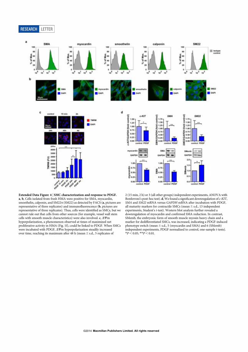
Extended Data Figure 4 | SMC characterization and response to PDGF.a, b, Cells isolated from fresh HMA were positive for SMA, myocardin,smoothelin, calponin, and SM22a (SM22) as detected by FACS (a, pictures arerepresentative of three replicates) and immunofluorescence (b, pictures arerepresentative of three replicates). Thus, cells were identified as SMCs, but wecannot rule out that cells from other sources (for example, vessel wall stemcells with smooth muscle characteristics) were also involved. c, DYmhyperpolarization, a phenomenon observed at times of maximized netproliferative activity in HMA (Fig. 1f), could be linked to PDGF. When SMCswere incubated with PDGF, DYm hyperpolarization steadily increasedover time, reaching its maximum after 48 h (mean 6 s.d., 5 replicates of
2 (15 min, 2 h) or 3 (all other groups) independent experiments, ANOVA withBonferroni’s post-hoc test). d, We found a significant downregulation of c-KIT,SMA and SM22 mRNA versus GAPDH mRNA after incubation with PDGF,all maturity markers for contractile SMCs (mean 6 s.d., 13 independentexperiments, Student’s t-test). Western blot analysis further revealed adownregulation of myocardin and confirmed SMA reduction. In contrast,SMemb, the embryonic form of smooth muscle myosin heavy chain and amarker for dedifferentiated SMCs, was increased, indicating a PDGF-inducedphenotype switch (mean 6 s.d., 5 (myocardin and SMA) and 6 (SMemb)independent experiments, PDGF normalized to control, one-sample t-tests).*P , 0.05; **P , 0.01.
RESEARCH LETTER
Macmillan Publishers Limited. All rights reserved©2014

Extended Data Figure 5 | DCA reduces DYm hyperpolarization andfacilitates apoptosis in SMCs. a, To assess the kinetics of DCA action in vitro,SMCs were pre-incubated with DCA for different time periods and thenstimulated with PDGF for 48 h (mean 6 s.d., 10 replicates of 3 independentexperiments per group, ANOVA with Bonferroni’s post-hoc test). DCAreduced PDGF-induced DYm hyperpolarization irrespective of thepre-incubation period. b, Atherosclerotic plaques from heavily calcified andatherosclerotic human coronary arteries were scraped off the media andcultured in SMC medium. Outgrowing SMCs were picked and expanded.PDGF significantly increased DYm, an effect that was prevented by DCA(mean 6 s.d., 10 replicates of 3 independent experiments, ANOVA withBonferroni’s post-hoc test). c–e, Apoptosis induction with staurosporinesignificantly increased the number of control SMCs that showed cytochrome Cleakage, as identified by diffuse cytochrome C staining throughout the cell(c, mean 6 s.d., 10 replicates of 2 independent experiments per group, ANOVA
with Bonferroni’s post-hoc test). PDGF significantly reduced cytochrome Crelease in SMCs and DCA counteracted this PDGF effect. Cytoplasmatic (C)and mitochondrial fractions (M) were isolated for immunoblotting (d, dataare representative of 3 independent experiments). Separation was confirmedby the contents of mitochondrial complex V (CV) and cytosolic GAPDH.Cytochrome C was retained inside the mitochondria in control SMCs and wasreleased with staurosporine. PDGF reduced the cytoplasmatic fraction,indicating reduced cytochrome C release, and DCA reversed the PDGF effect.SMC apoptosis was detected by TUNEL staining (e, mean 6 s.d., 10 replicatesof 2 (column 1) or 3 (columns 2–4) independent experiments, ANOVAwith Bonferroni’s post-hoc test). Staurosporine significantly increased thenumber of apoptotic control SMCs. PDGF treatment decreased the percentageof apoptotic SMCs and DCA reversed this anti-apoptotic effect. *P , 0.05;**P , 0.01.
LETTER RESEARCH
Macmillan Publishers Limited. All rights reserved©2014

Extended Data Figure 6 | DCA lowers medial and myointimal DYm andfacilitates apoptosis in balloon-injured rat aortas. a, b, Trichrome (a) andimmunofluorescence (b) confirmed abundant SMCs in the aortic media ofDCA-treated animals 48h after injury. c, Similar to control vessels, the infiltratemainly contained macrophages and neutrophils, but no CD31 lymphocytes.d, DCA reduced DYm of medial cells (mean 6 s.d., n 5 6 animals (control at48 h) and 4 animals (DCA at 48 h), Student’s t-test). e, Macrophages werethe main inflammatory cell population in the developing myointima ofimmunocompetent DCA animals at 7 days. Very few neutrophils and nolymphocytes were observed. Leukocyte infiltration was markedly alleviated at28 days (black arrows, internal elastic lamina). f, g, DCA effectively lowered the
elevated DYm of myointimal cells on day 7 (f, mean 6 s.d., n 5 4 animals pergroup, Student’s t-test), but had little effect on the already reduced DYm at 28days (g, mean 6 s.d., n 5 6 animals per group, Student’s t-test). h, i, Thepercentages of proliferating (Ki671) and apoptotic (TUNEL1) cells in themyointima were calculated (representative cropped pictures of the myointimaare presented). The few myointimal cells in control- and DCA-vessels at 7 daysshowed high proliferative activity (red, positive y axis), but apoptosis (green,negative y axis) was only observed in DCA-treated animals (h, mean 6 s.e.m.,n 5 4 animals per group). Proliferation and apoptosis were low after 28 days inboth groups (i, mean 6 s.e.m., n 5 5 animals (control at day 28) and 7 animals(DCA at day 28)). *P , 0.05.
RESEARCH LETTER
Macmillan Publishers Limited. All rights reserved©2014

Extended Data Figure 7 | DCA does not impair endothelial cell migrationor vessel re-endothelialization. The effect of DCA on endothelial cell (EC)migration was assessed in vitro. a, b, A scratch (width 338mm) was made acrossa confluent human EC monolayer. The number of cells that migrated into a340 mm 3 338 mm scratch rectangle within 5 h was counted (a, mean 6 s.d.,triplicates of 3 independent experiments, Student’s t-test) and the time neededto close the scratch was recorded (b, mean 6 s.d., n 5 5 (EC), 4 (EC 1 DCA)independent experiments, Student’s t-test). There were no differencesbetween untreated and DCA-treated ECs. c, d, To evaluate vesselre-endothelialization, rat aortas underwent mechanical endothelial denudationby balloon injury. Aortas were recovered after 28 days and stained for ratendothelial cell antigen (c, black arrows, RECA-1). Re-endothelialization wasquantified (d, mean 6 s.d., n 5 5 animals (control at day 28) and 6 animals
(DCA at day 28), Student’s t-test) and was similar in both groups.e, f, Endothelial function in both 28-day groups was further assessed inrelaxation studies and compared to denuded aortas three days after ballooninjury (mean 6 s.e.m., n 5 9 animals (control at day 28), 11 animals (DCAat day 28) and 3 animals (denuded at day 3)). Freshly recovered aortic segmentswere pre-constricted and DCA-treated and control vessels, as well as denudedaortas, showed similar endothelium-independent relaxation capacities usingnitroglycerin (NTG) as vasodilator (e). However, denuded aortas largely failedto show endothelium-dependent relaxation with acetylcholine (Ach; f). BothDCA-treated and control vessels demonstrated similar and physiologicendothelium-dependent relaxation, indicating functional integrity of thevascular endothelium.
LETTER RESEARCH
Macmillan Publishers Limited. All rights reserved©2014

Extended Data Figure 8 | PDK2 knockdown in SMCs, HMAs and HCAs.a, PDK1, PDK2, PDK3 and PDK4 mRNA expression was assessed in controlSMCs and normalized to GAPDH (mean 6 s.d., 3 independent experiments).PDK1, PDK2 and PDK3 mRNA was detected as shown. Also, PDK1, PDK2 andPDK3 but not PDK4 were detectable in immunoblot analyses (data arerepresentative of 3 independent experiments). b, To confirm in vitro PDK2knockdown, PDK1 and PDK2 expressions were assessed in immunoblotanalyses (data are representative of 3 independent experiments). PDK2 wasmarkedly reduced in PDK2-knockdown SMCs (PDK2-KD-SMC), but not inscramble shRNA-transduced SMCs (scramble-SMC). PDK1 expressionremained unaffected by PDK2- or scramble-shRNA constructs. c, To excludethat lentiviral shRNA transduction changed the DYm response to PDGF,scramble-SMCs were stimulated with PDGF (mean 6 s.d., 10 replicates of 2independent experiments, Student’s t-test). Both the baseline potential and thePDGF-triggered increase in DYm were similar to SMCs (see Extended Data
Fig. 5a). d, Fresh HMA vessels underwent balloon injury, were divided, and onehalf was sampled (day -1). The other half was incubated with lentiviral particlescontaining PDK2 or scrambled shRNA. On the next day, transduced vesselswere transplanted into RNU rats. Two days later, vessels were recovered (day 2)for immunoblotting. PDK2 could be detected in untreated HMA vessels(day -1). On day 2, only scrambled control HMAs, but not PDK2-KD vesselsshowed a PDK2 signal (data are representative of 3 independent experiments).e, f, After 28 days, HMA and HCA samples were stained for DAPI, PDK1,PDK2 and SMA. PDK1 was similarly detectable in all control, scramble andPDK2-KD sections at day 28 (e, pictures are representative of 3 independentexperiments). At day 28, HMA control and scramble as well as HCA controlshowed detectable PDK2 protein, whereas PDK2 fluorescence in HMA andHCA PDK2-KD at day 28 was negligible (f, pictures are representative of 3independent experiments), demonstrating selective knockdown. **P , 0.01.
RESEARCH LETTER
Macmillan Publishers Limited. All rights reserved©2014

Extended Data Figure 9 | PDK2 knockdown mimics the DCA effect on SMCDYm and apoptosis. a, PDK2-KD-SMCs were pre-incubated with DCAand/or stimulated with PDGF. PDK2-KD-SMCs maintained lowDYm despitePDGF stimulation and DCA did not further depolarize DYm (mean 6 s.d.,10 replicates of 3 (columns 1 and 3) or 4 (column 2) independent experiments,ANOVA with Bonferroni’s post-hoc test). b, c, Apoptosis was induced withstaurosporine. Control PDK2-KD-SMCs showed mainly mitochondria-housed cytochrome C in fluorescence stainings (b, mean 6 s.d., 10 replicates of2 independent experiments, ANOVA with Bonferroni’s post-hoc test) andcompartment-separated immunoblot analyses (c, data are representative of
2 independent experiments). After induction of apoptosis, cytochrome Cleaked into the cytoplasm and PDGF did not suppress cytochrome C leakage.d, PDGF did not induce resistance to apoptosis in PDK2-KD-SMCs(mean 6 s.d., 10 replicates of 2 independent experiments per group, ANOVAwith Bonferroni’s post-hoc test). e, To establish a link between PDH andDYm,PDH was knocked down (data are representative of 2 independentexperiments). f, DYm was significantly increased in PDH siRNA-transfectedSMCs and PDGF and DCA no longer affectedDYm (mean 6 s.d., 10 replicatesof 2 (column 2) or 3 (columns 1, 3, 4 and 5) independent experiments, ANOVAwith Bonferroni’s post-hoc test). **P , 0.01.
LETTER RESEARCH
Macmillan Publishers Limited. All rights reserved©2014

Extended Data Figure 10 | VDAC controls DYm and apoptosis. a, PDGFstimulation of SMCs significantly increased phosphorylated AKT, and ERK1and ERK2, and increased the expression of HK2 (mean 6 s.d., 5 (pAKTand pERK1/2) and 8 (HK2) independent experiments, PDGF normalized tocontrol, one-sample t-tests). b, Immunoblot analysis revealed PDGF-inducedupregulation of HK2 expression in SMCs (mean 6 s.d., 3 (SMC) independentexperiments, data normalized to control, one-sample t-test (control versusPDGF), Student’s t-test (PDGF versus PDGF 1 DCA)). HK2 upregulationwas alleviated by DCA or PDK2-KD (mean 6 s.d., 8 (PDK2-KD-SMC)independent experiments, PDGF normalized to control, one-sample t-test).c, Confocal images with mitochondrial (MitoTracker, red), HK2 (green),and nuclear staining (DAPI, blue, data are representative of 2 independentexperiments) were captured. Co-localization of green HK2 with redmitochondria generated a yellow colour. SMCs incubated with PDGF increasedtheir yellow signal, an effect that was prevented by DCA. PDGF did not induceHK2-mitochondrial association in PDK2-KD-SMCs. d, Cytoplasmatic (C)and mitochondrial fractions (M) were isolated for immunoblotting (dataare representative of 2 independent experiments). In control SMCs and
PDK2-KD-SMCs, HK2 was mainly detected in the cytoplasmatic fraction.In SMCs, PDGF caused a partial translocation of HK2 to the mitochondrialcompartment. DCA markedly alleviated this translocation. PDGF did notinduce HK2 translocation in PDK2-KD-SMCs. e, In the presence ofHXK2VBD-cpm, PDGF failed to induce both DYm hyperpolarization(mean 6 s.d., 10 replicates of 3 independent experiments, ANOVA withBonferroni’s post-hoc test) and apoptosis resistance (mean 6 s.d., 10 replicatesof 3 independent experiments, ANOVA with Bonferroni’s post-hoc test).f, VDAC inhibition by DIDS caused DYm hyperpolarization (mean 6 s.d.,10 replicates of 2 independent experiments, Student’s t-test) and renderedSMCs resistant to staurosporine-induced apoptosis (mean 6 s.d., 10 replicatesof 3 independent experiments, ANOVA with Bonferroni’s post-hoc test).g, The depolarizing effect of DCA on PDGF-treated SMCs was counteractedby DIDS (mean 6 s.d., 10 replicates of 3 independent experiments, ANOVAwith Bonferroni’s post-hoc test). Although PDGF did not increase DYm inPDK2-KD-SMC, DIDS caused marked mitochondrial hyperpolarization(mean 6 s.d., 10 replicates of 2 independent experiments, ANOVA withBonferroni’s post-hoc test). *P , 0.05; **P , 0.01.
RESEARCH LETTER
Macmillan Publishers Limited. All rights reserved©2014

Brief Report
SCNT-Derived ESCs with Mismatched MitochondriaTrigger an Immune Response in Allogeneic Hosts
Graphical Abstract
HighlightsNT-ESCs with mismatched mitochondria possess
alloantigenicity
This alloantigenicity is weaker than that of an MHC mismatch
The provoked alloimmune response is adaptive and amenable
for tolerance induction
Authors
Tobias Deuse, Dong Wang, ...,
Irving L. Weissman, Sonja Schrepfer
In BriefDeuse et al. report that SCNT-derived
mouse ESCs trigger an immune rejection
in spite of their matching with the entire
nuclear genome of the recipient mouse
strain. Their findings reveal that mis-
matched mitochondria can be sufficient
to elicit an allogeneic response.
Deuse et al., 2015, Cell Stem Cell 16, 1–6January 8, 2015 ª2015 Elsevier Inc.http://dx.doi.org/10.1016/j.stem.2014.11.003

Cell Stem Cell
Brief Report
SCNT-Derived ESCs with Mismatched MitochondriaTrigger an Immune Response in Allogeneic HostsTobias Deuse,1,2,3 Dong Wang,1,2 Mandy Stubbendorff,1,2 Ryo Itagaki,1,2 Antje Grabosch,1,2 Laura C. Greaves,4
Malik Alawi,5,6 Anne Grunewald,7 Xiaomeng Hu,1,2 Xiaoqin Hua,1,2 Joachim Velden,8 Hermann Reichenspurner,2,3
Robert C. Robbins,9 Rudolf Jaenisch,10 Irving L. Weissman,11,12 and Sonja Schrepfer1,2,9,12,*1TSI Laboratory, University Heart Center Hamburg, Martinistrasse 52, 20246 Hamburg, Germany2Cardiovascular Research Center Hamburg (CVRC) and DZHK (German Center for Cardiovascular Research), partner siteHamburg/Kiel/Luebeck, Martinistrasse 52, 20246 Hamburg, Germany3Cardiovascular Surgery, University Heart Center Hamburg, Martinistrasse 52, 20246 Hamburg, Germany4Newcastle University Centre for Brain Ageing and Vitality, Newcastle University, Newcastle upon Tyne, NE2 4HH, UK5Bioinformatics Service Facility, University Medical Center Hamburg-Eppendorf, Martinistrasse 52, 20246 Hamburg, Germany6Heinrich-Pette Institute, Leibniz Institute for Experimental Virology, Virus Genomics, Martinistrasse 52, 20246 Hamburg, Germany7Wellcome Trust Centre for Mitochondrial Research, Institute of Neuroscience, Newcastle University, Newcastle upon Tyne, NE2 4HH, UK8Department of Nephropathology, Institute of Pathology, University Hospital Erlangen, Maximiliansplatz 2, 91054 Erlangen, Germany9Stanford Cardiovascular Institute and Department of Cardiothoracic Surgery, Stanford University School of Medicine, 300 Pasteur Drive,Stanford, CA 94305, USA10Whitehead Institute for Biomedical Research, Massachusetts Institute of Technology, 9 Cambridge Center, Cambridge, MA 02142, USA11Department of Developmental Biology, Stanford Institute for Stem Cell Biology and Regenerative Medicine, Stanford University School ofMedicine, 300 Pasteur Drive, Stanford, CA 94305, USA12Co-senior author*Correspondence: [email protected]://dx.doi.org/10.1016/j.stem.2014.11.003
SUMMARY
The generation of pluripotent stem cells by somaticcell nuclear transfer (SCNT) has recently beenachieved in human cells and sparked new interestin this technology. The authors reporting thismethodical breakthrough speculated that SCNTwould allow the creation of patient-matched embry-onic stem cells, even in patients with hereditarymitochondrial diseases. However, herein we showthat mismatched mitochondria in nuclear-transfer-derived embryonic stem cells (NT-ESCs) possessalloantigenicity and are subject to immune rejection.In a murine transplantation setup, we demonstratethat allogeneic mitochondria in NT-ESCs, which arenucleus-identical to the recipient, may trigger anadaptive alloimmune response that impairs the sur-vival of NT-ESC grafts. The immune response isadaptive, directed against mitochondrial content,and amenable for tolerance induction. Mitochondrialalloantigenicity should therefore be consideredwhendeveloping therapeutic SCNT-based strategies.
In somatic cell nucleus transfer (SCNT), the nucleus of a somaticcell is transplanted into an enucleated oocyte (Noggle et al.,2011). It has been envisioned that by this means, autologouspluripotent stem cells can be generated for specific patient-matched therapies. Embryonic stem cells by nuclear transfer(NT-ESCs) were first successfully generated from mice (Munsieet al., 2000) and primates (Byrne et al., 2007). After substantial
optimization of the SCNT technique, the derivation of NT-ESCsfrom human fetal fibroblasts (Tachibana et al., 2013), and mostrecently, from adult somatic cells (Chung et al., 2014; Yamadaet al., 2014), was reported. These latter studies suggest thatage-associated changes in the nucleus donor cell are not neces-sarily an impediment to generating human NT-ESCs (Chunget al., 2014). Furthermore, the differentiation of insulin-producingbeta cells derived from NT-ESCs generated from a subject withlongstanding diabetes proved that specialized cell types that arelost in diseased patients can be reestablished (Yamada et al.,2014). Because NT-ESCs acquire healthy mitochondria fromthe selected oocyte donor, inherited or acquired mtDNA dis-eases aremost amenable for correction using the SCNT technol-ogy (Tachibana et al., 2013). The recent advancements in SCNTmethodology have increased expectations for this approachto lead to human-assisted reproductive technologies (Cibelli,2014).A so far underappreciated obstacle for clinical application of
SCNT-derived cell therapy may be the immunogenicity of mis-matched mitochondria in NT-ESCs. After transplantation ofNT-ESC-derived cells or tissue back into the nucleus donor, mis-matched mtDNA-coded proteins may induce alloimmunity. Wehere show that allogeneic mitochondria in murine NT-ESCsmay trigger an adaptive immune response that impairs the sur-vival of NT-ESC grafts.To assess mitochondria-specific alloantigenicity, transplanta-
tion studies with SCNT-derived murine NT-ESCs were con-ducted across defined immunological barriers. NT-ESCs wereproduced previously by Kirak et al. (2010) using enucleatedBDF1 (B6D2F1/J) oocytes (C57BL/6J 3 DBA/2NCrl) F1 and iso-lated BALB/c (BALB/cAnNCrl; major histocompatibility complex[MHC] H2d) fibroblast nuclei (Figure 1A). Both oocytes and NT-ESCs demonstrated high mitochondrial abundance (Figure S1A
Cell Stem Cell 16, 1–6, January 8, 2015 ª2015 Elsevier Inc. 1
Please cite this article in press as: Deuse et al., SCNT-Derived ESCs with Mismatched Mitochondria Trigger an Immune Response in Allogeneic Hosts,Cell Stem Cell (2015), http://dx.doi.org/10.1016/j.stem.2014.11.003

available online). The high repetition of mtDNA copies and thehigh amount of mitochondrial proteins contribute to their rele-vance as possible immunogenic antigens. Culture-expandedNT-ESCs expressed markers related to pluripotency (Fig-ure S1B). NT-ESCs grew tumors in immunodeficient NODSCID mice (NOD.CB17-Prkdcscid/J). These tumors containedtissues of endodermal, ectodermal, and mesodermal origin (Fig-ures S1C and S1D), thus meeting the criteria for teratomas. Thegenerated NT-ESCs thus contain maternally inherited C57BL/6J(B6) mitochondria and BALB/c nuclei of the H2d haplotype. Sus-pensions of 106 NT-ESCs were intramuscularly injected intothe thighs of recipient mice. BALB/c recipients are matched forthe entire nuclear genome but are mitochondria mismatched.
mtDNA sequence analysis comparing B6 mitochondria of theNT-ESCs with BALB/c mitochondria revealed two nonsynony-mous nucleotide substitutions within the mt-Co3 and mt-Cytbgenes, which resulted in amino acid substitutions in the associ-ated proteins (Figure S2A). These two nonsynonymous singlenucleotide polymorphisms (SNPs) were therefore the only differ-ences in the entire cellular DNA between donor cells and recip-ient. According to dbSNP (Sherry et al., 2001), both variantsare common in mouse. The affected loci are conserved amongvertebrates (Figure S2B) and for one of the two observed variantsa human counterpart is described in dbSNP. BDF1 (H2b/d) recip-ients share the H2d alleles with the NT-ESCs and are mitochon-dria matched but minor histocompatibility antigen (MiHA)
Figure 1. NT-ESCs Possess Mitochondria-Related Antigenicity(A) NT-ESCs were generated using enucleated BDF1 oocytes and isolated BALB/c fibroblast nuclei. NT-ESCs were transplanted into mitochondria-mismatched
(MM) BALB/c, MiHA-MM BDF1, MHC-MM B6, immunodeficient NOD SCID, or combined MHC-MM and mitochondria-MM CBA. See also Figure S1.
(B) Recipient-specific Th1 ELISPOT results against NT-ESCs showed significantly lower IFNg spot frequencies for BALB/c (n = 18) and BDF1 (n = 17) recipients
compared to B6 (n = 24). Overall, the CBA IFNg response (n = 15) was the strongest and the NOD SCID response (n = 6) was negligible.
(C) IL-4 (Th2) ELISPOT responses revealed similar relations among groups (BALB/c, n = 12; BDF1, n = 17; B6, n = 24; NOD SCID, n = 8; CBA, n = 10) as described
in (B) for Th1 response.
(D) The NT-ESC-specific IgM antibody production after 5 days was significantly stronger in B6 (n = 22) than in BALB/c (n = 31) or BDF1 (n = 15). No IgM antibodies
against NT-ESCs were detected in NOD SCID (n = 6).
(E) Isogeneic BALB/c ESCs (isoESCs) were transplanted either into isogeneic BALB/c or recipients with combined mitochondria- and MiHA-MM (BDF1) or
mitochondria- and MHC-MM (B6).
(F) The IFNg ELISPOT responses against isoESC grafts were negligible in BALB/c (n = 5) and significantly stronger in BDF1 (n = 22) or B6 (n = 6). Th1 activation in
MHC-MM B6 was significantly stronger than in MiHA-MM BDF1.
(G) The isoESC-specific IL-4 ELISPOT responses were significantly stronger in BDF1 (n = 23) and B6 (n = 10) than in BALB/c (n = 5).
(H) BALB/c nude mice received NT-ESC (mitochondria-MM) or isoESC grafts. Because of the T cell deficiency, the nude model assesses NK cell responses.
(I) The IFNg NK cell response against NT-ESCs (n = 9) was low, similar to the response against isoESCs (n = 8).
(J and K) Teratoma growth diagrams for NT-ESCs (J, n = 5) and isoESCs (K, n = 5) in BALB/c nudemice are shown. The overall teratoma development is depicted
in separate adjacent graphs.
On each box blot, the central mark is the median, the edges of the box are the 25th and 75th percentiles, and the whiskers extend to the most extreme data points.
MM, mismatch; *p < 0.05.
Cell Stem Cell
Mitochondria Possess Alloantigenicity
2 Cell Stem Cell 16, 1–6, January 8, 2015 ª2015 Elsevier Inc.
Please cite this article in press as: Deuse et al., SCNT-Derived ESCs with Mismatched Mitochondria Trigger an Immune Response in Allogeneic Hosts,Cell Stem Cell (2015), http://dx.doi.org/10.1016/j.stem.2014.11.003

mismatched (Figure 1A). B6 recipients possess the H2b haplo-type and are therefore MHC mismatched but mitochondriamatched to the NT-ESCs. The severely immunodeficient NODSCID strain was used as a negative control because it cannotmount any antigen-specific cellular immune response. TheCBA (CBA/J; H2k) strain is both MHC and mitochondria mis-matched to the NT-ESCs. mtDNA sequencing revealed identicalmtDNA genomes for CBA and BALB/c (not shown), as was pre-viously reported in the context of mouse mtDNA phylogeny(Goios et al., 2007). Therefore, besides the MHC differences,NT-ESC transplantations into BALB/c and CBA cross the samemitochondrial mismatch.Previous studies on the immunogenicity of murine mitochon-
drial peptides were mostly restricted to cytotoxic T lymphocytes(CTLs) and MHC class Ia and Ib presentation (Dabhi and Lin-dahl, 1995). We herein used assays that quantify activation ofT helper cells (Th) because this entity plays a central role inactivating CTLs and B cells, thus orchestrating graft rejection.Five-day ELISPOT assays for Th1-specific g-interferon (IFNg)showed a substantial immune activation in the MHC-mis-matched B6 recipients, which was significantly stronger thanthose in mitochondria-mismatched BALB/c or MiHA-mis-matched BDF1 (Figure 1B). The MHC- and mitochondria-mis-matched CBA strain demonstrated the highest spot frequenciesof all strains, which were approximately additive of the fre-quencies of the isolated MHC-mismatched B6 and the isolatedmitochondria-mismatched BALB/c. The immunodeficient NODSCID recipients showed immunological Th1 anergy. The Th2response, although generally generating lower IL-4 spot fre-quencies, showed a similar relation between the recipientstrains (Figure 1C). There was also a surge in NT-ESC-specificIgM antibodies in B6 recipients 5 days after immunization,which was significantly stronger than that in BALB/c or BDF1(Figure 1D). There were no NT-ESC-specific IgM antibodies inNOD SCID.In an additional experiment, we could exclude that there were
general differences among BALB/c, BDF1, and B6 in the degreeof immune responsiveness (Figure S2C). Splenocytes of thesethree strains were unspecifically stimulated and ELISPOT assaysdemonstrated similar Th1 and Th2 activation. This result permitsus to attribute the above ELISPOT results to the mismatchedproteins.To exclude methodological bias caused by the state of
pluripotency, isogeneic BALB/c ESCs (isoESCs) were used(Figure 1E). Transplantation experiments were performed withfully nuclear DNA- and mtDNA-matched, isogeneic BALB/crecipients, MiHA- and mitochondria-mismatched BDF1, andMHC- and mitochondria-mismatched B6. There was negligibleTh1 and Th2 activation in isogeneic BALB/c recipients (Figures1F and 1G). Between mitochondria-mismatched recipients, anadditional MHC mismatch (B6) generated a significantly stron-ger Th1 activation than an additional MiHA mismatch (BDF1,Figure 1F). Together, these results demonstrate that an isolatedmitochondria mismatch is sufficient to initiate marked Th acti-vation and NT-ESC-directed antibody production. These im-mune responses are not caused by pluripotency featuresthemselves. Second, an MHC mismatch causes a significantlystronger immune activation than an MiHA mismatch, in thesetting of both additional mitochondrial match and mismatch.
And third, the Th1 activation is additive for an MHC and a mito-chondria mismatch.Based on the results by Ishikawa (Ishikawa et al., 2010), which
assumed that the innate immunity was responsible for rejectionof mitochondria-mismatched cell grafts, the T cell-deficientnude mouse model was used to study the NK cell response.BALB/c nude (BALB/c nu/nu, CAnN.Cg-Foxn1 < nu > /Crl;H2d) mice were used as recipients for either mitochondria-mis-matched NT-ESCs or fully isogeneic isoESCs (H2d, Figure 1H).Our results show that there was no increase in NK-cell-drivenIFNg ELISPOT frequency in response to NT-ESCs as comparedto the response to isoESCs (Figure 1I). Also, the survival of cellgrafts was similar for NT-ESCs and isoESCs and all animalsformed teratomas, thus excluding relevant innate rejection pro-cesses (Figures 1J and 1K). Importantly, the transplantationexperiment by Ishikawa was specifically designed to be mis-matched for the maternally transmitted mitochondrial antigen(Mta) previously described by Fischer Lindahl (Fischer Lindahlet al., 1980). Our transplant setting avoids this incompatibilityand, as a consequence, we did not observe relevant innateimmunity-driven rejection.Survival of NT-ESC grafts and teratoma development were
assessed in the transplantation settings of Figure 1A. As ex-pected, all NT-ESC transplants survived and formed teratomasin NOD SCID recipients (Figure 2A), whereas all grafts wererejected in fully mismatched CBAs (Figure 2B). The rathermild Th1 activation in MiHA-mismatched BDF1 still allowed90% teratoma development of rapidly proliferating NT-ESCgrafts (Figure 2C), whereas there was only 20% teratomagrowth in MHC-mismatched B6 (Figure 2D). In mitochondria-mismatched BALB/c, teratomas developed in 60% of the recip-ients, but the tumor growth was markedly slowed (Figure 2E).Half of the tumors did not develop until day 50 to 90. Interest-ingly, there was a strong correlation between graft survival andTh1 activation in individual BALB/c animals (Figure 2F). OnlyBALB/c recipients with very low spot frequencies developedteratomas, whereas animals with at least moderate Th1 activa-tion rejected their grafts. This further supports the notion of aT cell-mediated rejection process of mitochondria-mismatchedgrafts.To confirm a mitochondrial location of the immunogenic pep-
tides, mitochondria of NT-ESCs were separated from theremainder of the cell (cytosol) and splenocytes of NT-ESC-immunized BALB/c were restimulated in vitro with either mis-matched mitochondria or isogeneic cytosol (Figure 2G). Onlythe mismatched mitochondria caused an IFNg response,whereas the spot frequencies with the isogeneic cytosol frac-tions were low, similar to those of unstimulated responder sple-nocytes (Figure 2H). Of note, the ELISPOT frequencies of theseparated cell compartments were markedly lower than thoseinvolving whole cells. Although their precise presentation mech-anism remains elusive, the detection ofmitochondrial peptides inthe context of MHC (Dabhi and Lindahl, 1995; Duvvuri et al.,2014) suggests that they enter conventional MHC antigenpresentation pathways. The dependence on indirect antigenpresentation in this assaymay explain this observation. In accor-dancewith the ELISPOT results, in a delayed-type hypersensitiv-ity test, only mismatched mitochondria generated paw swelling,whereas isogeneic cytosol did not (Figure 2I). The location of the
Cell Stem Cell
Mitochondria Possess Alloantigenicity
Cell Stem Cell 16, 1–6, January 8, 2015 ª2015 Elsevier Inc. 3
Please cite this article in press as: Deuse et al., SCNT-Derived ESCs with Mismatched Mitochondria Trigger an Immune Response in Allogeneic Hosts,Cell Stem Cell (2015), http://dx.doi.org/10.1016/j.stem.2014.11.003

antigens for BALB/c immune activation against NT-ESCs wasthus restricted to the mitochondrial fraction.
We next tested whether acquired immunological toleranceagainst allogeneic mitochondria can be induced. In accordancewith the Medawar experiments (Billingham et al., 1953), neonatalBALB/c mice were immunized with mitomycin-inhibited NT-ESCs (Figure 2J). In adulthood, these animals were reinjectedwith NT-ESCs (BALB/creinject). In contrast to adult BALB/c recip-ients that received their first NT-ESC grafts, BALB/creinjectshowed no IFNg response in ELISPOT assays and all animalsdeveloped teratomas (Figures 2K and 2L). This experiment notonly shows that tolerance against allogeneic mitochondria canreliably be induced, it is also further proof for the relevance of
the adaptive immune system in rejecting mitochondria-mis-matched grafts.There have been few publications on the transplantation of
mitochondria-mismatched cells or tissues with conflicting re-sults. Although prolonged hematopoietic chimerism (Lanzaet al., 2005) and acceptance of cardiac and renal cell transplants(Lanza et al., 2002) in cows receiving SCNT-derived mitochon-dria-mismatched cells was reported, this may not guarantee uni-versal success. Pigs rejected kidney grafts from SCNT-derivedmitochondria-mismatched clones (Kwak et al., 2013). In miceand rats, it was shown that a single nonsynonymous nucleotidesubstitution in the mitochondrial ND1 gene or the ATPase 6gene, respectively, generated an aberrant peptide and resulted
Figure 2. The Rejection of Mitochondria-MM NT-ESCs Is Adaptive, Directed against Mitochondrial Content, and Amenable for ToleranceInduction(A–E) Teratoma growth diagrams for NT-ESC grafts in immunodeficient NOD SCID (A, n = 5), combined MHC- and mitochondria-MM CBA (B, n = 5), MiHA-MM
BDF1 (C, n = 10), MHC-MM B6 (D, n = 10), and mitochondria-MM BALB/c (E, n = 10) recipients. The overall teratoma development is depicted in separate
adjacent graphs. See also Figure S2.
(F) Within the BALB/c recipient group, the IFNg ELISPOT frequencies of animals that did not (no teratoma, n = 20) or did (teratoma, n = 8) show teratoma growth
were compared.
(G) BALB/c receivedmitochondria-MMNT-ESC grafts. Subsequent ELISPOT assays did not use NT-ESCs but used isolatedmitochondria or the remainder of the
cells (referred to as cytosol) for stimulation.
(H) IFNg ELISPOT analyses show some BALB/c response against isolated NT-ESC mitochondria (NT-ESCmito, n = 8), whereas no reactivity against the
mitochondria-depleted NT-ESC remnants (NT-ESCcytosol, n = 4) was detectable.
(I) There also was a paw swelling response in BALB/c recipients challenged with cutaneous injections of isolated NT-ESCmitochondria (n = 9), but not in BALB/c
challenged with the mitochondria-depleted NT-ESC remnants (n = 5).
(J) One group of BALB/cmice received their first NT-ESC grafts at neonatal stage and were reinjected during adulthood (BALB/creinject). The other group received
NT-ESC grafts only after entering adulthood (BALB/c).
(K) In ELISPOT assays, BALB/creinject (n = 14) showed a significantly lower IFNg response than BALB/c (n = 7).
(L) The overall teratoma development of NT-ESC grafts in BALB/creinject (n = 27) and BALB/c animals (n = 7) is presented.
On each box in ELISPOT blots, the central mark is themedian, the edges of the box are the 25th and 75th percentiles, and the whiskers extend to themost extreme
data points. In each scatter blot, the central mark is the mean. MM, mismatch; yp < 0.01.
Cell Stem Cell
Mitochondria Possess Alloantigenicity
4 Cell Stem Cell 16, 1–6, January 8, 2015 ª2015 Elsevier Inc.
Please cite this article in press as: Deuse et al., SCNT-Derived ESCs with Mismatched Mitochondria Trigger an Immune Response in Allogeneic Hosts,Cell Stem Cell (2015), http://dx.doi.org/10.1016/j.stem.2014.11.003

in the loss of histocompatibility (Bhuyan et al., 1997; Davies et al.,1991; Loveland et al., 1990). Neopeptides from human mtDNAharboring large-scale frameshift deletions were recently re-ported to bind to MHC molecules and can be recognized byT cells from other individuals (Duvvuri et al., 2014). Interestingly,the Duvvuri et al. results further suggest that some neopeptidescould bind promiscuously to both MHC class I and class IIalleles, activate CD4 and CD8 T cells, and be recognized byCD8 memory T cells. Aberrant mitochondrial peptides mayeven provoke MHC overexpression and thus amplify their immu-nogenicity. In both mice and humans, increased MHC class Iexpression in cells harboring mtDNA deletions was shown (Guet al., 2003). Mitochondrial peptides can thus serve as immuno-gens (Morse et al., 1996) and the recipient immune responsemay depend on the alloantigenicity of the aberrant mitochondrialprotein. Although thought to be beneficial for immune surveil-lance and clearance of cells with mitochondrial mutations(Duvvuri et al., 2014; Gu et al., 2003), this immune feature ispotentially hazardous in the context of SCNT-derived celltransplantation.Artificial reprograming using viral or nonviral plasmid-based
approaches seems to alter the expression of a multitude ofgenes in induced pluripotent stem cells (iPSCs) that are differen-tially expressed in ESCs (de Almeida et al., 2014; Zhao et al.,2011). Specific factors being suppressed (de Almeida et al.,2014) or overexpressed (Zhao et al., 2011) in the pluripotent statehave been suggested to contribute to the antigenicity of iPSCs.With differentiation, the antigenicity was reported to vanish astranscriptome profile and surface antigen expression convergedwith those of corresponding somatic cells (de Almeida et al.,2014). Terminally differentiated cells derived from iPSCs arenow believed to possess no or negligible immunogenicity (Arakiet al., 2013). The described immunogenicity is therefore differen-tiation dependent and not due to antigenic somatic codingmutations, which have recently been reported to occur during re-programming (Gore et al., 2011). When compared to autologousiPSCs that are fully matched for both nuclear and mitochondrialDNA, NT-ESCs with mtDNA differences to the nucleus donorpossess additional allogeneic peptides that may potentially beantigenic. These antigenic proteins inmitochondria-mismatchedNT-ESCs are maintained with differentiation and the mtDNAcopies, if anything, are increased in energy-consuming differen-tiated cells like cardiomyocytes. However, it remains to bedetermined if the results presented herein are transferrable todifferentiated cells and tissues from NT-ESCs.The level of heterogeneity in human mitochondria is consider-
ably higher than that in laboratory mice (Goios et al., 2007);dbSNP lists 2,066 active humanmitochondrial RefSNPs. At least64 of these are common (global minor allele frequency 1% orhigher) and nonsynonymously coding. MT-CYTB is affected by26 such variants andMT-CO3 by two, including the human coun-terpart of the murine variant we observed. A recent study hints atthe level of heterogeneity in human mitochondrial genomes(Ridge et al., 2014). Within a data set of 1,007 full mitochondrialgenomes, 899 single nucleotide variants, 26 insertions, and 20deletions were identified. Compared to the human referencesequence, each individual carried on average 25.3 variants. Sin-gle individuals marked the extremes with 2 and 52 variants,respectively.
In the field of xenotransplantation, the immunity against theaGal-antigen was long considered to be the one obstaclethat needed to be overcome to clear the path for subsequentclinical application. However, when aGal was knocked out,xenoimmune responses were still evident and new immunetargets surfaced that previously were overshadowed by thestrong aGal response (Miyata and Platt, 2003). Similarly,when nuclear DNA is matched in SCNT-derived cells, antigeniccompetition (Johnson et al., 1981) may allow mitochondrialantigens to emerge as immunogens. The immunogenicity ofmitochondria should thus not be neglected when advancingSCNT technology.
SUPPLEMENTAL INFORMATION
Supplemental Information for this article includes two figures and Supple-
mental Experimental Procedures and can be found with this article online at
http://dx.doi.org/10.1016/j.stem.2014.11.003.
AUTHOR CONTRIBUTIONS
T.D., R.C.R., R.J., I.L.W., and S.S. designed the study. T.D., D.W., M.A.,
and S.S. analyzed the data and performed the statistics. D.W. performed
the live cell mitochondrial staining, characterized the cells, and provided
all confocal data. M.S., R.I., A. Grabosch., and X. Hu performed the
ELISPOT assays and/or generated the cell survival data. M.S. contributed
to the FACS and DTH data. L.C.G. and A. Grunewald performed the
mtDNA sequencing. M.A. performed bioinformatics analyses of mtDNA se-
quences. X. Hua and J.V. generated the teratoma data. R.J. generated the
SCNT cells and ESCs. H.R. edited the manuscript. S.S. performed or su-
pervised cell transplantation and all immunologic assays. T.D., D.W., M.A.,
I.L.W., and S.S. wrote the manuscript and all authors edited the
manuscript.
ACKNOWLEDGMENTS
We thank Kirsten Fischer Lindahl for her critical comments and editing of the
manuscript. We further thank Christiane Pahrmann for performing all cell
cultures and for her overall technical assistance and Oktay Kirak and
Hidde Ploegh for generating the experimental cells. We extend special thanks
to the UKE Imaging Facility (UMIF, Bernd Zobiak) and the UKE Animal
Facility. This study was supported by the Else-Kroner-Fresenius-Stiftung
(2012_EKES.04; T.D.), a grant from the Fondation Leducq (CDA 2013-2015;
S.S.), and the German Research Foundation (Deutsche Forschungsgemein-
schaft; DFG: SCHR992/3-1 and SCHR992/4-1; S.S.).
Received: July 6, 2014
Revised: September 27, 2014
Accepted: November 7, 2014
Published: November 20, 2014
REFERENCES
Araki, R., Uda, M., Hoki, Y., Sunayama, M., Nakamura, M., Ando, S., Sugiura,
M., Ideno, H., Shimada, A., Nifuji, A., and Abe, M. (2013). Negligible immuno-
genicity of terminally differentiated cells derived from induced pluripotent or
embryonic stem cells. Nature 494, 100–104.
Bhuyan, P.K., Young, L.L., Lindahl, K.F., and Butcher, G.W. (1997).
Identification of the rat maternally transmitted minor histocompatibility anti-
gen. J. Immunol. 158, 3753–3760.
Billingham, R.E., Brent, L., and Medawar, P.B. (1953). Actively acquired toler-
ance of foreign cells. Nature 172, 603–606.
Byrne, J.A., Pedersen, D.A., Clepper, L.L., Nelson, M., Sanger, W.G., Gokhale,
S., Wolf, D.P., and Mitalipov, S.M. (2007). Producing primate embryonic stem
cells by somatic cell nuclear transfer. Nature 450, 497–502.
Cell Stem Cell
Mitochondria Possess Alloantigenicity
Cell Stem Cell 16, 1–6, January 8, 2015 ª2015 Elsevier Inc. 5
Please cite this article in press as: Deuse et al., SCNT-Derived ESCs with Mismatched Mitochondria Trigger an Immune Response in Allogeneic Hosts,Cell Stem Cell (2015), http://dx.doi.org/10.1016/j.stem.2014.11.003

Chung, Y.G., Eum, J.H., Lee, J.E., Shim, S.H., Sepilian, V., Hong, S.W., Lee, Y.,
Treff, N.R., Choi, Y.H., Kimbrel, E.A., et al. (2014). Human somatic cell nuclear
transfer using adult cells. Cell Stem Cell 14, 777–780.
Cibelli, J.B. (2014). Human somatic cell nuclear transfer is alive and well. Cell
Stem Cell 14, 699–701.
Dabhi, V.M., and Lindahl, K.F. (1995). MtDNA-encoded histocompatibility
antigens. Methods Enzymol. 260, 466–485.
Davies, J.D., Wilson, D.H., Hermel, E., Lindahl, K.F., Butcher, G.W., and
Wilson, D.B. (1991). Generation of T cells with lytic specificity for atypical
antigens. I. A mitochondrial antigen in the rat. J. Exp. Med. 173, 823–832.
de Almeida, P.E., Meyer, E.H., Kooreman, N.G., Diecke, S., Dey, D., Sanchez-
Freire, V., Hu, S., Ebert, A., Odegaard, J., Mordwinkin, N.M., et al. (2014).
Transplanted terminally differentiated induced pluripotent stem cells are
accepted by immune mechanisms similar to self-tolerance. Nat. Commun.
5, 3903.
Duvvuri, B., Duvvuri, V.R., Wang, C., Chen, L., Wagar, L.E., Jamnik, V., Wu, J.,
Yeung, R.S., Grigull, J., Watts, T.H., and Wu, G.E. (2014). The human immune
system recognizes neopeptides derived from mitochondrial DNA deletions.
J. Immunol. 192, 4581–4591.
Fischer Lindahl, K., Bocchieri, M., and Riblet, R. (1980). Maternally transmitted
target antigen for unrestricted killing by NZB T lymphocytes. J. Exp. Med. 152,
1583–1595.
Goios, A., Pereira, L., Bogue, M., Macaulay, V., and Amorim, A. (2007). mtDNA
phylogeny and evolution of laboratory mouse strains. Genome Res. 17,
293–298.
Gore, A., Li, Z., Fung, H.L., Young, J.E., Agarwal, S., Antosiewicz-Bourget, J.,
Canto, I., Giorgetti, A., Israel, M.A., Kiskinis, E., et al. (2011). Somatic coding
mutations in human induced pluripotent stem cells. Nature 471, 63–67.
Gu, Y., Wang, C., Roifman, C.M., and Cohen, A. (2003). Role of MHC class I in
immune surveillance of mitochondrial DNA integrity. J. Immunol. 170, 3603–
3607.
Ishikawa, K., Toyama-Sorimachi, N., Nakada, K., Morimoto, M., Imanishi, H.,
Yoshizaki, M., Sasawatari, S., Niikura, M., Takenaga, K., Yonekawa, H., and
Hayashi, J. (2010). The innate immune system in host mice targets cells with
allogenic mitochondrial DNA. J. Exp. Med. 207, 2297–2305.
Johnson, L.L., Bailey, D.W., andMobraaten, L.E. (1981). Antigenic competition
between minor (non-H-2) histocompatibility antigens. Immunogenetics 13,
451–455.
Kirak, O., Frickel, E.M., Grotenbreg, G.M., Suh, H., Jaenisch, R., and Ploegh,
H.L. (2010). Transnuclear mice with predefined T cell receptor specificities
against Toxoplasma gondii obtained via SCNT. Science 328, 243–248.
Kwak, H.H., Park, K.M., Teotia, P.K., Lee, G.S., Lee, E.S., Hong, S.H., Yang,
S.R., Park, S.M., Ahn, C., Park, C.K., et al. (2013). Acute rejection after swine
leukocyte antigen-matched kidney allo-transplantation in cloned miniature
pigs with different mitochondrial DNA-encoded minor histocompatibility anti-
gen. Transplant. Proc. 45, 1754–1760.
Lanza, R.P., Chung, H.Y., Yoo, J.J., Wettstein, P.J., Blackwell, C., Borson, N.,
Hofmeister, E., Schuch, G., Soker, S., Moraes, C.T., et al. (2002). Generation of
histocompatible tissues using nuclear transplantation. Nat. Biotechnol. 20,
689–696.
Lanza, R., Shieh, J.H., Wettstein, P.J., Sweeney, R.W., Wu, K., Weisz, A.,
Borson, N., Henderson, B., West, M.D., and Moore, M.A. (2005). Long-term
bovine hematopoietic engraftment with clone-derived stem cells. Cloning
Stem Cells 7, 95–106.
Loveland, B., Wang, C.R., Yonekawa, H., Hermel, E., and Lindahl, K.F. (1990).
Maternally transmitted histocompatibility antigen of mice: a hydrophobic pep-
tide of a mitochondrially encoded protein. Cell 60, 971–980.
Miyata, Y., and Platt, J.L. (2003). Xeno—still stuck without alphaGal. Nat.
Biotechnol. 21, 359–360.
Morse, M.C., Bleau, G., Dabhi, V.M., Hetu, F., Drobetsky, E.A., Lindahl, K.F.,
and Perreault, C. (1996). The COI mitochondrial gene encodes a minor histo-
compatibility antigen presented by H2-M3. J. Immunol. 156, 3301–3307.
Munsie, M.J., Michalska, A.E., O’Brien, C.M., Trounson, A.O., Pera, M.F., and
Mountford, P.S. (2000). Isolation of pluripotent embryonic stem cells from re-
programmed adult mouse somatic cell nuclei. Curr. Biol. 10, 989–992.
Noggle, S., Fung, H.L., Gore, A., Martinez, H., Satriani, K.C., Prosser, R., Oum,
K., Paull, D., Druckenmiller, S., Freeby, M., et al. (2011). Human oocytes repro-
gram somatic cells to a pluripotent state. Nature 478, 70–75.
Ridge, P.G., Maxwell, T.J., Foutz, S.J., Bailey, M.H., Corcoran, C.D., Tschanz,
J.T., Norton, M.C., Munger, R.G., O’Brien, E., Kerber, R.A., et al. (2014).
Mitochondrial genomic variation associated with higher mitochondrial copy
number: the Cache County Study on Memory Health and Aging. BMC
Bioinformatics 15 (7), S6.
Sherry, S.T., Ward, M.H., Kholodov, M., Baker, J., Phan, L., Smigielski, E.M.,
and Sirotkin, K. (2001). dbSNP: the NCBI database of genetic variation.
Nucleic Acids Res. 29, 308–311.
Tachibana, M., Amato, P., Sparman, M., Gutierrez, N.M., Tippner-Hedges, R.,
Ma, H., Kang, E., Fulati, A., Lee, H.S., Sritanaudomchai, H., et al. (2013).
Human embryonic stem cells derived by somatic cell nuclear transfer. Cell
153, 1228–1238.
Yamada, M., Johannesson, B., Sagi, I., Burnett, L.C., Kort, D.H., Prosser,
R.W., Paull, D., Nestor, M.W., Freeby, M., Greenberg, E., et al. (2014).
Human oocytes reprogram adult somatic nuclei of a type 1 diabetic to diploid
pluripotent stem cells. Nature 510, 533–536.
Zhao, T., Zhang, Z.N., Rong, Z., and Xu, Y. (2011). Immunogenicity of induced
pluripotent stem cells. Nature 474, 212–215.
Cell Stem Cell
Mitochondria Possess Alloantigenicity
6 Cell Stem Cell 16, 1–6, January 8, 2015 ª2015 Elsevier Inc.
Please cite this article in press as: Deuse et al., SCNT-Derived ESCs with Mismatched Mitochondria Trigger an Immune Response in Allogeneic Hosts,Cell Stem Cell (2015), http://dx.doi.org/10.1016/j.stem.2014.11.003

Cell Stem Cell, Volume 16 Supplemental Information
SCNT-Derived ESCs with Mismatched Mitochondria Trigger an Immune Response in Allogeneic Hosts Tobias Deuse, Dong Wang, Mandy Stubbendorff, Ryo Itagaki, Antje Grabosch, Laura C. Greaves, Malik Alawi, Anne Grünewald, Xiaomeng Hu, Xiaoqin Hua, Joachim Velden, Hermann Reichenspurner, Robert C. Robbins, Rudolf Jaenisch, Irving L. Weissman, and Sonja Schrepfer

Supplemental Information
Inventory of Supplemental Information:
Figure S1, related to Figure 1
Figure S2, related to Figure 2
Experimental Procedures
Supplemental References


Figure S1: Characterization of SCNT-derived NT-ESCs and proof of pluripotency, Related to
Figure 1.
(A) A murine oocyte (upper row) and an NT-ESC (lower row) were stained for nuclear (Hoechst)
and mitochondrial content (Mitotracker). Confocal immunofluorescence shows the defined
nucleus and the great abundance of mitochondria throughout the cells.
(B) SCNT-derived NT-ESCs demonstrated pluripotency markers as they were positive for
alkaline phosphatase (AP), and expressed SSEA-1, Sox2, and Oct4.
(C) NT-ESCs were transplanted into immunodeficient NOD SCID mice and grew tumors within
3-4 weeks. Recovered tumors stained positive for tissues of all three germ layers and were thus
identified as teratomas. Endodermal (cytokeratin 8), mesodermal (brachyury), and ectodermal
(GFAP) lineages were demonstrated by confocal immunofluorescence microscopy.
(D) Hematoxylin-eosin staining of NT-ESC-derived teratomas revealed their composition of
various types of tissues. Upper row (left to right): primitive neural tube differentiation (ectoderm),
squamous epithelium (ectoderm), brain tissue (ectoderm), bronchial and clear cell bronchogenic
epithelium and bone (endoderm and mesoderm). Lower row (left to right): hair follicles with
sebaceous glands (ectoderm), intestine (endoderm), smooth muscle (mesoderm), striated
muscle (mesoderm).


Figure S2: Mitochondrial DNA (mtDNA) sequencing, level of conservation among vertebrates,
and unspecific immune responsivenesses of different mouse strains, Related to Figure 2.
(A) The comparison of mtDNA sequences between isolated B6 mitochondria and BALB/c
mitochondria revealed two non-synonymous nucleotide substitutions.
(B) Multiple sequence alignments of mt-Co3 (upper part) and mt-Cytb (lower part). The level of
conservation for each position is indicated by bars and background colors. The positions
affected by variants observed in this study are highlighted. Alignments were calculated using
Muscle (v3.8.31) (Edgar, 2004) with default parameters. For visualization, UGENE (v1.11.5)
(Okonechnikov et al., 2012) was employed.
(C) BALB/c (n=10), BDF1 (n=10 for IFNγ and n=5 for IL-4), and B6 (n=10) splenocytes were
stimulated with PMA and ionomycin for 24h. The IFNγ and IL-4 ELISPOT frequencies were
similar among the mouse strains. In box blot graphs, the median is shown, the edges of the box
are the 25th and 75th percentiles, and the whiskers extend to the most extreme data points.

Experimental Procedures
Generation and culture of NT-ESCs and isoESCs
Somatic cell nuclear transfer (SCNT)
Oocytes from superovulated BDF1 mice were metaphase-II arrested and enucleated. Nuclei
from BALB/c were transferred into the enucleated oocytes to create NT-ESCs.
isoESCs
The BALB/c ESC line V39.7 was previously generated as described (Eggan et al., 2001).
Cell culture
NT-ESCs and isoESCs were cultured on mitomycin-inhibited CF1 feeders using standard ES
cell media (Gibco, Darmstadt, Germany) containing LIF (Millipore,!Billerica, MA). Before in vivo
cell injection and in vitro immunology experiments, NT-ESCs and isoESCs were cultured on
gelatin (Millipore) without feeders using standard media containing LIF. Cell cultures were
regularly screened for mycoplasma infections (Lonza, Cologne, Germany).
Mice
BALB/c (BALB/cAnNCrl, H2d), BDF1 (B6D2F1/J, H2b/d), C57BL/6 (C57BL/6J, B6, H2b), NOD
SCID (NOD.CB17-Prkdcscid/J), CBA (CBA/J, H2k), and BALB/c nude (BALB/c NU/NU, CAnN.Cg-
Foxn1<nu>/Crl, H2d) (all 6-10 weeks of age) were used as recipients for NT-ESC or isoESC
grafts. Mice were purchased from Charles River Laboratories (Sulzfeld, Germany) and received
humane care in compliance with the Guide for the Principles of Laboratory Animals. Mouse
studies were approved by the Hamburg “Amt für Gesundheit und Verbraucherschutz”. The
numbers of mice per experimental group are presented with each figure.
Immunostaining in vitro
The mouse ESC/iPSC Characterization Kit (Applied StemCell, Sunnyvale, CA) was used to
demonstrate the expression of the pluripotency markers alkaline phosphatase (AP), SSEA-1,
Sox2, and Oct4. Staining was performed according to the manufacturer’s protocol.
In vivo teratoma assay
Teratoma formation was observed within 4 weeks after injecting 107 NT-ESCs intramuscularly
into NOD SCID mice and teratomas were fixed with 4% paraformaldehyde and paraffin-
embedded for confocal immunofluorescence and histopatholgy.
To demonstrate pluripotency using immunostainings, 5µm sections underwent heat-induced
antigen retrieval with Dako antigen retrieval solution (Dako, Glostrup, Denmark) in a steamer

followed by blocking with Image-iT® FX signal enhancer (Invitrogen, Carlsbad, CA). Sections
were incubated with a primary antibody against brachyury (ab20680, Abcam, Cambridge,
England). For detection, a goat anti-rabbit antibody labeled with Alexa Fluor 555 (Invitrogen)
was used. Subsequently, sections were incubated with FITC- and Alexa Fluor 647-conjugated
antibodies against cytokeratin 8 (SB37b, Abcam) and GFAP (GA5, Cell signaling, Cambridge,
UK), respectively. After nuclei staining with DAPI, images were obtained and analyzed using
the Nikon Eclipse TiE microscope (Nikon, Tokyo, Japan) equipped with Perkin Elmer UltraVIEW
VoX confocal imaging system (Perkin Elmer, Waltham, MA). Hematoxylin and eosin (H&E)
stained slides were interpreted by an expert pathologist (J.V.) blinded to the study.
Live cell mitochondrial staining
Living cells were incubated in Mitotracker FM staining solution (0.1µM; Invitrogen) for 15 min at
37°C. Cell nuclei were stained with Hoechst 33342 (0.3µM; Invitrogen). Immediately after
staining, images were taken with a Nikon Eclipse TiE microscope equipped with Perkin Elmer
UltraVIEW VoX confocal imaging system (Perkin Elmer). Cropped pictures of single cells are
presented.
mtDNA sequencing
B6, BALB/c, and CBA mtDNA sequencing was performed as previously described (Trifunovic et
al., 2004). Sequence data reported in this publication have been submitted to the European
Nucleotide Archive (ENA) and are available at http://www.ebi.ac.uk/ena/data/view/PRJEB7710.
Results were compared to known SNPs in the Database of Single Nucleotide Polymorphisms
(dbSNP; National Center for Biotechnology Information, National Library of Medicine, Bethesda,
MD). dbSNP build IDs: 141 (human) and 140 (mouse) are available from
http://www.ncbi.nlm.nih.gov/SNP/.
Cell transplantation
Before transplantation, NT-ESCs and isoESCs were cultured off-feeder for 1 passage to avoid
contamination with feeder cells. NT-ESCs and isoESCs were trypsinated and re-suspended in
sterile saline for immunobiology experiments (106 cells per 80µl saline for ELISPOT, 107 cells
per 80µl saline for DTH) and NT-ESC graft survival studies (106 cells per 80µl saline). Cell
viability was approximately 95% as determined by trypan blue staining. Cell transplantation was
performed by direct injection of cell suspensions into the right thigh muscle of recipient mice
using a 27-gauge syringe.

ELISPOT assays
For uni-directional Enzyme-Linked ImmunoSpot (ELISPOT) assays, recipient splenocytes were
isolated from fresh spleen 5 days after NT-ESC or isoESC injection and used as responder
cells. NT-ESCs or isoESCs were mitomycin-inhibited and served as stimulator cells. 105
stimulator cells were incubated with 5x10⁵ recipient responder splenocytes for 24h and IFNγ
and IL-4 spot frequencies were automatically enumerated using an ELISPOT plate reader.
Duplicates or quadruplicates were performed in most assays.
To measure the ELISPOT response to cell compartments (mitochondria or cytosol),
mitochondria were isolated from 3x10⁷ NT-ESCs and served as stimulators. The remainder of
the cells comprised the cytosol fraction. Successful separation of mitochondria and
mitochondria-depleted cytosol was confirmed by immunoblots. Mitochondrial complex IV and
GAPDH were used to confirm mitochondrial and cytosol content, respectively, and to rule out
cross-contamination.
In accordance to the Medawar experiments, neonatal BALB/c mice were immunized with 105
mitomycin-inhibited NT-ESCs. In adulthood, the animals were re-injected with 106 NT-ESCs
(Balb/Cre-inject) and uni-directional ELISPOT assays were performed as describe above.
Unspecifically stimulated BALB/c, BDF1, and B6 splenocytes served as ELISPOT controls. For
these assays, 3x10⁴ responder splenocytes were stimulated with PMA (phorbol 12-myristate 13-
acetate, Sigma, 1ng/ml) and ionomycin (Sigma, 500ng/ml) for 24 h.
Delayed-type hypersensitivity response (DTH)
DTH is a rapid T cell-dependent in vivo immune response to a foreign antigen, which the host
immune system has experienced in the recent past. In the sensitization phase, 2x10⁶ NT-ESCs
were injected subcutaneously into the flank of BALB/c mice. Seven days after sensitization,
heated mitochondria or cytosol fractions, obtained from 106 NT-ESCs, were intradermally
injected into the footpad during the challenge phase and paw swelling was measured after 24h
using a digimatic micrometer (Mitutoyo, Aurora, IL). Results are demonstrated as swelling
response to the antigen challenge in comparison to the PBS-injected contralateral control
footpad of the animals.
NT-ESC-specific antibody assay using flow cytometry (FACS)
NT-ESC-specific mouse antibodies were detected by FACS analysis as previously described
(Deuse et al., 2008). Briefly, the serum of BALB/c, BDF1, and B6 mice 5 days after

transplantation of NT-ESC grafts was incubated with NT-ESCs and the binding of antibodies
was quantified. Only IgM antibodies were analyzed because of their known rapid surge within 5
days after allogeneic stimulation. IgM antibodies were stained by incubation of the cells with a
PE-conjugated goat antibody specific for the Fc portion of mouse IgM (BD Biosciences, Franklin
Lakes, NJ). Cells were washed and then analyzed on a FACSCalibur system (BD Biosciences).
Fluorescence data were expressed as mean fluorescence intensity (MFI).
Survival of cell grafts
Survival of cell grafts and teratoma development were assessed in vivo by injecting 107 NT-
ESCs or isoESCs in 80µl saline intramuscularly into the indicated recipients. Cell survival
leading to teratoma formation was monitored every third day for 30 days using the digimatic
caliper (Mitutoyo). Usually, teratoma formation occured within 30 days. However, animals not
developing teratomas were followed for 150 days to exclude late teratoma development. The
overall fractions of animals that developed teratomas are shown in separate adjacent graphs.
Statistics
In box blot graphs, the median is shown, the edges of the box are the 25th and 75th percentiles,
and the whiskers extend to the most extreme data points. In scatter blots, the central mark is the
mean. Intergroup differences were appropriately assessed by either unpaired Student’s t test or
one-way analysis of variance (ANOVA) with Bonferroni’s postHoc test. * p<0.05, ! p<0.01.
Supplemental References
Deuse, T., Hoyt, G., Koyanagi, T., Robbins, R.C., and Schrepfer, S. (2008). Prevention and inhibition but not reversion of chronic allograft vasculopathy by FK778. Transplantation 85, 870-877.
Edgar, R.C. (2004). MUSCLE: a multiple sequence alignment method with reduced time and space complexity. BMC Bioinformatics 5, 113.
Eggan, K., Akutsu, H., Loring, J., Jackson-Grusby, L., Klemm, M., Rideout, W.M., 3rd, Yanagimachi, R., and Jaenisch, R. (2001). Hybrid vigor, fetal overgrowth, and viability of mice derived by nuclear cloning and tetraploid embryo complementation. Proc Natl Acad Sci USA 98, 6209-6214.
Okonechnikov, K., Golosova, O., Fursov, M., and team, U. (2012). Unipro UGENE: a unified bioinformatics toolkit. Bioinformatics 28, 1166-1167.
Trifunovic, A., Wredenberg, A., Falkenberg, M., Spelbrink, J.N., Rovio, A.T., Bruder, C.E., Bohlooly, Y.M., Gidlof, S., Oldfors, A., Wibom, R., et al. (2004). Premature ageing in mice expressing defective mitochondrial DNA polymerase. Nature 429, 417-423.