effect of gamma radiation on growth, oxidative stress
TRANSCRIPT

International Journal of Research Studies in Biosciences (IJRSB)
Volume 3, Issue 3, March 2015, PP 161-174
ISSN 2349-0357 (Print) & ISSN 2349-0365 (Online)
www.arcjournals.org
©ARC Page | 161
“Effect of Gamma Radiation on Growth, Oxidative Stress,
Antioxidant System, and Alliin Producing Gene Transcripts in
Allium sativum”
1Rashad Kebeish (Ph.D)*,
2Hanan, E. Deef (Ph.D),
3Nagwa El-Bialy (M.Sc)
Faculty of Science/Botany Department/Plant Biotechnology Laboratory (PBL)
Zagazig University, Zagazig, Egypt [email protected], [email protected], [email protected]
Abstract: The cloves of garlic (Allium sativum L.) were exposed to variable doses of gamma rays ranging from
10 to 150 Gy in order to assess their effects on plant growth, morphological variation, biochemical, and
molecular traits. There was a clear correlation between gamma radiation doses and plant growth. Pigments
fractions and total carbohydrate contents were also decreased with increasing γ- radiation doses. The level of
Proline contents and the activity of antioxidant enzymes; CAT, POD, PPO, and SOD showed gradual increase
with increasing the level of γ- radiation up to 100 Gy and thereafter decline. It is interesting to note that
abundance of Alliinase gene transcripts which was gradually reduced with the increase of γ-radiation doses.
Keywords: Allium sativum L, Gamma radiation, Antioxidant enzymes, Alliinase transcripts.
1. INTRODUCTION
Garlic (Allium sativum L.), belongs to the Liliaceae family, is a common food spice, used widely in
many parts of the world. For many centuries, various species of genus Allium have been used as
vegetables, spices and as folk medicines for curing of various diseases [1]. Garlic has been a subject
of considerable interest as a medicine world-wide since ancient times. Since ancient times, garlic has
been used worldwide as a seasoning spice and herbal remedy [2]. Garlic is known to possess a vast
variety of biological functions. It was reported as an antimicrobial [3], antithrombotic [4], anticancer
[5], antioxidant [6], and it could improve the immune-system [7]. Garlic has the capacity to lower
serum lipid, glucose levels [8] and blood pressure [9]. Garlic has demonstrated beneficial effects in a
large number of pathological conditions, including hyperlipidemia [10], cardiovascular disorders and
arteriosclerosis [11]. Cancer preventative properties of garlic have also been reported [12].
Epidemiologic studies have revealed the lower risk of stomach cancer in people with high garlic
intake [13].
Gamma rays, belong to ionizing radiation, can be energetically charged particles such as electrons, or
high-energy photons. The biological effect of gamma rays is based on the interaction with atoms or
molecules in the cell particularly with water to produce free radicals in cells. These radicals can
damage or modify important components of plant cells and have been reported to affect differentially
the morphology, anatomy, biochemistry and physiology of plants. These effects include changes in
the plant cellular structure and metabolism e.g. dilation of thylakoid membranes, alteration in
photosynthesis, modulation of the antioxidative system and accumulation of phenolic compounds
[14,15]. The primary effects of ionizing radiation are ionization, dissociation and excitation. The
excitation cause a weak interaction, whereas the ionization and dissociation resulted in strong
interaction. Absorption of ionizing radiation in biological materials acts directly on critical targets in
the cell [16]. The effects observed after exposure were deeply influenced by several factors, some
related to plant characteristics (e.g., species, cultivar, stage of development, tissue architecture and
genome organization) and some related to radiation features (e.g., quality, dose, duration of exposure)
[17].
The objectives of the current study are to investigate growth, pigments fractions, proline,
carbohydrate content, antioxidant enzyme activities, and Alliinase gene expression in garlic plants
(Allium sativum L) after exposure of their bulbs to different doses of γ-radiation.

Kebeish et al.
International Journal of Research Studies in Biosciences (IJRSB) Page | 162
2. MATERIALS AND METHODS
2.1. Plant Materials
The garlic cloves were obtained from the Agricultural Research Center, Giza, Egypt. It’s genotypes,
namely ‘Balady’, a locally adapted garlic cultivar widely grown in Egypt. It is an early cultivar with
large number of relatively small clove per bulb (60).
2.2. Plant Cultivation
γ-irradiation pretreatment by Indian Co-60 gamma cell at National Center for Radiation Research and
Technology (NCRRT), Nasr City, Cairo , Egypt. Bulbs were pachaged in paper bag, coverd with
aluminum foil , and exposed to low doses (10, 20, 30, 40, 50, 70, 100, 120, and 150 Gy). Cloves of
garlic used in this investigation were germinated in plastic pots filled with about 1cm of tap water in a
place illuminated by natural light.
2.3. Methods
2.3.1. Shoot growth measurements
The length of shoot system of the germinated bulbs of Allium sativum was recorded after 15 days of
growth. The mean of the length of the shoot system was calculated:The mean length of the shoot
system (at each γ-radiation dose) = ( The total length of germinated bulbs after 15 days / Total number
of germinated bulbs).
2.3.2. Determination of total pigments
The chlorophyll content was measured as described by [18,19]. 100 mg of fresh plant leaves were
harvested in a 2 ml eppendorf tube and immediately frozen in liquid nitrogen. The leaf samples were
ground and 1 ml of 80% acetone were added. Samples were mixed vigorously then spun down (30000
x g/10 min). The basic extinction at 663, 645, and 440 nm corresponding to Chl A, Chl B, and
Carotenoids; respectively were measured.
2.3.3. Determination of total carbohydrates content
80% hot ethanol was added to a known fresh weight of plant material then boils till the leaf become
colorless .Discard the leaf rests and evaporates all ethanol. Dissolve in 0.5 ml dist. H2O
[20].Carbohydrate content was estimated according to the method described by [21]. In a clean test
tube 1ml of 5% phenol solution was added to 0.1 ml of the extract was mixed well, with 5 ml of conc.
H2SO4 The tubes were equally agitated during the acid addition . After 10 minutes, the tubes were re-
shaken and placed in a water bath at 25-30°C for 20 minutes. The absorbance of the developed yellow
brown color was measured at 560nm and compared with calibration standard curve. Glucose was used
as a standard.
2.3.4. Determination of proline contents
Extraction and determination of proline was performed according to the method of [22]. Plant cells
were homogenized in 10 ml of 3% sulphosalicylic acid .Supernatant was obtained by centrifugation at
5000 rpm for 10 minutes.2 ml of supernatant was reacted with 2 ml acid ninhydrin and 2 ml glacial
acetic acid in a test tube for 1 hour at 100°C, and the reaction was then terminated by placing the
tubes in an ice bath. The reaction mixture was extracted with 4 ml toluene mixed vigorously for 15-20
sec. The absorbance of the developed blue color was measured at 520 nm. Proline content was
calculated as µmol/dry weight. Proline was used as a standard.
2.3.5. Antioxidant enzyme activities
Estimation of the activities of oxidative enzymes (catalase, peroxidase and polyphenol oxidase):
2.3.5.1. Enzyme extraction
Frozen leaves were ground in liquid nitrogen to a fine powder with a mortar and pestle. plant material
was homogenized in 0.005M cold phosphate buffer (KH2PO4 , K2HPO4 ) (PH 6.5) and centrifuged at
10.000 rpm for 10 min. The supernatant was completed to a total known volume and used as enzyme
source [23].
Effects of Gamma Radiation on Growth, Oxidative Stress, Antioxidant System, and Alliin Producing
Gene Transcripts in Allium sativum
International Journal of Research Studies in Biosciences (IJRSB) Page | 163
2.3.5.2. Assay of catalase activity
Catalase activity (CAT) was determined spectrophotometrically at 25°C according to the methods
described by [24]. The reaction mixture containing (3ml) of 50mM phosphate buffer, PH 7.0 ,to
which 10Mm, 30%(w/v) H2O2 was added until reaching an absorbance at 240 nm. The reaction was
started by adding the reaction solution to 10µl of crude extract and the activity followed by
monitoring the decrease in absorbance at 240 nm as a consequence of H2O2 consumption.Catalase
activity was expressed as µ mol H2O2 destroyed/mg protein/minute.
2.3.5.3. Assay of Peroxidase enzyme
The assay mixture of peroxidase (POX) contained 2.3 ml of 0.1M of phosphate buffer (pH 6.0) at
4°C. The reaction mixture (0.5 ml) consisted of 0.01 M pyrogallol and 0.1 ml of 0.025 M hydrogen
peroxide. The addition of 0.1 ml of crude enzyme extract initiated the reaction, which was measured
spectrophotometrically at 420 nm. The enzyme activity was expressed as the change in the optical
density/mg protein/minute according to [25] and [23].
2.3.5.4. Assay of Polyphenoloxidase enzyme
Polyphenol oxidase (PPO) assay was performed according to the method described by [25] and [23]
The assay mixture contained 1.5 ml of 0.1 M phosphate buffer (pH 6.0) at 4°C. The reaction mixture
(0.5 ml) consisted of 0.01 M pyrogallol. The addition of 1.0 ml of crude enzyme extract initiated the
reaction, which was measured spectrophotometrically at 420 nm at 30 s interval for 3 min. The
enzyme activity was expressed as the change in the optical density/mg protein/minute.
2.3.5.5. Measurement of Superoxide Dismutase activity (SOD)
SOD activity was measured according to [26]. Three milliliters of the reaction mixture contained
50mM phosphate buffer (pH 7.8), 0.1 mM EDTA-Na2, 13 mM methionine, 75 μM nitroblue
tetrazolium chloride (NBT), 2 μM riboflavin, and 50 μl of the enzyme extract. Riboflavin was added
last, The reaction started by placing the tubes two 40-W fluorescent lamps for 10 min. The reaction
was finished by keeping the tubes in the dark for 10 min. The developed purple colour was then
measured at 560 nm using a UV/VIS spectrophotometer (PG Instrument, UK). One unit of SOD
activity was defined as the corrected amount of enzyme required to result in a 50% inhibition of the
rate of NBT reduction measured at 560 nm in comparison with the positive control under the assay
conditions described. The activity was expressed as units/ mg protein.
2.3.5.6. RNA extraction and Real-time RT-PCR analysis
RNA was prepared from garlic leaves following the BCP (1-bromo-3-chlorpropane) protocol [27].
Preparation of first strand cDNA was performed as described by [28]. Quantitative PCRs were
performed on an ABI PRISM® 7300 Sequence Detection System (Applied Biosystems, USA)
following the manufacturer’s instructions. Amplifications were performed in the presence of SYBR
Green (SYBR® GreenER™ qPCR SuperMixes; Invitrogen), and oligonucleotides were purchased
from Metabion, Planegg, Germany. For the detection of Alliinase transcripts, primers were 5’-
TGACCTCAACACATTCGGTTT -3’ and 5’- CGTTTCAAACCCAGAGCAGT -3’. For the
detection of ACTIN2 transcripts, primers were 5’-GGTAACATTGTGCTCAGTGGTGG-3’ and 5’-
GGTGCAACGACCTTAATCTTCAT-3’. The final primer concentration was 200 nM in the reaction
mixture. Amplification conditions were 10 min of initial denaturation at 95 ˚C, followed by 40 cycles
each of 15 s denaturation at 95 ˚C and 1 min combined annealing and extension at 60 ˚C.
2.3.5.7. HPLC analysis of Alliin production in Allium sativum
Alliin production in irradiated cloves of Allium sativum after 15 days of growth. Alliin was quantified
by HPLC analysis of methanol extracts; 100 mg fresh leaf materials were directly frozen in liquid
nitrogen and homogenized to a fine powder using mortar and pestles and extracted in 500 µl of
methanol: water (9:1, v/v). Extracts and cell debris were separated by centrifugation (13000 rpm) for
20 min at 4 ºC. Cell extracts were concentrated in vacuum, and residues were taken up in methanol:
water (9:1, v/v). HPLC analysis was performed using a Thermo Scientific Surveyor PlusTM HPLC
System (Thermo Scientific Co, USA). The system was completed with PDA Plus detector set at 350
nm. Metabolites and parent compound were separated on Hypersil gold C18 (10µm, 100X, 4.6 mm)
columns (Surveyor, Thermo scientific co, USA) using acetonitrile: water (1:1, v/v) as mobile phase
Kebeish et al.
International Journal of Research Studies in Biosciences (IJRSB) Page | 164
with flow rate of 1 ml/min at 25 °C and injection volume of 20 μl. Metabolites were identified by
comparison with reference compound, Alliin (Sigma Aldrich, Germany) peak at a retention time of
130 seconds. The amount of Alliin produced in the leaf samples was quantified based on a standard
curve of serial dilutions of Alliin.
2.4. Statistical Analysis
The data were represented as mean ± stander error (SE) of at least three independent experiments.
Students t-test was use to determine significant differences among the data. Differences were
considered significant when P ≤ 0.01. All statistical analyses were carried out using the Microsoft
Excel software.
3. RESULTS AND DISCUSSION
Bulbs of Allium sativum that exposed to different doses of γ- radiation were planted and irrigated for
15 days. The length of shoot system germinated bulbs of Allium sativum were recorded and measured
after 15 days of growth. Exposure of garlic bulbs to γ- radiation caused obvious changes in plant
growth. Variable biochemical, physiological, and molecular parameters have been extensively
studied.
3.1. Effect of γ-Irradiation on Germination and Growth of Allium sativum L Plants
The cloves of Allium sativum exposed to different doses of gamma radiation recorded highly
significant changes in seed germination. Length of germinated plants decreased by increasing the dose
of γ- radiation Fig1. It is obvious that the length of shoot system decreased with increasing the γ-
radiation dose. The results of the plant length analyses revealed that exposure to gamma radiation
have negative effects on the germination rates and plant growth in garlic. The tallest plants were
observed for plants resulted from bulbs that were exposed to the low doses of γ- rays (10, 20, 30 and
40 Gy). Extra doses of gamma radiation led to sharp decrease in the plant height, by increasing
radiation dose to 50, 70 and 100 Gy. The maximum deceased in plant height was observed when
cloves were exposed to 120 and 150 Gy. Above 150 Gy, a complete inhibition in plant growth was
obvious. Exposure to low doses of γ- radiation of (10, 20, 30 and 40 Gy) caused 11.36%, 14.34%,
15.88%, and 23.01% reduction; respectively in total plant height. Gamma radiation doses of 50, 70
and 100 Gy show a reduction of 33.72%, 39.57%, and 45.94; respectively in the total plant height.
Higher doses of γ- radiation (i.e. 120 and 150 Gy) led to significant decrease in the total shoot length
by 56.29% and 57.14%; repetitively when compared to untreated control plants (Fig1). γ-rays are a
part of electromagnetic spectrum belonging to ionizing radiation with energetically charged particles,
such as electrons, or high-energy photons [14]. The biological effect of gamma rays is based on the
interaction with atoms or molecules in the cell, particularly with water to produce free radicals. These
radicals can damage or modify important components of plant cells and have been reported to affect
differentially the morphology, anatomy, biochemistry and physiology of plants depending on the
irradiation level. These effects include changes in the plant cellular structure and metabolism, e.g.,
dilation of thylakoid membranes, alteration in photosynthesis, modulation of the antioxidative system
and accumulation of phenolic compounds [15]. The effects observed after exposure were deeply
influenced by several factors, some related to plant characteristics (e.g., species, cultivar, stage of
development, tissue architecture and genome organization) and some related to radiation features
(e.g., quality, dose, duration of exposure) [17].The irradiation of seeds with high doses of gamma rays
disturbs the synthesis of protein, hormone balance, leaf gas-exchange, water exchange and enzyme
activity [29]. Some authors refer to the concept of hormesis, the stimulation of different biological
processes (e.g., faster germination, increased growth of roots and leaves, that accurse when seeds are
subjected to pre-irradiation with low doses of a radiation source [30]. The stimulatory effects of γ-rays
on germination may be attributed to the activation of RNA synthesis as observed in castor bean
(Ricinus communis L.) [31]. The inhibition of seed germination at high doses could be due to the
damage in seed tissue, chromosomes and subsequent mitotic retardation and the severity of the
damage depend on the doses used [30]. In contrast, the growth inhibition induced by high-dose
irradiation has been attributed to the cell cycle arrest at the G2/M phase during somatic cell division
and (or) varying damage to the entire genome [32]. The relationship between growth of irradiated
plants and the dose of γ-irradiation has been manifested by investigating the morphological changes
and seedling growth of irradiated plants. Growth inhibition by γ-irradiation in the current study may
be related to auxin and DNA biogenesis.

Effects of Gamma Radiation on Growth, Oxidative Stress, Antioxidant System, and Alliin Producing
Gene Transcripts in Allium sativum
International Journal of Research Studies in Biosciences (IJRSB) Page | 165
Fig1. Effect of γ- radiation on shoot growth of Allium sativum upon exposure to different doses of gamma
radiation. Vertical bars represent standard error. Data are means of three independent measurements ± SE. *,
P < 0.05; **, P < 0.01 and ***, P < 0.001 according to Student’s t-test.
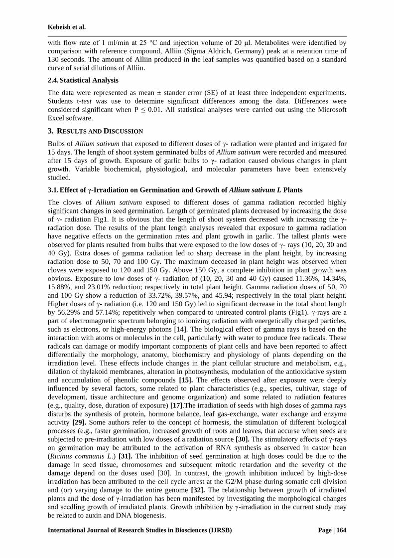
3.2. Effect of Gamma Radiation on Chlorophyll Content
The bioassay results, as illustrated in Fig2, show clear differences in chlorophyll A, B and carotenoids
in the leaves of garlic seedlings resulted from bulbs that were exposed to different doses of γ-
radiation. There is a gradual decrease in the measured chlorophyll contents. Higher doses of γ-
radiation cause a great reduction in chlorophyll contents in Allium sativum. It is obvious that total
chlorophyll pigments and carotenoid pigments were decreased with increasing the doses of γ-
radiation. All the irradiated plantlets exhibited the amount of chlorophyll content as compared to the
non-irradiated plantlets. Exposure to low doses of γ-radiation at 20 Gy caused 19.31% and 11.55%
reduction in both chlorophyll A and B; respectively when compared to untreated control plants.
However, higher doses of γ- radiation at 120 Gy led to sharp decrease in chlorophyll A and B by ~
64.04% and 58.48; respectively. Carotenoids content of garlic plant also show subsequent decrease as
the doses of γ-radiation increased. The lowest level of total carotenoids was obtained in garlic
seedlings irradiated with 120 Gy, hence the carotenoids contents decreased by 73.70%. Exposure to
low doses of γ-radiation (i.e. 20 Gy) caused only a reduction of 18.96% in the total carotenoids
contents. These results indicate that pigments fractions were decreased regularly by increasing the
dose of gamma. The chlorophyll content showed irregular distribution among the irradiated plantlets.
It can be observed that the concentration of chl. A was relatively higher than chl. B in both irradiated
and non-irradiated plantlets. It has been reported that γ-irradiation resulted in greater reduction in the
amount of chlorophyll B as opposed to chlorophyll A [33]. These effects include changes in the plant
cellular structure and metabolism [38]. Plastids were affected by irradiation in two ways: (i) inhibition
of senescence and (ii) dedifferentiation into a granal stage [14]. The developmental regression of
chloroplasts can be assumed primarily from destruction of grana [34]. The irradiation of seeds with
high doses of γ- rays disturbs the synthesis of protein, hormone balance, leaf gas exchange, water
exchange, and enzyme activity as previously reported [29]. Higher γ-irradiation inhibits chlorophyll
Kebeish et al.
International Journal of Research Studies in Biosciences (IJRSB) Page | 166
synthesis in wheat [16]. Therefore, it can be concluded that the decrease in pigment fractions in
irradiated plants is due to the destructive effects of γ-radiation on DNA and protein synthesis in
irradiated Allium sativum plantlets.
Fig2. Effect of γ-radiation on Chl. A, Chl. B, and Carotenoids content (µg/g fresh weight) in Allium sativum.
Data are means of three independent measurements. Vertical bars represent standard error. (*), (**) and (***)
represent statistically significant differences when compared with untreated control at p < 0.05, at p < 0.01,
and at p < 0.001 levels; respectively.
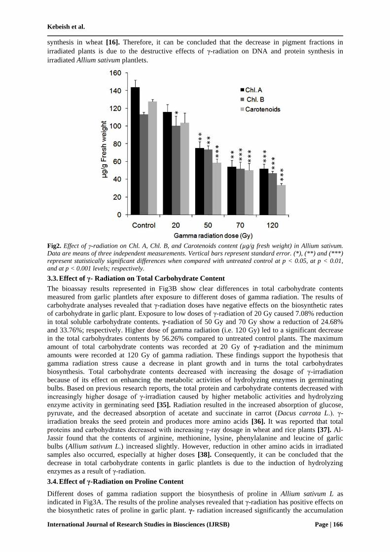
3.3. Effect of γ- Radiation on Total Carbohydrate Content
The bioassay results represented in Fig3B show clear differences in total carbohydrate contents
measured from garlic plantlets after exposure to different doses of gamma radiation. The results of
carbohydrate analyses revealed that γ-radiation doses have negative effects on the biosynthetic rates
of carbohydrate in garlic plant. Exposure to low doses of γ-radiation of 20 Gy caused 7.08% reduction
in total soluble carbohydrate contents. γ-radiation of 50 Gy and 70 Gy show a reduction of 24.68%
and 33.76%; respectively. Higher dose of gamma radiation (i.e. 120 Gy) led to a significant decrease
in the total carbohydrates contents by 56.26% compared to untreated control plants. The maximum
amount of total carbohydrate contents was recorded at 20 Gy of γ-radiation and the minimum
amounts were recorded at 120 Gy of gamma radiation. These findings support the hypothesis that
gamma radiation stress cause a decrease in plant growth and in turns the total carbohydrates
biosynthesis. Total carbohydrate contents decreased with increasing the dosage of γ-irradiation
because of its effect on enhancing the metabolic activities of hydrolyzing enzymes in germinating
bulbs. Based on previous research reports, the total protein and carbohydrate contents decreased with
increasingly higher dosage of γ-irradiation caused by higher metabolic activities and hydrolyzing
enzyme activity in germinating seed [35]. Radiation resulted in the increased absorption of glucose,
pyruvate, and the decreased absorption of acetate and succinate in carrot (Dacus carrota L.). γ-
irradiation breaks the seed protein and produces more amino acids [36]. It was reported that total
proteins and carbohydrates decreased with increasing γ-ray dosage in wheat and rice plants [37]. Al-
Jassir found that the contents of arginine, methionine, lysine, phenylalanine and leucine of garlic
bulbs (Allium sativum L.) increased slightly. However, reduction in other amino acids in irradiated
samples also occurred, especially at higher doses [38]. Consequently, it can be concluded that the
decrease in total carbohydrate contents in garlic plantlets is due to the induction of hydrolyzing
enzymes as a result of γ-radiation.
3.4. Effect of γ-Radiation on Proline Content
Different doses of gamma radiation support the biosynthesis of proline in Allium sativum L as
indicated in Fig3A. The results of the proline analyses revealed that γ-radiation has positive effects on
the biosynthetic rates of proline in garlic plant. γ- radiation increased significantly the accumulation
Effects of Gamma Radiation on Growth, Oxidative Stress, Antioxidant System, and Alliin Producing
Gene Transcripts in Allium sativum
International Journal of Research Studies in Biosciences (IJRSB) Page | 167
of proline in Allium sativum at almost all γ- radiation doses. Exposure to low doses of γ- radiation of
20 Gy shows induction in total proline content, higher dose of gamma radiation (i.e. 70 Gy) led to a
significant increase in the total proline contents by compared to untreated control plants. The
maximum amount of total proline contents was recorded at 70 Gy of gamma after that the total proline
contents decreased. γ-radiation dose of 120 Gy show an increase in the total proline contents but still
higher than untreated control plants. The overall effects indicate that gamma radiation induces stress
in garlic cells with the elevation of proline contents. Gamma radiation was reported to induce
oxidative stress with overproduction of reactive oxygen species (ROS) such as superoxide radicals,
hydroxyl radicals and hydrogen peroxide, which react rapidly with almost all structural and functional
organic molecules, including proteins, lipids and nucleic acids causing disturbance of cellular
metabolism [39]. To avoid oxidative damage, plants have evolved various protective mechanisms to
counteract the effects of reactive oxygen species in cellular compartments [36]. Proline is a
compatible osmolyte and it may interact with enzymes to preserve enzyme structure and activities.
Indeed, proline has been shown to reduce In vitro enzyme denaturations caused due to heat, NaCl
stress, and gamma radiation stress [40].
Fig3. Effect of Gamma radiation on total proline contents (A) and total Carbohydrate contents (B) in Allium
sativum L. Data are means of three independent measurements. Vertical bars represent standard error. (*) and
(**) represent statistically significant differences when compared with untreated control plants at p < 0.05, and
p < 0.01 levels; respectively.
3.5. Effect of Gamma Irradiation on the Antioxidant Enzymes Activity
Different doses of gamma radiation support the biosynthesis of antioxidant enzymes in Allium sativum
L. Gamma radiation causes a dramatic variation in the activity of the different antioxidant enzymes
Catalase (CAT), Peroxidase (POD), Polyphenol oxidase (PPO), and Superoxide dismutase (SOD).
Different doses of gamma radiation support the increase in the activity of catalase in Allium sativum L
and showed highly significant changes in total catalase activity as indicated in Fig4. Irradiation dose
at 10 Gy to 70 Gy enhance the catalase activity of Allium sativum L plants. The maximum activity
was recorded in Irradiation dose of 70 Gy compared with corresponding control. After irradiation
dose 70 Gy, the activity of catalase enzyme declined. The maximum amount of total catalase activity
was recorded at 70 Gy of γ-radiation and the minimum amounts were recorded at 150 Gy of gamma
radiation.
Similar effects were observed after measuring the peroxidase activity in irradiated garlic plantlets.
The maximum peroxidase activity was recorded in at irradiation dose of 100 Gy compared with
corresponding control. After irradiation dose 100 Gy, the activity of peroxidase enzyme declined.
Kebeish et al.
International Journal of Research Studies in Biosciences (IJRSB) Page | 168
Exposure to γ-irradiation cause changes in biochemical activity through various metabolites produced
as function of γ-irradiation. The most important of these metabolites are peroxyl radicals. The
radiation protective effect of peroxidase is due to removal of H2O2, peroxides, and especially lipid
hydrogen peroxides. This accounts for the greater effectiveness of peroxidases than catalase [41]. The
induction of POD by the irradiation would be one of the defense systems activated through the ROS-
mediated cellular signaling [42]. It was suggested therefore that the increase in gamma doses lead to
an increase in the specific activity of peroxidase enzyme [43].
The results of the PPO activity measurements illustrated in Fig4 revealed that doses of gamma
radiation increased the activity of polyphenol oxidase in garlic plants. The maximum activity was
recorded at irradiation dose of 70 Gy compared with corresponding control. After irradiation dose 70
Gy, the activity of PPO enzyme declined. Jimenez et al, found that green onion exposure to γ-
radiation produces slight increases in the polyphenol concentrations [44]. On the other hand, other
studies indicated no significant differences in the residual PPO activity after the radiation dose [45,
46].
Different doses of gamma radiation support the activity of superoxide dismutase in Allium sativum L
as indicated in Fig4. The maximum activity was recorded in irradiation dose at 70 Gy compared with
corresponding control. After irradiation dose 70 Gy, the activity of superoxide dismutase enzyme
declined. The minimum SOD activity was recorded at 150 Gy of γ-radiation.
Superoxide dismutase (SOD) isozymes are compartmentalized in higher plants and play a major role
in combating oxygen radical mediated toxicity. Cells exhibiting high levels of SOD, catalase, and
peroxidase activity are relatively less vulnerable to secondary effects of radiation. Since superoxide is
sufficiently stable to permit diffusion within the cell. It is possible that it acts as an electron donor to
transition metals in irradiated tissue and may account for sensitization of cells to the effects of H2O2
by radiation exposure and the protection afforded by SOD to irradiating cells [47]. Significant
differences in the changes in the two antioxidant enzymes activities were noted according to the
developmental stage. Superoxide dismutase activity was conversely increased from 2.9 U/mg protein
(non-irradiation) to 11.3 U/mg protein (800 Gy treatment) at the reproductive stage [48]. However,
SOD levels of irradiated plants were lower than non-irradiated plants at vegetative stage. The activity
of free radical scavenging enzymes peroxidase, catalase and superoxide dismutase showed inverse
relationships with ageing period and direct proportion to reductions in the seed germination level[49].
A major, direct target of γ-irradiation that is probably the most important one is the water molecule,
which is omnipresent in organisms. The primary reactions are excitation and ionization, which
produce ionized water molecules (H2O˙+) and the radicals H˙ and ˙OH. However, in biological tissue
these ionizations are induced all along the path of the radiation and lead to chain reactions, which
produce secondary reactive oxygen species (ROS) as a result of H˙ and eaq– becoming trapped. The
most important ROS is H2O2; singlet O2– is produced to very low extent, depending on the O2
concentration [50]. The ˙OH radical can react rapidly with various types of macromolecules,
including lipids, proteins and, in particular, DNA. However, some of the resulting injuries can be
readily repaired and recovered, depending on the dose range. One result of oxidative stress is cellular
damage by hydroxyl radical attack. The amount and rate of hydroxyl radicals generation from ROS is
controlled partly by the cellular antioxidant status and partly by the availability of systems capable of
reducing (or activating) superoxide or hydrogen peroxide. It has been shown that the effects of H2O2
resemble those of ionizing radiation [51]. Therefore, the increased level of antioxidant enzymes in
Allium sativum L plantlets is strongly dependent on the gamma irradiation doses.

Effects of Gamma Radiation on Growth, Oxidative Stress, Antioxidant System, and Alliin Producing
Gene Transcripts in Allium sativum
International Journal of Research Studies in Biosciences (IJRSB) Page | 169
Fig4. Effect of gamma radiation on the activity of Catalase (A), Peroxidise (B), Polyphenol oxidase (C), and
Superoxide dismutase (D) in Allium sativum. Catalase (CAT), Peroxidase (POD), Polyphenol oxidase (PPO),
and Superoxide dismutase (SOD) enzymatic activities measured from Allium sativum exposed to different doses
of gamma radiation. Data are means of three independent measurements. Vertical bars represent standard
error.
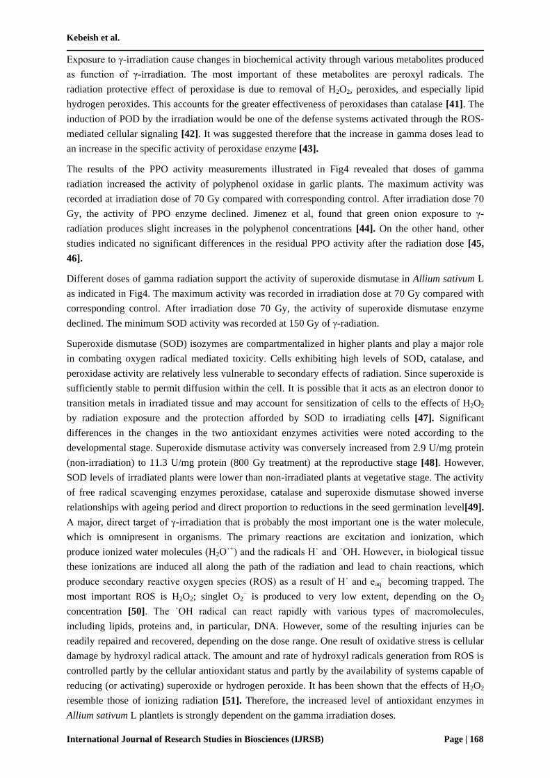
3.6. Quantitative Real-time RT-PCR Analysis of Alliinase Transcripts
Alliinase is the basic enzyme involved in Alliin biosynthetic pathway in galic plants. Thus, the
Alliinase gene expression was tested in A. Sativum L plants generated from garlic bulbs that was
irradiated with variable doses of gamma radiation (i.e. 20, 50, 70, and 120 Gy). For this purpose,
Alliinase mRNA transcript accumulation was measured by Real-Time RT-PCR as shown in Fig5. The
amount of Alliinase signal in the different RNA preparations was standardized for the abundance of
the transcript from the house keeping gene, Actin2. Variable amounts of Alliinase signals in the
different RNA preparations isolated from the A. Sativum L plants were observed based on the level of
gamma irradiation doses. It was obvious that gamma irradiation has inhibitory effects on Alliinase
gene expression. Interestingly, the expression level of Alliinase gene was correlated with the strength
of gamma radiation doses (Fig5). The highere levels of Alliinase expression were observed at low
irradiation doses (i.e. 20 and 50 Gy) whereas the maximum inhibition of Alliinase gene expression
was obsreved to be at 120 Gy. Similar approaches have been performed to study the effect of heavy
metals like copper on photosynthetic related gene transcripts in some algae species [52]. The authors
observed a great reduction in phtosynthetic related gene transcripts upon exposure to heavy metal
stress. Based on these findings, it can be concluded that Alliinase gene expression is decreased by
increasing the gamma irradiation dose.
Kebeish et al.
International Journal of Research Studies in Biosciences (IJRSB) Page | 170
Fig5. Reverse transcriptase RT-PCR analysis of Alliinase gene expression in A. sativum L plants.
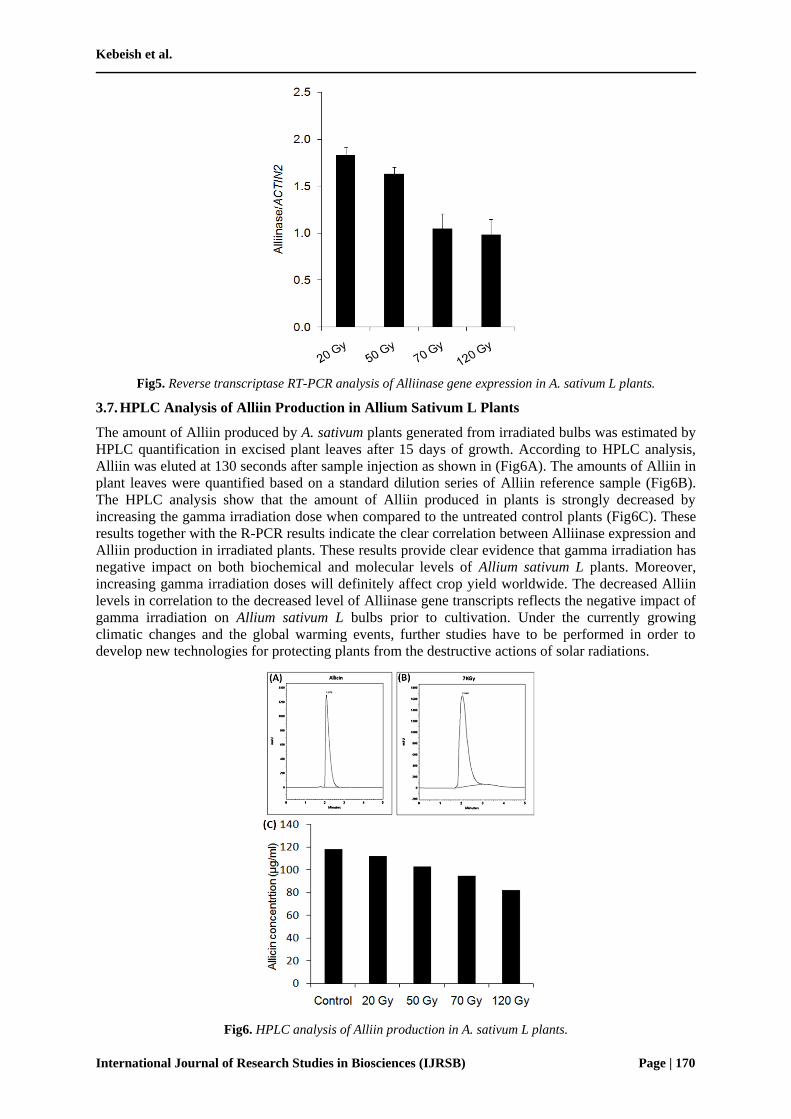
3.7. HPLC Analysis of Alliin Production in Allium Sativum L Plants
The amount of Alliin produced by A. sativum plants generated from irradiated bulbs was estimated by
HPLC quantification in excised plant leaves after 15 days of growth. According to HPLC analysis,
Alliin was eluted at 130 seconds after sample injection as shown in (Fig6A). The amounts of Alliin in
plant leaves were quantified based on a standard dilution series of Alliin reference sample (Fig6B).
The HPLC analysis show that the amount of Alliin produced in plants is strongly decreased by
increasing the gamma irradiation dose when compared to the untreated control plants (Fig6C). These
results together with the R-PCR results indicate the clear correlation between Alliinase expression and
Alliin production in irradiated plants. These results provide clear evidence that gamma irradiation has
negative impact on both biochemical and molecular levels of Allium sativum L plants. Moreover,
increasing gamma irradiation doses will definitely affect crop yield worldwide. The decreased Alliin
levels in correlation to the decreased level of Alliinase gene transcripts reflects the negative impact of
gamma irradiation on Allium sativum L bulbs prior to cultivation. Under the currently growing
climatic changes and the global warming events, further studies have to be performed in order to
develop new technologies for protecting plants from the destructive actions of solar radiations.
Fig6. HPLC analysis of Alliin production in A. sativum L plants.

Effects of Gamma Radiation on Growth, Oxidative Stress, Antioxidant System, and Alliin Producing
Gene Transcripts in Allium sativum
International Journal of Research Studies in Biosciences (IJRSB) Page | 171
A) representative chromatogram of Alliin reference sample, B) representative chromatogram of HPLC analysis
of Alliin from Allium sativum L plant leaves extracts isolated from irradiated bulbs with 70 Gy gamma, and C)
Alliin amounts produced in Allium sativum L plants at all gamma irradiation doses.
4. CONCLUSION
The results of the current study indicated the negative effects of gamma radiation on biochemical and
molecular characteristics. Gamma radiation increased the level of antioxidant enzyme system, proline
contents. However, it causes a great reduction in plant growth, chlorophyll contents, and Alliin levels
in irradiated Allium sativum L plantlets.
ACKNOWLEDGEMENTS
This study was supported partly by the Egyptian Science and Technology Development Fund (STDF),
Botany Department, Faculty of Science, Zagazig University, Egypt, and also supported by Biology
Department, Faculty of Science Yanbu, Taibah University, KSA. The authors are thankful to M.Sc:
Emad Hamdy, Plant Biotechnology Lab., Botany Department, Faculty of Science, Zagazig University,
for his valuable help with cDNA synthesis and RT-PCR analysis.
REFERENCES
[1] Akgu l, A. Spice science and technology.Turkish Association of Food Technologists, Ankara,
Publ. 15, 451(1993).
[2] Ahmad J. Garlic-a panacea for health and good taste. Nutrition and Food Science 5, 32-
35(1996).
[3] Kim J.Y. Alliinase-independent inhibition of Staphylococcus aureus B33 by heated garlic.
Journal of Food Science. 67, 780-785(2002).
[4] Block E., Ahmad S., Catalfamo J.L., Jain M.K. and Apitz C.R. Antithrombotic organo
sulfur compounds from garlic: structural, mechanistic. and synthetic studies. Journal of the
American Chemical Society. 108, 7045-7055. (1986).
[5] Mousa A.S. Discovery of angiogenesis inhibition by garlic ingredients: Potential anti-cancer
benefits. FASEB 15, A117(2001).
[6] Wu C.C., Sheen L.Y., Chen H.W., Tsai S.J. and Lii, C.K. Effects of organo-sulfur compounds
from garlic oil on the antioxidation system in rat liver and red blood cells. Food and Chemical
Toxicology. 39, 563-569(2001).
[7] Kang N.S., Moon E.Y., Cho C.G. and Si P. Immunomodulating effect of garlic component.
allicin. on murine peritoneal macrophages. Nutrition Research. 21, 61 61-626(2001).
[8] Lawson L.D., Wang Z.J. and Papadimitnou, D. Allicin release under simulated
gastrointestinal conditions from garlic powder tablets employed in clinical trials on serum
cholesterol. Pluntu Medica. 67, 13-18(2001).
[9] Sovova Effect of allicin from garlic powder on serum lipids and blood pressure in rats fed with a
high cholesterol diet. Prostaglandins Leukotrienes & Essential Fatty Acids. 62, 253-259(2000).
[10] Jabbari A., Argani H., Ghorbanihaghjo A. and Mahdavi R. Comparison between swallowing
and chewing of garlic on levels of serum lipids, cyclosporine, creatinine and lipid peroxidation in
renal transplant recipients. Lipids Health Diseases. 4, 1-4(2005).
[11] Rahman K. and Lowe G.M. Garlic and cardiovascular disease. A critical review. Journal of
Nutrition 136, 736-740(2006).
[12] Ejaz S., Woong L.C. and Ejaz A. Extract of garlic (Allium sativum) in cancer chemoprevention.
Experimental Oncology. 25, 93-97(2003).
[13] Galeone C., Pelucchi C., Levi F., Negri E., Franceschi S. and Talamini R. Onion and garlic
use and human cancer. American Journal of Clinical Nutrition. 84, 1027-1032(2006).
[14] Kim J.H., Baek M.H., Chung B.Y., Wi S.G. and Kim J.S. Alterations in the photosynthetic
pigments and antioxidant machineries of red pepper (Capsicum annuum L.) seedlings from
gamma irradiated seeds. Plant Biology 47, 314-321(2004).

Kebeish et al.
International Journal of Research Studies in Biosciences (IJRSB) Page | 172
[15] Wi S.G., Chung B.Y., Kim J.H., Baek M.H., Yang D.H. and Lee J.W. Ultrastructural changes
of cell organelles in Arabidopsis stem after gamma irradiation. J. Plant Biol. 48(2),195
200(2005).
[16] Kovacs E. and Keresztes A. Effect of gamma and UV-B/C radiation on plant cells. Micron 33,
199-210(2002).
[17] Jan S., Parween T., Siddiqi T.O. and Uzzafar M. Effect of gamma radiation on
morphological, biochemical and physiological aspects of plants and plant products. Environment
Review 20, 17-39(2012).
[18] Nybom N. The pigment characteristics of chlorophyll mutations in barley. Heriditas 41, 510
512(1955).
[19] Rau, M. H., and Senger, H. (1965). Untersuchungen zur Synchronisierbarkeit einzlner
Pigmentmangel-Mutanten von Chlorella. Planta 65, 186-194.
[20] Stitt, M., Lilley, R., Gerhardt, R., and Heldt, H. (1989). Determination of metabolite levels in
specific cells and subcellular compartments of plant leaves. Methods in Enzymology 174, 518-
552.
[21] Dubois M., Gills K.A., Hamilton J.K., Rebers P.A. and Smith F. Colorimetric method for
determination of suger and related substances. Anal. Chem. 28, 350-356(1956).
[22] Bates L.S., Walds R.P. and Teare I.D. Rapid determination of free proline for water stress
studies. Plant and Soil, 39, 205-207(1973).
[23] Kar M. and Mishra D. Catalas, peroxidase, and polyphenol oxidase activities during rice leaf
senescence. Plant Physiol. 57, 315-319(1976).
[24] Aebi, H. (1984). Catalase in Vitro. Methods in Enzymology 105, 121-126.
[25] Shannon L., Key E. and Lew J. Peroxidase isoenzymes from horseradish roots.1.Isolation and
physical properties Biol. Chem. 241, 2166-2175(1966).
[26] Beauchamp C. and Fridovich I. Superoxide Dismutase: Improve assays and an assay
applicable to acrylamide gels. Anal.Biochem. 44, 276-287(1971).
[27] Niessen M, Thiruveedhi K, Rosenkranz R, Kebeish R, Hirsch H-J, Kreuzaler F,
Peterhansel C Mitochondrial glycolate oxidation contributes to photorespiration in higher
plants. J. Exp. Bot. 58: 2709-2715(2007)
[28] Chomczynski P, Mackey K Substitution of chloroform by bromo-chloropropane in the single
step method of RNA isolation. Anal Biochem 225: 163-164(1995)
[29] Hameed A., Shah T.M., Atta B.M., Haq M.A. and Sayed H. Gamma irradiation effects on
seed germination and growth, protein content, peroxidase and protease activity, lipid
peroxidation in desi and kabuli chickpea. Pakistan Journal Botany 40(3), 1033-1041(2008).
[30] Thapa C.B. Effect of acute exposure of gamma rays on seed germination of Pinus kesiya Gord
and P. wallichiana A.B. Jacks. Botanica Orientalis. Journal Plant Science 2, 120-121(1999).
[31] Kuzin A.M., Vagabova M.E. and Revin A.F. Molecular mechanisms of the stimulating action
of ionizing radiation on seeds. 2. Activation of protein and high molecular RNA synthesis.
Radiobiology 16, 259-261(1976).
[32] Preussa S.B. and Britta A.B. A DNA-damage-induced cell cycle checkpoint in Arabidopsis.
Genetics, 164, 323-334(2003).
[33] Strid A., Chow W.S. and Anderson J.M. Effects of supplementary gamma irradiation on
photosynthesis in Pisum sativum. Biochem., 1020(1), 206 - 268(1990).
[34] Kovács E. and Keresztes A. The effect of irradiation on starch content in Golden Delicious
apples. Food Microbiol. 8:67–74(1989).
[35] Maity J.P., Chakraborty A., Saha A., Santra S.C. and Chanda S. Radiation induced effects
on some common storage edible seeds in India infested with surface microflora. Radiat. Phys.
Chem. 71(5): 1065–1072(2004).
[36] Kiong A., Ling Pick A., Grace Lai S.H. and Harun A.R. Physiological responses of
Orthosiphon stamineus plantlets to gamma irradiation. Am-Eurasian J. Sustain. Agric 2(2), 135-
149(2008).

Effects of Gamma Radiation on Growth, Oxidative Stress, Antioxidant System, and Alliin Producing
Gene Transcripts in Allium sativum
International Journal of Research Studies in Biosciences (IJRSB) Page | 173
[37] Inoue M., Hasegawa H. and Hori S. Physiological and biochemical changes in gamma
irradiated rice. Radiat. Bot. 15(4): 387–395(1975).
[38] Al-Jassir M.S. Chemical composition and microflora of black cumin (Nigella sativa L.) seeds
growing in Saudi Arabia. Food Chem. 45(4): 239–242(1992).
[39] Noreen Z. and Ashraf M. Changes in antioxidant enzymes and some key metabolites in some
genetically diverse cultivars of radish (Raphanus sativus L.). Environ. Exp. Bot., 67, 395-
402(2009).
[40] Ashraf M. and Foolad M.R. Roles of glycine betaine and proline in improving plant abiotic
stress resistance. Environ. Exp. Bot., 59(2), 206-216(2007).
[41] Croute F., Delaporte E., Bonnefoy J.Y., Fertin C., Thivolet J. and Nicolas J.F. Interleukin-
1 beta stimulates fibroblast elastase activity. Br. J. Dermatol, 124: 538–541(1991).
[42] Wi S.G., Chung B.Y., Kim J.S., Kim J.H., Baek M.H., Lee J.W. and Kim Y.S. Effects of
gamma irradiation on morphological changes and biological responses in plants. Micron 38(1),
553-564(2006).
[43] Sah N.K., Prramanik S. and Raychaudhuri, S.S. Peroxidase changes in barley induced by
ionizing and thermal radiation. . Institute J. Radiology Biol. 69(1), 107-117(1996).
[44] Jimenez L., Alarcon E., Trevithick-Sutton C., Gandhi N., and Scaiano J.C. Effect of
gamma-radiation on green onion DNA integrity: Role of ascorbic acid and polyphenols against
nucleic acid damage. Food Chem., 128, 735-741(2011).
[45] Falguera V., Pagan J., Garza S., Garvin A., and Ibarz A. Inactivation of polyphenol oxidase
by ultraviolet irradiation: Protective effect of melanins. Food Eng. 110(2), 305-309(2011).
[46] Latorre M.E., Narvaiz P., Rojas A.M. and Gerschenson L.N. Effects of gamma irradiation on
bio-chemical and physico-chemical parameters of fresh-cut red beet (Beta vulgaris L. var.
conditiva) root. Food Eng. 98, 178-191(2010).
[47] Petkau A. Role of superoxide dismutase in modification of radiation injury. Br. J. Cancer Suppl.
8:87–95(1987).
[48] Kim D.S., Kim J.B., Goh E.J., Kim W.J., Kim S.H. and Seob Y.W. Antioxidant response of
Arabidopsis plants to gamma irradiation: Genome-wide expression profiling of the ROS
scavenging and signal transduction pathways. Plant Physiology. 168, 1960-1971(2011).
[49] Rao R.G.S., Singh P.M. and Rai M. Storability of onion seeds and effects of packaging and
storage conditions on viability and vigour. Sci. Horticult . 110, 1-6(2006).
[50] Lee M.H., Moon Y.R., Chung B.Y., Kim J.S., Lee K.S., Cho J.Y. and Kim J.H. Practical use
of chemical probes for reactive oxygen species produced in biological systems by g- irradiation.
Radiat. Phys. Chem. 78(5): 323–327(2009).
[51] Riley P.A. Free radicals in biology: Oxidative stress and the effects of ionizing radiation. Int. J.
Radiat. Biol. 65(1): 27–33(1994).
[52] Kebeish R., El-Ayouty Y., Hussain A. Effect of copper on growth, bioactive metabolites,
antioxidant enzymes and photosynthesis-related gene transcription in Chlorella vulgaris, World
Journal of Biology and Biological Sciences, Vol. 2 (2), 34-43 (2014).

Kebeish et al.
International Journal of Research Studies in Biosciences (IJRSB) Page | 174
AUTHORS’ BIOGRAPHY
Rashad Kebeish (Ph.D)
- Ph. D in Plant Molecular Biology from RWTH-Aachen Germany 2006.
- Assistant Professor degree in Plant Molecular Biology and plant
Physiology from Zagazig University, Egypt 2012.
Permanent Affiliation:
Botany Department, Faculty of Science, Zagazig University, Egypt
Current Affiliation: Faculty of Science, Yanbu, Taibah University, KSA
Hanan, E. Deef (Ph.D)
- Ph.D in Plant Physiology from Zagazig University, Egypt.
- Assistant Professor degree in Plant Physiology from Zagazig University, Egypt.
Affiliation: Botany Department, Faculty of Science, Zagazig University, Egypt
Nagwa El-Bialy (M.Sc)
- MSc in Plant Ecology from Zagazig University, Egypt
Affiliation: Botany Department, Faculty of Science, Zagazig University, Egypt