the protective role of septilin against gamma radiation
TRANSCRIPT
The Egyptian Journal of Hospital Medicine (2007) Vol., 27: 176– 187
The Protective Role of Septilin Against Gamma Radiation-Induced
Testicular Toxicity in Rats
Omaima Soliman Eissa* and Nehal Aly Moustafa** *National Center for Radiation Research and Technology, Atomic Energy Authority:
** Faculty of Science , Al-Azhar University, Cairo, Egypt
Abstract
Backgrounds: This study deals with evaluation of the histological and some
histochemical alterations in rat testes induced by whole body gamma irradiation as well as evaluation of the protective effect of septilin (a herbal preparation) against these effects.
Results : The obtained results indicated that doses of (3 Gy and 6 Gy) gamma radiation
have testicular toxic effects in rats. The histological lesions observed in the testes varied
between vacuolation, swelling, pyknosis and even necrosis in some spermatogenic cells as well as significant depletion in the number of spermatogonia, primary spermatocytes, secondary
spermatocytes and spermatids. The histochemical observations revealed diminution in the
polysaccharides content and increase in the collagen fibres in the testis of irradiated animals. These effects were mostly perceptive with the high dose of the radiation than with the lower
one. Treatment with septilin (a herbal preparation) for one week followed by gamma radiation
proved that septilin has a protective effect against gamma radiation-induced toxic effects in the testes of rats; where most of the histological and histochemical changes observed due to
irradiation were minimized to a large extent; however there was no complete protection.
Conclusion: Thus, this study concluded that gamma-irradiation exerts toxic effects in the
testes of rats and pre-treatment with septilin inhibits these toxic effects, which in turn advocate using such herbal extract as a radioprotector.
Key words: Septilin, γ-radiation, Histology, Testis, Rats.
Introduction
Radiation can change the charact-
eristics of the cell nucleus and cytoplasm, as mammalian germ cells are very sensitive
to ionizing radiation (Dobson and Felton,
1983). Ionized radiation, being one of the
environmental cytotoxic factors, causes death of the germinal cells and therefore,
sterility (Meistrich, 1993 and Georgieva et
al., 2005). The testis is a main target organ of
radiation damage; it has been reported that
irradiation induces several histopathological consequences in the organs and tissues,
including the increase of loose fibrous
tissue (Matalka et al., 1994 and Yang et al.,
2000). Moreover, ionizing radiation of normal tissue results in fibrosis, which is
perhaps the most effect of radiation (Geraci
et al., 1992), in the testis, resulting in depletion of more advanced spermatogenic
cells (Kangasniemi et al., 1990). The
differentiating spermatogonia are very
sensitive to radiation and are killed under
the effect of doses less than 3 Gy in the Sprague-Dawley rat (Kangasniemi et al.,
1990), in humans (Rowley et al., 1974), and
in mice (Mian et al., 1977).
The radioprotective agents are compounds that are administered before
exposure to ionizing radiation to reduce its
damaging effects, including radiation-induced lethality (Satoh et al., 2003). The
discovery of radioprotectors for the first
time seemed to be very promising (in radiotherapy) and has attracted the interest
of a number of radiobiologists. Although
synthetic radioprotectors such as the
aminothiols have yielded the highest protective factors; typically they are more
toxic (Rades et al., 2004) than naturally
occurring protectors (Weiss and Landauer, 2003). Thereafter different plant extracts
were tested against radiation effects and
showed potential radioprotective activities
176
Omaima Soliman Eissa & Nehal Aly Moustafa
177
in mammals (Uma Devi et al., 2000;
Lamartiniere et al., 2002; Jagetia et al.,
2004 and Landauer et al., 2005). The effect of various doses (5, 10, 20, 40, 60, 80, 100,
120, 140 and 160 mg/kg b. wt.) of 50%
ethanolic extract of Septilin (a herbal
preparation) on the radiation-induced mortality was studied in mice exposed to 10
Gy of gamma-irradiation daily. Treatment
of mice with different doses of septilin, for 5 days before irradiation, delayed the onset
of mortality and reduced the symptoms of
radiation sickness when compared with the
drug untreated irradiated controls (Jagetia and Baliga, 2004). The present work aimed
to investigate whether septilin pretreatment
could prevent gamma radiation-induced histological damage in the testes of rats.
Material And Methods
The experimental animals in the present investigation were 40 male albino
rats (Rattus norvegicus) with average body
weights 100-130 g., obtained from the
Egyptian Organization for Vaccine and Biological Preparations at Helwan. Animals
allowed a period of two weeks pre-
experimentation to be adapted to the laboratory conditions. Food and water were
supplied daily ad libitum. Animals were
divided into four main groups; the first one (10 rats) sited as control. The second group
(6 rats) was treated with septilin in a daily
oral dose (100mg/kg b. wt.) for one week
(positive control). The third group (12 rats) was divided into two equal subgroups; the
first one was subjected to whole body γ -
irradiation at a sub-lethal single dose of 3Gy and the second one subjected to whole
body γ -irradiation at a sub-lethal single
dose of 6Gy. The fourth main group (12
animals) received septilin in a daily oral dose of 100mg/kg b. wt. for one week, and
then divided into two equal subgroups; the
first one subjected to whole body γ -irradiation at a sub-lethal single dose of
3Gy and the second one was subjected to
whole body γ -irradiation at a sub-lethal single dose level of 6Gy. All the groups
were sacrificed after one week post
irradiation.
Whole body γ-irradiation was
performed using Cesium 137 Biological γ-
cell-40 irradiation facility belonging to The National Center for Radiation Research and
Technology (NCRRT). The dose rate was 1
Gy/1.42 min at the time of the experiment.
The herb used in the present investigation was septilin Himalaya (a
product of heral healthcare), Drug Co.
Private Ltd; it is an Ayurvedic herbal preparation containing various herbs and
minerals. Its main ingredients are (in mg):
Balsamodendron mukul 162; Sank Bhasma
32: Maharasnadi qoath, 65; Tinospora cordifolia, 49; Rubia cordifolia, 32;
Emblica officinalis, 16; Moringa pterigos-
perma, 16: Glycyrrhiza glabra, 6. It has been reported to have anti-bacterial, anti-
inflammatory, anti-exudative and immuno-
stimulatory effect (Rom, 1984; Kumar et al., 1993 and Sharma and Ray, 1997). The
dose used in this study was 100mg/kg b.
wt./day as oral administration using a
stomach tube for 7 days (Jagetia and Baliga, 2004).
The testes of control and treated
animals were excised, washed with normal saline and fixed in Bouin’s solution for 24
to 48 hours. They were dehydrated in gra-
ded ethanol series, embedded in paraplast, cut in sections at a thickness of 6un, stained
with Harris haematoxylin and eosin.
Masson's trichrome stain was applied for
demonstration of collagen fibres (Pearse, 1972). PAS stain was used for demonstr-
ation of the polysaccharides (Mc Manus,
1946). Statistical significancy among the
groups was performed using Student’s t-
test.
Results
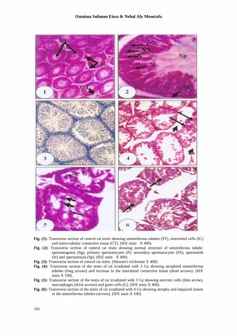
The control group:
The parenchyma of the testis of albino
rat, Rattus norvegicus is composed of many seminiferous tubules of varying sizes; each
is surrounded by an outer thin layer of
connective tissue and lined with sperma-togenic epithelium that surrounds a central
lumen. The epithelium consists of Sertoli
cells and spermatogenic cells. The
The Protective Role of Septilin Against Gamma…….
178
interstitial cells are found mostly in groups
between the seminiferous tubules; they are
large and ovoid with rounded nuclei (Fig. 1). The normal structures of Sertoli cells,
spermatogenic cells and spermatozoa are
depicted in figure (2). Table (1) and
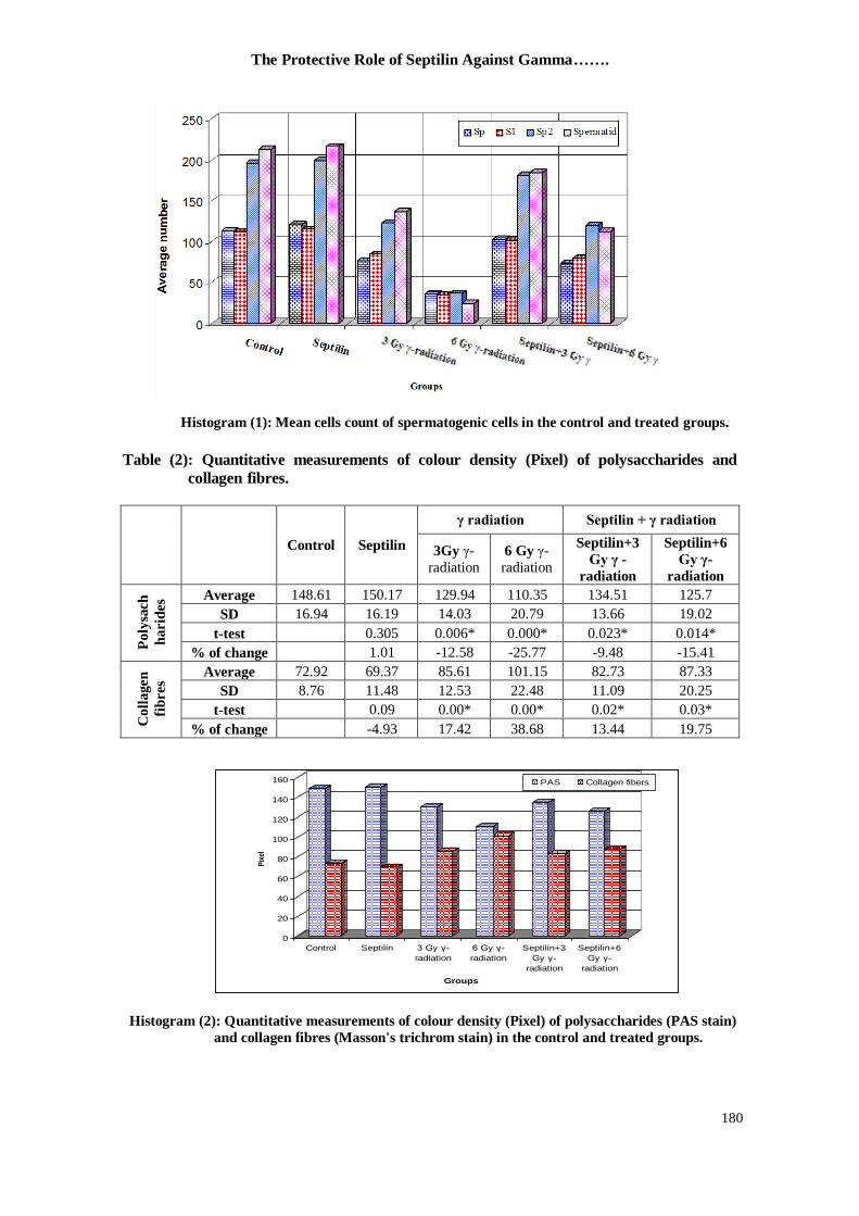
histogram (1) show the mean count of the spermatogenic series in the control and
treated groups. Figure (3) displays the
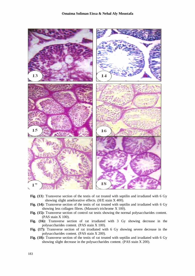
collagen fibres in the testes of the control animals. The average of the polysaccharides
content of the testes of the control animals
(Fig. 15), indicated by PAS stain, recorded
148.61 (Table 2 and Histogram 2).
Septilin- treated group:
Investigated testes sections obtained from rats treated with septilin at a daily oral
dose of 100 mg/kg b. wt. exhibited closely
similar structure to those of the control animals denoting that septilin has no
cytotoxic effects on the testes of rats.
Nonsignificant increases in the numbers of
the spermatogonia, primary spermatocytes, secondary spermatocytes and spermatids
were recorded (Table 1). The amount of the
collagen fibres in the testis recorded insignificant reduction in comparison with
control. The average of the polysaccharides
content of the testis in this group recorded insignificant increase in comparison with
control (Table 2).
Effect of γ –irradiation: Different histopathological lesions
were noticed in the testes of rats one week
post exposure to a single dose (3 Gy) of gamma irradiation. The seminiferous
tubules were mostly atrophied and the
interstitial connective was widened with
less number of interstitial cells (Fig. 4). The spermatogenic cells were irregularly
distributed within the seminiferous tubules
and the basement membrane of the tubules was abnormal. Moreover, clumped
chromatin in the nuclei and vacuolated
cytoplasm were encountered in most of the spermatogenic cells; some cells appeared
necrotic and tissue infiltration with
macrophages was perceptive (Fig. 5). In
addition, depletion in spermatozoa, presence of giant cells, congestion in the
blood vessels and haemorrhage in the
interstitial tissue were noticed. The present
data showed significant reduction in the
number of spermatogonia, primary spermat-ocytes, secondary spermatocytes and
spermatids as a resuts of exposure to 3Gy
gamma irradiation (Table 1). Significant
increase in the collagen fibres in the testes of rats irradiated with 3Gy was noticed
(Fig. 8); the percentage of increment
recorded 17.42%. Significant reduction in the polysaccharides content was recorded as
compared with the control animals; the
percentage of reduction was 12.58% (Fig.
16 and Table 2). Severe histological changes were
observed in the testes of rats one week post
exposure to a single dose (6Gy) of gamma radiation. Atrophy and obliterated lumen
with absence of spermatozoa in the
seminiferous tubules, increase of the interstitial connective tissue and depletion
in the interstitial cells (Leyding cells) were
the most prominent features in the testes
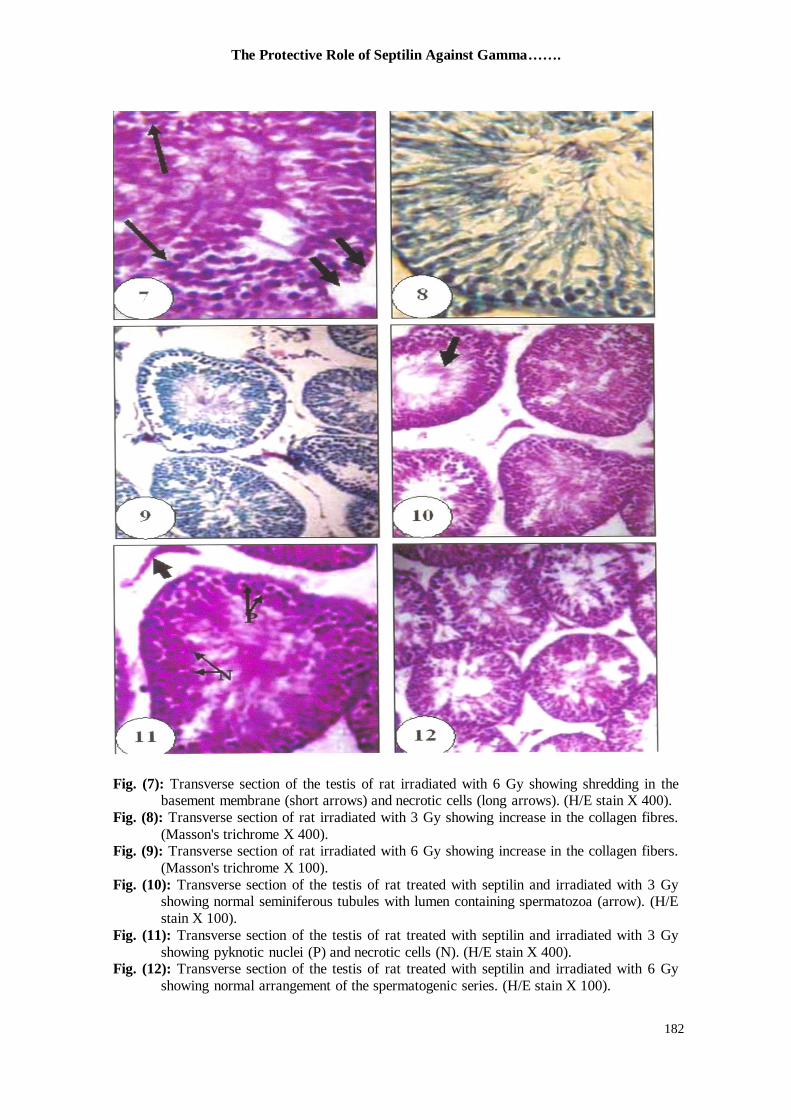
(Fig. 6). Also, degenerated spermatogenic cells, shredding in the basement membrane
that detached from the overlying cells,
depletion in the spermatogonia and prevailing necrosis were ratified in the
testicular tissue (Fig. 7). Significant
reduction in the number of spermatogonia, primary spermatocytes, secondary
spermatocytes and spermatids were found
as results of exposure to 6Gy gamma
irradiation, the percentages of these reduction were 68.25%, 68.87%, 81.31%
and 88.43%, respectively (Table 1).
Obvious significant increase in the collagen fibres in the testes of rats irradiated with
6Gy was recorded and reached 38.68%
(Fig. 9). Significant reduction in the
polysaccharides content was recorded as compared with control animals; the
percentage of reduction was 25.77% (Fig.
17 and Table 2).
Septilin plus γ –irradiation:
Obvious ameliorative effect was detected in the testes of 3 Gy gamma-
irradiated rats treated with septilin one week
prior to exposure. Most of the histopatho-
logical lesions observed in the testes of irradiated rats were disappeared and the
seminiferous tubules (Fig.10) had a feature
Omaima Soliman Eissa & Nehal Aly Moustafa
179
approximately similar to those of the
control. The spermatogenic cells appeared
regularly arranged within the tubules, nor-mal tubular lumen contained spermatozoa
was greatly encountered and the basement
membrane of the seminiferous tubules was
mostly normal. Otherwise, the interstitial connective tissue was still increased and
exhibited vanish in the interstitial cells.
Also, few of the seminiferous tubules showed some histopathological changes that
included pyknotic and necrotic cells in the
spermatogenic series (Fig. 11). The number
of the cells in the spermatogenic series exhibited a high increase when compared
with those of irradiated rats but showed
insignificant decrease as compared with the control group (Table 1). The amount of the
collagen fibres decreased as compared with
those of the irradiated rats while it was still more than the control by 13.44%. The
average of the polysaccharides content sho-
wed large amelioration as compared with
those of irradiated animals, but still decre-ased than the control by 9.48 % (Table 2).
Slight ameliorations were noticed in
the testes of 6 Gy gamma-irradiated rats
treated with septilin daily for one week prior
to exposure. The ameliorative effects were
represented by the normal appearance of the seminiferous tubules, normal arrangement
of the spermatogenic cells, and presence of
spermatozoa within the lumen of some
tubules (Fig. 12). Whereas the spermatog-enic cells also showed different histological
alterations manifested by pyknotic nuclei,
necrotic cells and vacuolated cytoplasm (Fig. 13). In addition, the interstitial conne-
ctive tissue was still abnormally increased
and contained less number of the interstitial
cells. The number of the cells in the sperm-atogenic series exhibited large increase
when compared with those of irradiated rats
and showed significant decrease as comp-ared with the control group (Table 1). The
amount of the collagen fibres decreased as
compared to those of the irradiated rat while they recorded significant increase when
compared with the control (Fig. 14 and
Table 2). The average of the polysaccha-
rides content showed increase as compared to those of irradiated animals while it was
still less than that of the control group by
15.41% (Fig. 18 and Table 2).
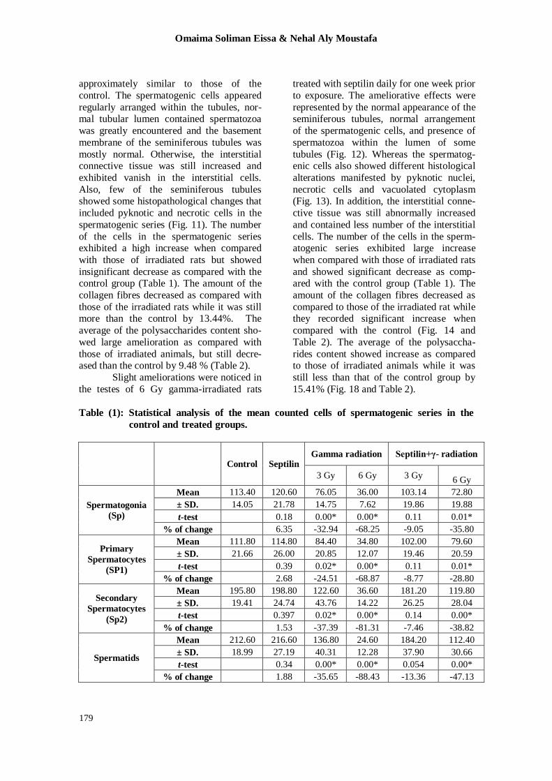
Table (1): Statistical analysis of the mean counted cells of spermatogenic series in the
control and treated groups.
Control Septilin Gamma radiation Septilin+γ- radiation
3 Gy 6 Gy 3 Gy 6 Gy
Spermatogonia
(Sp)
Mean 113.40 120.60 76.05 36.00 103.14 72.80
± SD. 14.05 21.78 14.75 7.62 19.86 19.88
t-test 0.18 0.00* 0.00* 0.11 0.01*
% of change 6.35 -32.94 -68.25 -9.05 -35.80
Primary
Spermatocytes (SP1)
Mean 111.80 114.80 84.40 34.80 102.00 79.60
± SD. 21.66 26.00 20.85 12.07 19.46 20.59
t-test 0.39 0.02* 0.00* 0.11 0.01*
% of change 2.68 -24.51 -68.87 -8.77 -28.80
Secondary
Spermatocytes
(Sp2)
Mean 195.80 198.80 122.60 36.60 181.20 119.80
± SD. 19.41 24.74 43.76 14.22 26.25 28.04
t-test 0.397 0.02* 0.00* 0.14 0.00*
% of change 1.53 -37.39 -81.31 -7.46 -38.82
Spermatids
Mean 212.60 216.60 136.80 24.60 184.20 112.40
± SD. 18.99 27.19 40.31 12.28 37.90 30.66
t-test 0.34 0.00* 0.00* 0.054 0.00*
% of change 1.88 -35.65 -88.43 -13.36 -47.13
The Protective Role of Septilin Against Gamma…….
180
Table (2): Quantitative measurements of colour density (Pixel) of polysaccharides and
collagen fibres.
Control Septilin
γ radiation Septilin + γ radiation
3Gy γ-
radiation
6 Gy γ-
radiation
Septilin+3
Gy γ -
radiation
Septilin+6
Gy γ-
radiation
Po
lysa
ch
ha
rid
es
Average 148.61 150.17 129.94 110.35 134.51 125.7
SD 16.94 16.19 14.03 20.79 13.66 19.02
t-test 0.305 0.006* 0.000* 0.023* 0.014*
% of change 1.01 -12.58 -25.77 -9.48 -15.41
Co
lla
gen
fib
res
Average 72.92 69.37 85.61 101.15 82.73 87.33
SD 8.76 11.48 12.53 22.48 11.09 20.25
t-test 0.09 0.00* 0.00* 0.02* 0.03*
% of change -4.93 17.42 38.68 13.44 19.75
Histogram (2): Quantitative measurements of colour density (Pixel) of polysaccharides (PAS stain)
and collagen fibres (Masson's trichrom stain) in the control and treated groups.
0
20
40
60
80
100
120
140
160
Pix
el
Control Septilin 3 Gy γ-
radiation
6 Gy γ-
radiation
Septilin+3
Gy γ-
radiation
Septilin+6
Gy γ-
radiation
Groups
PAS Collagen fibers
Histogram (1): Mean cells count of spermatogenic cells in the control and treated groups.
Omaima Soliman Eissa & Nehal Aly Moustafa
181
Fig. (1): Transverse section of control rat testis showing seminiferous tubules (ST), interstitial cells (IC)
and inters-tubular connective tissue (CT). (H/E stain X 400).
Fig. (2): Transverse section of control rat testis showing normal structure of seminiferous tubule:
spermatogonia (Sg); primary spermatocytes (P); secondary spermatocytes (SS); spermatids
(St) and spermatozoa (Sp). (H/E stain X 400).
Fig. (3): Transverse section of control rat testis. (Masson's trichrome X 400).
Fig. (4): Transverse section of the testis of rat irradiated with 3 Gy showing atrophied seminiferous
tubules (long arrows) and increase in the interstitial connective tissue (short arrows). (H/E
stain X 100).
Fig. (5): Transverse section of the testis of rat irradiated with 3 Gy showing necrotic cells (thin arrow),
macrophages (thick arrows) and giant cells (G). (H/E stain X 400).
Fig. (6): Transverse section of the testis of rat irradiated with 6 Gy showing atrophy and impaired lumen in the seminiferous tubules (arrows). (H/E stain X 100).
The Protective Role of Septilin Against Gamma…….
182
Fig. (7): Transverse section of the testis of rat irradiated with 6 Gy showing shredding in the basement membrane (short arrows) and necrotic cells (long arrows). (H/E stain X 400).
Fig. (8): Transverse section of rat irradiated with 3 Gy showing increase in the collagen fibres.
(Masson's trichrome X 400). Fig. (9): Transverse section of rat irradiated with 6 Gy showing increase in the collagen fibers.
(Masson's trichrome X 100).
Fig. (10): Transverse section of the testis of rat treated with septilin and irradiated with 3 Gy showing normal seminiferous tubules with lumen containing spermatozoa (arrow). (H/E
stain X 100).
Fig. (11): Transverse section of the testis of rat treated with septilin and irradiated with 3 Gy
showing pyknotic nuclei (P) and necrotic cells (N). (H/E stain X 400). Fig. (12): Transverse section of the testis of rat treated with septilin and irradiated with 6 Gy
showing normal arrangement of the spermatogenic series. (H/E stain X 100).
Omaima Soliman Eissa & Nehal Aly Moustafa
183
Fig. (13): Transverse section of the testis of rat treated with septilin and irradiated with 6 Gy
showing slight ameliorative effects. (H/E stain X 400).
Fig. (14): Transverse section of the testis of rat treated with septilin and irradiated with 6 Gy showing less collagen fibres. (Masson's trichrome X 100).
Fig. (15): Transverse section of control rat testis showing the normal polysaccharides content.
(PAS stain X 100).
Fig. (16): Transverse section of rat irradiated with 3 Gy showing decrease in the polysaccharides content. (PAS stain X 100).
Fig. (17): Transverse section of rat irradiated with 6 Gy showing severe decrease in the
polysaccharides content. (PAS stain X 200). Fig. (18): Transverse section of the testis of rat treated with septilin and irradiated with 6 Gy
showing slight decrease in the polysaccharides content. (PAS stain X 200).
The Protective Role of Septilin Against Gamma…….
184
Discussion
In the present study, histopathological
changes of the testes were assessed after one week of gamma-irradiation with or
without pretreatment of septilin in male
albino rats. In this study, severe atrophy of the seminiferous tubules with necrotic cells,
pyknotic nuclei and complete loss of germ
cells were observed in most seminiferous
tubules in the rats irradiated with 3 or 6 Gy of gamma-radiation. However, other
tubules showed normal spermatogenesis
with a few degenerating germ cells. The dose and time response of rat testis to
gamma-irradiation was studied with use of
single doses from 2.5 Gy to 6.0 Gy.
Germ cells were initially depleted as a result of killing the radiosensitive differen-
tiating spermatogonia (Kangasniemi et al.,
1996). Spermatogonial depletion due to testicular toxicants and seminiferous tubule
atrophy by impairment of spermatogenesis
was documented (Son et al., 1999). In irradiated mice, (8 Gy) the germinal
epithelium was highly disorganized with
shrinkage of tubules, absence of sperm and
spermatids shrinkage in their size of sertoli cells and Leydig cells (Kumar et al., 2006).
However, in the present study necrosis
appeared at earlier stages even in the spermatogonia. Otherwise, Ghanem et al.
(1995) showed that the occurrence of many
pathological changes in primary and secondary spermatocytes could be due to
the fact that those cells were derived from
injured spermatogonia. In agreement with
the present study, gamma irradiation resulted in death of the spermatogenic cells
and this death was more obvious in the
spermatogonia in the testes of rats (Pinon-Lataillade et al., 1991).
Moreover, radiotherapy is widely
used for cancer therapy; although the
treatments could be successful, patients often complain of azoospermia or infertility
(Taksey et al., 2003). Radiation doses as
low as 0.1-0.2 Gy exerted detectable effects on spermatogenesis in adult men (Centola
et al., 1994) and high doses over 4 Gy
caused in permanent azoospermia (Relander et al., 2000). The vacuolation
observed in the spermatogenic cells as a
result of gamma radiation is attributed in part to a progressive hypoxia and partly to
fatty accumulation as a result of cell degen-
eration (Bhatavdekar et al., 1977). In respect to giant cells observed in the testis
of gamma-irradiated animals, Labib (1995)
suggested that the multinucleated giant cells
were derived from the fusion of two or three primary spermatocytes. The present
data showed that the interstitial spaces in
the testis of irradiated animals appeared larger than those in the control animals and
most Leyding cells became degenerated.
Comparable observations were reported by
Labib (1995). Whoever, the exposure of the mammalian testis to ionizing radiation at
even low dose might cause significant
disturbance in the spermatogenesis process and differentiation of the spermatozoa
(Khattab et al., 1995). The present data
showed significant reduction in the number of spermatogonia, primary spermatocytes,
secondary spermatocytes and spermatids
due to gamma irradiation; this decrease was
more pronounced with the high dose than the low one. In agreement with these data,
significant decrease in the number of sper-
matogonia, primary spermatocytes, second-ary spermatocytes and spermatids in irradi-
ated mice was noticed (Kumar et al., 2006).
According to Filippi and Mela (1958) gradual decrease in the carbohydrate
content of the spermatogenic cells, espec-
ially in the spermatids which are the main
site of carbohydrates in the testis, as a results of exposure to different xenobiotics.
Harman (1983) suggested that the
accumulated free radicals in the cells attack. The DNA and lead to cross-linkages with
the molecules and these result in reduction
of their biological activity. The present
study showed that gamma irradiation induced significant reduction in the polys-
accharides content and significant increase
in the collagen fibres in the testes of rats. Harmful effects of ionizing radiation
in mammals are known for a long time and
attempts have been made to find out suitable protective agents against these
Omaima Soliman Eissa & Nehal Aly Moustafa
185
effects. Radioprotective agents are defined
as compounds that help to diminish the
biological effects of ionizing radiation when administered before exposure to
radiation (Satoh et al., 2003). Various other
experimental approaches, aimed at
preventing radiation-induced quantitative damage to the gonads, have been explored
(Jegou et al., 1991; Lee et al., 1998 and
Landauer et al., 2005). The present study showed that spetilin has strong protective
effects against gamma-radiation induced
testicatular histological alterations in rats.
Septilin in oral daily dose of 100 mg/kg b. wt. for one week prior to gamma-irradiation
could attenuate the histological lesions
observed under the effect of gamma-irradiation especially with the low dose of
this radiation. Similarly, in mice treated
with extract of Adhatoda vasica and then exposed to radiation dose, the ameliorative
effect determined by intact germinal
epithelium, no pyknosis, necrosis, karyol-
ysis present, less cytoplasmic vacuolization and number of germ cells increased by day
30 (Kumar et al., 2006).
Conclusion The present study proved that septilin
exerts a strong effect against whole body
gamma irradiation-induced testicular histo-pathological changes, where it efficiently
ameliorated the deleterious influences of
ionizing radiation when administered pre-
exposure. The results have implication for the potential use of septilin as a
radioprotector.
References 1. Bhatavdekar J M, Aravinda B K. and
Shah V C. (1977) Effect of low dose X-
irradiation on nucleic acids and protein
content of guinea pig, rat and mouse tissue.
Ind. J. Exp. Biol., 15: 908-912.
2. Centola G M, Keller J W, Henzler M.
and Rubin P. (1994) Effect of low-dose
testicular irradiation on sperm count and
fertility in patients with testicular semi-
noma. J. Androl., 15: 608-613.
3. Dobson R L. and Felton J S. (1983): Female germ cell loss from radiation and
chemical exposures. Am. J. Ind. Med.,
4:175–190.
4. Filippi B. and Mela V. (1958) Malformation congenitales et antibiotique.
Arch. Fr. Pediat., 15: 565.
5. Geraci J P, Mariano M S. and Jackson
K L. (1992) Radiation hepatology of the
rat: Microvascular fibrosis and enhance-
ment of liver dysfunction by diet and drugs.
Radiat. Res., 129: 322-332.
6. Ghanem N F, Attia S I, Risk A M. and
Shwaireb M H. (1995) Effect of
antithyroid drug cabimazole on the fertility and testicular structure of mice. J. Egypt.
Ger. Soc. Zool., 18(C): 1-12.
7. Georgieva T S, Yablanskii P, Georgiev
D, Yakov M. and Oblakova M. (2005) Reciprocal translocation and reproductive
capacity in rabbits following external
gamma irradiation. Bulgarian J. Vet. Med.,
8(4): 227-232.
8. Jagetia G C. and Baliga M S. (2004) Polyherbal extract of septilin protects mice
against whole body lethal dose of gamma radiation. Phytother. Res., 18(8): 619-623.
9. Jagetia G C, Venkatesh P. and Baliga M
S. (2004) Evaluation of the radioprote-ctive
effect of A. vasica leaf (Avasicagle
marmelos) extract in mice. Int. J. Radiat.
Biol., 80: 281–90.
10. Jegou B, Callet J. and Baucho F. (1991) Protective effect of medroxyproges-terone
acetate plus testosterone against radiation-
induced damage to the reproduc-tive
function of male rats and their offspr-ing. Proc. Natl. Acad. Sci. USA, 88: 8710-8714.
11. Harman D. (1983) Free radical theory of
aging consequences of mitochondrial
aging. Age, 6: 86-94.
12. Kangasniemi M, Veromaa T I, Kulmala
J, Kaipia A, Parvinen M. and Toppari
J. (1990) DNA-flow cytom-etry of defined
stages of rat seminiferous epithelium:
effects of 3 Gy of high-energy X-
irradiation. J. Androl., 11: 312-317.
13. Kangasniemi M, Huhtaniemi I. and
Meistrich M. (1996) Failure of permato-genesis to Recover Despite the Presence of
A Spermatogonia in the Irradiated LBNF1
Rat. Biol. Repr., 54: 1200-1208.
14. Khattab F, Roushdy H. and El-Dawy H.
(1995) Effect of ionizing radiation on
sperm of mice. Egypt. J. Rad. Sci. Applic.,
8(2): 141-148.
15. Kumar P V, Kuttan G. and Luttan R.
(1993) Immunomodulatory activity of
septilin. Probe; 33: 1-5.
16. Kumar M, Samarth R, Kumar M,
Senthamil R, Begraj S. and Kumar A.
(2006) Protective effect of Adhatoda vascia
Nees against radiation-induced damage at
The Protective Role of Septilin Against Gamma…….
186
cellular, biochemical and chromosomal
levels in Swiss albino mice. Compl. and
Altern. Med., 5: 1- 8.
17. Labib M M. (1995) Histological changes
and withdrawal in gonads of young rats
response to the administration of heroin.
J. Union Arab Biol., 3(A) : 135-157.
18. Lamartiniere C A, Cotroneo M S, Fritz
W A, Wang J, Mentor-Marcel R. and
Elgavish A. (2002) Genistein chemopreve-
ntion: timing and mechanisms of action in murine mammary and prostate. J. Nutr.,
132: 552S–558S.
19. Landauer M R, Srinivasan V, Grace M
B, Chang C M, Parekh V, Clarke T K.
and Jackson W.E. (2005) Prevention of
Gamma Radiation-Induced Mortality in
Mice by the Isoflavone Genistein. NATO
RTG-099.
20. Lee Y, Change W, Kimi J K. and Yoon
Y. (1998) Effects of gamma radiation on
ovarian follicles. Arh. Hig. Rada. Toksikol., 49 (2): 147–153.
21. Matalka K Z, Barth R F, Bailey M Q,
Koestner A D. and Hopewell J W. (1994) Radiation effect of boron neutron capture
therapy on brain, skin and eye of rats. Int. J.
Radiat. Oncol. Biol. Phys., 28: 1089- 1097.
22. Mc Manus, J.P.A. (1946) Histological
demonstration of mucin after periodic acid.
Nature (London), 158: 202.
23. Mian T A, Suzuki N, Glenn H J, Haynie
T P. and Meistrich M L. (1977) Radiation damage to mouse testis cells from (99mTc)
pertechnetate. J. Nucl. Med., 18: 1116-
1122.
24. Meistrich M L. (1993) Effects of chemot-
herapy and radiotherapy on spermato-
genesis. European Urology, 23: 136_142.
25. Pearse A G. (1972) Histochemistry,
theoretical and applied. 3rded. Vol. 11, Little,
Brownand com. Boston.
26. Pinon-Lataillade G, Viguier-Martinez M
C, Touzalin A M, Maas J. and Jegou B.
(1991) Effect of an acute exposure of rat testes to gamma rays on germ cells and on
Sertoli and Leydig cell functions. Reprod.
Nutr. Dev., 31(6): 617-629. 27. Rades D, Fehlauer F, Bajrovic A,
Mahlmann B, Richter E. and Alberti
W. (2004) Serious adverse effects of
amifostine during radiotherapy in head and
neck cancer patients. Radiother Oncol., 70:
261-264.
28. Relander T, Cavallin-Stahl E. Garwicz S,
Olsson A M. and Willen M. (2000) Gonadal and sexual function in men treated
for childhood cancer. Med. Pediatr. Oncol.,
35: 52-63.
29. Rom D G. (1984) The anti-infective and
anti bacterial efficacy of septilin. Probe, 23:
84-87.
30. Rowley M J, Leach D R, Warner G A. and Heller C G. (1974) Effect of graded
doses of ionizing radiation on the human
testis. Radiat. Res., 59: 665-678.
31. Sharma S B. and Ray S. (1997) Effect of
herbal preparation on immune response of
immunosuppressed mice. Ind. J. Physiol.
Pharmacol., 41: 293-296.
32. Son H Y, Kim Y B, Kang B H, Cho S W,
Ha C S. and Roh J K. (1999) Effect of 2-
bromopropane on spermatoge-nesis in the
Spargue-Dawley rat. Reprod. Toxicol., 13: 179-187.
33. Satoh H, Nishikawa K, Suzuki K,
Asano R, Virgona N, Ichikawa T,
Hagiwara K. and Yano T. (2003) Genistein, a soy isoflavone, enhances
necrotic-like cell death in a breast cancer
cell treated with a chemotherapeuticagent,
Res. Commun. Mol. Pathol. Pharmacol.,
113-114: 149-158.
34. Taksey J, Bissada N K. and Chaudhary
U B. (2003) Fertility after chemotherapy for testicular cancer. Arch. Androl., 49:
389-395.
35. Uma Devi P, Ganasoundari A, Vrinda B,
Srinivasan K K. and Unnikrishnan M K.
(2000) Radiation protection by ocimum
flavonoids orientin and vicenin: mechanism
of action. Radiat. Res., 154: 455–60.
36. Weiss J F, Landauer M R. (2003) Protection against ionizing radiation by
antioxidant nutrients and phytochemicals,
Toxicology. 189: 1-20.
37. Yang W, Barth R F, Rotaru J H, Boesel
C P, Wilkie D A, Bresnahan J,
Hadjiconstantinou M, Goettl V M, Joel D
D. and Nawrocky M M. (2000) Boron
neutron capture therapy of brain tumors:
functional and neuropathhological effects
of blood brain barrier disruption and
intracarotid injection of sodium borocaptate
and boronopheny-lalanine. J. Neurooncol.,
48: 179-190.
Omaima Soliman Eissa & Nehal Aly Moustafa
187
دور الىقائى للسيبتليه ضد الأضرار التى تحدثها أشعة جاما فى خصية ال
الجرذان
**وهال على مصطفى* أميمه سليمان عيسى
هيئخ الطبقخ السشيخ -الوصكط القيه لجحيس ىركيليجيب الإشعبع*
كليخ العليم للجبد جبهعخ الأضهص**
لزهيتتصاد الوبتتزيكيويب يخ ربىلتتذ هتتس الرشاقتتخ رقيتتين الهيتتصاد الوبتتزيليجيخ ىثعتتط ا
الوحرصخ ف خصيخ الجصزاى زيجخ الزعصض لأشتعخ جبهتب ثبلإظتبفخ لت رقيتين الترىش اليقتب رن اقتزدرام عترذ بشثعتيي هتي زكتيش الجتصزاى . ظر هس الزأصيصاد( رحعيص عشج)للبيجزليي
لجذ عي( 6)اقزدرهذ كوجويعخ ظبثطخ، الضبيخ ( 10)الأىل : قبويا ل بشثع هجويعبد
( 12)لوترح بقتجيع، الوجيعتخ الضبلضتخ ( كيليجصام هتي ىضى الجبتن/ مهليجصا 100)ثبلبيجزليي
جتصا، 3قبوذ ل هجويعزيي هببىيزيي الأىل رعصظذ لجصعخ هي بشعخ جبهب هقرشاهب
عيلجتذ ثبلبتيجزليي ( 12)جتصا، ىالوجويعتخ الصاثعتخ 6ىالضبيخ رعصظذ لجصعخ هقراشهب
لوترح بقتجيع صتتن قبتوذ لوجوتيعزيي هزبتتبىييي ( صام هتي ىضى الجبتتنكيلتيج/ مهليجتصا 100)
جصا ىالضبيخ رعصظذ لجصعخ هقراشهب 3ىعصظذ الأىل لجصعخ هي بشعخ جبهب هقرشاهب
.جصا 6بىظتتحذ الزتتب و تترىس رهيتتصاد عريتترح فتت الزصكيتتت البتتيج لدصتتيخ الجتتصزاى
الزحلت الكبهت ىووتيش خ يتب الوعصظخ لأشعخ جبهب روضلذ ف رىس الفجتياد ىالزكتصض ى
عو قتخ، ىرحلتت فت الد يتتب الجييتخ هتتع ضيتتبذح فت هبتتب خ البتيو العتتبم الجيت، كوتتب تترس
عتتية زى ذةلتتخ صتتب يخ فتت بعتتراذ بهوتتبد الوتت ىالد يتتب الوييتتخ اةثزرا يتتخ ىالضبييتتخ
كويتخ الأليتب بيعب بووصد الزب و رىس ضيبذح زاد ذةلخ صتب يخ فت. ىالط ع الوييخ
، ىكسا قص زى ذةلخ صب يخ ف هحزي الدصتيخ هتي الوتياذ عريترح الزبتكص، خالكية جيي
.عىقر كبذ هس الزهيصاد بكضص ىظي ب هع الجصعخ العبليخ هي الإشعب
بىظحذ الزب و بى الوعبلجخ ثبلبيجزليي لوترح بقتجيع قجت الزعتصض لأشتعخ جبهتب يقلت رحرس ف الدصيخ زيجخ الزعصض للإشتعبع، يتش اخزفتذ هع تن ثشك كجيص الزهيصاد الز
الزهيتتصاد الوبتتزيليجيخ ىرحبتتذ بعتتراذ بهوتتبد الوتت ىالد يتتب الوييتتخ اةثزرا يتتخ ىالضبييتتخ
، ىرحبتتي هحزتتي الدصتتيخ هتتي الوتتياذ خىالط تتع الوييتتخ ىقلتتذ كويتتخ الأليتتب الكتتية جييتت
. ل هبزي الوجويعخ العبثطخ عريرح الزبكص، غيص بى هس الزحبيبد لن رص